Embed Size (px)
Citation preview

Cancer Cell
Article
Disrupting the Interaction of BRD4with Diacetylated Twist SuppressesTumorigenesis in Basal-like Breast CancerJian Shi,1,4 Yifan Wang,1,4,5 Lei Zeng,6 Yadi Wu,2,4 Jiong Deng,7 Qiang Zhang,6 Yiwei Lin,1,4 Junlin Li,1,4 Tiebang Kang,5
Min Tao,8 Elena Rusinova,6 Guangtao Zhang,6 Chi Wang,4 Haining Zhu,1 Jun Yao,9 Yi-Xin Zeng,5 B. Mark Evers,1,3,4
Ming-Ming Zhou,6,* and Binhua P. Zhou1,4,5,*1Department of Molecular and Cellular Biochemistry, College of Medicine, University of Kentucky, Lexington, KY 40506, USA2Department of Molecular and Biomedical Pharmacology, College of Medicine, University of Kentucky, Lexington, KY 40506, USA3Department of Surgery, College of Medicine, University of Kentucky, Lexington, KY 40506, USA4Markey Cancer Center, College of Medicine, University of Kentucky, Lexington, KY 40506, USA5Sun Yat-sen University Cancer Center, State Key Laboratory of Oncology in South China, and Collaborative Innovation Center of CancerMedicine, Guangzhou 510060, China6Department of Structural and Chemical Biology, Icahn School of Medicine at Mount Sinai, New York, NY 10029, USA7Department of Pathophysiology, Shanghai Key Laboratory for TumorMicroenvironment and Inflammation, and TranslationMedicine Center,
Shanghai Chest Hospital, Shanghai Jiao Tong University School of Medicine, Shanghai 200025, China8Department of Oncology, the First Affiliated Hospital of Soochow University, Suzhou 215006, China9Department of Molecular and Cellular Oncology, University of Texas M.D. Anderson Cancer Center, Houston, TX 77030, USA
*Correspondence: [email protected] (M.-M.Z.), [email protected] (B.P.Z.)
http://dx.doi.org/10.1016/j.ccr.2014.01.028
SUMMARY
Twist is a key transcription activator of epithelial-mesenchymal transition (EMT). It remains unclear how Twistinduces gene expression. Here we report a mechanism by which Twist recruits BRD4 to direct WNT5Aexpression in basal-like breast cancer (BLBC). Twist contains a ‘‘histone H4-mimic’’ GK-X-GK motif that isdiacetylated by Tip60. The diacetylated Twist binds the second bromodomain of BRD4, whose first bromo-domain interacts with acetylated H4, thereby constructing an activated Twist/BRD4/P-TEFb/RNA-Pol IIcomplex at the WNT5A promoter and enhancer. Pharmacologic inhibition of the Twist-BRD4 associationreducedWNT5A expression and suppressed invasion, cancer stem cell (CSC)-like properties, and tumorige-nicity of BLBC cells. Our study indicates that the interaction with BRD4 is critical for the oncogenic function ofTwist in BLBC.
INTRODUCTION
Recruitment and activation of RNA-Pol II at gene promoters are
two key steps required for a productive transcription (Zhou et al.,
2012). After RNA-Pol II recruitment to a gene promoter, TFIIH
phosphorylates serine 5 of the heptapeptide repeats in the C-ter-
minal domain (CTD) of RNA-Pol II, resulting in initial synthesis of
short RNA species. However, RNA-Pol II pauses in the proximal
promoter and requires a second phosphorylation event on serine
Significance
BLBC is associated with an aggressive clinical history, developsurvival. BLBC contains abundant EMT transcription factor Twithat the Twist-activated EMT program confers growth advantagdomain in Twist creates a formidable hurdle toward developinginteracts with and recruits the BRD4/P-TEFb/RNA-Pol II transcvation. BET-specific inhibitors disrupted the Twist-BRD4 interainhibition of invasion and tumorigenicity of BLBC in vitro and inaction provides an effective approach for treating BLBC.
210 Cancer Cell 25, 210–225, February 10, 2014 ª2014 Elsevier Inc.
2 of the CTD that is carried out by the pause release factor
P-TEFb, a complex composed of CDK9 and cyclin T1/2. Impor-
tantly, the recruitment of P-TEFb to RNA-Pol II is mediated, in
part, by BRD4 (Jang et al., 2005).
BRD4 is a member of the BET (bromodomain and extra termi-
nal domain) family proteins that are characteristic of two tandem
bromodomains (BDs) located in the N terminus. The BDs of
BET proteins recognize acetylated-lysine residues in nucleo-
somal histones (Filippakopoulos et al., 2012), facilitating the
ment of recurrence, distant metastasis, and shorter patientst and possessesmany CSC-like characteristics, suggestinges to BLBC. However, the absence of a clear ligand-bindinginhibitors that can suppress its function.We found that Twistription complex to theWNT5A superenhancer for gene acti-ction and resulted in significant Wnt5a reduction, leading tovivo. Our study indicates that targeting the Twist-BRD4 inter-

Cancer Cell
Twist-BRD4 Interaction in Gene Transcription
recruitment of transcriptional proteins to chromatin. Recent
studies have shown that pharmacologic inhibition of BRD4
with BET-specific BD inhibitors effectively blocks MYC expres-
sion in multiple myeloma (Delmore et al., 2011), Burkitt’s
lymphoma, and acute myeloid leukemia (Dawson et al., 2011;
Zuber et al., 2011). However, many mechanistic questions about
BRD4 functions as a chromatin regulator in gene transcription
are still unanswered, including (1) howBRD4 interacts and works
with transcription factors at the target gene promoter and
enhancer sites and (2) whether and how the two BDs in BRD4
function differently in gene transcription.
Breast cancer is a heterogeneous disease that can be divided
into four major subtypes based on gene expression profiling:
luminal A, luminal B, ErbB2, and basal like. Basal-like breast
cancer (BLBC) is characterized by the lack of expression of es-
trogen receptor (ER), progesterone receptor (PR), and epidermal
growth factor receptor 2 (HER2) and positive expression of basal
markers (Cytokeratin 5/6 [CK5/6] and CK14) (Rakha et al., 2008).
The absence of effective targeted therapies and poor response
to standard chemotherapy often results in a rapidly fatal clinical
outcome for this disease. Notably, BLBC has activated the
epithelial-mesenchymal transition (EMT) program, which pro-
vides cells with increased plasticity and stem-cell-like properties
required during embryonic development, tissue remodeling,
wound healing, and metastasis (Thiery et al., 2009).
Twist and Snail are two key members of EMT-activating
transcriptional factors. During mesoderm development in
Drosophila, Snail functions as a transcriptional repressor to
prevent expression of genes that belong to ectoderm, whereas
Twist serves as a transcriptional activator to induce mesodermal
gene expression (Leptin, 1991). A complete loss of all meso-
dermal characteristics occurs only when both Snail and Twist
are absent. These results suggest that Snail and Twist work
synergistically, controlling distinct sets of genes, to coordinate
EMT induction and mesoderm formation (Zeitlinger et al., 2007).
We previously showed that Snail interacts with several tran-
scriptional repressive complexes to suppress gene expression.
However, the mechanism underlying gene transcriptional acti-
vation by Twist has remained elusive. In this study, we sought
to identify Twist-interacting proteins and determine the mecha-
nism by which Twist controls gene transcriptional activation in
EMT and BLBC.
RESULTS
BRD4-BD2 Interacts with Lysine-Acetylated TwistWe sought to identify Twist-interacting proteins from a stable
HeLa S3 cell line expressing Flag-Twist. Affinity protein purifica-
tion, followed by SDS-PAGE and analysis by mass spectrom-
etry, revealed the presence of BRD4 and TRRAP/EP400 (data
not shown). To validate the interaction between Twist and
BRD4, we coexpressed hemagglutinin (HA)-Twist and Flag-
BRD4 in HEK293 cells in the presence or absence of the histone
deacetylase inhibitor Trichostatin (TSA). After immunoprecipitat-
ing Twist, we detected the associated BRD4, and vice versa (Fig-
ure 1A and Figure S1A available online). Although similar
amounts of Twist were immunoprecipitated from cells with and
without TSA treatment, Twist was more acetylated and inter-
acted with more BRD4 in cells treated with TSA. In addition,
C
immunoprecipitation with a pan-acetylated-lysine (pan-AcK)
antibody pulled down Twist and BRD4 in cells treated with
TSA. Similar observations were made in Twist-expressing
HeLa S3 cells (Figures 1B and S1B). We further confirmed the
interaction between the endogenous Twist and BRD4 and acet-
ylation of the endogenous Twist in four BLBC cell lines, both of
which were substantially enhanced with TSA treatment (Figures
1C and S1C). The Twist-BRD4 interaction is specific because
Twist did not associate with other BET members (BRD2,
BRD3, and BRDT) or lysine specific demethylase 1 (LSD1), and
BRD4 did not associate with TCF4 (Figure S1D). The increased
Twist-BRD4 interaction by TSA could not be due to an altered
subcellular localization of these two proteins as TSA did not
affect their localization (Figure S1E).
We next generated BRD4 deletion constructs and coex-
pressed them with Twist in HEK293 cells. We found that only
N-terminal fragments containing both BDs, but not other regions
of BRD4, retained the ability to interact with Twist (Figure 1D).
When BD1 or BD2 was coexpressed with Twist in HEK293 cells,
only BD2WT, but not BD1WT, bound to Twist (Figures 1E and
S1F). Mutation of the conserved tyrosine and asparagine resi-
dues in the acetyllysine binding pocket of BD2 to alanine
(BD2YN) reduced its binding to Twist. The Twist-BRD4 inter-
action was readily disrupted when JQ1, a BET-specific BD inhib-
itor, was added to the immunoprecipitation reaction (Figures 1F,
1G, S1G, and S1H). Similarly, MS417, a BET-specific BD inhibi-
tor with approximately 10-fold higher binding affinity than JQ1
(Zhang et al., 2012), effectively blocked the Twist-BRD4 inter-
action in four BLBC cell lines (Figures S1I and S1J). These results
indicate that the Twist-BRD4 interaction is mediated by the BD2
of BRD4 binding to lysine-acetylated Twist.
Twist Diacetylation at K73 and K76 by Tip60 Is Requiredfor Twist-BRD4 InteractionThe N-terminal half of Twist contains an acidic segment and two
lysine/arginine-rich basic motifs that share high sequence simi-
larity to histones H2B and H4, respectively (Figure S2A). We
generated Twist deletion constructs DL1 (residues 15–202),
DL2 (residues 31–202), and DL3 (residues 47–202) and coex-
pressed them individually with Flag-BRD4 in HEK293 cells.
DL1 retained, whereas DL2 and DL3 lost, interaction with
BRD4 (Figures 2A and S2B). Surprisingly, in contrast to DL1,
DL2 and DL3 also completely lost acetylation (Figures 2B and
S2C). Because the first 30 N-terminal residues in Twist do not
contain lysine, the loss of acetylation in DL2 and DL3 suggests
that the N-terminal region is critical for Twist acetylation. In our
mass spectrometry analysis, the NuA4 histone acetyltransferase
complex proteins, including TRRAP and EP400 (Doyon and
Cote, 2004), were identified as Twist association partners. We
postulated that Tip60, the acetyltransferase of NuA4 complex,
was responsible for Twist acetylation. Indeed, when TwistWT,
DL1, DL2, and DL3 were coexpressed with Tip60 in HEK293
cells, we found that TwistWT and DL1, but not DL2 or DL3, inter-
acted with Tip60 (Figures 2C and S2D). Endogenous Twist-Tip60
interaction was confirmed in three BLBC cell lines, which was
markedly enhanced by TSA treatment (Figure S2E). We further
observed that ectopic expression of Tip60 in BT549 and
SUM1315 cells resulted in enhanced acetylation of Twist and
the association of Twist with BRD4 even in the absence of
ancer Cell 25, 210–225, February 10, 2014 ª2014 Elsevier Inc. 211

A
D
F G
E
B C
Figure 1. BD2 of BRD4 Is Required for Its Interaction with Acetylated Twist
(A) HA-Twist and Flag-BRD4 were coexpressed in HEK293 cells. After treatment of cells with TSA (2 mM) for 12 hr, Twist, BRD4, and acetylated Twist were
immunoprecipitated with HA, Flag, and pan-acetylated-lysine (pan-AcK) antibodies, respectively, and analyzed by western blotting.
(B) HeLa cells stably expressing Flag-Twist were treated with TSA as in (A). Flag-Twist, endogenous BRD4, and acetylated Twist were immunoprecipitated and
examined by western blotting.
(C) Cells were treated as described in (A). Endogenous Twist, BRD4, and acetylated Twist were immunoprecipitated and examined by western blotting.
(D) Schematic depiction of the functional domains of BRD4 and deletion constructs used (top). ET, extraterminal domain. Flag-tagged wild-type (WT) or deletion
mutants of BRD4 were coexpressed with HA-Twist in HEK293 cells. After being immunoprecipitated with HA or Flag antibody, the bound BRD4 or Twist was
examined by western blotting (bottom).
(legend continued on next page)
Cancer Cell
Twist-BRD4 Interaction in Gene Transcription
212 Cancer Cell 25, 210–225, February 10, 2014 ª2014 Elsevier Inc.

Cancer Cell
Twist-BRD4 Interaction in Gene Transcription
TSA, whereas knockdown of Tip60 yielded opposite effects even
in the presence of TSA (Figures 2D and S2F).
Twist contains five lysine residues (K33, K38, K73, K76, and
K77) in its N-terminal region, which are highly conserved among
different species (Figure S2A). Point mutation of K33R, K73R,
and K76R showed a reduced level of acetylation compared to
that of TwistWT (Figures 2E and S2G). TwistWT, K33R, K38R,
and K77R showed a similar interaction with BD2, whereas
K73R and K76R exhibited clearly weaker binding to BD2 (Figures
2F and S2H). The K73R/K76R double mutant showed an almost
complete loss of acetylation and interaction with BD2 (Figures
2G and S2I). We further confirmed the Tip60-mediated Twist
acetylation on K73/K76 by mass spectrometry analysis (Figures
2H and S2J). There are 12 nuclear histone acetyltransferases
(HATs), divided into three major groups: (1) the GNAT family
(e.g., PCAF), (2) the MYST family (e.g., Tip60), and (3) the
p300/CBP family (e.g., p300 and CBP) (Rekowski and Giannis,
2010). To examine whether other HATs can also acetylate Twist,
we knocked down the expression of p300, CBP, PCAF, or Tip60
individually in BT549 and SUM1315 cells (Figure S2K). We found
that knockdown of Tip60, but not p300, CBP, or PCAF, sup-
pressed Twist acetylation at K73/K76. Taken together, these
data support our contention that Tip60 is the major HAT respon-
sible for the K73/K76 acetylation on Twist and that diacetylation
of Twist is required for its association with BRD4-BD2.
Histone H4 Mimicry in Twist Is Responsible for ItsInteraction with BRD4Diacetylations of the N-terminal tail of H4 at K5 and K8 are often
required for the interaction of H4 with BDs of BET family proteins
(Filippakopoulos et al., 2012; Moriniere et al., 2009). The Twist
sequence at K73 and K76 shares high similarity to the N-terminal
tail of H4 at K5 and K8 (Figure 3A). To investigate this ‘‘histone
mimicry’’ in the interaction between Twist and BRD4, we per-
formed a pull-down study using biotinylated H4 and Twist
peptides and lysate of HEK293 cells expressing BRD4-BD2.
We observed that biotinylated H4-K5ac/K8ac peptide (residues
1–21) was bound to BD2 and that this interaction was disrupted
by nonbiotinylated H4-K5ac/K8ac peptide (Figure 3B, lane 2
versus lane 1). This interaction was also markedly reduced by
a Twist-K73ac/K76ac peptide (residues 61–80) but not by the
unacetylated corresponding peptide (Figure 3B, lanes 3 and 4
versus lane 1). Similarly, the interaction of a biotinylated Twist-
K73ac/K76ac peptide with BD2 was disrupted by a Twist-
K73ac/K76ac, but not unacetylated, peptide (Figure 3B, lanes
7 and 8 versus lane 5). Notably, acetylated H4 peptide also
disrupted the acetylated Twist and BD2 association (Figure 3B,
lane 6 versus lane 5), indicating that diacetylated K5/K8 in H4
and diacetylated K73/K76 in Twist function similarly as a recog-
nition motif for BRD4-BD2.
(E) Schematic diagram showing the double bromodomain (BD1+BD2) of BRD4 an
were coexpressed with HA-Twist in HEK293 cells treated with TSA as in (A). Twis
and analyzed by western blotting (bottom).
(F) HA-Twist and Flag-BRD4 were coexpressed in HEK293 cells treated with TS
bodies, respectively, in the presence or absence of JQ1 (1 mM) and analyzed by
(G) Cells were treated with TSA as in (A). Endogenous Twist and BRD4were immun
absence of JQ1 (1 mM) and examined by western blotting.
See also Figure S1.
C
We then developed a specific antibody against Twist-K73ac/
K76ac. Twist recognition by this antibody was disrupted by a
Twist-K73ac/K76ac peptide, but not the corresponding nonace-
tylated peptide (Figures 3C and S3A). This antibody recognized
immunoprecipitated TwistWT, but not TwistK73R/K76R that
harbored mutated K73 and K76 (Figures 3D and S3B). In line
with our contention that Tip60 acetylates Twist at K73/K76,
both Twist and H4 acetylated by purified Tip60 in vitro were
recognized by this Twist-K73ac/K76ac antibody and a pan-acet-
ylated antibody (Figure 3E). Furthermore, this antibody readily
detected endogenous Twist-K73ac/K76ac that was immuno-
precipitated from four BLBC cell lines (Figures 3D and S3B).
Although immunoprecipitation of the endogenous Twist by this
antibody was weak, it was robustly increased by the addition
of JQ1 to the binding buffer, indicating that the K73ac/K76ac
site is masked by binding to BRD4 in cells (Figure 3F).
We further characterized the functional importance of this
diacetylation-dependent Twist-BRD4 interaction in human
mammary epithelial (HMLE) cells. Ectopic expression of TwistWT
resulted in an induction of EMT, as indicated by the downregula-
tion of E-cadherin and upregulation of vimentin (Figures 3G and
S3C). While localized in the nucleus (Figure S3D), TwistK73R/K76R
expression failed to induce EMT, indicating that the interaction
with BRD4 is critical for the function of Twist.
Molecular Basis of BRD4 Binding to Lysine-AcetylatedTwistTo determine the molecular basis of diacetylation-dependent
Twist-BRD4 association, we characterized binding of the two
BDs of BRD4 to a series of Twist peptides (residues 68–79)
bearing no, single-acetylated, or diacetylated lysine at K73 and
K76 by nuclear magnetic resonance (NMR) titration. As shown in
2D 1H-15NHSQCspectra (Figure S4A), BRD4-BD2exhibited sub-
stantially more extended chemical shift perturbations upon bind-
ing to the single-acetylated, and even more to the diacetylated,
Twist peptides than those produced by BRD4-BD1, confirming
that BRD4-BD2 is largely responsible for BRD4 association
with the diacetylated Twist. The preferred recognition of Twist-
K73ac/K76ac by BRD4-BD2 was supported in a fluorescence
anisotropy competition binding study using a fluorescein-labeled
H4K5ac/K8ac peptide as an assay probe, yielding a Ki of 800 mM
and >3,000 mM for BRD4-BD2 and BRD4-BD1, respectively
(Figure S4B). Furthermore, BRD4-BD1 and other BDs, including
those from CBP and PCAF, showed almost no interaction with
the single- or diacetylated Twist peptides (data not shown).
We next solved the 3D structure of BRD4-BD2 bound to Twist-
K73ac/K76ac peptide using NMR spectroscopy to determine
the molecular basis of this selective interaction (Figures 4A,
4B, and S4C and Table S1). As revealed in the 3D structure,
the Twist-K73ac/K76ac peptide is bound in the protein across
d individual BD constructs used (top). Flag-BD1WT, BD1YN, BD2WT, and BD2YN
t and BDs were immunoprecipitated with HA and Flag antibodies, respectively,
A as in (A). Twist and BRD4 were immunoprecipitated with HA and Flag anti-
western blotting.
oprecipitated with Twist and BRD4 antibodies, respectively, in the presence or
ancer Cell 25, 210–225, February 10, 2014 ª2014 Elsevier Inc. 213

DL3
DL1
DL2WT
IgG
Twis
t pan-AcK
TSA + + + + +A B D
L3
DL1
DL2
Twist
Tip60
IP: T
wis
t
TSA + + + + +
WT
IgGC
Twist (WT)DL1(15-202)DL2(31-202)DL3 (47-202)TW (K33R)TW (K38R)TW (K73R)
HLH Binding++
++
--
IP:
pan-
AcK
Twist
Twist IP: T
ip60
Tip60
TwistTW (K77R)
TW (K73R)TW (K76R)
+
±±
35
25
DL1
4
TSA - + - + - + - + +WT DL2 DL3 IgG MW
(kDa)
IP:
D
BRD4TSA - + - + - + + - + - + - + +
Cont Tip60 IgGsiTipSUM1315BT549
Cont Tip60 IgGsiTip15
BRD4
Twist
IP: B
RD
4
10
WT 77
R
38R
33R
WT 33
R38
R
73R
73R
76R
76R
77R
WT
BRD4-BD1 BRD4-BD2 IgG
Twist
pan-AcK
F
BRD4
TwistIP: T
wis
t
35
25
15
P: T
wis
t
K33
RK
38R
K73
RK
76R
K77
R
pan-AcKTSA + + + + + + +
WT
IgGE
IP: T
wis
t2
Twist
BD1/2
W K7
K3K3 W K3
K3K7
K7
K7
K7
K7 W
T 3R 6R /76R
G
BRD4-BD2
IPIP
: AcK
Twist
Twist IP: B
D1/
2 Twist
BD1/2
HG
danc
e K73ac
IP: B
D2
wis
t
BD2
BD2
Twist
WT
K73 K76
K73
/
IgG
Rel
ativ
e A
bund
K76ac
IP: T
wIP
: AcK
Twist
pan-AcK
Twist
BD2
K76K76aacc
Rel
ativ
e A
bund
ance
K76ac
Figure 2. Twist Diacetylation at K73/K76 by Tip60 Is Required for Interaction with BRD4(A) Schematic diagram showing the domain organization of Twist, with deletion and mutation constructs used (top). HA-tagged WT or deletion mutants of Twist
were coexpressed with Flag-BRD4 in HEK293 cells treated with or without TSA. Twist and BRD4 were immunoprecipitated with HA and Flag antibodies,
respectively, and analyzed by western blotting (bottom).
(B) HA-tagged WT or deletion mutants of Twist were expressed in HEK293 cells treated with TSA. Twist and acetylated Twist were immunoprecipitated with HA
and pan-AcK antibodies, respectively, and analyzed by western blotting.
(C) Flag-tagged WT or deletion mutants of Twist were coexpressed with HA-Tip60 in HEK293 cells treated with TSA. Twist and Tip60 were immunoprecipitated
with Flag and HA antibodies, respectively, and analyzed by western blotting.
(legend continued on next page)
Cancer Cell
Twist-BRD4 Interaction in Gene Transcription
214 Cancer Cell 25, 210–225, February 10, 2014 ª2014 Elsevier Inc.

Cancer Cell
Twist-BRD4 Interaction in Gene Transcription
an elongated cavity formed between the ZA and BC loops of this
left-handed four-helical bundle structure. Specifically, acety-
lated K73 is bound in the canonical acetyllysine binding site,
forming a hydrogen bond between its carbonyl oxygen and the
side-chain nitrogen of the conserved Asn433. Acetylated K76
is recognized, next to K73ac, by the BD2 in a small hydrophobic
cavity that is lined with Trp374, Val380, Leu385, and Val439.
While the overall recognition of the diacetylated K73/K76 in Twist
by BD2 is similar to that of the diacetylated K5/K8 in H4 by the
BD1 of BRD4, several additional interactions observed in the
former complex explain its selectivity. For instance, the imid-
azole nitrogen atom of His437 of BD2 is within hydrogen bond
distance to the backbone carbonyl oxygen of the K73ac (Figures
4A and 4B). Notably, within this highly conserved acetyllysine
binding pocket, His437 in BRD4-BD2, which corresponds to
Asp144 in BRD4-BD1, is unique. Asp144 was not engaged in
any interaction with the H4 peptide as shown in the crystal struc-
ture of the BD1/H4-K5ac/K8ac peptide complex (Filippakopou-
los et al., 2012), explaining the failed binding of BRD4-BD1 to
Twist-K73ac/K76ac. To further examine the role of His437 in
BRD4/Twist association, we engineered two point mutants by
switching His437 and Asp144 in the two BDs, generating BD2-
H437D and BD1-D144H mutants. Remarkably, we found that
BD2-H437D almost completely lost its ability to bind to the diac-
etylated Twist, whereas BD1-D144H gained binding ability for
the acetylated Twist (Figure 4C), confirming the important func-
tion of His437 in the Twist-K73ac/K76ac recognition.
We observed additional intermolecular interactions in the
complex structure that contribute to the selectivity of BRD4-
BD2/Twist recognition. For instance, the methyl group of Ala70
of Twist interacts with the aromatic side chain of the conserved
Tyr432 in BRD4-BD2, whereas side chains of Ser78 of Twist and
Glu438 of the BD2 form electrostatic interactions. Importantly,
both Ala70 and Ser78 in Twist are located outside the diacetyla-
tion GK-X-GK motif and are not conserved in H4. Ala70 has no
corresponding residue in H4, whereas the corresponding resi-
due for Ser78 in H4 is Leu10, which could not form a favorable
interaction with Glu438 in BD2, or even Asp145 in BD1. Collec-
tively, our structural insights provide a detailed understanding
of the molecular basis for the selective recognition of Twist-
K73ac/K76ac by BRD4-BD2.
Histone H4 and Twist Synergistically Interact with BRD4Because single BD2 of BRD4 can interact with H4 or Twist, we
examined their interactions in a cellular context by expressing
Twist or TwistK73R/K76R with single or double BDs of BRD4 in
HEK293 cells. After immunoprecipitation of Twist, BDs, or H4
individually, the association and acetylation of the other twomol-
(D) Ectopic expression of Tip60 or knockdown of endogenous Tip60 was perfor
cipitated, acetylation of Twist and the bound BRD4 were examined by western b
(E) HA-tagged WT and mutant Twist were expressed in HEK293 cells, acetylated
and examined by western blotting.
(F) HA-tagged WT or mutant Twist was coexpressed with Flag-tagged BD1 and B
with HA and Flag antibodies, respectively, and examined by western blotting.
(G) HA-tagged WT or mutant Twist was coexpressed with Flag-tagged BD2
immunoprecipitated with HA, Flag, and pan-AcK antibodies, respectively, and a
(H) Determination of Tip60 catalyzed acetylation sites in Twist by mass spectro
bottom, respectively.
See also Figure S2.
C
ecules were analyzed by western blotting (Figures 4D and S4D).
First, we immunoprecipitated Twist and examined the presence
of other two molecules (Figure 4D, left panel). We found that
TwistK73R/K76R did not associate with any of the BDs (Figure 4D,
left panel, lanes 7–9), whereas Twist associated with BD2 and
BD1+BD2 but not BD1. Notably, the associated BD1+BD2 also
contained H4 (Figure 4D, left panel, lanes 4–6). Second, we
immunoprecipitated BD1, BD2, or BD1+BD2 and examined the
presence and acetylation of Twist and H4 (Figure 4D, middle
panel). BD1 associated with H4 but not Twist, whereas BD2
interacted with both Twist and H4, indicating that Twist and H4
can compete for interaction with BD2 (Figure 4D, middle panel,
lanes 4–6). BD1+BD2 also associated with Twist and H4. Intrigu-
ingly, the amount of H4 associatedwith single BD (BD1 or BD2) is
similar to that with double BDs in BD1+BD2 (Figure 4D, middle
panel, lanes 2 and 3 versus lane 1), suggesting that only one
BD in BD1+BD2 binds to H4. In the presence of Twist, the bind-
ing of BD1+BD2 to H4 did not alter (Figure 4D, middle panel, lane
4 versus lane 1). Because Twist interacts with BD2 but not BD1,
H4 likely only interacts with BD1 when BD1+BD2, Twist, and H4
are all present. Consistent with this contention, the amount of
Twist associated with BD1+BD2 was more than that with BD2
(Figure 4D, middle panel, lane 4 versus lane 5), where Twist
and H4 competed for the binding to the BD2. Lastly, we immuno-
precipitated H4 and examined the association of Twist and BDs
(Figure 4D, right panel). We found that levels of BD1, BD2, and
BD1+BD2 appeared to be equivalent, suggesting that H4 inter-
acted equally with BD1, BD2, and BD1+BD2 and that only one
BD interacted with H4 in BD1+BD2. In the presence of Twist,
the immunoprecipitated BD2 was reduced (Figure 4D, right
panel, lane 5 versus lane 2), suggesting that Twist and H4
compete for the interaction with single BD2. However, when
the double BDs (BD1+BD2) were present, only one BD was
engaged in interaction with H4, since the intensity of immunopre-
cipitated BD1+BD2 was about equal to that of Twist (Figure 4D,
right panel, lane 1 versus lane 4). Consistent with the results from
immunoprecipitated Twist, the immunoprecipitated BD1+BD2
by H4 contained Twist, reaffirming the association of three
protein molecules in cells. Taken together, these results indicate
that Twist and H4 can simultaneously interact with the double
BDs of BRD4, in which BD1 binds to H4, whereas BD2 associ-
ates with Twist. This distinct binding selectivity of the two BDs
of BRD4 is supported by our structural analysis.
Twist-BRD4 Interaction Is Required for WNT5A
ExpressionTo identify the transcriptional target of the Twist-BRD4 complex,
we performed cDNA microarray analysis of HMLE and luminal
med in BT549 and SUM1315 cells. After endogenous Twist was immunopre-
lotting.
Twist was immunoprecipitated with HA and pan-AcK antibodies, respectively,
D2 in HEK293 cells treated with TSA. Twist and BDs were immunoprecipitated
in HEK293 cells treated with TSA. Twist, BRD4, and acetylated Twist were
nalyzed by western blotting.
metry. Peptides contain K73 and K76 acetylations are shown at the top and
ancer Cell 25, 210–225, February 10, 2014 ª2014 Elsevier Inc. 215

A B
Con
trol
Ac-
H4
Ac-
TW
TW Ac-
H4
Ac-
TWTW A
c-H
4
Ac-
TW
TW
Bio-Ac-H4 Bio-Ac-TW Bio-TW
BD2
Con
trol
Con
trol
bead
sCompetedpeptides
H4(H) SG-RGKGGKGLGKGGAH4(M) SG-RGKGGKGLGKGGATW1(H) SPAQGKRGKKSAGCGGTW1(Ch) SPAQGKRGKKSAGCGGTW1(Mk) SPAQGKRGKKSAGCGG
H4K5 H4K8
K73/76ac pepK73/76 pepD
InputBD2Lane 1 2 3 4 5 6 7 8 9 10 11 12 13
BT549 MDA157 SUM1315 SUM149HEK293
TW1(Mk) SPAQGKRGKKSAGCGGTW1(M) SPAQGKRGKKSAGGGGTW1(R) SPAQGKRGKKSAGGGGTW1(B) SPAQGKRGKKSAGCGGTW1(Sh) SPAQGKRGKKSAGCGG
K73/76ac pepK73/76 pep
Twist
K73/76ac
TSA - + + - + +IgG IgG
C BT549 MDA157 SUM1315 SUM149IgG IgG IgG IgGK73/76R
K73/76ac
Twist
WT IgGHEK293
HistoneH4
pan AcK
Acyl CoA - + - +Tip60 + + + +
PurifiedTwist
E F
JQ1 + - + - - - + + - + + - + + - + +IgG IgG IgG IgG
BT549 MDA157 SUM1315 SUM149IgG
HEK293
K73/76ac
Input
pan-AcK
G
JQ1 TSA - - + + + + + + + + + + + + + + +
TwistInputTwist
Vec
tor
E-cadherin tsiwTa5tnWesahP Vimentin
Twis
tWT
Twis
tK73
//76R
TSA - + - + + - + + - + + - + + - + +
Figure 3. K73ac/K76ac Twist and BRD4 Interaction Is Critical for the Function of Twist
(A) Sequence alignment between K5/8 of histone H4 and K73/76 of Twist. H, human; M, mouse; Ch, chimpanzee; Mk, monkey; R, rat; B, bovine; Sh, sheep.
(B) The indicated biotinylated peptides weremixedwith lysates fromHEK293 cells expressing BD2without or with indicated nonbiotinylated competing peptides.
The bound BD2 was analyzed by western blotting after pull-down of the biotinylated peptides.
(C) HA-Twist was expressed in HEK293 cells treated with or without TSA. The immunoprecipitated Twist was analyzed on western blots using indicated antibody
in the presence of Twist-K73/K76 or Twist-K73ac/K76ac peptides.
(D) HA-tagged or endogenous Twist was immunoprecipitated from cells treated with or without TSA using HA and Twist antibodies, respectively, and analyzed by
K73ac/K76ac antibody.
(legend continued on next page)
Cancer Cell
Twist-BRD4 Interaction in Gene Transcription
216 Cancer Cell 25, 210–225, February 10, 2014 ª2014 Elsevier Inc.

Cancer Cell
Twist-BRD4 Interaction in Gene Transcription
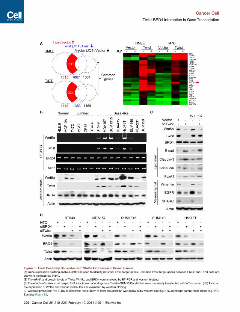
T47D cells that have undergone Twist-mediated EMT (Figures
3G and S5A). We reasoned that genes that are transcriptionally
active in Twist/HMLE and Twist/T47D cells but are downregu-
lated by JQ1 in these cells but not in vector control cells are likely
targets of the Twist-BRD4 complex (Figure 5A). Among the 29
overlapping genes, WNT5A is noted to encode a critical ligand
of both canonical (controlling pluripotency) and noncanonical
(regulating motility and planar cell polarity) Wnt pathways. Upre-
gulation of Wnt5a is correlated with an aggressive phenotype in
melanoma, as well as breast, lung, and prostate tumors (Witze
et al., 2008). We thus selectedWNT5A as an example to charac-
terize the transcriptional mechanism of Twist. We noticed that
TwistWT but not TwistK73R/76R induced Wnt5a expression (Fig-
ures 3G and S3C). Similarly, TwistWT induced EMT and Wnt5a
expression in T47D cells (Figure S5A). In addition, TWIST expres-
sion positively correlates withWNT5A expression in eight micro-
array data sets from human breast cancer (Figure S5B). Using
Twist and Wnt5a antibodies that detect Twist and Wnt5a,
respectively, in xenograft tumors derived from SUM1315 cells,
which express high levels of Twist and Wnt5a, but not MCF7
cells, which express low levels of Twist and Wnt5a (Figure S5C),
we found that Twist is also positively correlated with Wnt5a
expression in breast cancer specimens, with both increased
expression found predominantly in estrogen receptor negative
(ER�) breast cancer (Figure S5D). Further, in 14 breast cell lines
(Figure 5B), both the mRNA and protein levels of Twist and
Wnt5a were found to be largely correlated, with elevated expres-
sions found in BLBC cell lines. BRD4 expression is relatively
constant among normal breast, luminal, and BLBC cell lines
(Figure 5B). Consistently, no significant difference in BRD4
mRNA was found between ER+ and ER� breast cancers from a
477 sample microarray data set (Figure S5E).
We generated a clone of SUM1315 cells with stable knock-
down of Twist. Twist knockdown reduced the mesenchymal
phenotype as these cells were clustered together; cells also
gained expression of epithelial markers and reduced the expres-
sion of mesenchymal markers (Figures 5C and S5F). Ectopic
expression of TwistWT, but not TwistK73R/K76R, restored the
mesenchymal phenotype in these cells. Twist knockdown
also resulted in suppression of Wnt5a expression. Ectopic
expression of TwistWT, but not TwistK73R/K76R, recovered
Wnt5a expression (Figure 5C) and restored the invasiveness
and mammosphere formation in these cells (Figure S5G). These
results are in line with observations in HMLE cells (Figure 3G) and
indicate that the Twist-BRD4 interaction is critical in maintaining
mesenchymal phenotype/characteristics in BLBC cells. Consis-
tently, JQ1 suppressed Wnt5a expression in both Twist/HMLE
and Twist/T47D EMT cells (Figure S5H). In addition, knockdown
of Twist or BRD4 in five BLBC cell lines resulted in reduced
Wnt5a expression; double knockdown of these two molecules
almost completely abolished Wnt5a expression (Figure 5D).
(E) Purified human Twist or histone H4 was incubated with purified Tip60 in the ab
examined by pan-AcK and anti-K73ac/K76ac antibodies.
(F) K73ac/K76ac Twist was immunoprecipitated with K73ac/K76ac antibody in
blotting.
(G) HMLE cells expressing the vector or the WT or K73/76R Twist were examine
expression of E-cadherin, vimentin, Wnt5a, and Twist (green) by immunofluoresc
See also Figure S3.
C
The downregulation of Wnt5a by BRD4-knockdown is specific,
because knockdown of other BET members did not alter
Wnt5a expression (Figure S5I). In addition, knockdown of
BRD4 did not change the expression of either epithelial or
mesenchymal markers (Figure S5J). The downregulation of
Wnt5a correlated with inhibition of invasiveness in BLBC cells;
addition of recombinant Wnt5a partially restored invasiveness
(Figure S5K). Collectively, these results indicate that the Twist-
BRD4 interaction is most likely conserved in HMLE and BLBC
cells for EMT and that this interaction is required for the expres-
sion ofWnt5a, whichmay represent as a bona fide target of Twist
for promoting tumorigenicity in BLBC.
Twist-BRD4 Interaction Is Required for the Recruitmentof BRD4 at the WNT5A SuperenhancerTo delineate how the Twist-BRD4 complex activates WNT5A
expression, we constructed a Twist-Gal4 fusion protein by fusing
Twist N-terminal residues 1–100 to Gal4 DNA-binding domain
(DBD). We also generated several N-terminal deletion mutants
of Twist fused with Gal4-DBD, including DL1-Gal4, DL2-Gal4,
DL3-Gal4, and KR-Gal4 (Figure S6A). When these Twist-Gal4
constructs were coexpressed with the Gal4-luciferase reporter,
Gal4-luciferase activity was moderately increased by about
2-fold compared to the control; coexpression of BRD4 with the
Twist-Gal4 fusion constructs that contain the N-terminal region
required for Tip60-mediated acetylation (i.e., TW and DL1)
greatly enhanced luciferase activity to approximately 8-fold,
suggesting that the N-terminal half of Twist contains transactiva-
tion activity and its interaction with BRD4 boosts this activity.
TheWNT5A promoter contains two Twist-responsive E boxes,
conserved in human and mouse, and located at �160 bp
and �67 bp from transcription start site (TSS) (Figure 6A). We
cloned the human WNT5A promoter (�2,000 bp upstream of
the translation start site) and generated several deletion and E
box mutants of the promoter-luciferase constructs, including
Luc1 (�2,000 bp), Luc2 (�760) and LucEM (�760, two E box
mutations). As expected, Twist alone induced Luc1 and Luc2
promoter luciferase activity; coexpression of BRD4 further
enhanced the Twist-induced Luc1 and Luc2 promoter luciferase
activities (Figure 6A, left panel). In addition, mutation of each E
box (E1M and E2M) in this region reduced, whereas mutation of
both E boxes (EM) completely abolished, Twist-BRD4-mediated
activation of theWNT5A promoter luciferase activity, suggesting
that both E boxes are required for Twist-BRD4-induced tran-
scriptional activation. TwistK73R/K76R mutant partially decreased
WNT5A promoter luciferase activity and was completely insen-
sitive to BRD4-mediated transcriptional activation (Figure 6A,
right panel). BRD4-mediated enhancement of Twist transcrip-
tional activity is specific because other BET members did not
possess this capability, and treatmentwith JQ1orMS417disrup-
ted this BRD4-mediated enhancement (Figure S6B).
sence or presence of acetyl-CoA. The acetylation of Twist and histone H4 was
the presence or absence of JQ1. The bound Twist was examined by western
d for morphological changes indicative of EMT by phase microscopy and the
ence staining. Nuclei were stained with DAPI (red). Scale bars, 50 mM.
ancer Cell 25, 210–225, February 10, 2014 ª2014 Elsevier Inc. 217

CA
D1
(WT)
D1
(YN
)D
1 (D
144H
)D
2 (W
T)D
2 (Y
N)
D2
(H43
7D)
G
BD1 BD2
BDs
Twist
Twist
BD BD
BD
BD BD BD
IgG
IP: T
wis
tB
Ds
B
BDs IP: B
Actin
Twist
BDs
Inpu
t
D
WT 73/76R G
IP: Twist IP: H4IP: BDsWT 73/76R G WT 73/76R G
BD1+2
BD1+2 + - - + - - + - - + + - - + - - + - - + + - - + - - + - - +BD2 - + - - + - - + - - - + - - + - - + - - - + - - + - - + - -BD1 - - + - - + - - + - - - + - - + - - + - - - + - - + - - + -
Vector TwistWT K73/76R
IgG Vector TwistWT K73/76R
IgG Vector TwistWT K73/76R
IgG
Blot:
H4
AcK-H4
BD1/2
Twist
AcK H4(H4ac4)
1 2 3 4 5 6 7 8 9 10 1 2 3 4 5 6 7 8 9 10 1 2 3 4 5 6 7 8 9 10
AcK-Twist(K73ac/76ac)
Figure 4. The Structural and Molecular Basis of Twist-K73ac/K76ac Recognition by BRD4
(A) Stereo ribbon diagram of the 3D solution structure of the BRD4-BD2 bound to a diacetylated K73ac/K76ac Twist peptide (yellow). Side chains of key residues
engaged at the protein/peptide interactions are depicted and color coded by atom type.
(B) Surface electrostatic potential (left) or space-filled (right) representation of the BRD4-BD2/Twsit-K73ac/K76ac complex structure highlights His437 (red) at the
acetyllysine binding site that is responsible for the BRD4-BD20 specificity of this molecular recognition.
(legend continued on next page)
Cancer Cell
Twist-BRD4 Interaction in Gene Transcription
218 Cancer Cell 25, 210–225, February 10, 2014 ª2014 Elsevier Inc.

Cancer Cell
Twist-BRD4 Interaction in Gene Transcription
Chromatin immunoprecipitation (ChIP) analysis revealed that
Twist, BRD4, and acetylated H4 associated at the WNT5A pro-
moter in BT549 and SUM1315 cells, together with P-TEFb and
RNA-Pol II (Figure 6B). A recent study indicated that BRD4prefer-
entially occupied a small subset of superenhancers in transcrip-
tional active key oncogenes that are critical for proliferation and
survival of tumor cells (Loven et al., 2013). Intriguingly, WNT5A
is one of these key oncogenes that contain BRD4-associated
superenhancer, which covers exon1, promoter, and a region up
to 30 kb upstream of the TSS in the WNT5A genomic sequence
in chromosome 3. To examine whether Twist and BRD4 also
bind theWNT5A superenhancer in BLBC, we designed two sets
of ChIP primers that are 22 and 28 kb upstream of the TSS.
ChIP experiments indicated that Twist and BRD4 indeed occu-
pied the WNT5A enhancer together with H3K27ac, a mark of
active enhancer (Figure 6C). Knockdown of Twist or JQ1 treat-
ment inhibited the association of BRD4 at the WNT5A enhancer
(data not shown). Knockdown of Twist or JQ1 treatment also
reduced the presence of BRD4, P-TEFb, and RNA-Pol II at the
WNT5A promoter (Figure 6D). However, JQ1 treatment did not
affect the association of Twist at theWNT5A promoter, suggest-
ing that Twist is required for the recruitment of the BRD4/P-TEFb/
RNA-Pol II complex to the WNT5A promoter. Consistent
with these observations, TwistKR, which could not interact with
BRD4 and failed to rescue Wnt5a expression (Figure 5C), was
unable to recruit the BRD4/P-TEFb/RNA-Pol II complex to the
WNT5A promoter (data not shown). In addition, ectopic expres-
sion of BRD4 increased Twist interaction with P-TEFb and RNA-
Pol II, whereas knockdown of BRD4 reduced the association of
Twist with P-TEFb andRNA-Pol II (Figure 6E). Our results indicate
that Twist recruitsBRD4andacts togetherwithP-TEFbandRNA-
Pol II at theWNT5A promoter/enhancer to activate transcription.
The direct transcriptional activation of WNT5A by the Twist-
BRD4 complex prompted us to investigate the stimuli respon-
sible for Twist acetylation and WNT5A expression. We found
that several stimuli, including TNFa and EGF plus insulin, could
induce Twist acetylation at K73/K76 (Figure S6C). TNFa and
EGF/insulin treatments greatly enhanced the interaction of Twist
with BRD4 and with Tip60, increased K73ac/K76ac of Twist, and
promoted Wnt5a expression (Figure 6F, left panel); JQ1 blocked
the interaction of Twist with BRD4 and thus suppressed Wnt5a
expression (Figure 6F, right panel). Consistent with these find-
ings, TNFa or EGF/insulin treatment greatly enhanced the asso-
ciation of Twist, BRD4, P-TEFb, and RNA-Pol II at the WNT5A
promoter (Figure S6D). Knockdown of Twist suppressed the
association of BRD4 and Twist at the WNT5A promoter; how-
ever, JQ1 treatment did not inhibit the binding of Twist at the
WNT5A promoter (Figure S6E). These data suggest that the as-
sociation of BRD4 at theWNT5A promoter is mediated by Twist.
Although JQ1 was reported to reduce c-Myc expression, we
noticed that JQ1 (1 mM) caused a decrease of c-Myc expression
only in one of five examined BLBC cell lines (Figure 6G). How-
ever, JQ1 reduced Wnt5a expression in all cell lines. The low
(C) HA-tagged Twist was coexpressed with Flag-taggedWT or mutant BD1 and B
antibodies, respectively, and the bound BDs and Twist were analyzed by wester
(D) HA-tagged Twist was coexpressed with Flag-tagged BD1, BD2, and BD1+
association and acetylation of these molecules were examined by western blotti
See also Figure S4 and Table S1.
C
sensitivity to JQ1 in Hs578T cells is likely due to the remarkably
high expression levels of Twist and Wnt5a in this particular cell
line (Figure 5B). Increased JQ1 concentration resulted in
Wnt5a downregulation in a dose-dependent manner in this cell
line (Figure S6F). The downregulation of Wnt5a by JQ1 corre-
lated with its inhibition of invasion and tumorsphere formation
of these cells; addition of recombinant Wnt5a could partially
restore this inhibitory effect (Figures 6H, S6F, and S6G).
Together, these data indicate that the Twist-BRD4 interaction,
enhanced by extracellular signals, is required for the recruitment
of P-TEFb/RNA-Pol II complex to theWNT5A superenhancer for
transcription of WNT5A, which executes, at least in part, the
oncogenic function of Twist. JQ1 disrupts this interaction and
thereby suppresses WNT5A expression in BLBC.
The Twist-BRD4-Wnt5a Axis Is Critical forTumorigenicity in Breast CancerTo further examine the oncogenic role of the Twist-BRD4-Wnt5a
axis and explore the therapeutic potential of BET-specific inhib-
itors for targeting this axis in BLBC in vivo, we established two
Wnt5a knockdown clones in SUM1315 cells. Knockdown of
Wnt5a inhibited the noncanonical Wnt pathway, exemplified by
the downregulation of JNK phosphorylation (Figure 7A). Wnt5a
knockdown also suppressed the canonical Wnt/b-catenin
pathway, indicated by the downregulation of b-catenin and the
suppression of Akt/GSK-3b phosphorylation. These effects
were further confirmed by b-catenin reporter assay (Figure S7A).
Although Wnt5a knockdown did not alter the expression of
epithelial or mensenchymal markers, it did reduce the expres-
sion of several pluripotent molecules (CD44, Sox2, and Oct4).
Consistently, Wnt5a knockdown suppressed invasion and tu-
morsphere formation in these cells (Figure 7B).
In vivo studieswere performed by injection of SUM1315 vector
control cells or Wnt5a knockdown clones into the mammary fat
pads of NOD-SCID mice. When control tumors were approxi-
mately 100 mm3, mice were divided into three groups to receive
daily treatments of JQ1 (50mg/kg), MS417 (20mg/kg), or solvent
control for 2 weeks. We found that knockdown of Wnt5a
completely inhibited tumor growth in SUM1315 cells and
that both JQ1 and MS417 treatments significantly inhibited
tumor growth (Figure 7C). The growth inhibitory effect of JQ1
and MS417 correlated with suppression of Wnt5a expression
and downregulation of proliferative marker Ki67 in these tumors
(Figure S7B). These results suggest that Wnt5a is critical for the
tumorigenicity of BLBC. BET-specific inhibitors suppress the
tumorigenicity of BLBC by inhibiting the Twist-BRD4 interaction
and WNT5A expression.
DISCUSSION
Our study provides several mechanistic insights into how Twist
and BRD4 function cooperatively to activate gene transcription
in EMT and BLBC. First, we show that Twist uses a unique
D2 in HEK293 cells. Twist and BDs were immunoprecipitated with HA and Flag
n blotting.
BD2 in HEK293 cells. After immunoprecipitation of Twist, BDs, and H4, the
ng.
ancer Cell 25, 210–225, February 10, 2014 ª2014 Elsevier Inc. 219
Twist/vectorTwist (JQ1)/Twist
Vector (JQ1)/VectorHMLE
211
JQ1 - + - + - + - + Vector VectorTwist Twist
HMLE T47DA
T47D
Common genes1315 1067 1301
334
CB
570A 4 315
359 31498T 59
Normal Luminal Basal-like
Vector + - - -hT i t
WT KR
1713 1353 1169M
DA
1
Wnt5a
Twist
HM
LE
MC
F10
T47D
MC
F7
ZR75
BT4
74
SU
M1
MD
A4
BT5
4 9
MD
A2
SU
M1
Hs5
7 8
SU
M1
PC
R
Twist
Wnt5a
BRD4
shTwist - + + +
E-cad
BRD4
Actin
Wnt5a
RT-
P E-cad
Claudin-3E
pith
elia
l
FoxA1
Occlaudin
Actin
Twist
BRD4
Wes
tern
blo
ts
Vimentin
EGFR
SPARCMes
ench
ymal
D
Wnt5a
NTC + - - - + - - - + - - - + - - - + - - -siBRD4 - + - + - + - + - + - + - + - + - + - +siTwist - - + + - - + + - - + + - - + + - - + +
MDA157BT549 SUM1315 SUM149 Hs578T
Actin
Wnt5a
Twist
BRD4
Actin
Figure 5. Twist Positively Correlates with Wnt5a Expression in Breast Cancer
(A) Gene expression profiling analysis (left) was used to identify potential Twist target genes. Common Twist target genes between HMLE and T47D cells are
shown in the heatmap (right).
(B) The mRNA and protein levels of Twist, Wnt5a, and BRD4 were analyzed by RT-PCR and western blotting.
(C) The effects of stable small hairpin RNA knockdown of endogenous Twist in SUM1315 cells that were transiently transfected with WT or mutant (KR) Twist on
the expression of Wnt5a and various molecules was evaluated by western blotting.
(D)Wnt5a expression in fiveBLBCcell lineswith knockdownof Twist and/or BRD4was analyzed bywestern blotting. NTC, nontarget control small interfering RNA.
See also Figure S5.
Cancer Cell
Twist-BRD4 Interaction in Gene Transcription
220 Cancer Cell 25, 210–225, February 10, 2014 ª2014 Elsevier Inc.

A SUM1315
RNA-PolII
NTC + - +siBRD4 - + -
IgGTSS
Luc1 (-2000bp)
Luc2 (-760bp)
ATG-67-160
E2E1
H:ACATTTGGM:ACATTTGG
H:ACAGTTGAM:ACAGTTGA
ATG RNA-PolII
HEK293
t
Flag-BRD4 - + +HA-Twist + + +
IgGE
E1 E2
Luc1 Luc2 Luc2
BRD4
Twist
BRD4
P-TEFb
Flag-BRD4
HA-Twist
Flag-BRD4IP: T
wis
t
P-TEFb
nt5a
luci
fera
se VectorTWWT
BRD4TWWT+BRD4
TWKR
TWKR+BRD43
6
9
Luc1WT E1M E2M EM
Luc2 Luc2
1
2
3
4
5
2.0
2.5
0.8
1.0 SP CPBT549 SUM1315
t
SP CP
RNA-PolII
Twist
P-TEFbB
RNA-PolII
HA-Twist
Inpu
t
P-TEFb
s
TwistBRD4
Wn
00
1
0.0
0.5
1.0
1.5
0.0
0.2
0.4
0.6
% In
put
EG
F+In
s
BRD4
K73/76acC
tr
FGF
TNFα
IgG
IgG
+JQ
1
+JQ
1
-JQ
1
-JQ
1
Ctr TNFα: T
wis
tF
BRD4
Pol IIP-TEFbIgG
H4ac4
C SUM1315BT549
Twist
Twist
Wnt5a
IPnp
ut
Tip60
% In
put
0.6
1.2
1.8
0.6
1.2
1.8
2.4 SP CPSP CPTwistBRD4
H3K27acIgG
H4ac4
D
BRD4In
Tip60
G
1.0
1.5
2.0
nput
SP CPControl siTwist JQ1
SP CP SP CP
0.0 0.0
TwistBRD4
Pol IIH4ac4
BT549 MDA157 1315 SUM149JQ1 - + - + - + - + - +
c-Myc
Hs578T
0.0
0.5% I
H
Pol IIP-TEFbIgG
NTC JQ1SUM149BT549 Hs578TSUM1315MDA157
(%)
)
SUM149BT549 Hs578TSUM1315MDA157NTC JQ1
Wnt5a
Actin
100
125 150
Tum
orsp
here
(
Wnt5a - + - + - + - + - +
Inva
sion
(%
Wnt5a - + - + - + - + - +0
25
50
75
100
0
50
100
Figure 6. The Twist-BRD4 Complex Directly Activates WNT5A Transcription
(A) Schematic depiction of theWNT5A promoter andWNT5A reporter luciferase constructs used (top). Enhancement ofWnt5a luciferase activity by coexpression
of Twist and BRD4 in HEK293 cells is shown (bottom).
(B) Twist, BRD4, H4ac4, RNA-Pol II, and P-TEFb (CDK9) association at theWNT5A promoter as assessed by ChIP. SP, specific primer; CP, control primer (5 kb
downstream of the 30 untranslated region).
(C) Twist, BRD4, H4ac, and H3K27ac association at the WNT5A enhancer as assessed by ChIP. SP, specific primer (22 kb upstream of the TSS); CP, control
primer (5 kb downstream of the 30 untranslated region).
(legend continued on next page)
Cancer Cell
Twist-BRD4 Interaction in Gene Transcription
Cancer Cell 25, 210–225, February 10, 2014 ª2014 Elsevier Inc. 221

Cancer Cell
Twist-BRD4 Interaction in Gene Transcription
mechanism for recruiting BRD4 in gene transcription (Figure 7D).
Although BRD4 is the key transcriptional regulator, it lacks
specific DNA binding motif. How BRD4 and its associated tran-
scriptional complex are recruited to gene-specific promoters/
enhancers remains elusive. We found that Twist contains an
‘‘H4-mimic’’ GK-X-GK motif and becomes diacetylated by
Tip60, which also acetylates multiple lysine residues in histone
H4 including K5 and K8. By binding to BRD4-BD2 via the
K73ac/K76ac motif, Twist recruits BRD4 to target gene pro-
moters/enhancers through the recognition of and interaction
with E boxes by its bHLH domain. Once localized in the chro-
matin, BRD4-BD1 binds with acetylated H4-K5ac/K8ac to facil-
itate the docking of the BRD4 complex on promoters/enhancers
and thereby activates pause release factor P-TEFb to phosphor-
ylate and release RNA-Pol II for WNT5A transcription.
Our study demonstrates that the two BDs of BRD4 have
distinct functions and binding specificities for acetylated pro-
teins in transcription. Although a single BD1 or BD2 of BRD4 is
individually capable of interacting with acetylated H4 in vitro,
only BD1 is engaged in the binding with acetylated H4 in the
tandem BD1+BD2. This is consistent with the observation that
a single BD1 of Brdt binds to acetylated histone H4 nearly as
well as Brdt (full length; contains BD1+BD2) (Moriniere et al.,
2009) and that BRD4-BD1 specifically recognizes acetylation
marks on H4, whereas BRD4-BD2 has broad binding specificity
for diacetylated substrates (Filippakopoulos et al., 2012). In line
with this contention, only BRD4-BD2 interacts with Twist-
K73ac/K76ac. We found that charged amino acid residues
(D144 in BD1, H437 in BD2) surrounding the acetyllysine-binding
pocket of BDs contributed to the binding specificity of BD1 and
BD2. Additional residues beyond the diacetylation motif further
contribute to Twist’s association with BRD4-BD2. Notably, it
has recently been reported that BRD4 is phosphorylated by
CK2 on several Ser residues in the C-terminal region of BD2
and that these phosphorylations were suggested to affect the
interaction of BRD4 with acetylated histones and transcriptional
cofactors (Wu et al., 2013). Although a single BD2 of BRD4 can
interact with either acetylated H4 or Twist-K73ac/K76ac individ-
ually, the tandem BD1+BD2 of BRD4 apparently form a ternary
complex with two acetylated proteins in that diacetylated H4 is
bound to BD1 and diacetylated Twist is bound to BD2. These
results suggest that BRD4 utilizes its tandem BDs as an integra-
tion platform to cooperatively interact with H4 and Twist in
assembling the integrated transcriptional complex containing
P-TEFb and RNA-Pol II at target gene promoters/enhancers.
Notably, several transcription-associated proteins that contain
(D) Effects of Twist knockdown or JQ1 treatment on the association of Twist, B
assessed by ChIP in SUM1315 cells. SP and CP primers are same as in (B).
(A–D) Statistical analysis (mean ± SD) from three separate experiments in triplica
(E) Assessment of the effects of transient expression of HA-Twist and/or Flag-BRD
and assessment of the effect of endogenous BRD4 knockdown on the associatio
shown.
(F) SUM1315 cells were serum starved for overnight followed by stimulation with F
Twist was immunoprecipitated and K73ac/K76ac of Twist and the association of
were also examined by western blotting (left).
(G) BLBC cells were treated with JQ1. Expression of c-Myc and Wnt5a was ana
(H) Invasion (left) and tumorsphere formation (right) assays of cells treated as in (G
Statistical analysis (mean ± SD) from three independent experiments with duplic
See also Figure S6.
222 Cancer Cell 25, 210–225, February 10, 2014 ª2014 Elsevier Inc.
tandem binding modules have been shown to engage in combi-
natorial recognition of different posttranslational modifications
(PTMs) in histones for the assembly of transcriptional complexes
(Zeng et al., 2010). For example, the tandem PHD-BD module in
BPTF specifically recognizes a combination of H3K4me3 and
H4K16ac in gene activation (Ruthenburg et al., 2011). Our results
not only support this notion, but also extend the functionality of
these tandem binding modules in directing gene transcriptional
activation as exemplified by the tandem BDs of BRD4 in bridging
histone and nonhistone transcription factor.
Notably, these histone-mimic sequences contain lysine/argi-
nine-rich resides, which are often viewed conventionally as a
nuclear-localization signal (NLS), as in the case of Twist. How-
ever, TwistKR mutant, which cannot be acetylated but still re-
sides in the nucleus, fails to interact with BRD4 and is unable
to induce EMT and WNT5A expression. Our results suggest
that theses lysine/arginine-rich ‘‘potential’’ NLS motifs in tran-
scription factors may have previously unrecognized histone-
mimic functions. Consistent with our findings, histone-mimic
sequences are deployed by influenza nonstructural protein 1
(NS1) in inhibiting human transcription elongation complex in
the antivirus response and by HP1 in forming HP1-chromatin
complex (Canzio et al., 2013; Marazzi et al., 2012). We believe
that PTMs on histone-mimic sequences present in nonhistone
proteins likely play an important role, via conserved molecular
mechanisms as seen with those PTMs in histone, in governing
the assembly and function of transcriptional complexes in
chromatin.
Second, our study demonstrates that Twist is a transcriptional
activator responsible for WNT5A expression in BLBC. Twist has
been shown to bind to the E-cadherin promoter to repress
transcription in a way similar to that of Snail. However, this
contradicts the role of Twist in development, where it acts as a
transcriptional activator to upregulate mesoderm-specific genes
in Drosophila. When the bHLH domain of Twist was replaced
with Gal4-DBD, we found that the Twist-Gal4-DBD fusion was
sufficient to activate gene expression, indicating that Twist
functions as a transcriptional activator. We further show that
Twist recruits BRD4 and the associated P-TEFb and RNA-Pol
II to the WNT5A promoter/enhancer to directly activate WNT5A
expression, which is required for invasion and the maintenance
of CSC-like properties of BLBC. Notably, Wnt5a is induced in
epithelial cells during EMT and required for maintenance of
CSC-like properties in the resulting mesenchymal cells (Scheel
et al., 2011). In addition, Wnt5a expression is required for the
loss of cell-cell contacts, allowing cells to migrate to the edge
RD4, RNA-Pol II, and P-TEFb (CDK9) and H4ac4 at the WNT5A promoter as
tes is shown.
4 on their association with RNA-Pol II and P-TEFb (CDK9) in HEK293 cells (left)
n of Twist, BRD4, RNA-Pol II, and P-TEFb (CDK9) in SUM1315 cells (right) are
GF, TNFa, or EGF plus insulin for 3 hr in the absence or presence of JQ1 (right).
Tip60 and BRD4 were analyzed. Expression of Wnt5a, Twist, BRD4 and Tip60
lyzed by western blotting.
) were examined in the absence or present of recombinant Wnt5a (100 ng/ml).
ates is shown.

A C
Wnt5a
Vec
tor
shR
1
shR
2
SUM1315
p JNKnoni
cal B
efor
e
Control JQ1 MS417 shR1 shR2
1200 Ctr
β-catenin
JNK
p-JNK
pAkt
Akt
Non
-can
onic
al
Afte
r
400
800Tu
mor
siz
e (m
m3 ) JQ1
MS417shR1shR2
CD44
GSK3β
Akt
pGSK3βCan
Sox2pote
ncy
0
T
15 45 60 63 65 67 69 71 73 (day)
Sox2
Oct4
E-cad
Claudin-3
Occlaudinpith
elia
lP
luri p
Control
g)
Ctr2.0
Vimentin
Occlaudin
EGFR
SPARC
Ees
ench
ymal
FoxA1 JQ1
MS417
shR1
shR2
Tum
or w
eigh
t (g JQ1
MS417shR1shR2
0.5
1.0
1.5
B D
%)
e (%
)
Vector90
120
90
120
Actin
SPARCM 0.0
Inva
sion
(%
Tum
orsp
here Vector
shR1shR2
0
30
60
0
30
60
Vector shR1 shR2
Figure 7. The Twist-BRD4-Wnt5a Axis Is Critical for Tumorigenicity In Vitro and In Vivo
(A) Expression of various molecules in SUM1315 cells with Wnt5a knockdown.
(B) Invasion and tumorsphere formation in SUM1315 cells with Wnt5a knockdown. Data are presented as a percentage of vector control values (mean ± SD in
three separate experiments in duplicates). Representative pictures of tumorspheres are shown at the bottom. Scale bar, 100 mM.
(C) Vector control and Wnt5a knockdown SUM1315 cells were injected into the mammary fat pad of NOD-SCID mice. When tumors from mice injected with
control cells reached 100mm3,micewere divided into three groups and treatedwith JQ1 (50mg/Kg), MS417 (20mg/Kg), or solvent control, respectively. The size
(legend continued on next page)
Cancer Cell
Twist-BRD4 Interaction in Gene Transcription
Cancer Cell 25, 210–225, February 10, 2014 ª2014 Elsevier Inc. 223

Cancer Cell
Twist-BRD4 Interaction in Gene Transcription
of wounds, and is also necessary for intestinal epithelial stem
cells to regenerate damage tissues during wound healing and
tissue repair (Miyoshi et al., 2012). The correlated expression
of Twist and Wnt5a in BLBC supports our contention that the
Twist-BRD4-Wnt5a signaling axis plays a critical role in the
development and progression of BLBC.
Third, our study indicates that the Twist-BRD4 interaction
represents a druggable target for treating BLBC. Although Twist
is highly expressed in BLBC, the absence of a clear ligand-bind-
ing domain in Twist creates a formidable obstacle toward devel-
oping small molecules that inhibit its activity as a transcription
factor. We found that BET-specific BD inhibitors disrupted the
Twist-BRD4 interaction and resulted in significant Wnt5a reduc-
tion, leading to inhibition of invasion and tumorigenicity of BLBC
cells in vitro and in vivo. Based on our mechanistic understand-
ing of Twist-BRD4 interaction in gene transcription, we predict
that selective chemical inhibition of BRD4/H4 interaction would
result in a broad inhibition of BRD4 functions as chromatin regu-
lator in gene transcription, whereas selective inhibition of the
BRD4/transcription factor association might affect specific tran-
scription factor’s ability in their target gene activation. BD inhib-
itors selectively target BD2 over BD1 of BRD4 are needed to
address these questions; they will also further functionally
validate the effectiveness and therapeutic benefits of targeting
BRD4 for treating BLBC.
EXPERIMENTAL PROCEDURES
Protein Purification and Mass Spectrometry Analysis
We generated a clone of HeLa S3 cells with stable expression of Flag-Twist (Li
and Zhou, 2011). After enriching the nuclear extracts from 40 l of suspension
culture, we carried out affinity protein purification with Flag affinity columns.
The final eluted immunocomplexes were separated on SDS-PAGE, and the
bound proteins were excised from the gel and subjected to nano-liquid chro-
matography-tandem mass spectrometry (nano-LC-MS/MS) analysis (Applied
Biomics). For identification of the acetylated lysine residues on Twist, the acet-
ylated Twist was digested with trypsin, and the tryptic peptides were analyzed
by LC-MS/MS using an LTQVelos Orbitrapmass spectrometer (Thermo Fisher
Scientific) coupled with a nano-LC Ultra/CHiPLC Nanoflex high-performance
liquid chromatography system (Eksigent) through a nanoelectrospray ioniza-
tion source (Li et al., 2013). MS/MS data were acquired using CID fragmenta-
tion of selected peptides during the information-dependent acquisition. The
LC-MS/MS results were subjected to protein identification and acetylation
sites determination using ProteomeDiscoverer 1.3 software (Thermo Fisher
Scientific) and MASCOT server.
Protein Structure Analysis by NMR
The NMR spectral collection, analysis, and structure determination of the
BRD4-BD2 with Twist-K73ac/K76ac were performed as previously reported
(Zhang et al., 2012). In brief, NMR samples contained a protein/peptide com-
plex of 0.5 mM in a 100 mM sodium phosphate buffer (pH 6.5) that contains
5 mM perdeuterated dithiothreitol and 0.5 mM EDTA in H2O/2H2O (9/1) or2H2O. All NMR spectra were collected at 30�C on NMR spectrometers of
800, 600, or 500 MHz. The 1H, 13C, and 15N resonances of the protein in the
complex were assigned by triple-resonance NMR spectra collected with a13C/15N-labeled and 75% deuterated BRD4-BD2 bound to an unlabeled Twist
of tumor was recorded by bioluminescence imaging before or after 2 week treatm
from five mice.
(D) A proposed model illustrating the interaction of Twist and BRD4 at the enhanc
expression in EMT and BLBC.
See also Figure S7.
224 Cancer Cell 25, 210–225, February 10, 2014 ª2014 Elsevier Inc.
peptide (Clore and Gronenborn, 1994). The distance restraints were obtained
from 3D 13C-NOESY or 15N-NOESY spectra. Protein structures were calcu-
lated with a distance geometry-simulated annealing protocol using X-PLOR
(Brunger, 1993) that was aided with iterative automated NOE assignment by
ARIA for refinement (Nilges and O’Donoghue, 1998). Structure quality was
assessed by PROCHECK-NMR (Laskowski et al., 1996). The structure of the
protein/ligand complex was determined using intermolecular NOE-derived
distance restraints that were obtained from 13C-edited (F1),13C/15N-filtered
(F3) 3D NOESY spectra.
Immunoprecipitation, Immunoblotting, Immunofluorescence,
Immunohistochemical Staining, RT-PCR, and ChIP
Detailed methods are provided in the Supplemental Experimental Procedures.
Tumorigenesis Assay
All procedures were approved by the Institutional Animal Care and Use
Committee at the University of Kentucky College of Medicine and conform
to the legal mandates and federal guidelines for the care and maintenance
of laboratory animals. Animals were maintained and treated under path-
ogen-free conditions. Female NOD-SCID mice (6–8 weeks old; Taconic)
were injected with breast cancer SUM1315 (2 3 106 cells/mouse) cells via
mammary fat pad, and mice had three groups: vector control and two stable
clones with Wnt5a-knockdown expression. Tumor growth was monitored
with caliper measurements. When tumors were approximately 1.0 cm in
size, mice were euthanized and tumors excised. Data were analyzed by Stu-
dent’s t test; p < 0.05 was considered significant.
Statistical Analysis
Data are presented as mean ± SD. A Student’s t test (two-tailed) was used to
compare two groups (p < 0.05 was considered significant) unless otherwise
indicated.
ACCESSION NUMBERS
Microarray data of Twist expression in HMLE and T47D cells with or without
JQ1 treatment were deposited at the Gene Expression Omnibus database
with the accession number GSE53222. Structure factors and coordinates for
the second bromodomain of BRD4 in complex with K73ac/K76ac diacetylated
Twist peptide were deposited at the Protein Data Bank under ID code 2MJV,
and the NMR spectral data were deposited at the BioMagResBank (BMRB)
under BMRB accession number 19738.
SUPPLEMENTAL INFORMATION
Supplemental Information includes Supplemental Experimental Procedures,
seven figures, and one table and can be found with this article online at
http://dx.doi.org/10.1016/j.ccr.2014.01.028.
ACKNOWLEDGMENTS
We thank Cathy Anthony for critical reading and editing of this manuscript. We
also thank Dr. Jing Chen for technical assistance on mass spectrometry anal-
ysis. We are grateful to Dr. James E. Bradner for providing JQ1, Dr. Robert A.
Weinberg for HMLE cells, Dr. Michael Rosenblatt for SUM1315 cells, and Dr.
Bruno Amati for Tip60 antibodies as valuable reagents for this study. This
work was supported in part by grants from the National Institute of Health
(CA125454 to B.P.Z., P20CA153043 to B.M.E., and HG004508 and
CA87658 to M.-M.Z.), the American Cancer Society (RSG13187 to Y.W.),
and the Nature Science Foundation of China (91129303 to J.D.). We acknowl-
edge the University of Kentucky Proteomics Core, which is partially supported
ent. Tumor weight was also measured. Data are represented as a mean ± SEM
er/promoter ofWNT5A, which leads to the transcriptional activation ofWNT5A

Cancer Cell
Twist-BRD4 Interaction in Gene Transcription
by grants from the National Cancer Institute (P30CA177558) and the National
Institute of General Medical Sciences (P20GM103486).
Received: June 9, 2013
Revised: September 23, 2013
Accepted: January 24, 2014
Published: February 10, 2014
REFERENCES
Brunger, A.T. (1993). X-PLOR version 3.1: A system for X-ray Crystallography
and NMR (version 3.1 edit.). (New Haven: Yale University Press).
Canzio, D., Liao, M., Naber, N., Pate, E., Larson, A., Wu, S., Marina, D.B.,
Garcia, J.F., Madhani, H.D., Cooke, R., et al. (2013). A conformational switch
in HP1 releases auto-inhibition to drive heterochromatin assembly. Nature
496, 377–381.
Clore, G.M., and Gronenborn, A.M. (1994). Multidimensional heteronuclear
nuclear magnetic resonance of proteins. Methods Enzymol. 239, 349–363.
Dawson, M.A., Prinjha, R.K., Dittmann, A., Giotopoulos, G., Bantscheff, M.,
Chan, W.I., Robson, S.C., Chung, C.W., Hopf, C., Savitski, M.M., et al.
(2011). Inhibition of BET recruitment to chromatin as an effective treatment
for MLL-fusion leukaemia. Nature 478, 529–533.
Delmore, J.E., Issa, G.C., Lemieux, M.E., Rahl, P.B., Shi, J., Jacobs, H.M.,
Kastritis, E., Gilpatrick, T., Paranal, R.M., Qi, J., et al. (2011). BET bromodo-
main inhibition as a therapeutic strategy to target c-Myc. Cell 146, 904–917.
Doyon, Y., and Cote, J. (2004). The highly conserved andmultifunctional NuA4
HAT complex. Curr. Opin. Genet. Dev. 14, 147–154.
Filippakopoulos, P., Picaud, S., Mangos, M., Keates, T., Lambert, J.P.,
Barsyte-Lovejoy, D., Felletar, I., Volkmer, R., Muller, S., Pawson, T., et al.
(2012). Histone recognition and large-scale structural analysis of the human
bromodomain family. Cell 149, 214–231.
Jang, M.K., Mochizuki, K., Zhou, M., Jeong, H.S., Brady, J.N., and Ozato, K.
(2005). The bromodomain protein Brd4 is a positive regulatory component of
P-TEFb and stimulates RNA polymerase II-dependent transcription. Mol.
Cell 19, 523–534.
Laskowski, R.A., Rullmannn, J.A., MacArthur, M.W., Kaptein, R., and
Thornton, J.M. (1996). AQUA and PROCHECK-NMR: programs for checking
the quality of protein structures solved by NMR. J. Biomol. NMR 8, 477–486.
Leptin, M. (1991). twist and snail as positive and negative regulators during
Drosophila mesoderm development. Genes Dev. 5, 1568–1576.
Li, J., and Zhou, B.P. (2011). Activation of b-catenin and Akt pathways by Twist
are critical for the maintenance of EMT associated cancer stem cell-like char-
acters. BMC Cancer 11, 49.
Li, X., Zhou, Q., Sunkara, M., Kutys, M.L., Wu, Z., Rychahou, P., Morris, A.J.,
Zhu, H., Evers, B.M., and Huang, C. (2013). Ubiquitylation of phosphatidylino-
sitol 4-phosphate 5-kinase type I g by HECTD1 regulates focal adhesion
dynamics and cell migration. J. Cell Sci. 126, 2617–2628.
Loven, J., Hoke, H.A., Lin, C.Y., Lau, A., Orlando, D.A., Vakoc, C.R., Bradner,
J.E., Lee, T.I., and Young, R.A. (2013). Selective inhibition of tumor oncogenes
by disruption of super-enhancers. Cell 153, 320–334.
Marazzi, I., Ho, J.S., Kim, J., Manicassamy, B., Dewell, S., Albrecht, R.A.,
Seibert, C.W., Schaefer, U., Jeffrey, K.L., Prinjha, R.K., et al. (2012).
C
Suppression of the antiviral response by an influenza histone mimic. Nature
483, 428–433.
Miyoshi, H., Ajima, R., Luo, C.T., Yamaguchi, T.P., and Stappenbeck, T.S.
(2012). Wnt5a potentiates TGF-b signaling to promote colonic crypt regenera-
tion after tissue injury. Science 338, 108–113.
Moriniere, J., Rousseaux, S., Steuerwald, U., Soler-Lopez, M., Curtet, S., Vitte,
A.L., Govin, J., Gaucher, J., Sadoul, K., Hart, D.J., et al. (2009). Cooperative
binding of two acetylation marks on a histone tail by a single bromodomain.
Nature 461, 664–668.
Nilges, M., and O’Donoghue, S. (1998). Ambiguous NOEs and automated NOE
assignment. Prog. Nucl. Magn. Reson. Spectrosc. 32, 107–139.
Rakha, E.A., Reis-Filho, J.S., and Ellis, I.O. (2008). Basal-like breast cancer: a
critical review. J. Clin. Oncol. 26, 2568–2581.
Rekowski, Mv., and Giannis, A. (2010). Histone acetylation modulation by
small molecules: a chemical approach. Biochim. Biophys. Acta 1799,
760–767.
Ruthenburg, A.J., Li, H., Milne, T.A., Dewell, S., McGinty, R.K., Yuen, M.,
Ueberheide, B., Dou, Y., Muir, T.W., Patel, D.J., and Allis, C.D. (2011).
Recognition of a mononucleosomal histone modification pattern by BPTF
via multivalent interactions. Cell 145, 692–706.
Scheel, C., Eaton, E.N., Li, S.H., Chaffer, C.L., Reinhardt, F., Kah, K.J., Bell, G.,
Guo,W., Rubin, J., Richardson, A.L., andWeinberg, R.A. (2011). Paracrine and
autocrine signals induce andmaintainmesenchymal and stem cell states in the
breast. Cell 145, 926–940.
Thiery, J.P., Acloque, H., Huang, R.Y., and Nieto, M.A. (2009). Epithelial-
mesenchymal transitions in development and disease. Cell 139, 871–890.
Witze, E.S., Litman, E.S., Argast, G.M., Moon, R.T., and Ahn, N.G. (2008).
Wnt5a control of cell polarity and directional movement by polarized redistri-
bution of adhesion receptors. Science 320, 365–369.
Wu, S.Y., Lee, A.Y., Lai, H.T., Zhang, H., and Chiang, C.M. (2013). Phospho
switch triggers Brd4 chromatin binding and activator recruitment for gene-
specific targeting. Mol. Cell 49, 843–857.
Zeitlinger, J., Zinzen, R.P., Stark, A., Kellis, M., Zhang, H., Young, R.A., and
Levine, M. (2007). Whole-genome ChIP-chip analysis of Dorsal, Twist, and
Snail suggests integration of diverse patterning processes in the Drosophila
embryo. Genes Dev. 21, 385–390.
Zeng, L., Zhang, Q., Li, S., Plotnikov, A.N., Walsh, M.J., and Zhou,M.M. (2010).
Mechanism and regulation of acetylated histone binding by the tandem PHD
finger of DPF3b. Nature 466, 258–262.
Zhang, G., Liu, R., Zhong, Y., Plotnikov, A.N., Zhang, W., Zeng, L., Rusinova,
E., Gerona-Nevarro, G., Moshkina, N., Joshua, J., et al. (2012). Down-regula-
tion of NF-kB transcriptional activity in HIV-associated kidney disease by
BRD4 inhibition. J. Biol. Chem. 287, 28840–28851.
Zhou, Q., Li, T., and Price, D.H. (2012). RNA polymerase II elongation control.
Annu. Rev. Biochem. 81, 119–143.
Zuber, J., Shi, J., Wang, E., Rappaport, A.R., Herrmann, H., Sison, E.A.,
Magoon, D., Qi, J., Blatt, K., Wunderlich, M., et al. (2011). RNAi screen iden-
tifies Brd4 as a therapeutic target in acute myeloid leukaemia. Nature 478,
524–528.
ancer Cell 25, 210–225, February 10, 2014 ª2014 Elsevier Inc. 225