Embed Size (px)
Citation preview

BioMed CentralC
TIONALINTERNACANCER CELLCancer Cell International
ss
Open AccePrimary researchIntrinsic anticarcinogenic effects of Piper sarmentosum ethanolic extract on a human hepatoma cell lineShahrul Hisham Zainal Ariffin*1, Wan Haifa Haryani Wan Omar1, Zaidah Zainal Ariffin*2, Muhd Fauzi Safian2, Sahidan Senafi1 and Rohaya Megat Abdul Wahab3Address: 1School of Bioscience and Biotechnology, Faculty of Science and Technology, Universiti Kebangsaan Malaysia, 43600, Selangor Darul Ehsan, Malaysia, 2Department of Microbiology, Faculty of Applied Science, Universiti Teknologi MARA, 40450, Shah Alam, Selangor, Malaysia and 3Department of Orthodontic, Faculty of Dentistry, Universiti Kebangsaan Malaysia, 50300, Kuala Lumpur, Malaysia
Email: Shahrul Hisham Zainal Ariffin* - [email protected]; Wan Haifa Haryani Wan Omar - [email protected]; Zaidah Zainal Ariffin* - [email protected]; Muhd Fauzi Safian - [email protected]; Sahidan Senafi - [email protected]; Rohaya Megat Abdul Wahab - [email protected]
* Corresponding authors
AbstractBackground: Piper sarmentosum, locally known as kaduk is belonging to the family of Piperaceae.It is our interest to evaluate their effect on human hepatoma cell line (HepG2) for the potential ofanticarcinogenic activity.
Results: The anticarcinogenic activity of an ethanolic extract from Piper sarmentosum in HepG2and non-malignant Chang's liver cell lines has been previously determined using (3-[4,5-dimethylthiazol-2-yl]-2,5-diphenyl-tetrazolium bromide) (MTT) assays, where the IC50 value wasused as a parameter for cytotoxicity. The ethanolic extract that showed anticarcinogenicproperties in HepG2 cells had an IC50 of 12.5 μg mL-1, while IC50 values in the non-malignantChang's liver cell line were greater than 30 μg mL-1. Apoptotic morphological changes in HepG2cells were observed using an inverted microscope and showed chromatin condensation, cellshrinkage and apoptotic bodies following May-Grunwald-Giemsa's staining. The percentage ofapoptotic cells in the overall population (apoptotic index) showed a continuously significantincrease (p < 0.05) in 12.5 μg mL-1 ethanolic extract-treated cells at 24, 48 and 72 hours comparedto controls (untreated cells). Following acridine orange and ethidium bromide staining, treatmentwith 10, 12 and 14 μg mL-1 of ethanolic extracts caused typical apoptotic morphological changes inHepG2 cells. Molecular analysis of DNA fragmentation was used to examine intrinsic apoptosisinduced by the ethanolic extracts. These results showed a typical intrinsic apoptoticcharacterisation, which included fragmentation of nuclear DNA in ethanolic extract-treatedHepG2 cells. However, the non-malignant Chang's liver cell line produced no DNA fragmentation.In addition, the DNA genome was similarly intact for both the untreated non-malignant Chang'sliver and HepG2 cell lines.
Conclusion: Therefore, our results suggest that the ethanolic extract from P. sarmentosuminduced anticarcinogenic activity through an intrinsic apoptosis pathway in HepG2 cells in vitro.
Published: 3 March 2009
Cancer Cell International 2009, 9:6 doi:10.1186/1475-2867-9-6
Received: 9 December 2008Accepted: 3 March 2009
This article is available from: http://www.cancerci.com/content/9/1/6
© 2009 Ariffin et al; licensee BioMed Central Ltd. This is an Open Access article distributed under the terms of the Creative Commons Attribution License (http://creativecommons.org/licenses/by/2.0), which permits unrestricted use, distribution, and reproduction in any medium, provided the original work is properly cited.
Page 1 of 9(page number not for citation purposes)

Cancer Cell International 2009, 9:6 http://www.cancerci.com/content/9/1/6
BackgroundHuman hepatocellular carcinoma is the fifth most com-mon cancer in the world and the fourth most commoncause of cancer-associated mortality [1]. Surgical resectionand local treatment are frequently limited due to metasta-sis, cirrhosis, and other pathological changes in the liverparenchyma. The synchronous occurrence of humanhepatocellular carcinoma may be due to different risk fac-tors such as chronic viral hepatitis B or hepatitis C infec-tion, aflotoxin explosure, alcohol consumption and ironoverload [2]. The development of chemotherapeutic orchemopreventive agents for hepatocellular carcinoma isimportant in order to help reduce the mortality caused bythis disease [3]. Thus, significant research efforts havefocused on novel chemotherapeutic drugs from the plantkingdom in search of cancer inhibitors and cures [4].
Plants have many phytochemicals with various bioactivities,including antioxidant, anti-inflammatory and anticancerfunctions. For example, some studies have reported thatextracts from natural products such as fruits, vegetables andmedicinal herbs have positive effects against cancer com-pared with chemotherapy or recent hormonal treatments[5]. The family of Piperaceae belonging to superorder Nym-phaeifloraea, order Piperales [6], comprises about 10 generaand 2,000 species [7]. The genus Piper (Piperaceae) is largelydistributed in tropical and subtropical regions of the world.Chemical studies have shown that the genus Piper has manycomponents including unsaturated amides, flavonoids, lign-ans, aristolactams, long and short chain esters, terpenes, ster-oids, prophenylphenols, and alkaloids [8,9]. Some Piperspecies are used in folk medicine to treat many diseases,including fever, jaundice, rheumatism and neuralgia [7]. InMalaysia, P. sarmentosum is locally known as kaduk and iscommonly used in folk medicine as a carminative. Theleaves and roots of this plant are used for the treatment oftoothaches, fungal dermatitis on the feet, asthmatic cough-ing and pleurisy [10]. In addition, the plant and its fruits areused as an expectorant [11].
Previous studies have investigated other biological activi-ties including the anti-inflammatory effects of Peperomiapellucida [12] and the antimicrobial effects of Piper anducum[13,14]. Chloroform extracts from Piper sarmentosum havealso shown considerable antimalarial activity against Plas-modium falciparum (in vitro) and Plasmodium berghei (in vivo)[15]. The water extract of the entire plant showed ahypoglycaemic effect in rats [16], while the methanolicextract from the leaves of Piper sarmentosum exhibited peakantioxidant activity [17]. However, the properties of thisplant, and especially its anticarcinogenic activity, have notyet been investigated. The objective of this study was toevaluate the anticarcinogenic properties and mode ofaction of the Piper sarmentosum ethanolic extract in ahuman hepatoma cell line (HepG2). The Chang's liver cellline was used as a non-malignant cell for cytotoxic activity.
Results and discussionCytotoxic activity of P. sarmentosum ethanolic extract on cellsIn this study, we investigated the effects of a crude eth-anolic extract from P. sarmentosum in HepG2 and non-malignant Chang's liver cell lines. In the first part of thisstudy, the antiproliferative properties of the ethanolicextract from P. sarmentosum were predetermined using anMTT assay. The principle of this assay is based on thereduction of a soluble tetrazolium salt, by mitochondrialdehydrogenase activity of viable tumour cells, into a solu-ble coloured formazan product that can be measuredspectrophotometrically after dissolution [18]. The IC50value was used as a parameter for cytotoxicity.
The criterion for cytotoxicity for the crude extracts, as estab-lished by the National Cancer Institute (NCI), is an IC50value lower than 30 μg mL-1 [19]. Figure 1 showed that theP. sarmentosum ethanolic extract was able to exert antiprolif-erative effects in the HepG2 cell line tested in dose-depend-ent manner. The IC50 value of the ethanolic extract forHepG2 cells viability was 12.5 μg mL-1 after exposure for 72hours (Figure 1). Our results show that the normal counter-part cells (non-malignant Chang's liver) treated with 200 μgmL-1 of ethanolic extract still retained > 50% viable cells, i.e.,55.6% viability. On the other hand, IC50 values for the eth-anolic extract in non-malignant Chang's liver cells were morethan 30 μg mL-1 (Figure 1). Therefore, P. sarmentosum eth-anolic extract predetermination by MTT assay induced cyto-toxicity activity in the hepatoma cell line (HepG2), but notin the non-malignant cell line (Chang's liver).
Comparatively, tamoxifen, a drug with anti-oestrogenicactivity, was used in this study as a positive control.Tamoxifen imposed an inhibitory effect in the HepG2 cellline with an IC50 value of 3 μg mL-1 and in the non-malig-nant Chang's liver cell line with a value of 18.6 μg mL-
1(Figure 2). Therefore, tamoxifen induced cytotoxic activ-ity in both carcinoma (HepG2) and non-carcinoma (non-malignant Chang's liver) cells. Both cells induced IC50below 30 μg mL-1 and were thus considered to inducecytotoxic activity to the treated cells, as recommended byNational Cancer Institute (NCI) [19]. NCI recommendedthat any extract generates IC50 below than 30 μg mL-1 isconsidered possess cytotoxic activity. As a result, MTTassay analysis showed that the ethanolic extract of P. sar-mentosum induced cytotoxic activity in HepG2 cells, butnot in the non-malignant Chang's liver cells. In contrast,an anticarcinogenic drug (tamoxifen) induced cytotoxicactivity in hepatocellular carcinoma, HepG2 and non-malignant Chang's liver cell lines.
Morphological observationLight microscopic observation of the P. sarmentosum eth-anolic extract-treated HepG2 cell line after 72 hours ofexposure showed typical morphological features of
Page 2 of 9(page number not for citation purposes)

Cancer Cell International 2009, 9:6 http://www.cancerci.com/content/9/1/6
apoptosis. The characterisation of morphological changesobserved were reduction in cell volume, cell shrinkage,reduction in chromatin condensation and formation ofcytoplasmic blebs [20]. Figure 3B shows that the HepG2cells treated with ethanolic extract at 12.5 μg mL-1werechanged into round shapes as compared to untreatedHepG2 cells (Figure 3A). The untreated cells (HepG2) alsoshowed a high confluency of monolayer cells (Figure 3A)compared to ethanolic extract-treated cells, which showeda reduction in cell volume and cell shrinkage (Figure 3B).Figure 3C shows that the morphology of the untreated non-malignant Chang's liver cell line is a confluent monolayer.The non-malignant Chang's liver cell line was then treatedwith 12.5 μg mL-1 of P. sarmentosum ethanolic extract. After72 hours of incubation, the morphology of the treated non-malignant Chang's liver cell line (Figure 3D) showed simi-lar morphology to that untreated non-malignant Chang'sliver cell line (Figure 3C).
Morphological observation by May-Grunwald Giemsa's stainingEthanolic extracts from P. sarmentosum can induce apop-tosis in HepG2 cells, as proven using May-Grunwald-Giemsa's staining (Figure 4). The apoptotic morphologi-cal pictures clearly show the appearance of apoptotic bod-ies (indicated as white arrow) when using an inverted
microscope at 100× actual magnification. Marked mor-phological changes of the apoptotic cells are representedby apoptotic bodies (indicated as white arrow), which areeasily determined by May-Grunwald-Giemsa's staining(Figure 4A, C and 4E). These apoptotic cells can be seenwhen the cells are exposed to 12.5 μg mL-1 of the ethanolicextract for 24, 48 and 72 hours.
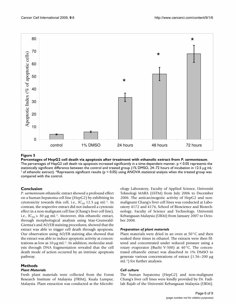
Apoptotic Index (AI)The apoptotic index (AI) was calculated to confirm thatethanolic-treated cell death was via apoptosis. AI isdescribed as the percentage of apoptotic cells and apop-totic bodies within the overall population of cells [21]. Anapoptotic index was determined as the percentage ofapoptotic cells from at least 400 counted cells underobservation using an inverted microscope. The statisticaldifferences between the control group and treated group(1% DMSO, 24 hours, 48 hours and 72 hours) were ana-lysed using ANOVA, and p values less than 0.05 were con-sidered as significant. The percentages of apoptotic cellsafter treatment were increased in a time-dependent man-ner with less than 50% at 24 hours, more than 50% at 48hours and even higher at 72 hours. Untreated cells are rep-resented as the control, i.e., the HepG2 cell line culturedin complete media for 72 hours. The control cells showedthat only 4% of these cell deaths produced a typical mor-
MTT assaying of the P. sarmentosum ethanolic extract in HepG2 and non-malignant Chang's liver cellsFigure 1MTT assaying of the P. sarmentosum ethanolic extract in HepG2 and non-malignant Chang's liver cells. Both cells were treated at various concentrations, i.e., 1.56–200 μg mL-1. The IC50 value for HepG2 was 12.5 μg mL-1, while the IC50 value for non-malignant Chang's liver cells was > 30 μg mL-1. Each data point represents values from three independent exper-iments (n = 3).
0
25
50
75
100
125
0 0.2 0.4 0.6 0.8 1 1.2 1.4 1.6 1.8 2 2.2 2.4 2.6
log [extract (μg mL-1)]
perc
enta
ge o
f vi
abili
ty (
%)
HepG2
Chang's liver
Page 3 of 9(page number not for citation purposes)

Cancer Cell International 2009, 9:6 http://www.cancerci.com/content/9/1/6
phological apoptotic feature (Figure 5). On the otherhand, cells treated with 1% DMSO (negative control) for72 hours produced only 8% cell death and showed no sig-nificant difference (p > 0.05) when compared to untreatedcells (control). In contrast, Figure 5 also showed that theAI percentage of HepG2 cells increased significantly (p <0.05) when the HepG2 cell line was treated with 12.5 μgmL-1 of ethanolic extract from P. sarmentosum at 24 hourscompared to the control. The AI percentage of the HepG2cell line also continued to increase significantly (p < 0.05)when the HepG2 cell line was treated with ethanolicextracts at 48 and 72 hours compared to the control (Fig-ure 5). This observation indicated that the apoptotic activ-ity was gradually increased when the ethanolic extract wasincubated longer in carcinoma HepG2 cells.
Morphological observation by acridine orange and ethidium bromide (AO/EB) stainingStaining cells with fluorescent dyes, including acridineorange and ethidium bromide, is used in evaluating thenuclear morphology of apoptotic cells. To corroboratethat apoptosis has been induced by P. sarmentosum eth-anolic plant extract, HepG2 cells were analysed in thepresence of acridine orange and ethidium bromide stain-ing (AO/EB staining). Acridine orange is a vital dye thatwill stain both live and dead cells, whereas ethidium bro-mide will stain only those cells that have lost their mem-brane integrity [22]. Three different concentrations were
chosen based on the IC50 values determined by MTT assay,which were 10, 12 and 14 μg mL-1. As a control, HepG2cells were cultured in complete media and stained withAO/EB (Figure 6A). The figure shows that the ethanolicextract from P. sarmentosum induced apoptosis after 72hours incubation at all concentrations of plant extracttested. Cells stained green represent viable cells, whereasyellow staining represented early apoptotic cells, and red-dish or orange staining represents late apoptotic cells. Asshown in Figure 6B, HepG2 cells treated with 10 μg mL-1
of ethanolic extract showed changes in cellular morphol-ogy, including chromatin condensation, membrane bleb-bing, and fragmented nuclei. On the other hand, Figures6C and 6D show similar features for cells treated with 10μg mL-1 of ethanolic extract (Figure 6B), but with extra fea-tures of late stage apoptotic activity with apoptotic bodieswhen HepG2 cells were treated with 12 μg mL-1 and 14 μgmL-1 of ethanolic extract from P. sarmentosum. Therefore,using the AO/EB staining procedure, the morphologicalfeatures of a hepatoma cell line in apoptosis were dosedependent, i.e., a stronger apoptosis signal was inducedwith higher concentrations of the respective extract.
Determination of intrinsic apoptosis by DNA fragmentationDNA fragmentation occurs in cells that produce intrinsicapoptosis activity when induced by a variety of agents.This cleavage produces ladders of DNA fragments that are
MTT assaying of tamoxifen in HepG2 and non-malignant Chang's liver cellsFigure 2MTT assaying of tamoxifen in HepG2 and non-malignant Chang's liver cells. Both cells were treated at various con-centrations (1.56–25 μg mL-1). The IC50 value for HepG2 is 3 μg mL-1 and 18.6 μg mL-1 for non-malignant Chang's liver cells. Each data point represent values from three independent experiments (n = 3).
0
25
50
75
100
125
150
0 0.2 0.4 0.6 0.8 1 1.2 1.4 1.6
log [extract (μg mL-1)]
perc
enta
ge o
f vi
abili
ty (
%)
HepG2
Chang's liver
Page 4 of 9(page number not for citation purposes)

Cancer Cell International 2009, 9:6 http://www.cancerci.com/content/9/1/6
the size of integer multiples of a nucleosome length (180–200 bp) [23]. The DNA fragmentation is initiated by cas-pase 3 activation of inactive CAD (caspase activated deox-yribonuclease) through removal of its inhibitors, i.e.,ICAD [6]. As a biochemical hallmark of intrinsic apop-totic cell death, DNA fragmentation was used to deter-mine whether the antiproliferative effect of P. sarmentosumethanolic extract on cells acts through the respective apop-tosis pathway [24]. As shown in Figure 7, the treatment ofHepG2 cells with ethanolic extract resulted in the induc-tion of intrinsic apoptosis activity at concentrations as lowas 10 μg mL-1. HepG2 cells were treated with three differ-ent concentrations of ethanolic extract (10, 12 and 14 μgmL-1) based on the IC50 that was predetermined by MTTassay. HepG2 cells treated with different concentrations ofethanolic extract (Lane 1–3; Figure 7) for 72 hoursshowed typical features of DNA laddering on an agarosegel, whereas untreated cells produced intact genomes(Lane 5; Figure 8). In contrast, the non-malignant Chang'sliver cell line when treated with the various concentra-tions of ethanolic extract (Lane 1–3; Figure 8) produced
similar genomic DNA features as in untreated non-malig-nant Chang's liver (Lane 4; Figure 8) and HepG2 (Lane 5;Figure 8) cell lines. Therefore, the ethanolic extract at aconcentration as low as 10 μg mL-1 can induce nucleo-somal DNA fragmentation of HepG2 due to intrinsicapoptosis processes, but not in the non-malignantChang's liver cell line. In this study, the reason of using"HepG2" and "Chang" liver cells because HepG2 are themodel of hepatocellular carcinoma while Chang liver cellsare considered as an in vitro model of non-malignant ornon-tumor liver cells. This is based on other studies suchas Antonin et al. and Teck et al. stated that Chang as non-malignant cells [25,26].
Morphological studies by inverted microscope at actual mag-nification 100×Figure 3Morphological studies by inverted microscope at actual magnification 100×. HepG2 and non-malignant Chang's liver cell line were treated without (A, C) and with 12.5 μg mL-1 of P. sarmentosum ethanolic extract (B, D) for 72 hours. Both types of treatment (C and D) produced similar cellular morphology and antiproliferative effect. However, in HepG2 cells, the confluency appeared to be reduced from 90% in untreated cells to 10% in treated cells. Similar cellular morphology was observed in three independent experiments (n = 3).
Morphological observation with May-Grunwald-Giemsa's staining at actual magnification 100×Figure 4Morphological observation with May-Grunwald-Giemsa's staining at actual magnification 100×. HepG2 cells were treated for 24 (A), 48 (C) and 72 (E) hours with 12.5 μg mL-1 of P. sarmentosum ethanolic extract while untreated HepG2 cells were grow in complete medium for 24 (B), 48 (D) and 72 (F) hours. The white arrows indicated apoptotic bodies. The figures shown are representative of three independent experiments (n = 3).
Page 5 of 9(page number not for citation purposes)
Cancer Cell International 2009, 9:6 http://www.cancerci.com/content/9/1/6
ConclusionP. sarmentosum ethanolic extract showed a profound effecton a human hepatoma cell line (HepG2) by exhibiting itscytotoxicity towards this cell, i.e., IC50 12.5 μg mL-1. Incontrast, the respective extract did not induced a cytotoxiceffect in a non-malignant cell line (Chang's liver cell line),i.e., IC50 > 30 μg mL-1. Moreover, this ethanolic extract,through morphological analysis using May-Grunwald-Giemsa's and AO/EB staining procedures, showed that theextract was able to trigger cell death through apoptosis.Our observation using AO/EB staining also showed thatthe extract was able to induce apoptotic activity at concen-trations as low as 10 μg mL-1. In addition, molecular anal-ysis through DNA fragmentation revealed that the celldeath mode of action occurred by an intrinsic apoptosispathway.
MethodsPlant MaterialsFresh plant materials were collected from the ForestResearch Institute of Malaysia (FRIM), Kuala Lumpur,Malaysia. Plant extraction was conducted at the Microbi-
ology Laboratory, Faculty of Applied Science, UniversitiTeknologi MARA (UiTM) from July 2006 to December2006. The anticarcinogenic activity of HepG2 and non-malignant Chang's liver cell lines was conducted at Labo-ratory 4172 and 4176, School of Bioscience and Biotech-nology, Faculty of Science and Technology, UniversitiKebangsaan Malaysia (UKM) from January 2007 to Octo-ber 2008.
Preparation of plant materialsPlant materials were dried in an oven at 50°C and thensoaked three times in ethanol. The extracts were then fil-tered and concentrated under reduced pressure using arotary evaporator (Buchi V-500) at 40°C. The concen-trated ethanolic extract was dissolved in 1% DMSO togenerate various concentrations of extract (1.56–200 μgmL-1) for further analyses.
Cell cultureThe human hepatoma (HepG2) and non-malignantChang's liver cell lines were kindly provided by Dr. Fadi-lah Rajab of the Universiti Kebangsaan Malaysia (UKM).
Percentages of HepG2 cell death via apoptosis after treatment with ethanolic extract from P. sarmentosumFigure 5Percentages of HepG2 cell death via apoptosis after treatment with ethanolic extract from P. sarmentosum. The percentages of HepG2 cell death via apoptosis increased significantly in a time-dependent manner. p < 0.05 represents the statistically significant difference between the control and treated group (1% DMSO, 24–72 hours of incubation in 12.5 μg mL-
1 of ethanolic extract). *Represents significant results (p < 0.05) using ANOVA statistical analysis when the treated group was compared with the control.
0
10
20
30
40
50
60
70
80
control 1% DMSO 24 hours 48 hours 72 hours
Apo
ptot
ic I
ndex
(%
of
apop
totic
cel
ls)
*
*
*
Page 6 of 9(page number not for citation purposes)

Cancer Cell International 2009, 9:6 http://www.cancerci.com/content/9/1/6
Both cells were cultured in RPMI 1640 (Flowlab) supple-mented with 10% foetal bovine serum (FBS; Gibco), pen-icillin (50 U mL-1) and streptomycin (50 μg mL-1)(Gibco). Cells were maintained in humidified air with 5%CO2 at 37°C. Cells were harvested using 0.25% trypsin(Hyclone) when they were 70–80% confluent in culture.
MTT assayBriefly, 200 μL of cells (1 × 10⁴ cells) were seeded into 96-well plates and incubated overnight. The following day,cells were then treated with 20 μL of various concentra-tions of extract (1.56–200 μg mL-1) and tamoxifen (posi-tive control) before further incubation for 72 hours. At theend of this incubation, 20 μL of MTT (Sigma) (2 mg mL-1
in PBS) was added to each well and incubated for another4 hours at 37°C. The formazan crystals were dissolved in100 μL dimethylsulphoxide (DMSO) and the absorbancewas determined at 540 nm using a multi-plate reader(BIO-RAD model 680). The absorbance value that wasdetermined for cells cultured in complete media without
plant extract was based on 100% viable cells. Each con-centration of the extract was assayed in triplicate.
Cell observation using an inverted microscopeHepG2 cell lines were grown in 6-well plates and treatedwith P. sarmentosum ethanolic extract. The cells were thenwashed with 1× Phosphate Buffer Saline (PBS) (Sigma).Morphological and confluency changes in the cells inboth the treated group (12.5 μg mL-1 of ethanolic treated-cells incubated for 24, 48 and 72 hours and 1% DMSOtreated-cells for 72 hours) and untreated group wereobserved using an inverted microscope (Nikon TMS).
Apoptosis analysisGiemsa stainingBriefly, the HepG2 cell line was seeded at 1 × 10⁵ cells/wellin 6-well plates (BD Labware, England), and the plateswere then incubated overnight at 37°C. After incubation,ethanolic extracts of various concentrations (10, 12 and14 μg mL-1) were added and incubated for an additional24 hours. The plates were washed with 1× Phosphate
Morphological observation with acridine orange and ethid-ium bromide (AO/EB) staining at actual magnification 400×Figure 6Morphological observation with acridine orange and ethidium bromide (AO/EB) staining at actual magni-fication 400×. HepG2 cells were treated without (A) and with P. sarmentosum ethanolic extract, 10 μg mL-1 (B), 12 μg mL-1 (C) and 14 μg mL-1 (D) for 72 hours. Dashed arrow indicated cells with chromatin condensation; rounded dotted arrow indicated cells with fragmented nuclei; dashed dotted arrow indicated cells with membrane blebbing and full white arrow indicated the presence of apoptotic bodies. Each experiment was performed in triplicate (n = 3) and gener-ated similar morphological features.
Gel electrophoresis of DNA genomes extracted from vari-ous HepG2 cells following treatmentFigure 7Gel electrophoresis of DNA genomes extracted from various HepG2 cells following treatment. Cells were incubated with various concentrations of ethanolic extract for 72 hours. DNA fragments were separated using 1.5% aga-rose gel electrophoresis and visualised under UV light after staining with ethidium bromide. M: 100 bp DNA ladder marker, lane 1: HepG2 treated with 10 μg mL-1 of P. sarmen-tosum ethanolic extract, lane 2: HepG2 treated with 12 μg mL-1 of P. sarmentosum ethanolic extract and lane 3: HepG2 treated with 14 μg mL-1 of P. sarmentosum ethanolic extract. Each experiment was performed in triplicate (n = 3).
Page 7 of 9(page number not for citation purposes)

Cancer Cell International 2009, 9:6 http://www.cancerci.com/content/9/1/6
Buffer Saline (PBS), and the cells were stained with May-Grundwald (BDH Chemical Ltd) for 4 minutes. The slideswere then rinsed with sterile water and flooded withfreshly prepared Giemsa's stain solution (BDH ChemicalLtd) for 6 minutes. Dyestuff was discarded and rinsedagain three times with sterile water. Morphologicalchanges were examined using an inverted microscopy(Nikon, TMS) with 100× actual magnification.
Acridine orange and ethidium bromide staining (AO/EB staining)For this purpose, cells were seeded in 6-well plates for 24hours and then treated with different concentrationranges (10, 12 and 14 μg mL-1) for 72 hours. After harvest-ing by trypsinisation, cells were washed with 1× PBS once.Twenty-five microlitres of the cell suspension was thenmixed with 1 μL of the dye mixture, containing 100 mgmL-1 of acridine orange (Sigma) and 100 mg mL-1 ofethidium bromide (Sigma) in 1× PBS. After staining, cellswere visualised immediately under a fluorescence micro-scope (Leica DM 2500).
Intrinsic apoptosis as determined by DNA fragmentationCells were lysed with lysis buffer (10 mM Tris-HCL, 5 mMEDTA, 200 mM NaCl, 0.2% SDS) and incubated at 60°Cfor 5 minutes. The sample was digested with 2.5 μL of pro-teinase K (more than 3 U μL-1) (Sigma) and 5 μL of RNaseA (1 U μL-1) (Fermentas) and was further incubated at60°C for 1 hour. After this, 250 μL of 5 M NaCl was addedand mixed and then incubated on ice for 5 minutes to pre-cipitate proteins. Cells were then centrifuged for 15 min-utes at 10,000 rpm and the supernatant was transferred toa fresh tube, to which an equal volume of isopropanolwas added to precipitate the DNA, and the sample wascentrifuged for 10 minutes at 10,000 rpm. The superna-tants were then discarded, and the pellets were washedwith 70% cold ethanol. DNA samples were electro-phoresed on a 1.5% agarose gel for 1 hour and 30 minutesat 70 V. Finally, the gel was examined under UV light fol-lowing ethidium bromide staining to determine apoptoticDNA fragmentation.
Data analysisThe analysis of variance (ANOVA) was used to determinedifferences between treated and control groups usingMicrosoft™ Excel 2007 software. p values less than 0.05 (p< 0.05) were considered statistically significant.
AbbreviationsHepG2: Human hepatoma cell line; MTT: (3-(4,5-dimeth-ylthiazol-2-yl)-2,5-diphenyl-tetrazolium bromide); AI:apoptotic index; CO2: carbon dioxide; DMSO: dimethyl-sulfoxide; ANOVA: analysis of variance; AO/EB: acridineorange and ethidium bromide; CAD: caspase activateddeoxyribonuclease; ICAD: inhibitor caspase activateddeoxyribonuclease; UV: ultraviolet; IC50: inhibition con-centration to kill 50% of cells population; RPMI: RoswellPark Memorial Institute; FBS: foetal bovine serum; PBS:Phosphate Buffered Saline; HCl: hydrochloride acid;EDTA: ethylenediaminetetraacetic acid disodium saltdehydrate; NaCl: natrium chloride; DNA: deoxyribonu-cleic acid; NCI: National Cancer Institute.
Competing interestsBefore this, there are other researchers working on this P.sarmentosum extract. However they are working towardsantimalarial, antioxidant and hypoglycaemic effect.
Authors' contributionsSHZA involved in scientific approach on cell culturesanalysis and molecular analysis. He is the head of this par-ticular project and supervisor of WHHWO. Therefore, heacts as the first corresponding author of this manuscript.RMAW and SS are research associate who was involved incell culture analysis, molecular analysis (advised by SS)and also clinical potential of the generated extract(advised by RMAW as clinician). WHHWO is the post-
Gel electrophoresis of DNA genomes extracted from vari-ous treated Chang's and untreated HepG2 cellsFigure 8Gel electrophoresis of DNA genomes extracted from various treated Chang's and untreated HepG2 cells. Cells were incubated without or with various concentrations of ethanolic extract for 72 hours. The DNA fragments were separated using 1.5% agarose gel electrophoresis and visual-ised under UV light after staining with ethidium bromide. M: 100 bp DNA ladder marker, lane 1: non-malignant Chang's liver treated with 10 μg mL-1 of P. sarmentosum ethanolic extract, lane 2: non-malignant Chang's liver treated with 12 μg mL-1 of P. sarmentosum ethanolic extract, lane 3: non-malignant Chang's liver treated with 14 μg mL-1 of P. sarmen-tosum ethanolic extract, lane 4: untreated Chang's liver cells and lane 5: untreated HepG2 cell line. Each experiment was performed in triplicate (n = 3).
Page 8 of 9(page number not for citation purposes)

Cancer Cell International 2009, 9:6 http://www.cancerci.com/content/9/1/6
Publish with BioMed Central and every scientist can read your work free of charge
"BioMed Central will be the most significant development for disseminating the results of biomedical research in our lifetime."
Sir Paul Nurse, Cancer Research UK
Your research papers will be:
available free of charge to the entire biomedical community
peer reviewed and published immediately upon acceptance
cited in PubMed and archived on PubMed Central
yours — you keep the copyright
Submit your manuscript here:http://www.biomedcentral.com/info/publishing_adv.asp
BioMedcentral
graduate student that doing the laboratory work and gen-erating all the results. ZZA involvement as the headresearcher on scientific approached in determine the plantto be analyses and extraction procedures of the plant. Herresearch associate is MFS. Therefore her involvementswarrant her to become second corresponding author ofthis manuscript on the plant extraction approached.
AcknowledgementsThis project was funded by UKM-OUP-TKP-18-84/2008 and UKM-GUP-BTK-07-15-197 through the Universiti Kebangsaan Malaysia, Malaysia.
References1. McGlynn KA, Tsao L, Hsing AW, Devesa SS, Fraumeni JF Jr: Interna-
tional trends and patterns of primary liver cancer. Int J Cancer2001, 94(2):290-296.
2. Rocken C, Carl-McGrath S: Pathology and pathogenesis ofhepatocellular carcinoma. Dig Dis 2001, 19(4):269-278.
3. Kaufmann SH, Earnshaw WC: Induction of apoptosis by cancerchemotherapy. Exp Cell Res 2000, 256(1):42-49.
4. Pezzuto JM: Plant-derived anticancer agents. Biochem Pharmacol1997, 53(2):121-133.
5. Wu J, Wu Y, Yang BB: Anticancer activity of Hemsleya amabilisextract. Life Sci 2002, 71(18):2161-2170.
6. Dahlgren RMT: A revised system of classification of theangiosperms. Bot J Linn Soc 1980, 80(2):9-124.
7. Christophe W: Medicinal Plants of Asia and Pacific USA: CRC Press;2006.
8. Navickiene HMD, Alécio AC, Kato MJ, Bolzani VS, Young MCM, Cav-alheiro AJ, Furlan M: Antifungal amides from Piper hispidum andPiper tuberculatum. Phytochemistry 2000, 55(6):621-626.
9. Facundo VA, Silveira ASP, Morais SM: Constituents of Piper alata-bacum Trel & Yuncker (Piperaceae). Biochem Syst Ecol 2005,33(7):753-756.
10. Perry LM: Medicinal Plants of East and Southeast Asia Cambridge: MITPress; 1981.
11. Thitima R, Puttan S, Kanchanawadee S, Chanika W, Phongpan R,Phaopong W, Apichart S: Chemical constituents and bioactivityof Piper sarmentosum. J Ethnopharmacol 2004, 93:173-176.
12. Arrigoni BM, Dmitrieva EG, Franzotti E, Antoniolli AR, Andrade MR,Marchioro M: Anti-inflammatory and analgesic activity of Pep-eromia pellucida (L.) HBK (Piperaceae). J Ethnopharmacol 2004,91(2–3):215-218.
13. Lentz DL, Clark AM, Hufford CD, Meurer-Grimes B, Passreiter CM,Cordero J, Ibrahimi O, Okunade AL: Antimicrobial properties ofHonduran medicinal plants. J Ethnopharmacol 1998,63(3):253-263.
14. Lemos GCS, Oliveira LO, Eberli BB, Motta OV, Folly MM: Bacteri-cidal activity of macela (Achyrocline satureioides (Lam) DC.)and jaborandi-falso (Piper aduncum L.) against strains ofStaphlococcus aureus isolated from subclinical bovine masti-tis. Revista Brasileira de Plantas Medicinais 2000, 3(1):67-72.
15. Nik NAR, Takahisa F, Somei K, Kikuchi T, Mustafa AM: Antimalarialactivity of extracts of Malaysian medicinal plants. J Ethnophar-macol 1999, 64(3):249-254.
16. Peungvicha P, Thirawarapan S, Temsiririrkkul R, Watanabe H, KumarPrasain J, Kadota S: Hypoglycemic effect of the water extract ofPiper sarmentosum in rats. J Ethnopharmacol 1998, 60(1):27-32.
17. Anchana C, Aphiwat T, Nuansri R: Screening of antioxidant activ-ity and antioxidant compounds of some edible plants of Thai-land. Food Chem 2005, 92(3):491-497.
18. Edrini S, Rahmat A, Ismail P, Hin TYY: Anticarcinogenic proper-ties and antioxidant activity of henna (Lawsonia inermis). JMed Sci 2002, 2(4):194-197.
19. Suffness M, Pezzuto JM: Assays related to cancer drug discov-ery. In Methods in Plant Biochemistry: Assay for Bioactivity Volume 6.Edited by: Hostettmann K. London: Academic Press; 1990.
20. Sarveswaran S, Marati RV, Maruthaiveeran PB: Effects of Terminaliaarjuna bark extract on apoptosis of human hepatoma cellline HepG2. World J Gastroenterol 2006, 12(7):1018-1024.
21. Soini Y, Paakko P, Letho VP: Histopathological evaluation ofapoptosis in cancer. Am J Pathol 1998, 153(4):1041-1053.
22. Jayadev R, Jagan MRP, Malisetty VS, Chinthapally VR: Diosgenin, asteroid saponin of Trigonella foenum graecum (Fenugreek),inhibits Azoxymethane-induced aberrant crypt foci forma-tion in F344 rats and induces apoptosis in HT-29 humancolon cancer cells. Cancer Epidemiol Biomarkers and Prev 2004,13(8):1392-1398.
23. Hui L, Cheng-Yong Q, Guo-Qing H, Hong-Wei X, Mei M, Zhen Y:Mechanism of apoptotic effects induced selectively by urso-deoxycholic acid on human hepatoma cell lines. World J Gas-troenterol 2007, 13(11):1652-1658.
24. Frederick LK, Xinbo Z: Apoptosis: biochemical aspects and clin-ical implications. Clin Chim Acta 2002, 326:27-45.
25. Antonin L, Cristina F, Vito L, Massimo V, Pier LA, Frantisek S, ClaudioT, Lorella P: Molecular determinants in the transport of bileacid-derived diagnostic agent in tumoral and nontumoralcell lines of human liver. The Journal of Pharmacology and Experi-mental Therapeutics 2006, 319(2):809-814.
26. Teck KS, Rosa CMYL, Chon KL, Maxey CMC: Hepatocellular car-cinoma: From bedside to proteomics. Proteomics 2001,1:1249-1263.
Page 9 of 9(page number not for citation purposes)


![Establishment and Identification of Small Cell Lung Cancer Cell … · [CANCER RESEARCH 45, 2913-2923, June 1985] Establishment and Identification of Small Cell Lung Cancer Cell Lines](https://img.dokumen.tips/doc/110x75/60347fa5d25195593e3efdb8/establishment-and-identification-of-small-cell-lung-cancer-cell-cancer-research.jpg)


![BIOCHEMICAL UNDERSTANDING OF CANCER CELL … · international conference on biochemical understanding of cancer cell survival and progression [icbuccsp'18] 5th - 7th february, 2018](https://img.dokumen.tips/doc/110x75/5b24f1387f8b9a992e8b495d/biochemical-understanding-of-cancer-cell-international-conference-on-biochemical.jpg)