Embed Size (px)
Citation preview

C6 pyridinium ceramide influences alternativepre-mRNA splicing by inhibiting proteinphosphatase-1Chiranthani Sumanasekera1, Olga Kelemen1, Monique Beullens2, Brandon E. Aubol3,
Joseph A. Adams3, Manjula Sunkara1, Andrew Morris1, Mathieu Bollen2,
Athena Andreadis4 and Stefan Stamm1,*
1Department of Molecular and Cellular Biochemistry, University of Kentucky, Lexington, Kentucky 40536, USA,2Laboratory of Biosignaling and Therapeutics, Department of Molecular Cell Biology, University of Leuven,B-3000 Leuven, Belgium, 3University of California, San Diego La Jolla, CA 92093 and 4UMASS Medical School,55 Lake Avenue North, Worcester, MA 01655-0106, USA
Received August 5, 2011; Revised December 6, 2011; Accepted December 14, 2011
ABSTRACT
Alternative pre-mRNA processing is a centralelement of eukaryotic gene regulation. The cellfrequently alters the use of alternative exons inresponse to physiological stimuli. Ceramides arelipid-signaling molecules composed of sphingosineand a fatty acid. Previously, water-insoluble cera-mides were shown to change alternative splicingand decrease SR-protein phosphorylation byactivating protein phosphatase-1 (PP1). To gainfurther mechanistical insight into ceramide-mediated alternative splicing, we analyzed the effectof C6 pyridinium ceramide (PyrCer) on alternativesplice site selection. PyrCer is a water-solubleceramide analog that is under investigation as acancer drug. We found that PyrCer binds to the PP1catalytic subunit and inhibits the dephosphorylationof several splicing regulatory proteins containing theevolutionarily conserved RVxF PP1-binding motif(including PSF/SFPQ, Tra2-beta1 and SF2/ASF). Incontrast to natural ceramides, PyrCer promotesphosphorylation of splicing factors. Exons that areregulated by PyrCer have in common suboptimalsplice sites, are unusually short and share two 4-ntmotifs, GAAR and CAAG. They are dependent onPSF/SFPQ, whose phosphorylation is regulated byPyrCer. Our results indicate that lipids can influencepre-mRNA processing by regulating the phosphoryl-ation status of specific regulatory factors, which ismediated by protein phosphatase activity.
INTRODUCTION
All polymerase-II transcripts undergo pre-mRNAprocessing and at least 95% of the transcriptional unitsare alternatively spliced (1). The exact regulation of alter-native splicing events is physiologically important, asevidenced by an increasing number of diseases that arecaused by the selection of the wrong splice site (2). Theproper recognition of exons is regulated by the transientformation of protein complexes on the pre-mRNA thatidentify a sequence for its recognition by the spliceosome.The signals on the pre-mRNA that mark an exon for itsinclusion in the mRNA are highly degenerate, most likelyto avoid interference with coding requirements. Therefore,exons are recognized by the formation of a complexbetween the pre-mRNA and various hnRNPs and SRproteins. Since these proteins generally bind to RNAwith low selectivity, a higher specificity is achieved bysimultaneous binding of the proteins to each other.The interaction between these proteins is regulated in
part by their phosphorylation status. For example,protein phosphatase-1 (PP1)-mediated dephosphorylationpromotes the interaction between Tra2-beta1 andSF2/ASF (also called SRSF1) in vitro (3). Reversible phos-phorylation of SR proteins and hnRNPs is achieved by aninterplay between protein kinases and phosphatases.Several SR proteins contain an evolutionarily highlyconserved PP1 binding element (RVxF) in theirRNA recognition motif which allows PP1 to influencethe phosphorylation status of proteins bound to pre-mRNA. In addition to the interaction between individualSR proteins, the affinity between components of thespliceosome, such as U1-70K and proteins that associatewith pre-mRNA, including PSF/SFPQ (4), SRp38 (5) and
*To whom correspondence should be addressed. Tel: +859 323 0897; Fax: +859 323 1037; Email: [email protected]
The authors wish it to be known that, in their opinion, the first two authors should be regarded as joint First Authors.
Published online 30 December 2011 Nucleic Acids Research, 2012, Vol. 40, No. 9 4025–4039doi:10.1093/nar/gkr1289
� The Author(s) 2011. Published by Oxford University Press.This is an Open Access article distributed under the terms of the Creative Commons Attribution Non-Commercial License (http://creativecommons.org/licenses/by-nc/3.0), which permits unrestricted non-commercial use, distribution, and reproduction in any medium, provided the original work is properly cited.
Downloaded from https://academic.oup.com/nar/article-abstract/40/9/4025/1128692by gueston 04 April 2018

SF2/ASF (6) is influenced by their phosphorylation status.This suggests that PP1 activity can regulate the recogni-tion of pre-mRNA substrates by the spliceosome andinfluences the selection of alternative exons by alteringprotein affinities. In agreement with this model, achange in PP1 activity has been shown to alter splice siteselection in vivo (3,7,8).Ceramides are a class of sphingolipids that are
composed of sphingosine and a fatty acid moiety. In thecell, ceramide can be generated by sphyingomyelinhydrolysis by sphingomyelinase or by de novo synthesis.Sphingomyelin and sphingomyelinase are both present inthe nucleus, which led to the suggestion that they partici-pate in nuclear signaling (9). Natural ceramides containlong alkyl groups (C14–26) and are therefore hydropho-bic. It has previously been shown that endogenousceramides alter bcl-x splicing via a purine-rich splicingenhancer (10). To improve delivery and water solubility,cationic-ceramide analogs containing pyridinium moietieswere synthesized. Owing to their ability to induce apop-tosis, these water-soluble ceramide analogs have beentested as anti-cancer drugs (11,12). Despite their usage inclinical studies, the effect of these pyridinium ceramides onalternative splicing has not been determined.Here, we investigate the role of a water-soluble
ceramide analog, C6 pyridinium ceramide (PyrCer), inalternative splicing. We found that PyrCer treatmentinhibits the dephosphorylation of splicing regulatoryproteins by PP1, which is opposite to the activationreported for water-insoluble short and long-chainceramides (13). In addition to SR proteins known to beaffected by natural ceramides, PyrCer treatment increasesthe phosphorylation of other proteins involved in splicing,such as PSF/SFPQ, SAP155 and UAP56. It changes thealternative splicing patterns of exons that are short anddependent on splicing enhancers. These findings suggestthat the dephosphorylation of splicing factors by PP1 isa molecular link between lipids and alternative splice siteselection. The difference between PyrCer and naturalceramides suggest that subclasses of lipids have distincitveeffects on splice site selection.
MATERIALS AND METHODS
Cell culture and transfection
HEK 293T cells were grown in DMEM (Invitrogen)supplemented with 10% (v/v) fetal bovine serum(Invitrogen) for 24 h to 80% confluence prior to ceramidetreatment and to 60–70% confluence prior to calciumphosphate transfections. Transfection of the minigenesand RT-PCR analyses was performed as described (14).
Ceramide treatment
Four microliters of HeLa nuclear extract (Dundee CellProducts) was incubated in 1mM MnCl2 and 1� PNKbuffer (70mM Tris–HCl pH 7.6, 10mM MgCl2, 5mMdithiothreitol). Twenty micromolar of C6 pyridiniumceramide (D-erythro-2-N-[60-(1-pyridinium)-hexanoyl]-sphingosine bromide) (Avanti Polar Lipids), or 20 mM1-(2-oxopropyl) pyridinium bromide (Sigma-Aldrich)
was added. After 5min at 37�C, 4 mCi of [g-32P] ATP(7000Ci/mmol, MP Biochemical) was added. The 50 mlreactions were then incubated for 30min and thereaction was stopped by adding 6� SDS sample buffer(0.375M Tris–HCl pH 6.8, 30% glycerol, 12% SDS,12% 2-b mercaptoethanol and 0.075% bromophenolblue) followed by 10% SDS–PAGE. Proteins that weredifferentially phosphorylated in the ceramide lanes wereexcised from the gel and analyzed by MALDI TOF/TOF.
Minigene construction
Minigenes were constructed as described (14). The primersfor the minigenes and the insert sequences are shown inSupplementary Figure S1.
Phosphatase assays
Hydrolysis of p-nitrophenylphosphate (p-NPP) wasperformed in 25 ml reactions containing 1� phosphatebuffer (0.1M sodium acetate, 0.05M bis–Tris, 0.05MTris–HCl, 2mM dithiothreitol, at pH 8.0), 50mMpNPP, 1mM MnCl2 and 0.1U of purified PP1 enzymemix (Upstate, #14-110) at 37�C for 30min. A PP1 unitreleases 1 nmol of phosphate/min from 15 mM phosphor-ylase at 30�C. The reaction was terminated by the additionof 100 ml of 0.25M NaOH and absorbance was measuredat 410 nm. The activity of PP1 on Phosphorylase b wasmeasured as previously described (15).
C6 pyridinium ceramide treatment of HeLa nuclearextracts
Ten micrograms of HeLa nuclear extract (NE) wasincubated with 4 mCi [g32P] ATP for 30min at 37�Cin 1� PNK buffer (New England Bio Labs) plus or10 mM C6 pyridinium ceramide in a 50 ml final volume.Radiolabeled-NE proteins were then subjected toimmunoprecipitation overnight with 2 mg of indicatedantibodies, 10 ml proteins A/G beads (Santa Cruz) and200 ml RIPA-rescue buffer containing phosphatase andprotease inhibitors. Immunoprecipitates were subjectedto SDS–PAGE, proteins were transferred on to nitrocel-lulose membranes and analyzed by phosphor-imaging.The membranes were then blotted with the indicatedantibodies for total protein expression. Representative32P signals and western blot results of triplicate experi-ments for SF2/ASF, Tra2-beta1, PSF, UAP56 andSAP155 are shown.
Protein expression and purification
Recombinant proteins were overexpressed in Escherichiacoli BL21 (DE3) cells. Expression was induced with IPTGin a final concentration of 0.4mM for 4 h at room tem-perature. The His-Tra2-beta1 protein and His-PSFprotein were purified under denaturing conditions onNi-NTA resin (QIAGEN). The cells were lysed in buffer#1 [20mM Tris, 300mM NaCl, 100mM MOPS, 15%glycerol and 4M guanidine hydrochloride pH (7.5)]using a French press. The cell lysate was clarified by cen-trifugation at 10 000� g for 30min at 4�C. The super-natant was incubated with Ni-NTA resin for 1 h at room
4026 Nucleic Acids Research, 2012, Vol. 40, No. 9
Downloaded from https://academic.oup.com/nar/article-abstract/40/9/4025/1128692by gueston 04 April 2018

temperature. The resin was poured into a column andwashed with buffer #1 containing 20mM imidazolefollowed by washing steps with refolding buffers [buffer#1 containing decreasing concentration of GuHCl (pH6.5)]. The proteins were eluted using buffer #5 [20mMTris–HCl, 300mM NaCl, 100mM MOPS, 15% glycerol(pH 2.2)].
GST-tagged SF2/ASF transformed E. coli cells werelysed in buffer A [50mM Tris–HCl (pH 7.5), 300mMNaCl, 0.5% Triton X-100, 0.1% 2-b mercaptoethanol,1mM DTT, 1mM MnCl2] using a French press. Thesupernatant was incubated with Glutathion sepharose4B (GE Healthcare) beads for 2 h at room temperature.The slurry was packed into a column and washed withbuffer B [50mM Tris–HCl (pH 7.5), 150mM NaCl,1mM DTT, 0.1% 2-b mercaptoethanol, 1mM MnCl2].The protein was eluted with buffer C [100mM Tris (pH8), 10mM glutathione] and was dialyzed against buffer D[50mM Tris (pH 7.5), 100mM NaCl, 1mM MnCl2, 1mMDTT, 0.1% 2-b mercaptoethanol and 10% glycerol]. Theproteins were stored at �80�C.
Dephosphorylation of purified Tra2-beta1-His6
Seven micrograms of purified recombinantTra2-beta1-His6 bound to Ni-NTA beads (Qiagen) wasphosphorylated in HeLa nuclear extract in the presenceof 5 mCi [g32P] ATP for 30min at 37�C in 1� PNKbuffer. Beads were then washed with RIPA buffer andaliquoted into tubes containing the indicated amounts ofPP1, C6 pyridinium ceramide, oxo-pyridinium, calyculinA or ethanol as indicated in a 30 ml volume containing1� PP1 buffer. The dephosphorylation reactions wereincubated for 30min at 37�C and then boiled inSDS-sample loading buffer. Samples were resolved bySDS–PAGE and gels were coomassie stained and sub-jected to phosphor imaging. The 32P signals and totalprotein in coomassie stained gels were used to determinethe extent of Tra2-beta1 dephosphorylation.
Measurement of ceramides and sphingolipids by HPLCtandem mass spectrometry
Ceramides were quantitated by HPLC electrospray ioniza-tion tandem mass spectrometry using an ABI 4000 Q-Traphybrid linear ion trap triple quadrupole mass spectrometeroperated in multiple reaction monitoring (MRM) mode.Ion currents associated with lipid species specific precursorproduct ion pairs were determined as described by otherswith minor adaptations (16). In brief, lipids were extractedusing acidified organic solvents (17), including 50 pmolC17 ceramide as a recovery standard. Ceramide molecularspecies were resolved using a 3mm� 100mm X-TerraXDB-C8 column (3.5 mm particle size, Waters, Milford,MA, USA) and a gradient of methanol/water/formicacid (61:39:0.5, v/v) containing 5mM ammonium formate(solvent A) and acetonitrile/chloroform/water/formic acid(90:10:0.5:0.5, v/v) with 5mM ammonium formate(solvent B) at a flow rate of 0.5ml/min.
The instrument was operated in positive mode withoptimal ion source settings determined empirically usinga set of synthetic ceramides. MRM transitions monitored
for the elution of ceramide molecular species were asfollows: m/z 510> 264, 14:0-Cer; m/z 538:264, 16:0-Cer;m/z 540:284, 16:0-DHCer; m/z 552:264, 17:0-Cer(internal standard); m/z 564> 264, 18:1-Cer; m/z566> 284, 18:1-DHCer; m/z 566> 264, 18:0-Cer; m/z568> 284, 18:0-DHCer; m/z 594> 264, 20:0-Cer;m/z 596> 284, 20:0-DHCer; m/z 624> 284, 22:0-DHCer;m/z 650>264, 24:1-Cer; m/z 652> 284, 24:1-DHCer; m/z652> 264, 24:0-Cer; m/z 654> 284, 24:0-DHCer;m/z 680> 264, 26:1-Cer; m/z 682> 264, 26:0-Cer; m/z708> 264, 28:1-Cer; m/z 710> 264, 28:0-Cer. Quan-titation was accomplished by reference to standardcurves generated using synthetic standards obtainedfrom Avanti Polar Lipids (Alabaster AL) that were inde-pendently determined by accurate mass measurements.Measurements of C6 pyridinium ceramide and oxo-pyridinium were accomplished using the extractionmethod detailed above.C6 pyridinium ceramide was separated using the
chromatographic methods used for natural ceramidesexcepet that the MRM transitions monitored were m/z476–379; 476–266 and 476–253. Precursor ion scanningexperiments indicated that the m/z 379 species wasderived by loss of the pyridinium group and the mz 266and 253 species were derived by sequential loss of thehydroxy groups from the resulting ion. Oxo-pyridiniumwas separated by hydrophilic interaction chromatographyusing a 4.6mm� 100mm Luna HILIC column (3mMparticle size, Phenomenex, Torrance, CA, USA) withmonitoring of MRM transitions m/z 136> 93, m/z136> 79, m/z 136> 65, m/z 137> 57.
Immunoprecipatation and western blots
For immunoprecipitation (IP) of PP1 isoforms, HEK293Tcells were grown to 70% confluencency in 6-well platesand transfected with 2 mg of EGFP-C2 vectors expressingPP1 alpha, PP1 beta, PP1 gamma or Tra2-beta1-RATA[constructs described in (3)]. After 12–14 h, cells weretreated with 0 and 10 mM C6 pyridinium ceramide for8 h before preparation of cell extracts. Cells werelysed for 30min at 4�C in 200 ml of RIPA buffer(20mM Na3PO4, 150mM NaCl, 1% NP-40, 1% sodiumdeoxycholate, 5mM2-b mercaptoethanol, 0.1% SDS,1mM EDTA, 1mM phenylmethylsulfonyl fluoride,5mg/ml of leupeptin, pepstatin A, antipain and aprotinin)supplemented with 75U/ml Benzonase (Sigma-Aldrich).RIPA-Rescue buffer (10mM sodium phosphate pH 7.2,20mM NaCl, and freshly added 1mM dithiothreitol,1mM phenylmethylsulfonyl fluoride, and 20mg/ml apro-tinin) was added to each reaction to a final volume of460 ml and subsequently 40 ml of pre-cleared 50% proteinA sepharose CL-4 bead suspension (GE HealthCare) wereadded. The beads were washed once with 1ml RIPAbuffer and three times with RIPA-rescue buffer (10mMsodium phosphate pH 7.2, 20mM NaCl, and freshlyadded 1mM dithiothreitol, and 1mM phenylmethyl-sulfonyl fluoride) and were resuspended in a 60 ml finalvolume. Thirty microliters of these beads were used forwestern blotting while the remaining half was used forthe HPLC assay.
Nucleic Acids Research, 2012, Vol. 40, No. 9 4027
Downloaded from https://academic.oup.com/nar/article-abstract/40/9/4025/1128692by gueston 04 April 2018

RESULTS
C6 pyridinium ceramide (PyrCer) changes alternativesplicing of endogenous genes
Ceramides have previously been implicated in regulationof splice site selection (7,8,18). However, the molecularmechanism of ceramide action on pre-mRNA processingis not clear, partly because ceramides are poorly soluble inwater. We therefore concentrated on C6 pyridiniumceramide, a water-soluble ceramide analog which is cur-rently being tested for the treatment of squamous cell car-cinomas (19,20). To determine the influence of ceramideaction on alternative pre-mRNA splicing, we used amedium-throughput RT-PCR screen (21) using RNAfrom HEK293 cells treated with C6 pyridinium ceramide.First, we used HEK293 cells in an MTT cytotoxicity
assay to determine a suitable concentration of PyrCerfor this screen. As a control, we used oxo-pyridinium, amolecule that only differs from PyrCer by its propyl groupthat replaces the short chain ceramide moiety. Our dataindicate that, at a 10 mM concentration, there was<5% cell death compared to higher concentrations ofPyrCer which lead to >80% cell death following a 24 htreatment (Supplementary Figure S2). In contrast,increasing concentrations of oxo-pyridinium had noeffect on cell viability. These data also indicate that thecytotoxicity of PyrCer is due to the ceramide group andnot due to the cationic pyridinium group which results inits water solubility. Based on these studies, we treated cellsfor 24 h with 10 mM PyrCer and analyzed the changes inalternative splicing compared to oxo-pyridiniumtreatment.In total, 92 alternative splicing events were tested and
29 splicing events showed a significant change in theiralternative splicing patterns. We validated these 29events by performing RT-PCR with mRNA extractedfrom HEK293 cells treated with or without 10 mM C6pyridinium ceramide. From these, we selected five exonswith the most significant changes for further analysis.These exons were present in the DRF1, TIAF1, POL-b,TAU and SYK pre-mRNAs. As shown in Figure 1, treat-ment with 10 mM PyrCer promoted both inclusion andskipping of exons. As a negative control, we validatedthe absence of a PyrCer effect on the pre-mRNAs ofCCNE1, ECT2, SHC1 and STM1 genes which showedno change in the primary screen (Supplementary FigureS3). Similar to other signal-dependent splicing systems(22), exon inclusion differs around 10–20% between thetreated cells and controls. However, these differences arestatistically significant, with P-values from the student’stest <0.01.The alternative exon usage in the DRF1 (Dbf4-related
factor 1) and POL-b (DNA polymerase beta) genes causeda frameshift in the encoded pre-mRNAs. Alternativesplicing of the other exons in the TIAF1 (TGFB1-induced antiapoptotic factor 1), TAU (microtubuleassociated protein tau) and SYK (spleen tyrosine kinase)transcripts retained their reading frames. The inclusion oftau exon 10 has been shown previously to increase theaffinity of the resulting tau isoform to microtubules andaberrant exon 10 usage results in frontotemporal dementia
(23). These data suggest that a treatment with PyrCerchanges alternative splicing of several pre-mRNAs andcould have functional consequences for the cellularproteome.
C6 pyridinium ceramide (PyrCer) changes alternativesplicing of exons in a heterologous context
To determine whether the sequence elements that respondto PyrCer treatment are localized in the vicinity of theregulated exons, we introduced the alternatively splicedexons into reporter minigenes. We used a recentlydeveloped recombination technique that introduces theexon of interest between two constitutively splicedinsulin exons (14). Depending on the intron length, theresulting reporter minigenes contained one to threeexons cloned between the constitutive insulin exons.Their structures are schematically shown in Figure 2.
These reporter constructs were transfected intoHEK293 cells and the cells were subsequently treatedwith C6 pyridinium ceramide. Total RNA was isolatedand analyzed using minigene-specific primers. In allcases, we observed a C6 pyridinium ceramide-specificchange that was congruent with the changes observedwith the endogenous genes. However, the magnitude ofthe effect on exon usage was larger in the transfectionstudies, which was expected, as the reporter genes wereoverexpressed. As an additional negative control, wecompared cells treated with the inactive Oxo-Pyr(Supplementary Figure S2) with naıve cells and foundno effect on alternative splicing of the DRF1, TIAF1,POL-b, TAU and SYK pre-mRNAs (SupplementaryFigure S4). The data suggest that C6 pyridiniumceramide-responsive sequences work in a heterologouscontext and reside in the alternative exons or their imme-diate flanking exons and introns.
C6 pyridinium ceramide (PyrCer) inhibits PP1
It has been previously reported that ceramides promotePP1 activity (13) and that exogenous D-(e)-C6-ceramidecauses SR-protein dephosphorylation (24). We thereforetested whether the PyrCer has an influence on PP1 activityusing the classical p-nitrophenyl phosphate phosphataseassay. An equimolar mixture of all three PP1 isoformspurified from rabbit was used as the PP1 source. Asshown in Figure 3A, PyrCer blocks PP1 activity at100 mM concentration, when 0.1U PP1 are present. Incontrast, oxo-pyridinium that represents the water-solublemoiety of PyrCer had no significant effect. We usedcalyculin A as a positive control for a PP1 inhibitor andfound that it inhibits PP1 activity at a 10 nM concentra-tion (Figure 3A and B). We next tested the influence ofPyrCer on the natural PP1 substrate phosphorylase phos-phatase and again found an inhibition starting from25 mM (Figure 3C).
These data indicate that PyrCer inhibits PP1, althoughat a >1000-fold higher concentration than establishedphosphatase inhibitors like calyculin A or okadaicacid. Collectively, these data show that unlike C6ceramide which activates PP1 and promotes SR protein
4028 Nucleic Acids Research, 2012, Vol. 40, No. 9
Downloaded from https://academic.oup.com/nar/article-abstract/40/9/4025/1128692by gueston 04 April 2018
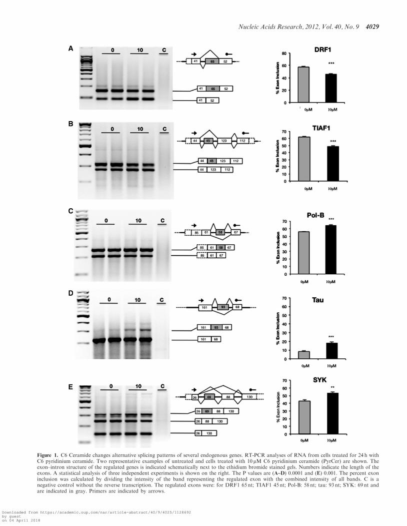
Figure 1. C6 Ceramide changes alternative splicing patterns of several endogenous genes. RT-PCR analyses of RNA from cells treated for 24 h withC6 pyridinium ceramide. Two representative examples of untreated and cells treated with 10 mM C6 pyridinium ceramide (PyrCer) are shown. Theexon–intron structure of the regulated genes is indicated schematically next to the ethidium bromide stained gels. Numbers indicate the length of theexons. A statistical analysis of three independent experiments is shown on the right. The P values are (A–D) 0.0001 and (E) 0.001. The percent exoninclusion was calculated by dividing the intensity of the band representing the regulated exon with the combined intensity of all bands. C is anegative control without the reverse transcription. The regulated exons were: for DRF1 65 nt; TIAF1 45 nt; Pol-B: 58 nt; tau: 93 nt; SYK: 69 nt andare indicated in gray. Primers are indicated by arrows.
Nucleic Acids Research, 2012, Vol. 40, No. 9 4029
Downloaded from https://academic.oup.com/nar/article-abstract/40/9/4025/1128692by gueston 04 April 2018

Figure 2. Reporter gene constructs recapitulate the effect of C6 pyridinium ceramide. The alternative exons regulated by C6 pyridinium phosphatewere cloned between two constitutively spliced insulin exons into a heterologous splice reporter construct. One microgram of the constructs wastransfected into HEK293 cells and the cells were treated with PyrCer for 24 h. Representative semi-quantitative RT-PCR analysis is shown. Thestatistical evaluation is shown on the right and was performed as described for Figure 1.
4030 Nucleic Acids Research, 2012, Vol. 40, No. 9
Downloaded from https://academic.oup.com/nar/article-abstract/40/9/4025/1128692by gueston 04 April 2018

dephosphorylation (13) its water-soluble pyridiniumanalog, C6 pyridinium ceramide, inhibits PP1 activity.
C6 pyridinium ceramide treatment changes thephosphorylation of several splicing factors in vitro
Numerous nuclear proteins contain putative PP1 bindingsites, and we recently showed that the splicing factors
SF2/ASF, Tra2-beta1 and SRp30c (also called SRSF9)bind directly to PP1 using an evolutionarily conservedRVxF motif located in their RRMs (3). PSF/SFPQ isinvolved in several nuclear processes. It acts early inspliceosome assembly, is involved in transcriptional regu-lation and could play a role in homologous DNA recom-bination (26–28). Importantly, PSF/SFPQ contains a PP1binding site in its RRM, which regulates its ability
Figure 3. C6 pyridinium ceramide inhibits protein phosphatase-1. (A) An equimolar mixture of PP1 isoforms (alpha, beta and gamma) wasincubated with the concentrations of C6 pyrimidinim ceramide indicated and the activity on p-nitrophenylphosphate was determined.1-(2-Oxopropyl) pyridinium was used as a negative control. (B) The effect on the PP1/PP2A inhibitor calyculin A is shown for comparison.(C) PyrCer effect on PP1 dephosphorylating phosphorylase phosphatase. (D) PyrCer effect on the phophorylation of splicing factors. HeLanuclear extract was incubated with [g-32P] ATP with or without 10 mM PyrCer and the proteins indicated were immunoprecipated. The top lanes(32P) show the signal from overnight autoradiography. The signal form western blot using the antisera employed in immunorecipitations are shownbelow. Tra2-beta1 and PSF migrate as doublet in western blots, which is due to different phosphorylation states (24). The numbers indicate themolecular weights. (E) Effect of 10 mM PyrCer on PP1-mediated dephosphorylation of purified Tra2-beta1 in vitro.
Nucleic Acids Research, 2012, Vol. 40, No. 9 4031
Downloaded from https://academic.oup.com/nar/article-abstract/40/9/4025/1128692by gueston 04 April 2018

to change alternative splicing (3,29,30). To determinewhether PyrCer influences the phosphorylation of othernuclear proteins involved in splicing, we tested its effecton a splicing competent HeLa nuclear extract. Weincubated HeLa nuclear extract with 4 mCi [g-32P] ATPin the presence or absence of 10 mM PyrCer and thensubjected the reactions to SDS–PAGE. Proteins with adifference in phosphorylation signal were determined byautoradiography. The differentially phosphorylated bandswere then identified by mass-spectrometry.We next validated candidate genes by immunopre-
cipitation from extracts incubated with [g-32P] ATP priorto the addition of 10 mM C6 pyridinium ceramide. Thephosphorylation of the immunoprecipated proteins wasdetected by autoradioagraphy and their general abun-dance was determined by by western blot. As shown inFigure 3D, PyrCer treatment increases the phosphoryl-ation of SF2/ASF, Tra2-beta1, PSF/SFPQ, UAP56 andSAP155, but does not change overall abundance of theseproteins. Again, this effect is opposite of the water-solubleC6-ceramide that promotes SF2/ASF dephosphorylation(24).We previously showed that PP1 dephosphorylated
Tra2-beta1 by binding to its RRM (3). As PyrCer in-hibited Tra2-beta1 dephosphorylation in nuclearextracts, we tested if it could inhibit PP1-mediated dephos-phorylation of the splicing factor when using recombinantproteins. His6-tagged Tra2-beta1 purified from insect cellswas 32P-labeled and treated with 10 mM C6 pyridiniumceramide or 10 nM Okadaic acid in the presence orabsence of 0.2U of PP1. As expected, labeledTra2-beta1 (Figure 3E, lane 1) was dephosphorylated byPP1 (Figure 3E, lane 2) but the presence of 10 mM C6pyridinium ceramide inhibited the dephosphorylation ofTra2-beta1 by PP1 (Figure 3E, lane 3), similar tookadaic acid (Figure 3D, lane 4). Thus, PyrCer directlyinterferes with the dephosphorylation of a splicing factorby PP1.Together, these data suggest that PyrCer treatment
increased the phosphorylation of several splicing factors,which is likely caused by blocking PP1 activity. This effectis the opposite of the water-insoluble natural and shortchain ceramides.
C6 Pyridinium Ceramide binds directly to PP1 in vivo
The inhibition of PP1 by PyrCer suggests that the twomolecules interact with each other. To explore this inter-action in vivo, we developed a mass-spectrometric assayfor PyrCer detection. In brief, cells were treated withpyridinium ceramide for 24 h to allow PyrCer and PP1to interact in vivo. Next, cells were washed and lysed,PP1-bound lipids were isolated with antibodies againstPP1 and lipids were extracted from immune-complexeswith chloroform, separated with HPLC and detected byion trap triple quadrupole mass spectrometry(Supplementary Figure S5A). This method gives a linearresponse to PyrCer as shown in Supplementary FigureS5B and S5C. To test whether C6 ceramide binds to PP1in vivo, we immunoprecipitated endogenous PP1 from cellsthat were treated with with or without 10 mMPyrCer using
a pan-PP1 antibody (Figure 4A and B). The ceramide sig-nal obtained from the mass-spectrometry was normalizedto the amount of PP1 present in the immunoprecipitations(Figure 4B).
We found that that PyrCer associates with PP1 in vivo.In addition, we performed a similar experiment withEGFP-tagged PP1 isoforms that were transfected intocells (Figure 4C). We immunoprecipitated the PP1isoforms alpha and gamma using an antiserum againstthe GFP tag. As shown in Figure 4C, both isoformsbound PyrCer about equally. This interaction appears tobe specific, as we could not detect C6 ceramide inimmunoprecipitates of Tra2-beta1-RATA, a Tra2-betamutant that does not interact with PP1 (3) (Figure 4C).As a further internal control, we analyzed the presence ofendogenous C18 ceramide in the immunoprecipitates. As
Figure 4. C6 pyridinium ceramide binds to protein phosphatase-1in vivo. (A) Detection of PyrCer and C18 ceramide bound to PP1.HEK293 cells were treated with 0 and 10 mM C6 ceramide for 14 hand PP1 was isolated by immunoprecipitation with a pan-PP1antibody. From the immunoprecipitates, bound lipids were extractedwith chloroform and analyzed by mass-spectrometry. Themass-spectrometry signal was normalized to the amount of immunopre-cipitated PP1, which was detected by western blot. The antiserum pre-cipitates the catalytic subunit of PP1. (B) Detection of PyrCer bound toPP1 variants. (C) EGFP-tagged constructs expressing PP1 gamma, PP1alpha, Tra2-beta1-RATA were transfected into HEK293 cells andimmunoprecipitated with an anti EGFP antiserum. PyrCer bound tothe immunoprecipitates was determined by mass-spectrometry andnormalized to the signal obtained by western blot using anti EGFP.
4032 Nucleic Acids Research, 2012, Vol. 40, No. 9
Downloaded from https://academic.oup.com/nar/article-abstract/40/9/4025/1128692by gueston 04 April 2018

shown in Figure 4A, C18 ceramide is associated with PP1in untreated cells. However, the amount of C18 ceramideis reduced upon PyrCer addition, suggesting that bothcompounds might compete for the same binding site.
Finally, we determined whether PyrCer is taken up bycells or exerts its action by stimulating signal transductioncascades emenating from the cell membrane. We added10 mM PyrCer to the cell medium and determined its intra-cellular distribution by cell fractionation. As shown inSupplemental Figure S6, about half of the lipid isaccumulated in the cells after 24 h, with 40% in thecytosol and 10% in nuclear fractions. This argues thatPyrCer can directly interact with cellular PP1 that ispresent in both the cytosol and nucleus.
In summary, these data show that cells take up C6pyridinium ceramide, where it binds directly to PP1 andinhibits the phosphatase.
C6 ceramide responsive exons have common features
At a concentration of 10 mM, PyrCer changes alternativesplicing of several exons in vivo (Figures 1 and 2). Thiscould indicate that protein complexes forming aroundcertain exons are more sensitive to PyrCer action andare changed at 10 mM concentration, which results in thepreferential usage of another splicing pathway. To deter-mine what features these exons have in common, weperformed component analysis of the exons. Allceramide responsive cassette exons that we identified areshorter than the mammalian consensus length for cassetteexons, which is around 150 nt. The mean length of theceramide-responsive exons is 66 nt, with a SD of 20 nt(Supplemental Figure S7B). It is therefore likely thatthese exons depend on enhancer sequences to overcometheir short length.
In order to determine common elements of PyrCer re-sponsive exons, we performed MEME (MultipleExpectation matrices for Motif Elicitation) analysis ofthe sequences (31). We found that each PyrCer-regulatedexon contains a GAAR and a CAAR motif (SupplementalFigure S7A). In addition, all exons contained SF2/ASFbinding motifs predicted by ESE finder (32). The nextcommon feature shared by all ceramide-responsive exonsis suboptimal splice sites. The combined 30 and 50 splicesites flanking PyrCer-responsive exons have an averagesplice-site-strength score of 7.5, whereas the control con-stitutive exons have a score of 15, when calculated by a lodscore algorithm based on mammalian splice sites (33)(Supplemental Figure S7C).
We next asked whether some of the regulated exonsbind directly to PSF/SFPQ, SF2/ASF and Tra2-beta1,proteins that are dephosphorylated by PP1 and predictedto bind to PyrCer-reponsive exons. We purifiedPSF/SFPQ, SF2/ASF and Tra2-beta1 from bacteria andperformed in vitro gel retardation assays using probes cor-responding to the parts of POL-B and TIAF1 exons thatcontain the GAAR and CAAR motifs. As controls, weused arteficial oligonucleotides where these motifs weredeleted. As shown in Figure 5, all three proteins boundto TIAF1 exon sequences. However Tra2-beta1 did notbind to the POL-B-exon RNA. This was surprising,
since the exon containes one AGAA binding motif thatis the high affinity binding site for Tra2-beta1 (34). Thissuggests that an unknown sequence context contributes toTra2-beta1 binding. The binding of Tra2-beta1, SF2/ASFand PSF to tau exon 10 has been reported earlier (35–37).We conclude that C6 pyridinium-dependent alternative
exons have suboptimal length, suboptimal splice sites andcontain binding sites characteristic for splicing enhancers.It is therefore likely that their regulation is dependenton regulatory protein complexes that form on thepre-mRNA.
Splicing regulators with a PP1 binding site influence theregulation of C6 pyridinium ceramide responsive exons
We found that PyrCer treatment of HeLa nuclear extractcaused an increase in the levels of phosphorylation ofsplicing factors PSF/SFPQ, SF2/ASF and Tra2-beta1(Figure 3D). All these splicing factors are known tointeract with PP1 (3). Therefore, we investigated the pos-sibility that these splicing factors could effect splicing ofthe PyrCer-regulated exons.As shown in Figure 6, the usage of all exons is changed
by PSF. With the exception of the DRF1 pre-mRNA, PSFpromotes exon skipping or inclusion of the tested reportergenes in a similar way as C6 pyridinium ceramide. SELEXexperiments showed that PSF binds to sequences contain-ing GAA-motifs (38) that are enriched in C6 pyridiniumceramide-responsive exons (Supplemental Figure S7A).Bioinformatic analysis using ESE finder (32)
indicates SF2/ASF binding sites in the regulated exons.Furthermore, all exons with the exception of DRF1contain the high affinity Tra2-beta1 binding site AGAA.However, DRF1 contains the lower affinity Tra2-beta1site TGAA. In vitro, Tra2-beta1 binds to AGAA andTGAA with 2.2 and 4.5mM affinity, respectively (34).We therefore analyzed the ceramide-responsive minigenesin co-transfection experiments together with constructsexpressing SF2/ASF and Tra2-beta1.As shown in Figure 7, all exons respond to SF2/ASF
and Tra2-beta1, which supports the bioinformatic predic-tion. It is noticeable that SF2/ASF promotes exonskipping in most cases, and Tra2-beta1 promotesskipping of an exon in the TIAF1 pre-mRNA, which isin contrast to their usual function as splicing activators.This unusual effect of SF2/ASF on tau exon 10 has beenpreviously observed (39). The effect of tra2-beta1 on thePOL-B alternative exons was much weaker than onTIAF1, which correlates with the strong binding oftra2-beta1 in vitro to the TIAF1 pre-mRNA and our in-ability to detect binding of tra2-beta1 to POL-B(Figure 5). Both SF2/ASF and Tra2-beta1 are direct sub-strates for PP1 and PP1-mediated dephosphorylation thatpromotes their interaction (3) and it is possible thatPyrCer affects exons that are dependent on both factors.These data indicate that ceramide responsive alternative
exons are dependent on splicing regulatory proteins thatcarry PP1 binding sites. The exons show the strongestresponse to PSF, suggesting that this factor is central tomediating the signaling between PyrCer and the splicingmachinery.
Nucleic Acids Research, 2012, Vol. 40, No. 9 4033
Downloaded from https://academic.oup.com/nar/article-abstract/40/9/4025/1128692by gueston 04 April 2018

DISCUSSION
Role of lipids in RNA metabolism
Lipids have been long known for their role as signalingmolecules that are important for human health. Theirbest-understood mode of action is the regulation ofprotein phosphorylation by modulating the activity ofkinases and phosphatases (40). However, there isemerging evidence that lipids directly interfere withRNA metabolism. For example, high levels of palmitaicacid are toxic for cells, which is mediated through an inter-action with snoRNAs in the cytosol, where snoRNAs arenormally not detectable (41). Similar to our findings,different lipids show various degrees of toxcicity andRNA association. RNAs were shown to directly bind to
membranes where it associates with liquid-ordered rafts(42) and RNA induced silencing complexes (RISC) areassociated with endosomal membranes (43,44), suggestingthat lipid composition regulates RNA processingcomplexes. The function of the lipids could be structuralby providing scaffolds for protein and RNA assembly aswell as catalytic by interfering with the activity of RNAprocessing enzymes, which was tested here.
In contrast to long-chain ceramides, C6 pyridiniumceramide blocks endogenous protein phosphatase-1 activity
Previously, it was found that short chain D-erythro-C6ceramide (24) and long-chain D-erythro-C18-ceramideactivate PP1 (13) and cause a decrease in SR-protein
Figure 5. PSF, SF2/ASF and Tra2-beta1 bind to motifs enriched in PyrCer dependent exons. (A) Gel retardation assay with TIAF1 and controlprobes. (B) Gel retardation assay with POL-B and control probes. (C) Sequence of the RNA probes. The GAAR motif is underlined, the CAAGmotif is underlined with zigzag. The SF2/ASF site is indicated by a dotted superscript. The concentration of all oligonucleotides was 0.4 mM in allreactions in a total volume of 10 ml. The protein concentrations for Tra2-beta1 were 0, 2.4, 3.6, 4.8 and 7.2 mM in lanes 1–5, respectively. Theconcentrations for PSF/SFPQ were 0, 0.5, 1.1, 2.1 and 3.2 mM in lanes 1–5, respectively. The concentration for SF2/ASF was 0, 1.1, 2.8, 4.0 and5.7 mM and in lanes 1–5, respectively.
4034 Nucleic Acids Research, 2012, Vol. 40, No. 9
Downloaded from https://academic.oup.com/nar/article-abstract/40/9/4025/1128692by gueston 04 April 2018
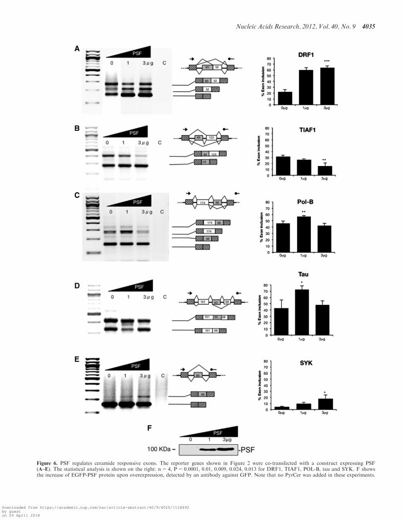
Figure 6. PSF regulates ceramide responsive exons. The reporter genes shown in Figure 2 were co-transfected with a construct expressing PSF(A–E). The statistical analysis is shown on the right: n=4, P=0.0001, 0.01, 0.009, 0.024, 0.013 for DRF1, TIAF1, POL-B, tau and SYK. F showsthe increase of EGFP-PSF protein upon overexpression, detected by an antibody against GFP. Note that no PyrCer was added in these experiments.
Nucleic Acids Research, 2012, Vol. 40, No. 9 4035
Downloaded from https://academic.oup.com/nar/article-abstract/40/9/4025/1128692by gueston 04 April 2018

Figure 7. Splicing regulators with a PP1 binding site regulate ceramide responsive exons. The reporter genes shown in Figure 2 were co-transfectedwith constructs expressing SF2/ASF or Tra2-beta1. RNA was isolated and analyzed by RT-PCR (A–E). The statistical analysis of at least threeindependent experiments is shown on the right. Note that no PyrCer was added in these experiments. (F) Detection of GFP-SF2/ASF and GFP-Tra2-beta1 by Western Blot analysis.
4036 Nucleic Acids Research, 2012, Vol. 40, No. 9
Downloaded from https://academic.oup.com/nar/article-abstract/40/9/4025/1128692by gueston 04 April 2018

phosphorylation. As water-soluble ceramides are used asanti-cancer agents, we tested the effect of the water-solublederivative PyrCer on PP1 activity. Surprisingly we foundthat PyrCer inhibits the phosphatase, both when usingrecombinant proteins and nuclear extract. In agreementwith an inhibition of PP1, PyrCer causes an increase ofphosphorylation of SF2/ASF, the opposite of its naturalderivatives, and the short non-cationic D-erythro-C6ceramide. Purified PP1 is inhibited starting at 50 mM,whereas proteins in nuclear extract are influenced at10 mM C6 pyridinium ceramide. As PP1 is located inprotein complexes, it is likely that allosteric effects thatinterfere with these complexes play a role in blocking thedephosphorylation of splicing factors. We showed thatPyrCer is taken up by the cells and found it inPP1 immunprecipitates, which indicates a direct inter-action with PP1, rather than a signal that ismediated through other pathways. These differences inPP1 inhibitions have functional consequences, as PyrCerand related short chain ceramides have different effects onalternative splicing: D-e-C6-ceramide changes bcl-x alter-native splicing (7), whereas PyrCer has no effect(Sumanasekera, C. et al., unpublished data).
The finding that chemically related lipids have oppo-sing effects on PP1 activity and splicing factor phosphor-ylation indicates that the cell could use lipid modificationto control the phosphorylation of PP1 targets, amongthem splicing factors. Such lipid modifications have beenwell documented for the glycerophospholipids, where theelectric charge of the water-soluble group is subject tomodification, as exemplified by the conversion of phos-photidylinositols to negatively charged phosphoinositidesthat have distinct signaling properties (45).
C6 pyridinium ceramide influences the phosphorylationof splicing factors
We used a mass-spectrometry approach to identifysplicing factors that change their phosphorylation due toPyrCer treatment. The rationale was that both PP1 andsplicing factor kinases are present in nuclear extract,where they are required for splicing activity (46,47). Thescreen resulted in the identification of five proteins thathad an increase in their phosphorylation upon PyrCertreatment: PSF/SFPQ, SF2/ASF, Tra2-beta1, UAP56and SAP155. Whereas the first three proteins are knownto interact with PP1 and contain an evolutionaryconserved binding site for PP1 in their RRMs, UAP56and SAP155 are not known to directly bind to PP1.However, it was shown that PP1 is directed to SAP155via an intermediate protein, NIPP-1 (48). It is possiblethat PyrCer interferes with the PP1-mediateddephosphorylation of SAP155 by blocking catalysis in aNIPP-1/PP1/SAP155 complex.
Using recombinant proteins, we tested the effect onPyrCer on the dephosphorylation of Tra2-beta1 byPP1. We found the PyrCer blocks the dephosphorylationin this system, indicating a direct influence on thephosphatase-substrate complex. Surprisingly, we did notdetect a PyrCer-mediated change of phosphorylation inRNA processing proteins that contain a PP1 binding
motif in their RRM, such as SRp30c, RBM15b, SFRS11and ROD1. In in vitro assays, SR proteins are often verysimilar, whereas in vivo analysis shows that they havediscrete functions (49). Our findings indicate that chem-ically related lipids can have opposing effects on the phos-phorylation and activity of SR proteins, which couldcontribute to their physiological regulation.
The C6 pyridinium ceramide signal is transducedby PSF/SFPQ, not only SR proteins
A medium-throughput PCR screen revealed several alter-native splicing events that are regulated by C6 pyridiniumceramide. All exons identified were cassette exons thatwere significantly shorter than constitutive exons andwere surrounded by weak splice sites. A search formotifs revealed the occurrence of GAAR and CAARmotifs in each exon. Each motif is expected to bepresent once in every 128 nt by chance, and should occur2.5 times when all exons are considered. In the regulatedexons there are 7 GAAR and 11 CAAG motifs, suggestingthat these motifs are 7-fold enriched in C6 pyridiniumceramide-dependent exons. The motifs overlap withpredicted PSF, SF2/ASF and Tra2-beta1 binding sites.The role of these proteins in regulating ceramide-
responsive exons was tested by cotransfection experimentsand direct protein binding and PSF showed the strongesteffect. This was surprising, as PSF was not known to beregulated by ceramide and is not an SR protein, a proteinclass previously shown to be regulated by ceramides (18).It was also unexpected that SF2/ASF promoted mostly
exon skipping, which is another unusual feature of PyrCerdependent exons. A possible mechanism could be arecruitment of PP1 by SF2/ASF to the pre-mRNA. Theoverexpression of SF2/ASF could recruit too much PP1 tothe pre-mRNA, which would result in aberrantprotein:RNA complexes. In agreement with this model,we observed skipping of C6-ceramide dependent exonswhen PP1 gamma was overexpressed in transfectionstudies (data not shown).The stronger effect of PSF on PyrCer dependent exons
could indicate that different lipids prefer distinct splicingfactors as targets, suggesting that lipid composition couldregulate alternative splice site selection in vivo.
SUPPLEMENTARY DATA
Supplementary Data are available at NAR Online:Supplementary Figures S1–S7.
ACKNOWLEDGEMENTS
We thank Carrol Beach, Matthew Gentry and RobertLester for discussions and thank Roscoe Klinck andBenoit Chabot for the RT-PCR screen.
FUNDING
European Alternative Splicing Network of Excellence(EURASNET) (LSHG-CT-2005-518238) and NationalInstitutes of Health (R21HD056195, 2P20 RR020171,
Nucleic Acids Research, 2012, Vol. 40, No. 9 4037
Downloaded from https://academic.oup.com/nar/article-abstract/40/9/4025/1128692by gueston 04 April 2018
P20RR021954 and RO1GM083187, GM50388,GM67969) as well as an ARRA supplement to J.A.A.Funding for open access charge: National Institutes ofHealth (RO1GM083187).
Conflict of interest statement. None declared.
REFERENCES
1. Pan,Q., Shai,O., Lee,L.J., Frey,B.J. and Blencowe,B.J. (2008)Deep surveying of alternative splicing complexity in the humantranscriptome by high-throughput sequencing. Nat. Genet., 40,1413–1415.
2. Tazi,J., Bakkour,N. and Stamm,S. (2009) Alternative splicing anddisease. Biochim. Biophys. Acta, 1792, 14–26.
3. Novoyatleva,T., Heinrich,B., Tang,Y., Benderska,N.,Butchbach,M.E., Lorson,C.L., Lorson,M.A., Ben-Dov,C.,Fehlbaum,P., Bracco,L. et al. (2008) Protein phosphatase 1 bindsto the RNA recognition motif of several splicing factors andregulates alternative pre-mRNA processing. Hum. Mol. Genet.,17, 52–70.
4. Shav-Tal,Y., Cohen,M., Lapter,S., Dye,B., Patton,J.G.,Vandekerckhove,J. and Zipori,D. (2001) Nuclear relocalization ofthe pre-mRNA splicing factor PSF during apoptosis involveshyperphosphorylation, masking of antigenic epitopes, and changesin protein interactions. Mol. Biol. Cell, 12, 2328–2340.
5. Shin,C., Feng,Y. and Manley,J.L. (2004) DephosphorylatedSRp38 acts as a splicing repressor in response to heat shock.Nature, 427, 553–558.
6. Xiao,S.H. and Manley,J.L. (1997) Phosphorylation of theASF/SF2 RS domain affects both protein-protein and protein-RNA interactions and is necessary for splicing. Genes Dev., 11,334–344.
7. Chalfant,C.E., Rathman,K., Pinkerman,R.L., Wood,R.E.,Obeid,L.M., Ogretmen,B. and Hannun,Y.A. (2002) De novoceramide regulates the alternative splicing of caspase 9 and Bcl-xin A549 lung adenocarcinoma cells. Dependence on proteinphosphatase-1. J. Biol. Chem., 277, 12587–12595.
8. Ghosh,N., Patel,N., Jiang,K., Watson,J.E., Cheng,J.,Chalfant,C.E. and Cooper,D.R. (2007) Ceramide-activated proteinphosphatase involvement in insulin resistance via Akt, serine/arginine-rich protein 40, and ribonucleic acid splicing in L6skeletal muscle cells. Endocrinology, 148, 1359–1366.
9. Ledeen,R.W. and Wu,G. (2008) Nuclear sphingolipids:metabolism and signaling. J. Lipid Res., 49, 1176–1186.
10. Massiello,A., Salas,A., Pinkerman,R.L., Roddy,P., Roesser,J.R.and Chalfant,C.E. (2004) Identification of two RNA cis-elementsthat function to regulate the 50 splice site selection of Bcl-xpre-mRNA in response to ceramide. J. Biol. Chem., 279,15799–15804.
11. Senkal,C.E., Ponnusamy,S., Rossi,M.J., Sundararaj,K., Szulc,Z.,Bielawski,J., Bielawska,A., Meyer,M., Cobanoglu,B., Koybasi,S.et al. (2006) Potent antitumor activity of a novel cationicpyridinium-ceramide alone or in combination with gemcitabineagainst human head and neck squamous cell carcinomas in vitroand in vivo. J. Pharmacol. Exp. Ther., 317, 1188–1199.
12. Separovic,D., Saad,Z.H., Edwin,E.A., Bielawski,J., Pierce,J.S.,Buren,E.V. and Bielawska,A. (2011) C16-ceramide analogcombined with Pc 4 photodynamic therapy evokes enhancedtotal ceramide accumulation, promotion of DEVDase activationin the absence of apoptosis, and augmented overall cell killing.J. Lipids, 2011, 713867.
13. Chalfant,C.E., Kishikawa,K., Mumby,M.C., Kamibayashi,C.,Bielawska,A. and Hannun,Y.A. (1999) Long chain ceramidesactivate protein phosphatase-1 and protein phosphatase-2A.Activation is stereospecific and regulated by phosphatidic acid.J. Biol. Chem., 274, 20313–20317.
14. Kishore,S., Khanna,A. and Stamm,S. (2008) Rapid generation ofsplicing reporters with pSpliceExpress. Gene, 427, 104–110.
15. Beullens,M., Van Eynde,A., Stalmans,W. and Bollen,M. (1992)The isolation of novel inhibitory polypeptides of protein
phosphatase 1 from bovine thymus nuclei. J. Biol. Chem., 267,16538–16544.
16. Sullards,M.C., Allegood,J.C., Kelly,S., Wang,E., Haynes,C.A.,Park,H., Chen,Y. and Merrill,A.H. Jr (2007) Structure-specific,quantitative methods for analysis of sphingolipids by liquidchromatography-tandem mass spectrometry: ‘‘inside-out’’sphingolipidomics. Methods Enzymol., 432, 83–115.
17. Pamuklar,Z., Federico,L., Liu,S., Umezu-Goto,M., Dong,A.,Panchatcharam,M., Fulerson,Z., Berdyshev,E., Natarajan,V.,Fang,X. et al. (2009) Autotaxin/lysopholipase D andlysophosphatidic acid regulate murine hemostasis and thrombosis.J. Biol. Chem., 284, 7385–7394.
18. Massiello,A. and Chalfant,C.E. (2006) SRp30a (ASF/SF2)regulates the alternative splicing of caspase-9 pre-mRNA and isrequired for ceramide-responsiveness. J. Lipid Res., 47, 892–897.
19. Karahatay,S., Thomas,K., Koybasi,S., Senkal,C.E., Elojeimy,S.,Liu,X., Bielawski,J., Day,T.A., Gillespie,M.B., Sinha,D. et al.(2007) Clinical relevance of ceramide metabolism in thepathogenesis of human head and neck squamous cell carcinoma(HNSCC): attenuation of C(18)-ceramide in HNSCC tumorscorrelates with lymphovascular invasion and nodal metastasis.Cancer Lett., 256, 101–111.
20. Separovic,D., Bielawski,J., Pierce,J.S., Merchant,S., Tarca,A.L.,Bhatti,G., Ogretmen,B. and Korbelik,M. (2011) Enhanced tumorcures after Foscan photodynamic therapy combined with theceramide analog LCL29. Evidence from mouse squamous cellcarcinomas for sphingolipids as biomarkers of treatment response.Int. J. Oncol., 38, 521–527.
21. Venables,J.P., Klinck,R., Bramard,A., Inkel,L., Dufresne-Martin,G., Koh,C., Gervais-Bird,J., Lapointe,E., Froehlich,U.,Durand,M. et al. (2008) Identification of alternative splicingmarkers for breast cancer. Cancer Res., 68, 9525–9531.
22. Stamm,S. (2002) Signals and their transduction pathwaysregulating alternative splicing: a new dimension of the humangenome. Hum. Mol. Genet., 11, 2409–2416.
23. Andreadis,A. (2005) Tau gene alternative splicing: expressionpatterns, regulation and modulation of function in normal brainand neurodegenerative diseases. Biochem. Biophys. Acta, 1739,91–103.
24. Chalfant,C.E., Ogretmen,B., Galadari,S., Kroesen,B.J., Pettus,B.J.and Hannun,Y.A. (2001) FAS activation inducesdephosphorylation of SR proteins; dependence on the de novogeneration of ceramide and activation of protein phosphatase 1.J. Biol. Chem., 276, 44848–44855.
25. Daoud,R., Mies,G., Smialowska,A., Olah,L., Hossmann,K. andStamm,S. (2002) Ischemia induces a translocation of the splicingfactor tra2-beta1 and changes alternative splicing patterns in thebrain. J. Neurosci., 22, 5889–5899.
26. Patton,J.G., Porro,E.B., Galceran,J., Tempst,P. andNadal-Ginard,B. (1993) Cloning and characterization of PSF,a novel pre-mRNA splicing factor. Genes Dev., 7, 393–406.
27. Urban,R.J., Bodenburg,Y., Kurosky,A., Wood,T.G. and Gasic,S.(2000) Polypyrimidine tract-binding protein-associated splicingfactor is a negative regulator of transcriptional activity of theporcine p450scc insulin-like growth factor response element.Mol. Endocrinol., 14, 774–782.
28. Akhmedov,A.T. and Lopez,B.S. (2000) Human 100-kDahomologous DNA-pairing protein is the splicing factor PSF andpromotes DNA strand invasion. Nucleic Acids Res., 28,3022–3030.
29. Liu,L., Xie,N., Rennie,P., Challis,J.R., Gleave,M., Lye,S.J. andDong,X. (2011) Consensus PP1 binding motifs regulatetranscriptional corepression and alternative RNA splicingactivities of the steroid receptor coregulators, p54nrb and PSF.Mol. Endocrinol., 25, 1197–1210.
30. Hirano,K., Erdodi,F., Patton,J.G. and Hartshorne,D.J. (1996)Interaction of protein phosphatase type 1 with a splicing factor.FEBS Lett., 389, 191–194.
31. Bailey,T.L. and Elkan,C. (1994) Fitting a mixture model byexpectation maximization to discover motifs in biopolymers.Proc. Int. Conf. Intell. Syst. Mol. Biol., 2, 28–36.
32. Smith,P.J., Zhang,C., Wang,J., Chew,S.L., Zhang,M.Q. andKrainer,A.R. (2006) An increased specificity score matrix for the
4038 Nucleic Acids Research, 2012, Vol. 40, No. 9
Downloaded from https://academic.oup.com/nar/article-abstract/40/9/4025/1128692by gueston 04 April 2018

prediction of SF2/ASF-specific exonic splicing enhancers.Hum. Mol. Genet., 15, 2490–2508.
33. Stamm,S., Zhu,J., Nakai,K., Stoilov,P., Stoss,O. and Zhang,M.Q.(2000) An alternative-exon database and its statistical analysis.DNA Cell Biol., 19, 739–756.
34. Clery,A., Jayne,S., Benderska,N., Dominguez,C., Stamm,S. andAllain,F.H. (2011) Molecular basis of purine-rich RNArecognition by the human SR-like protein Tra2-beta1.Nat. Struct. Mol. Biol., 18, 443–450.
35. Jiang,Z., Tang,H., Havlioglu,N., Zhang,X., Stamm,S., Yan,R. andWu,J.Y. (2003) Mutations in tau gene exon 10 associated withFTDP-17 alter the activity of an exonic splicing enhancer tointeract with Tra2 beta. J. Biol. Chem., 278, 18997–19007.
36. Ray,P., Kar,A., Fushimi,K., Havlioglu,N., Chen,X. and Wu,J.Y.(2011) PSF suppresses tau Exon 10 inclusion by interacting witha stem-loop structure downstream of exon 10. J. Mol. Neurosci.,45, 453–466.
37. Kondo,S., Yamamoto,N., Murakami,T., Okumura,M., Mayeda,A.and Imaizumi,K. (2004) Tra2 beta, SF2/ASF and SRp30cmodulate the function of an exonic splicing enhancer in exon 10of tau pre-mRNA. Genes Cells, 9, 121–130.
38. Peng,R., Dye,B.T., Perez,I., Barnard,D.C., Thompson,A.B. andPatton,J.G. (2002) PSF and p54nrb bind a conserved stem in U5snRNA. RNA, 8, 1334–1347.
39. Wang,J., Gao,Q.-S., Wang,Y., Lafyatis,R., Stamm,S. andAndreadis,A. (2004) Tau exon 10, whose missplicing causesfrontotemporal dementia, is regulated by an intricate interplay ofCis elements and Trans factors. J. Neurochem., 88, 1078–1090.
40. Wymann,M.P. and Schneiter,R. (2008) Lipid signalling in disease.Nat. Rev. Mol. Cell Biol., 9, 162–176.
41. Michel,C.I., Holley,C.L., Scruggs,B.S., Sidhu,R., Brookheart,R.T.,Listenberger,L.L., Behlke,M.A., Ory,D.S. and Schaffer,J.E. (2011)Small nucleolar RNAs U32a, U33, and U35a are criticalmediators of metabolic stress. Cell Metab., 14, 33–44.
42. Janas,T. and Yarus,M. (2006) Specific RNA binding to orderedphospholipid bilayers. Nucleic Acids Res., 34, 2128–2136.
43. Siomi,H. and Siomi,M.C. (2009) RISC hitches onto endosometrafficking. Nat. Cell Biol., 11, 1049–1051.
44. Gibbings,D.J., Ciaudo,C., Erhardt,M. and Voinnet,O. (2009)Multivesicular bodies associate with components of miRNAeffector complexes and modulate miRNA activity. Nat. Cell Biol.,11, 1143–1149.
45. Martin,T.F. (1998) Phosphoinositide lipids as signaling molecules:common themes for signal transduction, cytoskeletal regulation,and membrane trafficking. Annu. Rev. Cell. Dev. Biol., 14,231–264.
46. Mermoud,J.E., Cohen,P. and Lamond,A.I. (1992) Ser/Thr-specificprotein phosphatases are required for both catalytic steps ofpre-mRNA splicing. Nucleic Acids Res., 20, 5263–5269.
47. Stamm,S. (2008) Regulation of alternative splicing by reversiblephosphorylation. J.Biol. Chem., 283, 1223–1227.
48. Tanuma,N., Kim,S.E., Beullens,M., Tsubaki,Y., Mitsuhashi,S.,Nomura,M., Kawamura,T., Isono,K., Koseki,H., Sato,M. et al.(2008) Nuclear inhibitor of protein phosphatase-1 (NIPP1) directsprotein phosphatase-1 (PP1) to dephosphorylate the U2 smallnuclear ribonucleoprotein particle (snRNP) component,spliceosome-associated protein 155 (Sap155). J. Biol. Chem., 283,35805–35814.
49. Shepard,P.J. and Hertel,K.J. (2009) The SR protein family.Genome Biol., 10, 242.
Nucleic Acids Research, 2012, Vol. 40, No. 9 4039
Downloaded from https://academic.oup.com/nar/article-abstract/40/9/4025/1128692by gueston 04 April 2018
![Bis[4-(dimethylamino)pyridinium] octaaquachloridolanthanum ...journals.iucr.org/e/issues/2012/11/00/su2504/su2504.pdfBis[4-(dimethylamino)pyridinium] octaaquachloridolanthanum(III)](https://img.dokumen.tips/doc/110x75/5e0610443af6f93e3057972f/bis4-dimethylaminopyridinium-octaaquachloridolanthanum-4-dimethylaminopyridinium.jpg)