Embed Size (px)
Citation preview

C H
A P
T E
R
Professor Doug Boliver
Human Anatomy & Physiology II
16The Endocrine System
Endocrine System: Overview
Endocrine system – the body’s second great controlling system which influences metabolic activities of cells by means of hormones

Endocrine System: Overview
Endocrine glands – pituitary, pineal, thyroid, parathyroid, thymus, and adrenal
The pancreas and gonads produce both hormones and exocrine products
Other tissues and organs that produce hormones –adipose cells, pockets of cells in the walls of the small intestine, stomach, kidneys, and heart
Endocrine System: Overview
The hypothalamus has both neural functions and releases hormones
Interacts with the nervous system & immune system to regulate & coordinate body activities (maintains homeostasis)
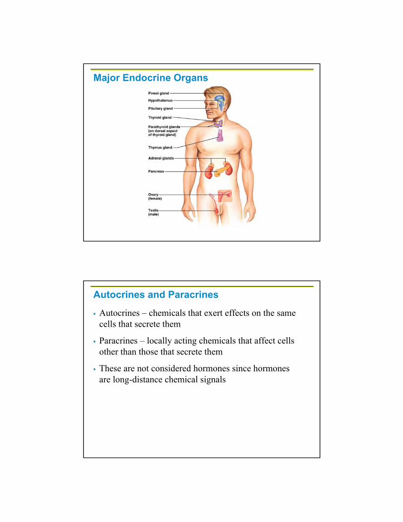
Major Endocrine Organs
Autocrines and Paracrines
Autocrines – chemicals that exert effects on the same cells that secrete them
Paracrines – locally acting chemicals that affect cells other than those that secrete them
These are not considered hormones since hormones are long-distance chemical signals
Hormones
Hormones – chemical substances secreted by cells into the extracellular fluids
Regulate the metabolic function of other cells via receptors
Have lag times ranging from seconds to hours
Tend to have prolonged effects
Are classified as amino acid-based hormones, or steroids
Eicosanoids – biologically active lipids with local hormone–like activity
Types of Hormones
Amino acid based
Amines, thyroxine, peptide, and protein hormones
Steroids – gonadal and adrenocortical hormones
Eicosanoids – include
Leukotrienes
Prostacyclin
Thromboxanes
Prostaglandins

Eicosanoid Synthesis & Related Drug Actions
Hormone Action
Hormones alter target cell activity by one of two mechanisms
Second messengers:
Regulatory G proteins
Amino acid–based hormones
Direct gene activation
Steroid & thyroid hormones
Tyrosine Kinase activation
The precise response depends on the type of the target cell
cAMP mechanism
PIP2 – Ca 2+ mechanism
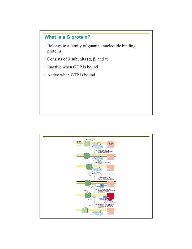
What is a G protein?
Belongs to a family of guanine nucleotide binding proteins
Consists of 3 subunits (α, β, and γ)
Inactive when GDP is bound
Active when GTP is bound

What is cAMP?
Formation and breakdown of cAMP↑ cAMP levels by activating adenylate cyclase↓ cAMP levels by inhibiting adenylate cyclase
2nd messenger cAMP mechanism (↑ cAMP)
Hormone (first messenger) binds to its receptor, which then binds to a G protein (α subunit)
The G protein is then activated as it binds GTP, which displaces GDP
Activated G protein activates the effector enzyme adenylate cyclase (AC)
AC generates cAMP (second messenger) from ATP
cAMP activates protein kinases, which then cause cellular effects
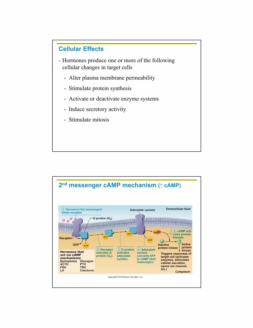
Cellular Effects
Hormones produce one or more of the following cellular changes in target cells
Alter plasma membrane permeability
Stimulate protein synthesis
Activate or deactivate enzyme systems
Induce secretory activity
Stimulate mitosis
2nd messenger cAMP mechanism (↑ cAMP)
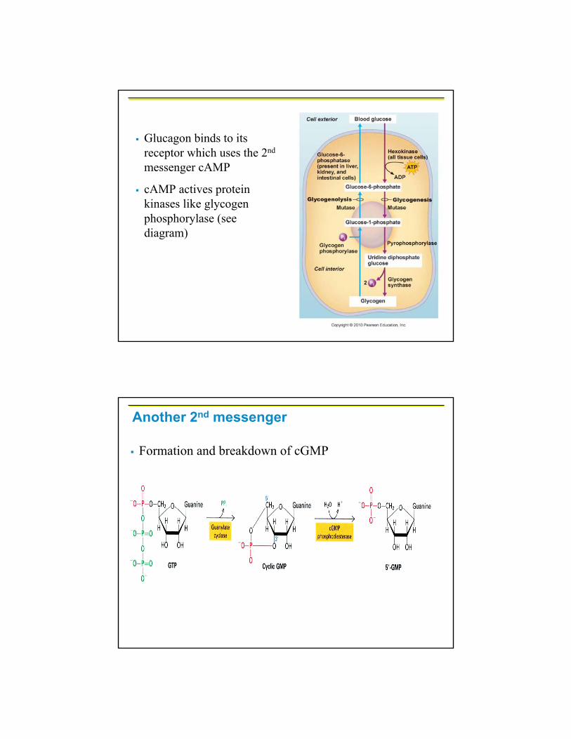
Glucagon binds to its receptor which uses the 2nd
messenger cAMP
cAMP actives protein kinases like glycogen phosphorylase (see diagram)
Another 2nd messenger
Formation and breakdown of cGMP
ReceptorGTP
GTP GTP
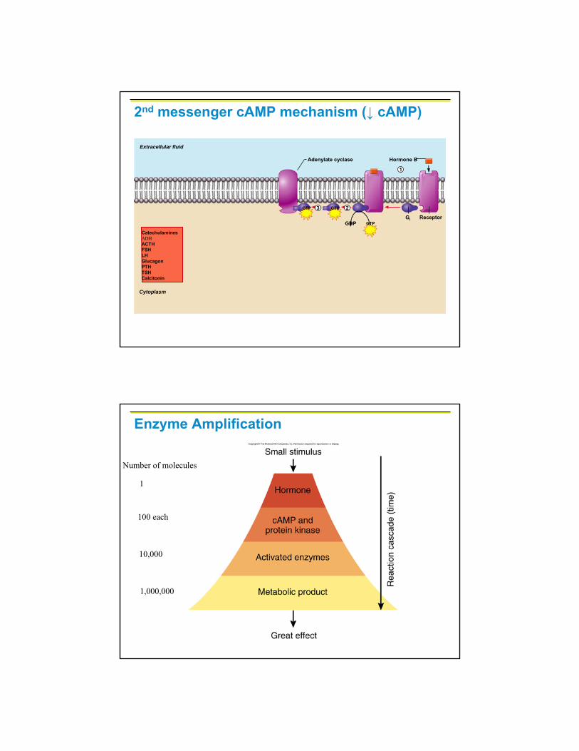
CatecholaminesADHACTHFSHLHGlucagonPTHTSHCalcitonin
Adenylate cyclase Hormone B
GDP
Extracellular fluid
Cytoplasm
Gi
3 2
1
2nd messenger cAMP mechanism (↓ cAMP)
Enzyme Amplification
1
100 each
Number of molecules
10,000
1,000,000

Protein phosphorylationis a common means of information transfer. Many second messengers elicit responses by activating protein kinases.These enzymes transfer phosphoryl groups from ATP to specific serine, threonine, and tyrosine residues in proteins.

Hormone binds to the receptor and activates G protein (α subunit)
G protein binds and activates phospholipase C (PL-C)
PL-C splits the phospholipid PIP2 into diacylglycerol(DAG) and IP3 (both act as second messengers)
DAG activates protein kinases (PKC) which phosphorylates & activates proteins
IP3 triggers release of Ca2+ stores
Ca2+ (third messenger) alters cellular responses
2nd messenger PIP2 – Ca2+mechanism
GTP PIP2
IP3
ReceptorGTP
GTP
CatecholaminesTRHGnRHOxytocin
Triggers responses of target cell
GDP
Extracellular fluid
Cytoplasm
Inactiveprotein kinase C
Activeprotein kinase C
Phospholipase C
Gq
Ca2+ Ca2+- calmodulin
Hormone
Endoplasmicreticulum
DAG1
2 34 5
5
6
2nd messenger PIP2 – Ca2+mechanism
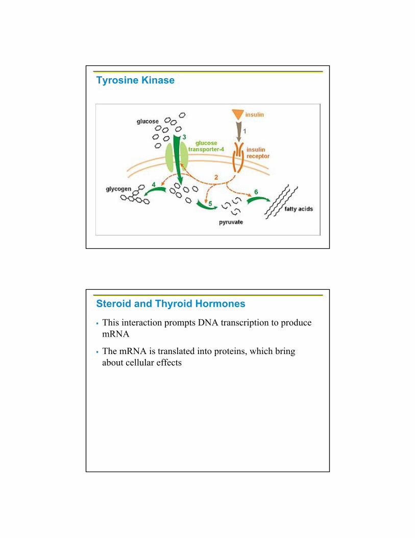
Tyrosine Kinase
Steroid and Thyroid Hormones
This interaction prompts DNA transcription to produce mRNA
The mRNA is translated into proteins, which bring about cellular effects

Hormone Effects on Gene Activity

Target Cell Specificity
Hormones circulate to all tissues but only activate cells referred to as target cells
Target cells must have specific receptors to which the hormone binds
These receptors may be intracellular or located on the plasma membrane
Target Cell Specificity
Examples of hormone activity
ACTH receptors are only found on certain cells of the adrenal cortex
Thyroxin receptors are found on nearly all cells of the body
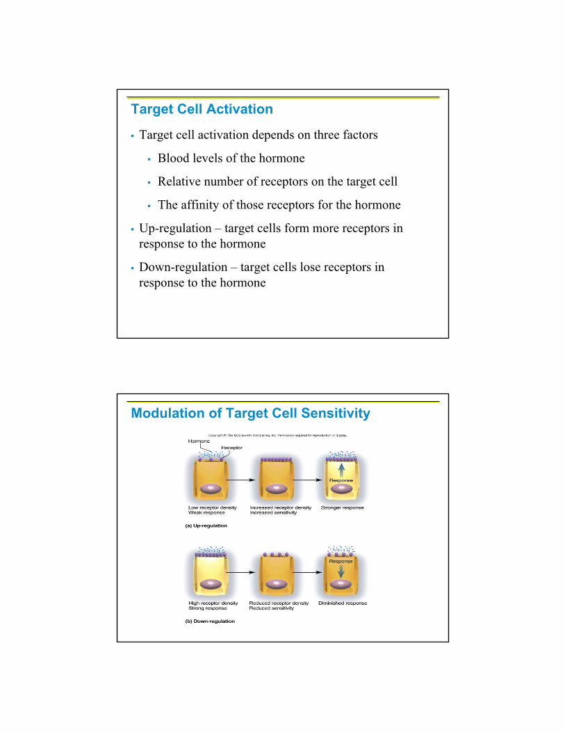
Target Cell Activation
Target cell activation depends on three factors
Blood levels of the hormone
Relative number of receptors on the target cell
The affinity of those receptors for the hormone
Up-regulation – target cells form more receptors in response to the hormone
Down-regulation – target cells lose receptors in response to the hormone
Modulation of Target Cell Sensitivity

Hormone Concentrations in the BloodHormones circulate in the blood in two forms –free or bound
Steroids and thyroid hormone are attached to plasma proteins
All others are unencumbered or free
Hormone Concentrations in the Blood
Concentrations of circulating hormone reflect:
Rate of release
Speed of inactivation and removal from the body
Hormones are removed from the blood by:
Degrading enzymes
The kidneys
Liver enzyme systems

Interaction of Hormones at Target Cells
Three types of hormone interaction
Permissiveness – one hormone cannot exert its effects without another hormone being present
Synergism – more than one hormone produces the same effects on a target cell
Antagonism – one or more hormones opposes the action of another hormone
Control of Hormone ReleaseBlood levels of hormones:
Are controlled by negative feedback systems
Vary only within a narrow desirable range
Hormones are synthesized and released in response to:
Humoral stimuli
Neural stimuli
Hormonal stimuli
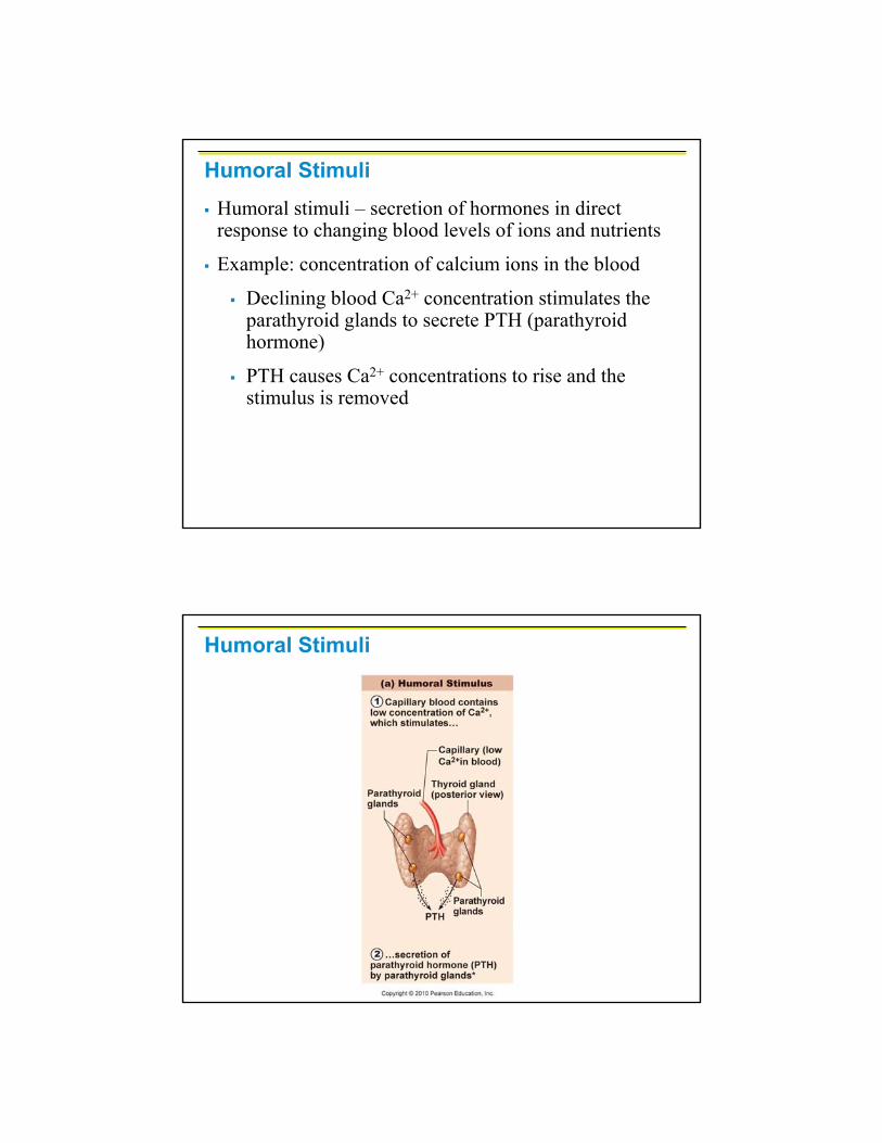
Humoral StimuliHumoral stimuli – secretion of hormones in direct response to changing blood levels of ions and nutrients
Example: concentration of calcium ions in the blood
Declining blood Ca2+ concentration stimulates the parathyroid glands to secrete PTH (parathyroid hormone)
PTH causes Ca2+ concentrations to rise and the stimulus is removed
Humoral Stimuli

Neural Stimuli
Neural stimuli – nerve fibers stimulate hormone release
Preganglionicsympathetic nervous system (SNS) fibers stimulate the adrenal medulla to secrete catecholamines
Hormonal Stimuli
Hormonal stimuli – release of hormones in response to hormones produced by other endocrine organs
The hypothalamic hormones stimulate the adenohypophysis
In turn, pituitary hormones stimulate targets to secrete still more hormones
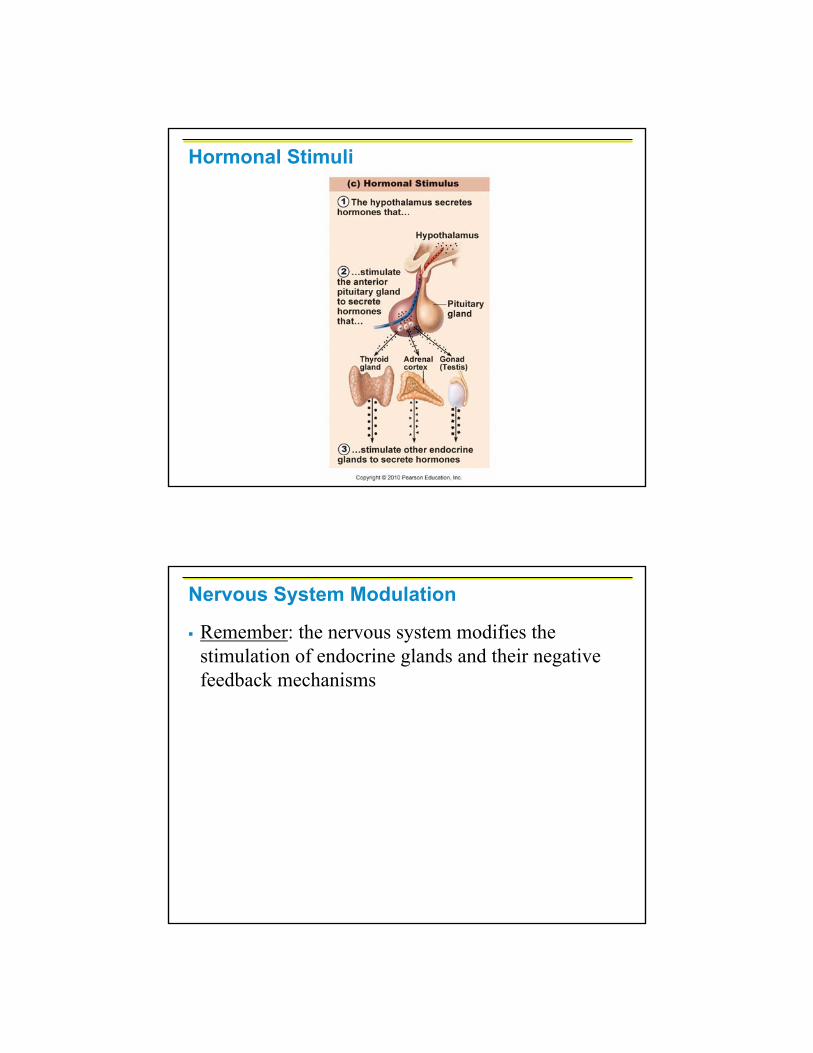
Hormonal Stimuli
Nervous System Modulation
Remember: the nervous system modifies the stimulation of endocrine glands and their negative feedback mechanisms
Nervous System Modulation
The nervous system can override normal endocrine controls
For example, control of blood glucose levels
Normally the endocrine system maintains blood glucose
Under stress, the body needs more glucose
The hypothalamus and the sympathetic nervous system are activated to supply ample glucose
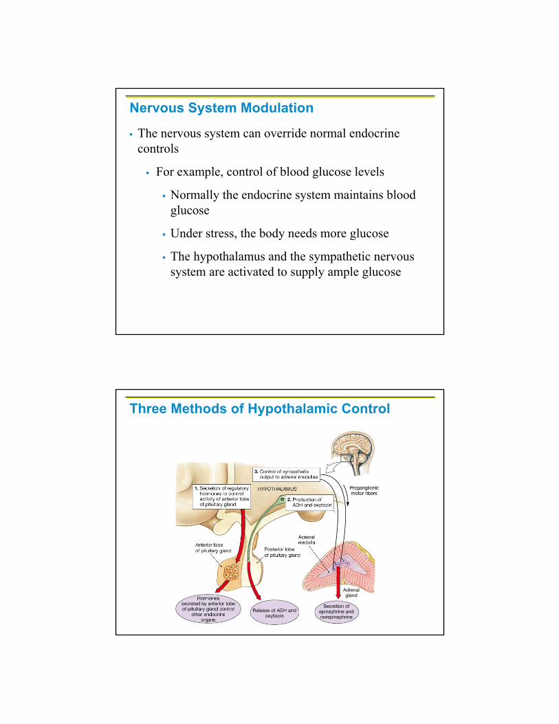
Three Methods of Hypothalamic Control
Major Endocrine Organs: Hypophysis
Hypophysis (pituitary gland) – two-lobed organ that secretes nine major hormones
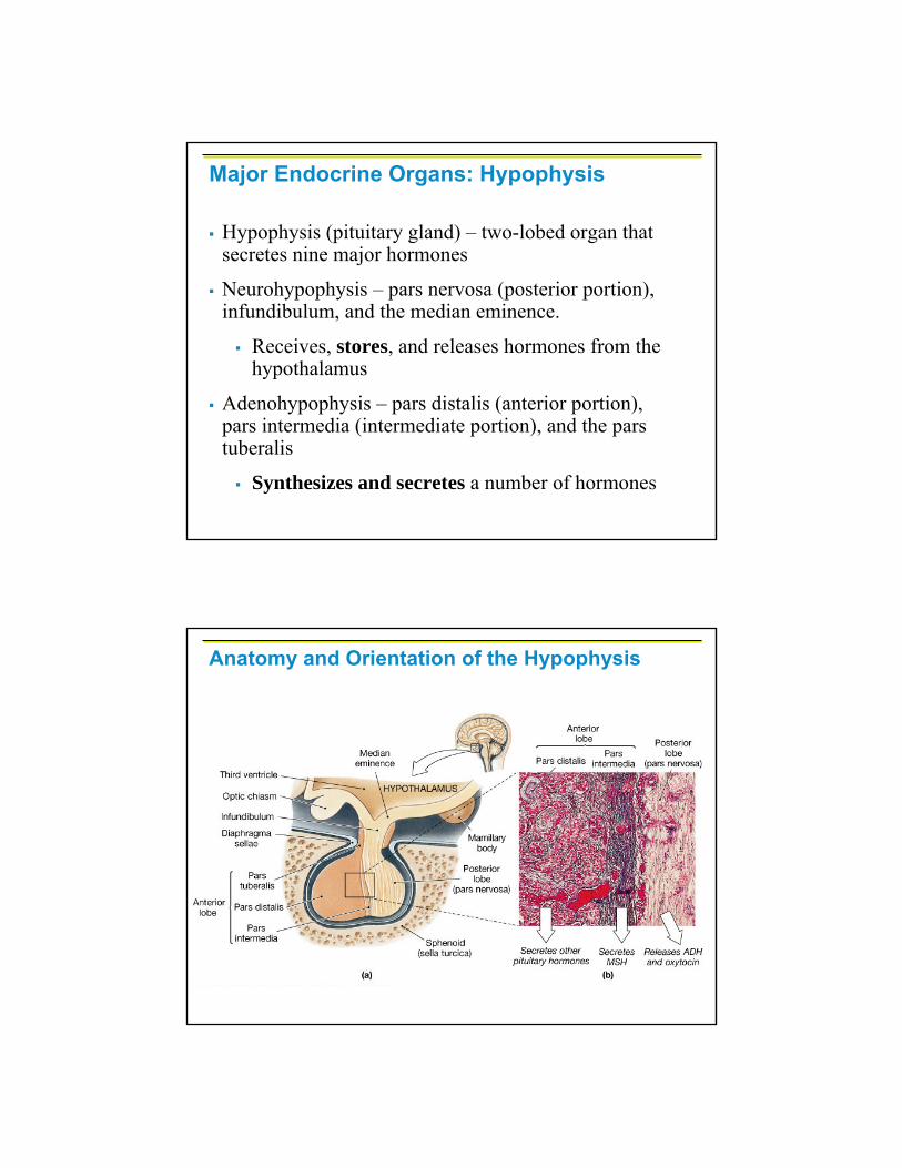
Neurohypophysis – pars nervosa (posterior portion), infundibulum, and the median eminence.
Receives, stores, and releases hormones from the hypothalamus
Adenohypophysis – pars distalis (anterior portion), pars intermedia (intermediate portion), and the pars tuberalis
Synthesizes and secretes a number of hormones
Anatomy and Orientation of the Hypophysis

Anatomy and Orientation of the Hypophysis
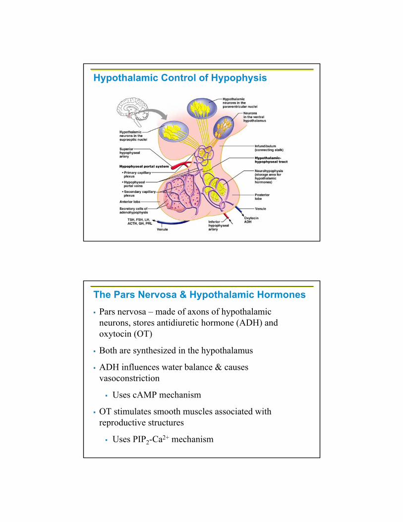
Hypothalamic Control of Hypophysis

Pituitary-Hypothalamic Relationships: Neurohypophysis (posterior portion)
The neurohypophysis is a downgrowth of hypothalamic neural tissue
Has a neural connection with the hypothalamus (hypothalamic-hypophyseal tract)
Nuclei of the hypothalamus synthesize oxytocin and antidiuretic hormone (ADH)
These hormones are transported to the posterior pituitary
Pituitary-Hypothalamic Relationships: Adenohypophysis (anterior portion)
The adenohypophysis of the pituitary is an outpocketing of the oral mucosa
There is no direct neural contact with the hypothalamus

Embryonic Development of the Hypophysis
Embryonic Development of the Hypophysis

There is a vascular connection, the hypophyseal portal system, consisting of:
The primary capillary plexus
The hypophyseal portal veins
The secondary capillary plexus
Pituitary-Hypothalamic Relationships: Adenohypophysis

Hypothalamic Control of Hypophysis
Adenophypophyseal Hormones
The six hormones of the adenohypophysis:
Abbreviated as GH, TSH, ACTH, FSH, LH (ICSH), and PRL
Regulate the activity of other endocrine glands
In addition, pro-opiomelanocortin (POMC):
Has been isolated from the pituitary
Is split into ACTH, opiates, and MSH
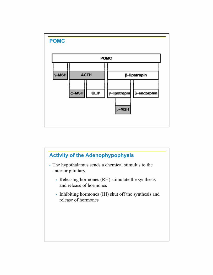
POMC
Activity of the Adenophypophysis
The hypothalamus sends a chemical stimulus to the anterior pituitary
Releasing hormones (RH) stimulate the synthesis and release of hormones
Inhibiting hormones (IH) shut off the synthesis and release of hormones
Activity of the Adenophypophysis
The tropic hormones that are released are:
Thyroid-stimulating hormone (TSH)
Adrenocorticotropic hormone (ACTH)
Follicle-stimulating hormone (FSH)
Luteinizing hormone (LH) also known as interstitial cell stimulating hormone (ICSH)
Growth Hormone (GH)
Produced by somatotropic cells of the pars distalis that:
Stimulate most cells, but target bone and skeletal muscle
Promote protein synthesis and encourage the use of lipids for fuel (glucose sparing)
Most effects are mediated indirectly by somatomedins

Growth Hormone (GH)
Antagonistic hypothalamic hormones regulate GH
Growth hormone–releasing hormone (GHRH) stimulates GH release
Growth hormone–inhibiting hormone (GHIH) inhibits GH release
Metabolic Action of Growth Hormone
GH stimulates liver, skeletal muscle, bone, and cartilage to produce insulin-like growth factors
Direct action promotes lipolysis and inhibits glucose uptake
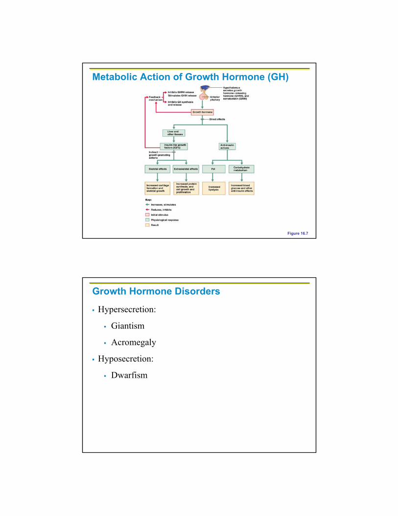
Metabolic Action of Growth Hormone (GH)
Figure 16.7
Growth Hormone Disorders
Hypersecretion:
Giantism
Acromegaly
Hyposecretion:
Dwarfism
Growth Hormone Abnormalities
Thyroid Stimulating Hormone (Thyrotropin)
Stimulates the normal development and secretory activity of the thyroid
Triggered by hypothalamic peptide thyrotropin-releasing hormone (TRH)
Rising blood levels of thyroid hormones act on the pituitary and hypothalamus to block the release of TSH

Adrenocorticotropic Hormone (Corticotropin)
Stimulates the adrenal cortex to release corticosteroids
Triggered by hypothalamic corticotropin-releasing hormone (CRH) in a daily rhythm
Internal and external factors such as fever, hypoglycemia, and stressors can trigger the release of CRH
GonadotropinsGonadotropins – follicle-stimulating hormone (FSH) and luteinizing hormone (LH)
Regulate the function of the ovaries and testes
FSH stimulates gamete (egg or sperm) production
Absent from the blood in prepubertal boys and girls
Triggered by the hypothalamic gonadotropin-releasing hormone (GnRH) during and after puberty
Functions of Gonadotropins
In females
LH works with FSH to cause maturation of the ovarian follicle
LH works alone to trigger ovulation (expulsion of the egg from the follicle)
LH promotes synthesis and release of estrogens and progesterone
Functions of Gonadotropins
In males
LH stimulates interstitial cells of the testes to produce testosterone
LH is also referred to as interstitial cell-stimulating hormone (ICSH)

Prolactin (PRL)
In females, stimulates the development of mammary glands, initiates & maintains milk production by the breasts
In males, enhances testosterone production
Triggered by the hypothalamic prolactin-releasing hormone (PRH)
Inhibited by prolactin-inhibiting hormone (PIH)
Prolactin (PRL) cont.
Blood levels rise toward the end of pregnancy
Suckling stimulates PRH release and encourages continued milk production
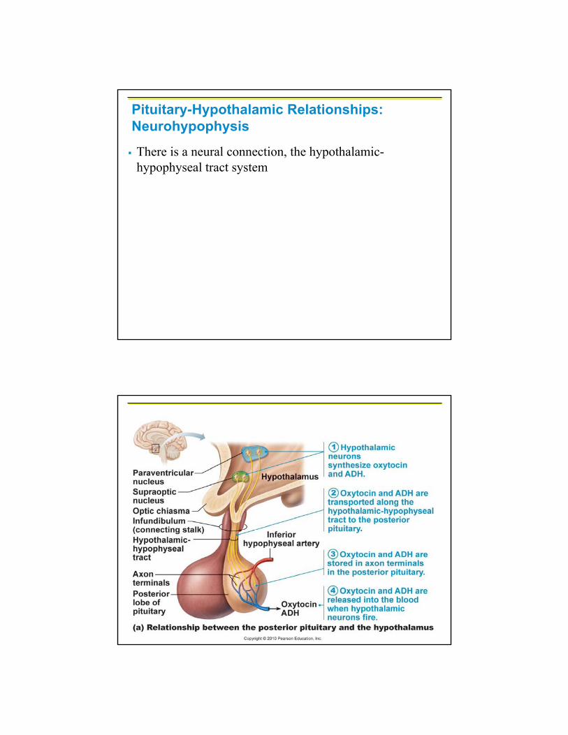
Pituitary-Hypothalamic Relationships: Neurohypophysis
There is a neural connection, the hypothalamic-hypophyseal tract system
Hypothalamic Control of Hypophysis
The Pars Nervosa & Hypothalamic HormonesPars nervosa – made of axons of hypothalamic neurons, stores antidiuretic hormone (ADH) and oxytocin (OT)
Both are synthesized in the hypothalamus
ADH influences water balance & causes vasoconstriction
Uses cAMP mechanism
OT stimulates smooth muscles associated with reproductive structures
Uses PIP2-Ca2+ mechanism
ADH
ADH helps to avoid dehydration or water overload
Prevents urine formation
Osmoreceptors monitor the solute concentration of the blood
With high solutes, ADH preserves water
With low solutes, ADH is not released, thus causing water loss
Alcohol inhibits ADH release and causes copious urine output
ADH Disorders
Hypersecretion:
Syndrome of Inappropriate ADH (SIADH)
Hyposecretion:
Diabetes Insipidus
Oxytocin
Regulated by a positive feedback mechanism to OT in the blood
In women:
This leads to increased intensity of uterine contractions, ending in birth
OT triggers milk ejection (“letdown” reflex)
Synthetic and natural OT drugs can be used to induce or hasten labor
Oxytocin
In males:
Involved in ejaculation and sperm transport
Plays a role in sexual arousal and satisfaction in males and nonlactating females
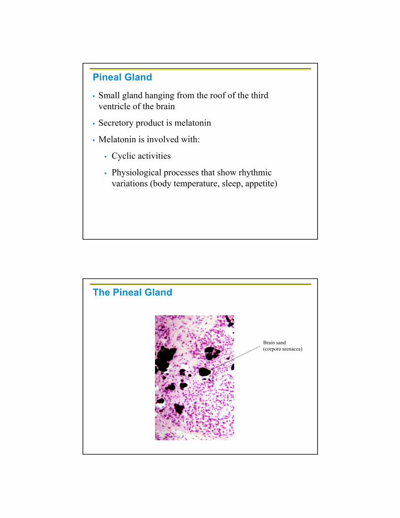
Small gland hanging from the roof of the third ventricle of the brain
Secretory product is melatonin
Melatonin is involved with:
Cyclic activities
Physiological processes that show rhythmic variations (body temperature, sleep, appetite)
Pineal Gland
The Pineal Gland
Brain sand (corpora arenacea)

Thyroid Gland
The largest endocrine gland, located in the anterior neck, consists of two lateral lobes connected by a median tissue mass called the isthmus
Composed of follicles that produce the glycoprotein thyroglobulin
Colloid (thyroglobulin + iodine) fills the lumen of the follicles and is the precursor of thyroid hormone
Other endocrine cells, the parafollicular cells (C cells), produce the hormone calcitonin
Thyroid Gland
Figure 16.8
Thyroid hormone – major metabolic hormone
Consists of two related iodine-containing compounds
T4 – thyroxine; has two tyrosine molecules plus four bound iodine atoms
T3 – triiodothyronine; has two tyrosines with three bound iodine atoms
Thyroid Hormone
Effects of Thyroid HormoneTH is concerned with:
Glucose oxidation
Increasing metabolic rate
Heat production
TH plays a role in:
Maintaining blood pressure
Regulating tissue growth
Developing skeletal and nervous systems
Maturation and reproductive capabilities
T4 and T3 bind to thyroxine-binding globulins (TBGs) produced by the liver
Both bind to target receptors, but T3 is more potent and has a shorter half life than T4
Peripheral tissues convert T4 to T3
Mechanisms of activity are similar to steroids
Regulation is by negative feedback
Hypothalamic thyrotropin-releasing hormone (TRH) can overcome the negative feedback
Transport and Regulation of TH
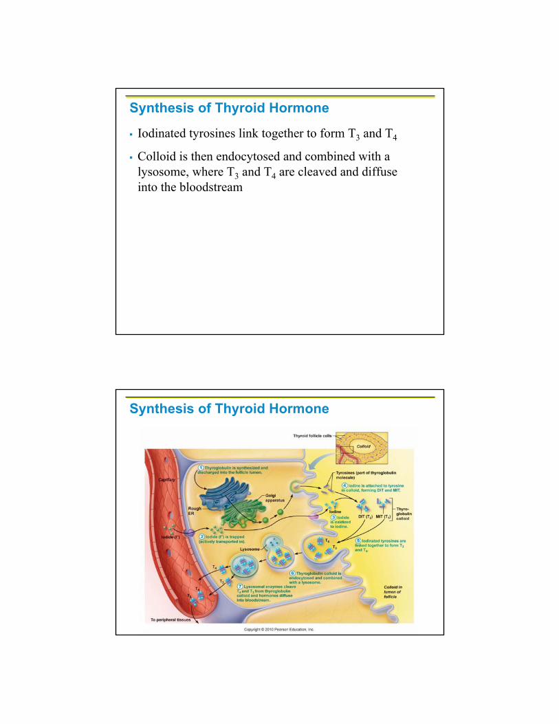
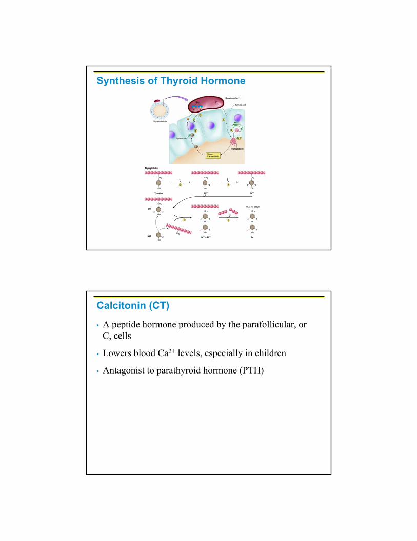
Synthesis of Thyroid Hormone
Thyroglobulin is synthesized and discharged into the lumen
Iodides (I–) are actively taken into the cell, oxidized to iodine (I2), and released into the lumen
Iodine attaches to tyrosine, mediated by peroxidaseenzymes, forming T1 (monoiodotyrosine, or MIT), and T2 (diiodotyrosine, or DIT)
Synthesis of Thyroid Hormone
Iodinated tyrosines link together to form T3 and T4
Colloid is then endocytosed and combined with a lysosome, where T3 and T4 are cleaved and diffuse into the bloodstream
Synthesis of Thyroid Hormone
Synthesis of Thyroid Hormone
A peptide hormone produced by the parafollicular, or C, cells
Lowers blood Ca2+ levels, especially in children
Antagonist to parathyroid hormone (PTH)
Calcitonin (CT)
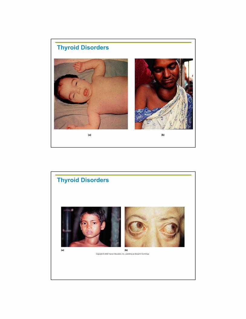
Thyroid Disorders
Thyroid Disorders

Thyroid Disorders – Endemic Goiter
CT targets the skeleton, where it:
Inhibits osteoclast activity (and thus bone resorption) and release of Ca2+ from the bone matrix
Stimulates Ca2+ uptake and incorporation into the bone matrix
Regulated by a humoral (Ca2+ ion concentration in the blood) negative feedback mechanism
Calcitonin

Parathyroid Glands
Tiny glands embedded in the posterior aspect of the thyroid
Cells are arranged in cords containing oxyphil and chief cells
Chief (principal) cells secrete PTH also referred to as parathormone
PTH increases blood Ca2+ levels
Parathyroid Glands
Figure 16.11

PTH release increases Ca2+ in the blood as it:
Stimulates osteoclasts to digest bone matrix
Enhances the reabsorption of Ca2+ and the secretion of phosphate by the kidneys
Increases absorption of Ca2+ by intestinal mucosal
Rising Ca2+ in the blood inhibits PTH release
Effects of Parathyroid Hormone
Effects of Parathyroid Hormone
Figure 16.12
Thymus
Lobulated gland located deep to the sternum
Major hormonal products are thymopoietins and thymosins
These hormones are essential for the maturation and proliferation of T lymphocytes (T cells) of the immune system
A triangular gland, which has both exocrine and endocrine cells, located behind the stomach
Acinar cells produce an enzyme-rich juice used for digestion (exocrine product)
Pancreatic islets (islets of Langerhans) produce hormones (endocrine products)
Pancreas
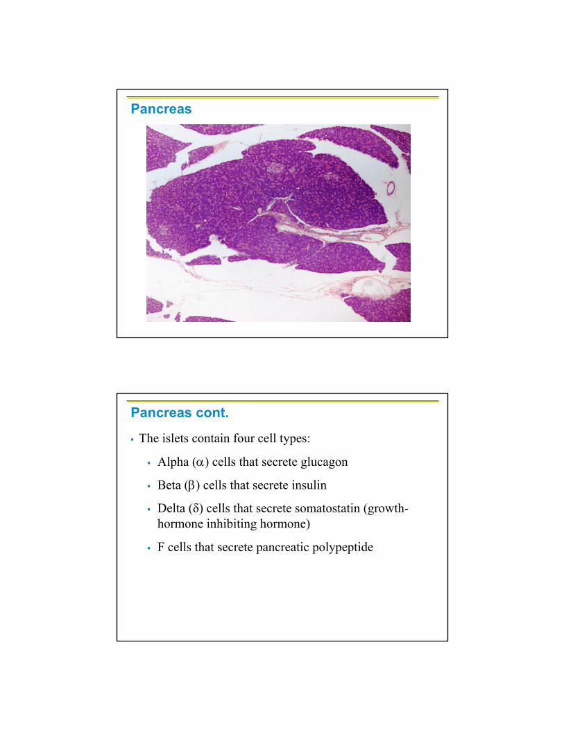
Pancreas
Pancreas cont.
The islets contain four cell types:
Alpha (α) cells that secrete glucagon
Beta (β) cells that secrete insulin
Delta (δ) cells that secrete somatostatin (growth-hormone inhibiting hormone)
F cells that secrete pancreatic polypeptide
A 29-amino-acid polypeptide hormone that is a potent hyperglycemic agent
Its major target is the liver, where it promotes:
Glycogenolysis – the breakdown of glycogen to glucose
Gluconeogenesis – synthesis of glucose from lactic acid and noncarbohydrates
Release of glucose to the blood from liver cells
Glucagon
A 51-amino-acid protein consisting of two amino acid chains linked by disulfide bonds
Synthesized as part of proinsulin and then excised by enzymes, releasing functional insulin
Insulin:
Lowers blood glucose levels
Enhances transport of glucose into body cells
Counters metabolic activity that would enhance blood glucose levels
Insulin
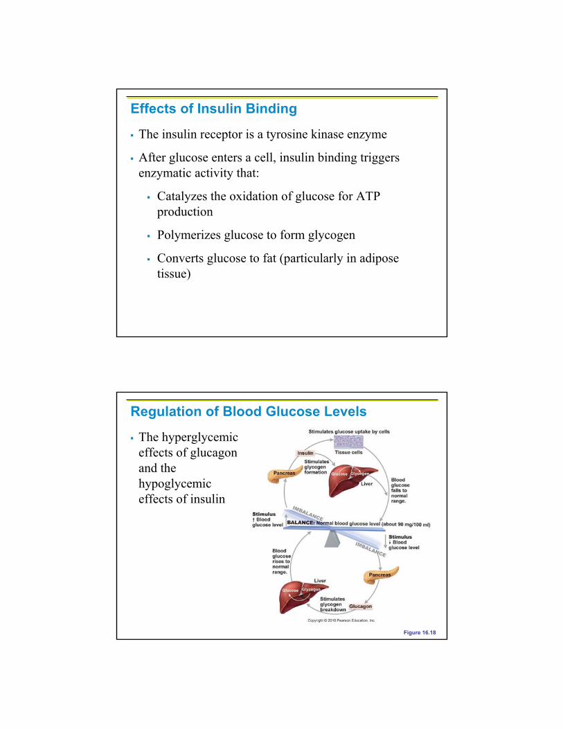
The insulin receptor is a tyrosine kinase enzyme
After glucose enters a cell, insulin binding triggers enzymatic activity that:
Catalyzes the oxidation of glucose for ATP production
Polymerizes glucose to form glycogen
Converts glucose to fat (particularly in adipose tissue)
Effects of Insulin Binding
Regulation of Blood Glucose Levels
Figure 16.18
The hyperglycemic effects of glucagon and the hypoglycemic effects of insulin
Insulin DisordersHyperinsulinism – excessive insulin secretion, resulting in hypoglycemia
Symptoms can include such things as headache, dizziness, weakness, and emotional instability. In severe cases there may be convulsions, coma, and death.
The cause of oversecretion of insulin may be organic, i.e., a tumor of the pancreas, impaired liver function, or endocrine disorders, or it may be functional, e.g., unusual muscular exertion, pregnancy, or lactation.
Insulin Disorders
Pre-diabetes → where fasting glucose levels are elevated above normal, but not high enough to be considered diabetes
Fasting results are between 100 to 125 mg/dl
Normal blood glucose levels in the range of 70 –110 mg/dl (90 – 100 mg/dl is average)
Also referred to as impaired fasting glucose
If no intervention takes place, then type 2 diabetes will develop
Insulin DisordersDiabetes Mellitus (DM)
Results from hyposecretion or hypoactivity of beta cells (insulin)
The three cardinal signs of DM are:
Polyuria – huge urine output
Polydipsia – excessive thirst
Polyphagia – excessive hunger and food consumption
3 further signs revealed by blood & urine tests include: hyperglycemia, glycosuria, ketonemia, ketonuria
Fasting blood glucose is 126 mg/dl or higher on three occasions
3 types:
Type 1 or insulin-dependent diabetes mellitus (IDDM)
Type 2 or non-insulin-dependent diabetes mellitus (NIDDM)
Gestational
Diabetes Mellitus (DM)

Diabetes Mellitus (DM)
Table 16.4
Adrenal glands – paired, pyramid-shaped organs atop the kidneys
Structurally and functionally, they are two glands in one
Adrenal medulla – neural tissue that acts as part of the SNS
Adrenal cortex – glandular tissue derived from embryonic mesoderm
Adrenal (Suprarenal) Glands

Adrenal CortexSynthesizes and releases steroid hormones called corticosteroids
Different corticosteroids are produced in each of the three layers
Zona glomerulosa – mineralocorticoids(chiefly aldosterone)
Zona fasciculata – glucocorticoids(chiefly cortisol)
Zona reticularis – gonadocorticoids(chiefly androgens)
Adrenal Gland

Adrenal Cortex – Zona Glomerulosa
Adrenal Cortex – Zona Fasciculata

Adrenal Cortex – Zona Reticularis
Adrenal Medulla
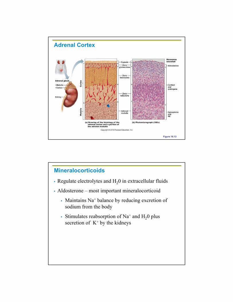
Adrenal Cortex
Figure 16.13
Regulate electrolytes and H20 in extracellular fluids
Aldosterone – most important mineralocorticoid
Maintains Na+ balance by reducing excretion of sodium from the body
Stimulates reabsorption of Na+ and H20 plus secretion of K+ by the kidneys
Mineralocorticoids

Aldosterone secretion is stimulated by:
Rising blood levels of K+
Low blood Na+
Decreasing blood volume or pressure
Mineralocorticoids
The Four Mechanisms of Aldosterone Secretion
Renin-angiotensin mechanism – kidneys release renin, which is converted into angiotensin II that in turn stimulates aldosterone release
Plasma concentration of sodium and potassium –directly influences the zona glomerulosa cells
ACTH – causes small increases of aldosterone during stress
Atrial natriuretic peptide (ANP) – inhibits activity of the zona glomerulosa
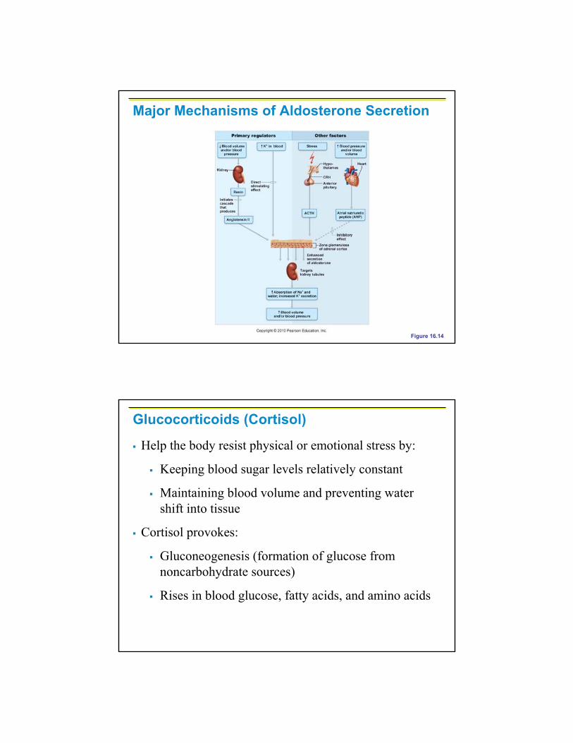
Major Mechanisms of Aldosterone Secretion
Figure 16.14
Glucocorticoids (Cortisol)
Help the body resist physical or emotional stress by:
Keeping blood sugar levels relatively constant
Maintaining blood volume and preventing water shift into tissue
Cortisol provokes:
Gluconeogenesis (formation of glucose from noncarbohydrate sources)
Rises in blood glucose, fatty acids, and amino acids

Excessive Levels of Glucocorticoids
Excessive levels of glucocorticoids:
Depress cartilage and bone formation
Inhibit inflammation
Depress the immune system
Promote changes in cardiovascular, neural, and gastrointestinal function
Gonadocorticoids (Sex Hormones)Most gonadocorticoids secreted are androgens (male sex hormones), and the most important one is testosterone
Androgens contribute to:
The onset of puberty
The appearance of secondary sex characteristics
Sex drive in females
Androgens can be converted into estrogens after menopause

Adrenal Medulla
Made up of chromaffin cells that secrete catecholamines (epinephrine & norepinephrine)
Secretion of these hormones causes:
Blood glucose levels to rise
Blood vessels to constrict
The heart to beat faster
Blood to be diverted to the brain, heart, and skeletal muscle
Adrenal Medulla
Epinephrine (E) is the more potent stimulator of the heart and metabolic activities
Norepinephrine (NE) is more influential on peripheral vasoconstriction and blood pressure
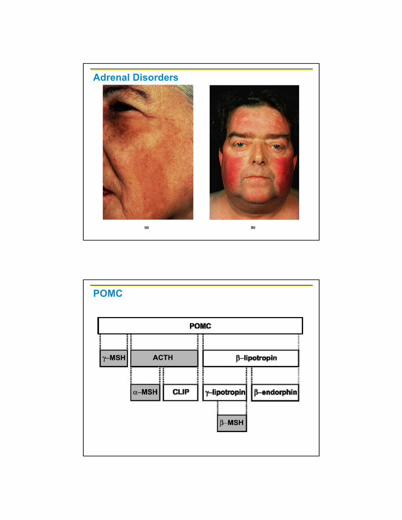
Adrenal Disorders
POMC

Adrenal Disorders
Adrenal Disorders
The same boy, only 4 months later.
Paired ovaries in the abdominopelvic cavity produce estrogens and progesterone
They are responsible for:
Maturation of the reproductive organs
Appearance of secondary sexual characteristics
Breast development and cyclic changes in the uterine mucosa
Gonads: Female
Testes located in an extra-abdominal sac (scrotum) produce testosterone
Testosterone:
Initiates maturation of male reproductive organs
Causes appearance of secondary sexual characteristics and sex drive
Is necessary for sperm production
Maintains sex organs in their functional state
Gonads: Male

Stress and the Adrenal Gland
General Adaptation Syndrome (GAS)
Alarm phase – short-term responses
Resistance phase – long-term metabolic adjustments
Exhaustion phase – collapse of vital organs
Stress and the Adrenal Gland
Figure 16.16
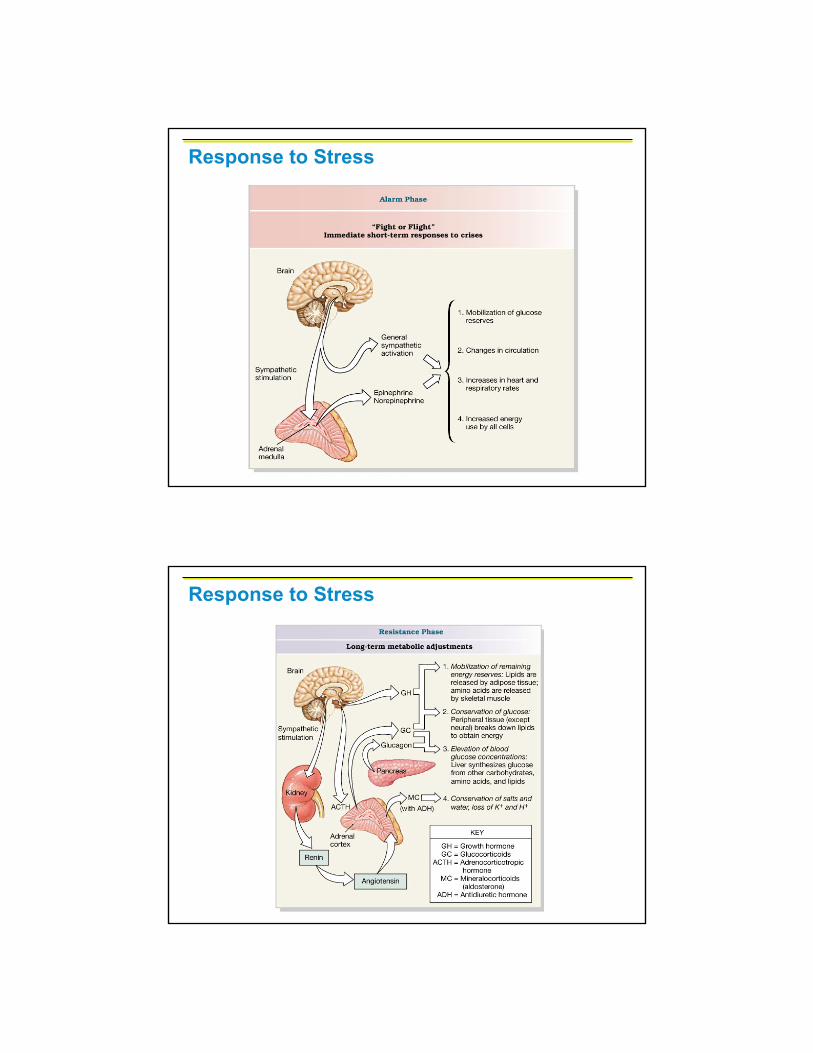
Response to Stress
Response to Stress
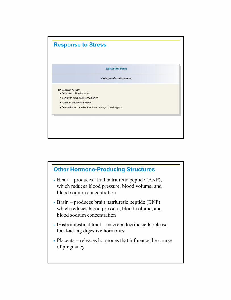
Response to Stress
Heart – produces atrial natriuretic peptide (ANP), which reduces blood pressure, blood volume, and blood sodium concentration
Brain – produces brain natriuretic peptide (BNP), which reduces blood pressure, blood volume, and blood sodium concentration
Gastrointestinal tract – enteroendocrine cells release local-acting digestive hormones
Placenta – releases hormones that influence the course of pregnancy
Other Hormone-Producing Structures

Kidneys – secrete erythropoietin, which signals the production of red blood cells, renin and calcitriol
Skin – produces cholecalciferol, the precursor of vitamin D
Adipose tissue – releases leptin, which is involved in the sensation of satiety, and stimulates increased energy expenditure
Other Hormone-Producing Structures
Endocrine Functions of the Kidneys
Calcitriol

Endocrine Functions of the Kidneys
Angiotensinconverting enzyme (ACE)
Plus a vasoconstrictor