Embed Size (px)
Citation preview
Clinical Presentation of Stroke Syndromes
By Ken Hui Yee for PBL group 7Case 24

Ischaemic Stroke
Causes: Thrombosis & Embolism (65% of strokes)▪ Artery-to-artery▪ Cardioembolic▪ Thrombosis in-situ
Small vessel (lacunar) strokes (20% of strokes)▪ atherothrombotic or lipohyalinotic occlusion
of a small intracranial artery▪ Often symptomless

Artery-to-Artery Embolic Stroke
Thrombus formation on atherosclerotic plaques embolize to intracranial arteries▪ Carotid bifurcation ▪ most common site (10% of ischaemic strokes)
Diseased vessel may acutely thrombose▪ Including aortic arch, common carotid,
internal carotid, vertebral, and basilar a.
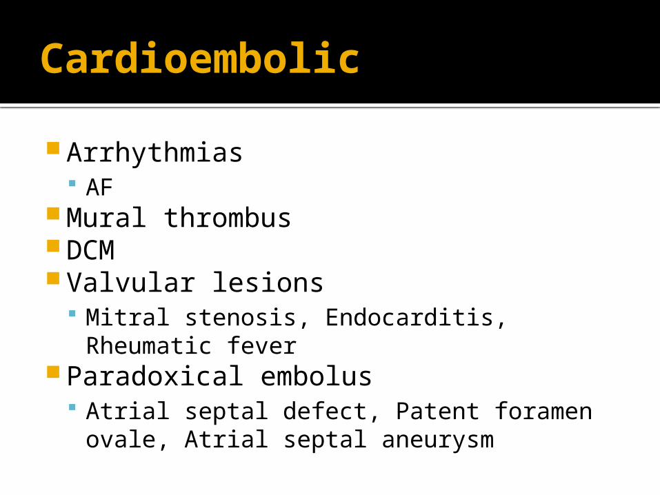
Cardioembolic
Arrhythmias AF
Mural thrombus DCM Valvular lesions
Mitral stenosis, Endocarditis, Rheumatic fever
Paradoxical embolus Atrial septal defect, Patent foramen ovale,
Atrial septal aneurysm

Less Common Causes of Ischaemic Stroke
Venous sinus thrombosis Complication of:▪ OCP▪ Pregnancy & the postpartum period▪ Inflammatory bowel disease▪ Intracranial infections (meningitis)▪ Dehydration

Haemorrhagic Stroke
Less common (only 15% of all strokes)
Higher mortality rate than Ischaemic
Haemorrhagic Stroke
Causes: Head trauma▪ Most common cause of SAH
Hypertensive haemorrhage Aneurysm
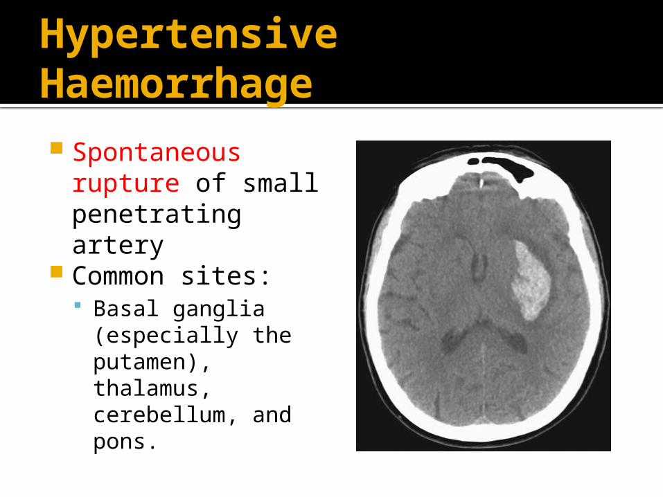
Hypertensive Haemorrhage Spontaneous
rupture of small penetrating artery
Common sites: Basal ganglia
(especially the putamen), thalamus, cerebellum, and pons.
Aneurysm
SAH from berry aneurysm▪ AcomA, PcomA, MCA (locations from most
common to less common) Mycotic aneurysm▪ Eg. Endocarditis
Other Causes of Hemorrhage Stroke
Amyloid angiopathy▪ Degen of intracranial vessels▪ Rare in <60
Tumour Drugs (eg. Cocaine)▪ Young pts
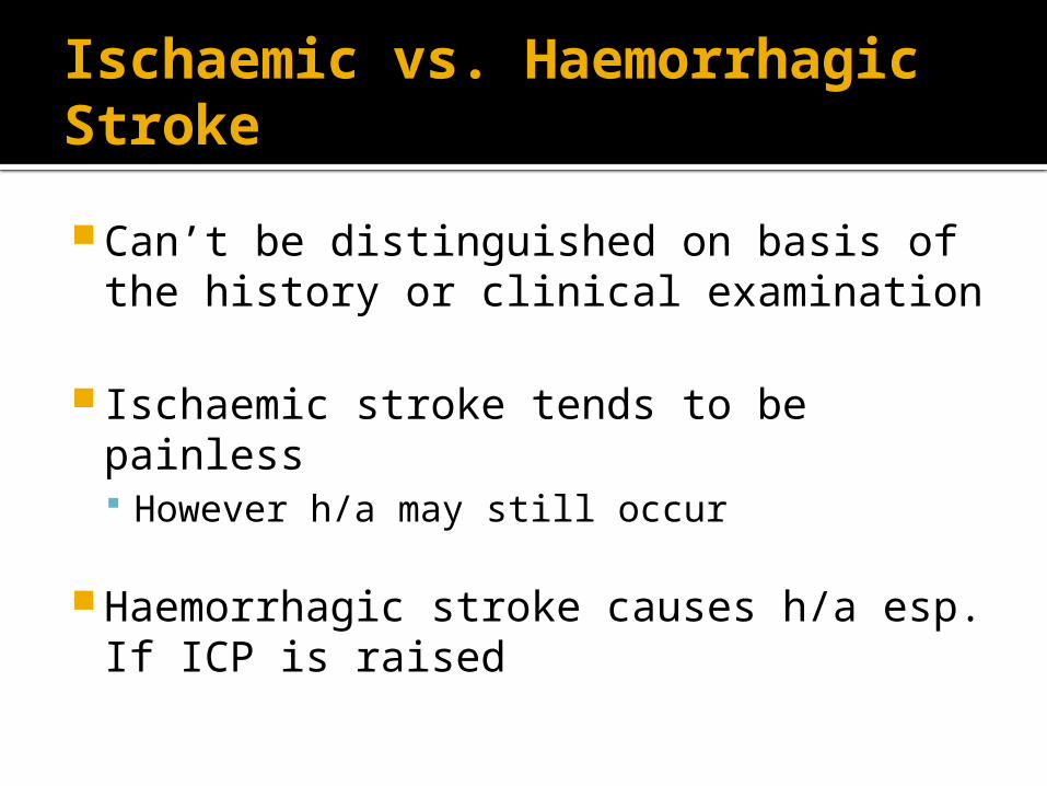
Ischaemic vs. Haemorrhagic Stroke
Can’t be distinguished on basis of the history or clinical examination
Ischaemic stroke tends to be painless However h/a may still occur
Haemorrhagic stroke causes h/a esp. If ICP is raised

Ischaemic vs. Haemorrhagic Stroke
Investigations: Determine between ischaemic and
haemorrhagic
CT MRI CSF

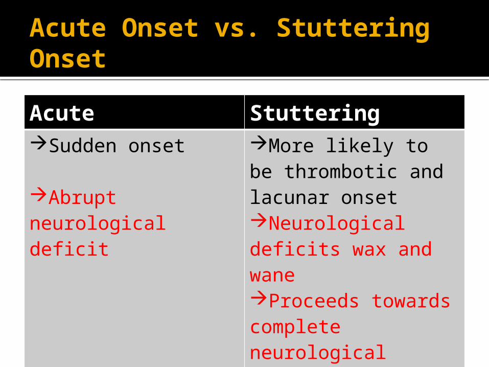
Acute Onset vs. Stuttering Onset
Acute StutteringSudden onset
Abrupt neurological deficit
More likely to be thrombotic and lacunar onsetNeurological deficits wax and waneProceeds towards complete neurological deficits

Case 1
HOPC:▪ Pt describes a shade or curtain being pulled
over the front of the eye (right)▪ Vision in right eye is lost only for a short time
(seconds to minutes)▪ On examination patient has carotid bruits▪ Painless
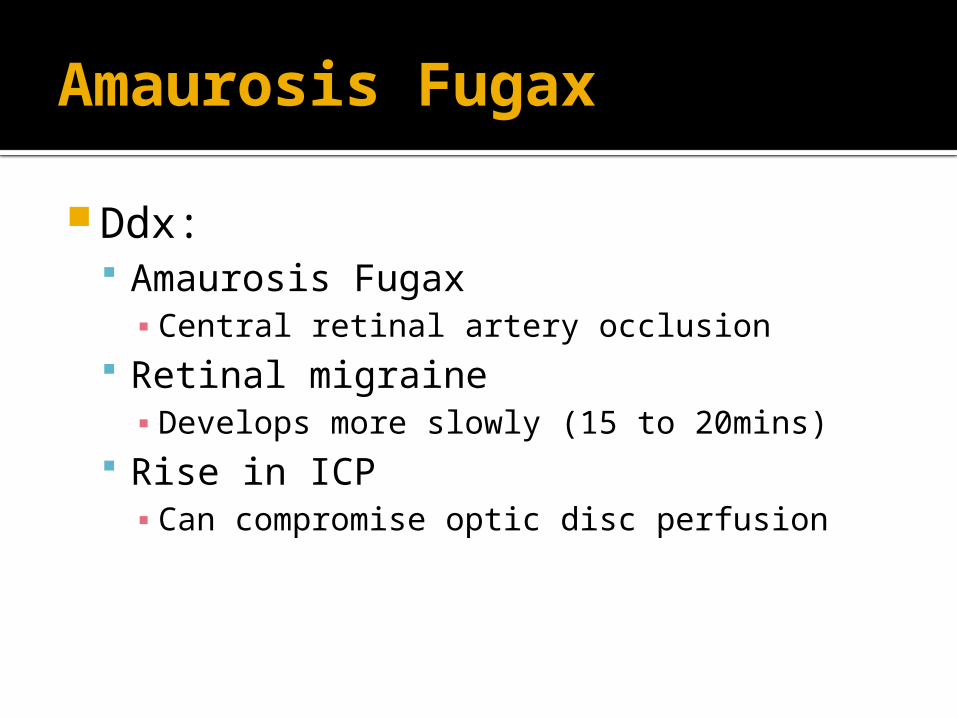
Amaurosis Fugax
Ddx: Amaurosis Fugax▪ Central retinal artery occlusion
Retinal migraine▪ Develops more slowly (15 to 20mins)
Rise in ICP▪ Can compromise optic disc perfusion

Case 2
HOPC:▪ Sudden onset of headache with aura▪ Nausea and vomiting▪ Tingling, numbness and vague weakness on
the right side of the body▪ Patient prefers a dark room▪ Patient reports that the aura has persisted for
more than a week. IX:▪ CT and MRI show focal ischaemia

Migrainous Infarction
Rare complication of migraines
Definition: Aura and a migraine headache, with the
aura symptom persisting > 7/7 + neuroimaging focal ischaemia

Complete vs Incomplete Strokes
Complete IncompleteTotal area of the brain supplied by an occluded vessel is damagedFurther prophylaxis Rx is pointless
some cellular damageAdditional tissue in the affected vascular distribution is at riskProphylaxis Rx is useful
Not that practical as distinction based on clinical findings can be impossible
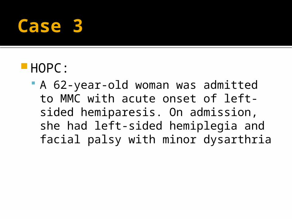
Case 3
HOPC: A 62-year-old woman was admitted to
MMC with acute onset of left-sided hemiparesis. On admission, she had left-sided hemiplegia and facial palsy with minor dysarthria
Case 3
IX: CT▪ right MCA mainstem occlusion but no early
ischemic changes Thrombolysis commenced pt
improved initially but then developed sudden decline of consciousness
Case 3
Repeat CT Ruled out ICH
MRI New occlusion in Left MCA discovered
Underlying cause was due to cardioembolic ischaemic stroke due to AF
Case 4
HOPC: Pt presents to ED with global aphasia Pt’s partner reports that pt is right
handed

MCA

Case 5
HOPC: Pt presents to ED with right leg and foot
paralysis Sensory impairment (pain, temperature)
over right lower limb Examination of upper limb = normal Impairment of gait

ACA

Case 6
HOPC: Pt presents with homonymous
hemianopia Has a failure to see to-and-fro
movements, inability to perceive objects not centrally located
Case 6
HOPC: Pt presents with homonymous
hemianopia Has a failure to see to-and-fro
movements, inability to perceive objects not centrally located
Reports peduncular hallucinosis

PCA

PCA – Specific Named Syndromes
Midbrain – Subthalamic -Thalamic Weber Syndrome▪ Contralateral hemiplegia
Thalamic Dejerine-Roussy▪ Contralateral hemisensory loss
Claude’s Syndrome▪ Third nerve palsy Contralateral ataxia

PCA – Specific Named Syndromes
Anton's syndrome Bilateral infarction in the distal PCAs
producing cortical blindness Pt maybe unaware of blindness and may
deny it Balint’s syndrome
Watershed infarction between PCA and MCA
Disorder of the orderly visual scanning of the environment

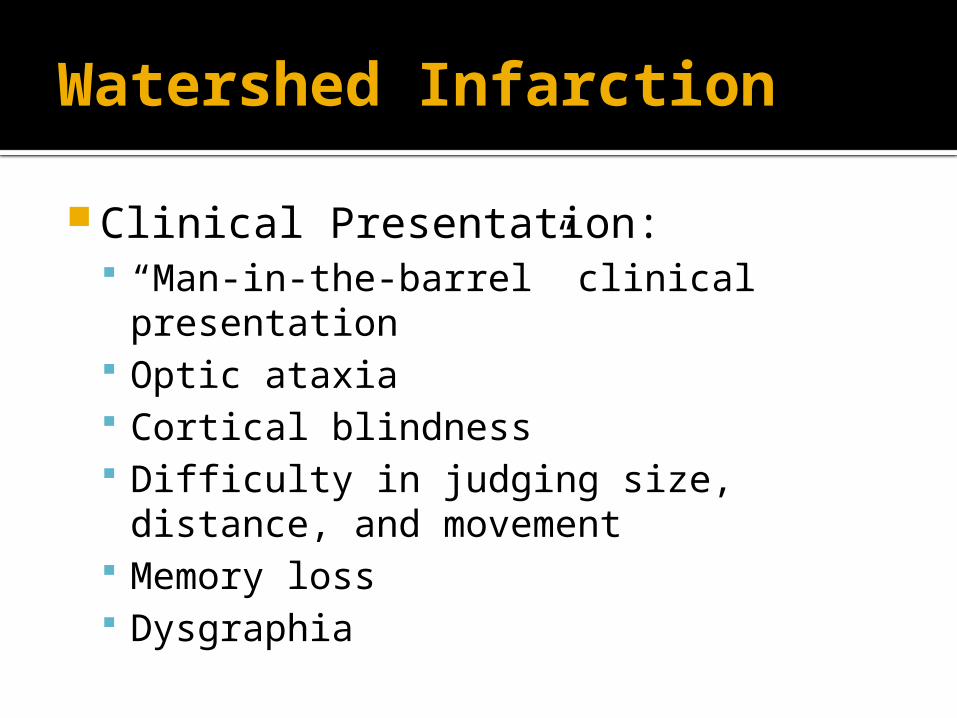
Watershed Infarction
Hypotension due to eg. AMI low perfusion in borderzones/junctional territories of the cerebral end arteries
Watershed Infarction
Clinical Presentation: “Man-in-the-barrel” clinical presentation Optic ataxia Cortical blindness Difficulty in judging size, distance, and
movement Memory loss Dysgraphia
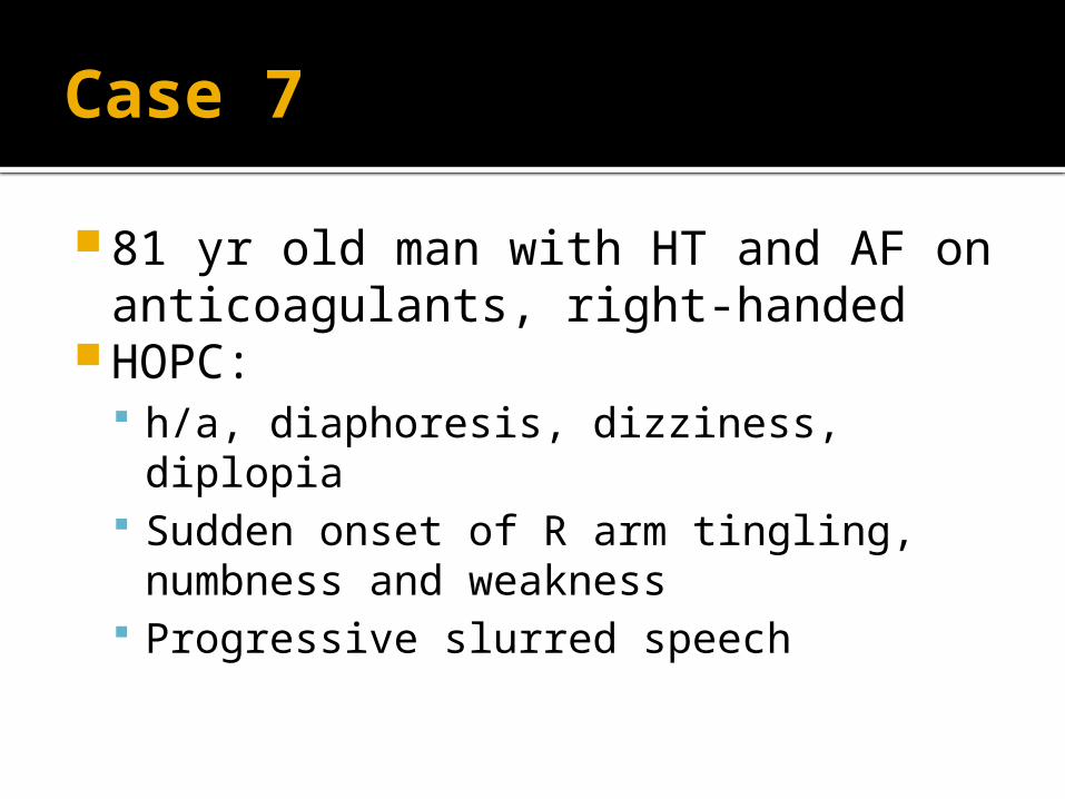
Case 7
81 yr old man with HT and AF on anticoagulants, right-handed
HOPC: h/a, diaphoresis, dizziness, diplopia Sudden onset of R arm tingling,
numbness and weakness Progressive slurred speech
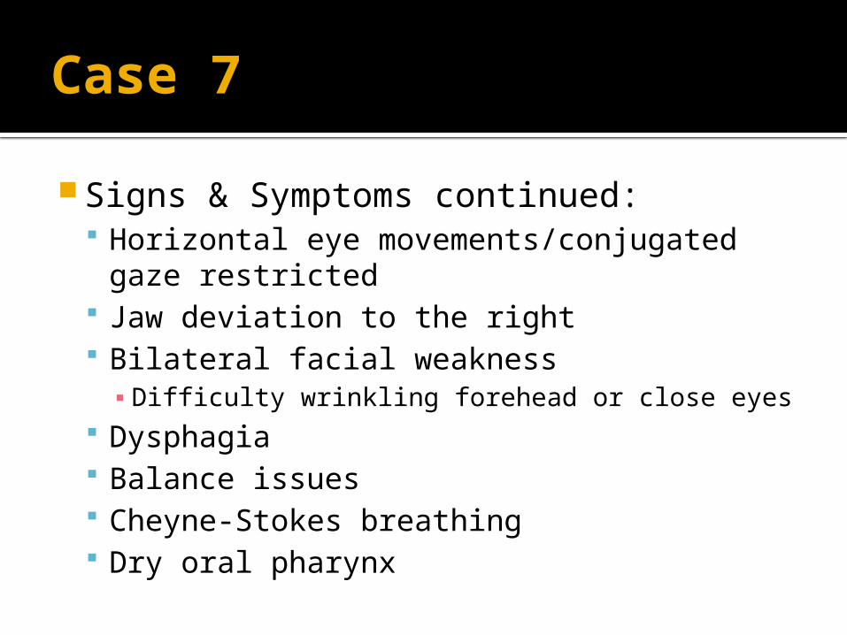
Case 7
Signs & Symptoms continued: Horizontal eye movements/conjugated
gaze restricted Jaw deviation to the right Bilateral facial weakness▪ Difficulty wrinkling forehead or close eyes
Dysphagia Balance issues Cheyne-Stokes breathing Dry oral pharynx

Case 7
IX: CT - progressive hemorrhagic stroke
intrinsic to the pontine tegmentum of the brain stem, with rupture into the fourth ventricle
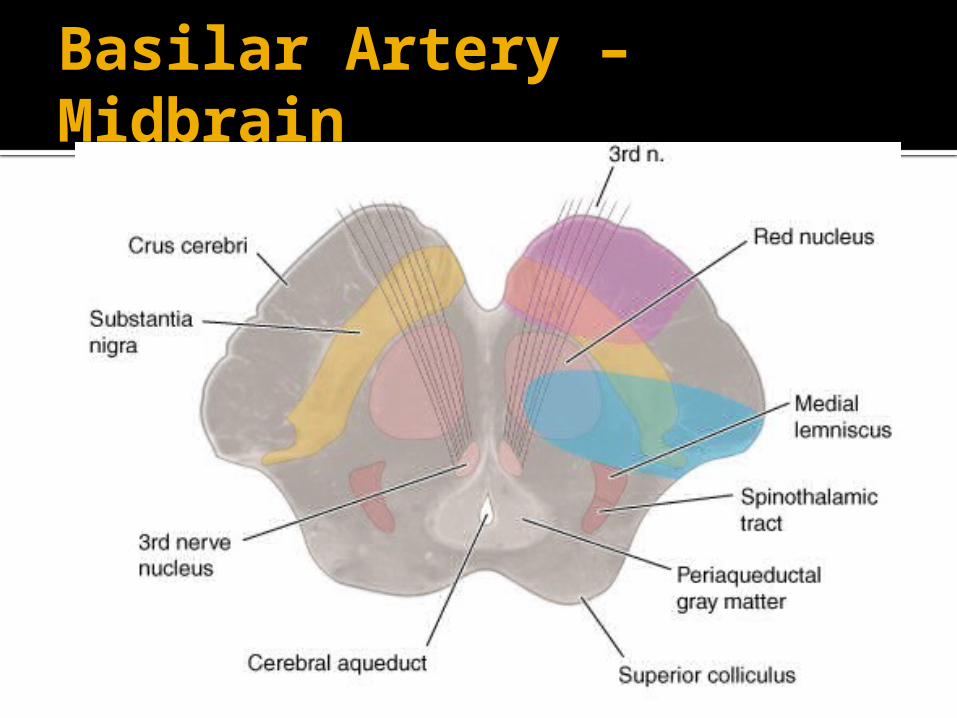
Basilar Artery – Midbrain

Basilar Artery Mid pons

Basilar Artery Inferior Pons

Vertebral and Posterior Inferior Cerebellar Arteries Medulla

Presentation of Brainstem Infarction
Clinical Feature Structure InvolvedHemiparesisSensory lossDiplopiaFacial numbnessFacial weaknessNystagmus & vertigoDysphagia & dysarthria

Presentation of Brainstem Infarction
Clinical Feature Structure Involved
Hemiparesis Corticospinal tracts Medial midpontine syndrome,Medial inferior pontine syndrome
Sensory loss Medial lemniscus and spinothalamic tracts
Lateral midpontine syndrome
Diplopia Oculomotor/Adducens
Medial inferior pontine syndrome
Facial numbness Trigeminal Lateral midpontine syndrome,Lateral inferior pontine syndrome
Facial weakness Facial Lateral inferior pontine syndrome
Nystagmus & vertigo
Vestibular Medial inferior pontine syndrome
Dysphagia & dysarthria
Glossopharyngeal & vagus
Medullary Syndrome
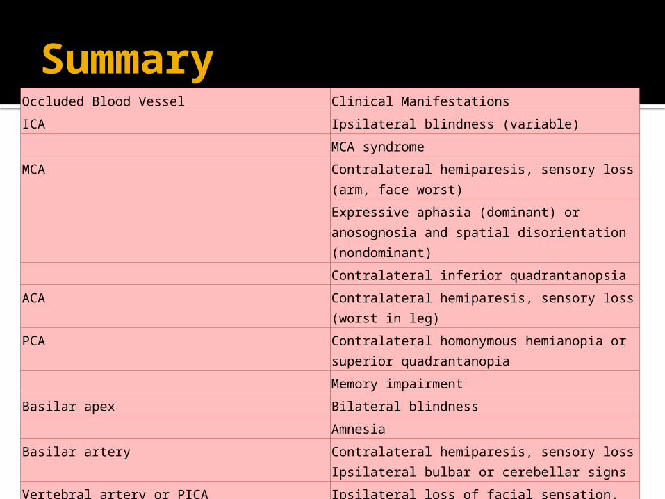
SummaryOccluded Blood Vessel Clinical Manifestations
ICA Ipsilateral blindness (variable)
MCA syndrome
MCA Contralateral hemiparesis, sensory loss (arm, face worst)
Expressive aphasia (dominant) or anosognosia and spatial disorientation (nondominant)
Contralateral inferior quadrantanopsia
ACA Contralateral hemiparesis, sensory loss (worst in leg)
PCA Contralateral homonymous hemianopia or superior quadrantanopia
Memory impairment
Basilar apex Bilateral blindness
Amnesia
Basilar artery Contralateral hemiparesis, sensory loss Ipsilateral bulbar or cerebellar signs
Vertebral artery or PICA Ipsilateral loss of facial sensation, ataxia, contralateral hemiparesis, sensory loss
Superior cerebellar artery Gait ataxia, nausea, dizziness, headache progressing to ipsilateral hemiataxia, dysarthria, gaze paresis, contralateral hemiparesis, somnolence