Embed Size (px)
Citation preview
By A Hollingworth
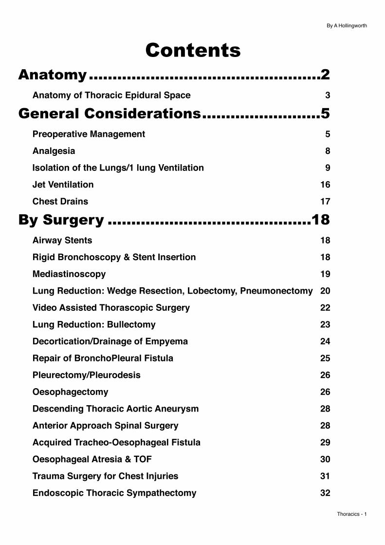
Contents Anatomy 2 .................................................
Anatomy of Thoracic Epidural Space 3
General Considerations 5 .........................Preoperative Management 5
Analgesia 8
Isolation of the Lungs/1 lung Ventilation 9
Jet Ventilation 16
Chest Drains 17
By Surgery 18 ...........................................Airway Stents 18
Rigid Bronchoscopy & Stent Insertion 18
Mediastinoscopy 19
Lung Reduction: Wedge Resection, Lobectomy, Pneumonectomy 20
Video Assisted Thorascopic Surgery 22
Lung Reduction: Bullectomy 23
Decortication/Drainage of Empyema 24
Repair of BronchoPleural Fistula 25
Pleurectomy/Pleurodesis 26
Oesophagectomy 26
Descending Thoracic Aortic Aneurysm 28
Anterior Approach Spinal Surgery 28
Acquired Tracheo-Oesophageal Fistula 29
Oesophageal Atresia & TOF 30
Trauma Surgery for Chest Injuries 31
Endoscopic Thoracic Sympathectomy 32
Thoracics - �1
By A Hollingworth
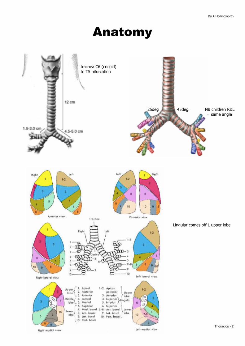
Anatomy
Thoracics - �2
Lingular comes off L upper lobe
25deg 45deg. NB children R&L = same angle
trachea C6 (cricoid) to T5 bifurcation
By A Hollingworth
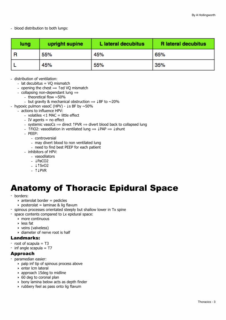
- blood distribution to both lungs:
- distribution of ventilation: - lat decubitus = VQ mismatch - opening the chest ⟹ ↑ed VQ mismatch - collapsing non-dependant lung ⟹
- theoretical flow ~50% - but gravity & mechanical obstruction ⟹ ↓BF to ~20%
- hypoxic pulmon vasoC (HPV) - ↓s BF by ~50% - actions to influence HPV:
- volatiles <1 MAC = little effect - IV agents = no effect - systemic vasoCs ⟹ direct ↑PVR ⟹ divert blood back to collapsed lung - ↑FiO2: vasodilation in ventilated lung ⟹ ↓PAP ⟹ ↓shunt - PEEP:
- controversial - may divert blood to non ventilated lung - need to find best PEEP for each patient
- inhibitors of HPV: - vasodilators - ↓PaCO2 - ↓↑SvO2 - ↑↓PVR
Anatomy of Thoracic Epidural Space - borders:
‣ anterolat border = pedicles ‣ posterolat = laminae & lig flavum
- spinous processes orientated steeply but shallow lower in Tx spine - space contents compared to Lx epidural space:
‣ more continuous ‣ less fat ‣ veins (valveless) ‣ diameter of nerve root is half
Landmarks: - root of scapula = T3 - inf angle scapula = T7 Approach - paramedian easier:
‣ palp inf tip of spinous process above ‣ enter lcm lateral ‣ approach 15deg to midline ‣ 60 deg to coronal plan ‣ bony lamina below acts as depth finder ‣ rubbery feel as pass onto lig flavum
Thoracics - �3
By A Hollingworth
Insertion Height - low thoracic epidural will spread more cranial - high Tx epidural will spread mostly caudal ↳ due to progressive ↑in epidural width moving down ↳ ∴ high Tx Epi insert high in incision site & low Tx epi insert middle of incision site
Thoracics - �4
By A Hollingworth
General Considerations
Preoperative Management - tend to be older, less fit patients - assess functional capacity - CXR and CT:
- ?signs of difficulty with DLT = airway obstruction, tracheal or carinal distortion/compression - ?other lung pathology eg bullae or effusions
↳ discuss scans with surgeon - co-morbid conditions; smoking, bronchial carcinoma, pleural effusion, cardiac disease, empyema, oesophageal obstruction, cachexia - suitability for lung resection:
- tumour type = non-small cell eg squamous, adenocarcinoma - surgically resectable eg 4 Ms: mass effect, metabolic effect, metastasis, medication - fitness for surgery - CVS/resp, nutrition, smoking, ex tol
- classify pts into risk categories based on Hx, exam & PFTs: - low risk = clinically fit with good ex tol & normal PFTs - mod risk = ↓ex tol & abnormal PFTs +/- moderate co-existing disease - high risk = minimal ex tol, grossly impaired PFTs, major med problems
- good communication with surgeon very important -> nature of operation, positioning, potential complications - optimise lung function:
- stop smoking 6-8 weeks prior (see PS12) - preoperative physio and incentive spirometry - optimise bronchodilators - manage infections - consider dose of oral steroids - home exercise plan
- optimise nutrition - CPET useful for borderline cases - MDT Ax vital PFTs - often used to determine suitability for lung resection - always consider results in context of general health & proposed surgery - spirometry = bellows of system - Diffusion capacity (DLCO) = lung gas transfer function - NB very possible to have normal spirometry & severely impaired lung function - Accepted minimum FEV1 values by surgery:
- Pneumonectomy >60% - lobectomy >40% - wedge resection >30%
- Predicted post operative (ppo) value of PFTs = preop value x (5 - number of lobes resected)/5 or
prep value x (1-% functional tissue to be removed/100) ↳ guided by VQ scan
- goal to avoid need for post op ventilation = is : - ppoFEV1 >40% of predicted normal = ~ >800mls - ppoDLCO >40% pred normal
Thoracics - �5
By A Hollingworth
Practical Pre-Assessment - use stepped approach to assessing patient - if meets stage 1 criteria then stop otherwise continue through stages:
Spirometry Screening - used to assess suitability for surgery and predict difficulty with ventilation post resection
- assess FEV1 & DLCO: ‣ straight forward patient = both >60% of predicted ‣ Marginal patient = either 30-60% predicted ‣ if marginal should work out ppo values
FEV1 Surgery Minimum FEV1%
Pneumonectomy >60% (or >2L) Lobectomy >40% (or 1.5L) Wedge Resection >30% (or 1L)
DLCO - - diffusion capacity of lungs - aim >60% predicted
Adjuncts ABG- hypoxia or hypercarbia on RA bad prognostic sign
Xe or Technetium V/Q scan - works out regional blood flow to both lungs -> and then we can calculate a more accurate FEV1 and DLCO - Normal VQ = L45% & R 55%
CPET Testing
- AT >11ml/kg/min - VO2 max
‣ >20ml/kg/min = low risk (1% mortality) ‣ 15-20 = medium risk ‣ <15 = high risk (10% mortality)
OTHER TESTS- functional CVS testing:
- breath hold for 30 sec - stair climbing
- = 20steps @ 6 inches = 1 flight. - <2 flights = high risk - >3 flights = low risk
- 6min walk: - correlates well with VO2 max. <2000feet = <15ml/kg/min - should monitor SpO2 to exclude (otherwise high risk):
- <4% drop - >90% after
- FBC - polycythaemia from chronic hypoxia, WCC for infection - CXR – sizing of DLT, gross pathology - CT – assessment of airway and degree of pathology - unilateral pulmonary artery occlusion test; blocking off of one pulmonary artery and pressure measured in PA (if PAP >35mmHg or PaO2 < 45mmHg -> cancel surgery)
Thoracics - �6
By A Hollingworth
Intraoperative Management
Issues:
1. controlled ventilation of lungs independently 2. shared airway and lung 3. sub-glottic obstruction of tracheal or carina from extrinsic compression or invasion by carcinoma 4. dynamic hyperinflation of lungs following IPPV in patients with lung pathology - eg bullae, cysts, tumour with flap valve: - ⟹ can lead to progressive distension ⟹ ↑ITP ⟹ ↓VR - “if in doubt, let air out” 5. significant mediastinal shifts - can ⟹ ↓↓CO ⟹ prompt recognition & correction 6. Surgical manipulation ⟹ obstruction of VR ⟹ volume load & ask surgeon to stop squashing the heart 7. Arrhythmia = AF
Postoperative Management - avoid prolonged ventilation if possible -
- stresses pulmonary suture lines - ↑air leaks - ↑infection risk
- physio - O2 (humidified) - thoracotomy very painful esp if more posterior -> good analgesia very important
Thoracics - �7
By A Hollingworth
Analgesia Anatomy & PathoPhys - 4 routes for C & Aδ fibres:
‣ intercostal nerves = skin & intercostal mms ‣ vagus nerve = lung & mediastinum ‣ intercostal & phrenic nerve = parietal pleura (v sensitive)
↳ visceral pleura = insensitive ‣ thoracodorsal & long thoracic nerves (C5-7 brachial plexus roots) = lat dorsi & serratus anterior
- chest drain sites often not covered by epidural ∴ most painful
• thoracotomy = very painful • poor pain relief ⟹
‣ CVS complications = ↑SNS ⟹ ↑cardiac O2 demand ‣ ↓mobilisation = risk of DVT/PE ‣ ↑resp complications = ↓respiration ⟹ atelectasis, secretions, ↓PaO2, ↑PaCO2
• chronic pain syndrome post thoracic surgery in 25-60% • techniques:
‣ simple multimodal analgesia ‣ gabapentin = 600-1200mg then post op: 400mb bd for 1/52 ‣ regional block:
- thoracotomy: epidural or paraverterbal catheters ↳ equal pain relief but avoiding epidural = ↓bp, ↓retention, ↓PONV
- intrapleural catheter = anaesthetist or surgeon. limited effect - thoracoscopic procedures: intercostal (limited coverage) or paravertebral nerve blocks
‣ intraop opioids ‣ post op PCA
• Epidural (thoracic) regime: ‣ = gold standard ‣ LA + opioid ‣ establish cautiously to avoid ↓bp ‣ 10mls 0.25% bupivacaine ‣ advs:
- CVS: SNS blockade ⟹ optimise O2 supply & demand to heart - metabolic ⟹ ↓plasma glucose by preventing catabolism - GI: ↑blood flow, ↓ileus
‣ disadv: - failure rate up to 15% (even in experts) - difficult insertion - post op care & complication monitoring - hypotension - from vasoD mesenteric vessels ⟹ ↓VR - contraindicated in active empyema/systemic sepsis - motor blockade of intercostals ⟹ ↓cough
• Percutaneous paravertebral techniques: ‣ = unilateral epidural ‣ can do single shot or catheter ‣ paravertebral space =
- wedge shaped space lat to intervertebral foramen - communicates above & below with paravertebral spaces - some communication with epidural space - intercostal nerve passes through here with no fascial sheath - nerve is outside parietal pleura
‣ adv over epidural: - unilat block ⟹ ↓complications of bp, stress response, resp impairment
‣ lacks evidence of epidural ‣ 0.3ml/kg of 0.5% bupivacaine ‣ post op infusion 0.1ml/kg/hr of 0.25-0.5% bupiv
Thoracics - �8
By A Hollingworth
• post-thoracotomy shoulder pain: ‣ 21-97% incidence ‣ unresponsive to epidural ‣ mod severe pain over deltoid/post shld at rest ‣ usually improves 4-48hours post ‣ possible causes:
- phrenic nerve irritiation - post ligament strain from lateral position intraop - chest drain irritating diaphragm - prox incision not covered by epidural
‣ Rx: - as above - consider interscalene block
Isolation of the Lungs/1 lung Ventilation - potential for complications should always be considered Advantages- protection of dependent lung from blood and secretions - allows independent control of ventilation to each lung - improves surgical access - reduces lung trauma during surgery Disadvantages- creates shunt -> hypoxaemia - sizing and placement of DLT can be difficult - Acute lung injury in 2-5% cases Indications - absolute (lung isolation):
- avoid contamination of lung in cases of infection, massive pulmon haemorrhage, bronchopulmon lavage - control distribution of ventilation eg giant bullae, lung cysts - VATS - unilat bronchopulmon lavage
- relative (lung separation): - improve surgical access - although this can often be achieved by careful retraction - non thoracic surgery - severe hypoxaemia due to unilat lung disease
Physiology VQ mismatch in Lat Decibitus- 60% blood to dependent lung - gravity - ventilation
‣ prefers non-dependent lung ‣ dependent lung= ↓compliance & ↓FRC due to mediastinal weight, elevated diaphragm
↳ can ameliorate with PEEP - when chest opened:
‣ bad = ↑ed compliance non-dependent lung ⟹ ↑↑ed VQ mismatch ‣ this is overcome by collapsing non-dependent lung
- see slow ↑in CO2 due to V/Q mismatch - theory of shunt fraction up to 50% but compensatory mechanisms keep it lower:
‣ gravity - ↑flow to dependent lung ‣ mechanical obstruction to non-dependent lung ‣ HPV -
- triggered by hypoxia or ↑PaCO2⟹ ↓50% flow - volatiles inhibit HPV by 20-30% (propofol has nearly no effect)
↳ ∴ actual blood flow through non-dependant lung = 20%
Techniques - DLT (most common) - Bronchial blockers (occasionally helpful in anatomically difficult patients)
Thoracics - �9
By A Hollingworth
- Single lumen bronchial intubation (rarely used): ‣ are specific tubes - smaller cuff, closer to end, no Murphy eye ‣ paeds: need to intubate L main stem = turn head to right & insert ETT with concavity facing posterior
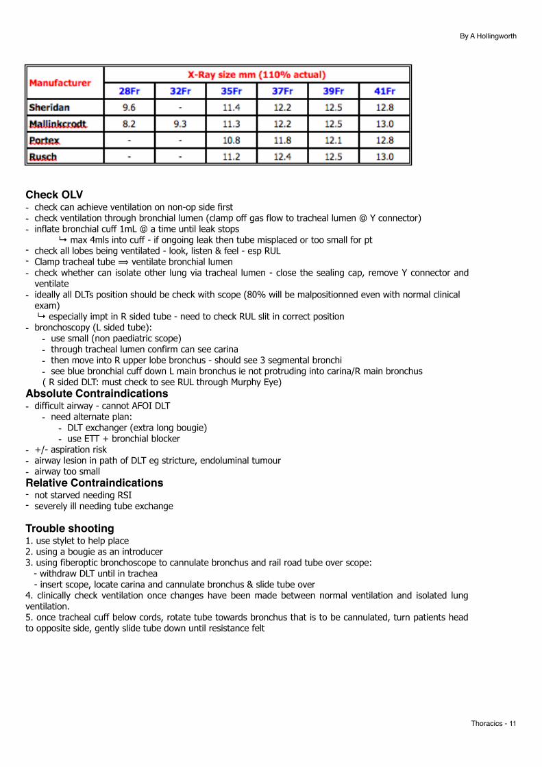
Double Lumen Endobronchial Tubes - right or left depending on bronchus they are designed to intubate - right sided tubes have a slit to be positioned to facilitate ventilation of RUL - size = external diameter of tube (French Gauge – 26-41 Fr) ↳ 39Fr = 13mm - internal lumens are small eg 39Fr = 6mm, 35Fr = 4.5mm - bronchoscopic check requires a narrow scope <4mm in diameter - guide:
- males; 39-41 Fr - females; 37-39 Fr - children <30kg -> use bronchial blocker technique
- remember in an emergency (ie. pulmonary haemorrhage) a standard ETT can be advanced into non-diseased lung
- types of DLET: - Carlens (left sided) = carinal hook to aid placement - Whites (right side) = carinal hook & slit in tube wall for RUL - Robertshaw (R & L) = traditionally R rubber, reusuable - single use PVC (R&L) Mallinckrodt:
- high volume, low pressure cuffs - bronchial cuff & pilot tube coloured blue - radioopaque strip runs to tip of bronchial lumen - sizes 28-41
- Table below give diameter of bronchus (usually left) measured on PA CXR (magnifies air bronchogram by 10%) - should chose largest DLT that will pass through glottis - common to use L sided tube unless:
- Carinal or prox L mainstem lesion - abnormal bronchial anatomy
- L DLT advs: - ↓chance of blocking lobar bronchus (R bronchus is short & variable & RUL easy to occlude) - ↑ed tolerance to shifts in tube position
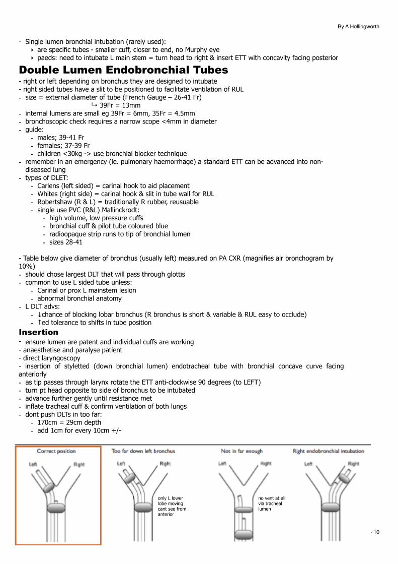
Insertion - ensure lumen are patent and individual cuffs are working - anaesthetise and paralyse patient - direct laryngoscopy - insertion of styletted (down bronchial lumen) endotracheal tube with bronchial concave curve facing anteriorly - as tip passes through larynx rotate the ETT anti-clockwise 90 degrees (to LEFT) - turn pt head opposite to side of bronchus to be intubated - advance further gently until resistance met - inflate tracheal cuff & confirm ventilation of both lungs - dont push DLTs in too far:
- 170cm = 29cm depth - add 1cm for every 10cm +/-
Thoracics - �10
only L lower lobe moving cant see from anterior
no vent at all via tracheal lumen
By A Hollingworth
Check OLV- check can achieve ventilation on non-op side first - check ventilation through bronchial lumen (clamp off gas flow to tracheal lumen @ Y connector) - inflate bronchial cuff 1mL @ a time until leak stops ↳ max 4mls into cuff - if ongoing leak then tube misplaced or too small for pt - check all lobes being ventilated - look, listen & feel - esp RUL - Clamp tracheal tube ⟹ ventilate bronchial lumen - check whether can isolate other lung via tracheal lumen - close the sealing cap, remove Y connector and
ventilate - ideally all DLTs position should be check with scope (80% will be malpositionned even with normal clinical
exam) ↳ especially impt in R sided tube - need to check RUL slit in correct position - bronchoscopy (L sided tube):
- use small (non paediatric scope) - through tracheal lumen confirm can see carina - then move into R upper lobe bronchus - should see 3 segmental bronchi - see blue bronchial cuff down L main bronchus ie not protruding into carina/R main bronchus
( R sided DLT: must check to see RUL through Murphy Eye) Absolute Contraindications- difficult airway - cannot AFOI DLT
- need alternate plan: - DLT exchanger (extra long bougie) - use ETT + bronchial blocker
- +/- aspiration risk - airway lesion in path of DLT eg stricture, endoluminal tumour - airway too small Relative Contraindications- not starved needing RSI - severely ill needing tube exchange
Trouble shooting1. use stylet to help place 2. using a bougie as an introducer 3. using fiberoptic bronchoscope to cannulate bronchus and rail road tube over scope: - withdraw DLT until in trachea - insert scope, locate carina and cannulate bronchus & slide tube over 4. clinically check ventilation once changes have been made between normal ventilation and isolated lung ventilation. 5. once tracheal cuff below cords, rotate tube towards bronchus that is to be cannulated, turn patients head to opposite side, gently slide tube down until resistance felt
Thoracics - �11
By A Hollingworth
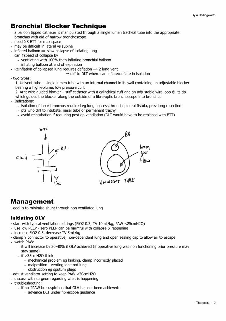
Bronchial Blocker Technique - a balloon tipped catheter is manipulated through a single lumen tracheal tube into the appropriate
bronchus with aid of narrow bronchoscope - need ≥8 ETT for max space - may be difficult in lateral vs supine - inflated balloon ⟹ slow collapse of isolating lung - can ↑speed of collapse by
- ventilating with 100% then inflating bronchial balloon - inflating balloon at end of expiration
- Reinflation of collapsed lung requires deflation ⟹ 2 lung vent ↳ diff to DLT where can inflate/deflate in isolation - two types: 1. Univent tube – single lumen tube with an internal channel in its wall containing an adjustable blocker bearing a high-volume, low pressure cuff. 2. Arnt wire-guided blocker – stiff catheter with a cylindrical cuff and an adjustable wire loop @ its tip which guides the blocker along the outside of a fibre-optic bronchoscope into bronchus - Indications:
- isolation of lobar bronchus required eg lung abscess, bronchopleural fistula, prev lung resection - pts who diff to intubate, nasal tube or permanent trachy - avoid reintubation if requiring post op ventilation (DLT would have to be replaced with ETT)
Management - goal is to minimise shunt through non ventilated lung
Initiating OLV - start with typical ventilation settings (FiO2 0.3, TV 10mL/kg, PAW <25cmH2O) - use low PEEP - zero PEEP can be harmful with collapse & reopening - increase FiO2 0.5, decrease TV 5mL/kg - clamp Y connector to operative, non-dependent lung and open sealing cap to allow air to escape - watch PAW:
- it will increase by 30-40% if OLV achieved (if operative lung was non functioning prior pressure may stay same)
- if >35cmH2O think - mechanical problem eg kinking, clamp incorrectly placed - malposition - venting lobe not lung - obstruction eg sputum plugs
- adjust ventilator setting to keep PAW <30cmH2O - discuss with surgeon regarding what is happening - troubleshooting:
- if no ↑PAW be suspicious that OLV has not been achieved: - advance DLT under fibrescope guidance
Thoracics - �12
By A Hollingworth
- lung not collapsed but DLT in good position: - suction operative lung ⟹ speed collapse
Hypoxia During OLV - = <90% SpO2 despite 100% O2 - frequent complication esp if R lung collapsed - usually occurs a few minutes into OLV as O2 in collapsed lung absorbs - SpO2 ↓s & then few mins later ↑s as shunt is minimised by ↑ed VQ matching - causes: 1. Mechanical/machine problems
- O2 supply, anaesthetic machine, circuit leaks 2. Airway:
- tube position, secretions, blood 3. Lung problems:
- Pneumonia, effusion, Ptx etc - V/Q mismatch and shunt:
- spont vent = similar to pt in sitting: - vent = greater in dependant lung - perfusion = greater in dependant lung - +ve pressure vent in 2 lung = ↑VQ mismatch - vent = greater in non-dep (upper) lung - perfusion = greater in dependant (bottom) lung - +ve pressure vent in 1 lung: - only vent in dependant lung (none in upper) - perfusion still occurs to non-vented upper lung (despite HPV) 4. Inadequate cardiac output (optimise haemodynamic parameters)
- anaemia - circulatory failure
- Predictors of problems: ‣ R side up ‣ higher prep FEV1 ‣ supine ‣ hypoxia pre-op ‣ ↑metabolic rate ‣ Anaemia
- inhibitors of HPV: ‣ ↑PAP from:
- atelectasis in vented lung ie under PEEP - over PEEP - vasoconstrictors - redirects blood from good lung ⟹ collapsed
‣ supine ⟹ ↑blood flow to unvented lung due to less gravity effect compared to side lying ‣ failure of lung collapse ⟹ ↑transpulmonary pressure splinting extra-alveolar vessels open ‣ vaso dilators ‣ volatiles > 1MAC (incl N2O)
↳ NB TIVA better as no effect ‣ ↑FiO2
- Treatment: - prevent - preoxygenation prior to OLV - deliver O2 - increase FiO2 - Ax causes of ↑airway pressure:
- check DLT position and patency (may need scope) - suction debris - PTX - bronchospasm
- ensure adequate CO & Hb - Physiological hypoxaemia: - if partial collapse of vent’ed lung ⟹ PEEP to that lung. Try PCV to that lung ↳ NB over PEEP can ↑shunt by diverting blood to non-vented lung - warn surgeon -> partially reinflate operative lung and apply 5-10cmH2O CPAP via reservior bag/APL
Thoracics - �13
By A Hollingworth
valve arrangement with O2 supplied from axillary supply - intermittent re-inflation/insufflation of operative lung (coordinated with surgical activity) - high frequency jet ventilation into non-vented lung - return to two lung ventilation - clamp the operative lungs pulmonary artery (in pneumonectomy) ⟹ ↓shunt NB continuing OLV with ongoing SpO2 <90% is rarely justified ↳ can consider ECMO, CPB Weaning to 2 Lung Ventilation - gently suction non-ventilated lung - close sealing cap on lumen to non-ventilated lung and remove clamp on Y connector - manually ventilate to re-expand collapsed lung under direct vision ↳ (may need long sustained inflation breaths with pressures up to 35-40cmH2O) - return to mechanical ventilation - be ready to go back to OLV at any point
Post Op Care ICU - risk factors for prolonged stay or ICU admission:
‣ >70yrs ‣ poor performance ‣ FEV1 <45%, DLCO <50% ‣ smoker ‣ BMI >38 ‣ DM, renal disease, SOB, chronic pain ‣ pneumonectomy
• ICU stay means: ‣ ↓trend morbidity (mortality same) ‣ ↑LOS ‣ ↑costs ‣ bed pressure
- routine lobectomies generally ⟹ ward - pneumonectomies ⟹ ICU
Effects of Intra-Thoracic Malignancy - systemic:
‣ cachexia ‣ Eaton-Lambert syndrome: autoimmune ⟹ ↓presynpatic release of Ach ⟹ pt sensitive to NMBs ‣ endocrine: hyperparathyroid, SIADH, Cushings ‣ venous thrombosis ‣ immunosuppresive
- local: ‣ SVC obstruction:
- severe cases should be Rx’ed prior to GA eg stented - Hx of syncope on valsalva ≈ pulmon artery/pericardial involvement - if ongoing SVC obstruction canulate LL in order to give rapid IVF for preload - mild cerebral oedema ⟹ prolonging recovery - neck surgery complicated by venous oozing
‣ airway obstruction: - above cords - below cords
‣ vocal cord innervation damage ⟹ hoarseness ‣ radiotherapy ⟹ DI
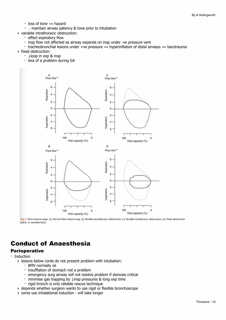
Flow Volume Loops - diff types:
‣ variable extra-thoracic obstruction = - affect inspiration - see stridor & ↑insp effort
Thoracics - �14
By A Hollingworth
- loss of tone ⟹ hazard - ∴ maintain airway patency & tone prior to intubation
‣ variable intrathoracic obstruction: - effect expiratory flow - insp flow not affected as airway expands on insp under -ve pressure vent - tracheobronchial lesions under +ve pressure ⟹ hyperinflation of distal airways ⟹ barotrauma
‣ fixed obstruction: - ↓loop in exp & insp - less of a problem during GA
Conduct of Anaesthesia Perioperative - Induction
‣ lesions below cords do not present problem with intubation: - BMV normally ok - insufflation of stomach not a problem - emergency surg airway will not resolve problem if stenosis critical - minimise gas trapping by ↓insp pressures & long exp time - rigid bronch is only reliable rescue technique
‣ depends whether surgeon wants to use rigid or flexible bronchoscope ‣ some use inhalational induction - will take longer
Thoracics - �15
By A Hollingworth
‣ sevo good for bronchodilation & ↓coughing - Maintenance
‣ if using rigid bronch - - use intermittent jet ventilation via side port - TIVA - cant control FiO2 well - high is bad for laser - remifentanil to control ‘pressure response’ to rigid scope - high risk of awareness - highly stimulating, shared airway, compromised pt, NMB ⟹ use BIS
- Extubation ‣ risk of bleeding, oedema, PtX, spreading of pneumonia ‣ give steroids, neb adrenaline ‣ fully reverse & sit upright before waking
- Heliox: ‣ available in 80/20 or 70/30 mixtures of H/O ‣ helium has lower density to O2 or nitrogen ‣ put into Reynolds number means more likely for laminar flow ‣ use for ventilation problem ⟹ ↓WOB ‣ disadvs: delivers fixed FiO2 but generally pts need ventilation not oxygenation ie tolerate hypoxia well ‣ if impending total obstruction should move back to 100% O2
Postop - central airway syndrome:
‣ complete obstruction ‣ can mimic partial reversal of NMB ‣ cause often new sputum or blood clot ‣ reintubation with rigid scope & vigorous suction required
Special Points - generally immediate relief given - anaesthesia for unrelated surgery:
‣ identify if stent is secure & its locations ‣ LMA if possible ‣ if ETT required must use fibrescope to check position of ETT & ?dislodgement of stent
Jet Ventilation - small, high pressure jets of O2 - entrains flow but not enough to increase flow above dead space. - ∴ mechanism of oxygenation:
‣ laminar flow - jet vent ⟹ spike in rapidly moving air down centre axis of airway, which gas in margins moves outwards
‣ Molecular diffusion (Taylor type dispersion) = mixing of air in small airways ‣ pendelluft or collateral vent = regional variation in resistance & compliance ⟹
- some areas fill, some empty - flow from adjacent units into others - effect ↑ed by HFJVs
‣ cardiogenic mixing - agitation & mixing next to beating heart - types of jet ventilation - categorised on frequency:
‣ high - special equip needed ‣ low eg manujet
- frequency of 8-10min allows time for expiration - TV = jet + entrainment
- unable to measure CO2 - indications:
‣ emergency - surg cannula cricothyroidotomy ‣ elective -
- diff airway - elective cannulation of cric incase of issues - thoracic surgery:
• major conducting airway surgery • into non-dep lung (instead of PEEP alone)
Thoracics - �16
By A Hollingworth
• bronchopleural fistula ⟹ leads to ↓air leak ‣ ICU - use in ARDS not supported by trials
- problems = hypercapnia
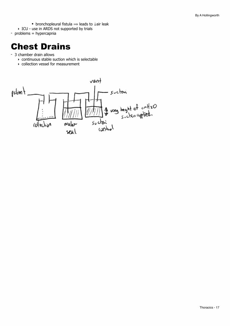
Chest Drains - 3 chamber drain allows
‣ continuous stable suction which is selectable ‣ collection vessel for measurement
Thoracics - �17
By A Hollingworth
By Surgery Airway Stents - airway stents used for:
‣ intrinsic airway lesions ‣ extrinsic compression ‣ tracheomalacia
- most commonly used to palliate malignant disease - metal expanding mesh which becomes epitheliased
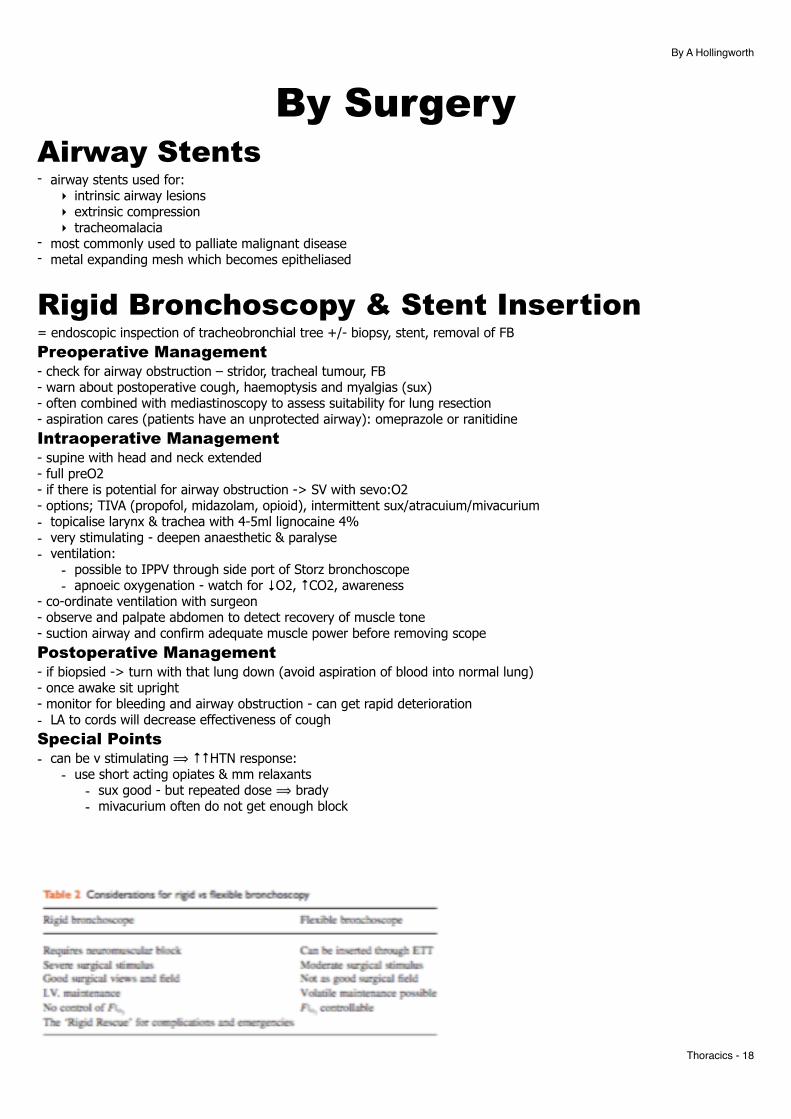
Rigid Bronchoscopy & Stent Insertion = endoscopic inspection of tracheobronchial tree +/- biopsy, stent, removal of FB Preoperative Management - check for airway obstruction – stridor, tracheal tumour, FB - warn about postoperative cough, haemoptysis and myalgias (sux) - often combined with mediastinoscopy to assess suitability for lung resection - aspiration cares (patients have an unprotected airway): omeprazole or ranitidine Intraoperative Management - supine with head and neck extended - full preO2 - if there is potential for airway obstruction -> SV with sevo:O2 - options; TIVA (propofol, midazolam, opioid), intermittent sux/atracuium/mivacurium - topicalise larynx & trachea with 4-5ml lignocaine 4% - very stimulating - deepen anaesthetic & paralyse - ventilation:
- possible to IPPV through side port of Storz bronchoscope - apnoeic oxygenation - watch for ↓O2, ↑CO2, awareness
- co-ordinate ventilation with surgeon - observe and palpate abdomen to detect recovery of muscle tone - suction airway and confirm adequate muscle power before removing scope Postoperative Management - if biopsied -> turn with that lung down (avoid aspiration of blood into normal lung) - once awake sit upright - monitor for bleeding and airway obstruction - can get rapid deterioration - LA to cords will decrease effectiveness of cough Special Points - can be v stimulating ⟹ ↑↑HTN response:
- use short acting opiates & mm relaxants - sux good - but repeated dose ⟹ brady - mivacurium often do not get enough block
Thoracics - �18
By A Hollingworth
- roc & suggamadex
Mediastinoscopy = inspection and biopsy of tumours and lymph nodes in superior and anterior mediastinum via a small suprasternal or anterior intercostal incision Anatomy - region between 2 pleural cavities from thoracic inlet to diaphragm - divided into superior & inferior by thoracic plane (imaginary line horizontally from sternal angle or T4) - superior =
‣ thymus ‣ aortic arch ‣ SVC ‣ areolar tissue ‣ lymph nodes
- inf mediastinum sub divided by pericardium of heart: ‣ ant ‣ middle = heart & pericardium, tracheal bifurcation, main bronchi, lung hllar, phrenic nerve, lymphs ‣ posterior = descending aorta, oesophagus, vagus, symph chain, thoracic duct, azygous & hemiazygous,
lymph Preoperative Management - r/v CT ?location of tumour - flow volume loops in sitting & lying useful - day case issues - check for
- SVC obstruction, - large mediastinal masses ⟹ tracheal deviation =/- compression of airway - previous operation - make access very difficult! - Tx aortic aneurysm - tracheal deviation
- systemic effects: - endocrine secretion - ACTH, ADH, PTH - neuromuscular effects -
- MG - thymic tumours - sensitive to NDNMBs & variable response to depolarising - Eaton Lambert
- prox myopathy (mostly legs) assoc with small cell carcinoma - ↓AcH release ⟹ sensitivity to all NMBs - exercise ↓s mm weakness (opposite to MG)
- Haematology - Hb, plts, thrombosis - other - nephrotic, amyloid
- often do a rigid bronch first Intraoperative Management - if tracheobronchial compression:
- difficult ventilation - may worsen with induction/IPPV esp in paeds ↳ ↓chest wall tone & cephalad displacement of diaphragm ⟹ loss of distending transmural pressure ⟹ complete obstruction
- keep breathing spont - v impt - ?AFOI - have rigid bronchoscope handy as rescue
- SVC syndrome: - =↓venous drainage from head ⟹ large tongue, laryngeal oedema, hoarse, ↑bleeding - Rx: elevate head, steroids, diuretics
- must avoid coughing ∴ when tube secured ensure deep paralysis - IPPV via a reinforced ETT - TIVA - supine, head up, arms by side, head ring and bolster under shoulders - pulse oximeter on right (brachiocephalic artery can be compressed by scope ⟹
- ↓flow R arm & R carotid ⟹ cerebral ischaemia) - BP on left arm
Thoracics - �19
By A Hollingworth
- IV fentanyl during OT - good IV access ->
- place leg cannula - catastrophic bleeding can occur - may require immediate sternontomy
- watch for - surgical compression of airway - monitor Vt - PTX
Postoperative Management - simple analgesia - monitor for pneumothorax, bleeding and cerebral ischaemia
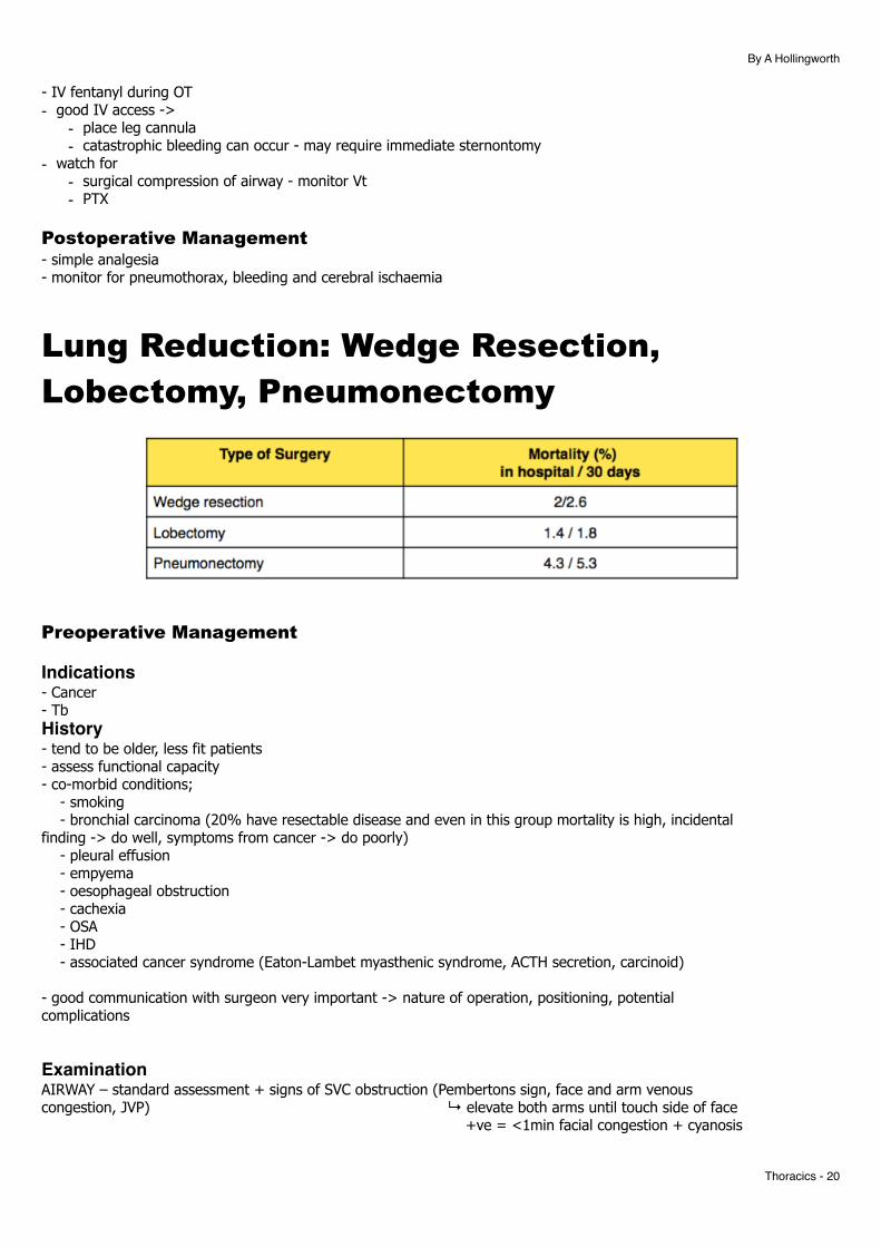
Lung Reduction: Wedge Resection, Lobectomy, Pneumonectomy
Preoperative Management
Indications- Cancer - Tb History- tend to be older, less fit patients - assess functional capacity - co-morbid conditions; - smoking - bronchial carcinoma (20% have resectable disease and even in this group mortality is high, incidental finding -> do well, symptoms from cancer -> do poorly) - pleural effusion - empyema - oesophageal obstruction - cachexia - OSA - IHD - associated cancer syndrome (Eaton-Lambet myasthenic syndrome, ACTH secretion, carcinoid)
- good communication with surgeon very important -> nature of operation, positioning, potential complications
ExaminationAIRWAY – standard assessment + signs of SVC obstruction (Pembertons sign, face and arm venous congestion, JVP) ↳ elevate both arms until touch side of face +ve = <1min facial congestion + cyanosis
Thoracics - �20
By A Hollingworth
BREATHING – unilateral chest signs (consolidation, effusion, pneumothorax), RR, position of trachea, sputum quality, SpO2 on RA
CIRCULATION – signs of right heart failure (pulmonary hypertension) – elevated JVP, RV thrill, oedema, murmurs signs of left heart failure, murmur (TR ?carcinoid), liver enlargement.
Walk patient up 2 flights of stairs (40 stairs)
Investigation= as general considerations at beginning of document
Management- stop smoking 8 weeks prior - preoperative physio and incentive spirometry - bronchodilators and anti-cholinergics
- may need post operative ventilation if; 1. DLCO <40% predicted normal 2. estimated postop FEV1 <800mL 3. estimated post op FVC <15mL/kg
Intraoperative Management - lateral position with broken table - DLT - use a L sided tube unless surgery:
- prox L lobectomy or pneumonectomy - abnormal bronchial anatomy
- IV in non dep arm - art line in dependent arm - lung protective ventilation (PAP < 30cmH2O, prolonged expiratory phase = short I:E ratio) - aim for extubation post op as mechanical ventilation -> stress stitches and increases risk of infection
- blood loss 200-800mL - CVL generally not used and are unrealiable - OLV prevents soiling of dependant lung - bronchial suture line is leak tested under saline at 40cmH2O
Analgesia Options- epidural - preop but caution - paravertebral injection (preincision) and then surgically placed catheter - wound catheter - intrapleural catheter - intecostal nerve blocks - PCA - simple analgesia (NSAIDS, paracetamol)
Postoperative Management - physio - O2 (humidified) - CXR in recovery - chest drain NOT on suction if pneumonectomy
- thoracotomy very painful -> good analgesia very important -> prevent respiratory complications, chronic pain and increases mobility
-- AF common (some people give prophylactic digoxin)
Thoracics - �21
By A Hollingworth
Complications - periop mortality pneumonectomy = 5% - ALI - see below - postpneumonectomy pulmonary oedema - see below - cardiac herniation - see below - arrhythmias - some use prophylactic digoxin loading - resp complications = 15-20% - CVS complications = 10-15% (arrhythmias, ischaemia) ALI- ALI 2-5% & x3 more likely post pneumonectomy ⟹ ↑mortality to 25-50% - RFs of ALI:
- preop: - chronic alcohol abuse - severe pulmon dysfunction
- intraop: - intraop plateau pressures >15cmH2O - large resection - overhydration = >4litres IVF in 1st 24hrs - R sided pneumonectomy
- causes: - multi hit: neutrophil hypothesis & epithelial hypothesis - operated lung = impaired surfactant & surgical manipulation - non operated = hyperoxic/mechanical injuries
- vent strategies: - protective lung vent & open lung approach:
- Vt <7ml/kg - PLV <30cmH2O - PEEP - with periodic recruitment - ↓ FiO2 - high FiO2 ⟹ atelectasis
Postpneumonectomy Pulmonary Oedema- incidence 9%. mortality 50% - RFs:
- large Vt, high airway pressures - too much fluid - R pneumonectomy
- occurs D2-4 although CXR changes seen ~24hrs earlier - unknown cause: = low pressure pulmon oedema. ?with fluid overload - Rx:
- low FiO2 - lung protection strategies - Vts 5-6ml/kg - fluid <20ml/kg/day
Cardiac Herniation- should avoid:
- lat position with operated side down - never suction drains - ↑IPPV
- Rx: - sit up or operated up - ↓all vent pressures - instill 1-2L air into empty hemithorax - ⟹ OT
Video Assisted Thorascopic Surgery = inspection of thoracic cavity via scope passed through intercostals incision (drainage of effusions, biopsy, pleurectomy, pleurodesis and pericardial biopsy/window
Thoracics - �22
By A Hollingworth
Preoperative Management - same assessment as for thoracotomy but less invasive - less deterioration in lung function - discuss RA and PCA
Intraoperative Management - lateral decubitus + broken table + arms flexed (FA in parallel with face) - IPPV and OLV via left DLT :
- commence OLV prior to insertion of trochar - good lung collapse required for surgical access
- art line - percutaneous paravertebral block +/- catheter (surgeons can insert under scope guidance)
Postoperative Management - extubate and sit up - CXR in recovery to ensure full lung expansion - PCA - epidural usually only needed if bilat surgery
Lung Reduction: Bullectomy = non-anatomical resection of regions of hyperinflated and poorly functioning lung (COPD with resectable bullous disease) - generally used in severe emphysema ie normally those who would avoid GA at all costs - National Emphysema TReatment Trial showed surgery good for upper lobe disease with low baselin ex
capacity - If isolated congenital bulla or cyst manage same but often much more robust
- aims: 1. to reduce amount of non-functional lung 2. improve diaphragm function 3. increase right heart function by decreasing tamponade effect
Preoperative Management - intensive assessment and careful selection: 1. very breathless 2. in homogeneity in lung disease (areas that can be resected) 3. no smoking for 3 months 4. age <75 5. no pHTN 6. FEV1 <20% predicted + predicted improvement post surgery
- cardiac assessment; angio, right heart catheterisation, ECHO, PAP - periop steroid supplementation as required
Intraoperative Management - surgical approach:
- thoracotomy – unilateral surgery - median sternotomy – bilateral surgery - VATS - less pain & less changes - severe pain - IPPV with DLT (verify position bronchoscopically) - deliberate hypoventilation and permissive hypercapnia - ventilate; TV 6ml/kg, RR 10, I:E 1:4, PAWP <30cmH2O - intermittently disconnect from vent ⟹ allow lungs to empty - GA with TIVA - high A-a gradient - thoracic epidural
Thoracics - �23
By A Hollingworth
- cautious ventilation as can burst bullae - cautious use of fluid - bronchospasm & sputum can be a problem
Postoperative Management - extubate quickly - accept high PaCO2, aim for SpO2 90-92% - watch closely for air leaks - physiotherapy - pulmonary rehab programme - complications;
- prolonged air leak (common) - 50% pts have leak >7days - 5-10% mortality - BPF, pneumothorax
Decortication/Drainage of Empyema = surgical removal of pus and organised thick fibrinous pleural membrane - causes:
- parapneumonic effusions = 50% - haemothorax - iatrogenic - ICDs, post surgery, oesophageal perf
- empyema assoc with pneumonia development stages: - simple para-pneumonic effusion = exudate with normal pH - fibropurulent stage = bacterial invasion ⟹ ↓pH - complicated para-pneumonic effusion = clear fluid but ↓pH - empyema = frank pus
- principle = - remove infected tissue - pleural peel - fully re-expand lung - obliterate infected pleural space
Preoperative Management - intrapleural infection usual causes = pneumonia, intercostals drains, prev chest surgery - quantify degree of sepsis and organ dysfunction - no consensus on timing of surgery but BTS suggest discussion sepsis with pleural collection ongoing for 5-7
days ↳ if iatrogenic do earlier - check whether has bronchopleural fistula ie pus into airways. ↳ Means:
- +ve pressure vent may ⟹ tension PTX - soiling of lung ⟹ serious morbidity - ∴ lung isolation prior to +ve pressure vent VERY impt in BPF
Intraoperative Management - lateral decubitus - very painful - GA, IPPV, DLT - invasive monitoring unless very F&W - rib may be resected - thoracoscopy to break down adhesions - decortication requires epidural (as paravertebral catheter contraindicated) - decortication frequently causes significant haemorrhage 500-2000mls
Postoperative Management - multi-modal analgesia - epidural - but often unable due to bacteraemia - HDU if crumbly patient - massive air leak can occur - complications:
Thoracics - �24
By A Hollingworth
- persistent air leak - severe parenchymal lung damage ↳ may need lobectomy
Repair of BronchoPleural Fistula = closure of a communication between trachea/bronchi -> pleural space (thoracotomy incision)
Preoperative Management - features:
- productive cough, - haemoptysis, - fever, - SOB, - SC emphysema, - falling fluid level in post-pneumonectomy space on CXR
- Common causes: - lung reduction surgery - others: pneumonia, lung abscess, empyema
- anaesthetic chart review (ease and size of DLT used) - CXR - ?obviously distorted anatomy from prev surgery - O2 - chest drain - in situ and working - IV antibiotics - IVF
Intraoperative Management - key aims:
- impt to protect good lung from contamination - control distribution of ventilation - isolate lungs quickly post induction
- keep sitting up (affected side tilted down) until affected lung isolated then ⟹ lateral decubitus - awake A line - risk of tension pneumonthorax on starting IPPV
- options for securing airway = (there is no great option!) 1. rapid IV induction and fiberoptically guided endobronchial intubation with DLT into contralal bronchus
(MRSI) ↳ risk of ↑ing fistula size with poor DLT placement 2. AFOI with double lumen tube ie SV 3. Deep inhalational or TIVA induction ie SV
- options if these DLT intubation fails/massive air leak = 1. intubate with standard 6mm tube -> use fiberoptic scope to pass ETT into normal bronchus & inflate cuff to isolate 2. surgeon with rigid fibrescope move into trachea -> pass a AEC through scope into normal lung -> removed bronchoscope -> intubate over AEC 3. pass a bronchial blocker or Fogarty embolectomy catheter into fistula via rigid bronchoscope to control leak temporarily
- watch for tension PTX - TIVA +/- ketamine - must isolate good lung quickly - small fistulae may be sealed with glue or sutures bronchoscopically
Postoperative Management - HDU/ICU - minimise AWP - extubate quickly
Thoracics - �25
By A Hollingworth
- good analgesia
Pleurectomy/Pleurodesis - = stripping of parietal pleura from inside chest wall, production of adhesions via chemical or physical
abrasion - procedures
- stripping of pleura from inside of chest wall - adhesions via
- chemical = talc, tetracycline - physical abrasion
Preoperative Management - can be
- young and otherwise healthy - check for asthma - severely debilitated elderly COPD patients - check PFTs, functional status
- review CXR for pneumothorax or effusion (needs ICD if pneumothorax present) - discuss and choose post-operative analgesic technique - effusions >⅔ hemithorax (2000ml) = massive ↳ should be drained at least >12hr prior to OT ↳ rapid intraop reinflation of collapsed ⟹ unilat post op re-expansion pulmon oedema
Intraoperative Management - lateral decubutus, broken table or supine - epidural if pleurectomy esp bilateral - may be done through open thoracotomy or VATS - IPPV, DLT and OLV - keep airway pressures as low as possible ↳ look for PTX - may tension even on good side - avoid N2O - collapse lung when surgeon instilling irritant - very painful - avoid NSAIDs though (pleurodesis less effective) - aim for full expansion of lung @ end of procedure ⟹ to oppose pleura - if large effusion -> cardiovascular collapse can occur when patient put into effusion side up ↳ MOA: mediastinal shift & ↑ITP ⟹ ↓VR
Postoperative Management - extubate and sit upright - CXR (ensure lung fully expanded) - monitor for re-expansion pulmonary oedema
Oesophagectomy = excision of some or all of oesophagus with mobilisation of stomach into chest - usually for oesopphageal Ca but can be for benign stricute/achalasia - one of highest peri-op mortality of all elective procedures - ~5% ↳ if <10 operations a year up to 20% mortality - 66% death from sepsis 2nd to anastomotic breakdown ⟹ resp infection
Preoperative Management - discuss with surgeon exact surgical approach:
- Transhiatal - laparotomy & cervical anastomosis - Ivor-Lewis - laparotomy & R thoracotomy (tumour upper ⅔) - thoracoabdominal - L throacotomy crossing costal margin & diaphragm (tumour lower ⅔) - minimally invasive - thorascopic oesophageal mobilisation, laparoscopic gastric mobilisation & cervical
anastomosis
Thoracics - �26
By A Hollingworth
- assess nutrition - often cachexia - assess cardio-respiratory function - preoperative chemo (residual immunosuppression) - book HDU/ICU - discuss analgesia; paravertebral catheter, epidural - get experienced anaesthetist who has done a lot of oesophagectomy anaesthesia
Intraoperative Management - consider epidural if possible - RSI - everyone - if thoracotomy required used a DLT and OLV - regional anaesthesia:
- thoracoabdominal = paravertebral LA infusion with PCA - laparotomy/thoracotomy = mid Tx epidural
- N/G - to be removed & reinserted later - invasive monitoring in all pts (liase with surgeon regarding CVL position) ↳ not for same side as cervical anastomosis - warming cares++ - careful fluid balance:
‣ stay ahead of fluid replacement ‣ too much fluid assoc with morbidity ‣ urine output may be ↓ed if ↑ed intra-abdo pressures from pneumoperitoneum ‣ CVP useless esp if prone, L lat, pneumoperiotneum ‣ ScvO2 - low ≈ ↑ed post op morbidity
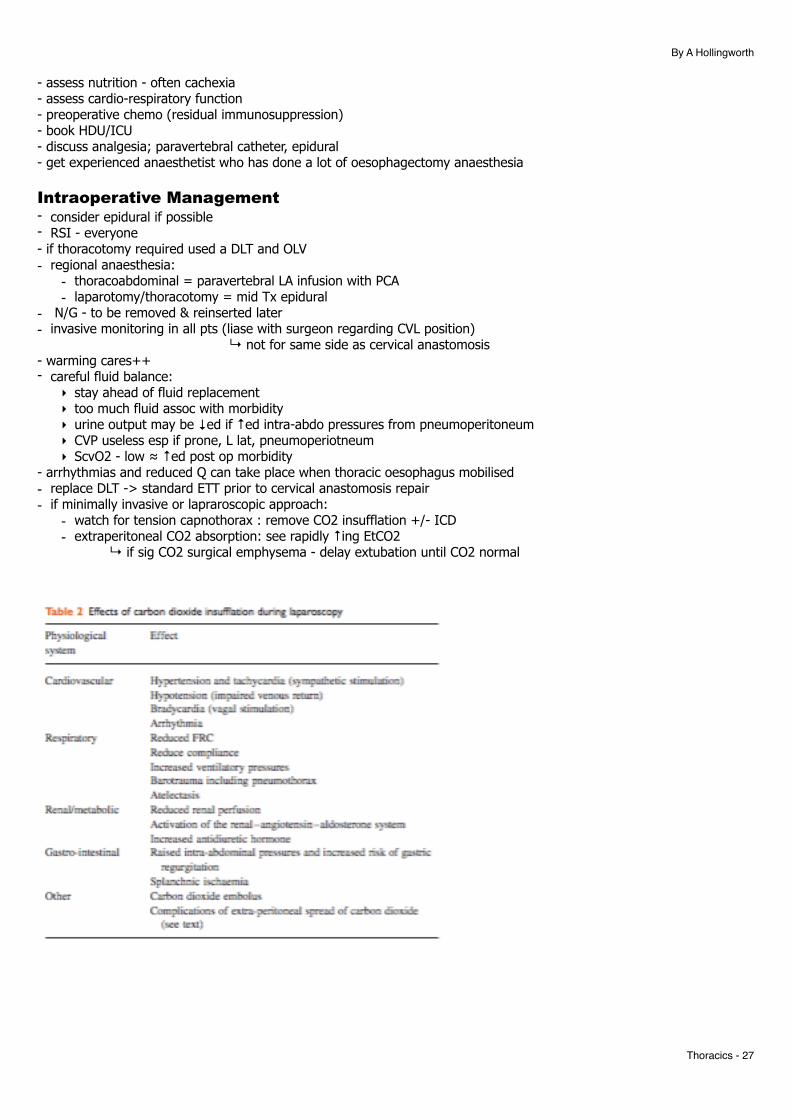
- arrhythmias and reduced Q can take place when thoracic oesophagus mobilised - replace DLT -> standard ETT prior to cervical anastomosis repair - if minimally invasive or lapraroscopic approach:
- watch for tension capnothorax : remove CO2 insufflation +/- ICD - extraperitoneal CO2 absorption: see rapidly ↑ing EtCO2
↳ if sig CO2 surgical emphysema - delay extubation until CO2 normal
Thoracics - �27
By A Hollingworth
Postoperative Management - WWWE - use jejunostomy or nasoduodenal tube for early enteral feeding - at high risk of infection and anastamotic breakdown - regional techniues:
‣ epidural - ↑pulmon complications from SNS blockade ⟹ ↑IVF admin (prevent using vasoconstrictor) ‣ paraverterbal block - placed by surgeon at end
Descending Thoracic Aortic Aneurysm Preoperative - usually elderly with multiple co-morbidities:
- TOE, CT, MRI - Cardiac risk: ECG, angio - ECHO - PFTs - CXR - look for L main bronchus
Perioperative • Induction
‣ R A line +/- femoral for distal perfusion ‣ TOE/PAC ‣ thoracotomy ⟹ OLV to improve surg access
• Maintenance ‣ expect blood loss ++
• cross clamping: ‣ suprarenal, supraceliac - anticipate haemodynamic changes
- proximal to clamp = ↑cardiac filling pressures ⟹↑wall stress ⟹ ↑O2 consumption & ↓CO - distal to clamp: ↓O2 supply⟹ ↑anaerobic metabolism ⟹ ↓lactate elimination
‣ consider distal perfusion techniques: - passive shunts - aorto-bifem bypass - partial CPB & DHCA
‣ ?CSF drainage ⟹ ↑spinal cord perfusion • declamping:
‣ anticipate: ↓MAP, ↓CO, ↑O2 consumption, ↑lactate production, ↓temp, met acidosis, ↑K ‣ Rx by
- ↑ing afterload = ↓volatiles, ↓vasoDs, ↑vasoconstrictors - ↑preload - gradual clamp release
Postop - ICU - check for renal impairment, spinal cord injury, mesenteric ischaemia, periph VTE
Anterior Approach Spinal Surgery - occasionally done for scoliosis repair in children - post approach is more common (↓ed morbidity) - scoliosis causes:
- idiopathic - most common - NM disease - tumour - infections - marfans - RA - injury
- surgery indicated if Cobb angle >40% ⟹ ↑posture & ↓progression of pulmon dysfunction
Thoracics - �28
By A Hollingworth
Preoperative - if idiopathic:
- PFTs - even if FVC <32% then surgery tolerated well - ECHO only if ↓ex function - 25% have MV prolapse
- if secondary - need ↑ed eval: - muscular dystrophies - cardiomyopathy ⟹ ECHO +/- stress ECHO - NM disease - bulbar palsy? vent capacity?
Perioperative • Induction
‣ DLT - for surgical access • Maintenance
‣ long procedure ‣ large blood loss ‣ avoid ↓temp ‣ keep depth anaesthesia stable ⟹ neurophysiological monitoring
• Extubation ‣ ICU ‣ extubate early for neuro assessment ‣ epidural by surgeon ‣ multimodal analgesia plan
Postop - careful fluids - ⅓ total blood loss is usually post op - vent failure - SIADH - pain +++ - ileus, sup mesenteric art syndrome
Acquired Tracheo-Oesophageal Fistula - big problem as no laryngeal reflex protection - causes:
- iatrogenic - post surgical - malignancy -
- 50% ∴ commonest - usually oesophageal tumours - median survival from diagnosis = 1-6weeks
- trauma: - ETT cuff post prolonged venting - (tracheostomy does not effect incidence) - RTCs - compression of trachea & oesophagus on spine - generally see in carinal area
- infection - signs:
- ‘Ono’s sign’ = uncontrolled coughing after swallowing - esp fizzy drinks - Ix:
- CXR soiling signs - barium swallow - endoscopy -
- although small leaks can be missed visually - methylene blue with leak into bronchoscopic view
- 2 Rx options: - repair - suturing oesophagus over a NG tube - stenting:
- done in Ca
Thoracics - �29
By A Hollingworth
- usually placed under sedation - self expanding metal stents well tolerated
Preoperative - principles:
- minimise further aspiration: - new ETT passed with cuff up distal to fistula - elevated bed head - draining gastrostomy tube only
- prevent & Rx infections - supportive therapy until repair:
- if pre-vent’ed should try to wean as much as possible to ↓chance of post correction IPPV - IPPV assoc with tracheal anastomotic leak & stenosis - nutrition
Perioperative • concerns as for bronchopulmon fistula repair • avoid cricoid pressure • avoid sux - fasiculations may ↑risk of tracheal soiling • NG tube awake & draining • if confirmed high prox tracheal TOF:
‣ bronchoscope to place ETT cuff below fistula • if low TOF:
‣ OLV with isolation: - endobronchial intubation - DLT
• TIVA • aim for immediate extubation Postop - proximal TOFS require standard analgesia - distal TOFs may require regional blocks eg intrapleural, paravertebral, epidural
Oesophageal Atresia & TOF - 92% of neonates with oesophageal atresia have a TOF History- 1:3,000 births - copious secretions in mouth - pulmonary aspiration of feeding - presents with choking and cyanosis on feeding - inability to pass a N/G - constant risk of aspiration (can pass a double lumen tube that allows irrigation and suction) - high incidence of cardiac disease and prematurity - 5 types -> most common = blind loop oesophagus + distal oesophagus to carina - 50% of newborns with this have other congenital abnormalities:
‣ V ertebral ‣ A norectal ‣ C ardiac ‣ T racheoesophageal ‣ R enal ‣ L imb abnormalities
Exam- standard + for cardiac abnormalities Investigations- passing a radio-opaque N/G tube + CXR - ECHO - electrolytes Preoperative - IVF for hydration as neonatal fluids - nurse head up
Thoracics - �30
By A Hollingworth
- continual suction on NG - prophylactic Abx - should not delay T/F to specialist centre - rapid surgery better prognosis - ECHO ?congen cardiac problems - routine bloods Perioperative • Induction
‣ suction pouch where Ng sitting & remove ‣ inhalational induction with spont vent ‣ avoid BMV if possible ‣ 2IVs & A line
• Maintenance ‣ removal of ETT & bronchosocpic exam to identify TOF ‣ place T piece on side port of bronch to allow spont vent during scoping ‣ reinsertion of ETT so that to occlude TOF - use small bronchoscope - may be difficult ‣ only then allow to give NMBs ‣ access via R thoracotomy ‣ manual ventilation after ligation of fistula to assist in repair of oesophagus
• Extubation ‣ extubate as soon as possible
Postop - complications:
‣ early = - tracheomalacia of varying severity - rep’ed infections - anastomotic leak in 10-20% - GORD - antacids initially +/- fundoplication later
‣ late = - resp problems: pneumonia, tracheomalacia, recurrent TOF, recurrent stricture
Special Points - prognosis based on weight & other cardiac problems
Trauma Surgery for Chest Injuries - <30% of pts with thoracic trauma require a thoracotomy - 1.5L or >200ml/hr from ICD ⟹ thoracotomy - must have good access - exsanguination is unacceptable - look for tension PTX - massive air leak = tracheobronchial injury
Aortic Disruption - CXR signs:
- wide mediastinum - obliterated aortic knob - tracheal deviation to R - no space between aorta & PA - depressed L main bronchus - deviation to R of oesophagus - paratracheal stripe - apical cap - L haemothorax - 1st or 2nd rib #s
- Ct if stable - TOE in theatre if unstable
Ruptured Diaphragm - varied presentation:
- chronic missed condition
Thoracics - �31
By A Hollingworth
- intestinal obstruction of herniated bowel - check preop bloods - repair is not an emergency - approach via lat thoracotomy or thoracoabdominal incision - manage as fundoplication - DLT & OLV facilitate surgical access - NGT to decompress stomach
Repair of Ruptured Oesophagus - presentation:
- surg emphysema - pleural effusions
- causes: - trauma - excessive abdo straining & vomiting = Boerhaave’s - iatrogenic - endoscopy - FBs
- sequelae: - mediastinits ⟹ sepsis ⟹ SIRS ⟹ shock & MODS
- surgery goal is to drain & prevent more contamination - if frail conservative management may be tried - NG suction & ICD - ICU preoptimisation - Anaesthetic plan as oesophagectomy - surgery:
- upper oesophagectomy = R thoracotomy - lower oesophagectomy = L thoracotomy - primary closure or oesophagectomy
- AF very common - early jejunostomy or PN feeding - high risk for many days postop
Repair of Tracheobronchial Injury - most die prehospital - 100% O2 & bilat ICDs - adequate +ve pressure vent may be impossible with a single lumen tube ↳ fibreoptic guided intubation of contralat bronchus with DLT - single lumen ETT can be guided past tracheal tear with bronchoscope & cuff inflated - once airway secured ⟹ thoracotomy for repair - carinal disruption requires CPB while repairing
Endoscopic Thoracic Sympathectomy - main indication = palmar hyperhidrosis (1% people) - generally younger ASA 1/2 patients - 2 paravertebral symp chains arise from ventro-lateral grey matter T1-L2 - short pre-ganglionic fibres & long post ganglionic adrenergic fibres to effector sites (cholinergic sweat
glands) - lower cervical & upper thoracic ganglia normally fuse = stellate ganglion - supply:
‣ head & neck = T1-2 ‣ UL = T2-5 ‣ thoracic viscera = T1-4
- symp chain runs beneath parietal pleura ⟹ over neck of ribs - surgery performed by resection, cutting or clipping (reversible) - Surg plan = division based on symptoms:
‣ T2-3 = palmar hyperhydrosis ‣ T2-4 = axillary hyperhydrosis ‣ T2-4/5 = angina & long QT syndrome
Thoracics - �32
By A Hollingworth
Preoperative - check if being done for angina/long QT Perioperative - Induction
‣ 2 options: - DLT - if slow surgeon - normal ETT +/- bronchial blocker - if quick surgeon
‣ small volumes of CO2 insufflated into thorax during apnoea/lung down to create visualisation of chain - Maintenance
‣ A line only if other co-morbidities ‣ back up plan for massive haemorrhage ‣ beach chair - control head ‣ prep for open thoracotomy if required ‣ 10-15min procedure per side ‣ ensure positioning to avoid branchial plexus injury ‣ partial lung collapse may ⟹ shunt 10-15%
- Extubation Postop
Special Points - complications:
‣ intraop: - severe hypoxia:
• using DLTs • partial collapse on CO2 insufflation
- cerebral ischaemia - severe haemorrhage from rupture of major subclavian vessels
‣ post op: - compensatory sweating - persistent PTX - Horners syndrome
Thoracics - �33