Embed Size (px)
Citation preview
RESEARCH Open Access
Brain injury unmasking Ehlers-Danlossyndromes after trauma: the fiber printClaude Hamonet1,2, Daniel Frédy3, Jérémie H. Lefèvre4,5, Sacha Bourgeois-Gironde6 and Jean-David Zeitoun7,8*
Abstract
Background: The role of physical trauma in the onset of symptoms in Ehlers-Danlos syndrome (EDS) has neverbeen characterized. We sought to search and describe brain lesions EDS patients also having personal history ofphysical trauma. We systematically performed brain magnetic resonance imaging in a first cohort of patients witha hypermobility type of EDS which described the onset of their disease or its worsening after a physical trauma.Unexpected yet consistent findings that were thought to be related to the reported traumas led to perform brainimaging in all subsequent patients with similar symptoms regardless of a history of trauma and to search for aprior trauma by active questioning.
Results: Fifty-nine patients were recruited and analyzed, among which 53 (89.8 %) were women. Overall, 26(44.1 %) reported a personal history of physical trauma. Six signs pertaining to subcortical lesions and affectingwhite matter tracts were identified. Those included lesions of the reticular formation, the two lenticular nuclei,the corpus callosum and the arcuate fasciculus. Thirty-six patients (61.0 %) had at least 5 of the 6 imagingsigns. In case of a trauma before 18, patients had significantly more lesions of the reticular formation (100 % vs.50 %; p = 0.0035).
Conclusions: Patients with EDS, hypermobility type, were found to have consistent and specific brain lesionsinvolving white matter tracts. Moreover, the record of a physical trauma in a substantial proportion of casessuggests that these lesions could be post-trauma consequences. Therefore, physical trauma could be a triggeringfactor in EDS.
Keywords: Ehlers-Danlos syndrome, Brain trauma, Brain injury
BackgroundEhlers-Danlos syndromes (EDSs) are a heterogeneousgroup of connective tissue disorders mainly character-ized by joint hypermobility, skin hyperextensibility andtissue fragility [1]. Despite the fact that the first clinicaldescription dates back more than a century [2, 3], EDSsare still poorly known, their genetic background is largelyignored and their pathophysiology is unclear. Additionally,we dramatically lack efficacious treatments. Last, althoughthe Villefranche’s classification is still the benchmark [4],their nosology and semiology are still a matter of debate[5]. For instance, we recently described that the frequencyand type of digestive symptoms in EDS were much more
common and different than previously reported [6].Yet, many subjective symptoms of EDS are not under-stood, in particular those that could be related to thenervous system.Anecdotal observation collated in the frame of our
routine clinical activity suggested that several patientsreported their symptoms being unmasked or worsenedfollowing a physical traumatism. This primarily appearedcounter-intuitive for a genetic disease to be triggered bya possible injury. Yet, we aimed to search and evaluatepossible lesions of the central nervous system (CNS) andmagnetic resonance imaging (MRI) examinations wereperformed that showed abnormalities which were thoughtto be attributable to numerous symptoms described bypatients. Subsequently, MRIs were also performed in otherpatients with similar subjective signs that could be linkedto CNS lesions but with no recorded history of traumaand those showed brain lesions analogous to the ones that
* Correspondence: [email protected] of Gastroenterology and Nutrition, Saint-Antoine Hospital,APHP, 284, rue du Faubourg Saint-Antoine, 75012 Paris, France8Department of Proctology, Croix Saint-Simon Hospital, Paris, FranceFull list of author information is available at the end of the article
© 2016 Hamonet et al. Open Access This article is distributed under the terms of the Creative Commons Attribution 4.0International License (http://creativecommons.org/licenses/by/4.0/), which permits unrestricted use, distribution, andreproduction in any medium, provided you give appropriate credit to the original author(s) and the source, provide a link tothe Creative Commons license, and indicate if changes were made. The Creative Commons Public Domain Dedication waiver(http://creativecommons.org/publicdomain/zero/1.0/) applies to the data made available in this article, unless otherwise stated.
Hamonet et al. Orphanet Journal of Rare Diseases (2016) 11:45 DOI 10.1186/s13023-016-0428-9
had been previously identified, suggesting that minor trau-mas could have occurred but gone unnoticed duringchildhood. We herein report the results of this seriesof 59 patients.
MethodsAll patients described in the current report were re-cruited from February 2010 through August 2013 fol-lowing a regular visit in our tertiary center dedicatedfor EDS. They were clinically assessed at least once bya single expert practitioner in EDS (CH) and were for-mally diagnosed has having a hypermobility type ofEDS according to Villefranche criteria [4]. All of themhad joint hypermobility, skin hyperextensibility and/orsmooth, velvety skin, and joint disorders. Joint hyper-mobility was specifically assessed using the Beightonscale [7], and all recruited patients had a score above thethreshold of 5 points. All patients had a familial history ofEDS, either for their ascendants or descendants.This study was conducted in accordance with the French
bioethics laws which do not require any approval by anethic committee for clinical and imaging examinations aslong as they are performed in the frame of standard cares.All patients provided written informed consent.
Sample 1: patients with symptoms and personal historyof physical traumaInitially, a first subset of 25 patients was identified fortheir claim of exacerbation of neurologic symptomsfollowing a physical trauma (hit, car accident, physicalabuse, etc.), especially because some of them neededobjective data proving their disorders as they were suing forcompensation. The reported signs included proprioceptivedisorders, dystonia, altered vigilance and sleep disorders,pain syndrome, dysautonomic syndrome, chilliness, sweat-ing, impaired thermoregulation. Therefore, they were re-ferred for brain imaging in order to search for a cause totheir complaints.
Sample 2: subsequent patients with no spontaneouslyreported history of physical traumaFollowing the intriguing findings of multiple, seeminglyspecific and systematic abnormalities in patients of thesample 1, subsequent patients (n = 34) seen in consultationwith similar clinical symptoms but no reported history ofphysical trauma were also systematically referred for brainimaging in the attempt of searching for analog brain injury.
Magnetic resonance imagingMRI scans were all performed by the same investigator(DF) according to the same techniques. MRI evalua-tions were made with a 1.5 tesla machine (GeneralElectric®) following Flair, T2*, Fiesta, and 3D-SPGR T1
sequences. Tensor diffusion imaging was also part ofthe MRI assessment.
Statistical analysisCategorical variables are described as frequencies andpercentages, and continuous variables are described asthe means ± standard deviation. Comparisons amonggroups were performed using the Chi-squared or Fisher’sexact test for discrete variables and by unpaired t-testsor the Wilcoxon rank-sum test for continuous variables.All tests were two sided with a level of significance set atp < 0.05. All statistical tests were performed using SASsoftware version 9.3; SAS Institute Inc., Cary, NC, USA.
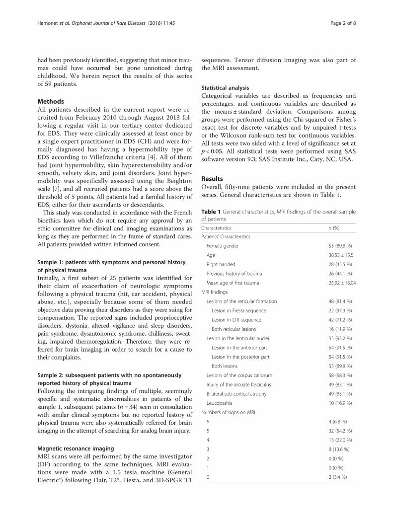
ResultsOverall, fifty-nine patients were included in the presentseries. General characteristics are shown in Table 1.
Table 1 General characteristics, MRI findings of the overall sampleof patients
Characteristics n (%)
Patients’ Characteristics
Female gender 53 (89.8 %)
Age 38.53 ± 15.5
Right handed 28 (45.5 %)
Previous history of trauma 26 (44.1 %)
Mean age of first trauma 25.92 ± 16.04
MRI findings
Lesions of the reticular formation 48 (81.4 %)
Lesion in Fiesta sequence 22 (37.3 %)
Lesion in DTI sequence 42 (71.2 %)
Both reticular lesions 16 (11.9 %)
Lesion in the lenticular nuclei 55 (93.2 %)
Lesion in the anterior part 54 (91.5 %)
Lesion in the posterior part 54 (91.5 %)
Both lesions 53 (89.8 %)
Lesions of the corpus callosum 58 (98.3 %)
Injury of the arcuate fasciculus 49 (83.1 %)
Bilateral sub-cortical atrophy 49 (83.1 %)
Leucopathia 10 (16.9 %)
Numbers of signs on MRI
6 4 (6.8 %)
5 32 (54.2 %)
4 13 (22.0 %)
3 8 (13.6 %)
2 0 (0 %)
1 0 (0 %)
0 2 (3.4 %)
Hamonet et al. Orphanet Journal of Rare Diseases (2016) 11:45 Page 2 of 8

Magnetic resonance imaging findingsSix main criteria pertaining to subcortical lesions wereidentified. Those lesions affected white matter tracts,essentially made of myelin coated axons.
� The first criterion was the presence of lesions of thereticular formation, which is a posterior medial cordextending downwards the whole length of thebrainstem. It is a network of longitudinal nervefibers that regulates several autonomic functionssuch as consciousness and sleep. Two types oflesions that were thought to be specific wereidentified with respect to this first criterion. Thefirst lesions were observed in the medial part ofthe reticular formation and perpendicular to theback of the floor of the 4th ventricle. As shown ina Fiesta sequence equally weighing T1/T2 ratios,the lesions exhibited a post-traumatic aspect(Fig. 1a). The second lesion affecting the reticularformation consisted in a bilateral rarefaction ofvertical cortical-spinal (spinal-thalamic) fibers.Those fibers plunge within the lenticular nucleiand go through the two cerebral peduncles. Besidestheir sensitive function, they collaterally support thereticular formation. Tridimensional diffusion tensorimaging (DTI) revealed the fibers rarefaction (Fig. 1b).
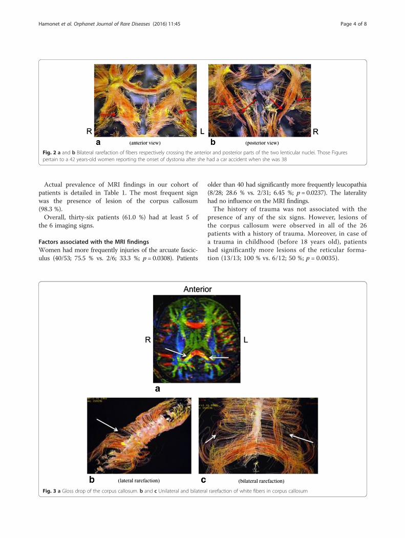
� The second criterion was a similar lesion patternin the two lenticular nuclei. Tridimensionalsequences showed a quasi-symmetrical bilateralrarefaction of fibers crossing the anterior [Fig. 2a]and posterior [Fig. 2b] parts of the two lenticularnuclei. This fiber tract appeared cut off after acerebra trauma. Since these fibers spread overthe two frontal lobes and the basal ganglia, theirrupture and rarefaction tended to be associatedwith attentional and motivational troubles.
� The third retained criterion was the presence of lesionsof the corpus callosum exhibiting a post-traumaticaspect. Bidimensional-DTI showed a gloss drop of thecorpus callosum which was bilateral and morepronounced in its posterior part [Fig. 3a]. Thisaspect could be correlated with functional impairment,consistently with previous reports. Indeed, lesions ordysmorphia of corpus callosum have been shown to beassociated with extreme plasticity in auditory andmotor scaffolding [8] and tactile anomia [9] (Fig. 3a).3D-DTI reveals in addition a lateral [Fig. 3b] or bilateral[Fig. 3c] rarefaction of white fibers in this structure.
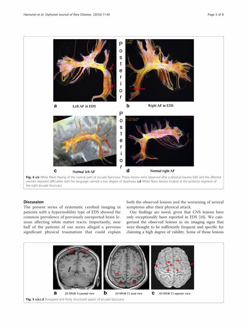
� The fourth criterion was made of arcuate fasciculus(AF) injuries. Tridimensional-DTI typically showedwhite fibers fraying in this structure. Whitefibers located at the junction between the upperand posterior segments of the left AF weremarked by a neat fraying out their normal path[Fig. 4ab]. In the case of that right-handedpatient a shock also slightly sprayed out whitefibers located at the posterior segment of herright arcuate fasciculus [Fig. 4cd]. Other possiblepatterns were that of a supple, elongated andfinely structured aspect,especially in the right AF [Fig. 5abcd].
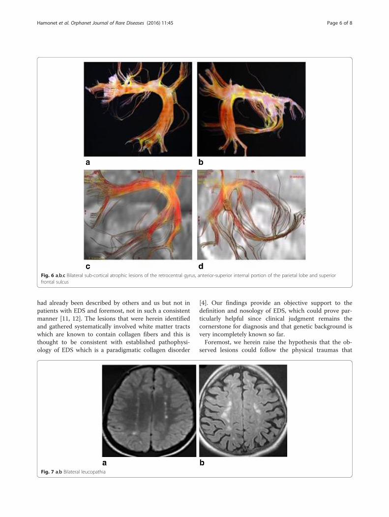
� The fifth retained criterion involved a bilateralsub-cortical atrophic aspect frequently observedat the following locations: retrocentral gyrus,anterior-superior internal portion of the parietallobe, superior frontal sulcus displaying a markedbilateral abnormal width [Fig. 6abc].
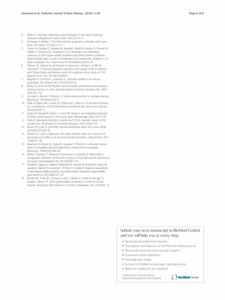
� The sixth criterion was an aspect of leucopathia,typical for its bilateral para-medial location presentingtwo strings of nodules made visible in axial hyperflashFlair sequence at the level of the two semi-ovalhemi-centers [Fig. 7a and 7b].
Fig. 1 a Post-traumatic aspect of the reticular formation in a Fiesta sequence equally weighing T1/T2 ratios. This Figure comes from a 34 years-oldwomen reporting the onset of vigilance and sleep disorders following a car accident ten years before. b Rarefaction of vertical cortical-spinal fibers ofthe reticular formation in 3D sequence
Hamonet et al. Orphanet Journal of Rare Diseases (2016) 11:45 Page 3 of 8
Actual prevalence of MRI findings in our cohort ofpatients is detailed in Table 1. The most frequent signwas the presence of lesion of the corpus callosum(98.3 %).Overall, thirty-six patients (61.0 %) had at least 5 of
the 6 imaging signs.
Factors associated with the MRI findingsWomen had more frequently injuries of the arcuate fascic-ulus (40/53; 75.5 % vs. 2/6; 33.3 %; p = 0.0308). Patients
older than 40 had significantly more frequently leucopathia(8/28; 28.6 % vs. 2/31; 6.45 %; p = 0.0237). The lateralityhad no influence on the MRI findings.The history of trauma was not associated with the
presence of any of the six signs. However, lesions ofthe corpus callosum were observed in all of the 26patients with a history of trauma. Moreover, in case ofa trauma in childhood (before 18 years old), patientshad significantly more lesions of the reticular forma-tion (13/13; 100 % vs. 6/12; 50 %; p = 0.0035).
Fig. 2 a and b Bilateral rarefaction of fibers respectively crossing the anterior and posterior parts of the two lenticular nuclei. Those Figurespertain to a 42 years-old women reporting the onset of dystonia after she had a car accident when she was 38
Fig. 3 a Gloss drop of the corpus callosum. b and c Unilateral and bilateral rarefaction of white fibers in corpus callosum
Hamonet et al. Orphanet Journal of Rare Diseases (2016) 11:45 Page 4 of 8
DiscussionThe present series of systematic cerebral imaging inpatients with a hypermobility type of EDS showed thecommon prevalence of previously unreported brain le-sions affecting white matter tracts. Importantly, nearhalf of the patients of our series alleged a previoussignificant physical traumatism that could explain
both the observed lesions and the worsening of severalsymptoms after their physical attack.Our findings are novel, given that CNS lesions have
only exceptionally been reported in EDS [10]. We cate-gorized the observed lesions as six imaging signs thatwere thought to be sufficiently frequent and specific forclaiming a high degree of validity. Some of those lesions
Fig. 4 a,b White fibers fraying of the normal path of arcuate fasciculus. Those lesions were observed after a physical trauma (fall) and the affectedwomen reported difficulties with her language, namely a low degree of dysphasia. c,d White fibers lesions located at the posterior segment ofthe right arcuate fasciculus
Fig. 5 a,b,c,d Elongated and finely structured aspect of arcuate fasciculus
Hamonet et al. Orphanet Journal of Rare Diseases (2016) 11:45 Page 5 of 8
had already been described by others and us but not inpatients with EDS and foremost, not in such a consistentmanner [11, 12]. The lesions that were herein identifiedand gathered systematically involved white matter tractswhich are known to contain collagen fibers and this isthought to be consistent with established pathophysi-ology of EDS which is a paradigmatic collagen disorder
[4]. Our findings provide an objective support to thedefinition and nosology of EDS, which could prove par-ticularly helpful since clinical judgment remains thecornerstone for diagnosis and that genetic background isvery incompletely known so far.Foremost, we herein raise the hypothesis that the ob-
served lesions could follow the physical traumas that
Fig. 6 a,b,c Bilateral sub-cortical atrophic lesions of the retrocentral gyrus, anterior-superior internal portion of the parietal lobe and superiorfrontal sulcus
Fig. 7 a,b Bilateral leucopathia
Hamonet et al. Orphanet Journal of Rare Diseases (2016) 11:45 Page 6 of 8

were recorded in patients’ personal medical histories.The link between the trauma and the unmasking orworsening of EDS symptoms seems plausible for at leastthree reasons. First, although it is not widely recognized,some authors have described a certain degree of tissuefragility in EDS. It is therefore likely that any physicalimpact could provoke injuries, perhaps even potentiallyirreversible if one considers that the ability of connectivetissue to heal is impaired in EDS. Second, many of ourpatients reported a clear temporal relationship betweenthe trauma and exacerbation of their EDS symptoms.Last, most reported symptoms were thought to be con-sistent with the newly identified CNS lesions. Indeed,vigilance decrement, sleep disorders and fatigue can berelated to the lesions observed of the reticular formation[13, 14]. Dystonia can plausibly be linked to the lesionsof the two lenticular nuclei [15, 16].Our study has limitations. First, our report is observa-
tional and causal link between observed lesions and clin-ical symptoms remains speculative. In effect, many ofthe alleged symptoms are subjective and non-specific.Moreover, those signs could be linked to serotoninergicmedications even though our records do not suggestthat those drugs were commonly taken by the patientsof our cohort. Also, headaches are known to be commonin EDS [17, 18] and we cannot state with certainty thatthe observed brain features were responsible for them.Last, one can always dispute the reliability of the diagno-sis of EDS since there is currently no objective test toconfirm the disease. However, our primary author has agreat clinical expertise in the field and all patients wereassessed through the commonly accepted clinical tools.We believe our findings should prompt a debate in
many respects regarding EDS. First, it should now beconsidered that some patients had their disease clinicallyinduced or worsened by a physical trauma. This opens away for compensation and many of our patients in theFrench national cohort are still struggling with Justice toobtain recognition of their prejudice. Second, further re-search, ideally with control groups, will be needed toconfirm our findings and determine whether MRI couldbe considered as a possible tool in EDS assessment. Third,physical trauma should be prevented for those patientssince we cannot exclude the risk of worsening of severalsubjective symptoms likely to impair their quality of life.
ConclusionsWe herein showed that patients with a hypermobilitytype of EDS were very frequently found to have specificbrain lesions involving white matter tracts. Moreover, asignificant proportion of our patients reported that aphysical trauma had unmasked or worsened their EDS,suggesting that brain tissue characteristics explain theformation of those lesions following the alleged shock.
One could imagine that the similar lesions observed inpatients not remembering any trauma could be due tominor and forgotten injuries. In any case, our findingsmust be seen as a preliminary report that should propelmore research on pathophysiology, imaging diagnosisand both preventive and therapeutic management re-garding those patients.
Ethics approval and consent to participateThis study was conducted in accordance with the Frenchbioethics laws which do not require any approval by anethic committee for clinical and imaging examinationsas long as they are performed in the frame of standardcares. All patients provided written informed consent.
Consent for publicationAll patients gave informed consent for publication.
Availability of data and materialAny material related to this work is available on request.
AbbreviationsEDS: Ehlers-Danlos syndrome; MRI: magnetic resonance imaging.
Competing interestsThe authors declare that they have no competing interests.
Authors’ contributionsCH is an expert practitioner in EDS. He performed clinical examinations to allrecruited patients and managed their follow-up. He drafted the manuscript. DFperformed the imaging examinations. DF and SB-G provided interpretation ofimaging findings. JL conducted the data analysis. J-DZ was responsible for theconception and design of this work. All authors participated in the analysis andinterpretation of the data and critically revised the manuscript for importantintellectual content. All authors read and approved the final manuscript.
AcknowledgementsNone.
FundingNone.
Author details1Department of Physical and Rehabilitation Medicine, Hôtel Dieu Hospital,APHP, Paris, France. 2Department of Medicine, University Paris-East-Créteil(UPEC), Créteil, France. 3Department of Neuroradiology, Saint-Anne Hospital,Paris, France. 4Department of General and Digestive Surgery, Saint-AntoineHospital, APHP, Paris, France. 5University Paris VI, Paris, France. 6Laboratory ofModern Economics, Paris II University, Paris, France. 7Department ofGastroenterology and Nutrition, Saint-Antoine Hospital, APHP, 284, rue duFaubourg Saint-Antoine, 75012 Paris, France. 8Department of Proctology,Croix Saint-Simon Hospital, Paris, France.
Received: 23 January 2016 Accepted: 17 April 2016
References1. Beighton P, De Paepe A, Steinmann B, Tsipouras P, Wenstrup RJ. Ehlers-Danlos
syndromes: revised nosology, Villefranche, 1997. Ehlers-Danlos NationalFoundation (USA) and Ehlers-Danlos Support Group (UK). Am J Med Genet.1998;77(1):31–7.
2. Danlos M. Un cas de cutis laxa avec tumeurs par contusion chronique descoudes et des genoux (xanthome juvénile pseudo-diabétique de MMHallopeau et Marc de Lépinay). Bull soc Franc Derm Syph. 1908;19:70–2.
Hamonet et al. Orphanet Journal of Rare Diseases (2016) 11:45 Page 7 of 8
3. Ehlers E. Cutis laxa, neigung zu haemorrhagien in der haut, lockerungmehrerer artikulationen. Derm Zschr. 1901;18:173–5.
4. De Paepe A, Malfait F. The Ehlers-Danlos syndrome, a disorder with manyfaces. Clin Genet. 2012;82(1):1–11.
5. Castori M, Dordoni C, Valiante M, Sperduti I, Ritelli M, Morlino S, Chiarelli N,Celletti C, Venturini M, Camerota F et al. Nosology and inheritancepattern(s) of joint hypermobility syndrome and Ehlers-Danlos syndrome,hypermobility type: a study of intrafamilial and interfamilial variability in 23Italian pedigrees. Am J Med Genet A. 2014;164A(12):3010–20.
6. Zeitoun JD, Lefevre JH, de Parades V, Sejourne C, Sobhani I, Coffin B,Hamonet C. Functional digestive symptoms and quality of life in patientswith Ehlers-Danlos syndromes: results of a national cohort study on 134patients. PLoS One. 2013;8(11):e80321.
7. Beighton P, Solomon L, Soskolne CL. Articular mobility in an Africanpopulation. Ann Rheum Dis. 1973;32(5):413–8.
8. Bailey JA, Penhune VB. Rhythm synchronization performance and auditoryworking memory in early- and late-trained musicians. Exp Brain Res. 2010;204(1):91–101.
9. Schnider A, Benson F, Rosner LJ. Callosal disconnection in multiple sclerosis.Neurology. 1993;43(6):1243–5.
10. Pajak M, Majos MA, Szubert W, Stefanczyk L, Majos A. Acute brain ischemiaas a complication of the Ehlers-Danlos syndrome, the case series. Vascular.2014;22(5):341–5.
11. Catani M, Howard RJ, Pajevic S, Jones DK. Virtual in vivo interactive dissectionof white matter fasciculi in the human brain. Neuroimage. 2002;17(1):77–94.
12. Frédy D, Bourgeois-Gironde S, Dardennes R. Post traumatic lesions of thearcuate tract. Psychiatrie Sci humaines Neurosci. 2014;12(3):41-59.
13. Brown EN, Lydic R, Schiff ND. General anesthesia, sleep, and coma. NEJM.2010;363(27):2638–50.
14. Watson CJ, Lydic R, Baghoyan HA. Sleep duration varies as a function ofglutamate and GABA in rat pontine reticular formation. J Neurochem. 2011;118(4):571–80.
15. Naumann M, Becker G, Toyka KV, Supprian T, Reiners K. Lenticular nucleuslesion in idiopathic dystonia detected by transcranial sonography.Neurology. 1996;47(5):1284–90.
16. Walter U, Buttkus F, Benecke R, Grossmann A, Dressler D, Altenmüller E.Sonographic alteration of lenticular nucleus in focal task-specific dystonia ofmusicians. Neurodegener Dis. 2012;9(2):99–103.
17. Puledda F, Viganò A, Celletti C, Petolicchio B, Toscano M, Vicenzini E, Castori M,Laudani G, Valente D, Camerota F, Di Piero V. A study of migraine characteristicsin joint hypermobility syndrome a.k.a. Ehlers-Danlos syndrome, hypermobilitytype. Neurol Sci. 2015;36(8):1417–24.
18. Bendik EM, Tinkle BT, Al-shuik E, Levin L, Martin A, Thaler R, Atzinger CL,Rueger J, Martin VT. Joint hypermobility syndrome: A common clinicaldisorder associated with migraine in women. Cephalagia. 2011;31(5):603–13.
• We accept pre-submission inquiries
• Our selector tool helps you to find the most relevant journal
• We provide round the clock customer support
• Convenient online submission
• Thorough peer review
• Inclusion in PubMed and all major indexing services
• Maximum visibility for your research
Submit your manuscript atwww.biomedcentral.com/submit
Submit your next manuscript to BioMed Central and we will help you at every step:
Hamonet et al. Orphanet Journal of Rare Diseases (2016) 11:45 Page 8 of 8