Embed Size (px)
Citation preview
©Ken L Schreibman, PhD/MD 10/8/11 www.schreibman.info
page 1 of 15 What to Order When: (WOW) Bony Pelvis
Anatomy Radiographs CT Protocols Pelvic Ring Fx AP Force Lateral Vertical Acetabular Fx Walls Columns Letournel Judet View WOW
© 2011 Ken L Schreibman, PhD/MD www.schreibman.info
Bony Pelvis Trauma WOW conference: 10/8/11
Slide 1 of 90 Jump to next slide
Jump to last slide viewed
Topics Anatomy Radiographs CT Protocols Pelvic Ring Fx AP Force Lateral Vertical Acetabular Fx Walls Columns Letournel Judet View WOW
© 2011 Ken L Schreibman, PhD/MD www.schreibman.info
Bony Pelvis Trauma WOW conference: 10/8/11
Slide 2 of 90 Jump to next slide
Jump to last slide viewed
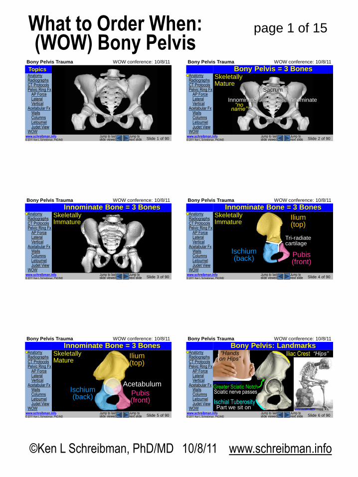
Bony Pelvis = 3 Bones
Sacrum
Innominate Innominate
Skeletally Mature
“no name”
Anatomy Radiographs CT Protocols Pelvic Ring Fx AP Force Lateral Vertical Acetabular Fx Walls Columns Letournel Judet View WOW
© 2011 Ken L Schreibman, PhD/MD www.schreibman.info
Bony Pelvis Trauma WOW conference: 10/8/11
Slide 3 of 90 Jump to next slide
Jump to last slide viewed
Bony Pelvis = 3 Bones Skeletally Immature
Innominate Bone = 3 Bones Anatomy Radiographs CT Protocols Pelvic Ring Fx AP Force Lateral Vertical Acetabular Fx Walls Columns Letournel Judet View WOW
© 2011 Ken L Schreibman, PhD/MD www.schreibman.info
Bony Pelvis Trauma WOW conference: 10/8/11
Slide 4 of 90 Jump to next slide
Jump to last slide viewed
Innominate Bone = 3 Bones Skeletally Immature
Ilium (top)
Ischium (back)
Pubis (front)
Tri-radiate cartilage
Anatomy Radiographs CT Protocols Pelvic Ring Fx AP Force Lateral Vertical Acetabular Fx Walls Columns Letournel Judet View WOW
© 2011 Ken L Schreibman, PhD/MD www.schreibman.info
Bony Pelvis Trauma WOW conference: 10/8/11
Slide 5 of 90 Jump to next slide
Jump to last slide viewed
Innominate Bone = 3 Bones Skeletally Mature
Acetabulum
Ilium (top)
Ischium (back)
Pubis (front)
Anatomy Radiographs CT Protocols Pelvic Ring Fx AP Force Lateral Vertical Acetabular Fx Walls Columns Letournel Judet View WOW
© 2011 Ken L Schreibman, PhD/MD www.schreibman.info
Bony Pelvis Trauma WOW conference: 10/8/11
Slide 6 of 90 Jump to next slide
Jump to last slide viewed
Bony Pelvis: Landmarks Iliac Crest
Ischial Tuberosity
www.sciencephoto.com
“Hips” “Hands on Hips”
Part we sit on www.zazzle.com
Greater Sciatic Notch Sciatic nerve passes
©Ken L Schreibman, PhD/MD 10/8/11 www.schreibman.info
page 2 of 15 What to Order When: (WOW) Bony Pelvis
Anatomy Radiographs CT Protocols Pelvic Ring Fx AP Force Lateral Vertical Acetabular Fx Walls Columns Letournel Judet View WOW
© 2011 Ken L Schreibman, PhD/MD www.schreibman.info
Bony Pelvis Trauma WOW conference: 10/8/11
Slide 7 of 90 Jump to next slide
Jump to last slide viewed
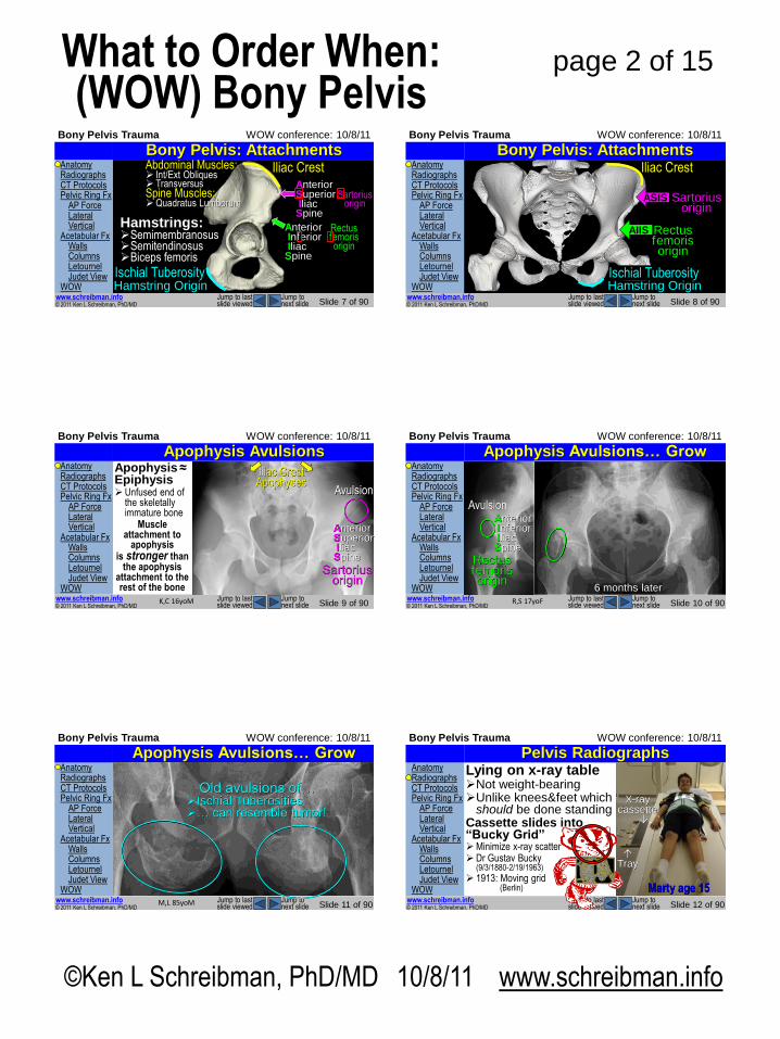
Bony Pelvis: Attachments Iliac Crest
Ischial Tuberosity Hamstring Origin
Hamstrings: Semimembranosus Semitendinosus Biceps femoris
Anterior Superior Iliac Spine
Anterior Inferior Iliac Spine
Sartorius origin
Rectus femoris origin
Abdominal Muscles: Int/Ext Obliques Transversus Spine Muscles: Quadratus Lumborum
Anatomy Radiographs CT Protocols Pelvic Ring Fx AP Force Lateral Vertical Acetabular Fx Walls Columns Letournel Judet View WOW
© 2011 Ken L Schreibman, PhD/MD www.schreibman.info
Bony Pelvis Trauma WOW conference: 10/8/11
Slide 8 of 90 Jump to next slide
Jump to last slide viewed
Bony Pelvis: Attachments Iliac Crest
ASIS
AIIS
Sartorius origin
Rectus femoris origin
Ischial Tuberosity Hamstring Origin
Anatomy Radiographs CT Protocols Pelvic Ring Fx AP Force Lateral Vertical Acetabular Fx Walls Columns Letournel Judet View WOW
© 2011 Ken L Schreibman, PhD/MD www.schreibman.info
Bony Pelvis Trauma WOW conference: 10/8/11
Slide 9 of 90 Jump to next slide
Jump to last slide viewed
Apophysis Avulsions Apophysis ≈ Epiphysis Unfused end of
the skeletally immature bone
Muscle attachment to
apophysis is stronger than
the apophysis attachment to the rest of the bone
K,C 16yoM
Iliac Crest Apophyses
Anterior Superior Iliac Spine
Sartorius origin
Avulsion
Anatomy Radiographs CT Protocols Pelvic Ring Fx AP Force Lateral Vertical Acetabular Fx Walls Columns Letournel Judet View WOW
© 2011 Ken L Schreibman, PhD/MD www.schreibman.info
Bony Pelvis Trauma WOW conference: 10/8/11
Slide 10 of 90 Jump to next slide
Jump to last slide viewed
Apophysis Avulsions… Grow
R,S 17yoF
6 months later
Anterior Inferior Iliac Spine
Rectus femoris origin
Avulsion
Anatomy Radiographs CT Protocols Pelvic Ring Fx AP Force Lateral Vertical Acetabular Fx Walls Columns Letournel Judet View WOW
© 2011 Ken L Schreibman, PhD/MD www.schreibman.info
Bony Pelvis Trauma WOW conference: 10/8/11
Slide 11 of 90 Jump to next slide
Jump to last slide viewed
Apophysis Avulsions… Grow
B,J 15yoM
Avulsions of… Ischial Tuberosities Hamstring Origin
M,L 85yoM
Old avulsions of… Ischial Tuberosities … can resemble tumor!
Anatomy Radiographs CT Protocols Pelvic Ring Fx AP Force Lateral Vertical Acetabular Fx Walls Columns Letournel Judet View WOW
© 2011 Ken L Schreibman, PhD/MD www.schreibman.info
Bony Pelvis Trauma WOW conference: 10/8/11
Slide 12 of 90 Jump to next slide
Jump to last slide viewed
Pelvis Radiographs Lying on x-ray table Not weight-bearing Unlike knees & feet which
should be done standing Cassette slides into “Bucky Grid” Minimize x-ray scatter Dr Gustav Bucky
(9/3/1880-2/19/1963)
1913: Moving grid (Berlin)
X-ray cassette
Tray
Marty age 15
©Ken L Schreibman, PhD/MD 10/8/11 www.schreibman.info
page 3 of 15 What to Order When: (WOW) Bony Pelvis
Anatomy Radiographs CT Protocols Pelvic Ring Fx AP Force Lateral Vertical Acetabular Fx Walls Columns Letournel Judet View WOW
© 2011 Ken L Schreibman, PhD/MD www.schreibman.info
Bony Pelvis Trauma WOW conference: 10/8/11
Slide 13 of 90 Jump to next slide
Jump to last slide viewed
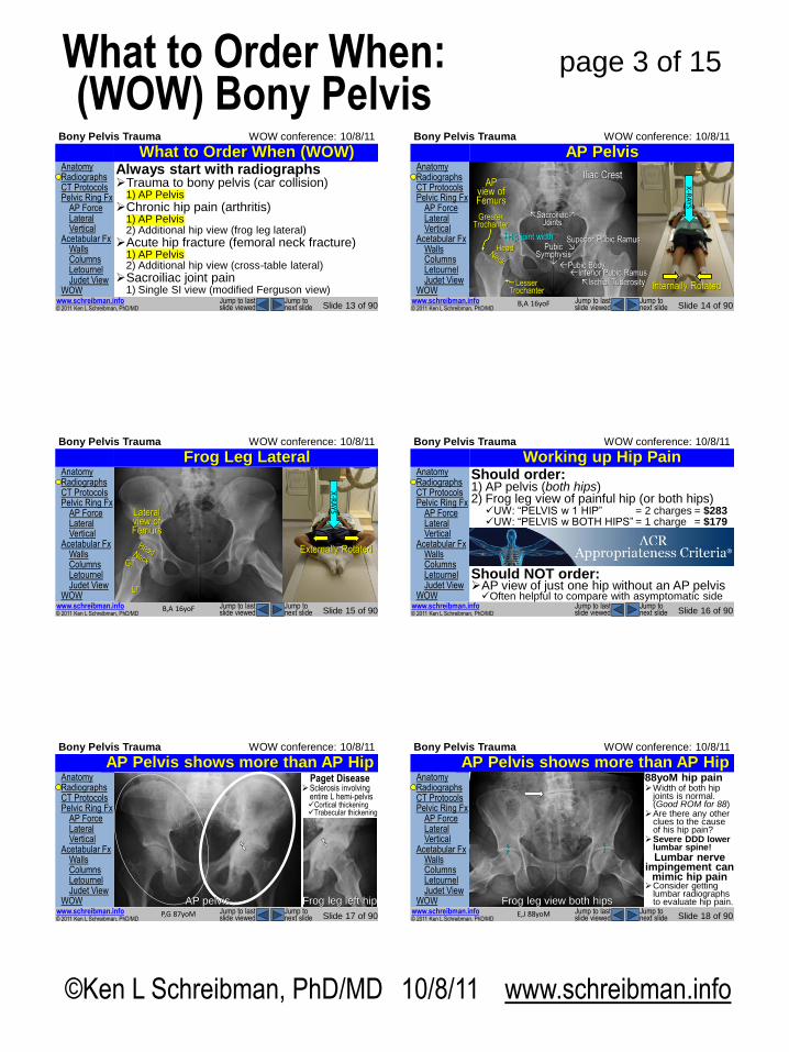
Always start with radiographs Trauma to bony pelvis (car collision)
1) AP Pelvis
Chronic hip pain (arthritis) 1) AP Pelvis 2) Additional hip view (frog leg lateral)
Acute hip fracture (femoral neck fracture) 1) AP Pelvis 2) Additional hip view (cross-table lateral)
Sacroiliac joint pain 1) Single SI view (modified Ferguson view)
What to Order When (WOW) Anatomy Radiographs CT Protocols Pelvic Ring Fx AP Force Lateral Vertical Acetabular Fx Walls Columns Letournel Judet View WOW
© 2011 Ken L Schreibman, PhD/MD www.schreibman.info
Bony Pelvis Trauma WOW conference: 10/8/11
Slide 14 of 90 Jump to next slide
Jump to last slide viewed
AP Pelvis
X-R
AYS
B,A 16yoF
Internally Rotated
Head
Greater Trochanter
Lesser Trochanter
Iliac Crest
Ischial Tuberosity
Pubic Body
Superior Pubic Ramus
Inferior Pubic Ramus
Pubic Symphysis
Sacroiliac Joints
AP view of Femurs
Hip joint width
Anatomy Radiographs CT Protocols Pelvic Ring Fx AP Force Lateral Vertical Acetabular Fx Walls Columns Letournel Judet View WOW
© 2011 Ken L Schreibman, PhD/MD www.schreibman.info
Bony Pelvis Trauma WOW conference: 10/8/11
Slide 15 of 90 Jump to next slide
Jump to last slide viewed
Frog Leg Lateral X
-RA
YS
B,A 16yoF
Externally Rotated
Lateral view of Femurs
GT
LT
Anatomy Radiographs CT Protocols Pelvic Ring Fx AP Force Lateral Vertical Acetabular Fx Walls Columns Letournel Judet View WOW
© 2011 Ken L Schreibman, PhD/MD www.schreibman.info
Bony Pelvis Trauma WOW conference: 10/8/11
Slide 16 of 90 Jump to next slide
Jump to last slide viewed
Working up Hip Pain Should order: 1) AP pelvis (both hips) 2) Frog leg view of painful hip (or both hips) UW: “PELVIS w 1 HIP” = 2 charges = $283 UW: “PELVIS w BOTH HIPS” = 1 charge = $179
Should NOT order: AP view of just one hip without an AP pelvis Often helpful to compare with asymptomatic side
Anatomy Radiographs CT Protocols Pelvic Ring Fx AP Force Lateral Vertical Acetabular Fx Walls Columns Letournel Judet View WOW
© 2011 Ken L Schreibman, PhD/MD www.schreibman.info
Bony Pelvis Trauma WOW conference: 10/8/11
Slide 17 of 90 Jump to next slide
Jump to last slide viewed
AP Pelvis shows more than AP Hip 87 yo male, left hip pain Severe joint space narrowing Anything more going on?
P,G 87yoM
AP left hip Frog leg left hip
Paget Disease Sclerosis involving
entire L hemi-pelvis Cortical thickening Trabecular thickening
AP pelvis
Anatomy Radiographs CT Protocols Pelvic Ring Fx AP Force Lateral Vertical Acetabular Fx Walls Columns Letournel Judet View WOW
© 2011 Ken L Schreibman, PhD/MD www.schreibman.info
Bony Pelvis Trauma WOW conference: 10/8/11
Slide 18 of 90 Jump to next slide
Jump to last slide viewed
AP Pelvis shows more than AP Hip 88yoM hip pain Width of both hip
joints is normal. (Good ROM for 88) Are there any other
clues to the cause of his hip pain?
Severe DDD lower lumbar spine!
Lumbar nerve impingement can
mimic hip pain Consider getting
lumbar radiographs to evaluate hip pain.
E,J 88yoM
AP pelvis Frog leg view both hips
©Ken L Schreibman, PhD/MD 10/8/11 www.schreibman.info
page 4 of 15 What to Order When: (WOW) Bony Pelvis
Anatomy Radiographs CT Protocols Pelvic Ring Fx AP Force Lateral Vertical Acetabular Fx Walls Columns Letournel Judet View WOW
© 2011 Ken L Schreibman, PhD/MD www.schreibman.info
Bony Pelvis Trauma WOW conference: 10/8/11
Slide 19 of 90 Jump to next slide
Jump to last slide viewed
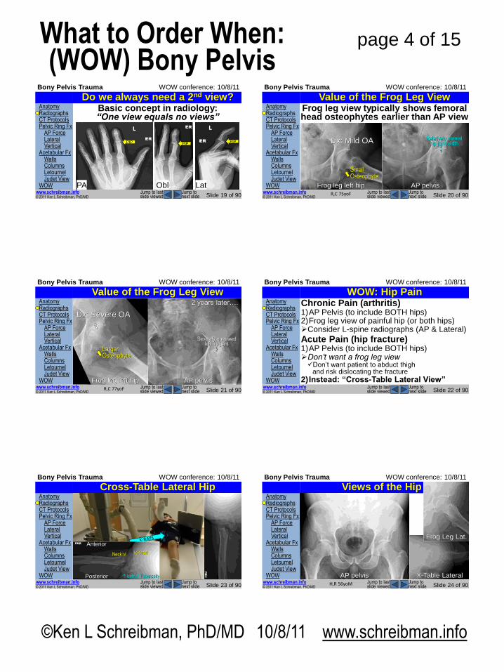
Do we always need a 2nd view?
PA
PIP
Obl
PIP
Lat
PIP
Basic concept in radiology: “One view equals no views”
Anatomy Radiographs CT Protocols Pelvic Ring Fx AP Force Lateral Vertical Acetabular Fx Walls Columns Letournel Judet View WOW
© 2011 Ken L Schreibman, PhD/MD www.schreibman.info
Bony Pelvis Trauma WOW conference: 10/8/11
Slide 20 of 90 Jump to next slide
Jump to last slide viewed
75 yo F L hip pain
Value of the Frog Leg View
R,C 75yoF
AP pelvis Frog leg left hip
Small Osteophyte
Frog leg view typically shows femoral head osteophytes earlier than AP view
Relatively normal hip joint width Dx: Mild OA
Anatomy Radiographs CT Protocols Pelvic Ring Fx AP Force Lateral Vertical Acetabular Fx Walls Columns Letournel Judet View WOW
© 2011 Ken L Schreibman, PhD/MD www.schreibman.info
Bony Pelvis Trauma WOW conference: 10/8/11
Slide 21 of 90 Jump to next slide
Jump to last slide viewed
Value of the Frog Leg View
R,C 77yoF
AP pelvis
2 years later….
Frog leg left hip
Dx: Severe OA
Severely narrowed left hip joint
Larger Osteophyte
Anatomy Radiographs CT Protocols Pelvic Ring Fx AP Force Lateral Vertical Acetabular Fx Walls Columns Letournel Judet View WOW
© 2011 Ken L Schreibman, PhD/MD www.schreibman.info
Bony Pelvis Trauma WOW conference: 10/8/11
Slide 22 of 90 Jump to next slide
Jump to last slide viewed
WOW: Hip Pain Chronic Pain (arthritis) 1)AP Pelvis (to include BOTH hips) 2)Frog leg view of painful hip (or both hips) Consider L-spine radiographs (AP & Lateral)
Acute Pain (hip fracture) 1)AP Pelvis (to include BOTH hips) Don’t want a frog leg view Don’t want patient to abduct thigh
and risk dislocating the fracture
2) Instead: “Cross-Table Lateral View”
Anatomy Radiographs CT Protocols Pelvic Ring Fx AP Force Lateral Vertical Acetabular Fx Walls Columns Letournel Judet View WOW
© 2011 Ken L Schreibman, PhD/MD www.schreibman.info
Bony Pelvis Trauma WOW conference: 10/8/11
Slide 23 of 90 Jump to next slide
Jump to last slide viewed
Cross-Table Lateral Hip
Anterior
Posterior Ischial Tuberosity
Head Neck
Anatomy Radiographs CT Protocols Pelvic Ring Fx AP Force Lateral Vertical Acetabular Fx Walls Columns Letournel Judet View WOW
© 2011 Ken L Schreibman, PhD/MD www.schreibman.info
Bony Pelvis Trauma WOW conference: 10/8/11
Slide 24 of 90 Jump to next slide
Jump to last slide viewed
Views of the Hip
H,R 56yoM
X-Table Lateral
Frog Leg Lat.
AP pelvis
©Ken L Schreibman, PhD/MD 10/8/11 www.schreibman.info
page 5 of 15 What to Order When: (WOW) Bony Pelvis
Anatomy Radiographs CT Protocols Pelvic Ring Fx AP Force Lateral Vertical Acetabular Fx Walls Columns Letournel Judet View WOW
© 2011 Ken L Schreibman, PhD/MD www.schreibman.info
Bony Pelvis Trauma WOW conference: 10/8/11
Slide 25 of 90 Jump to next slide
Jump to last slide viewed
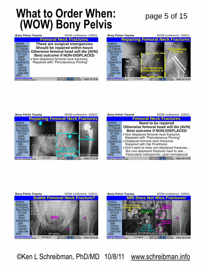
Femoral Neck Fractures These are surgical emergencies Should be repaired within hours
Otherwise femoral head will die (AVN) Best outcome if NON-DISPLACED
Non-displaced femoral neck fractures: Repaired with “Percutaneous Pinning”
Anatomy Radiographs CT Protocols Pelvic Ring Fx AP Force Lateral Vertical Acetabular Fx Walls Columns Letournel Judet View WOW
© 2011 Ken L Schreibman, PhD/MD www.schreibman.info
Bony Pelvis Trauma WOW conference: 10/8/11
Slide 26 of 90 Jump to next slide
Jump to last slide viewed
Repairing Femoral Neck Fractures
C,T 63yoM
AP pelvis
Non-displaced Femoral neck fx
Normal side
X-Table Lat Left
Percutaneous Pinning
Post Operative
Anatomy Radiographs CT Protocols Pelvic Ring Fx AP Force Lateral Vertical Acetabular Fx Walls Columns Letournel Judet View WOW
© 2011 Ken L Schreibman, PhD/MD www.schreibman.info
Bony Pelvis Trauma WOW conference: 10/8/11
Slide 27 of 90 Jump to next slide
Jump to last slide viewed
Repairing Femoral Neck Fractures
C,G 71yoF
AP pelvis
X-Table Lateral Left Hip
DISPLACED Femoral neck fx
Post Operative
Hip Prosthesis
Anatomy Radiographs CT Protocols Pelvic Ring Fx AP Force Lateral Vertical Acetabular Fx Walls Columns Letournel Judet View WOW
© 2011 Ken L Schreibman, PhD/MD www.schreibman.info
Bony Pelvis Trauma WOW conference: 10/8/11
Slide 28 of 90 Jump to next slide
Jump to last slide viewed
Femoral Neck Fractures Need to be repaired
Otherwise femoral head will die (AVN) Best outcome if NON-DISPLACED
Non-displaced femoral neck fractures: Repaired with “Percutaneous Pinning” Displaced femoral neck fractures:
Repaired with Hip Prosthesis Don’t want to miss non-displaced fractures… But non-displaced fractures hard to see… Particularly osteoporotic, post-menopausal
Anatomy Radiographs CT Protocols Pelvic Ring Fx AP Force Lateral Vertical Acetabular Fx Walls Columns Letournel Judet View WOW
© 2011 Ken L Schreibman, PhD/MD www.schreibman.info
Bony Pelvis Trauma WOW conference: 10/8/11
Slide 29 of 90 Jump to next slide
Jump to last slide viewed
Subtle Femoral Neck Fracture?
P,M 68yoF
AP pelvis Frog leg L Hip
68 yo F, chronic L hip pain Both views: Negative 68 yo F, 9 months later Fell on L hip, has new acute pain Walks into clinic a few days later… Surgeon: Not convinced How can we prove it?
MRI!
AP pelvis AP L Hip
Anatomy Radiographs CT Protocols Pelvic Ring Fx AP Force Lateral Vertical Acetabular Fx Walls Columns Letournel Judet View WOW
© 2011 Ken L Schreibman, PhD/MD www.schreibman.info
Bony Pelvis Trauma WOW conference: 10/8/11
Slide 30 of 90 Jump to next slide
Jump to last slide viewed
MRI Does Not Miss Fractures!
P,M 68yoF
AP L Hip
Coronal T1 (Fat is bright) Normal fatty
yellow marrow Marrow Edema
Coronal STIR (Fluid is bright)
Marrow Edema
High-Res T2-FatSat
(Fluid is bright)
Black line
Neck Fx!
©Ken L Schreibman, PhD/MD 10/8/11 www.schreibman.info
page 6 of 15 What to Order When: (WOW) Bony Pelvis
Anatomy Radiographs CT Protocols Pelvic Ring Fx AP Force Lateral Vertical Acetabular Fx Walls Columns Letournel Judet View WOW
© 2011 Ken L Schreibman, PhD/MD www.schreibman.info
Bony Pelvis Trauma WOW conference: 10/8/11
Slide 31 of 90 Jump to next slide
Jump to last slide viewed
AP L Hip
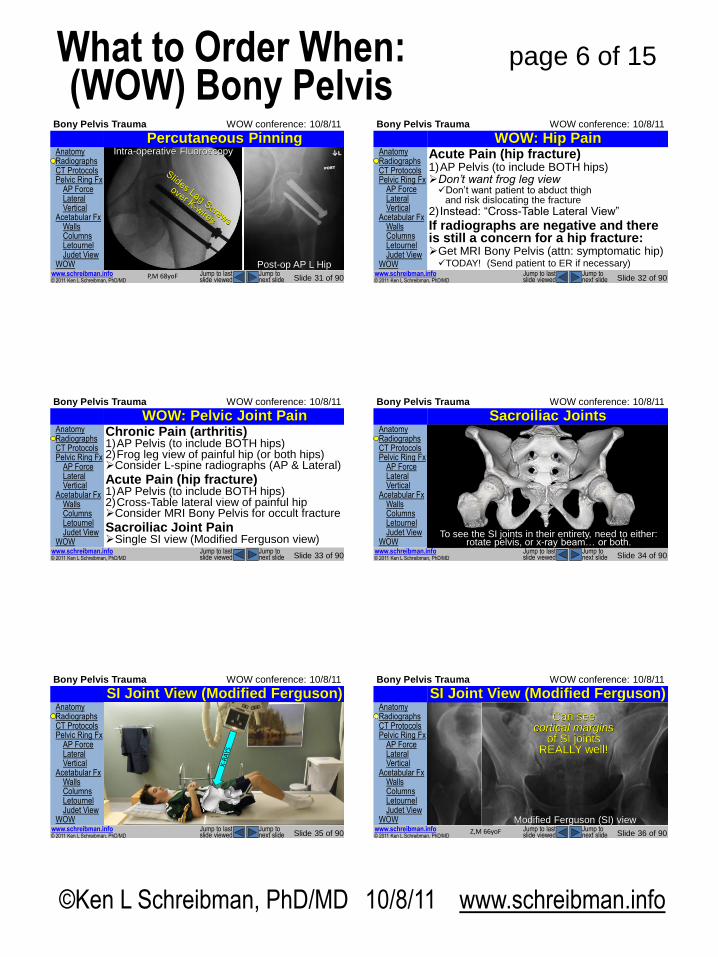
Percutaneous Pinning
P,M 68yoF
Intra-operative Fluoroscopy
Post-op AP L Hip
Anatomy Radiographs CT Protocols Pelvic Ring Fx AP Force Lateral Vertical Acetabular Fx Walls Columns Letournel Judet View WOW
© 2011 Ken L Schreibman, PhD/MD www.schreibman.info
Bony Pelvis Trauma WOW conference: 10/8/11
Slide 32 of 90 Jump to next slide
Jump to last slide viewed
WOW: Hip Pain Acute Pain (hip fracture) 1)AP Pelvis (to include BOTH hips) Don’t want frog leg view Don’t want patient to abduct thigh
and risk dislocating the fracture
2) Instead: “Cross-Table Lateral View”
If radiographs are negative and there is still a concern for a hip fracture: Get MRI Bony Pelvis (attn: symptomatic hip) TODAY! (Send patient to ER if necessary)
Anatomy Radiographs CT Protocols Pelvic Ring Fx AP Force Lateral Vertical Acetabular Fx Walls Columns Letournel Judet View WOW
© 2011 Ken L Schreibman, PhD/MD www.schreibman.info
Bony Pelvis Trauma WOW conference: 10/8/11
Slide 33 of 90 Jump to next slide
Jump to last slide viewed
WOW: Pelvic Joint Pain Chronic Pain (arthritis) 1)AP Pelvis (to include BOTH hips) 2)Frog leg view of painful hip (or both hips) Consider L-spine radiographs (AP & Lateral)
Acute Pain (hip fracture) 1)AP Pelvis (to include BOTH hips) 2)Cross-Table lateral view of painful hip Consider MRI Bony Pelvis for occult fracture
Sacroiliac Joint Pain Single SI view (Modified Ferguson view)
Anatomy Radiographs CT Protocols Pelvic Ring Fx AP Force Lateral Vertical Acetabular Fx Walls Columns Letournel Judet View WOW
© 2011 Ken L Schreibman, PhD/MD www.schreibman.info
Bony Pelvis Trauma WOW conference: 10/8/11
Slide 34 of 90 Jump to next slide
Jump to last slide viewed
Sacroiliac Joints
AP pelvis
Sacroiliac Joints
Slope Posteriorly
To see the SI joints in their entirety, need to either: rotate pelvis, or x-ray beam… or both.
Anatomy Radiographs CT Protocols Pelvic Ring Fx AP Force Lateral Vertical Acetabular Fx Walls Columns Letournel Judet View WOW
© 2011 Ken L Schreibman, PhD/MD www.schreibman.info
Bony Pelvis Trauma WOW conference: 10/8/11
Slide 35 of 90 Jump to next slide
Jump to last slide viewed
SI Joint View (Modified Ferguson) Anatomy Radiographs CT Protocols Pelvic Ring Fx AP Force Lateral Vertical Acetabular Fx Walls Columns Letournel Judet View WOW
© 2011 Ken L Schreibman, PhD/MD www.schreibman.info
Bony Pelvis Trauma WOW conference: 10/8/11
Slide 36 of 90 Jump to next slide
Jump to last slide viewed
SI Joint View (Modified Ferguson)
m
Z,M 66yoF
AP pelvis
Can see SI joints
pretty well…
Modified Ferguson (SI) view
Can see cortical margins
of SI joints REALLY well!
©Ken L Schreibman, PhD/MD 10/8/11 www.schreibman.info
page 7 of 15 What to Order When: (WOW) Bony Pelvis
Anatomy Radiographs CT Protocols Pelvic Ring Fx AP Force Lateral Vertical Acetabular Fx Walls Columns Letournel Judet View WOW
© 2011 Ken L Schreibman, PhD/MD www.schreibman.info
Bony Pelvis Trauma WOW conference: 10/8/11
Slide 37 of 90 Jump to next slide
Jump to last slide viewed
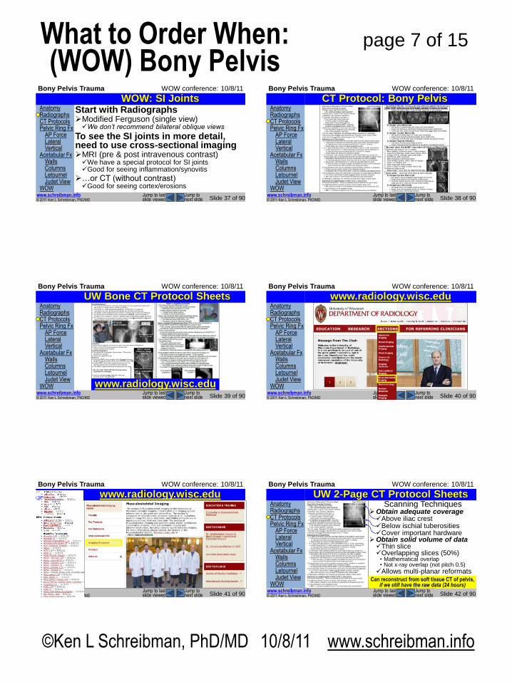
WOW: SI Joints Start with Radiographs Modified Ferguson (single view) We don’t recommend bilateral oblique views
To see the SI joints in more detail, need to use cross-sectional imaging MRI (pre & post intravenous contrast) We have a special protocol for SI joints Good for seeing inflammation/synovitis
…or CT (without contrast) Good for seeing cortex/erosions
Anatomy Radiographs CT Protocols Pelvic Ring Fx AP Force Lateral Vertical Acetabular Fx Walls Columns Letournel Judet View WOW
© 2011 Ken L Schreibman, PhD/MD www.schreibman.info
Bony Pelvis Trauma WOW conference: 10/8/11
Slide 38 of 90 Jump to next slide
Jump to last slide viewed
CT Protocol: Bony Pelvis
Anatomy Radiographs CT Protocols Pelvic Ring Fx AP Force Lateral Vertical Acetabular Fx Walls Columns Letournel Judet View WOW
© 2011 Ken L Schreibman, PhD/MD www.schreibman.info
Bony Pelvis Trauma WOW conference: 10/8/11
Slide 39 of 90 Jump to next slide
Jump to last slide viewed
UW Bone CT Protocol Sheets
www.radiology.wisc.edu
Anatomy Radiographs CT Protocols Pelvic Ring Fx AP Force Lateral Vertical Acetabular Fx Walls Columns Letournel Judet View WOW
© 2011 Ken L Schreibman, PhD/MD www.schreibman.info
Bony Pelvis Trauma WOW conference: 10/8/11
Slide 40 of 90 Jump to next slide
Jump to last slide viewed
www.radiology.wisc.edu
Anatomy Radiographs CT Protocols Pelvic Ring Fx AP Force Lateral Vertical Acetabular Fx Walls Columns Letournel Judet View WOW
© 2011 Ken L Schreibman, PhD/MD www.schreibman.info
Bony Pelvis Trauma WOW conference: 10/8/11
Slide 41 of 90 Jump to next slide
Jump to last slide viewed
www.radiology.wisc.edu
Anatomy Radiographs CT Protocols Pelvic Ring Fx AP Force Lateral Vertical Acetabular Fx Walls Columns Letournel Judet View WOW
© 2011 Ken L Schreibman, PhD/MD www.schreibman.info
Bony Pelvis Trauma WOW conference: 10/8/11
Slide 42 of 90 Jump to next slide
Jump to last slide viewed
UW 2-Page CT Protocol Sheets Scanning Techniques
Obtain adequate coverage Above iliac crest Below ischial tuberosities Cover important hardware
Obtain solid volume of data Thin slice Overlapping slices (50%)
• Mathematical overlap • Not x-ray overlap (not pitch 0.5)
Allows multi-planar reformats Can reconstruct from soft tissue CT of pelvis,
if we still have the raw data (24 hours)
©Ken L Schreibman, PhD/MD 10/8/11 www.schreibman.info
page 8 of 15 What to Order When: (WOW) Bony Pelvis
Anatomy Radiographs CT Protocols Pelvic Ring Fx AP Force Lateral Vertical Acetabular Fx Walls Columns Letournel Judet View WOW
© 2011 Ken L Schreibman, PhD/MD www.schreibman.info
Bony Pelvis Trauma WOW conference: 10/8/11
Slide 43 of 90 Jump to next slide
Jump to last slide viewed
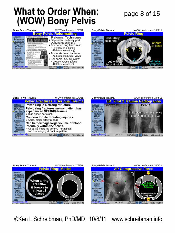
Bony Pelvis Reformatting Reformat Techniques
Depend upon body part Depend upon injury For pelvic ring fractures: Reformat in 3 planes
(Relative to anatomy)
For acetabular fractures: Add simulated Judet views
For sacral fxs, SI joints: Oblique coronal & axial
(Relative to sacrum)
Anatomy Radiographs CT Protocols Pelvic Ring Fx AP Force Lateral Vertical Acetabular Fx Walls Columns Letournel Judet View WOW
© 2011 Ken L Schreibman, PhD/MD www.schreibman.info
Bony Pelvis Trauma WOW conference: 10/8/11
Slide 44 of 90 Jump to next slide
Jump to last slide viewed
Pelvic Ring Structurally, solid ring…
…but with discontinuities
at both SI joints
at pubic symphysis
Anatomy Radiographs CT Protocols Pelvic Ring Fx AP Force Lateral Vertical Acetabular Fx Walls Columns Letournel Judet View WOW
© 2011 Ken L Schreibman, PhD/MD www.schreibman.info
Bony Pelvis Trauma WOW conference: 10/8/11
Slide 45 of 90 Jump to next slide
Jump to last slide viewed
Pelvic Fractures = Serious Trauma Pelvic ring is a strong structure. Pelvic ring fractures means patient has experienced SERIOUS trauma. High speed car crash
Concern for life threating injuries. Aorta, major artery rupture
Can hemorrhage large volume of blood internally within the pelvis. All pelvic fractures go to CT to assess
soft tissue injury & fracture pattern.
Anatomy Radiographs CT Protocols Pelvic Ring Fx AP Force Lateral Vertical Acetabular Fx Walls Columns Letournel Judet View WOW
© 2011 Ken L Schreibman, PhD/MD www.schreibman.info
Bony Pelvis Trauma WOW conference: 10/8/11
Slide 46 of 90 Jump to next slide
Jump to last slide viewed
ER: First 2 Trauma Radiographs Chest Pelvis
This patient did not survive
long enough to go to CT
S,J 36yoM
Anatomy Radiographs CT Protocols Pelvic Ring Fx AP Force Lateral Vertical Acetabular Fx Walls Columns Letournel Judet View WOW
© 2011 Ken L Schreibman, PhD/MD www.schreibman.info
Bony Pelvis Trauma WOW conference: 10/8/11
Slide 47 of 90 Jump to next slide
Jump to last slide viewed
Pelvic Ring: Model
Anterior
Posterior
Anterior
Posterior
When a ring breaks…
it breaks in at least 2
places
Anatomy Radiographs CT Protocols Pelvic Ring Fx AP Force Lateral Vertical Acetabular Fx Walls Columns Letournel Judet View WOW
© 2011 Ken L Schreibman, PhD/MD www.schreibman.info
Bony Pelvis Trauma WOW conference: 10/8/11
Slide 48 of 90 Jump to next slide
Jump to last slide viewed
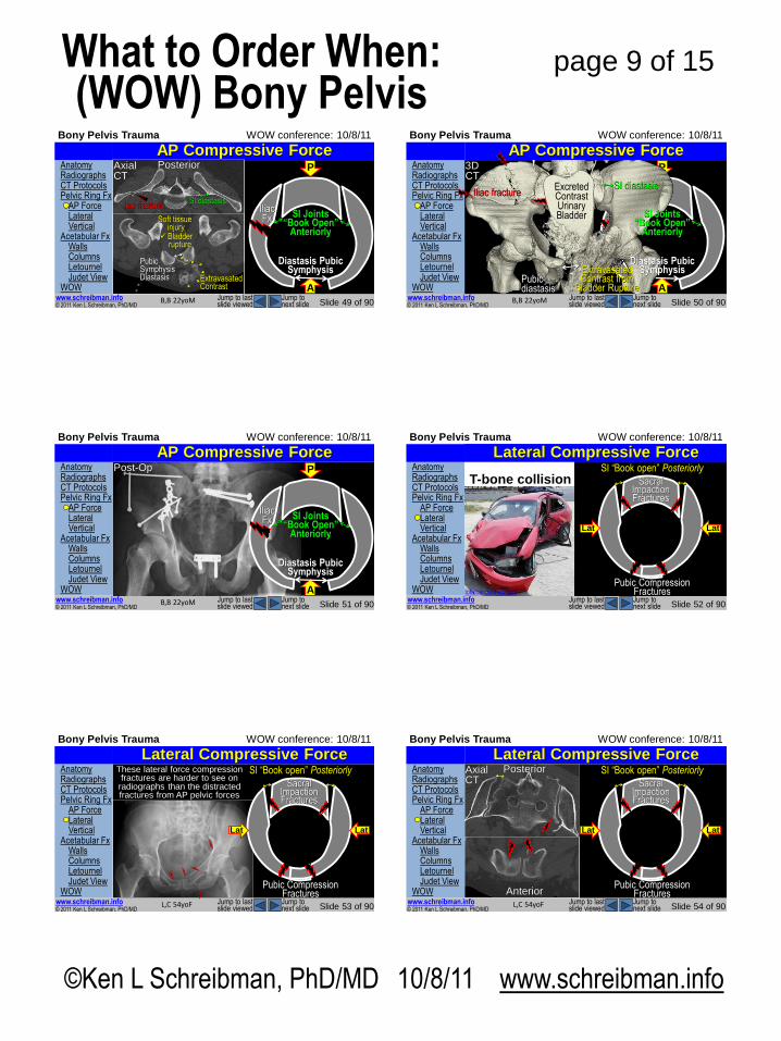
Understanding pelvic ring fracture patterns, caused by different types of external forces, helps us detect the second (and third) pelvic fractures.
www.youtube.com
Pelvic Ring Fracture Patterns AP Compressive Force P
A
P
A B,B 22yoM
Diastasis Pubic Symphysis
Front end collision
www.evworld.com
SI Joints “Book Open”
Anteriorly
Iliac Fx
©Ken L Schreibman, PhD/MD 10/8/11 www.schreibman.info
page 9 of 15 What to Order When: (WOW) Bony Pelvis
Anatomy Radiographs CT Protocols Pelvic Ring Fx AP Force Lateral Vertical Acetabular Fx Walls Columns Letournel Judet View WOW
© 2011 Ken L Schreibman, PhD/MD www.schreibman.info
Bony Pelvis Trauma WOW conference: 10/8/11
Slide 49 of 90 Jump to next slide
Jump to last slide viewed
AP Compressive Force P
A
Diastasis Pubic Symphysis
SI Joints “Book Open”
Anteriorly
Iliac Fx
B,B 22yoM
Axial CT
Posterior
Anterior
SI diastasis Iliac fracture
Pubic Symphysis Diastasis Extravasated
Contrast
Soft tissue injury
Bladder rupture
Anatomy Radiographs CT Protocols Pelvic Ring Fx AP Force Lateral Vertical Acetabular Fx Walls Columns Letournel Judet View WOW
© 2011 Ken L Schreibman, PhD/MD www.schreibman.info
Bony Pelvis Trauma WOW conference: 10/8/11
Slide 50 of 90 Jump to next slide
Jump to last slide viewed
AP Compressive Force P
A
Iliac Fx
B,B 22yoM
SI Joints “Book Open”
Anteriorly
3D CT
Pubic diastasis
SI diastasis Iliac fracture
Diastasis Pubic Symphysis Extravasated
Contrast from Bladder Rupture
Excreted Contrast Urinary Bladder
Anatomy Radiographs CT Protocols Pelvic Ring Fx AP Force Lateral Vertical Acetabular Fx Walls Columns Letournel Judet View WOW
© 2011 Ken L Schreibman, PhD/MD www.schreibman.info
Bony Pelvis Trauma WOW conference: 10/8/11
Slide 51 of 90 Jump to next slide
Jump to last slide viewed
AP Compressive Force P
A
Diastasis Pubic Symphysis
SI Joints “Book Open”
Anteriorly
Iliac Fx
B,B 22yoM
Post-Op Anatomy Radiographs CT Protocols Pelvic Ring Fx AP Force Lateral Vertical Acetabular Fx Walls Columns Letournel Judet View WOW
© 2011 Ken L Schreibman, PhD/MD www.schreibman.info
Bony Pelvis Trauma WOW conference: 10/8/11
Slide 52 of 90 Jump to next slide
Jump to last slide viewed
Pelvic Ring Fracture Patterns Lateral Compressive Force Understanding
pelvic ring fracture patterns,
caused by different types of external forces, helps us detect the second (and
third) pelvic fractures.
Lat Lat
T-bone collision
Lat Lat
Pubic Compression Fractures
Sacral Impaction Fractures
SI “Book open” Posteriorly
“Surrogates” (2009) Bruce Willis totalcarcrashes.com
Anatomy Radiographs CT Protocols Pelvic Ring Fx AP Force Lateral Vertical Acetabular Fx Walls Columns Letournel Judet View WOW
© 2011 Ken L Schreibman, PhD/MD www.schreibman.info
Bony Pelvis Trauma WOW conference: 10/8/11
Slide 53 of 90 Jump to next slide
Jump to last slide viewed
Lateral Compressive Force
L,C 54yoF
Lat
Pubic Compression Fractures
Sacral Impaction Fractures
SI “Book open” Posteriorly These lateral force compression fractures are harder to see on
radiographs than the distracted fractures from AP pelvic forces
Lat
Anatomy Radiographs CT Protocols Pelvic Ring Fx AP Force Lateral Vertical Acetabular Fx Walls Columns Letournel Judet View WOW
© 2011 Ken L Schreibman, PhD/MD www.schreibman.info
Bony Pelvis Trauma WOW conference: 10/8/11
Slide 54 of 90 Jump to next slide
Jump to last slide viewed
Lateral Compressive Force
L,C 54yoF
Lat
Pubic Compression Fractures
Sacral Impaction Fractures
SI “Book open” Posteriorly Axial CT
Posterior
Lat
Lateral Compression Urinary Bladder
Anterior
©Ken L Schreibman, PhD/MD 10/8/11 www.schreibman.info
page 10 of 15 What to Order When: (WOW) Bony Pelvis
Anatomy Radiographs CT Protocols Pelvic Ring Fx AP Force Lateral Vertical Acetabular Fx Walls Columns Letournel Judet View WOW
© 2011 Ken L Schreibman, PhD/MD www.schreibman.info
Bony Pelvis Trauma WOW conference: 10/8/11
Slide 55 of 90 Jump to next slide
Jump to last slide viewed
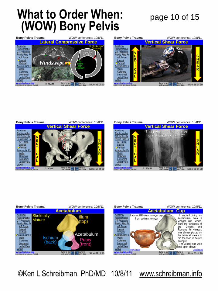
Lateral Compressive Force
Lat Lat
F,S 24yoM
SI “Books open”
Anteriorly
Windswept
SI “Books open” Posteriorly
Sacral Impaction Fracture
Anatomy Radiographs CT Protocols Pelvic Ring Fx AP Force Lateral Vertical Acetabular Fx Walls Columns Letournel Judet View WOW
© 2011 Ken L Schreibman, PhD/MD www.schreibman.info
Bony Pelvis Trauma WOW conference: 10/8/11
Slide 56 of 90 Jump to next slide
Jump to last slide viewed
Pelvic Ring Fracture Patterns Vertical Shear Force
S H E A R
V E R T I CA L
www.youtube.com
Anatomy Radiographs CT Protocols Pelvic Ring Fx AP Force Lateral Vertical Acetabular Fx Walls Columns Letournel Judet View WOW
© 2011 Ken L Schreibman, PhD/MD www.schreibman.info
Bony Pelvis Trauma WOW conference: 10/8/11
Slide 57 of 90 Jump to next slide
Jump to last slide viewed
Vertical Shear Force
S H E A R
V E R T I CA L
S,J 47yoF
Anatomy Radiographs CT Protocols Pelvic Ring Fx AP Force Lateral Vertical Acetabular Fx Walls Columns Letournel Judet View WOW
© 2011 Ken L Schreibman, PhD/MD www.schreibman.info
Bony Pelvis Trauma WOW conference: 10/8/11
Slide 58 of 90 Jump to next slide
Jump to last slide viewed
Vertical Shear Force
S H E A R
V E R T I CA L
S,J 36yoM
This patient did not survive
long enough to go to CT
Anatomy Radiographs CT Protocols Pelvic Ring Fx AP Force Lateral Vertical Acetabular Fx Walls Columns Letournel Judet View WOW
© 2011 Ken L Schreibman, PhD/MD www.schreibman.info
Bony Pelvis Trauma WOW conference: 10/8/11
Slide 59 of 90 Jump to next slide
Jump to last slide viewed
Acetabulum Skeletally Mature
Acetabulum
Ilium (top)
Ischium (back)
Pubis (front)
Anatomy Radiographs CT Protocols Pelvic Ring Fx AP Force Lateral Vertical Acetabular Fx Walls Columns Letournel Judet View WOW
© 2011 Ken L Schreibman, PhD/MD www.schreibman.info
Bony Pelvis Trauma WOW conference: 10/8/11
Slide 60 of 90 Jump to next slide
Jump to last slide viewed
Acetabulum: Cup
lmtorbust.com
Latin acētābulum, vinegar cup, from acētum, vinegar
In ancient dining, an acetabulum was a vinegar cup, which, from the fondness of the Greeks and Romans for vinegar, was always placed on the table at meals to dip the food in before eating it. The vessel was wide
and open above. wikipedia.org
©Ken L Schreibman, PhD/MD 10/8/11 www.schreibman.info
page 11 of 15 What to Order When: (WOW) Bony Pelvis
Anatomy Radiographs CT Protocols Pelvic Ring Fx AP Force Lateral Vertical Acetabular Fx Walls Columns Letournel Judet View WOW
© 2011 Ken L Schreibman, PhD/MD www.schreibman.info
Bony Pelvis Trauma WOW conference: 10/8/11
Slide 61 of 90 Jump to next slide
Jump to last slide viewed
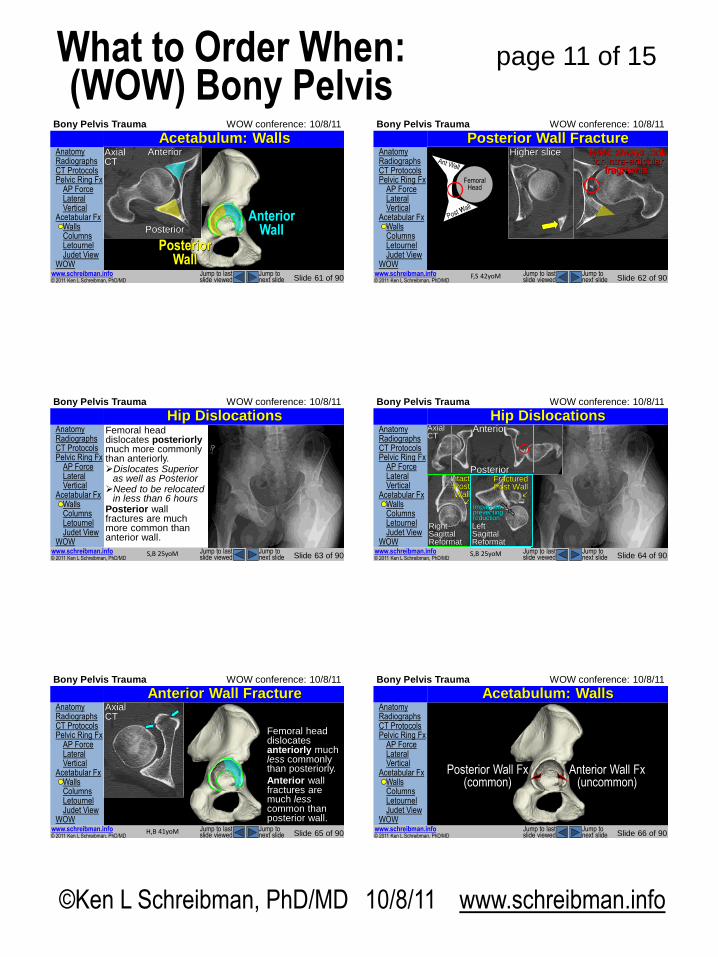
Acetabulum: Walls
Posterior Wall
Anterior Wall
Axial CT
Posterior
Anterior Anatomy Radiographs CT Protocols Pelvic Ring Fx AP Force Lateral Vertical Acetabular Fx Walls Columns Letournel Judet View WOW
© 2011 Ken L Schreibman, PhD/MD www.schreibman.info
Bony Pelvis Trauma WOW conference: 10/8/11
Slide 62 of 90 Jump to next slide
Jump to last slide viewed
Posterior Wall Fracture Axial CT
F,S 42yoM
Need always look for intra-articular
fragments!
Posterior
Anterior
Femoral Head
Higher slice
Anatomy Radiographs CT Protocols Pelvic Ring Fx AP Force Lateral Vertical Acetabular Fx Walls Columns Letournel Judet View WOW
© 2011 Ken L Schreibman, PhD/MD www.schreibman.info
Bony Pelvis Trauma WOW conference: 10/8/11
Slide 63 of 90 Jump to next slide
Jump to last slide viewed
Hip Dislocations Femoral head dislocates posteriorly much more commonly than anteriorly. Dislocates Superior
as well as Posterior Need to be relocated
in less than 6 hours Posterior wall fractures are much more common than anterior wall.
S,B 25yoM
Anatomy Radiographs CT Protocols Pelvic Ring Fx AP Force Lateral Vertical Acetabular Fx Walls Columns Letournel Judet View WOW
© 2011 Ken L Schreibman, PhD/MD www.schreibman.info
Bony Pelvis Trauma WOW conference: 10/8/11
Slide 64 of 90 Jump to next slide
Jump to last slide viewed
Hip Dislocations
S,B 25yoM
Axial CT
Left Sagittal Reformat
Right Sagittal Reformat
Intact Post Wall
Fractured Post Wall
Impaction preventing reduction
Posterior
Anterior
Anatomy Radiographs CT Protocols Pelvic Ring Fx AP Force Lateral Vertical Acetabular Fx Walls Columns Letournel Judet View WOW
© 2011 Ken L Schreibman, PhD/MD www.schreibman.info
Bony Pelvis Trauma WOW conference: 10/8/11
Slide 65 of 90 Jump to next slide
Jump to last slide viewed
Anterior Wall Fracture
Femoral head dislocates anteriorly much less commonly than posteriorly.
Anterior wall fractures are much less common than posterior wall.
H,B 41yoM
Axial CT
Anatomy Radiographs CT Protocols Pelvic Ring Fx AP Force Lateral Vertical Acetabular Fx Walls Columns Letournel Judet View WOW
© 2011 Ken L Schreibman, PhD/MD www.schreibman.info
Bony Pelvis Trauma WOW conference: 10/8/11
Slide 66 of 90 Jump to next slide
Jump to last slide viewed
Acetabulum: Walls
Anterior Wall Fx (uncommon)
Posterior Wall Fx (common)
©Ken L Schreibman, PhD/MD 10/8/11 www.schreibman.info
page 12 of 15 What to Order When: (WOW) Bony Pelvis
Anatomy Radiographs CT Protocols Pelvic Ring Fx AP Force Lateral Vertical Acetabular Fx Walls Columns Letournel Judet View WOW
© 2011 Ken L Schreibman, PhD/MD www.schreibman.info
Bony Pelvis Trauma WOW conference: 10/8/11
Slide 67 of 90 Jump to next slide
Jump to last slide viewed
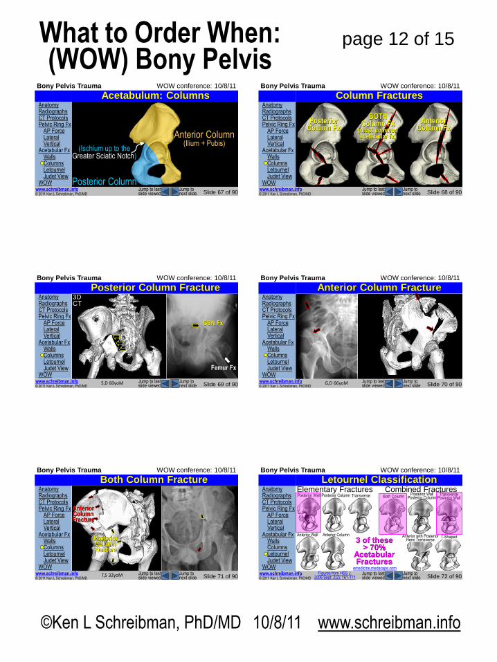
Acetabulum: Columns
Ilium (top)
Ischium (back)
Pubis (front)
Anterior Column (Ilium + Pubis)
(Ischium up to the Greater Sciatic Notch)
Posterior Column
Anatomy Radiographs CT Protocols Pelvic Ring Fx AP Force Lateral Vertical Acetabular Fx Walls Columns Letournel Judet View WOW
© 2011 Ken L Schreibman, PhD/MD www.schreibman.info
Bony Pelvis Trauma WOW conference: 10/8/11
Slide 68 of 90 Jump to next slide
Jump to last slide viewed
Column Fractures
Posterior Column Fx
Anterior Column Fx
BOTH Column Fx
(most common acetabular fx)
Anatomy Radiographs CT Protocols Pelvic Ring Fx AP Force Lateral Vertical Acetabular Fx Walls Columns Letournel Judet View WOW
© 2011 Ken L Schreibman, PhD/MD www.schreibman.info
Bony Pelvis Trauma WOW conference: 10/8/11
Slide 69 of 90 Jump to next slide
Jump to last slide viewed
Posterior Column Fracture
Posterior Column Fx
S,D 60yoM
GSN Fx
Femur Fx
3D CT
Anatomy Radiographs CT Protocols Pelvic Ring Fx AP Force Lateral Vertical Acetabular Fx Walls Columns Letournel Judet View WOW
© 2011 Ken L Schreibman, PhD/MD www.schreibman.info
Bony Pelvis Trauma WOW conference: 10/8/11
Slide 70 of 90 Jump to next slide
Jump to last slide viewed
Anterior Column Fracture
G,D 66yoM
Anatomy Radiographs CT Protocols Pelvic Ring Fx AP Force Lateral Vertical Acetabular Fx Walls Columns Letournel Judet View WOW
© 2011 Ken L Schreibman, PhD/MD www.schreibman.info
Bony Pelvis Trauma WOW conference: 10/8/11
Slide 71 of 90 Jump to next slide
Jump to last slide viewed
Both Column Fracture
T,S 32yoM
Anterior Column Fracture
GSN Fx= Posterior Column Fracture
Anterior Column Fracture
Posterior Column Fracture
Anatomy Radiographs CT Protocols Pelvic Ring Fx AP Force Lateral Vertical Acetabular Fx Walls Columns Letournel Judet View WOW
© 2011 Ken L Schreibman, PhD/MD www.schreibman.info
Bony Pelvis Trauma WOW conference: 10/8/11
Slide 72 of 90 Jump to next slide
Jump to last slide viewed
Letournel Classification Elementary Fractures Posterior Wall Posterior Column
Anterior Wall Anterior Column
Transverse Combined Fractures
Posterior Wall Posterior Column Both Column Transverse
Posterior Wall
T-Shaped Anterior with Posterior Hemi Transverse 3 of these
> 70% Acetabular Fractures
emedicine.medscape.com Figures from HSS J.
2006 Sept; 2(2): 161-171
©Ken L Schreibman, PhD/MD 10/8/11 www.schreibman.info
page 13 of 15 What to Order When: (WOW) Bony Pelvis
Anatomy Radiographs CT Protocols Pelvic Ring Fx AP Force Lateral Vertical Acetabular Fx Walls Columns Letournel Judet View WOW
© 2011 Ken L Schreibman, PhD/MD www.schreibman.info
Bony Pelvis Trauma WOW conference: 10/8/11
Slide 73 of 90 Jump to next slide
Jump to last slide viewed
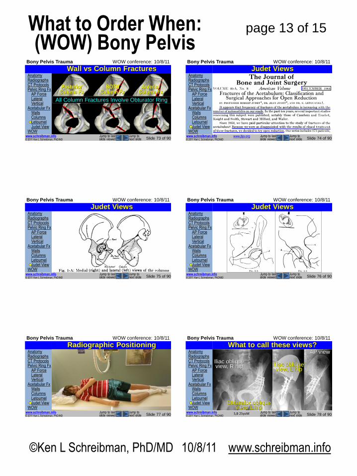
Wall vs Column Fractures
Posterior Column Fx
BOTH Column Fx
Anterior Column Fx
All Column Fractures Involve Obturator Ring
Anatomy Radiographs CT Protocols Pelvic Ring Fx AP Force Lateral Vertical Acetabular Fx Walls Columns Letournel Judet View WOW
© 2011 Ken L Schreibman, PhD/MD www.schreibman.info
Bony Pelvis Trauma WOW conference: 10/8/11
Slide 74 of 90 Jump to next slide
Jump to last slide viewed
Judet Views
www.jbjs.org
Anatomy Radiographs CT Protocols Pelvic Ring Fx AP Force Lateral Vertical Acetabular Fx Walls Columns Letournel Judet View WOW
© 2011 Ken L Schreibman, PhD/MD www.schreibman.info
Bony Pelvis Trauma WOW conference: 10/8/11
Slide 75 of 90 Jump to next slide
Jump to last slide viewed
Judet Views Anatomy Radiographs CT Protocols Pelvic Ring Fx AP Force Lateral Vertical Acetabular Fx Walls Columns Letournel Judet View WOW
© 2011 Ken L Schreibman, PhD/MD www.schreibman.info
Bony Pelvis Trauma WOW conference: 10/8/11
Slide 76 of 90 Jump to next slide
Jump to last slide viewed
Judet Views
Anatomy Radiographs CT Protocols Pelvic Ring Fx AP Force Lateral Vertical Acetabular Fx Walls Columns Letournel Judet View WOW
© 2011 Ken L Schreibman, PhD/MD www.schreibman.info
Bony Pelvis Trauma WOW conference: 10/8/11
Slide 77 of 90 Jump to next slide
Jump to last slide viewed
Radiographic Positioning Anatomy Radiographs CT Protocols Pelvic Ring Fx AP Force Lateral Vertical Acetabular Fx Walls Columns Letournel Judet View WOW
© 2011 Ken L Schreibman, PhD/MD www.schreibman.info
Bony Pelvis Trauma WOW conference: 10/8/11
Slide 78 of 90 Jump to next slide
Jump to last slide viewed
What to call these views? AP view
S,B 25yoM
Iliac oblique view, L hip
Obturator oblique view, L hip
Iliac oblique view, R hip
©Ken L Schreibman, PhD/MD 10/8/11 www.schreibman.info
page 14 of 15 What to Order When: (WOW) Bony Pelvis
Anatomy Radiographs CT Protocols Pelvic Ring Fx AP Force Lateral Vertical Acetabular Fx Walls Columns Letournel Judet View WOW
© 2011 Ken L Schreibman, PhD/MD www.schreibman.info
Bony Pelvis Trauma WOW conference: 10/8/11
Slide 79 of 90 Jump to next slide
Jump to last slide viewed
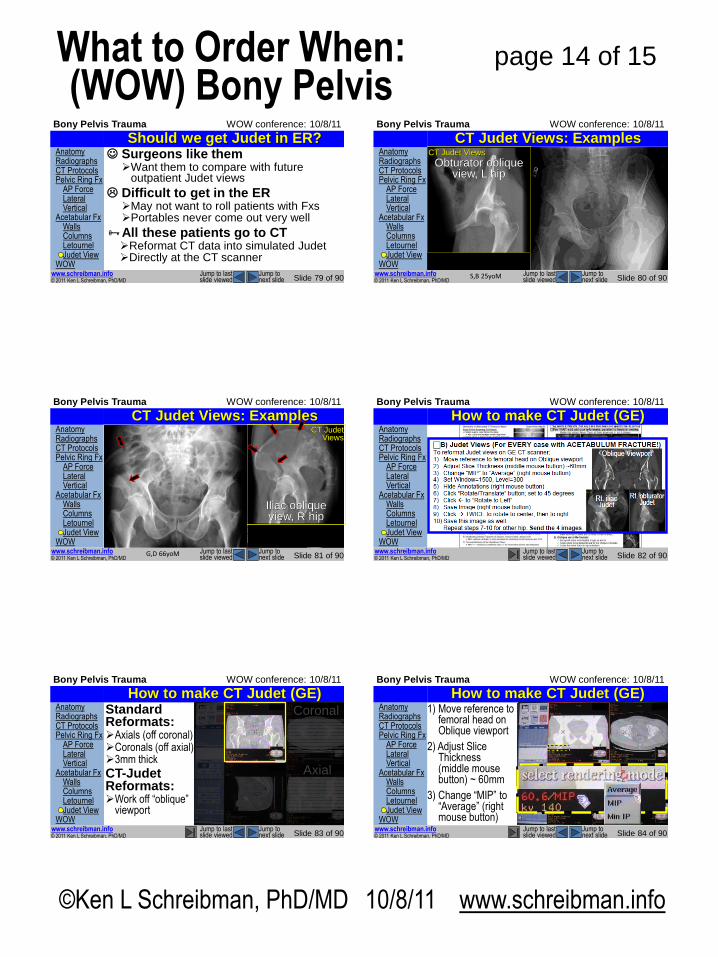
Should we get Judet in ER? Surgeons like them Want them to compare with future
outpatient Judet views
Difficult to get in the ER May not want to roll patients with Fxs Portables never come out very well
All these patients go to CT Reformat CT data into simulated Judet Directly at the CT scanner
Anatomy Radiographs CT Protocols Pelvic Ring Fx AP Force Lateral Vertical Acetabular Fx Walls Columns Letournel Judet View WOW
© 2011 Ken L Schreibman, PhD/MD www.schreibman.info
Bony Pelvis Trauma WOW conference: 10/8/11
Slide 80 of 90 Jump to next slide
Jump to last slide viewed
CT Judet Views: Examples
S,B 25yoM
Axial CT
Left Sagittal Reformat
Right Sagittal Reformat
Intact Post Wall
Fractured Post Wall
Impaction preventing reduction
Posterior
Anterior
Iliac oblique view, R hip
Obturator oblique view, R hip Iliac oblique view, L hip
Obturator oblique view, L hip
CT Judet Views
Anatomy Radiographs CT Protocols Pelvic Ring Fx AP Force Lateral Vertical Acetabular Fx Walls Columns Letournel Judet View WOW
© 2011 Ken L Schreibman, PhD/MD www.schreibman.info
Bony Pelvis Trauma WOW conference: 10/8/11
Slide 81 of 90 Jump to next slide
Jump to last slide viewed
CT Judet Views: Examples
G,D 66yoM
Obturator oblique view, L hip
Iliac oblique view, L hip
OA
Obturator oblique view, R hip Iliac oblique view, R hip
CT Judet Views
Anatomy Radiographs CT Protocols Pelvic Ring Fx AP Force Lateral Vertical Acetabular Fx Walls Columns Letournel Judet View WOW
© 2011 Ken L Schreibman, PhD/MD www.schreibman.info
Bony Pelvis Trauma WOW conference: 10/8/11
Slide 82 of 90 Jump to next slide
Jump to last slide viewed
How to make CT Judet (GE)
Anatomy Radiographs CT Protocols Pelvic Ring Fx AP Force Lateral Vertical Acetabular Fx Walls Columns Letournel Judet View WOW
© 2011 Ken L Schreibman, PhD/MD www.schreibman.info
Bony Pelvis Trauma WOW conference: 10/8/11
Slide 83 of 90 Jump to next slide
Jump to last slide viewed
How to make CT Judet (GE) Standard Reformats: Axials (off coronal) Coronals (off axial) 3mm thick
CT-Judet Reformats: Work off “oblique”
viewport
Axial
Coronal Anatomy Radiographs CT Protocols Pelvic Ring Fx AP Force Lateral Vertical Acetabular Fx Walls Columns Letournel Judet View WOW
© 2011 Ken L Schreibman, PhD/MD www.schreibman.info
Bony Pelvis Trauma WOW conference: 10/8/11
Slide 84 of 90 Jump to next slide
Jump to last slide viewed
How to make CT Judet (GE) 1) Move reference to
femoral head on Oblique viewport
2) Adjust Slice Thickness (middle mouse button) ~ 60mm
3) Change “MIP” to “Average” (right mouse button)
©Ken L Schreibman, PhD/MD 10/8/11 www.schreibman.info
page 15 of 15 What to Order When: (WOW) Bony Pelvis
Anatomy Radiographs CT Protocols Pelvic Ring Fx AP Force Lateral Vertical Acetabular Fx Walls Columns Letournel Judet View WOW
© 2011 Ken L Schreibman, PhD/MD www.schreibman.info
Bony Pelvis Trauma WOW conference: 10/8/11
Slide 85 of 90 Jump to next slide
Jump to last slide viewed
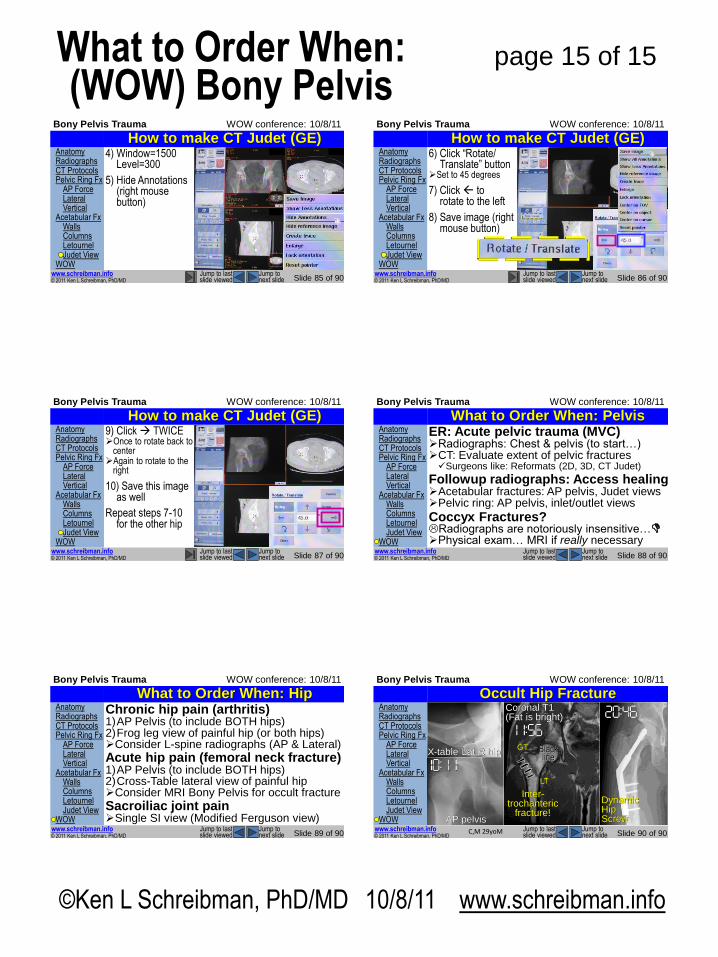
How to make CT Judet (GE) 4) Window=1500
Level=300
5) Hide Annotations (right mouse button)
Anatomy Radiographs CT Protocols Pelvic Ring Fx AP Force Lateral Vertical Acetabular Fx Walls Columns Letournel Judet View WOW
© 2011 Ken L Schreibman, PhD/MD www.schreibman.info
Bony Pelvis Trauma WOW conference: 10/8/11
Slide 86 of 90 Jump to next slide
Jump to last slide viewed
How to make CT Judet (GE) 6) Click “Rotate/
Translate” button Set to 45 degrees
7) Click to rotate to the left
8) Save image (right mouse button)
Anatomy Radiographs CT Protocols Pelvic Ring Fx AP Force Lateral Vertical Acetabular Fx Walls Columns Letournel Judet View WOW
© 2011 Ken L Schreibman, PhD/MD www.schreibman.info
Bony Pelvis Trauma WOW conference: 10/8/11
Slide 87 of 90 Jump to next slide
Jump to last slide viewed
How to make CT Judet (GE) 9) Click TWICE Once to rotate back to
center Again to rotate to the
right
10) Save this image as well
Repeat steps 7-10 for the other hip
Anatomy Radiographs CT Protocols Pelvic Ring Fx AP Force Lateral Vertical Acetabular Fx Walls Columns Letournel Judet View WOW
© 2011 Ken L Schreibman, PhD/MD www.schreibman.info
Bony Pelvis Trauma WOW conference: 10/8/11
Slide 88 of 90 Jump to next slide
Jump to last slide viewed
What to Order When: Pelvis ER: Acute pelvic trauma (MVC) Radiographs: Chest & pelvis (to start…) CT: Evaluate extent of pelvic fractures Surgeons like: Reformats (2D, 3D, CT Judet)
Followup radiographs: Access healing Acetabular fractures: AP pelvis, Judet views Pelvic ring: AP pelvis, inlet/outlet views
Coccyx Fractures? Radiographs are notoriously insensitive… Physical exam… MRI if really necessary
Anatomy Radiographs CT Protocols Pelvic Ring Fx AP Force Lateral Vertical Acetabular Fx Walls Columns Letournel Judet View WOW
© 2011 Ken L Schreibman, PhD/MD www.schreibman.info
Bony Pelvis Trauma WOW conference: 10/8/11
Slide 89 of 90 Jump to next slide
Jump to last slide viewed
What to Order When: Hip Chronic hip pain (arthritis) 1)AP Pelvis (to include BOTH hips) 2)Frog leg view of painful hip (or both hips) Consider L-spine radiographs (AP & Lateral)
Acute hip pain (femoral neck fracture) 1)AP Pelvis (to include BOTH hips) 2)Cross-Table lateral view of painful hip Consider MRI Bony Pelvis for occult fracture
Sacroiliac joint pain Single SI view (Modified Ferguson view)
Anatomy Radiographs CT Protocols Pelvic Ring Fx AP Force Lateral Vertical Acetabular Fx Walls Columns Letournel Judet View WOW
© 2011 Ken L Schreibman, PhD/MD www.schreibman.info
Bony Pelvis Trauma WOW conference: 10/8/11
Slide 90 of 90 Jump to next slide
Jump to last slide viewed
29 yo Male Bike hit curb Fell on right hip Right hip pain r/o fracture…
Radiographs negative
Still concerned for fracture…
Now what? MRI! Today! ASAP!
Occult Hip Fracture
C,M 29yoM
AP pelvis
X-table Lat R hip
Coronal T1 (Fat is bright)
Black line
GT
LT
Inter-trochanteric
fracture!
Dynamic Hip Screw