Embed Size (px)
Citation preview

Biochimica et Biophysica Acta 1858 (2016) 2468–2482
Contents lists available at ScienceDirect
Biochimica et Biophysica Acta
j ourna l homepage: www.e lsev ie r .com/ locate /bbamem
Efficient preparation and analysis of membrane and membraneprotein systems☆
Matti Javanainen a,⁎,1, Hector Martinez-Seara a,b,⁎,1a Department of Physics, Tampere University of Technology, Tampere, Finlandb Institute of Organic Chemistry and Biochemistry, Academy of Sciences of the Czech Republic, Prague, Czech Republic
☆ This article is part of a Special Issue entitled: Biosimulaand Tomasz Róg.⁎ Corresponding authors.
E-mail addresses: [email protected] (M. Javanain(H. Martinez-Seara).
1 Both authors contributed equally to the article.
http://dx.doi.org/10.1016/j.bbamem.2016.02.0360005-2736/© 2016 Elsevier B.V. All rights reserved.
a b s t r a c t
a r t i c l e i n f oArticle history:Received 30 December 2015Received in revised form 23 February 2016Accepted 25 February 2016Available online 4 March 2016
Molecular dynamics (MD) simulations have become a highly important technique to consider lipid membranesystems, and quite often they provide considerable added value to laboratory experiments. Rapid developmentof both software and hardware has enabled the increase of time and size scales reachable by MD simulationsto match those attainable by several accurate experimental techniques. However, until recently, the qualityand maturity of software tools available for building membrane models for simulations as well as analyzingthe results of these simulations have seriously lagged behind.Here, we discuss the recent developments of such tools from the end-users' point of view. In particular, wereview the software that can be employed to build lipid bilayers and other related structures with or withoutembeddedmembrane proteins to be employed inMD simulations. Additionally,we provide a brief critical insightinto force fields andMD packages commonly used formembrane andmembrane protein simulations. Finally, welist analysis tools that can be used to study the properties of membrane and membrane protein systems. In allthese points we comment on the respective compatibility of the covered tools.We also share our opinion on the current state of the available software. We briefly discuss the most commonlyemployed tools and platforms on which new software can be built. We conclude the review by providing a fewideas and guidelines on how the development of tools can be further boosted to catch up with the rapid pace atwhich thefield ofmembrane simulation progresses. This includes improving the compatibility between softwaretools and promoting the openness of the codes on which these applications rely.This article is part of a Special Issue entitled: Biosimulations edited by Ilpo Vattulainen and Tomasz Róg.
© 2016 Elsevier B.V. All rights reserved.
Keywords:Tools and softwareMembrane buildingProtein insertionMolecular dynamicsLipid bilayer
1. Introduction
The first simulation on soft matter was performed 30 years ago, andsince then the field of computational biophysics has expanded at anenormous pace. This first Monte Carlo simulation, studying the water–lipid interface [1], was followed by studies on micelles [2] and bilayers[3,4] using themolecular dynamics (MD)method. Simulations of mem-brane proteins took place soon after [5,6].
Since these ground-breaking studies in the early and mid 90s, bothcomputing power and the accuracy of the employed models have in-creased drastically, leading to a large number of studies on membranes(see e.g. [7,8]) andmembrane protein systems (see e.g. [9–11]).What ismore, experimental techniques have also improved, providing moreaccurate data against which the simulation models can be parameter-ized and optimized. Nowadays the knowledge required to perform
tions edited by Ilpo Vattulainen
en), [email protected]
MD simulations of membranes or membrane protein systems is easilyavailable for everyone via the internet. Such simulations can be performedwith numerous available software packages, including several free reliablealternatives, on any modern desktop computer to a certain extent.
However, except for the last few years, what has been seriouslylacking are publicly available user-friendly tools that aid the setting upand analysis of membrane or membrane protein simulations. Suchtools are necessary to make the field of computational biophysicsmore approachable to newcomers. Additionally, theywould also simpli-fy the tasks of experienced scientists, as automation and ease-of-use oftools would leave more time for the actual science. Luckily things arechanging and a number of new approaches have been introduced toboth building lipid membranes and inserting proteins into them, aswell as to analyzing the results of the simulations on these systems.
Most of these new tools have been made available since the lastthorough review on the topic almost ten years ago [12], which callsfor an update. In this paper we review the important aspects of settingup and analyzing membrane and membrane protein simulations. Itshould be noted that this review does not aim to provide step-by-stepinstructions for performing membrane or membrane protein simula-tions, yet such recipes are available in e.g. Refs. [13] and [14]. Instead,
2469M. Javanainen, H. Martinez-Seara / Biochimica et Biophysica Acta 1858 (2016) 2468–2482
we aim to provide a comprehensive list of the key software available.We comment the ease-of-use and generality of these tools and alsoprovide information on their compatibility with force fields and fileformats. This listing will aid both newcomers to select the proper toolsfor their project as well as inform more experienced users of newlypublished tools and techniques. It must be noted that the ever increas-ing user-friendliness of the applications and simulation softwaremight, however, introduce a new and a perhaps surprising issue. New-comers without the proper background knowledge on the underlyingalgorithms might nowadays be able to perform both simulations andanalyses. This might accidentally lead to incorrect conclusions that areextremely hard to catch during the peer-review process. Therefore, itis important that regardless of how easy to use scientific tools become,they should never be used as black boxes.
This review is structured as follows. We first introduce the mostcommon force fields employed in molecular dynamics simulations oflipids and proteins. Next, the numerous approaches used to build lipidbilayers are reviewed. This is followed by a thorough list of techniquesand tools for the insertion of proteins into membranes. After a briefexamination of the popular molecular dynamics software packages,we review tools available for the analysis of membrane and membraneprotein simulations. Finally, we raise issues related to the current para-digm of tool development and try to foresee how these issues could betackled in the near future.
2. Force fields for biomolecular simulations
A careful selection of the proper lipid and protein force fields is ofkey importance for every project considering MD simulations on bio-molecular systems. Most importantly, the level of detail of the chosenforce field, be it e.g. a fully atomistic or a coarse-grained one, shouldallow to sample time and size scales relevant for the problem at handyet still provide the required chemical accuracy. Another factor affectingthe selection of the force field is its compatibility with the availablesimulation software.What is more, the chosen force fields should eitherinclude the molecule parameters related to the research problem orprovide tools for parameterizing them. Lipid force fields seldom coverall possible head groups and tail types. Notably, certain head groups(such as phosphatidylcholine) and tails (such as palmitic acid or oleicacid) are often parameterized first and appear in almost everylipid force field. On the other hand, some head groups (such asphosphatidylinositol) or tails (such as linoleyl or linolenoyl) are rarelyavailable. Therefore, the desired membrane composition might limitthe number of plausible force fields. The choice of the lipid force fieldalso often sets limits to the available options for the protein force field,and vice versa. Sometimes the projects involve molecules beyond lipidand protein families (such as sugars or nucleotides) and in such casesthe selected force field should also cover these extra molecule types orbe compatible with a force field that contains them.
Some common force field models, which can be divided into differ-ent categories based on howmuch detail they provide, are briefly listedbelow. Formore thorough reviews and comparisons of lipid and proteinforce fields please see Refs. [15–19]. Notably, no thorough comparisonof the performance of the force fields in describing membrane proteinsystems exists in the literature to our knowledge.
2.1. Coarse-grained models
Coarse-grained models map multiple atoms into larger pseudoatomsor “beads”, which significantly reduces the number of degrees of freedomand therefore allows longer simulation times.
The Martini model has gained broad acceptance in the biomolecularsimulation community. It contains parameters for lipids [20], includingglycolipids [21], and proteins [22,23] as well as carbohydrates [24] andnucleic acids [25] among others. It is also compatible with a polarizablewater model [26]. The implicit solvent version of the Martini lipid force
field, titled Dry Martini, is also available [27]. One major advantage ofMartini, in addition to the large selection of parameterized moleculetypes, is the number and quality of tools provided on the Martiniwebsite.
The PLUM force field also relies on a solvent-free approach andcontains parameters for both proteins and lipids [28–30]. One key ad-vantage that PLUM has over Martini is that it describes protein folding,whereas secondary structures are fixed in Martini.
Furthermore, the ELBA force field [31] introduces dipoles into bothlipid molecules and water beads, which greatly improves the descrip-tion of electrostatics. However, the number of lipid types available isvery limited and proteins have not been parameterized at the time ofwriting this review.
2.2. United-atom force fields
United atommodels usually combinemethyl groups andmethylenebridges into pseudoatoms, thus effectively combining the properties ofthe hydrogen atoms into their host carbons. The most common ofsuch force fields, namely GROMOS, contains multiple parameter setsfor proteins with the newest one being 54A7/54B7 [32]. The multipleversions are also compatible with the corresponding lipid force fields[33–35] and contain parameters for many other molecule types, suchas carbohydrates and nucleic acids. Two automated web-based toolsexist for the parameterization of small molecules for GROMOS. Thelong-running and popular PRODRG server [36,37] has recently receivedcriticism, most importantly for its poor handling of charge groups [38].The more recent Automated Topology Builder (ATB) [39,40] aims totackle the charge group partitioning issue [41], in addition to otherimprovements.
Here, the commonly employed yet old Berger united atom lipidmodel [42] should bementioned. It combines parameters frommultiplesources and has been used together with atomistic protein force fields(see below). This parameterization was recently refined to correctlydescribe phase behavior [43].
The united atom TraPPE force field contains parameters for lipids[44] yet parameters for proteins are not available.
2.3. Atomistic force fields
Thanks to the rise in computing power, researchers can now waivethe performance provided by united atom approaches in favor to theimproved accuracy provided by fully atomistic force fields. Additionally,the interest towards membrane protein simulations has called for thedevelopment of high quality lipid force fields compatible with the pro-tein force fields previously employed in simulations of water-solubleproteins.
Various versions of the Amber protein force field are commonlyused,with ff99SB-ILDN [45] gainingwidespread acceptance. Additional-ly, the ff99SB force field was recently refined in the form of ff14SB [46].Further, another development branch entitled ff14ipq employedcharges derived in a new way [47] and has not yet been thoroughlytested. Even the old ff03 is still used to some extend [48] (note thatff03 is from 2003 whereas ff99SB-ILDN is from 2010).
Multiple Amber-compatible sets of lipid parameters also exist. TheGeneral Amber Force Field (GAFF) lipid parameters [49] were latercombined with the development of Lipid11 [50] resulting in theLipid14 parameter set [51,52]. Lipid14 contains parameters for severallipid types as well as cholesterol. Until Lipid14 all these force fieldsrequired the use of applied surface tension in order to maintain themembrane in a liquid phase. In addition, the Slipids parameter set[53–55] is compatible with the Amber protein force fields and hasparameters for multiple lipid types including sphingomyelin andcholesterol. However, polyunsaturated tails are not included in Slipids.Amber also supports theGlycamcarbohydrate forcefield [56]. Automatedways to parameterize molecules, such as drugs, for the GAFF [57] force

2470 M. Javanainen, H. Martinez-Seara / Biochimica et Biophysica Acta 1858 (2016) 2468–2482
field are available [58–61]. Additionally, a database containing a num-ber of parameterized molecules for GAFF (as well as OPLS-AA andCHARMM general force field) in the GROMACS format is availableonline [62].
The CHARMM22 protein force field [63] together with the CMAPcorrection [64] as well as the more recent CHARMM36 protein forcefield [65] have been employed extensively to studymembrane proteins.The recently launched CHARMM36 lipid force field also allows thesimulation lipid bilayers in a tensionless state. This CHARMM36 lipidparameter set [66] includes a great number of the most common celllipid types [67]. CHARMM also includes parameters for carbohydrates[68] and nucleic acids [69]. A MATCH server providing automatedatom typing [70] exists for CHARMM and the CHARMM general forcefield (CGenFF) [71] also supports ways to automate the force field pa-rameter generation [72,73,59]. A large set of parameterized moleculesfor the CGenFF is also available in the GROMACS format [62].
The OPLS-AA all atom protein force field [74] and the fresh update(OPLS-AA/M) [75] are compatible with the recently released lipidforce field [76,77]. Unfortunately, a very limited set of lipids is currentlyavailable. OPLS-AA is also compatible with a limited set of carbohy-drates [78], and a large set of parameterized molecules is available forOPLS-AA in the GROMACS format [62].
2.4. Hybrid approaches
To combine the speed and detail benefits of models at two levels ofresolution, multiple hybrid approaches have been developed. The unit-ed atom Berger force field [42], introduced earlier, has been combinedwith both Amber [79] and OPLS-AA [80] protein force fields.
The PACE force field was designed to combine a coarse-grainedmembrane model with a united atom protein model [81–83].
Similarly, the Multi-Scale Coarse-Grained (MS-CG) force field hasbeen used to combine a lower-detail membrane into a higher-detailprotein [84]. Similarly, the Martini force field has also been combinedwith higher-detail proteins [85].
2.5. Polarizable force fields
Polarizable force fields, as their name suggests, aim to account forthe polarization of themedia due to the presence of chargedmolecules.To our knowledge, only the polarizable CHARMM force field based onthe Drude oscillator contains parameters for both lipids [86] andproteins [87]. Additionally, nucleic acids have been parameterized forthis force field [88]. These models, however, have not been widelyadapted due to their computational cost and fairly limited accuracy.More information on them can be found in a number of very recentreviews [89–91].
2.6. Transformations between models
Tools allowing flexible transformations between different modelshave been developed recently. They allow the fine-graining of coarse-grainedmodels into either the fully atomistic or theunited atom scheme.To our knowledge three independent such tools exist [92–94], eachwiththeir own features and limitations. Additionally, Lipid Converter [95]allows conversions between lipid force fields (all-atom and unitedatom) as well as between lipid types within one force field.
3. Building membranes
Biological membranes are very complex entities. They are constitut-ed by a vast number of different lipids, proteins and sugarmoieties. Theproportions of these moieties and their spatial distribution dependlargely on the studied membrane [96] and may even change in time.With such complexity, it is understandable that one of the critical
tasks related tomembrane simulations is the construction of the systemthat is to be simulated.
So far, molecularmodeling of membranes has focused on simulatingsmall bilayer patches containing just few lipid moieties. Although sim-plified, these systems are believed to be representative models forsome aspects of real biological membranes. Within this framework,the construction of membranes was usually performed manually andeach researcher had his or her own protocol for this task. As long asthe membranes were simple enough and the number of systems re-quired for a project remained rather small, this approach was tolerable.However, this was a clear impediment for newcomers who wanted toinclude membrane simulations in their research toolkit. What is more,even experienced membrane researchers had to spend substantialamount of time on this task rather than on actual science.
This has drastically changed during the last few years. Several newtools and approaches that aim to simplify and avoid the pitfalls of previ-ously established protocols have appeared. Coupledwith the increase ofcomputing power, this has changed radically the complexity and size ofstudied membranes. It is nowadays not rare to see simulations of largemembranes including many lipid moieties in combination with othermolecular classes like proteins [97–99] and even polymers [100]. Suchsystems would be too complex to be set up using manual membranebuilding protocols.
This section aims to review the currently available methods forconstructing lipid-only membranes with emphasis on their knownstrengths and weaknesses. The construction of protein-containing sys-tems as well as insertion of proteins into membranes will be discussedin the next section.
We will start by introducing a prototype of the manual buildingprotocol. This should serve to understandwhy the other, more straight-forward methods introduced later in this section, are needed. We willalso point out the existence of several repositories where one can findspecific pre-built and even equilibrated membranes ready to be simu-lated. We will then present several tools which allow the constructionof custommembranes. Finally,wewill briefly discuss the improvementsexpected from future approaches.
3.1. Previous considerations
A few important points need to be considered before selecting themethod for building a membrane for a particular project. The first stepis to choose the membrane composition. Decision of the required lipidmoieties together with their desired proportions should be based onexperimental evidence of the systems that are to be mimicked [96].However, this information is not always fully available since the extrac-tion of specific membranes and the subsequent separation of lipidcomponents are challenging experimental tasks. What is more, thereare also technical aspects that need to be considered. Most importantly,not all possible lipids, i.e. naturally occurring or artificially synthesized,have been parameterized. This is a fundamental point because parame-terizing lipids is a time consuming and demanding task. Therefore,unless one is willing to devote the time required to parameterize theconsidered lipid, researchers usually stick to those currently availableeven if this results in small deviations from the desired optimal compo-sition. Moreover, not all available lipids have been parameterized for allforce fields. Therefore, to avoid large compromises in the desired mem-brane composition, the choice of the force field is often limited to fewoptions, and this choice also dictates the set of tools that can be usedto build and simulate the membrane, as discussed below.
Several approaches aim to overcome this problem, yet they have tobe considered with extreme caution. There are tools that, despite nevermeant to be used to parameterize lipids, can be used to perform thistask [49]. Another option is to adapt the missing parameters from adifferent force field. It is also common to build topologies for new lipidsby complementing existing ones using chemical analogy of knownmolecular fragments [101]. Although the agility of these shortcuts

2471M. Javanainen, H. Martinez-Seara / Biochimica et Biophysica Acta 1858 (2016) 2468–2482
might look attractive at first, they should be avoided. They rarely workproperly and even if they do, the quality does not likely match that ofthe carefully parameterized models. Therefore, extensive validation ofthe behavior of the produced lipids must be performed in order toavoid artifacts.
3.2. Structure and topology of lipids
In comparison with other biomolecules, naturally occurring lipidsare relatively small and present simple connectivity [96]. One can con-struct them easily using any molecular editor, e.g. Avogadro [102], incombination with any chemical software toolbox, e.g. Open Babel[103], that provides connectivity information required for themoleculartopology. As lipids contain stereochemical centers, which are oftenexclusively selected in nature, one must ensure that the constructedlipids match the required spatial configuration [96]. Despite the appar-ent simplicity of this process, the real challenge lies in finding suitableforce field parameters for the constructed lipid. Lipids are known to bevery challenging molecules to parameterize [104], as high-levelquantum chemistry calculations are required in order to obtain valuesfor the partial charges and dihedral potentials. Furthermore, theLennard-Jones interactions need to be iteratively adjusted againstavailable experimental data. In general, this iterative nature of theparameterization process restricts the use of the lipid force field to avery particular water model and to a very narrow set of parameters,such as the cut-off scheme of nonbonded interactions [104]. Further-more, when combined with other molecule types, the parametersshould be consistent, which in general means that the same protocolneeds to be followed in the parameterization of all associatedmolecules.This restriction renders the shortcut approaches described above fairlyuseless, unless the changes to existing lipid models are cosmetic, e.g.extension of acyl chains. In any other case one should download the re-quired structures and topology files from one of the several availableweb resources, such as lipidbook [105], CHARMM-GUI [106], as wellas Slipids [55] andMartini [20] websites. In some rare cases, force fieldscan also be provided by the used MD package [51].
Recently, taking advantage of the modular design of the CHARMMforce field, a new tool called LipidBuilder [101] has been developed.This tool can be used to generate certain customized lipid topologiesfor this force field by combining existing head groups into tails ofdesired length and level of saturation. Although, its use is rather limitedto NAMD MD package and just one force field, it points to the correctdirection by solving the problem of lipid structure and topologygeneration.
3.3. Manual construction of membranes
Building amembranemanually starting from the known structure ofthe desired lipids is usually a tedious, highly inefficient and even error-prone task, even in the case of simple single-component lipid bilayers[13]. All manual protocols share essentially the same steps, as shownin Ref. [13].
The process starts by spreading oriented lipid molecules in a grid.The coordinates of one lipid are replicated and translated to match thelocation of the assigned grid points. For this reason, the positions ofthe lipids in the resulting bilayer are usually highly correlated. This isfollowed by solvation and a subsequent long equilibration phase. Toavoid collisions between lipids and penetration of ring structures, theinitial distances between lipids need to be fairly large. This results in along and tricky equilibration simulation, during which the bilayershrinks until it relaxes to the correct area per lipid. In order to speedup this process, the equilibration can be performed without water mol-ecules by restricting the movement of some lipid atoms in the directionnormal to the membrane [107]. This trick also prevents lipid flip-flops,which are fairly common within this loosely packed initial structure.What is more, this approach also minimizes the amount of water that
ends up inside the membrane during the posterior solvation processsince the excess free volume within the membrane into which watermolecules could be accidentally placed has already been eliminated.
Another important problem arises with multicomponent mem-branes. In these systems the components should be carefully mixedfrom the very beginning since the lateral diffusion of lipids is slow[108]. Even for small membrane patches in a fluid phase the realmixingprocess might take close to a microsecond [109]. The mixing timeincreases very steeply with the number of lipids. Therefore, for largemembranes, the initial lipid positions need to be properly randomized.Otherwise, microseconds of simulation might be wasted just for equili-bration and lipid mixing.
Finally, on many occasions several initial structures of the samemembrane are generated and simulated to serve as independentreplicas of the studied system. This approach aims to tackle the limitedsampling problem that is expected in the currently reachable timescales. Unfortunately, when manual methods are used to generatemembranes, the construction of such independent configurationsessentially requires repetitive manual work.
All these problems clearly call for othermembrane buildingmethodsthat are free of the listed shortcomings. Such toolswill certainly not onlyimprove the efficiency of the researchworkflow, but also prevent sever-al typical and difficult-to-spot mistakes.
3.4. Pre-equilibrated bilayers
One of the first alternatives to manual construction of membraneswas the reuse of pre-equilibrated structures. Alternatively, largermem-branes could be constructed by simple multiplication of the coordinatesof these structures. One advantage of reusing membranes is that theirintegrity is often better cross-checked by other researchers. This re-duces the chance of errors and allows direct comparison with previous-ly published data obtained with the same membrane model. It is alsopossible to slightly change the lipid composition of pre-equilibratedmembranes by selectively removing some lipids. Further, modifyinglipid moieties to other ones is possible [95] and also very efficientwhen coarse-grained models are employed. However, such modifica-tions must be performed very carefully to atomistic systems in orderto avoid affecting the structural connectivity of the lipids such as theirstereochemistry.
Overall, these arguments convinced several research groups thathave been publishing the membrane structures used in their researchon their groupwebsites. Despite being appreciated initiatives, these col-lections provide a solution that is far from optimal as the membranestructures are scattered across numerous hard-to-find websites. Newprotein structures are stored without exceptions in the centralizedRCSB PDB repository [110] and a few initiatives such as lipidbook[105] or more general repositories (e.g. www.zenodo.org) might steeralso the membrane simulation community to this direction in the nearfuture.
Unfortunately, using pre-existing membranes has several draw-backs. To begin with, this approach is not flexible at all. Furthermore,the ordering of lipid atoms likely depends on the employed force field.Therefore, either the same force field with which the membranewas originally simulated needs to be used, or the atoms need to bereordered. Despite being a fairly simple task, the reordering process israther prone to errors that can lead to subtle and hard-to-spot changesin e.g. chirality of stereocenters and cis–trans isomerization.
3.5. Membrane builder applications
To solve the issues mentioned above, especially those related to thebuilding of custom membranes, new tools have recently started toemerge. These tools usually provide a fully automated mechanism todistribute different lipid moieties on top of a grid, with the exceptionof CHARMM-GUI [111] that provides membrane structures with lateral

2472 M. Javanainen, H. Martinez-Seara / Biochimica et Biophysica Acta 1858 (2016) 2468–2482
density already close to its equilibrium value. In all the tools mentionedbelow lipids are randomly distributed to improve their lateral mixing.
The tools can be divided into two categories: web services(CHARMM-GUI [111], Membuilder [112], LipidBuilder [101]) anddistributed applications (Packmol [113] and insane [114]). Web-basedapplications usually provide a user-friendly interface, and they oftengenerate not only the coordinate files but also all other files requiredto perform simulations on the system, i.e. topologies and simulationparameters. Their downside is that their performance is often low oreven limited bydesign to optimize theweb server resources. Generatinghighly optimized membranes is a resource-intensive process. Takinginto consideration the limited resources that a research group can divertto such a service, these problems might prove difficult to address.Another aspect of such web-based tools that restricts their usage ininnovative research is that only few people (i.e. the developers) canmodify them. Only if all the requirements of a project are fulfilled by aweb service, can full advantage be taken out of it. This is true evenwhen trivial modifications, such as the addition of a new lipid type,are required. In fact, this is a common problem of all these services.They can only be used for a limited number of MD packages, lipidmoieties and force fields.
The newly published LipidBuilder [101] partly addresses the limita-tion of fixed set of available lipid moieties by including a lipid builderapplication, yet it is only compatible with the CHARMM force fieldand the NAMD software. The limitations of another web service,Membuilder (with Membuilder II available as a beta version at thetime of writing this review), are related to MD software, as it only sup-plies files in the formats of GROMACS. However, a number of GROMOS(43a1,43A1-S3, and 53a6) and CHARMM (27 and 36) as well as theSlipids force fields are supported.
CHARMM-GUI [115] nowadays provides input files for multiple MDpackages including GROMACS, NAMD, Amber, OpenMM and CHARMM,yet in terms of atomistic force fields it is limited to CHARMM36 [66].Naturally, however, the structure provided by CHARMM-GUI can alsobe used with other force fields by simply altering the atom ordering ifit differs from that of CHARMM36. Finally, it should be mentioned thatthese web services are difficult to link to other tools, which furtherlimits their usability and adoption as a de facto tool.
On the other hand, distributed software tools for membrane gener-ation are still in their infancy. With the exception of insane [114] thatgenerates sophisticated membranes for the Martini force field [20],the features provided by membrane builder software fall short com-pared to those of theirweb-based counterparts. The only program reallyworth mentioning is Packmol [113] which allows the efficient genera-tion of any kind of densely packed structures, including sophisticatedmembranes. However, this software is blind to molecular details andconsiders molecules as rigid entities. Therefore, it cannot obtain similaroptimal packing densities as CHARMM-GUI.
3.6. Building micelles, liposomes and other non-planar structures
Besides planar membranes, other lipid structures bear biological in-terest. Micelles, vesicles, and hexagonal phases are notable examples ofsuch structures. Some tools, such as CHARMM-GUI [111], Membuilder[112], and Packmol [113] can also create such structures within theirlimitations.
3.7. Final remarks
To conclude, new tools that simplify the creation and simulation ofcomplexmembranes are constantly being developed. These tools repre-sent a significant improvement with respect to the previous paradigmwhere manual setup or reuse of existing membrane structures werethe only available options. Taking into consideration the number oftools that appeared recently and their constant improvement, we canexpect that running a membrane simulation will become a rather
simple task in the near future. However, building tools are useless ifthe used force field does not provide parameters for the desired lipids.So far, an automated way to reliably parameterize lipids does not existand its implementation seems like amonumental task. However, devel-opment in this direction would pave the way for the ultimate generalmembrane builder tool whichwould be free of all the numerous limita-tions listed above.
4. Insertion of membrane proteins
Asmembrane protein simulations have recently become increasing-ly popular, the quality and variety of tools for constructing the initialstructures for such simulations have also improved substantially.Notably, most of the tools and approaches reviewed in this sectionwere developed less than five years ago, which calls for an update tothe thorough review of Kandt et al. [12].
Having set up a lipid membrane following one of the approacheslisted in the previous section, a choice between the multiple proteinembedding approaches needs to be taken. Alternatively, the proteincontaining membrane can also be assembled from scratch. As in thecase of building a membrane, none of the possible approaches can bedeclared to be the best in every possible case. Instead, a choice needsto bemade based onmultiple criteria. The approachmust be compatiblewith the chosen lipid and protein models. Some approaches work withmultiple force fields, even covering both atomistic and coarse-grainedschemes, yet some were designed to work strictly with a single forcefield. Additionally, the file formats involved in the process need to becompatible with the ones at hand, or at least a straightforward conver-sion must be available.
The protein embedding approaches can be divided into three cate-gories. First, web-based tools provide a simple way to conduct thetask, yet they are often not adaptable to cases other than the ones forwhich theywere originally designed. Second category includesmethodsthat employ the functionality ofmolecular dynamics packages or are in-cluded exclusively within a certain MD code. Third, some approachesare based on independent software designed solely for the purpose ofprotein embedding. Itmust also bementioned that, likewithmembranestructures, some websites offer pre-equilibrated membrane structureswith membrane proteins embedded into them. Naturally, these avail-able structures seldom provide the exact desired composition and, asmentioned in the previous chapter, modifications to their compositionmust be performed with care.
In this section we list the existing approaches for protein insertionand evaluate them in terms of ease-of-use, computational cost and,most importantly, compatibility in terms of both simulation softwareand the employed lipid and protein models.
4.1. Early approaches
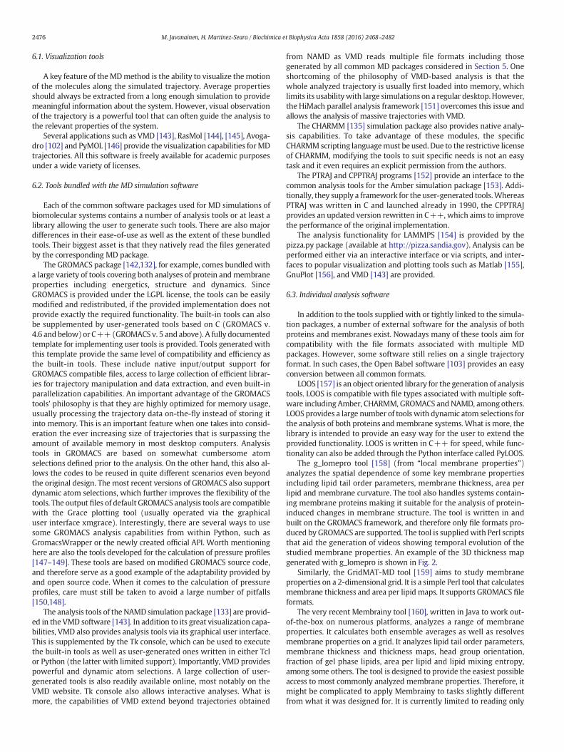
The first simulations of membrane protein systems [5,6] relied onassembling a membrane around a protein. With proper automationand with a sufficient collection of lipid conformations from which themembrane is built, this approach can be quite effective. However,since the packing algorithms are never perfect, the built system mightrequire extended energy minimization and equilibration simulationswith restraints on the protein structure. In case molecules with rings,such as sterols, are involved, care must be taken to avoid ring penetra-tion by e.g. lipid tails. Also, no advantage of a pre-built and equilibratedbilayer can be taken. It is noteworthy that the CHARMM-GUImembranebuilder, discussed later in this section, implements this approach.Another and more general tool which can be employed to build thebilayer around a membrane is Packmol [113], a software which placesmolecules in a system based on user-provided restrictions. Examplesof protein containing systems generated with Packmol and CHARMM-GUI can be found in Fig. 1.

Fig. 1.Outputs of several highly automated tools that can be used to build systems formembrane protein simulations. a) The coarse-grained structure generated by the insane tool. Proteinis shown in gray spheres, whereas the different lipid moieties are colored (pink, cyan, yellow, red, tan) to emphasize the tool's ability to build asymmetric membranes containingnumerous lipid types. Additionally, water (iceblue) and ions (red and blue) are also added. The initial structure is built on a regular grid from very ordered lipid structures, yet therelaxation during a simulation is very rapid thanks to the coarse-grained scheme. b) An atomistic bilayer generated by the CHARMM-GUI membrane builder. Coloring as on previouspanel. c) Multiple proteins (gray) embedded in a DPPC lipid vesicle (pink) in the coarse-grained scheme. The system is built with the Packmol tool. The shown structure was kindlyprovided by Petri Kaurola.
2473M. Javanainen, H. Martinez-Seara / Biochimica et Biophysica Acta 1858 (2016) 2468–2482
Another straightforward approach is to delete lipids from a bilayer,creating a void into which a protein can be placed. The removal can beperformed manually, or tools such as genbox/solvate of the GROMACSpackage can be employed to remove lipids which collide with the pro-tein. This simple lipid removal perturbs the lipid composition and the(a)symmetry of the membrane. A gentler approach is described byShen et al. [116], who first delete a few lipids to create an initial hole,which is then expanded by a cylindrical repulsive potential. Theresulting hole does not often match the shape of the protein, whichagain indicates that a long equilibration is required. This method wasdeveloped further by taking into account the protein shape in creatingthe hole. Approaches following this idea, namely make_hole andGRIFFIN are discussed later in this section.
The clear advantage of these twomethodologies is their universalityto both atomistic and coarse-grained models as well as independencyfrom the used MD package. However, these approaches are not veryuser-friendly, they require removal of lipids, and the generated struc-tures need a more careful equilibration than many of the more recentapproaches described in the following sections.
4.2. External software and modified MD code
Instead of a simple cylinder, the mdrun_hole (or make_hole) tool[117] creates a hole based on the shape of the inserted protein. Thisminimizes the empty space around the protein following its insertion,which results in a faster equilibration of the system. This approach,even though employed extensively in the past, comes short due to afew reasons. First, two external pieces of software are required to createthe input files for the tool itself. Second, the mdrun_hole relies onGROMACS code and the most recent version is based on an outdatedversion 3.1.4. This dependency on GROMACS also indicates that all in-volved files will need to be available in formats that are compatiblewithGROMACS. Additionally, like its counterpart that creates cylindricalholes mentioned earlier, this approach also requires the removal oflipids from the system, which will perturb the lipid composition and(a)symmetry of the membrane.
The InflateGRO method [12], later updated to InflateGRO2 [118], isalso based on creating a hole in the host membrane and placing theprotein into this hole. However, the perturbation of the membrane
structure is minimized by first inflating the membrane, i.e. scaling upits coordinates to create more space between lipids. The protein isthen inserted and only few lipids need to be removed due to the largeinter-lipid spacing. Finally, the coordinates are stepwise scaled back tothe original lipid density and the structure is energy-minimized ateach step. Alternatively, in case no pre-built bilayer is available, it canbe built with a large inter-lipid spacing and the procedure followedfrom there on. This approach also requires the removal of lipids, thoughthe number is smaller than in many other methods. Most importantly,the tool relies on theGROMACS engine to perform the energyminimiza-tion steps. Therefore, the force field files must be available in aGROMACS-compatible format. Additionally, the scaling script workson GROMACS-based “.gro” file format. However, many of these limita-tions can be overcome by tools which allow conversion of MD softwarespecific files from one format to another.
Despite its success, the original InflateGRO had a few issues as listedby Schmidt and Kandt [118]. First, the termination of the script after thecoordinate scaling was not well defined in many cases. Second, the toolwas only functional with atomistic systems. Third, the functionality wasbased on residue names leading to many complications with heteroge-neous lipid membranes. Fourth, the inflation of the membrane couldresult in loss of equilibrated lipid conformations. Finally, the verticalposition of the protein in the membrane had to be manually adjusted.These issues were removed with the update to InflateGRO2 [118]. Bun-dled with the new aligning tool LAMBADA, the procedure is now fullyautomated, applicable to coarse-grained systems, and functions basedon index files instead of residue names. However, the dependency onGROMACS and the requirement for lipid deletion still remain after theupdate. Furthermore, whereas the original InflateGRO provided a wayto insert lipids into the enclosed volume of donut-shaped proteins,such functionality is not present in InflateGRO2.
Another GROMACS-based approach, g_membed [119] (nowapart ofthe mdrun functionality), is currently included in the GROMACS pack-age. This tool is based on a clever approach to first embed a shrunkenprotein into a hole created by the removal of a few lipids. The proteinis then scaled up slowly to its full size during a short simulation,which causes the membrane to gently adapt to the presence of theprotein. Due to this smooth adaptation, the system should only requirea fairly short equilibration after the insertion process. Since the

2474 M. Javanainen, H. Martinez-Seara / Biochimica et Biophysica Acta 1858 (2016) 2468–2482
functionality is only available as a part of the GROMACS package, filesneed to be in a format supported by it. However, the tool is not restrict-ed to any specific force fields.
The idea behind the mdrun_hole methodology (see above) wasrefined in the GRIFFIN tool [120] that brought the whole procedureinto one stand-alone tool and simultaneously advanced its functionality.In addition to the shape-dependent forces employed in the earlier toolto push away lipids and solvent molecules, GRIFFIN also includesCoulombic and van der Waals forces in order to better optimize thelipid–protein interface, and therefore minimize the equilibration periodrequired after the insertion of the protein. Additional geometrical re-straints can also be provided for GRIFFIN in order to keep moleculesaway fromundesired locations such as prevent lipids fromenteringpro-tein pores. GRIFFINworks by providing theMD codewith forces that areused to push away lipid and solvent molecules. One important strengthof the tool is that it works also with donut-shaped proteins, i.e. proteinswhich encapsulate lipids in some hollow region within their structure.However, as with many methods, GRIFFIN also requires the removal oflipids in order to obtain the correct system density before applying theforces that make room for the protein. Additionally, the tool is limitedto work together with GROMACS and NAMD packages.
In the case of coarse-grained models, system equilibration is usuallyvery rapid, and therefore the initial structure does not need to be socarefully constructed. A Python-based tool insane [114] builds upmem-brane protein systems compatible with theMartini force field. A coarse-grained structure of the protein needs to be provided and it can be easilygenerated with the martinize tool. The output structure contains alllipids in a straight-tail conformation (see Fig. 1), which is not a problemdue to the rapid equilibration. Additionally, no care needs to be put onpreserving the secondary structure of the protein since this is fixed inthe Martini protein force field. The insane tool can build membraneswith complex lipid compositions and it can be employed to insertmultiple proteins by exploiting the functionality provided by the DAFTapproach [121]. In addition to being limited to only the Martini forcefield, insane also suffers from the lack of possibility to specify theexact number of lipid molecules of each type as only ratios of lipidcomponents can be provided. This might result in the need tomanuallyremove molecules from the system to reach the desired numbers of thelipidmolecules. Some functionality remains to be included at the timeofwriting this review. One ultimate benefit of this tool is that large com-plex lipid systems can be easily constructed and rapidly equilibratedin the coarse-grained scheme followed by the transformation into amodel providing atomistic detail via e.g. the backward tool [93].
To summarize, multiple protein insertion methodologies rely oneither external software or are directly built-in in the MD software.These tools are often fairly straightforward to use, yet the dependencyon the MD software or related file formats limits their generaladaptation. The next section discusses two tools which provide a moreuniversal approach.
4.3. Methods employing the functionality of the MD code
Some recent protein insertion approaches rely on the model-independent functionality available in all common MD software,which renders them essentially universal. In the approach describedby Javanainen [107], a protein is first placed next to a bilayer and theninserted into it by applying a high lateral pressure on the system. Thisapproach does not depend on the MD software nor the used lipid andprotein models. One additional strength of this method is that there isno need for lipid removal. This means that the lipid composition and(a)symmetry of the host bilayer aremaintained throughout the process.However, this also means that in the case of proteins that occupydifferent volumes in the two membrane leaflets, the number of lipidsin these leaflets need to bemanually adjusted in order to create a planarstress-free system. Since the method is based on running a simulationon an unmodified MD code using specific pressure coupling options
and position restraints, some manual work is required to set up thesesimulations. The approach also allows the insertion of multiple proteinsat once even though this is somewhat complicated. The method is lim-ited to planar geometries and cannot handle donut-shaped proteins.
Another approach, alchembed [122], relying on the built-in func-tionalities of MD codes pushes the lipids away from the protein volumeby slowly turning on the protein's interactionswith lipids and other sur-roundingmolecules. This is achieved by soft-core interactions originallydeveloped for free energy calculations. This functionality is thereforereadily available in all major MD packages. The method is also generalin terms of lipid and protein models, and both atomistic and coarse-grained models can be employed. Similar to the method based on highlateral pressure described above, alchembed also relies onmanually po-sitioning and aligning the protein prior to running the simulation duringwhich the interactions are turned on. As a downside, lipids have to beremoved manually from the volume into which the protein is inserted.Moreover, parameters to be employed for the soft-core potential aresuggested, yet their generality remains to be carefully tested. However,multiple proteins can be inserted at once and non-planar geometriescan be employed, even though the manual position and alignmentprocedures might become quite tedious in such systems.
As described above, the approaches relying on the functionality ofthe MD packages can be used with essentially all MD software andwith all lipid and protein force fields. However, this universalitycomes with a price: the procedures are not automated and lack theease-of-use of some other approaches. Quite a lot of manual input is re-quired yet for an experienced user this requirement should be balancedby the versatility of the approaches.
4.4. Web-based tools and data banks
For quite some time many research groups have shared both lipidand lipid–protein structures on their web pages. Even though suchsystems seldom directly match the requirements of other researchers,they can act as a good starting point from which a desired structure isobtained, for example, via changing lipid types, force fields or evenlevel of detail of the model (from atomistic to coarse-grained and viceversa).
A significant improvement to this recently surfaced, as the group ofProf. Mark Sansom initiated a thorough data bank [123] containing thestructures of all known membrane protein structures embedded in aDPPC bilayer. These pre-equilibrated structures are available in coarse-grained (Martini) and united atom (GROMOS) schemes. Via specializedtools, such as backward [93], the coarse-grained structure can be turnedinto an atomistic one corresponding to any force field provided that themapping between them is available or generated. The limitation to aDPPC bilayer is not a major one, since in the coarse-grained schemetransformations between lipid types can be easily performed via simplescripts and the equilibration following such modifications is rapid.
Another huge step forward in building membrane protein systemswas the introduction and subsequent development of the CHARMM-GUI website [124,125]. CHARMM-GUI enables the flexible constructionof various kinds of systems including lipid structures of various geome-tries as well as membrane protein systems. The lipid composition aswell as the type and amount of the solvent can be carefully adjusted.Output files containing the assembled and nearly equilibrated systemare provided in various formats compatible with CHARMM, NAMD,GROMACS, AMBER and OpenMM [115]. One downside of CHARMM-GUI is that only membranes containing a single protein can be built.Additionally, in the atomistic scheme only the CHARMM force field issupported. However, with tools such as the Lipid Converter [95] thetransformation of the lipid part of the coordinate file to another forcefield can be readily performed. Actually many all-atom force fieldsshare atom ordering, which often removes the need for such conver-sion. In addition to the atomistic CHARMM force field, CHARMM-GUInow also includes membrane builders for Martini [126] and PACE

2475M. Javanainen, H. Martinez-Seara / Biochimica et Biophysica Acta 1858 (2016) 2468–2482
[127] force fields. An example of a membrane protein system generatedby CHARMM-GUI is shown in Fig. 1.
In this section the web-based membrane protein orientation toolsand databases [128–131] should also be mentioned as they provideimportant information on how to position and align the proteins forthe embedding methodologies that do not solve the protein alignmentand positioning themselves. Contrary to the Protein Data Bank (PDB)[110], these databases provide complete membrane protein structuresin which missing atoms are provided. Further, proteins are providedin their complete functional form where all symmetric subunits are re-constructed from the information in the original Protein Data Bank files,if needed. Notably, CHARMM-GUI can directly download oriented andcomplete protein structures from the OPM database [128,129].
5. Molecular dynamics simulation packages
The ease of building a membrane or the quality of the underlyingforce fields are superfluous if we cannot accurately perform a longenough simulation using the desired molecular ensemble. Molecularmechanics mostly relies on the ergodic assumption stating that in thelimit of long simulation times, time averages match ensemble averages.Clearly, the longer we simulate, the higher are the chances that oursimulation fulfills the ergodic assumption, stating that thewhole config-uration space is visited properly. It is not surprising that the simulationcommunity tightly relies on a few highly advanced and complex MDpackages and the talented developers behind them. With constantlyincreasing complexity of the computational resources paradigms (bye.g. GPUs, SIMD, openMP, MPI…) and the need to fully exploit theircapabilities to the limit to tackle biologically relevant problems, thepossibility that each researcher creates their own MD engine vanishes.Instead, most of the currently available MD packages are the resultof decades of development by large communities. Their leaders areto thank for the golden age of membrane simulations we currentlylive in.
5.1. MD packages available for membrane simulations
Until recently most of the works involving membrane simulationshave used either GROMACS [132] or NAMD [133] simulation packages.The main reason behind this is that they are the fastest available MDpackages for such simulations [134]. However, things are changing asother MD packages are quickly catching up in performance, and there-fore their use for membrane simulations is expected to grow steadily[134]. In particular, CHARMM [134,135], Amber [136,137], OpenMM[138] and LAMPPS [139] have growing user bases in the field.
Another reason that determines which MD package to use is theavailability of force fields. Some MD packages, such as CHARMM andAmber, originally only supported their own force fields, e.g. CHARMM[135] and Amber [136]. The ones that do not provide their own forcefields had to import third-party ones or provide a mechanism to usetopology files from other MD packages. These strategies are also afundamental reason behind the preferential usage of GROMACS andNAMD for membrane simulations as they supplied a large set of forcefields to choose from. For these reasons otherMDpackages have follow-ed this example to various degrees, e.g. via CHAMBER [140]. Currently, itis very difficult to know which force fields are fully compatible witheach MD package. The instructions provided by the packages andthird-party conversion tools are often confusing and very technical atbest. This is becoming a real problem for users and for science itself asit leads to hard-to-track errors in the simulations. This could beovercome if the developers realized the criticality of this problem, andtherefore agreed on some sort of a universally supported standardformat for topology files.
Not all the MD packages provide the same functionality. For exam-ple, GROMACS offers a rather limited selection of free energy methodsand advanced sampling algorithms when compared to Amber, NAMD
or CHARMM. Importantly, the need to use one of these advancedmethods also often dictates the choice of the MD package. Third-partysoftware is aiming to tackle this very problem by providing genericimplementations of these methods that can be easily used togetherwith many MD packages. One well known example is the PLUMED li-brary [141] that provides a wide variety of free energy and enhancedsamplingmethods compatiblewith a large amount of collective variableoptions. Although this strategy in not ideal from the performance pointof view, it has many advantages. To begin with, it reduces the imple-mentation time of an algorithm as this work only needs to be doneonce to provide the functionality to numerous MD packages. Addition-ally, the approach of PLUMED provides a good workaround to providenew features to some MD packages whose proprietary license (suchas Amber, CHARMM, amd NAMD) restricts developers from modifyingthe code and redistributing their work.
Finally, another aspect of MD packages worth considering is theinput and output formats, notably the format of the trajectory file. Avail-able analysis tools can generally only deal with one or in the best case afew formats. Therefore, in case the aim is to perform a very particular orcomplex analysis on the simulation data (see Sections 6 and 7), thecompatibility of the output formats with the analysis tool becomes animportant factor affecting the choice of the MD package.
5.2. Are results dependent on the used MD package?
In the past, every MD package was able to handle a limited set offorce fields and algorithms, which precluded comparison betweenthem. With the increasing availability of the same force field and algo-rithms in different MD packages, the consistency of results betweenMD packages can finally be evaluated. As outlined above, MD packagesare very complex pieces of software and the precise implementation ofthe numerous algorithms together with propagation of errors mightaffect the obtained results for a particular system. Moreover, not allthe MD packages implement the same algorithms.
During the last few years increasing evidence has surfaced suggest-ing that a force field is not only the functional form and associatedparameters of the interaction between atoms, but also the used algo-rithms together with their implementation details, which might varybetween MD packages [115]. It is clear that only porting a force fieldwithout carefully considering these factorsmight lead to fundamentallydifferent behavior. As an example, GROMACS version 4.6 [142] and ear-lier did not provide the cut-off schemes employed in CHARMM36 forcefield [66]. A careful attempt to overcome this discrepancy by optimizingsimulation parameterswasmade [104], yet a satisfactorymatchwas notobtained.What is alarming is that despite the availability of what seemsto be the same algorithms (such as the CHARMM36-compatible cut-offschemes in GROMACS versions 5 and up), differences in behavior oflipid membranes still emerge due to implementation details that areextremely hard to track [115]. Finally, it should be noted that generallythe time from the first implementation of a force field in one software[66] to it being properly tested in other ones might be substantiallylong [115], and the validity of the results published during this timespan must be taken with a grain of salt.
6. Analysis of membranes
In this section we review the commonly employed tools for analyz-ing membrane properties. We leave for the next section some proteinanalysis tools which we consider to be useful for the analysis of mem-brane protein simulations. The tools are divided into sections based ontheir nature. First, most popular visualization tools are listed. This isfollowed by the tools and analysis interfaces provided with the MDsimulation packages. Third, the available external and independenttools for trajectory post-processing are reviewed.
2476 M. Javanainen, H. Martinez-Seara / Biochimica et Biophysica Acta 1858 (2016) 2468–2482
6.1. Visualization tools
A key feature of theMDmethod is the ability to visualize themotionof the molecules along the simulated trajectory. Average propertiesshould always be extracted from a long enough simulation to providemeaningful information about the system. However, visual observationof the trajectory is a powerful tool that can often guide the analysis tothe relevant properties of the system.
Several applications such as VMD [143], RasMol [144], [145], Avoga-dro [102] and PyMOL [146] provide the visualization capabilities forMDtrajectories. All this software is freely available for academic purposesunder a wide variety of licenses.
6.2. Tools bundled with the MD simulation software
Each of the common software packages used for MD simulations ofbiomolecular systems contains a number of analysis tools or at least alibrary allowing the user to generate such tools. There are also majordifferences in their ease-of-use as well as the extent of these bundledtools. Their biggest asset is that they natively read the files generatedby the corresponding MD package.
The GROMACS package [142,132], for example, comes bundled witha large variety of tools covering both analyses of protein andmembraneproperties including energetics, structure and dynamics. SinceGROMACS is provided under the LGPL license, the tools can be easilymodified and redistributed, if the provided implementation does notprovide exactly the required functionality. The built-in tools can alsobe supplemented by user-generated tools based on C (GROMACS v.4.6 andbelow) or C++(GROMACS v. 5 and above). A fully documentedtemplate for implementing user tools is provided. Tools generated withthis template provide the same level of compatibility and efficiency asthe built-in tools. These include native input/output support forGROMACS compatible files, access to large collection of efficient librar-ies for trajectory manipulation and data extraction, and even built-inparallelization capabilities. An important advantage of the GROMACStools' philosophy is that they are highly optimized for memory usage,usually processing the trajectory data on-the-fly instead of storing itinto memory. This is an important feature when one takes into consid-eration the ever increasing size of trajectories that is surpassing theamount of available memory in most desktop computers. Analysistools in GROMACS are based on somewhat cumbersome atomselections defined prior to the analysis. On the other hand, this also al-lows the codes to be reused in quite different scenarios even beyondthe original design. The most recent versions of GROMACS also supportdynamic atom selections, which further improves the flexibility of thetools. The output files of default GROMACS analysis tools are compatiblewith the Grace plotting tool (usually operated via the graphicaluser interface xmgrace). Interestingly, there are several ways to usesome GROMACS analysis capabilities from within Python, such asGromacsWrapper or the newly created official API. Worth mentioninghere are also the tools developed for the calculation of pressure profiles[147–149]. These tools are based on modified GROMACS source code,and therefore serve as a good example of the adaptability provided byand open source code. When it comes to the calculation of pressureprofiles, care must still be taken to avoid a large number of pitfalls[150,148].
The analysis tools of the NAMD simulation package [133] are provid-ed in the VMD software [143]. In addition to its great visualization capa-bilities, VMD also provides analysis tools via its graphical user interface.This is supplemented by the Tk console, which can be used to executethe built-in tools as well as user-generated ones written in either Tclor Python (the latter with limited support). Importantly, VMD providespowerful and dynamic atom selections. A large collection of user-generated tools is also readily available online, most notably on theVMD website. Tk console also allows interactive analyses. What ismore, the capabilities of VMD extend beyond trajectories obtained
from NAMD as VMD reads multiple file formats including thosegenerated by all common MD packages considered in Section 5. Oneshortcoming of the philosophy of VMD-based analysis is that thewhole analyzed trajectory is usually first loaded into memory, whichlimits its usabilitywith large simulations on a regular desktop.However,the HiMach parallel analysis framework [151] overcomes this issue andallows the analysis of massive trajectories with VMD.
The CHARMM [135] simulation package also provides native analy-sis capabilities. To take advantage of these modules, the specificCHARMMscripting languagemust be used. Due to the restrictive licenseof CHARMM, modifying the tools to suit specific needs is not an easytask and it even requires an explicit permission from the authors.
The PTRAJ and CPPTRAJ programs [152] provide an interface to thecommon analysis tools for the Amber simulation package [153]. Addi-tionally, they supply a framework for the user-generated tools.WhereasPTRAJ was written in C and launched already in 1990, the CPPTRAJprovides an updated version rewritten in C++, which aims to improvethe performance of the original implementation.
The analysis functionality for LAMMPS [154] is provided by thepizza.py package (available at http://pizza.sandia.gov). Analysis can beperformed either via an interactive interface or via scripts, and inter-faces to popular visualization and plotting tools such as Matlab [155],GnuPlot [156], and VMD [143] are provided.
6.3. Individual analysis software
In addition to the tools supplied with or tightly linked to the simula-tion packages, a number of external software for the analysis of bothproteins and membranes exist. Nowadays many of these tools aim forcompatibility with the file formats associated with multiple MDpackages. However, some software still relies on a single trajectoryformat. In such cases, the Open Babel software [103] provides an easyconversion between all common formats.
LOOS [157] is an object oriented library for the generation of analysistools. LOOS is compatible with file types associated with multiple soft-ware including Amber, CHARMM, GROMACS and NAMD, among others.LOOS provides a large number of tools with dynamic atom selections forthe analysis of both proteins andmembrane systems.What is more, thelibrary is intended to provide an easy way for the user to extend theprovided functionality. LOOS is written in C++ for speed, while func-tionality can also be added through the Python interface called PyLOOS.
The g_lomepro tool [158] (from “local membrane properties”)analyzes the spatial dependence of some key membrane propertiesincluding lipid tail order parameters, membrane thickness, area perlipid and membrane curvature. The tool also handles systems contain-ing membrane proteins making it suitable for the analysis of protein-induced changes in membrane structure. The tool is written in andbuilt on the GROMACS framework, and therefore only file formats pro-duced by GROMACS are supported. The tool is suppliedwith Perl scriptsthat aid the generation of videos showing temporal evolution of thestudied membrane properties. An example of the 3D thickness mapgenerated with g_lomepro is shown in Fig. 2.
Similarly, the GridMAT-MD tool [159] aims to study membraneproperties on a 2-dimensional grid. It is a simple Perl tool that calculatesmembrane thickness and area per lipid maps. It supports GROMACS fileformats.
The very recent Membrainy tool [160], written in Java to work out-of-the-box on numerous platforms, analyzes a range of membraneproperties. It calculates both ensemble averages as well as resolvesmembrane properties on a grid. It analyzes lipid tail order parameters,membrane thickness and thickness maps, head group orientation,fraction of gel phase lipids, area per lipid and lipid mixing entropy,among some others. The tool is designed to provide the easiest possibleaccess to most commonly analyzed membrane properties. Therefore, itmight be complicated to apply Membrainy to tasks slightly differentfrom what it was designed for. It is currently limited to reading only
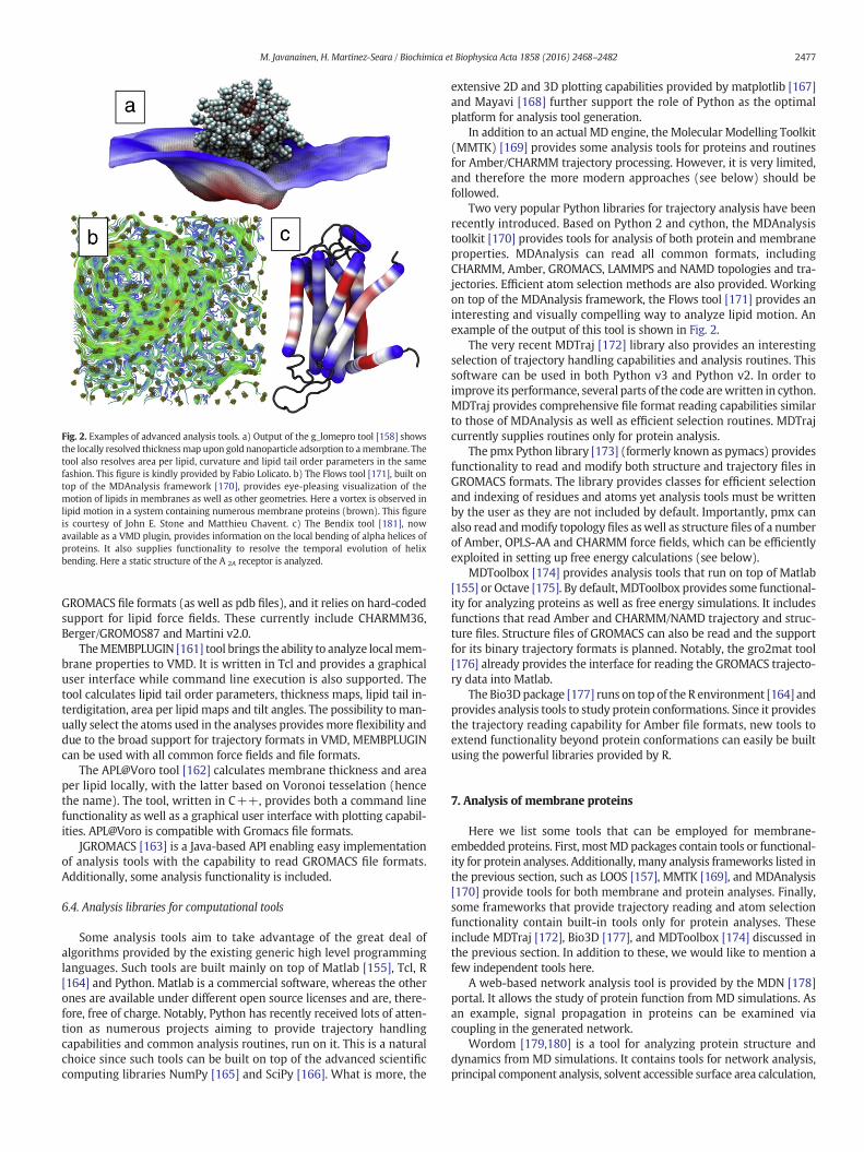
Fig. 2. Examples of advanced analysis tools. a) Output of the g_lomepro tool [158] showsthe locally resolved thicknessmap upon gold nanoparticle adsorption to amembrane. Thetool also resolves area per lipid, curvature and lipid tail order parameters in the samefashion. This figure is kindly provided by Fabio Lolicato. b) The Flows tool [171], built ontop of the MDAnalysis framework [170], provides eye-pleasing visualization of themotion of lipids in membranes as well as other geometries. Here a vortex is observed inlipid motion in a system containing numerous membrane proteins (brown). This figureis courtesy of John E. Stone and Matthieu Chavent. c) The Bendix tool [181], nowavailable as a VMD plugin, provides information on the local bending of alpha helices ofproteins. It also supplies functionality to resolve the temporal evolution of helixbending. Here a static structure of the A 2A receptor is analyzed.
2477M. Javanainen, H. Martinez-Seara / Biochimica et Biophysica Acta 1858 (2016) 2468–2482
GROMACS file formats (as well as pdb files), and it relies on hard-codedsupport for lipid force fields. These currently include CHARMM36,Berger/GROMOS87 and Martini v2.0.
TheMEMBPLUGIN [161] tool brings the ability to analyze localmem-brane properties to VMD. It is written in Tcl and provides a graphicaluser interface while command line execution is also supported. Thetool calculates lipid tail order parameters, thickness maps, lipid tail in-terdigitation, area per lipid maps and tilt angles. The possibility to man-ually select the atoms used in the analyses provides more flexibility anddue to the broad support for trajectory formats in VMD, MEMBPLUGINcan be used with all common force fields and file formats.
The APL@Voro tool [162] calculates membrane thickness and areaper lipid locally, with the latter based on Voronoi tesselation (hencethe name). The tool, written in C++, provides both a command linefunctionality as well as a graphical user interface with plotting capabil-ities. APL@Voro is compatible with Gromacs file formats.
JGROMACS [163] is a Java-based API enabling easy implementationof analysis tools with the capability to read GROMACS file formats.Additionally, some analysis functionality is included.
6.4. Analysis libraries for computational tools
Some analysis tools aim to take advantage of the great deal ofalgorithms provided by the existing generic high level programminglanguages. Such tools are built mainly on top of Matlab [155], Tcl, R[164] and Python. Matlab is a commercial software, whereas the otherones are available under different open source licenses and are, there-fore, free of charge. Notably, Python has recently received lots of atten-tion as numerous projects aiming to provide trajectory handlingcapabilities and common analysis routines, run on it. This is a naturalchoice since such tools can be built on top of the advanced scientificcomputing libraries NumPy [165] and SciPy [166]. What is more, the
extensive 2D and 3D plotting capabilities provided by matplotlib [167]and Mayavi [168] further support the role of Python as the optimalplatform for analysis tool generation.
In addition to an actual MD engine, the Molecular Modelling Toolkit(MMTK) [169] provides some analysis tools for proteins and routinesfor Amber/CHARMM trajectory processing. However, it is very limited,and therefore the more modern approaches (see below) should befollowed.
Two very popular Python libraries for trajectory analysis have beenrecently introduced. Based on Python 2 and cython, the MDAnalysistoolkit [170] provides tools for analysis of both protein and membraneproperties. MDAnalysis can read all common formats, includingCHARMM, Amber, GROMACS, LAMMPS and NAMD topologies and tra-jectories. Efficient atom selection methods are also provided. Workingon top of the MDAnalysis framework, the Flows tool [171] provides aninteresting and visually compelling way to analyze lipid motion. Anexample of the output of this tool is shown in Fig. 2.
The very recent MDTraj [172] library also provides an interestingselection of trajectory handling capabilities and analysis routines. Thissoftware can be used in both Python v3 and Python v2. In order toimprove its performance, several parts of the code arewritten in cython.MDTraj provides comprehensive file format reading capabilities similarto those of MDAnalysis as well as efficient selection routines. MDTrajcurrently supplies routines only for protein analysis.
The pmx Python library [173] (formerly known as pymacs) providesfunctionality to read and modify both structure and trajectory files inGROMACS formats. The library provides classes for efficient selectionand indexing of residues and atoms yet analysis tools must be writtenby the user as they are not included by default. Importantly, pmx canalso read andmodify topology files aswell as structure files of a numberof Amber, OPLS-AA and CHARMM force fields, which can be efficientlyexploited in setting up free energy calculations (see below).
MDToolbox [174] provides analysis tools that run on top of Matlab[155] or Octave [175]. By default, MDToolbox provides some functional-ity for analyzing proteins as well as free energy simulations. It includesfunctions that read Amber and CHARMM/NAMD trajectory and struc-ture files. Structure files of GROMACS can also be read and the supportfor its binary trajectory formats is planned. Notably, the gro2mat tool[176] already provides the interface for reading the GROMACS trajecto-ry data into Matlab.
The Bio3D package [177] runs on top of the R environment [164] andprovides analysis tools to study protein conformations. Since it providesthe trajectory reading capability for Amber file formats, new tools toextend functionality beyond protein conformations can easily be builtusing the powerful libraries provided by R.
7. Analysis of membrane proteins
Here we list some tools that can be employed for membrane-embedded proteins. First, mostMD packages contain tools or functional-ity for protein analyses. Additionally, many analysis frameworks listed inthe previous section, such as LOOS [157], MMTK [169], and MDAnalysis[170] provide tools for both membrane and protein analyses. Finally,some frameworks that provide trajectory reading and atom selectionfunctionality contain built-in tools only for protein analyses. Theseinclude MDTraj [172], Bio3D [177], and MDToolbox [174] discussed inthe previous section. In addition to these, we would like to mention afew independent tools here.
A web-based network analysis tool is provided by the MDN [178]portal. It allows the study of protein function from MD simulations. Asan example, signal propagation in proteins can be examined viacoupling in the generated network.
Wordom [179,180] is a tool for analyzing protein structure anddynamics from MD simulations. It contains tools for network analysis,principal component analysis, solvent accessible surface area calculation,

2478 M. Javanainen, H. Martinez-Seara / Biochimica et Biophysica Acta 1858 (2016) 2468–2482
and secondary structure determination. The tool is written in C but alsoprovides a Python interface.
The Bendix tool [181], now also a built-in feature of VMD, analyzesthe bending of helices in static structures and dynamic trajectories. Itprovides both visualizations like that shown in Fig. 2 aswell as temporaldata on evolution of helix conformations. The tool works with bothcoarse-grained and atomistic models and thanks to the broad supportof VMD, file formats of all common MD packages are supported.
The carma software [182] and the graphical user interface imple-mentation grcarma [183] provide tools for the analysis of the structureand dynamics of proteins. The dcd file format is supported makingcarma/grcarma readily compatible with CHARMM and NAMD. Carmaand grcarma are written in C and Perl/Tk, respectively.
8. Enhanced sampling and free energy techniques
In numerous cases the studied system is unable to sample theimportant parts of the phase space in the limited simulation time dueto high energy barriers separating the relevant conformations of thesystem. What follows is that the post-processing analysis techniquefails, and other approaches must be employed to accelerate thesampling. Such enhanced sampling methods include metadynamics,various replica exchange methods as well as simulated annealing,which all require specific runtime functionality. Luckily, the commonMD packages natively provide at least some of this functionality.
In case the simulated system is unable to sample the desired confor-mations, one is often interested in the free energy barriers separatingthese conformations. Numerous free energy methods, including slowgrowth methods, thermodynamic integration (TI), and free energyperturbation aim to overcome these limitations and provide as a resultthe excess free energy profile as a function of the selected reaction coor-dinate. This reaction coordinate can be either alchemical or geometrical.In the latter case, the tools are often referred to as potential of meanforce (PMF) methods (see Ref. [184]), and they include (adaptive) um-brella sampling as well as steered MD. Notably, these two are examplesof an equilibrium and a non-equilibrium method, respectively. In addi-tion, metadynamics can also provide the free energy profiles alongwiththe acceleration in sampling. At least some of the listed free energymethods are provided with all common MD packages.
The development of the enhanced sampling and free energy algo-rithms is nowadays very fast and the MD packages usually lag behindin implementing these new features. PLUMED [185,141] provides alibrary that rapidly adapts the newest introduced techniques. It iswritten in C++ and designed to work together with most of thecommon MD simulation packages. Thanks to this universality, the toolhas attracted a large user community. An active discussion forum is amajor advantage that PLUMED has over free energy implementationsspecific to certain MD engines. PLUMED provides tools for PMF calcula-tions including metadynamics, umbrella sampling and steered MD.Notably, different kinds of bias potentials can be applied to an extensivecollection of collective variables. METAGUI [186] is a VMD plugin de-signed to provide a graphical user interface for analyzingmetadynamicssimulations performed with PLUMED.
Additionally, some tools aim for the easy setup of free energy calcu-lations. The FESetup [187] Python tool provides an easy way to setup TIcalculations for both Amber and GROMACS simulation programs. It iscompatible with Amber force fields.
The pmx tool mentioned above as an analysis tool can also beemployed to setup free energy calculations for GROMACS using variousforce fields [173,188]. The tool is designed to generate topologies corre-sponding to amino acid mutations which can then be used with TI andother methods to calculate the associated free energy changes.
The highly mobile membrane-mimetic (HMMM) model [189]replaces lipid tails by small organic molecules, which results in one ortwo orders of magnitude larger lipid diffusion. This approach signifi-cantly reduces the time scale required to study adsorption of peripheral
proteins, and could, therefore, be considered as an enhanced samplingmethod, even though the Hamiltonian is not altered. Recently, the capa-bility of constructing systems for HMMM simulations was included inCHARMM-GUI [190].
9. Conclusions and future directions
This review provides the state of the art of the tools associated withmolecular dynamics simulations of membranes and membrane pro-teins. We began by introducing the most commonly employed lipidand protein force fields. Both of these have seen significant improve-ment in their accuracy during this last decade. In addition, the numberof supported lipid moieties has been constantly growing. We havealso pointed out that some of the lipid force fields were especiallydesigned to be compatible with protein force fields, hence allowingaccurate membrane simulations containing proteins. Such systems arefundamental biological environments, and therefore, these recentimprovements in computationalmodels commence to allow the combi-nation of simulation and experiment to tackle key questions in cellularmembrane biology.
However, we feel that the current way of force field development isfar from optimal. First of all, the numerous simulation programs providedifferent algorithms and their implementations. This results in thedependence of the outcome on the employed MD package. Therefore,introduction of methodologies that would eliminate such dependence,such as the cut-off free Lennard-Jones potential, would be highly appre-ciated. This naturally comes with the cost of reparameterizing the forcefields. However, this effort might still pay off in the long term.
We also think that the constant demand for more accurate forcefields would be best satisfied if the generation of lipid parameterscould be somehow automated. Such an automated platform would beof great importance as more and more lipid parameters as well asparameters for other types of molecules are required to account forthe vast diversity of real biological membranes.
We then reviewed the tools available for building lipid membranes.This procedure has shifted from error-prone manual approaches to-wards newly emerged user-friendly tools that are capable to constructcustom complex membranes in a highly automated fashion. Yet manyof these membrane builder tools are only compatible with a verysmall spectrum of force fields, lipid environments, and MD packages.Furthermore, we listed approaches for embedding membrane proteinsinto lipid membranes. Numerous new methods have been recentlyintroduced to take care of this procedure in away thatwould causemin-imal perturbation to the host membrane, which helps to minimize theequilibration phase after protein insertion. Some of the approachesaim to provide a very general embedding method independent offorce fields, file formats, and external software, yet they often fallshort in terms of ease-of-use. On the other hand, the straightforwardapproaches are often limited to work with few force fields and simula-tion platforms, andmight also require installation of additional externalsoftware. What is more, most approaches were designed to assemblelipids in planar geometries. However, these toolswill need to be extend-ed as simulations of vesicles, bicelles or even lipid droplets becomemore common.
We feel that the highly automated tools for setting up MD simula-tions are important as they can be readily adapted by newcomers inthe field. On the other hand, membrane simulation veterans still benefitfrom the adaptability of some seeminglymore complicated approaches.Therefore, both approaches are required to satisfy the ever growingmembrane and membrane protein simulation community.
Finally, we have discussed the emergence of new analysis tools thatallow researchers to study membranes and membrane proteins moreefficiently and employ more creative approaches. These tools are oftenopenly available, which allows researchers to dedicate more time tothe actual science rather than to tool development.

2479M. Javanainen, H. Martinez-Seara / Biochimica et Biophysica Acta 1858 (2016) 2468–2482
Analysis programs are usually slow even to the point at which someanalyses on their output trajectories are more time consuming than thewell parallelized simulations themselves. Currently available analysistools are usually not well parallelized yet rather use just one core or afew at best. This is clearly an areawherewewill see rapid improvementin the future cherished by the consolidation of development of universalanalysis environments which aim to be independent of the MD pack-ages, and therefore of file formats. Hardware development will alsohave a critical role in the analysis of MD simulations. Modern desktopcomputers are equipped with multi-core processors, which allows theuse of several cores simultaneously, thus drastically decreasing thetime required for analyses. Another point worth mentioning is theamount of memory, as several analyses require the whole trajectory tobe stored in memory at once. This is a real issue when considering thesize of the trajectories produced already nowadays, let alone in thefuture as developments in computing power will make the situationmuch worse.
Finally,wewould like to point out that only fewof the listed tools areversatile enough to be applied to other than planar membranes withsmall fluctuations. As simulated membranes grow in size, undulationswill increase drastically, which calls for smarter approaches for varioustasks related to analysis. These include e.g., separation of themembraneinto leaflets, determination of the local membrane normal, and calcula-tion of displacements along curved surfaces, to name a few. Again,analysis of vesicles, bicelles, or lipid droplets would also benefit greatlyfrom the development of more flexible and smart tools that would notrely on oversimplified assumptions.
These reviewed developments, even though still very scattered andhighly specific, clearly point towards a brighter future. The ultimate goalclearly is to have fully automated, user-friendly tools and error-freeprotocols that support all kinds of imaginable membranes interactingwith all kinds of molecules. Furthermore, automated molecule parame-terization, and intelligent, fast as well as universal analysis tools are alsodesired.
Though in our opinion we are still far from this ultimate goal, wecould possibly speed up our journey there by following a few guidelines.First, we need to encourage discussion between the developers of thedifferent MD packages so that file format compatibility stops being anissue like it is today. Perhaps it is time to consider adapting a commonstandard for the file formats, even though this would require majorwork from the developers. In the best case this could result in thecompatibility of every MD package with numerous membrane builderand protein embedding tools as well as analysis software. This wouldsubstantially enlarge the toolkits of researchers in the field. What ismore, this would allow easier cross-checking of the results providedby different MD packages.
Second,we need to promote initiatives and options that limit unnec-essary repetition of certain tasks. Nowadays, as seen from the extensivelist of tools above, several implementations of the same analysisalgorithm are frequently encountered often without clear improvementbetween them. With universally adapted file formats, each algorithmshould require only one implementation unless new features or substan-tial improvements in performance are added in new implementations inthe future. This second point, in our opinion, will require all scientificsoftware to be open source and covered with flexible licenses thatallow anybody to contribute to the code and freely redistribute theresulting version of the tool.
Summarizing, during the last years the membrane simulation fieldhas profited extensively from development of software. Indeed,performing a membrane simulation is nowadays a rather simple taskif compared to the situation a decade ago. This attracts researchers toengage in computational membrane biophysics and membrane proteinbiophysics, and therefore further contribute to the advance of the fieldvia the development of tools, models and force fields. However, wemust still educate these people of the possible pitfalls involved in themodern seemingly straightforward software. Overall, we are living in
the golden age of membrane and membrane-protein simulations andwe are confident that many great advances are still to appear andconsolidate the status of computational biophysics as an importantcounterpart to traditional laboratory experiments.
Transparency Document
The Transparency document associated with this article can befound, in online version.
Acknowledgments
We would like to thank Dr. Giray Enkavi and Dr. Maria Kalimeri forthe critical discussions about the content. We acknowledge financialsupport from the Academy of Finland through its Centre of ExcellenceProgramme.
References
[1] H. Scott, Monte Carlo studies of lipid/water interfaces, BBA-Biomembr. 814 (2)(1985) 327–332.
[2] J. Wendoloski, S. Kimatian, C. Schutt, F. Salemme, Molecular dynamics simulationof a phospholipid micelle, Science 243 (4891) (1989) 636–638.
[3] P.S. Charifson, R.G. Hiskey, L.G. Pedersen, Construction and molecular modeling ofphospholipid surfaces, J. Comput. Chem. 11 (10) (1990) 1181–1186.
[4] M.L. Berkowitz, K. Raghavan, Computer simulation of a water/membrane interface,Langmuir 7 (6) (1991) 1042–1044.
[5] T.B. Woolf, B. Roux, Molecular dynamics simulation of the gramicidin channel in aphospholipid bilayer, Proc. Natl. Acad. Sci. U. S. A. 91 (24) (1994) 11631–11635.
[6] O. Edholm, O. Berger, F. Jähnig, Structure and fluctuations of bacteriorhodopsin inthe purple membrane: a molecular dynamics study, J. Mol. Biol. 250 (1) (1995)94–111.
[7] T. Róg, M. Pasenkiewicz-Gierula, I. Vattulainen, M. Karttunen, Ordering effects ofcholesterol and its analogues, BBA-Biomembr. 1788 (1) (2009) 97–121.
[8] P.S. Niemelä, M.T. Hyvönen, I. Vattulainen, Atom-scale molecular interactions inlipid raft mixtures, BBA-Biomembr. 1788 (1) (2009) 122–135.
[9] P.C. Biggin, P.J. Bond, Molecular dynamics simulations of membrane proteins,Molecular Modeling of Proteins, Springer 2015, pp. 91–108.
[10] T. Baştuğ, S. Kuyucak, Molecular dynamics simulations of membrane proteins,Biophys. Rev. 4 (3) (2012) 271–282.
[11] K. Pluhackova, T.A. Wassenaar, R.A. Böckmann, Molecular dynamics simulations ofmembrane proteins, Membrane Biogenesis, Springer 2013, pp. 85–101.
[12] C. Kandt, W.L. Ash, D.P. Tieleman, Setting up and running molecular dynamicssimulations of membrane proteins, Methods 41 (4) (2007) 475–488.
[13] H. Martinez-Seara, T. Róg, Molecular dynamics simulations of lipid bilayers: simplerecipe of how to do it, Methods Mol. Biol. 924 (2013) 407–429.
[14] A. Kukol, Lipid membranes for membrane proteins, Molecular Modeling ofProteins, Springer 2015, pp. 73–90.
[15] K. Pluhackova, R.A. Böckmann, Biomembranes in atomistic and coarse-grainedsimulations, J. Phys. Condens. Matter 27 (32) (2015) 323103.
[16] P.E. Lopes, O. Guvench, A.D. MacKerell Jr., Current status of protein force fields formolecular dynamics simulations, Molecular Modeling of Proteins, Springer 2015,pp. 47–71.
[17] A. Botan, F. Favela-Rosales, P.F.J. Fuchs, M. Javanainen, Mf. Kanduč, W. Kulig, A.Lamberg, C. Loison, A. Lyubartsev, M.S. Miettinen, L. Monticelli, J. Määttä, O.H.S.Ollila, M. Retegan, T. Róg, H. Santuz, J. Tynkkynen, Toward atomistic resolutionstructure of phosphatidylcholine headgroup and glycerol backbone at differentambient conditions, J. Phys. Chem. B 119 (49) (2015) 15075–15088.
[18] E.A. Cino, W.-Y. Choy, M. Karttunen, Comparison of secondary structure formationusing 10 different force fields in microsecond molecular dynamics simulations, J.Chem. Theory Comput. 8 (8) (2012) 2725–2740.
[19] K. Lindorff-Larsen, P. Maragakis, S. Piana, M.P. Eastwood, R.O. Dror, D.E. Shaw,Systematic validation of protein force fields against experimental data, PLoS One7 (2) (2012), e32131.
[20] S.J. Marrink, H.J. Risselada, S. Yefimov, D.P. Tieleman, A.H. De Vries, The MARTINIforce field: coarse grained model for biomolecular simulations, J. Phys. Chem. B111 (27) (2007) 7812–7824.
[21] C.A. López, Z. Sovova, F.J. van Eerden, A.H. de Vries, S.J. Marrink, Martini force fieldparameters for glycolipids, J. Chem. Theory Comput. 9 (3) (2013) 1694–1708.
[22] L. Monticelli, S.K. Kandasamy, X. Periole, R.G. Larson, D.P. Tieleman, S.-J. Marrink,The MARTINI coarse-grained force field: extension to proteins, J. Chem. TheoryComput. 4 (5) (2008) 819–834.
[23] D.H. de Jong, G. Singh, W.D. Bennett, C. Arnarez, T.A. Wassenaar, L.V. Schafer, X.Periole, D.P. Tieleman, S.J. Marrink, Improved parameters for the martini coarse-grained protein force field, J. Chem. Theory Comput. 9 (1) (2012) 687–697.
[24] C.A. López, A.J. Rzepiela, A.H. De Vries, L. Dijkhuizen, P.H. Hunenberger, S.J. Marrink,Martini coarse-grained force field: extension to carbohydrates, J. Chem. TheoryComput. 5 (12) (2009) 3195–3210.
[25] J.J. Uusitalo, H.I. Ingólfsson, P. Akhshi, D.P. Tieleman, S.J. Marrink, Martini coarse-grained force field: extension to DNA, J. Chem. Theory Comput. 11 (8) (2015)3932–3945.

2480 M. Javanainen, H. Martinez-Seara / Biochimica et Biophysica Acta 1858 (2016) 2468–2482
[26] S.O. Yesylevskyy, L.V. Schäfer, D. Sengupta, S.J. Marrink, Polarizable water modelfor the coarse-grained MARTINI force field, PLoS Comput. Biol. 6 (6) (2010),e1000810.
[27] C. Arnarez, J.J. Uusitalo, M.F. Masman, H.I. Ingólfsson, D.H. de Jong, M.N. Melo, X.Periole, A.H. De Vries, S.J. Marrink, Dry Martini, a coarse-grained force field forlipid membrane simulations with implicit solvent, J. Chem. Theory Comput. 11(1) (2014) 260–275.
[28] T. Bereau, M. Deserno, Generic coarse-grainedmodel for protein folding and aggre-gation, J. Chem. Phys. 130 (23) (2009) 235106.
[29] Z.-J. Wang, M. Deserno, A systematically coarse-grained solvent-free model forquantitative phospholipid bilayer simulations, J. Phys. Chem. B 114 (34) (2010)11207–11220.
[30] T. Bereau, Z.-J. Wang, M. Deserno, More than the sum of its parts: coarse-grainedpeptide-lipid interactions from a simple cross-parametrization, J. Chem. Phys.140 (11) (2014) 115101.
[31] M. Orsi, J.W. Essex, The ELBA force field for coarse-grain modeling of lipid mem-branes, PLoS One 6 (12) (2011), e28637.
[32] N. Schmid, A.P. Eichenberger, A. Choutko, S. Riniker, M.Winger, A.E. Mark, W.F. vanGunsteren, Definition and testing of the GROMOS force-field versions 54 A7 and54B7, Eur. Biophys. J. 40 (7) (2011) 843–856.
[33] S.-W. Chiu, S.A. Pandit, H. Scott, E. Jakobsson, An improved united atom force fieldfor simulation of mixed lipid bilayers, J. Phys. Chem. B 113 (9) (2009) 2748–2763.
[34] A. Kukol, Lipid models for united-atom molecular dynamics simulations ofproteins, J. Chem. Theory Comput. 5 (3) (2009) 615–626.
[35] D. Poger, W.F. Van Gunsteren, A.E. Mark, A new force field for simulating phospha-tidylcholine bilayers, J. Comput. Chem. 31 (6) (2010) 1117–1125.
[36] D.M. van Aalten, R. Bywater, J.B. Findlay, M. Hendlich, R.W. Hooft, G. Vriend,PRODRG, a program for generating molecular topologies and unique moleculardescriptors from coordinates of small molecules, J. Comput. Aided Mol. Des. 10(3) (1996) 255–262.
[37] A.W. Schüttelkopf, D.M. Van Aalten, PRODRG: a tool for high-throughput crystal-lography of protein–ligand complexes, Acta Crystallogr. Sect. D Biol. Crystallogr.60 (8) (2004) 1355–1363.
[38] J.A. Lemkul, W.J. Allen, D.R. Bevan, Practical considerations for building GROMOS-compatible small-molecule topologies, J. Chem. Inf. Model. 50 (12) (2010)2221–2235.
[39] A.K. Malde, L. Zuo, M. Breeze, M. Stroet, D. Poger, P.C. Nair, C. Oostenbrink, A.E.Mark, An automated force field topology builder (ATB) and repository: version1.0, J. Chem. Theory Comput. 7 (12) (2011) 4026–4037.
[40] K.B. Koziara, M. Stroet, A.K. Malde, A.E. Mark, Testing and validation of the Auto-mated Topology Builder (ATB) version 2.0: prediction of hydration free enthalpies,J. Comput. Aided Mol. Des. 28 (3) (2014) 221–233.
[41] S. Canzar, M. El-Kebir, R. Pool, K. Elbassioni, A.K. Malde, A.E. Mark, D.P. Geerke, L.Stougie, G.W. Klau, Charge group partitioning in biomolecular simulation, J.Comp. Biol. 20 (3) (2013) 188–198.
[42] O. Berger, O. Edholm, F. Jähnig, Molecular dynamics simulations of a fluid bilayer ofdipalmitoylphosphatidylcholine at full hydration, constant pressure, and constanttemperature, Biophys. J. 72 (5) (1997) 2002.
[43] R. Tjörnhammar, O. Edholm, Reparameterized united atom model for moleculardynamics simulations of gel and fluid phosphatidylcholine bilayers, J. Chem.Theory Comput. 10 (12) (2014) 5706–5715.
[44] N. Bhatnagar, G. Kamath, J.J. Potoff, Biomolecular simulations with the transferablepotentials for phase equilibria: extension to phospholipids, J. Phys. Chem. B 117(34) (2013) 9910–9921.
[45] K. Lindorff-Larsen, S. Piana, K. Palmo, P. Maragakis, J.L. Klepeis, R.O. Dror, D.E. Shaw,Improved side-chain torsion potentials for the Amber ff99SB protein force field,Proteins Struct. Funct. Bioinf. 78 (8) (2010) 1950–1958.
[46] J.A. Maier, C. Martinez, K. Kasavajhala, L. Wickstrom, K.E. Hauser, C. Simmerling,ff14SB: improving the accuracy of protein side chain and backbone parametersfrom ff99SB, J. Chem. Theory Comput. 11 (8) (2015) 3696–3713.
[47] D.S. Cerutti, W.C. Swope, J.E. Rice, D.A. Case, ff14ipq: a self-consistent force field forcondensed-phase simulations of proteins, J. Chem. Theory Comput. 10 (10) (2014)4515–4534.
[48] Y. Duan, C. Wu, S. Chowdhury, M.C. Lee, G. Xiong, W. Zhang, R. Yang, P. Cieplak, R.Luo, T. Lee, et al., A point-charge force field for molecular mechanics simulations ofproteins based on condensed-phase quantum mechanical calculations, J. Comput.Chem. 24 (16) (2003) 1999–2012.
[49] C.J. Dickson, L. Rosso, R.M. Betz, R.C. Walker, I.R. Gould, GAFFlipid: a General AmberForce Field for the accurate molecular dynamics simulation of phospholipid, SoftMatter 8 (37) (2012) 9617–9627.
[50] Å.A. Skjevik, B.D. Madej, R.C. Walker, K. Teigen, LIPID11: a modular framework forlipid simulations using amber, J. Phys. Chem. B 116 (36) (2012) 11124–11136.
[51] C.J. Dickson, B.D. Madej, Å.A. Skjevik, R.M. Betz, K. Teigen, I.R. Gould, R.C. Walker,Lipid14: the amber lipid force field, J. Chem. Theory Comput. 10 (2) (2014)865–879.
[52] B.D. Madej, I.R. Gould, R.C. Walker, A parameterization of cholesterol for mixedlipid bilayer simulation within the Amber Lipid14 force field, J. Phys. Chem. B119 (38) (2015) 12424–12435.
[53] J.P. Jämbeck, A.P. Lyubartsev, Derivation and systematic validation of a refined all-atom force field for phosphatidylcholine lipids, J. Phys. Chem. B 116 (10) (2012)3164–3179.
[54] J.P. Jämbeck, A.P. Lyubartsev, An extension and further validation of an all-atomistic force field for biological membranes, J. Chem. Theory Comput. 8 (8)(2012) 2938–2948.
[55] J.P. Jämbeck, A.P. Lyubartsev, Another piece of the membrane puzzle: extendingslipids further, J. Chem. Theory Comput. 9 (1) (2012) 774–784.
[56] K.N. Kirschner, A.B. Yongye, S.M. Tschampel, J. González-Outeiriño, C.R. Daniels, B.L.Foley, R.J. Woods, GLYCAM06: a generalizable biomolecular force field. Carbohy-drates, J. Comput. Chem. 29 (4) (2008) 622–655.
[57] J.Wang, R.M.Wolf, J.W. Caldwell, P.A. Kollman, D.A. Case, Development and testingof a general amber force field, J. Comput. Chem. 25 (9) (2004) 1157–1174.
[58] J. Wang, W. Wang, P.A. Kollman, D.A. Case, Automatic atom type and bond typeperception in molecular mechanical calculations, J. Mol. Graphics Modell. 25 (2)(2006) 247–260.
[59] L. Huang, B. Roux, Automated force field parameterization for nonpolarizable andpolarizable atomic models based on ab initio target data, J. Chem. Theory Comput.9 (8) (2013) 3543–3556.
[60] R.M. Betz, R.C. Walker, Paramfit: automated optimization of force field parametersfor molecular dynamics simulations, J. Comput. Chem. 36 (2) (2015) 79–87.
[61] J. Wang, W. Wang, P.A. Kollman, D.A. Case, Antechamber: an accessory softwarepackage for molecular mechanical calculations, J. Am. Chem. Soc. 222 (2001) U403.
[62] D. van der Spoel, P.J. van Maaren, C. Caleman, GROMACS molecule & liquiddatabase, Bioinformatics 28 (5) (2012) 752–753.
[63] A.D. MacKerell, D. Bashford, M. Bellott, R. Dunbrack, J. Evanseck, M.J. Field, S.Fischer, J. Gao, H. Guo, S.A. Ha, et al., All-atom empirical potential for molecularmodeling and dynamics studies of proteins, J. Phys. Chem. B 102 (18) (1998)3586–3616.
[64] A.D. MacKerell, M. Feig, C.L. Brooks, Extending the treatment of backboneenergetics in protein force fields: limitations of gas-phase quantum mechanics inreproducing protein conformational distributions in molecular dynamics simula-tions, J. Comput. Chem. 25 (11) (2004) 1400–1415.
[65] R.B. Best, X. Zhu, J. Shim, P.E. Lopes, J. Mittal, M. Feig, A.D. MacKerell Jr., Optimiza-tion of the additive CHARMM all-atom protein force field targeting improved sam-pling of the backbone ɸ, ψ and side-chain χ1 and χ2 dihedral angles, J. Chem.Theory Comput. 8 (9) (2012) 3257–3273.
[66] J.B. Klauda, R.M. Venable, J.A. Freites, J.W. O′Connor, D.J. Tobias, C. Mondragon-Ramirez, I. Vorobyov, A.D. MacKerell Jr., R.W. Pastor, Update of the CHARMM all-atom additive force field for lipids: validation on six lipid types, J. Phys. Chem. B114 (23) (2010) 7830–7843.
[67] R.M. Venable, A.J. Sodt, B. Rogaski, H. Rui, E. Hatcher, A.D.MacKerell, R.W. Pastor, J.B.Klauda, Charmm all-atom additive force field for sphingomyelin: elucidation of hy-drogen bonding and of positive curvature, Biophys. J. 107 (1) (2014) 134–145.
[68] O. Guvench, S.S. Mallajosyula, E.P. Raman, E. Hatcher, K. Vanommeslaeghe, T.J.Foster, F.W. Jamison, A.D. MacKerell Jr., CHARMM additive all-atom force field forcarbohydrate derivatives and its utility in polysaccharide and carbohydrate–protein modeling, J. Chem. Theory Comput. 7 (10) (2011) 3162–3180.
[69] K. Hart, N. Foloppe, C.M. Baker, E.J. Denning, L. Nilsson, A.D. MacKerell Jr.,Optimization of the CHARMM additive force field for DNA: improved treatmentof the BI/BII conformational equilibrium, J. Chem. Theory Comput. 8 (1) (2011)348–362.
[70] J.D. Yesselman, D.J. Price, J.L. Knight, C.L. Brooks, MATCH: an atom-typingtoolset for molecular mechanics force fields, J. Comput. Chem. 33 (2) (2012)189–202.
[71] K. Vanommeslaeghe, E. Hatcher, C. Acharya, S. Kundu, S. Zhong, J. Shim, E. Darian,O. Guvench, P. Lopes, I. Vorobyov, et al., CHARMM general force field: a force fieldfor drug-like molecules compatible with the CHARMM all-atom additive biologicalforce fields, J. Comput. Chem. 31 (4) (2010) 671–690.
[72] K. Vanommeslaeghe, E.P. Raman, A.D. MacKerell Jr., Automation of the CHARMMGeneral Force Field (CGenFF) II: assignment of bonded parameters and partialatomic charges, J. Chem. Inf. Model. 52 (12) (2012) 3155–3168.
[73] K. Vanommeslaeghe, A.D. MacKerell Jr., Automation of the CHARMMGeneral ForceField (CGenFF) I: bond perception and atom typing, J. Chem. Inf. Model. 52 (12)(2012) 3144–3154.
[74] W.L. Jorgensen, J. Tirado-Rives, The OPLS [optimized potentials for liquidsimulations] potential functions for proteins, energy minimizations for crystals ofcyclic peptides and crambin, J. Am. Chem. Soc. 110 (6) (1988) 1657–1666.
[75] M.J. Robertson, J. Tirado-Rives, W.L. Jorgensen, Improved peptide and proteintorsional energetics with the OPLS-AA force field, J. Chem. Theory Comput. 11(7) (2015) 3499–3509.
[76] A. Maciejewski, M. Pasenkiewicz-Gierula, O. Cramariuc, I. Vattulainen, T. Rog,Refined OPLS all-atom force field for saturated phosphatidylcholine bilayers atfull hydration, J. Phys. Chem. B 118 (17) (2014) 4571–4581.
[77] W. Kulig, M. Pasenkiewicz-Gierula, T. Róg, Cis and trans unsaturated phosphatidyl-choline bilayers: a molecular dynamics simulation study, Chem. Phys. Lipids 195(2016) 12–20.
[78] D. Kony, W. Damm, S. Stoll, W.F. Van Gunsteren, An improved OPLS–AA force fieldfor carbohydrates, J. Comput. Chem. 23 (15) (2002) 1416–1429.
[79] A. Cordom, G. Caltabiano, L. Pardo, Membrane protein simulations using AMBERforce field and Berger lipid parameters, J. Chem. Theory Comput. 8 (3) (2012)948–958.
[80] D.P. Tieleman, J.L. MacCallum, W.L. Ash, C. Kandt, Z. Xu, L. Monticelli, Membraneprotein simulations with a united-atom lipid and all-atom protein model: lipid–protein interactions, side chain transfer free energies and model proteins, J. Phys.Condens. Matter 18 (28) (2006) S1221.
[81] W. Han, C.-K. Wan, F. Jiang, Y.-D. Wu, PACE force field for protein simulations. 1.full parameterization of version 1 and verification, J. Chem. Theory Comput. 6(11) (2010) 3373–3389.
[82] W. Han, C.-K. Wan, Y.-D. Wu, PACE force field for protein simulations. 2. foldingsimulations of peptides, J. Chem. Theory Comput. 6 (11) (2010) 3390–3402.
[83] C.-K. Wan, W. Han, Y.-D. Wu, Parameterization of pace force field for membraneenvironment and simulation of helical peptides and helix–helix association, J.Chem. Theory Comput. 8 (1) (2011) 300–313.

2481M. Javanainen, H. Martinez-Seara / Biochimica et Biophysica Acta 1858 (2016) 2468–2482
[84] Q. Shi, S. Izvekov, G.A. Voth, Mixed atomistic and coarse-grained moleculardynamics: simulation of a membrane-bound ion channel, J. Phys. Chem. B 110(31) (2006) 15045–15048.
[85] T.A. Wassenaar, H.I. Ingólfsson, M. Priess, S.J. Marrink, L.V. Schäfer, Mixing MARTI-NI: electrostatic coupling in hybrid atomistic-coarse-grained biomolecular simula-tions, J. Phys. Chem. B 117 (13) (2013) 3516–3530.
[86] J. Chowdhary, E. Harder, P.E. Lopes, L. Huang, A.D. MacKerell Jr., B. Roux, A polariz-able force field of dipalmitoylphosphatidylcholine based on the classical drudemodel for molecular dynamics simulations of lipids, J. Phys. Chem. B 117 (31)(2013) 9142–9160.
[87] P.E. Lopes, J. Huang, J. Shim, Y. Luo, H. Li, B. Roux, A.D. MacKerell Jr., Polarizableforce field for peptides and proteins based on the classical Drude oscillator, J.Chem. Theory Comput. 9 (12) (2013) 5430–5449.
[88] C.M. Baker, V.M. Anisimov, A.D. MacKerell Jr., Development of CHARMM polariz-able force field for nucleic acid bases based on the classical Drude oscillatormodel, J. Phys. Chem. B 115 (3) (2010) 580–596.
[89] C.M. Baker, Polarizable force fields for molecular dynamics simulations of biomol-ecules, Wiley Interdiscip. Rev. Comput. Mol. Sci. 5 (2) (2015) 241–254.
[90] J. Huang, P.E. Lopes, B. Roux, A.D.MacKerell Jr., Recent advances in polarizable forcefields for macromolecules: microsecond simulations of proteins using the classicalDrude oscillator model, J. Phys. Chem. Lett. 5 (18) (2014) 3144–3150.
[91] K. Vanommeslaeghe, A. MacKerell, CHARMM additive and polarizable force fieldsfor biophysics and computer-aided drug design, BBA-Biomembr. 1850 (5) (2015)861–871.
[92] P. Brocos, P. Mendoza-Espinosa, R. Castillo, J. Mas-Oliva, Á. Pineiro, Multiscalemolecular dynamics simulations of micelles: coarse-grain for self-assembly andatomic resolution for finer details, Soft Matter 8 (34) (2012) 9005–9014.
[93] T.A. Wassenaar, K. Pluhackova, R.A. Bockmann, S.J. Marrink, D.P. Tieleman, Goingbackward: a flexible geometric approach to reverse transformation from coarsegrained to atomistic models, J. Chem. Theory Comput. 10 (2) (2014) 676–690.
[94] P.J. Stansfeld, M.S. Sansom, From coarse grained to atomistic: a serial multiscaleapproach to membrane protein simulations, J. Chem. Theory Comput. 7 (4)(2011) 1157–1166.
[95] P. Larsson, P.M. Kasson, Lipid converter, a framework for lipid manipulations inmolecular dynamics simulations, J. Membr. Biol. 247 (11) (2014) 1137–1140.
[96] D. Marsh, Handbook of lipid bilayers, second ed. CRC Press, 2013, ISBN 978-1-4200-8833-5.
[97] H. Koldsø, M.S. Sansom, Organization and dynamics of receptor proteins in aplasma membrane, J. Am. Chem. Soc. 137 (46) (2015) 14694–14704.
[98] H.I. Ingólfsson, M.N. Melo, F.J. van Eerden, C. Arnarez, C.A. Lopez, T.A.Wassenaar, X.Periole, A.H. De Vries, D.P. Tieleman, S.J. Marrink, Lipid organization of the plasmamembrane, J. Am. Chem. Soc. 136 (41) (2014) 14554–14559.
[99] R. Ramon Guixà-González, M. Javanainen, M. Gómez-Soler, B.n. Cordobilla, J.C.Domingo, F. Sanz, M. Pastor, F. Ciruela, H. Martinez-Seara, J. Selent, Membraneomega-3 fatty acids modulate the oligomerisation kinetics of adenosine A2A anddopamine D2 receptors, Sci. Rep. 6 (2016) 19839.
[100] A.Y. Antipina, A.A. Gurtovenko, Molecular mechanism of calcium-induced adsorp-tion of DNA on zwitterionic phospholipid membranes, J. Phys. Chem. B 119 (22)(2015) 6638–6645.
[101] C. Bovigny, G. Tamo, T. Lemmin, N. Mano, M. Dal Peraro, LipidBuilder: a frameworkto build realistic models for biological membranes, J. Chem. Inf. Model. 55 (12)(2015) 2491–2499.
[102] M.D. Hanwell, D.E. Curtis, D.C. Lonie, T. Vandermeersch, E. Zurek, G.R. Hutchison,Avogadro: an advanced semantic chemical editor, visualization, and analysisplatform, J. Cheminform. 4 (1) (2012) 17.
[103] N.M. OLBoyle, M. Banck, C.A. James, C. Morley, T. Vandermeersch, G.R. Hutchison,Open babel: an open chemical toolbox, J. Cheminform. 3 (2011) 33.
[104] T.J. Piggot, Á. Piñeiro, S. Khalid, Molecular dynamics simulations of phosphatidyl-choline membranes: a comparative force field study, J. Chem. Theory Comput. 8(11) (2012) 4593–4609.
[105] J. Domański, P.J. Stansfeld, M.S. Sansom, O. Beckstein, Lipidbook: a public reposito-ry for force-field parameters used in membrane simulations, J. Membr. Biol. 236(3) (2010) 255–258.
[106] E.L. Wu, X. Cheng, S. Jo, H. Rui, K.C. Song, E.M. Dávila-Contreras, Y. Qi, J. Lee, V.Monje-Galvan, R.M. Venable, J.B. Klauda, W. Im, CHARMM-GUI membrane buildertoward realistic biological membrane simulations, J. Comput. Chem. 35 (27)(2014) 1997–2004.
[107] M. Javanainen, Universal method for embedding proteins into complex lipidbilayers for molecular dynamics simulations, J. Chem. Theory Comput. 10 (6)(2014) 2577–2582.
[108] H. Martinez-Seara, T. Róg, M. Karttunen, I. Vattulainen, R. Reigada, Why is the sn-2chain of monounsaturated glycerophospholipids usually unsaturated whereas thesn-1 chain is saturated? Studies of 1-stearoyl-2-oleoyl-sn-glycero-3-phosphatidyl-choline (SOPC) and 1-oleoyl-2-stearoyl-sn-glycero-3-phosphatidylcholine (OSPC)membranes with and without cholesterol, J. Phys. Chem. B 113 (24) (2009)8347–8356.
[109] C. Hong, D.P. Tieleman, Y. Wang, Microsecond molecular dynamics simulations oflipid mixing, Langmuir 30 (40) (2014) 11993–12001.
[110] H. Berman, J. Westbrook, Z. Feng, G. Gilliland, T. Bhat, H. Weissig, I. Shindyalov, P.Bourne, The Protein Data Bank, Nucleic Acids Res. 28 (1) (2000) 235–242.
[111] S. Jo, J.B. Lim, J.B. Klauda, W. Im, CHARMM-GUI membrane builder for mixedbilayers and its application to yeast membranes, Biophys. J. 97 (1) (2009) 50–58.
[112] M.M. Ghahremanpour, S.S. Arab, S.B. Aghazadeh, J. Zhang, D. van der Spoel,MemBuilder: a web-based graphical interface to build heterogeneously mixedmembrane bilayers for the GROMACS biomolecular simulation program, Bioinfor-matics 30 (3) (2013) 439–441.
[113] L. Martnez, R. Andrade, E.G. Birgin, J.M. Martnez, PACKMOL: a package for buildinginitial configurations for molecular dynamics simulations, J. Comput. Chem. 30(13) (2009) 2157–2164.
[114] T.A. Wassenaar, H.I. Ingólfsson, R.A. Bockmann, D.P. Tieleman, S.J. Marrink, Compu-tational lipidomics with insane: a versatile tool for generating custom membranesfor molecular simulations, J. Chem. Theory Comput. 11 (5) (2015) 2144–2155.
[115] J. Lee, X. Cheng, J.M. Swails, M.S. Yeom, P.K. Eastman, J.A. Lemkul, S. Wei, J. Buckner,J.C. Jeong, Y. Qi, CHARMM-GUI input generator for NAMD, GROMACS, AMBER,OpenMM, and CHARMM/OpenMM simulations using the CHARMM36 additiveforce field, J. Chem. Theory Comput. 12 (1) (2016) 405–413.
[116] L. Shen, D. Bassolino, T. Stouch, Transmembrane helix structure, dynamics, andinteractions: multi-nanosecond molecular dynamics simulations, Biophys. J. 73(1) (1997) 3.
[117] J.D. Faraldo-Gómez, G.R. Smith, M.S. Sansom, Setting up and optimization ofmembrane protein simulations, Eur. Biophys. J. 31 (3) (2002) 217–227.
[118] T.H. Schmidt, C. Kandt, LAMBADA and InflateGRO2: efficient membrane alignmentand insertion of membrane proteins for molecular dynamics simulations, J. Chem.Inf. Model. 52 (10) (2012) 2657–2669.
[119] M.G. Wolf, M. Hoefling, C. Aponte-Santamara, H. Grubmüller, G. Groenhof, g_membed: efficient insertion of a membrane protein into an equilibratedlipid bilayer with minimal perturbation, J. Comput. Chem. 31 (11) (2010)2169–2174.
[120] R. Staritzbichler, C. Anselmi, L.R. Forrest, J.D. Faraldo-Gómez, GRIFFIN: a versatilemethodology for optimization of protein–lipid interfaces for membrane proteinsimulations, J. Chem. Theory Comput. 7 (4) (2011) 1167–1176.
[121] T.A. Wassenaar, K. Pluhackova, A. Moussatova, D. Sengupta, S.J. Marrink, D.P.Tieleman, R.A. Böckmann, High-throughput simulations of dimer and trimerassembly of membrane proteins. the DAFT approach, J. Chem. Theory Comput.11 (5) (2015) 2278–2291.
[122] E. Jefferys, Z.A. Sands, J. Shi, M.S. Sansom, P.W. Fowler, Alchembed: a computation-al method for incorporating multiple proteins into complex lipid geometries, J.Chem. Theory Comput. 11 (6) (2015) 2743–2754.
[123] P.J. Stansfeld, J.E. Goose, M. Caffrey, E.P. Carpenter, J.L. Parker, S. Newstead, M.S.Sansom, MemProtMD: automated insertion of membrane protein structures intoexplicit lipid membranes, Structure 23 (7) (2015) 1350–1361.
[124] S. Jo, T. Kim, V.G. Iyer, W. Im, CHARMM-GUI: a web-based graphical user interfacefor CHARMM, J. Comput. Chem. 29 (11) (2008) 1859–1865.
[125] S. Jo, T. Kim, W. Im, Automated builder and database of protein/membrane com-plexes for molecular dynamics simulations, PLoS One 2 (9) (2007), e880.
[126] Y. Qi, H.I. Ingólfsson, X. Cheng, J. Lee, S.J. Marrink, W. Im, CHARMM-GUI Martinimaker for coarse-grained simulations with the Martini force field, J. Chem. TheoryComput. 11 (9) (2015) 4486–4494.
[127] Y. Qi, X. Cheng,W. Han, S. Jo, K. Schulten,W. Im, CHARMM-GUI PACE CG builder forsolution, micelle, and bilayer coarse-grained simulations, J. Chem. Inf. Model. 54(3) (2014) 1003–1009.
[128] M.A. Lomize, A.L. Lomize, I.D. Pogozheva, H.I. Mosberg, Opm: orientations ofproteins in membranes database, Bioinformatics 22 (5) (2006) 623–625.
[129] M.A. Lomize, I.D. Pogozheva, H. Joo, H.I. Mosberg, A.L. Lomize, OPM database andPPM web server: resources for positioning of proteins in membranes, NucleicAcids Res. 40 (D1) (2012) D370–D376.
[130] T. Kimmett, N. Smith, S. Witham, M. Petukh, S. Sarkar, E. Alexov, ProBLM webserver: protein and membrane placement and orientation package, Comput.Math. Methods Med. (2014).
[131] T. Nugent, D.T. Jones, Membrane protein orientation and refinement using aknowledge-based statistical potential, BMC Bioinf. 14 (1) (2013) 276.
[132] M.J. Abraham, T. Murtola, R. Schulz, S. Páll, J.C. Smith, B. Hess, E. Lindahl, GROMACS:high performance molecular simulations throughmulti-level parallelism from lap-tops to supercomputers, SoftwareX 1–2 (2015) 19–25.
[133] J.C. Phillips, R. Braun, W. Wang, J. Gumbart, E. Tajkhorshid, E. Villa, C. Chipot, R.D.Skeel, L. Kale, K. Schulten, Scalable molecular dynamics with NAMD, J. Comput.Chem. 26 (16) (2005) 1781–1802.
[134] A.-P. Hynninen, M.F. Crowley, New faster CHARMM molecular dynamics engine, J.Comput. Chem. 35 (5) (2013) 406–413.
[135] B.R. Brooks, C.L. Brooks, A.D. Mackerell, L. Nilsson, R.J. Petrella, B. Roux, Y. Won, G.Archontis, C. Bartels, S. Boresch, A. Caflisch, L. Caves, Q. Cui, A.R. Dinner, M. Feig, S.Fischer, J. Gao, M. Hodoscek,W. Im, K. Kuczera, T. Lazaridis, J. Ma, V. Ovchinnikov, E.Paci, R.W. Pastor, C.B. Post, J.Z. Pu, M. Schaefer, B. Tidor, R.M. Venable, H.L.Woodcock, X. Wu, W. Yang, D.M. York, M. Karplus, CHARMM: the biomolecularsimulation program, J. Comput. Chem. 30 (10) (2009) 1545–1614.
[136] D. Case, J. Berryman, R. Betz, D. Cerutti, T. Cheatham III, T. Darden, R. Duke, T. Giese,H. Gohlke, A. Goetz, et al., AMBER, University of California, San Francisco, 2015.
[137] R. Salomon-Ferrer, D.A. Case, R.C. Walker, An overview of the Amber biomolecularsimulation package, Wiley Interdiscip. Rev. Comput. Mol. Sci. 3 (2) (2012)198–210.
[138] P. Eastman, M.S. Friedrichs, J.D. Chodera, R.J. Radmer, C.M. Bruns, J.P. Ku, K.A.Beauchamp, T.J. Lane, L.-P. Wang, D. Shukla, T. Tye, M. Houston, T. Stich, C. Klein,M.R. Shirts, V.S. Pande, OpenMM 4: a reusable, extensible, hardware independentlibrary for high performance molecular simulation, J. Chem. Theory Comput. 9 (1)(2013) 461–469.
[139] S. Plimpton, Fast parallel algorithms for short-range molecular dynamics, J.Comput. Phys. 117 (1) (1995) 1–19.
[140] M.F. Crowley, M.J. Williamson, R.C. Walker, CHAMBER: comprehensive support forCHARMM force fields within the AMBER software, Int. J. Quantum Chem. 109 (15)(2009) 3767–3772.
[141] G.A. Tribello, M. Bonomi, D. Branduardi, C. Camilloni, G. Bussi, PLUMED 2: newfeathers for an old bird, Comput. Phys. Commun. 185 (2) (2014) 604–613.

2482 M. Javanainen, H. Martinez-Seara / Biochimica et Biophysica Acta 1858 (2016) 2468–2482
[142] S. Pronk, S. Pall, R. Schulz, P. Larsson, P. Bjelkmar, R. Apostolov, M.R. Shirts, J.C.Smith, P.M. Kasson, D. van der Spoel, B. Hess, E. Lindahl, GROMACS 4.5: a high-throughput and highly parallel open source molecular simulation toolkit,Bioinformatics 29 (7) (2013) 845–854.
[143] W. Humphrey, A. Dalke, K. Schulten, VMD: visual molecular dynamics, J. Mol.Graph. 14 (1) (1996) 33–38.
[144] R.A. Sayle, E.J. Milner-White, RASMOL: biomolecular graphics for all, TrendsBiochem. Sci. 20 (9) (1995) 374.
[145] D.S. Goodsell, Representing Structural Information with RasMol, John Wiley &Sons, Inc., 2002 (Ch. UNIT 5.4).
[146] L.L.C. Schrödinger, The PyMOL Molecular Graphics System, version 1.3r1, August2010.
[147] O.S. Ollila, H.J. Risselada, M. Louhivuori, E. Lindahl, I. Vattulainen, S.J. Marrink, 3Dpressure field in lipid membranes and membrane–protein complexes, Phys. Rev.Lett. 102 (7) (2009) 078101.
[148] J.M. Vanegas, A. Torres-Sánchez, M. Arroyo, Importance of force decomposition forlocal stress calculations in biomembrane molecular simulations, J. Chem. TheoryComput. 10 (2) (2014) 691–702.
[149] A. Torres-Sánchez, J.M. Vanegas, M. Arroyo, Examining the mechanical equilibriumof microscopic stresses in molecular simulations, Phys. Rev. Lett. 114 (25) (2015)258102.
[150] J. Sonne, F.Y. Hansen, G.H. Peters, Methodological problems in pressure profilecalculations for lipid bilayers, J. Chem. Phys. 122 (12) (2005) 124903.
[151] T. Tu, C. Rendleman, D.W. Borhani, R.O. Dror, J. Gullingsrud, M.Ø. Jensen, J.L.Klepeis, P. Maragakis, P. Miller, K. Stafford, et al., A scalable parallel frameworkfor analyzing terascale molecular dynamics simulation trajectories, High Perfor-mance Computing, Networking, Storage and Analysis, 2008. SC 2008, InternationalConference for, IEEE 2008, pp. 1–12.
[152] D.R. Roe, T.E. Cheatham III, PTRAJ and CPPTRAJ: software for processing andanalysis of molecular dynamics trajectory data, J. Chem. Theory Comput. 9 (7)(2013) 3084–3095.
[153] D.A. Case, T.E. Cheatham, T. Darden, H. Gohlke, R. Luo, K.M. Merz, A. Onufriev, C.Simmerling, B. Wang, R.J. Woods, The Amber biomolecular simulation programs,J. Comput. Chem. 26 (16) (2005) 1668–1688.
[154] S. Plimpton, Fast parallel algorithms for short-range molecular dynamics, J.Comput. Phys. 117 (1) (1995) 1–19.
[155] The Mathworks, Inc., Natick, Massachusetts, MATLAB version 8.5.0.197613(R2015a), 2015.
[156] T. Williams, C. Kelley, R. Lang, D. Kotz, J. Campbell, G. Elber, A. Woo, et al., Gnuplot5: an interactive plotting program, June 2015 (URL http://www.gnuplot.info).
[157] T.D. Romo, N. Leioatts, A. Grossfield, Lightweight object oriented structure analysis:Tools for building tools to analyze molecular dynamics simulations, J. Comput.Chem. 35 (32) (2014) 2305–2318.
[158] V. Gapsys, B.L. de Groot, R. Briones, Computational analysis of local membraneproperties, J. Comput. Aided Mol. Des. 27 (10) (2013) 845–858.
[159] W.J. Allen, J.A. Lemkul, D.R. Bevan, GridMAT-MD: a grid-based membrane analysistool for use with molecular dynamics, J. Comput. Chem. 30 (12) (2009)1952–1958.
[160] M. Carr, C.E. MacPhee, Membrainy: a ‘smart’, unified membrane analysis tool,Source Code Biol. Med. 10 (1) (2015) 3.
[161] R. Guixà-González, I. Rodriguez-Espigares, J.M. Ramrez-Anguita, P. Carrió-Gaspar,H. Martinez-Seara, T. Giorgino, J. Selent, MEMBPLUGIN: studying membranecomplexity in VMD, Bioinformatics 30 (10) (2014) 1478–1480.
[162] G. Lukat, J. Krüger, B. Sommer, APL@ Voro: a voronoi-based membrane analysistool for GROMACS trajectories, J. Chem. Inf. Model. 53 (11) (2013) 2908–2925.
[163] M. Münz, P.C. Biggin, JGromacs: a Java package for analyzing protein simulations, J.Chem. Inf. Model. 52 (1) (2012) 255–259.
[164] R Core Team, R: a language and environment for statistical computing, R Founda-tion for Statistical Computing, Vienna, Austria, 2015 (https://www.R-project.org).
[165] S. Van Der Walt, S.C. Colbert, G. Varoquaux, The NumPy array: a structure forefficient numerical computation, IEEE Comput. Sci. Eng. 13 (2) (2011) 22–30.
[166] E. Jones, T. Oliphant, P. Peterson, SciPy: open source scientific tools for Python,2001 (Online; accessed 2016-03-04) http://www.scipy.org/.
[167] J.D. Hunter, Matplotlib: a 2D graphics environment, IEEE Comput. Sci. Eng. 9 (3)(2007) 90–95.
[168] P. Ramachandran, G. Varoquaux, Mayavi: 3D visualization of scientific data, IEEEComput. Sci. Eng. 13 (2) (2011) 40–51.
[169] K. Hinsen, The molecular modeling toolkit: a new approach to molecular simula-tions, J. Comput. Chem. 21 (2) (2000) 79–85.
[170] N. Michaud-Agrawal, E.J. Denning, T.B. Woolf, O. Beckstein, MDAnalysis: a toolkitfor the analysis of molecular dynamics simulations, J. Comput. Chem. 32 (10)(2011) 2319–2327.
[171] M. Chavent, T. Reddy, J. Goose, A.C.E. Dahl, J.E. Stone, B. Jobard, M.S. Sansom, Meth-odologies for the analysis of instantaneous lipid diffusion in md simulations oflarge membrane systems, Faraday Discuss. 169 (2014) 455–475.
[172] R.T. McGibbon, K.A. Beauchamp, M.P. Harrigan, C. Klein, J.M. Swails, C.X.Hernández, C.R. Schwantes, L.-P. Wang, T.J. Lane, V.S. Pande, MDTraj: a modernopen library for the analysis of molecular dynamics trajectories, Biophys. J. 109(8) (2015) 1528–1532.
[173] V. Gapsys, S. Michielssens, D. Seeliger, B.L. de Groot, pmx: automated proteinstructure and topology generation for alchemical perturbations, J. Comput.Chem. 36 (5) (2015) 348–354.
[174] Y. Matsunaga, mdtoolbox documentation, August 2015 URL http://mdtoolbox.readthedocs.org/mdtoolbox.
[175] J.W. Eaton, D. Bateman, S. Hauberg, R. Wehbring, GNU Octave version 4.0.0manual: a high-level interactive language for numerical computations, 2015 URLhttp://www.gnu.org/software/octave/doc/interpreter.
[176] H. Dien, C.M. Deane, B. Knapp, Gro2mat: a package to efficiently read Gromacsoutput in Matlab, J. Comput. Chem. 35 (20) (2014) 1528–1531.
[177] B.J. Grant, A.P. Rodrigues, K.M. ElSawy, J.A. McCammon, L.S. Caves, Bio3d: an Rpackage for the comparative analysis of protein structures, Bioinformatics 22(21) (2006) 2695–2696.
[178] A.A. Ribeiro, V. Ortiz, MDN: a web portal for network analysis of moleculardynamics simulations, Biophys. J. 109 (6) (2015) 1110–1116.
[179] M. Seeber, M. Cecchini, F. Rao, G. Settanni, A. Caflisch, Wordom: a program forefficient analysis of molecular dynamics simulations, Bioinformatics 23 (19)(2007) 2625–2627.
[180] M. Seeber, A. Felline, F. Raimondi, S. Muff, R. Friedman, F. Rao, A. Caflisch, F. Fanelli,Wordom: a user-friendly program for the analysis of molecular structures, trajec-tories, and free energy surfaces, J. Comput. Chem. 32 (6) (2011) 1183–1194.
[181] A.C.E. Dahl, M. Chavent, M.S. Sansom, Bendix: intuitive helix geometry analysis andabstraction, Bioinformatics 28 (16) (2012) 2193–2194.
[182] N.M. Glykos, Software news and updates carma: a molecular dynamics analysisprogram, J. Comput. Chem. 27 (14) (2006) 1765–1768.
[183] P.I. Koukos, N.M. Glykos, Grcarma: a fully automated task-oriented interface for theanalysis of molecular dynamics trajectories, J. Comput. Chem. 34 (26) (2013)2310–2312.
[184] D. Trzesniak, A.-P.E. Kunz, W.F. van Gunsteren, a comparison of methods to com-pute the potential of mean force, Chem. Phys. Chem. 8 (1) (2007) 162–169.
[185] M. Bonomi, D. Branduardi, G. Bussi, C. Camilloni, D. Provasi, P. Raiteri, D. Donadio, F.Marinelli, F. Pietrucci, R.A. Broglia, et al., PLUMED: a portable plugin for free-energycalculations with molecular dynamics, Comput. Phys. Commun. 180 (10) (2009)1961–1972.
[186] X. Biarnés, F. Pietrucci, F. Marinelli, A. Laio, METAGUI. a VMD interface for analyzingmetadynamics and molecular dynamics simulations, Comput. Phys. Commun. 183(1) (2012) 203–211.
[187] H.H. Loeffler, J. Michel, C.J. Woods, FESetup: automating setup for alchemical freeenergy simulations, J. Chem. Inf. Model. 55 (12) (2015) 2485–2490.
[188] D. Seeliger, B.L. De Groot, Protein thermostability calculations using alchemical freeenergy simulations, Biophys. J. 98 (10) (2010) 2309–2316.
[189] Y.Z. Ohkubo, T.V. Pogorelov, M.J. Arcario, G.A. Christensen, E. Tajkhorshid, Acceler-ating membrane insertion of peripheral proteins with a novel membrane mimeticmodel, Biophys. J. 102 (9) (2012) 2130–2139.
[190] Y. Qi, X. Cheng, J. Lee, J.V. Vermaas, T.V. Pogorelov, E. Tajkhorshid, S. Park, J.B.Klauda, W. Im, CHARMM-GUI HMMM builder for membrane simulations withthe highly mobile membrane-mimetic model, Biophys. J. 109 (10) (2015)2012–2022.