Embed Size (px)
Citation preview

Biochimica et Biophysica Acta 1845 (2014) 136–154
Contents lists available at ScienceDirect
Biochimica et Biophysica Acta
j ourna l homepage: www.e lsev ie r .com/ locate /bbacan
Review
Targeting the STAT3 signaling pathway in cancer: Role of synthetic andnatural inhibitors
Kodappully Sivaraman Siveen a,1, Sakshi Sikka a,b,1, Rohit Surana a,b, Xiaoyun Dai a, Jingwen Zhang a,Alan Prem Kumar a,b,c,d, Benny K.H. Tan a, Gautam Sethi a,b,⁎, Anupam Bishayee e,⁎⁎a Department of Pharmacology, Yong Loo Lin School of Medicine, National University of Singapore, Singaporeb Cancer Science Institute of Singapore, National University of Singapore, Centre for Translational Medicine, Singaporec School of Biomedical Sciences, Faculty of Health Sciences, Curtin University, Western Australia, Australiad Department of Biological Sciences, University of North Texas, Denton, TX, USAe Department of Pharmaceutical Sciences, School of Pharmacy, American University of Health Sciences, Signal Hill, CA, USA
⁎ Correspondence to: G. Sethi, Department of Pharmac⁎⁎ Correspondence to: A. Bishayee, Department of PharmTel.: +1 562 988 2278.
E-mail addresses: [email protected] (G. Sethi), abisha1 Both the authors contributed equally to this article.
0304-419X/$ – see front matter © 2013 Elsevier B.V. All rhttp://dx.doi.org/10.1016/j.bbcan.2013.12.005
a b s t r a c t
a r t i c l e i n f oArticle history:Received 15 August 2013Received in revised form 24 December 2013Accepted 27 December 2013Available online 2 January 2014
Keywords:STAT3InhibitorsTumorigenesisInflammationProliferationMetastasis
Signal transducers and activators of transcription (STATs) comprise a family of cytoplasmic transcription factorsthat mediate intracellular signaling that is usually generated at cell surface receptors and thereby transmit it tothe nucleus. Numerous studies have demonstrated constitutive activation of STAT3 in a wide variety of humantumors, including hematological malignancies (leukemias, lymphomas, and multiple myeloma) as well asdiverse solid tumors (such as head and neck, breast, lung, gastric, hepatocellular, colorectal andprostate cancers).There is strong evidence to suggest that aberrant STAT3 signaling promotes initiation and progression of humancancers by either inhibiting apoptosis or inducing cell proliferation, angiogenesis, invasion, and metastasis.Suppression of STAT3 activation results in the induction of apoptosis in tumor cells, and accordingly its pharma-cological modulation by tyrosine kinase inhibitors, antisense oligonucleotides, decoy nucleotides, dominant neg-ative proteins, RNA interference and chemopreventive agents have been employed to suppress the proliferationof various human cancer cells in culture and tumorigenicity in vivo. However, the identification and developmentof novel drugs that can target deregulated STAT3 activation effectively remains an important scientific and clin-ical challenge. This review presents the evidence for critical roles of STAT3 in oncogenesis and discusses the po-tential for development of novel cancer therapies based on mechanistic understanding of STAT3 signalingcascade.
© 2013 Elsevier B.V. All rights reserved.
Contents
1. Introduction . . . . . . . . . . . . . . . . . . . . . . . . . . . . . . . . . . . . . . . . . . . . . . . . . . . . . . . . . . . . . . 1372. STAT3 activation by diverse agents . . . . . . . . . . . . . . . . . . . . . . . . . . . . . . . . . . . . . . . . . . . . . . . . . . . . 1383. Role of STAT3 in tumorigenesis . . . . . . . . . . . . . . . . . . . . . . . . . . . . . . . . . . . . . . . . . . . . . . . . . . . . . 139
3.1. Proliferation . . . . . . . . . . . . . . . . . . . . . . . . . . . . . . . . . . . . . . . . . . . . . . . . . . . . . . . . . . . 1403.2. Survival . . . . . . . . . . . . . . . . . . . . . . . . . . . . . . . . . . . . . . . . . . . . . . . . . . . . . . . . . . . . . 1413.3. Inflammation . . . . . . . . . . . . . . . . . . . . . . . . . . . . . . . . . . . . . . . . . . . . . . . . . . . . . . . . . . 1413.4. Invasion . . . . . . . . . . . . . . . . . . . . . . . . . . . . . . . . . . . . . . . . . . . . . . . . . . . . . . . . . . . . . 1423.5. Metastasis . . . . . . . . . . . . . . . . . . . . . . . . . . . . . . . . . . . . . . . . . . . . . . . . . . . . . . . . . . . . 1423.6. Angiogenesis . . . . . . . . . . . . . . . . . . . . . . . . . . . . . . . . . . . . . . . . . . . . . . . . . . . . . . . . . . 143
4. Selected natural inhibitors of the STAT3 signaling pathway . . . . . . . . . . . . . . . . . . . . . . . . . . . . . . . . . . . . . . . . . 1434.1. Betulinic acid . . . . . . . . . . . . . . . . . . . . . . . . . . . . . . . . . . . . . . . . . . . . . . . . . . . . . . . . . . 1434.2. Butein . . . . . . . . . . . . . . . . . . . . . . . . . . . . . . . . . . . . . . . . . . . . . . . . . . . . . . . . . . . . . 1444.3. Caffeic acid . . . . . . . . . . . . . . . . . . . . . . . . . . . . . . . . . . . . . . . . . . . . . . . . . . . . . . . . . . . 1454.4. Capsaicin . . . . . . . . . . . . . . . . . . . . . . . . . . . . . . . . . . . . . . . . . . . . . . . . . . . . . . . . . . . . 145
ology, Yong Loo Lin School of Medicine, National University of Singapore, Singapore. Tel.: +65 651 63267.aceutical Sciences, School of Pharmacy, AmericanUniversity of Health Sciences, Signal Hill, 1600 East Hill Street, CA 90755, USA.
[email protected] (A. Bishayee).
ights reserved.

137K.S. Siveen et al. / Biochimica et Biophysica Acta 1845 (2014) 136–154
4.5. Celastrol . . . . . . . . . . . . . . . . . . . . . . . . . . . . . . . . . . . . . . . . . . . . . . . . . . . . . . . . . . . . 1454.6. Cucurbitacins . . . . . . . . . . . . . . . . . . . . . . . . . . . . . . . . . . . . . . . . . . . . . . . . . . . . . . . . . . 1464.7. Curcumin . . . . . . . . . . . . . . . . . . . . . . . . . . . . . . . . . . . . . . . . . . . . . . . . . . . . . . . . . . . . 1464.8. Diosgenin . . . . . . . . . . . . . . . . . . . . . . . . . . . . . . . . . . . . . . . . . . . . . . . . . . . . . . . . . . . . 1474.9. Guggulsterone . . . . . . . . . . . . . . . . . . . . . . . . . . . . . . . . . . . . . . . . . . . . . . . . . . . . . . . . . 1474.10. Honokiol . . . . . . . . . . . . . . . . . . . . . . . . . . . . . . . . . . . . . . . . . . . . . . . . . . . . . . . . . . . . 147
5. Synthetic blockers of STAT3 activation cascade . . . . . . . . . . . . . . . . . . . . . . . . . . . . . . . . . . . . . . . . . . . . . . 1476. Conclusion and perspectives . . . . . . . . . . . . . . . . . . . . . . . . . . . . . . . . . . . . . . . . . . . . . . . . . . . . . . 149Conflict of interest statement . . . . . . . . . . . . . . . . . . . . . . . . . . . . . . . . . . . . . . . . . . . . . . . . . . . . . . . . . 149Acknowledgements . . . . . . . . . . . . . . . . . . . . . . . . . . . . . . . . . . . . . . . . . . . . . . . . . . . . . . . . . . . . . 149References . . . . . . . . . . . . . . . . . . . . . . . . . . . . . . . . . . . . . . . . . . . . . . . . . . . . . . . . . . . . . . . . . 149
1. Introduction
Cancer still remains a deadly disease and second leading cause ofmortality worldwide. It is well established that the inheritance ofmutated genes and somatic mutations are major causes for cancerdevelopment. Large-scale integrated cancer genome characteriza-tion efforts including the cancer genome atlas have provided criticalinsights into the genetic alterations and the potential to identifynovel therapeutic targets [1,2]. In addition, lifestyle related factorsand environmental agents for instance, obesity, alcohol, tobacco, ra-diation, environmental pollutants, chronic infections, and high-calorie diet can alter the expression of essential proteins involvedin multiple cellular pathways leading to changes in growth, differen-tiation and apoptosis [3–6]. The molecular mechanisms by whichthese risk factors induce cancer are becoming increasingly clear.One common phenomena through which these risk factors initiateand promote tumorigenesis is that of chronic inflammation. The crit-ical link between chronic inflammation and cancer is clearly illus-trated by the fact that major pro-inflammatory transcription factorsnuclear factor-κB (NF-κB) and signal transducers and activators of tran-scription 3 (STAT3), can be activated by several of these important can-cer risk factors [7]. These two oncogenic transcription factors have beenfound to play key roles in tumorigenesis and can thus be considered aspotential targets in strategies to prevent or treat cancer.
STAT3was initially discovered in the context of the specificity of theinterferon (IFN) signaling [3,8–11]. STAT3 was at first described ininterleukin-6 (IL-6) stimulated hepatocytes as a DNA-binding factorcapable of selectively interacting with an enhancer element in thepromoter region of acute-phase genes [12]. Later, it was found thatSTAT3 can be activated by the entire IL-6 family of cytokines andgrowth factors such as epidermal growth factor (EGF) [13–15]. Sub-sequently, the potential oncogenic role of STAT3 was established bythe expression of constitutively activated STAT3 in various tumorcell lines including breast, colon, gastric, lung, head and neck, skin,and prostate [16–19]. The STAT family comprises seven membersnamely, STAT1, STAT2, STAT3, STAT4, STAT5a, STAT5b, and STAT6.They range in size from 750 to 850 amino acids and, based on theirfunctions, the entire STAT family can be divided into two groups.The first group consists of STAT2, STAT4, and STAT6, which areactivated through a small number of cytokines and are involved inT-cell development and IFN-γ signaling. The second group includesSTAT1, STAT3, and STAT5, which are activated in different tissuesthrough various ligands and are involved in IFN-γ signaling, devel-opment of mammary glands, and embryogenesis. The latter groupof STATs plays a key role in oncogenesis since they control cell-cycle progression and apoptosis [20]. In the human genome, all theSTAT members are organized on 3 different chromosomes, for in-stance, STAT1 and STAT4 are clustered on chromosome 2, whereasSTAT3, STAT5a, and STAT5b are clustered on chromosome 17, whileSTAT2 and STAT6 are assembled on chromosome 12 [21]. STATs havealso been found to be mutated in certain malignancies, e.g. NAB2-STAT6gene fusions in solitary fibrous tumor (SFT)/hemangiopericytoma [22],STAT3 mutations in T large granular lymphocytic leukemia [23] and
STAT5b mutations in large granular lymphocytic leukemia [24]. Of theSTAT family of proteins, STAT3 has received greatest attention since it isinvolved in a number of oncogenic signaling pathways and intra-cellularsignal transduction pathways stimulated by several pro-inflammatorycytokines and growth factors [25].
The STAT3 family of transcription factors also controls numerousphysiological processes including development, differentiation, im-munity, metabolism, and is aberrantly expressed in pathologicalconditions such as cancer. Several cytokines and growth factors likeIL-6 and EGF family members, as well as hepatocyte growth factor(HGF) mediate activation of STAT3 by phosphorylation. The IL-6 familyof cytokines can regulate cell differentiation aswell as cell growth. Theyinduce the differentiation of B cells into antibody-forming plasma cellsand the differentiation of the myeloid leukemia cell line, M1 into mac-rophages. Several evidences support a central role of STAT3 in these dif-ferentiation processes [26]. STAT3 has also been implicated in B celldifferentiation. CD40 is a receptor that is critical for the survival, growth,differentiation, and isotype switching of B lymphocytes. Interestingly,CD40 stimulation can also induce the activation of JAK3 and STAT3 [27].
Initiation of STAT3 activation through ligand (such as IL-6 and IL-22)–receptor interaction results in dimerization of a signal transducerprotein, gp130 in the cytoplasm [28,29]. This is followed by inductionof Janus-kinase (JAK) phosphorylation and subsequently STAT3 phos-phorylation. The JAK family of tyrosine-kinases especially JAK1 medi-ates the activation of STAT3 [30]. Phosphorylated STAT3 monomerscombine to formdimers and translocate into the nucleus to induce tran-scription of genes involved in cell survival and proliferation [31–34].However, non-phosphorylated STAT3 is also capable of dimerizationand induction of transcription [35]. Once activated, STAT3 controls theexpression of anti-apoptotic, pro-proliferative and immune responsegenes. Moreover, due to up-regulation of upstream signaling pathwaysattributed to autocrine and paracrine factors that are produced withinthe tumor microenvironment, STAT3 is constitutively phosphorylatedin neoplastic cells [36]. STAT3 transcriptional activity and DNA bindingare further enhanced through serine (Ser 727) phosphorylation.
It has been found that cancer cells harboring aberrant STAT3 activityhave elevated levels of anti-apoptotic (Mcl-1 and Bcl-xL) and cell cycleregulating proteins (cyclin D1 and c-Myc) [37–39]. Thus, cancer cellsexpressing constitutively activated STAT3 are more resistant to apopto-sis and chemotherapies aimed at initiating apoptosis. Numerous groupshave shown that in vivo administration of STAT3 inhibitors have anti-tumor effects in human cancer xenograft mouse models. Indeed, agrowing number of pre-clinical studies conducted in numerous cancertypes, as well as the first Phase 0 clinical trial of oligonucleotide-baseddecoys of the STAT3 DNA-binding, suggest that STAT3 is a valid thera-peutic target for cancer therapy [37]. Phase 0 studies are exploratorystudies that often use only a few small doses of a new drug in each pa-tient. They are generally carried out to find whether the drug reachesthe tumor, how the drug acts in the human body, and how cancercells in the human body respond to the drug. For instance, in 2009, Xuand colleagues [40] from Baylor College of Medicine patented a promis-ing group of STAT3 inhibitors that emerged from in silico screeningusing docking software [41].

138 K.S. Siveen et al. / Biochimica et Biophysica Acta 1845 (2014) 136–154
The current knowledge of STAT3 signaling pathway allows the studyof numerous strategies to suppress STAT3 activation such as inhibitingthe receptor ligand complexes, blocking the kinases that phosphorylatethe cytoplasmic tail of the receptor, inducing the activity of thephosphatases that dephosphorylate STAT3, inhibiting JAK kinasesthereby stopping STAT3 dimerization, preventing nuclear transloca-tion of STAT3, blocking STAT3 DNA binding and transcriptional ac-tivity and application of STAT3 anti-sense strategies and decoyoligodeoxynucleotides [14,42]. Moreover, DNA-alkylating agentsthat are platinum complexes, such as cisplatin, are extensivelyused in chemotherapy and STAT3 was observed to be inhibited by cis-platin. In particular, Turkson et al. [43] generated a platinum (IV)-based compound, CPA-7, which was found to inhibit STAT3 at lowmicromolar concentrations. A second compound based on the sameprinciple, IS3 295, decreased expression of cyclin D1 and Bcl-xL and in-duced cell-growth arrest at G0/G1phase and apoptosis of tumor cells[44]. Meydan et al. [45] demonstrated that inhibition of JAK2 activityby a tyrosine kinase (TK) blocker, AG-490, selectively blocked leukemiccell growth in vitro and in vivo by inducing programmed cell death. Sev-eral studies have also established inhibition of STAT3 phosphorylationby pharmacological JAK2 blocker, AG-490 which resulted in inhibitionof the growth of tumor cells due to induction of apoptosis [46]. Further-more, the use ofmonoclonal antibodies against IL-6 is a promising strat-egy to combat STAT3 signaling pathway. In particular, Sant7, a potentIL-6 antagonist, has been tested in the human multiple myeloma cellline, U266, andwas shown to block IL-6 receptor signaling by inhibitingSTAT3 activation, thereby inducing apoptosis [47].
In the present review, we discuss firstly the possible role of STAT3signaling cascade in the initiation/development of several cancersand also analyze the role of various STAT3-regulated genes in cancerprogression, inflammation, survival, invasion, metastasis, and angio-genesis. Also, we have critically analyzed the role of few importantsynthetic and natural blockers of STAT3 activation that have exhibit-ed significant anticancer effects in tumor cell lines, preclinicalmodels and clinical samples in the last few years.
2. STAT3 activation by diverse agents
The STAT signaling pathway was originally discovered in the con-text of normal cytokine signaling. STAT3 activation by phosphoryla-tion is a highly regulated and transient process. More than 40different polypeptide ligands cause STAT phosphorylation. STAT3 isactivated in response to many cytokines such as IL-6 [48], leukemiainhibitory factor (LIF) [49], cardiotrophin-1 [50], ciliary neurotroph-ic factor (CNTF) [51], IL-5 [52], IL-9 [53], IL-10 [54], IL-11 [55], IL-12[56], IL-21 [57], IL-22 [58], IL-27 [59], IFN-γ [60], TNF-α [61],LIGHT, a member of the TNF superfamily [62], monocyte chemotacticprotein-1 (MCP-1) [63], macrophage inflammatory protein-1α (MIP-1α) [64], CCL-5/RANTES [64], stem cell factor (SCF) [65], oncostatin M(OSM) [66]), growth factors (e.g., EGF [67], TGFα [61], PDGF [68], IGF-1 [69], G-CSF [70],M-CSF [71] andGM-CSF [52]) and oncogenic proteinsthrough phosphorylation of tyrosine 705. In addition, numerous car-cinogens (e.g. nicotine in cigarette smoke [34], diesel exhaust parti-cles [72], hepatitis C virus core protein [73], lipopolysaccharide(LPS) [74]), environmental stress (e.g., ultraviolet light [75], osmoticshock [76], heat shock [77] and oxidative stress [78]) and other fac-tors such as EBV oncoprotein LMP1 [79], HIV-1 Nef protein [80], lep-tin [81], Ca2+/calmodulin-dependent protein kinase IIγ (CaMKIIγ)[82], bile acids and low pH [83], black soy peptides [84], diazoxide[85], isoliquiritigenin [86], and olanzapine [87] have been identifiedto activate STAT3 (Fig. 1).
The growth factor receptors that are known to cause the activa-tion of STAT3 include epidermal growth factor receptors (EGFRs), fi-broblast growth factor receptors (FGFRs), insulin-like growth factorreceptors (IGFRs), hepatocyte growth factor receptors (HGFRs),platelet-derived growth factor receptors (PDGFRs), and vascular
endothelial growth factor receptors (VEGFRs) [88]. The binding ofgrowth factors to the corresponding receptor leads to specific phos-phorylation of receptor tyrosine residues to create docking sites forthe recruitment of latent cytoplasmic STAT3 through its SH-2 do-main. The binding of STAT3 at receptor phosphotyrosine sites leadsto the phosphorylation of tyrosine 705 in the C-terminal domain ofSTAT3, and this phosphorylation activates STAT3 [89]. Other classesof non-receptor protein tyrosine kinases have also been reported tostimulate STAT3 activation. Src family of kinases (SFKs) that includeSrc, Lck, Hck, Lyn, Fyn, and Fgr can either activate STAT3 directly ormay function downstream of the activation of RTKs or GPCRs[90,91]. Receptors lacking intrinsic tyrosine-kinase activity recruitreceptor-associated tyrosine kinases, such as JAK and Src upon li-gand engagement, leading to STAT3 phosphorylation via the tyrosinephosphorylation cascade. In transformed cells, STAT3 can also be di-rectly activated by constitutively active non-receptor tyrosine ki-nases, such as Src [92]. Tyr705 phosphorylation converts STAT3from an inactive form to an active form, which undergoes dimeriza-tion via the interaction between the phospho-Tyr705 within onemonomer and the SH2 domain within the other.
STAT3-mediated signaling is typically transmitted through theformation of Tyr705 phosphorylated canonical STAT3 homodimers.But in immune cells, the complex cytokine signaling required forgenerating a robust and specialized immune response is mediatedthrough the use of limited number of STAT molecules, which involvesthe heterodimerization of STAT proteins [93]. STAT heterodimers hasalso been shown to be important in GM- and M-CSF signaling.While M-CSF was known to activate both STAT3 and STAT5, STAT5homodimers as well as STAT3:STAT5 heterodimers could be formedupon M-CSF stimulation, but only STAT3:STAT5 heterodimers couldbind to particular consensus sequences [94]. The IL-6 superfamilyof cytokines also utilizes STAT heterodimerization, in response toIL-6, STAT3 can utilize STAT1:STAT3 heterodimers, especially inlate phase of signaling cascade [95]. IL-27, which uses the IL-6 recep-tor subunit gp130 to induce STAT1 and STAT3 activation cascade,was also shown to form STAT1:STAT3 heterodimers, although themolecular consequences of that heterodimer are still not clear [96].
These homo-or hetero STAT3 dimers then dissociate from the recep-tor and translocate to the cell nucleus. Importin α5/NPI-1 mediates thenuclear transport of STAT3 [97] and has a STAT3-binding domain in Cterminus. In the nucleus, STAT3 dimer binds to DNA, in particular, fourloops per monomer contact the sugar-phosphate backbones of bothDNA strands and recognize bases in the major groove where they bindto specific DNA response elements such as the IFN-stimulated responseelement (ISRE) present in the promoter regions of responsive targetgenes that are involved in cell proliferation, differentiation, and apopto-sis to regulate their transcription. The STAT dimers recognize an 8- to10-base pair inverted repeat DNA element with a consensus sequenceof 5-TT(N)AA-3 [20].
Phosphorylation of a single serine (Ser727) in the C-terminaltransactivation domain of STAT3 by numerous serine kinases includingmTOR [98], protein kinase C delta (PKC) [99], CDK5 [100], and PKC-ε[101] allows for the maximal activation of transcription of respon-sive genes [102]. The transcriptional activation by STAT3 proteins re-quires the recruitment of co-activators such as CBP (CREB-bindingProtein)/p300, APE1/Ref-1 [103], and NCOA/Src1a [104]. Duringcytokine-mediated STAT3 activation, besides phosphorylation ontyrosine and serine sites within the carboxyl-terminal region,STAT3 is also acetylated on a single lysine residue 685 by histoneacetyltransferase p300 [105], which was found to be critical for theformation of stable dimers.
In addition, latent or non-phosphorylated STAT dimers have beenobserved and appear to play a vital role in various aspects of cell sig-naling. Unlike other STATs, STAT3 can enter the nucleus independentof its phosphorylation, while activated STATs shuttle more rapidlythan non-activated ones [20]. The high-resolution structures of
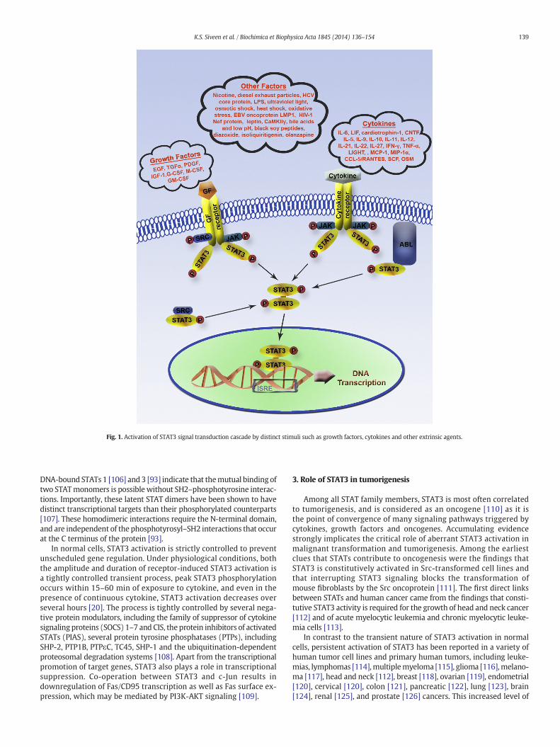
Fig. 1. Activation of STAT3 signal transduction cascade by distinct stimuli such as growth factors, cytokines and other extrinsic agents.
139K.S. Siveen et al. / Biochimica et Biophysica Acta 1845 (2014) 136–154
DNA-bound STATs 1 [106] and 3 [93] indicate that themutual binding oftwo STATmonomers is possible without SH2–phosphotyrosine interac-tions. Importantly, these latent STAT dimers have been shown to havedistinct transcriptional targets than their phosphorylated counterparts[107]. These homodimeric interactions require the N-terminal domain,and are independent of the phosphotyrosyl–SH2 interactions that occurat the C terminus of the protein [93].
In normal cells, STAT3 activation is strictly controlled to preventunscheduled gene regulation. Under physiological conditions, boththe amplitude and duration of receptor-induced STAT3 activation isa tightly controlled transient process, peak STAT3 phosphorylationoccurs within 15–60 min of exposure to cytokine, and even in thepresence of continuous cytokine, STAT3 activation decreases overseveral hours [20]. The process is tightly controlled by several nega-tive protein modulators, including the family of suppressor of cytokinesignaling proteins (SOCS) 1–7 and CIS, the protein inhibitors of activatedSTATs (PIAS), several protein tyrosine phosphatases (PTPs), includingSHP-2, PTP1B, PTPεC, TC45, SHP-1 and the ubiquitination-dependentproteosomal degradation systems [108]. Apart from the transcriptionalpromotion of target genes, STAT3 also plays a role in transcriptionalsuppression. Co-operation between STAT3 and c-Jun results indownregulation of Fas/CD95 transcription as well as Fas surface ex-pression, which may be mediated by PI3K-AKT signaling [109].
3. Role of STAT3 in tumorigenesis
Among all STAT family members, STAT3 is most often correlatedto tumorigenesis, and is considered as an oncogene [110] as it isthe point of convergence of many signaling pathways triggered bycytokines, growth factors and oncogenes. Accumulating evidencestrongly implicates the critical role of aberrant STAT3 activation inmalignant transformation and tumorigenesis. Among the earliestclues that STATs contribute to oncogenesis were the findings thatSTAT3 is constitutively activated in Src-transformed cell lines andthat interrupting STAT3 signaling blocks the transformation ofmouse fibroblasts by the Src oncoprotein [111]. The first direct linksbetween STATs and human cancer came from the findings that consti-tutive STAT3 activity is required for the growth of head and neck cancer[112] and of acute myelocytic leukemia and chronic myelocytic leuke-mia cells [113].
In contrast to the transient nature of STAT3 activation in normalcells, persistent activation of STAT3 has been reported in a variety ofhuman tumor cell lines and primary human tumors, including leuke-mias, lymphomas [114],multiplemyeloma [115], glioma [116],melano-ma [117], head and neck [112], breast [118], ovarian [119], endometrial[120], cervical [120], colon [121], pancreatic [122], lung [123], brain[124], renal [125], and prostate [126] cancers. This increased level of

140 K.S. Siveen et al. / Biochimica et Biophysica Acta 1845 (2014) 136–154
phosphorylated STAT3 is not due to mutations in STAT3 but arises fromoversupply of growth factors, such as TGFα or (IL6-family) cytokineswithin the tumor microenvironment that activate STAT3 in a paracrinemanner. The activation of oncogenes, inactivation of tumor-suppressorgenes, chromosomal rearrangement/amplification, deregulation ofmultiple potential upstream inputs such as elevated EGFR expressionlevels, EGFR mutations that result in constitutive RTK activation, over-expression of Src or other SFKs, mutations that hyperactivate JAKs[89] and other genetic events in neoplastic cells directly triggerSTAT3 activation or the release of inflammatory mediators as partof an autocrine pathway [127]. Hyper-activation of STAT3 can alsoresult from impairment mutations in any of the negative regulatoryproteins, which limit the extent of STAT3 activation in normal cells[128]. For example, epigenetic silencing of SOCS3 by hypermethylationin CpG islands of the functional SOCS-3 promoter in human lung can-cers [129] as well as mutations in STAT3-inactivating receptor proteintyrosine phosphatase delta in glioblastoma and other human cancers[130] leads to STAT3-mediated cell proliferation and survival.
Forced expression of a constitutively active form of STAT3 in mouseepidermis was found to shorten latency and enhance the number ofmalignant skin lesions progressing rapidly to squamous-cell carcinoma,induced by two-stage carcinogenesis [131]. Transfection of a dominant-negative form of STAT3 led to production of soluble factors that induceapoptosis and cell cycle arrest in the murine melanoma model [132].A growth promoting role for STAT3 in the mouse model of anaplasticlarge cell lymphomas mediated by the oncogenic fusion protein,nucleophosmin-anaplastic lymphoma kinase (NPM-ALK), was dem-onstrated in both in vitro and in vivomodels. Even though NPM-ALK-dependent tumor could develop in the absence of STAT3, STAT3 is re-quired for the growth and survival of NPM-ALK lymphoma T cellsin vitro and in vivo. Ablation of STAT3 in mice bearing xenograftedNPM-ALK-dependent T-cell lymphomas significantly impaired tumorgrowth in vivo [133], suggesting that tumor cells formed in the presenceof STAT3 become STAT3 addicted. Moreover the growth of murine B16melanoma cells that harbored activated STAT3 could be suppressed ef-ficiently in mice by introduction of a functionally deficient STAT3 vari-ant [134]. Constitutive activation of STAT3 is involved in many cellular
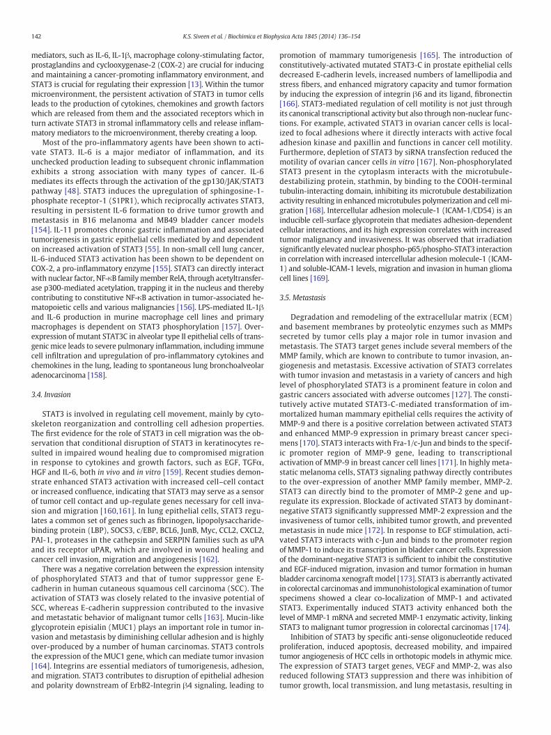
Fig. 2.Multifaceted role of STAT3 in
processes including proliferation, survival, inflammation, invasion,metastasis and angiogenesis, all of which favor tumor initiation andprogression (Fig. 2) and have been discussed below in detail.
3.1. Proliferation
STAT proteins have been shown to play important roles in cellproliferation induced by cytokines. The first evidence towards therole of STAT3 in survival was that STAT3 activation is essential forgp130-induced proliferation of the IL-3-dependent pro-B hematopoiet-ic cell line, BAF/B03 cells [135]. In breast carcinoma cells, autocrine-mediated STAT3 activationwas found to correlatewith cell proliferation[136]. Activated STAT3 promotes proliferation primarily by stimulatingtranscription of key cancer genes linked with proliferation of tumorcells, such as cyclin D1, cyclin B and cdc2, which are involved in the reg-ulation of cell cycle [127]. The active form of STAT3 was found to pro-mote the G1/S phase transition of the cell cycle through theexpression of cyclin D1, which can associate with cdk4 or cdk6 and con-trol progression from G1 to S phase in gastric [137] and colorectal [138]cancer cells. STAT3 was found to be phosphorylated in 19% of bladdercancer tissues as well as several bladder cancer cell lines. Targetingthe STAT3 signaling pathway in bladder cancer cells using anadenovirus-mediated dominant-negative STAT3 prohibited cell growthand induction of apoptosis in bladder cancer cell lines through down-regulation of cell cycle-regulating gene cyclin D1 [139]. In humanhead and neck squamous cell carcinoma, constitutive activation ofSTAT3 was found to play a causative role in over-expression of cyclinD1, and in clinical studies, STAT3 activation level provided a novel prog-nostic factor [140]. Cucurbitacin I-mediated inhibition of STAT3-induced cell-cycle arrest at the G2/M transition in cell lines derivedfrom laryngeal squamous cell carcinoma and glioblastomawas associat-ed with the downregulation of cyclin B1 and cdc2 [138]. Constitutivelyphosphorylated STAT3 has been found to induce over-expression of tar-get genes such as cdc2, cyclin B1, m-ras, and E2F-1 in colon and breastcarcinomas. E2F-1 is a transcription factor that activates the synthesisof mRNAs encoding proteins needed during the cell cycle. The productsof some E2F-1-regulated genes, such as cyclin D1 and cyclin E, cdc2 and
tumor invasion and metastasis.

141K.S. Siveen et al. / Biochimica et Biophysica Acta 1845 (2014) 136–154
cyclin B1 contribute to the G1/S and G2/M transitions and inhibit apo-ptosis [141]. STAT3 plays a key role in the G1 to S phase cell-cycle tran-sition induced by the cytokine receptor subunit gp130 through theupregulation of cyclins D2, D3 and A, and cdc25A, and the concomitantdownregulation of p21 and p27. The membrane-proximal region ofgp130, consisting of 133 amino acid residues, contains Tyr767, whichis required for the activation of STAT3. The repression of p21 is con-trolled by several signaling molecules whose expressions are regulatedby STAT3. For example, stimulation by gp130 scarcely induced p21 ex-pression in BAF-B03 (G133-bcl2 cells) consistent with the fact thatgp130 induces growth. However, the truncated gp130 containing onlythe membrane-proximal 68 amino acid residues (G68-bcl2) could in-duce p21 mRNA. The same was true for the cells expressing gp130 mu-tated at Tyr767 or expressing dominant-negative STAT3s. Theseobservations indicate that the region of gp130 consisting of themembrane-proximal 68 amino acid residues can induce p21 expres-sion, and that suppression of STAT3 activity leads to gp130-inducedgene expression of p21 through the membrane-proximal region [142].
In normal tissues, cell proliferation is controlled by irreversibleentry into post-mitotic, differentiated states. Emergent tumor cellsmust escape this terminal differentiation, which in part is regulatedby c-Myc [143]. The gp130-mediated STAT3 signaling has been shownto up-regulate the expression of several growth-promoting genes, suchas c-Myc, Pim-1 and Pim-2 [144]. STAT3-C (constitutively dimerizedand active form of STAT3) transformed cells were found to have 3- to5-fold elevated levels of cyclin D1 mRNA and c-myc mRNA when com-pared to untransformed cells [110]. Inhibition of STAT3 with the orallyactive JAK inhibitor, AZD1480, was found to decrease tumor growth inneuroblastoma and pediatric sarcomas through downregulation of cellcycle regulators, cyclin D1, cyclin D3 and cdc25A, anti-apoptotic genesBcl-2 and survivin, the metastasis-related factor TIMP-1, and c-Myc.Knockdown of STAT3 expression by RNAi inhibited the growth ofhuman ovarian cancer through downregulation of cyclin D1, c-Myc andBcl-2 [145].
3.2. Survival
STAT3 contributes to the tumorigenic process mainly by triggeringpro-survival and pro-proliferative signaling into cells. Apoptosis isessential for the maintenance of normal physiological functions, ascell death occurs during natural elimination of aging cells and inimmunoselection of T and B cells. One of the major characteristicsof tumor cells is the ability to avoid programmed cell death. TheBcl-2 family of proteins has been identified as important regulatorsof apoptotic cell death in many cell types, and aberrant expressionof Bcl-2 family members is frequently found in diverse malignancies.One of the first indications that STAT3 signaling contributes to malig-nancy by preventing apoptosis pathway came from studies in multiplemyeloma cells showing that increased expression of Bcl-xL, a memberof the anti-apoptotic Bcl-2-family, is dependent on constitutively acti-vated STAT3 [111]. Several anti-apoptotic proteins, such as survivinand members of the Bcl family (Bcl-xL, Bcl-2 and Mcl-1), which areknown to be crucial for tumor cell survival, are direct target genes ofSTAT3 and are down-regulated as a consequence of STAT3 inhibition[146]. STAT3 has also been found to be involved in colorectal cancercell growth, survival, invasion, and migration through regulation ofgene expression, such as Bcl-2, p21waf1/cip1, p27kip1, E-cadherin,VEGF, and MMPs [147].
Multiple myeloma is a disease characterized by slow-growingplasma cells in which IL-6 is an important growth and survival factor.STAT3 is constitutively activated in bone marrow mononuclear cellsfrom patients with myeloma, and in the IL-6-dependent humanmyeloma cell line, U266. Moreover, U266 cells are inherently resistantto Fas-mediated apoptosis and express high levels of the anti-apoptotic protein, Bcl-xL. Blocking IL-6 receptor signaling fromJanus kinases to the STAT3 protein inhibits Bcl-xL expression and
induces apoptosis, demonstrating that STAT3 signaling is essential forthe survival of myeloma tumor cells [113]. STAT3 activation is alsoinvolved in IL-6-dependent T-cell proliferation through prevention ofapoptosis, independently of Bcl-2 [148]. In epithelial cells, STAT3induces C-type lectin, RegIIIβ; that indirectly suppresses apoptosis,which is overexpressed in human colon cancer and inflammatory boweldisease [127]. Moreover, STAT3 was shown to repress transcription ofmurine p53 expression, thus impacting p53-mediated apoptosis and con-tributing to cell survival.
Mcl-1 is another anti-apoptotic gene of the Bcl-2 family that is atarget of STAT3. Blocking STAT3 in human tumor cells has beenshown to down-regulate Mcl-1 expression and induce tumor-cellapoptosis [111]. Survivin, a member of the small inhibitor of apopto-sis (IAP) family, has a bi-functional role by counteracting apoptosisas well as by controlling mitogenic progression. Survivin is selectivelyexpressed during G2/M transition, localizes to mitotic spindle microtu-bules, and is phosphorylated by physical association with cdc2 [138].STAT3 was constitutively activated in various human gastric cancercells and its inhibition by ectopic dominant-negative STAT3 or Januskinase inhibitor was found to induce apoptosis. Furthermore, STAT3inhibition markedly decreased survivin expression, while forced ex-pression of survivin rescued human gastric adenocarcinoma AGS cellsfrom apoptosis induced by STAT3 inhibition [137].
The proliferative effect of STAT3 in tumor cells is mediated indi-rectly through the downregulation of pro-apoptotic factors such asFas. The expression of dominant negative STAT3 or c-Jun in melano-ma cells was found to increase Fas expression efficiently [149]. De-creased Fas expression during tumor progression often results in aloss of Fas-ligand-mediated apoptosis and enables the progressionof cells through division. Activated STAT3 is also involved in the in-duction of the immediate early genes c-jun and c-fos and repressionof the cell cycle inhibitor p21 [127]. A positive correlation has beenobserved between the expression of p-STAT3 and c-jun in hepatocel-lular carcinomas (HCC) and their surrounding liver tissues [150].STAT3 was found to regulate the hepatocyte growth factor inducedproliferation of human endothelial cells through activation of the c-fospromoter [151]. p21 is known to induce G2/M phase arrest by inhibitingcyclin-B/cdc2. siRNA-mediated inhibition of STAT3 in human colorectalcancer cell lines induced p16-, p21- and p27-mediated cell cycle arrestand apoptosis. STAT3 inhibition in gastric cancer cells down-regulatedthe expression of S-phase kinase-associated protein-2 (Skp2), whichcatalyzes the ubiquitination of p21, thereby reducing the ability ofcells tomigrate and invade, by inhibiting the activity of RhoA, downreg-ulation of pFAK (phosphorylated focal adhesion kinase) expression, up-regulation of E-cadherin, and reducing the activities of matrixmetalloproteinase-2 (MMP-2) and MMP-9 [152].
Heat shock protein 70 (Hsp70) can protect cells from apoptosisby binding and modulating the activity of various pro- and anti-apoptotic proteins at transcriptional and post-translational level.Hsp70 prevents JNK-induced phosphorylation and inhibition of Bcl-2and Bcl-xL anti-apoptotic proteins and promotes cell survival throughthe maintenance of mitochondria stability. STAT3 can bind to specificsequences at the Hsp70 promoter (5′-CTGGRA-3′) and is responsiblefor Hsp70 synthesis in cancer cells, mediating the expression of anti-apoptotic proteins [153].
3.3. Inflammation
STAT3 signaling is a major intrinsic pathway for cancer inflamma-tion because it is often activated in tumor-associated immune andinflammatory cells as well as malignant cells and is capable of inducinga large number of genes that are crucial for inflammation including IL-6,10, 11, 17, 23, CXCL12, and COX-2 [127]. STAT3 was initially discoveredas an acute-phase response protein that selectively binds to the IL-6-responsive element within the acute-phase gene promoter, thus sug-gesting its link to inflammation [88]. Cytokines, chemokines and other
142 K.S. Siveen et al. / Biochimica et Biophysica Acta 1845 (2014) 136–154
mediators, such as IL-6, IL-1β, macrophage colony-stimulating factor,prostaglandins and cyclooxygenase-2 (COX-2) are crucial for inducingand maintaining a cancer-promoting inflammatory environment, andSTAT3 is crucial for regulating their expression [13]. Within the tumormicroenvironment, the persistent activation of STAT3 in tumor cellsleads to the production of cytokines, chemokines and growth factorswhich are released from them and the associated receptors which inturn activate STAT3 in stromal inflammatory cells and release inflam-matory mediators to the microenvironment, thereby creating a loop.
Most of the pro-inflammatory agents have been shown to acti-vate STAT3. IL-6 is a major mediator of inflammation, and itsunchecked production leading to subsequent chronic inflammationexhibits a strong association with many types of cancer. IL-6mediates its effects through the activation of the gp130/JAK/STAT3pathway [48]. STAT3 induces the upregulation of sphingosine-1-phosphate receptor-1 (S1PR1), which reciprocally activates STAT3,resulting in persistent IL-6 formation to drive tumor growth andmetastasis in B16 melanoma and MB49 bladder cancer models[154]. IL-11 promotes chronic gastric inflammation and associatedtumorigenesis in gastric epithelial cells mediated by and dependenton increased activation of STAT3 [55]. In non-small cell lung cancer,IL-6-induced STAT3 activation has been shown to be dependent onCOX-2, a pro-inflammatory enzyme [155]. STAT3 can directly interactwith nuclear factor, NF-κB familymember RelA, through acetyltransfer-ase p300-mediated acetylation, trapping it in the nucleus and therebycontributing to constitutive NF-κB activation in tumor-associated he-matopoietic cells and various malignancies [156]. LPS-mediated IL-1βand IL-6 production in murine macrophage cell lines and primarymacrophages is dependent on STAT3 phosphorylation [157]. Over-expression of mutant STAT3C in alveolar type II epithelial cells of trans-genic mice leads to severe pulmonary inflammation, including immunecell infiltration and upregulation of pro-inflammatory cytokines andchemokines in the lung, leading to spontaneous lung bronchoalveolaradenocarcinoma [158].
3.4. Invasion
STAT3 is involved in regulating cell movement, mainly by cyto-skeleton reorganization and controlling cell adhesion properties.The first evidence for the role of STAT3 in cell migration was the ob-servation that conditional disruption of STAT3 in keratinocytes re-sulted in impaired wound healing due to compromised migrationin response to cytokines and growth factors, such as EGF, TGFα,HGF and IL-6, both in vivo and in vitro [159]. Recent studies demon-strate enhanced STAT3 activation with increased cell–cell contactor increased confluence, indicating that STAT3 may serve as a sensorof tumor cell contact and up-regulate genes necessary for cell inva-sion and migration [160,161]. In lung epithelial cells, STAT3 regu-lates a common set of genes such as fibrinogen, lipopolysaccharide-binding protein (LBP), SOCS3, c/EBP, BCL6, JunB, Myc, CCL2, CXCL2,PAI-1, proteases in the cathepsin and SERPIN families such as uPAand its receptor uPAR, which are involved in wound healing andcancer cell invasion, migration and angiogenesis [162].
There was a negative correlation between the expression intensityof phosphorylated STAT3 and that of tumor suppressor gene E-cadherin in human cutaneous squamous cell carcinoma (SCC). Theactivation of STAT3 was closely related to the invasive potential ofSCC, whereas E-cadherin suppression contributed to the invasiveand metastatic behavior of malignant tumor cells [163]. Mucin-likeglycoprotein episialin (MUC1) plays an important role in tumor in-vasion and metastasis by diminishing cellular adhesion and is highlyover-produced by a number of human carcinomas. STAT3 controlsthe expression of the MUC1 gene, which can mediate tumor invasion[164]. Integrins are essential mediators of tumorigenesis, adhesion,and migration. STAT3 contributes to disruption of epithelial adhesionand polarity downstream of ErbB2-Integrin β4 signaling, leading to
promotion of mammary tumorigenesis [165]. The introduction ofconstitutively-activated mutated STAT3-C in prostate epithelial cellsdecreased E-cadherin levels, increased numbers of lamellipodia andstress fibers, and enhanced migratory capacity and tumor formationby inducing the expression of integrin β6 and its ligand, fibronectin[166]. STAT3-mediated regulation of cell motility is not just throughits canonical transcriptional activity but also through non-nuclear func-tions. For example, activated STAT3 in ovarian cancer cells is local-ized to focal adhesions where it directly interacts with active focaladhesion kinase and paxillin and functions in cancer cell motility.Furthermore, depletion of STAT3 by siRNA transfection reduced themotility of ovarian cancer cells in vitro [167]. Non-phosphorylatedSTAT3 present in the cytoplasm interacts with the microtubule-destabilizing protein, stathmin, by binding to the COOH-terminaltubulin-interacting domain, inhibiting its microtubule destabilizationactivity resulting in enhancedmicrotubules polymerization and cell mi-gration [168]. Intercellular adhesion molecule-1 (ICAM-1/CD54) is aninducible cell-surface glycoprotein that mediates adhesion-dependentcellular interactions, and its high expression correlates with increasedtumor malignancy and invasiveness. It was observed that irradiationsignificantly elevated nuclear phospho-p65/phospho-STAT3 interactionin correlation with increased intercellular adhesion molecule-1 (ICAM-1) and soluble-ICAM-1 levels, migration and invasion in human gliomacell lines [169].
3.5. Metastasis
Degradation and remodeling of the extracellular matrix (ECM)and basement membranes by proteolytic enzymes such as MMPssecreted by tumor cells play a major role in tumor invasion andmetastasis. The STAT3 target genes include several members of theMMP family, which are known to contribute to tumor invasion, an-giogenesis and metastasis. Excessive activation of STAT3 correlateswith tumor invasion and metastasis in a variety of cancers and highlevel of phosphorylated STAT3 is a prominent feature in colon andgastric cancers associated with adverse outcomes [127]. The consti-tutively active mutated STAT3-C-mediated transformation of im-mortalized human mammary epithelial cells requires the activity ofMMP-9 and there is a positive correlation between activated STAT3and enhanced MMP-9 expression in primary breast cancer speci-mens [170]. STAT3 interacts with Fra-1/c-Jun and binds to the specif-ic promoter region of MMP-9 gene, leading to transcriptionalactivation of MMP-9 in breast cancer cell lines [171]. In highly meta-static melanoma cells, STAT3 signaling pathway directly contributesto the over-expression of another MMP family member, MMP-2.STAT3 can directly bind to the promoter of MMP-2 gene and up-regulate its expression. Blockade of activated STAT3 by dominant-negative STAT3 significantly suppressed MMP-2 expression and theinvasiveness of tumor cells, inhibited tumor growth, and preventedmetastasis in nude mice [172]. In response to EGF stimulation, acti-vated STAT3 interacts with c-Jun and binds to the promoter regionof MMP-1 to induce its transcription in bladder cancer cells. Expressionof the dominant-negative STAT3 is sufficient to inhibit the constitutiveand EGF-induced migration, invasion and tumor formation in humanbladder carcinoma xenograftmodel [173]. STAT3 is aberrantly activatedin colorectal carcinomas and immunohistological examination of tumorspecimens showed a clear co-localization of MMP-1 and activatedSTAT3. Experimentally induced STAT3 activity enhanced both thelevel of MMP-1 mRNA and secreted MMP-1 enzymatic activity, linkingSTAT3 to malignant tumor progression in colorectal carcinomas [174].
Inhibition of STAT3 by specific anti-sense oligonucleotide reducedproliferation, induced apoptosis, decreased mobility, and impairedtumor angiogenesis of HCC cells in orthotopic models in athymic mice.The expression of STAT3 target genes, VEGF and MMP-2, was alsoreduced following STAT3 suppression and there was inhibition oftumor growth, local transmission, and lung metastasis, resulting in

143K.S. Siveen et al. / Biochimica et Biophysica Acta 1845 (2014) 136–154
significantly prolonged survival of tumor-bearing mice [175].Suppression of STAT3 activation by transfection with a dominant-negative STAT3 decreased brain metastasis of human melanomacells in animal models and melanoma cell invasion in vitro andsignificantly affected the expression of bFGF, VEGF and MMP-2in vivo and in vitro by transcriptionally regulating the promoteractivity in human melanoma cells [176]. Activation of STAT3 by IL-6 was also found to induce Twist expression in human breast cancercells by binding to the second proximal STAT3-binding site on thehuman Twist promoter and activating its transcription. Inhibitionof STAT3 by STAT3 small hairpin RNAs reduced migration and inva-sion of human breast cancer cells [177]. Twist is involved in tumormetastasis through the upregulation of MMP-2 and -9 [178] and in-hibition of TIMP-1 [179]. Moreover, STAT3-deficient keratinocytesdemonstrated increased cell adhesiveness and compromised growthfactor-inducible cell migration, accompanied with an increasednumber of tyrosyl-hyperphosphorylated focal adhesion and tyrosinephosphorylation of p130cas, suggesting that STAT3 in keratinocytesplays a critical role in modulating cell adhesiveness to the substra-tum leading to growth factor-dependent cell migration [180].
3.6. Angiogenesis
The intrinsic ability of a cancer cell to proliferate uncontrollablyand resist apoptosis is not sufficient for tumor development. Tumorscannot sustain their growth unless they are supplied with oxygenand nutrients from newly formed blood vessels. Activated oncogeneproducts play a crucial role in stimulating angiogenesis. One of themost potent angiogenesis-inducing signals is VEGF. STAT3 is consti-tutively activated in glioma and medulloblastoma tumors and isconsidered to play a central role in autocrine activation of VEGF[124]. One of the earliest evidence to suggest that STAT3 is linkedto angiogenesis was from granulocyte–macrophage colony-stimulating factor-induced angiogenic activity in chick chorioallan-toic membrane and GM-CSF induced vessel sprouting from chickenaorta rings [181]. Transfection of cells with the constitutively acti-vated STAT3-C mutant was found to be sufficient to increase VEGFexpression and induce angiogenesis in vivo [182]. Among the vari-ous pro-angiogenic factors, VEGF and hypoxia-inducible factor-1α(HIF-1α) stand out as prominent transcriptional targets for STAT3,and a requirement for STAT3 has been proposed for functionalityof HIF-1α [127]. It was shown that human breast cancer, head andneck carcinoma, melanoma [182], pancreatic cancer [122] and cervi-cal cancer cell lines [183] that display constitutively active STAT3also express VEGF and that STAT3 activity up-regulates VEGF ex-pression and tumor angiogenesis, suggesting that down-modulation of STAT3 activation can suppress the expression ofVEGF and inhibit angiogenesis. Blocking STAT3 signaling has beenshown to inhibit Src-mediated VEGF upregulation and a completeinhibition of VEGF-dependent vascular permeability of humantumor cells in vivo [184].
STAT3 activity in tumor-derived myeloid cells can up-regulatethe expression of a number of known STAT3 target genes, includingVEGF, bFGF, and MMP-9. VEGF and bFGF contribute to myeloidcell-mediated and STAT3-dependent angiogenesis. In addition,CCL2, CXCL2, and IL-1β, which can contribute to myeloid cell-medi-ated angiogenesis, required STAT3 for their elevated expression intumor-derived myeloid cells, suggesting that STAT3 activity inimmune cells can contribute to tumor angiogenesis at multiplelevels [185]. Expression of phospho-STAT3 had a positive correlationwith the VEGF and Bcl-2 in gastric cancer specimens, suggesting thatphospho-STAT3 expression might be associated with angiogenesis,anti-apoptosis, and tumor progression in this particular cancer[186]. STAT3 signaling pathway activation was found to medi-ate tumor angiogenesis in non-small cell lung cancer by the upregu-lation of VEGF and bFGF. The expression of p-STAT3 in human
samples correlated with VEGF and bFGF while knocking downSTAT3 reduced the expression of VEGF and bFGF in human lung can-cer cells [187].
Hypoxia-inducible factor 1α (HIF-1α) is a transcription factorthat controls oxygen homeostasis and is the final switch for VEGF up-regulation in cancer [188]. STAT3 can indirectly regulate VEGF by in-ducing the expression of HIF-1α, which drives VEGF transcriptionupon hypoxic stimulation. STAT3 is required for both basal andgrowth signal-induced expression of HIF-1α and targeting STAT3with small-molecule inhibitor blocks HIF-1α and VEGF expressionin vitro and inhibits tumor growth and angiogenesis in vivo [189]. Ac-tivated STAT3 in ischemic rat kidney and in hypoxic renal carcinomacells increased the cellular levels of HIF-1α either by blocking pro-tein degradation or by enhancing HIF-1α synthesis under hypoxicconditions. STAT3 also interacts directly with HIF-1α and is recruitedto the human VEGF promoter in response to hypoxia, suggesting thatSTAT3 is a target for hypoxia-induced angiogenesis in human renalcarcinoma [190]. As constitutively active STAT3 can inhibit p53 ex-pression by binding to the p53 promoter [191], interruption ofSTAT3 signaling may lower HIF-1α activity by promoting its degra-dation through p53-dependent mechanisms. Thus, STAT3 inhibitorscan block VEGF expression at several levels, generating a potentanti-angiogenic effect.
4. Selected natural inhibitors of the STAT3 signaling pathway
Themain goal of using a target-specific drug is to inhibit amoleculartarget central to a disease mechanism of interest. The first step towardthis goal is to identify individual molecular targets and validatetheir relevance to the human disease. This is followed in turn byidentification of specific chemical-or antibody based small moleculemodulators or inhibitors of the target [192]. In theory, targeting asingle molecular mechanism should be sufficient to achieve a signif-icant therapeutic effect; in reality, however, single-target drugs havehad very little therapeutic impact. In fact, they have generally beenhighly ineffective in treating complex diseases (e.g., cancer) [193]or diseases affecting multiple tissues or cell types (e.g., diabetesand immunoinflammatory disorders). Drugs aimed at multipletargets can be more efficacious and less vulnerable to acquired resis-tance because the disease system is less able to compensate for theaction of two or more drugs simultaneously. This multi-targetapproach can be particularly beneficial in cancers because oncogen-esis is known to be a multigenic process [194]. For example, theErbB2 (HER-2/neu) inhibitor trastuzumab (Herceptin) is now beingcombined with the anti-VEGF inhibitor bevacizumab (Avastin) totreat breast cancer.
As discussed above, aberrant activation of STAT3 plays a pivotalrole in the initiation, progression and metastasis of different cancersand thereby the abrogation of STAT3 activation using pharmacolog-ical inhibitors can form the basis of future cancer therapy. However,interestingly, a number of strategies, including the use of anti-senseoligonucleotide targeting STAT3, synthetic drugs, small moleculesderived from natural sources, and gene therapy techniques havebeen employed over the last decade to suppress deregulated STAT3signaling cascade in cancer. In the following sections, we will discussabout selected natural and synthetic agents that have shown signif-icant efficacy in blocking STAT3 activation and thereby inhibitingtumor growth and metastasis.
4.1. Betulinic acid
Betulinic acid (Fig. 3) is a pentacyclic triterpene, discovered in 1995from the stem bark of the plant Zizyphusmauritiana andwas initially re-ported to be a melanoma-specific cytotoxic agent [8]. In addition, nu-merous studies over the past decade showed that betulinic acid couldinduce apoptosis in thyroid, breast, lung and colon carcinomas,

Fig. 3. Chemical structures of selected natural blockers of STAT3 activation cascade.
144 K.S. Siveen et al. / Biochimica et Biophysica Acta 1845 (2014) 136–154
leukemias as well as multiple myeloma [8]. Pandey et al. [195] foundthat betulinic acid inhibited constitutive activation of STAT3, Src kinase,JAK1, and JAK2. Furthermore, betulinic acid induced the expression ofthe PTP SHP-1; silencing of the SHP-1 gene abolished the ability ofbetulinic acid to inhibit STAT3 activation. Betulinic acid also down-regulated the expression of STAT3-regulated gene products, such asBcl-xL, Bcl-2, cyclin D1, and survivin. It also induced apoptosis by anincrease in the sub-G1 cell population and an increase in caspase-3-induced PARP cleavage. Moreover, betulinic acid enhanced apoptosisin myeloma cells induced by thalidomide (from 10% to 55%) andbortezomib (from 5% to 70%). Overall, the results suggest that betulinicacid down-regulates STAT3 activation through upregulation of SHP-1,suggesting the potential role of betulinic acid as a chemopreventiveagent.
4.2. Butein
Butein (3,4,2,4-tetrahydroxychalcone) is derived from numerousplants including stem bark of Semecarpus anacardium, Rhus vernicifluaStokes, and the heartwood of Dalbergia odorifera [196]. Butein hasbeen traditionally used for the treatment of pain, thrombotic disease,gastritis, stomach cancer, and parasitic infections in Korea, Japan, andChina and so far no reports have suggested the toxicity of this polyphe-nol [3]. In Korea, butein is used as a food additive, thereby indicatingthat it is safe to be consumed by humans [197]. Reports have indicatedthat butein can suppress the proliferation of breast carcinoma, coloncarcinoma, osteosarcoma, lymphoma, acute myelogenous leukemia,chronic myeloid leukemia, multiple myeloma, melanoma, and hepaticstellate cells [198–200]. Moreover, it was found to suppress phorbol

145K.S. Siveen et al. / Biochimica et Biophysica Acta 1845 (2014) 136–154
ester-induced skin tumor formation [201,202], reduce antibody-associated glomerulonephritis [203], and suppress liver fibrosisinduced by carbon tetrachloride [204]. Furthermore, butein wasalso found to induce G2/M phase arrest and apoptosis in hepatocellularcarcinoma (HCC) cells through reactive oxygen species (ROS) genera-tion [205], suggesting that butein may have a great potential for HCCtreatment. Additionally, its effects on HCC cell lines were investigated[206]. HCC cell lines HepG2 were incubated with different concentra-tions of butein for 6 h, and the phosphorylation of STAT3was examinedby Western blot. It was observed that butein inhibited the constitutiveactivation of STAT3 in a dose-dependent manner, with maximum in-hibition occurring at 50 μM. The inhibition of STAT3 was also time-dependent, with maximum inhibition occurring at around 6 to 8 h.Since butein induces the generation of ROS that blocks STAT3 signaling[205,207], it was next determinedwhether NAC, a ROS scavenger, couldcounteract the inhibitory effect of butein on STAT3 activation. HCC(HepG2) cells were pre-treated with NAC for 1 h before treatmentwith butein and it was observed that decrease in phosphorylatedSTAT3 was ROS-dependent, as levels reverted to control levels in thepresence of NAC. Also, the phosphorylation of JAK2, which is consti-tutively active in HepG2 cells, was found to be suppressed in a time-dependent manner upon butein treatment [206]. The expression ofBcl-2, a marker of cell survival was down-regulated and that ofcaspase-3, which is the marker of apoptosis was increased in thebutein-treated cells compared with the control group. Overall, theresults suggested that butein suppresses HCC growth through mod-ulation of the STAT3 activation pathway. Further studies are requiredbefore its clinical potential is fully realized in HCC treatment.
4.3. Caffeic acid
Caffeic acid (CA) is a phenolic compound largely found in foodplants [208]. CA has been extensively studied for its potent anti-oxidant [209,210] as well as anti-inflammatory properties [211].Moreover, some caffeic acid derivatives, such as caffeic acid phenetylester (CAPE) have anti-oxidant properties that are similar to those ofCA [212]. In addition, CA and CAPEwere reported to inhibit NF-κB ac-tivation and iNOS expression [213] as well as inhibit angiogenesis,tumor invasion, and metastasis [214]. VEGF is known to play a cru-cial role in angiogenesis and tumor progression [215], and the inhi-bition of VEGF produced promising results as a tumor anti-angiogenesis strategy in both animal models and cancer patients[216]. Hypoxia develops within solid tumors due to a mismatch be-tween tumor cell proliferation, which is faster than the rate ofblood vessel formation, thereby leading to a compromised tumorblood supply [217]. Moreover, hypoxia stimulates VEGF expressionin tumors in order to promote angiogenesis to meet the metabolicrequirement for growth. Recently, STAT3 was found to be a directtranscriptional activator of the VEGF gene, and demonstrated toup-regulate VEGF expression in various human cancers [218], sug-gesting that STAT3 activity may contribute significantly to over-production of VEGF in tumors. Thus, it was considered that inhibitingSTAT3 phosphorylation at Tyr705 might reduce VEGF expression intumor cells. CA and its synthetic derivative, CADPE [3-(3,4-Dihydroxy-phenyl)-acrylic acid 2-(3,4-dihydroxy-phenyl)-ethyl ester] werefound to be actively involved in inhibiting STAT3 activity [219,220].Since STAT3 phosphorylation at Tyr705 is essential for its nuclear trans-location aswell as for binding to specific promoter sequences on its tar-get genes, the activities of CA and CADPE were investigated againstSTAT3 phosphorylation in vitro, using renal carcinoma Caki cells. Itwas noted that the phosphorylation of STAT3 at Tyr 705 increased in re-sponse to hypoxia and was decreased by CA or CADPE in a dose-dependent manner. To study the inhibitory effects of CA and CADPEon hypoxia-induced STAT3 translocalization, wild-type STAT3 wastransfected into COS7 (STAT3-deficient cell lines), which were thenfractioned into nuclear and cytosolic proteins. The Western blot results
showed that phosphorylated STAT3 was translocated from cytosol tothe nucleus in response to hypoxia, but STAT3 remained in the cyto-plasm of cells pre-treated with 100 μM CA or 30 μM CADPE. These re-sults thus suggested that CA or CADPE inhibited STAT3phosphorylation that was required for nuclear translocation of STAT3,and also inhibited the hypoxia-induced nuclear translocation ofSTAT3. The effects of CA or CADPE on suppression of VEGF expressionwere also studied. Firstly, luciferase assays were carried out in Caki-Icells using a VEGF promoter reporter plasmid. Itwas observed that hyp-oxia increased the reporter activity in Caki-I cells but not in cells pre-treated with 100 μM CA or 30 μM CADPE. Subsequently, in cellstransfected with STAT3-specific siRNA, the hypoxia-induced reporteractivity of VEGF promoter was found to be significantly reduced bythe siRNA-mediated repression of STAT3. Furthermore, in Caki-I cellspre-treated with CA or CADPE and thereafter subjected to hypoxia,the hypoxia-induced mRNA expression of VEGF decreased in a dose-dependentmanner. Thus, it can be concluded that CA or CADPE reducedhypoxia-induced transcriptional activity of VEGF by inhibiting STAT3,blocking STAT3 recruitment, and blocking the formation of a transcrip-tional unit between STAT3, HIF-1α, and p300 on VEGF promoter [215].
4.4. Capsaicin
Capsaicin (N-vanillyl-8-methyl-alpha-nonenamide) is a spicycomponent of hot pepper that induces certain cancer cells to undergoapoptosis. It has been reported to have a promising role in cancerchemoprevention [221]. Capsaicin has been reported to induceapoptosis in multiple myeloma cells through down-regulation ofSTAT3-regulated expression of Bcl-2, Bcl-xL, and survivin [222].The effect of capsaicin on both constitutive and IL-6-induced STAT3activation, STAT3-regulated gene products involved in proliferation,survival, and angiogenesis, was investigated in multiple myelomacells. It was found that capsaicin inhibited constitutive activation ofSTAT3 in multiple myeloma cells in a dose- and time-dependentmanner. Capsaicin also inhibited IL-6-induced STAT3 activation aswell as the activation of JAK1 and c-Src. Moreover, pervanadate re-versed the capsaicin-induced down-regulation of STAT3, suggestingthe involvement of a protein tyrosine phosphatase. Capsaicin down-regulated the expression of cyclin D1, Bcl-2, Bcl-xL, survivin, andVEGF. Moreover, capsaicin induced the accumulation of cells in G1
phase, inhibited proliferation, and induced apoptosis. Capsaicinalso significantly potentiated the apoptotic effects of bortezomiband thalidomide in multiple myeloma cells. Upon administration ofcapsaicin, the growth of human multiple myeloma xenograft tumorsin male athymic nu/nu mice was inhibited. These results collectivelysuggest that capsaicin is a novel blocker of the STAT3 activationpathway, with a potential role in the prevention and treatment ofmyeloma.
4.5. Celastrol
Celastrol derived from the Chinese medicinal plant Tripterygiumwilfordii, has attracted great attention recently for its potent anti-cancer effects [223,224]. Celastrol has been documented to inhibitproliferation, induce apoptosis, and suppress invasion/migrationand angiogenesis in a wide variety of tumor models both in vitro andin vivo [225]. Thus, Lu et al. [226] investigated the effects of celastrolon STAT3 activation in HCC cells and in a xenograft mouse model. C3Acells, a subclone of the hepatoma-derived HepG2 cell line, were incu-bated with different concentrations of celastrol for 6 h and Westernblot was used to examine the phosphorylation of STAT3. It was shownthat celastrol inhibited the constitutive activation of STAT3 in C3Acells in a dose-dependent manner with maximum inhibition occurringat 5 μM. Moreover, the inhibition induced by celastrol was found to betime-dependent, with maximum inhibition occurring at 4 to 6 h.Because tyrosine phosphorylation causes the dimerization and nuclear

146 K.S. Siveen et al. / Biochimica et Biophysica Acta 1845 (2014) 136–154
translocation of STAT3 [227], the authors also determined the ability ofcelastrol to modulate the DNA-binding activity of STAT3 which causesgene expression. Upon analyzing the nuclear extracts prepared fromC3A cells, it was shown that celastrol inhibited STAT3 DNA-binding ac-tivities in a time-dependent manner therefore suggesting that celastrolabrogates the DNA-binding ability of STAT3. The effects of celastrol onsuppressing nuclear translocation of STAT3 in C3A cells was also deter-mined, [228] and it was concluded that celastrol could indeed inhibitthe translocation of STAT3 to the nucleus, thereby inhibiting the func-tion of transcription factors. Furthermore, as it is well-known that IL-6induces STAT3 phosphorylation [229] therefore, the authors deter-mined whether celastrol could inhibit IL-6-induced STAT3 phosphory-lation. In HUH-7 human hepatocarcinoma cells, IL-6 was found toinduce phosphorylation of STAT3 as early as 5 min, with maximumphosphorylation observed at 30 to 60 min. IL-6 also induced phos-phorylation of STAT3 in a dose-dependent manner with initial acti-vation observed at a dose of 5 ng/mL. In HUH-7 cells, IL-6-inducedSTAT3 and JAK2 phosphorylation were suppressed by celastrol in atime-dependent manner. STAT3 activation regulates the expressionof various gene products involved in cell survival, proliferation, an-giogenesis, and chemoresistance [230]. It was found that expressionof cyclin D1, Bcl-2, Bcl-xL, survivin, Mcl-1, and VEGF decreased in atime-dependent manner upon treatment with celastrol, with maxi-mum suppression observed at around 24 h. Celastrol has been welltolerated with no toxicity in pre-clinical studies using various in-flammatory diseases and cancer models [231,232]. However,celastrol has never been tested in humans before and hence its clin-ically relevant doses are not known yet. Thus, overall, the in vitro andin vivo experimental findings clearly indicate that the anti-proliferative and pro-apoptotic effects of celastrol in HCC are due tothe suppression of STAT3 activation and provide a platform to ex-plore the potential of celastrol to overcome toxicity and enhancetreatment efficacy for patients with HCC. Moreover, celastrol hasbeen found to inhibit the proliferation and suppress the growth ofprostate, gliomas and chronic myeloid leukemia xenografts in nudemice models [225,233]. Also, the ability of celastrol to modulate theexpression of pro-inflammatory cytokines, adhesion molecules, po-tassium channels, NF-κB, transforming growth factor-activated ki-nase 1 (TAK1), CXCR4, VEGFR, proteasome and heat shock responsehas been reported [224,225,234]. In another study by Kannaiyan et al.[224], to investigate whether celastrol could suppress the proliferationand induce chemosensitization ofmultiplemyeloma cells by interferingwith STAT3 activation pathways, itwas observed that celastrol inhibitedthe proliferation of multiple myeloma cell lines regardless of their sen-sitivity or resistance to bortezomib and other conventional chemother-apeutic drugs. Celastrol inhibited both the constitutive and IL-6-induced activation of STAT3, which induced apoptosis leading to an in-crease in the accumulationof cells in the sub-G1 phase, and activation ofcaspase-3. This result indicates that future studies in animal models toestablish the anti-cancer effects of celastrol in vivo are warranted.
4.6. Cucurbitacins
Cucurbitacins are essential herbs used in a large number of tradi-tional Chinese medicines [235,236] and have been shown to possessanti-inflammatory, anti-microbial, and anti-cancer activities [237].Chemically, cucurbitacins are highly diverse and can be dividedinto twelve categories, ranging from cucurbitacins A–T [236,238].Cucurbitacin B is found in many Cucurbitaceae species and is one ofthe most abundant forms of cucurbitacins [239]. It has significantanti-inflammatory activity and has been used to treat hepatitis [240].Recently, it was reported that cucurbitacin B could inhibit the growthof numerous human cancer cell lines and tumor xenografts [241,242].The inhibitory effect of cucurbitacin B on STAT3was examined in leuke-mia cells. K562 cells, an immortalizedmyelogenous leukemia line, weretreated with cucurbitacin B at doses ranging from 1 μM to 50 μM,
Western blot analysis showed that cucurbitacin B 50 μM for 4 hinhibited STAT3 activation [243]. Furthermore, treatment withcurcubitacin B reduced the level of phosphorylated STAT3 and its down-stream targets, such as cyclin B1 and Bcl-2, in human HCC cell lines[244].
4.7. Curcumin
Curcumin (diferuloylmethane) is a dietary spice and is the activecompound of turmeric (Curcuma longa) [245]. This compound hasbeen shown to modify the expression of inflammatory cytokines andenzymes, transcription factors, and gene products linked to cell survival,proliferation, invasion, and angiogenesis. Curcumin has been found topossess anti-proliferative and pro-apoptotic effects in a number oftumor cell lines in vitro [246]. In murine carcinoma models, curcuminhas also been shown to reduce the number and size of tumors, andeven ameliorate tumor-induced immunodepletion of the host [247].Phase I clinical studies of curcumin have shown that this agent can beadministered safely at oral doses of up to 8 g per day with no dose-limiting toxicity [248]. A recent phase II study of curcumin has shownthat this compound has useful biological activities in patients with pan-creatic cancer [249]. Various approaches are being investigated to over-come the limitations of curcumin bioavailability, such as the synthesis ofcurcumin analogs and development of improved delivery platforms forthe parent compound, such as liposomal, nanoparticulated, and phos-pholipid complex formulations of curcumin [250].
The anti-tumor effects of curcumin on CTCL cell lines (cutaneous Tcell lymphoma-leukemia cells) and peripheral blood mononuclearcells (PBMCs) from Mycosis Fungoides/Sézary syndrome (MF/SS) pa-tients with a high percentage of circulating tumor T-cells were exam-ined. It was shown that constitutive activation of STAT3 contributed tooncogenesis by stimulating cell proliferation and preventing apoptosisin CTCL cell lines. Western blot was performed in order to determinewhether STAT signaling was involved in curcumin-induced apoptosis.The results showed that expression levels of both STAT3 and phosphor-ylated STAT3 decreased by a range of 21–55% and 38–47%, respectively,in CTCL cells compared with untreated controls. Moreover, mRNA ex-pression levels of Bcl-2 decreased by 22–94%, and protein and mRNAexpression levels of survivin decreased by 11–95% compared with un-treated controls. From these findings, it can be concluded that curcuminselectively induces apoptosis in CTCL cell lines and PBMCs of MF/SSpatients.
Glioblastomas are known to be the most common and most deadlyof primary brain tumors [251]. The anti-cancer effects of curcuminwere evaluated on glioma cells were evaluated in vitro and in vivousing an immunocompetent orthotopic mouse model. Curcumininhibited JAK1, JAK2, and STAT3 tyrosine-phosphorylation in adose-dependent manner in murine glioma cell lines. Real-time RT-PCR revealed that curcumin down-regulated the transcription ofSTAT3 target genes such as c-Myc, MMP-9, Snail, and Twist, andKi67. In addition, curcumin suppressed cell proliferation by inducinga G2/M phase arrest in a dose-dependent manner.
In addition, in a report by Bharti et al. [252], it was demonstratedthat curcumin, a pharmacologically safe agent in humans, inhibitedIL-6-induced STAT3 phosphorylation and consequent STAT3 nucleartranslocation. Whether curcumin affects the phosphorylation ofother STAT proteins in U266 cells was investigated. Besides STAT3,it was found that U266 cells expressed STAT5. Curcumin had no ef-fect on STAT5 phosphorylation, but inhibited the IFN-α-inducedSTAT1 phosphorylation. The constitutive phosphorylation of STAT3found in multiple myeloma U266 cells was also abrogated upon treat-ment with curcumin. Also, CD138+ cells obtained from the bone mar-row of 22 myeloma patients were examined for STAT3 and NF-κBactivation after exposure to 50 μM curcumin for 2 h [115]. The resultsindicated that exposure to curcumin down-regulate STAT3 activationand survival of CD138+cells derived from multiple myeloma patients.

147K.S. Siveen et al. / Biochimica et Biophysica Acta 1845 (2014) 136–154
4.8. Diosgenin
Diosgenin is a steroid sapogenin obtained from fenugreek (Trigonellafoenum graecum). Various studies have shown that diosgenin sup-presses proliferation and induces apoptosis in a wide variety of can-cer cells. Anti-proliferative effects of diosgenin aremediated throughcell-cycle arrest, disruption of Ca2+ homeostasis, activation of p53,release of apoptosis-inducing factor, andmodulation of caspase-3 activ-ity [253]. The effects of diosgenin on human HCC cell line, C3A wereinvestigated. C3A cells were incubated with different concentrationsof diosgenin for 6 h, and the phosphorylation of STAT3 was examined[254]. Diosgenin inhibited the constitutive activation of STAT3 in C3Acells in a dose-dependent manner, with maximum inhibition occurringat around 100 μM. It was also shown that IL-6-induced STAT3 phos-phorylation in HUH-7 cells was suppressed by diosgenin in a time-dependent manner. Additionally, it was found that diosgenin sup-pressed the constitutive phosphorylation of JAK1 and JAK2 in atime-dependent manner. The expression of cyclin D1, Bcl-2, Bcl-xL,survivin, Mcl-1, and the angiogenic gene product, VEGF, also decreasedin a time-dependent manner, with maximum suppression observed ataround 24 h. Further in vivo studies with diosgenin either alone oralongwith existing chemotherapeutic drugs are needed to demonstratethe potential application of diosgenin for treatment of cancers.
4.9. Guggulsterone
Guggulsterone (GS) is derived from Commiphora mukul and is usedto treat obesity, diabetes, hyperlipidemia, atherosclerosis, and osteoar-thritis [255,256]. GS has also been shown to have potent anti-cancerpotential due to its ability to suppress the proliferation of a wide va-riety of human tumor cell types, including leukemia, head and neckcarcinoma, multiple myeloma, lung carcinoma, melanoma, breastcarcinoma, prostate, and ovarian carcinoma [257,258]. A study byAhn et al. [255] further demonstrated the effects of GS on constitu-tive STAT3 activation in multiple myeloma cells. It was found thatGS inhibited the constitutive activation of STAT3 in U266 cells in adose-dependent manner with maximum inhibition occurring at 10 to25 μM. The inhibitionwas also time-dependent, with maximum inhibi-tion occurring at 4 h. Since tyrosine phosphorylation causes dimeriza-tion of STAT3 and its translocation to the nucleus, where it binds toDNA and regulates gene transcription, the question arose whether GScould suppress the DNA-binding activity of STAT3. The nuclear extractsprepared from U266 cells showed that GS decreased STAT3 DNA-binding activity in a dose- and time-dependent manner. Because IL-6is a growth factor for multiple myeloma cells and induces STAT3 phos-phorylation [259,260], it was also determined whether GS could inhibitIL-6-induced STAT3 phosphorylation in multiple myeloma cells pre-treated with GS for 4 h. Indeed, IL-6-induced STAT3 phosphorylationwas suppressed. Because protein tyrosine phosphatases have been im-plicated in STAT3 activation [261], it was further determined whetherGS could inhibit STAT3 tyrosine phosphorylation by activating proteintyrosine phosphatase (PTPase). Treatment of U266 cells with thetyrosine phosphatase inhibitor sodium pervanadate reversed theGS-induced inhibition of STAT3 activation. This suggests that tyro-sine phosphatases are involved in GS-induced inhibition of STAT3activation. STAT3 regulates cyclin D1which is required for cell prolif-eration and for transition from G1 to S phase of the cell cycle [262]. Itwas shown that GS treatment suppressed the expression of cyclin D1in a time-dependentmanner. GS also down-regulated the expressionof Bcl-2, Bcl-xL, and Mcl-1, and VEGF in a time-dependent manner[255]. Furthermore, a time-dependent activation of caspase-3 uponGS treatment was also observed. Activation of downstream caspasesled to the cleavage of PARP protein. Thus, these results clearly suggestthat GS induces caspase-3-dependent apoptosis in U266 cells. In anotherstudy, a dose-dependent decrease in cell viability was observed inhuman head and neck squamous cell carcinoma (HNSCC) cell lines
upon treatment with GS. GS was also observed to induce apoptosisand cell cycle arrest, inhibit invasion, and enhance the efficacy of erloti-nib, cetuximab and cisplatin in HNSCC cell lines. HIF-1α was also de-creased in response to GS treatment. In a xenograft model of HNSCC,GS treatment resulted in increased apoptosis and decreased expressionof STAT3. Therefore, these results suggest that the inhibition of STAT3and HIF-1α by GS provide a strong rationale for further clinical investi-gation of this compound in the treatment of HNSCC [263–265]. More-over, the reported safety of GS [266] makes it a good candidate forcomplementary and preventive therapy.
4.10. Honokiol
Honokiol is a small molecular weight natural product and is themain active compound of Magnolia officinalis, which is widely knownto ameliorate microbial infection, inflammation, and gastrointestinaldisorder and is used in traditional Chinese medicinal systems [267].The ability of honokiol to modulate constitutive STAT3 activation inHCC cells was investigated by Rajendran et al. [268]. HepG2 cells wereincubated with different concentrations of honokiol for 6 h, and thephosphorylation of STAT3 was examined by Western blot. The resultsshowed that honokiol inhibited the constitutive activation of STAT3 inHepG2 cells in a dose-dependentmanner, withmaximum inhibition oc-curring at 50 μM. The inhibition of STAT3 was time-dependent, withcomplete inhibition occurring at 6 h. Moreover, the analysis of nuclearextracts showed that honokiol inhibited STAT3-DNA binding activitiesin a time-dependent manner. In HUH-7 cells incubated with honokiol,IL-6-induced STAT3 and JAK2 phosphorylation was suppressed byhonokiol in a time-dependent manner. Phosphorylation of JAK2 wassuppressed in a time-dependent manner upon treatment of HepG2cells with honokiol. It was observed that honokiol induced the expres-sion of SHP-1 protein in HepG2 cells in a dose-dependent manner,with maximum expression at 25–50 μM [29]. This stimulation of SHP-1 expression by honokiol correlated with the downregulation of consti-tutive STAT3 activation in HepG2 cells. The expression of the cell cycleregulator, cyclin D1, the anti-apoptotic proteins Bcl-2, Bcl-xL, survivin,Mcl-1 and the angiogenic gene product VEGF decreased in a time-dependent manner upon honokiol treatment, with maximum suppres-sion observed at around 24 h. Additionally, honokiol potentiated theapoptotic effect of doxorubicin and paclitaxel in HepG2 cells [269] i.e.treatment of cells with honokiol together with either doxorubicin orpaclitaxel significantly enhanced the apoptotic effects of doxorubicinfrom 12% to 36% and of paclitaxel from 10% to 32%.
5. Synthetic blockers of STAT3 activation cascade
As indicated earlier, the JAK/STAT signaling pathway is involved inthe regulation of cell growth, differentiation, and apoptosis [270]. Per-sistently active STAT3 is found in a wide range of human cancer celllines and tissues where it was found to stimulate cell proliferation, pro-mote angiogenesis, mediate immune evasion, and confer increased re-sistance to apoptosis, thereby contributing to oncogenesis. Thus it canbe suggested that anti-STAT3 inhibitors hold great potential as promis-ing targets for anti-cancer therapy. All STAT proteins in mammalsconsist of six domains, as follows: N-domain (ND), coiled-coil, DNA-binding, linker, Src homology 2 (SH2), and transcriptional activationdomain [271]. These inhibitors exert their pharmacological effects bydiverse mechanisms such as blocking abnormally activated upstreamkinases such as JAK and Src or directly suppressing the STAT3phosphor-ylation [272]. Most of the currently available inhibitors act bypreventing STAT3 tyrosine phosphorylation i.e. they directly bind tothe SH2 domain of STAT3 and prevent tyrosine phosphorylation,protein dimerization, and transcriptional activity [273]. The ND ofSTAT3 is involved in many protein–protein interactions, such as theinteraction of two STAT dimers on neighboring sites to form a morestable tetramer, the interaction of STAT3 with other transcription

148 K.S. Siveen et al. / Biochimica et Biophysica Acta 1845 (2014) 136–154
factors and co-factors leading to formation of enchanceosomes andthe interaction with histone-modifier proteins to cause changes inchromatin structure [274,275]. Moreover, the ND is also involved ininteraction with receptors and in the nuclear translocation of STATs[276]. The domains are about 130 residues long and contain eighthelices, which comprise most of the NDs [106]. A number of targetedlibraries of cell-permeable analogs of the STAT3 second helix havebeen synthesized and are found to be potent inhibitors of STAT3-dependent cancer cell survival and growth. Cell-permeable derivativesof the STAT3 second helix were found to directly and specifically bindto STAT3. In addition, the inhibitors caused significant changes in themitochondrial potential of cancer cells, leading to cell death. Thus,these compounds are promising drug candidates and have provided abetter understanding of the crucial role of the ND in STAT3 function[277].
One approach to prevent STAT3 from promoting transcriptionwould be to inhibit the tyrosine kinase activities of JAKs that activateSTAT3; however, in most cancers, the inhibition of multiple kinasepathways has been found to be impractical [278–280]. Several small-molecule JAK inhibitors (such as AG490, LS-104, INCB018424, andCEP-701) have been tested in tumor xenograftmodels and some clinicaltrials for cancer [280]. Both in vitro and in vivo, AG490 was recorded toinhibit JAK2 activity, thereby reducing activated STAT3 levels, blockingSTAT3 DNA binding, and inhibiting the growth of cancer cells. Further-more, LS-104, a closely related analogue of AG490, has progressed tophase II clinical trials for acute lymphoblastic leukemia [281].INCB018424, a small-molecule inhibitor of both JAK1 and JAK2,suppresses levels of phosphorylated STAT3 in subjects with mutantand wild-type JAK2[282]. INCB018424 is observed to elicit a clinicalbenefit inmyelofibrosis patients while producingmildmyelosuppressionin 10% of patients. In a phase II study of CEP-701, levels of phosphorylatedSTAT3 have been reported to decrease upon treatment with the drug[280]. In multiple myeloma, JAKs are persistently activated due to aconstant stimulation by IL-6, which is produced in the bone marrowenvironment. INCB20 is a synthetic molecule that potently inhibitsall members of the JAK family. Treatment of multiple myeloma celllines and patient tumor cells with INCB20 resulted in a significantdose-dependent inhibition of IL-6-induced cell growth. INCB20 alsoabrogated the protective effect of IL-6 against dexamethasone byblocking phosphorylation of SHP-2 and AKT. In a severe combinedimmunodeficient mice xenografted with INA-6 human multiple my-eloma cells, INCB20 significantly delayed INA-6 tumor growth. Thesestudies show that disruption of JAKs and downstream signalingpathways may inhibit myeloma cell growth and survival as well asovercome cytokine-mediated drug resistance, thereby providingthe pre-clinical rationale for using JAK inhibitors as a novel thera-peutic approach in multiple myeloma [283].
The Philadelphia chromosome (Ph1), created by the reciprocaltranslocation between chromosomes 9 and 22, contains a gene calledBcr-abl that encoded for a fusion protein called bcr–abl, and is thehallmark of chronic myelogenous leukemia (CML). The tyrosine ki-nase activity of the bcr–abl protein is required for the transformationinto CML [284]. STAT3 was found to be specifically activated by bcr–ablin amurine embryonic stem (ES) cell model and in primary CD34+ CMLcells [285]. Bcr-abl substantially phosphorylated STAT3 at Ser727 resi-due and, to a lesser extent, Tyr705 residue in Bcr-abl expressing celllines such as UT7-p210 (human megakaryoblastic leukemia cell line),MO7E-p210 (human megakaryoblastic leukemia cell line), and K562(human chronic myelogenous leukemia cell line) and in primaryCD34+ CML cells. Inhibition of Bcr-abl with imatinib mesylate led to adose-dependent downregulation of total STAT3 protein and mRNA,thereby suggesting that Bcr-abl may also be involved in the transcrip-tional regulation of STAT3 [286].
A novel approach using peptide aptamers that inhibit EGFR-mediated phosphorylation of STAT3 have also been identified in amodified yeast two-hybrid screen [287]. However, a drawback of
peptide inhibitors is poor cell permeability and metabolic stability,thus prompting the search for peptidomimetics and small-moleculederivatives. Several small-molecule inhibitors have been identifiedthrough peptide- and peptidomimetic-inspired rational design includ-ing in silico computational approaches and in vitro high-throughputscreening [38,39]. A non-peptide small molecule called Stattic selective-ly inhibits STAT3 by directly binding to the SH2 domain [288]. The SH2domain of STAT3 is important for the activation of STAT3 and its nucleartranslocation, which is inhibited by Stattic. In a study by Turkson et al.[43], cell lysates prepared upon treatmentwith 10 μMof Stattic showeda 7.6-fold increase in the sub-G1 population in the STAT3-dependentMDA-MB-231 human breast adenocarcinoma cells and a 14.4-fold in-crease in MDA-MB-435S human melanoma cells. In contrast, only aslight increase (1.7-fold) in the sub-G1 population was observed inthe STAT3-independent MDA-MB-453 cells, suggesting the inductionof apoptosis in a STAT3-dependent manner. Moreover, terminaldeoxynucleotidyl transferase-mediated nick end labeling (TUNEL)assay revealed significant apoptosis in MDA-MB-435S cells after treat-mentwith 10 μMStattic for 24 h. To summarize, Stattic inhibits bindingof tyrosine-phosphorylated peptidemotif to the STAT3 SH2domain andabrogates dimerization and DNA binding of STAT3. It potently sup-presses STAT3 activation and nuclear translocation, and selectively in-duces apoptosis of STAT3-dependent cancer cell lines, thus making it apotentially useful tool for the inhibition of STAT3 activation.
Another strategy to block STAT3–protein interactions employsG-rich oligodeoxynucleotides that form potassium-dependentfour-stranded structures (i.e., G quartets) to occupy sites within theSTAT3 SH2 domains. Double-stranded oligodeoxynucleotides (dsODN)are designed and synthesized to mimic cis regulatory elements withingenes that are activated by STAT3, which then compete for activeSTAT3 dimers and block induction of STAT3-responsive gene expressionand tumor cell growth in vitro [39,287]. The inhibition of STAT3-mediated transcription, elicited by daily intratumoral injections of aSTAT3-specific dsODN decoy, has also been reported to reduce tumorcell growth in mouse xenograft models [289,290]. In a safety trial con-ducted in cynomolgus monkeys, intramuscular injection of 3.2 mg/kgof a STAT3 dsODN decoy produced no observable adverse effects de-spite reductions in STAT3 target gene expression at the site of injection[290]. Moreover, a sequence-specific RNA interference (RNAi) methodto switch off constitutive STAT3 expression has also been studied. Themost effective sequence of a synthetic small interfering RNA (siRNA)specific for STAT3 and its effect on suppression of the growth of oralsquamous cell carcinoma cells has been investigated. Thus, targetingSTAT3 using siRNA may represent a useful approach for the treatmentof oral squamous cell carcinoma [291]. Furthermore, the platinum com-pounds appear to disrupt STAT3 DNA binding activity and reducephosphotyrosine levels of STAT3 [292]. The fourth-generation platinumcomplex, CPA-1 and CPA-7, have been demonstrated to suppress tumorproliferation andmetastasis [43]. In addition, CPA-1 and CPA-7 interferewith STAT3 and disrupt its ability to bind to DNA in breast carcinoma,mouse melanoma, prostate cancer and lung carcinoma cells. In a studyby Liby et al. [293] myeloma and lung cancer cells were treatedwith the new synthetic triterpenoid, 2-cyano-3,12-dioxooleana-1,9(11)-dien-28-oicacid (CDDO)-Imidazolide (CDDO-Im), and theeffects on STAT3 phosphorylation and apoptosis were evaluated. Itwas reported that CDDO-Im suppressed constitutive or IL-6-induced STAT3 phosphorylation in human myeloma and lung cancercells within 30–60 min and at nanomolar levels. Furthermore, CDDO-Im up-regulated critical inhibitors of STATs, such as suppressor of cyto-kine signaling-1 and SH2-containing phosphatase-1. Thus, CDDO-Imidazolide is a potent suppressor of STAT signaling pathway andwarrants further evaluation of the mechanistic basis for future clinicaluse to control inflammation or cell proliferation [293,294]. In conclu-sion, it can be observed that it is important to develop new drugs tocontrol the activation of STAT signaling pathway, particularly theirphosphorylation state, which is required for the gene expression.

149K.S. Siveen et al. / Biochimica et Biophysica Acta 1845 (2014) 136–154
6. Conclusion and perspectives
STAT proteins were originally discovered in the context of normalcytokine signaling and were not considered as potential targets for can-cer therapy at that time. Recent studies provide evidence for persistentSTAT activation at high frequency in clinical tumor samples, so thatSTAT3 now emerges as an attractive target for tumor treatment. Persis-tent STAT3 activation in tumor cells seems to disrupt the normal phys-iological roles in controlling cell growth, survival, angiogenesis andimmune function while conversely, blockade of STAT3 in culturedtumor cells was found to induce apoptosis, inhibit cell proliferation,suppress angiogenesis and stimulate immune responses. InhibitingSTAT3 at the source of activating signaling pathwayswould be challeng-ing because healthy cells would also be affected, unless there is a way toaccurately target only tumor cells. As STAT3 is activated by dimerizationand then binds DNA to perform its functions, specific inhibitorstargeting the disruption of their protein–protein interactions or DNA-binding activity, are more promising agents. Inhibitors may target up-stream kinases, such as JAK2 or Src, or act as inducers of Src homology2 phosphatases. After a decade of preclinical evaluation of STAT3 inhib-itors, limited translational studies are currently in progress. The designand discovery of truly specific and potent STAT3 inhibitors is a challeng-ing process. There is an abundance of natural compounds with inhibito-ry properties against this transcription factor that could act as leadcompounds for the synthesis of more efficient molecules. The mostimportant hurdle for the development of a STAT3 inhibitor as potentialtherapeutics will be the demonstration of their anti-tumor efficacy inin vivo systems and lack of toxicity in relevant animal models ofhuman cancer.
Conflict of interest statement
None.
Acknowledgements
This research work was supported by grants from Singapore Minis-try of Health's National Medical Research Council to GS under its Indi-vidual research grants funding scheme. APK was supported by grantsfrom the National Medical Research Council of Singapore [Grant R-713-000-124-213] and Cancer Science Institute of Singapore, Experi-mental Therapeutics I Program [Grant R-713-001-011-271].
References
[1] Q. Mo, S. Wang, V.E. Seshan, A.B. Olshen, N. Schultz, C. Sander, R.S. Powers, M.Ladanyi, R. Shen, Pattern discovery and cancer gene identification in integratedcancer genomic data, Proc. Natl. Acad. Sci. U. S. A. 110 (2013) 4245–4250.
[2] B.S. Taylor, N. Schultz, H. Hieronymus, A. Gopalan, Y. Xiao, B.S. Carver, V.K. Arora, P.Kaushik, E. Cerami, B. Reva, Integrative genomic profiling of human prostate can-cer, Cancer Cell 18 (2010) 11–22.
[3] B.B. Aggarwal, R.V. Vijayalekshmi, B. Sung, Targeting inflammatory pathways forprevention and therapy of cancer: short-term friend, long-term foe, Clin. CancerRes. 15 (2009) 425–430.
[4] P. Anand, A.B. Kunnumakkara, C. Sundaram, K.B. Harikumar, S.T. Tharakan, O.S. Lai,B. Sung, B.B. Aggarwal, Cancer is a preventable disease that requires major lifestylechanges, Pharm. Res. 25 (2008) 2097–2116.
[5] D.M. Parkin, The global health burden of infection-associated cancers in the year2002, Int. J. Cancer 118 (2006) 3030–3044.
[6] D. Belpomme, P. Irigaray, L. Hardell, R. Clapp, L. Montagnier, S. Epstein, A.J. Sasco,The multitude and diversity of environmental carcinogens, Environ. Res. 105(2007) 414–429.
[7] W.W. Lin, M. Karin, A cytokine-mediated link between innate immunity, inflam-mation, and cancer, J. Clin. Invest. 117 (2007) 1175–1183.
[8] B.B. Aggarwal, G. Sethi, K.S. Ahn, S.K. Sandur, M.K. Pandey, A.B. Kunnumakkara, B.Sung, H. Ichikawa, Targeting signal-transducer-and-activator-of-transcription-3for prevention and therapy of cancer: modern target but ancient solution, Ann.N. Y. Acad. Sci. 1091 (2006) 151–169.
[9] J.E. Darnell Jr., I.M. Kerr, G.R. Stark, Jak-STAT pathways and transcriptional activa-tion in response to IFNs and other extracellular signaling proteins, Science 264(1994) 1415–1420.
[10] B.B. Aggarwal, A.B. Kunnumakkara, K.B. Harikumar, S.R. Gupta, S.T. Tharakan, C.Koca, S. Dey, B. Sung, Signal transducer and activator of transcription‐3, inflamma-tion, and cancer, Ann. N. Y. Acad. Sci. 1171 (2009) 59–76.
[11] B.B. Aggarwal, P. Gehlot, Inflammation and cancer: how friendly is the relationshipfor cancer patients? Curr. Opin. Pharmacol. 9 (2009) 351–369.
[12] S. Akira, Y. Nishio, M. Inoue, X.J. Wang, S. We, T. Matsusaka, K. Yoshida, T. Sudo, M.Naruto, T. Kishimoto, Molecular cloning of APRF, a novel IFN-stimulated gene fac-tor 3 p91-related transcription factor involved in the gp130-mediated signalingpathway, Cell 77 (1994) 63–71.
[13] H. Yu, D. Pardoll, R. Jove, STATs in cancer inflammation and immunity: a leadingrole for STAT3, Nat. Rev. Cancer 9 (2009) 798–809.
[14] J. Turkson, R. Jove, STAT proteins: novel molecular targets for cancer drug discov-ery, Oncogene 19 (2000) 6613–6626.
[15] S. Haftchenary, M. Avadisian, P.T. Gunning, Inhibiting aberrant Stat3 function withmolecular therapeutics: a progress report, Anticancer Drugs 22 (2011) 115–127.
[16] A. Yoshimura, T. Naka, M. Kubo, SOCS proteins, cytokine signalling and immuneregulation, Nat. Rev. Immunol. 7 (2007) 454–465.
[17] P. Sansone, G. Storci, S. Tavolari, T. Guarnieri, C. Giovannini, M. Taffurelli, C.Ceccarelli, D. Santini, P. Paterini, K.B. Marcu, P. Chieco, M. Bonafe, IL-6 triggers ma-lignant features in mammospheres from human ductal breast carcinoma and nor-mal mammary gland, J. Clin. Invest. 117 (2007) 3988–4002.
[18] W. Yin, S. Cheepala, J.N. Roberts, K. Syson-Chan, J. DiGiovanni, J.L. Clifford, ActiveStat3 is required for survival of human squamous cell carcinoma cells inserum-free conditions, Mol. Cancer 5 (2006) 15.
[19] D.E. Levy, C.-k. Lee, What does Stat3 do? J. Clin. Invest. 109 (2002) 1143–1148.[20] A. Subramaniam, M.K. Shanmugam, E. Perumal, F. Li, A. Nachiyappan, X. Dai, S.N.
Swamy, K.S. Ahn, A.P. Kumar, B.K. Tan, K.M. Hui, G. Sethi, Potential role of signaltransducer and activator of transcription (STAT)3 signaling pathway in inflamma-tion, survival, proliferation and invasion of hepatocellular carcinoma, Biochim.Biophys. Acta, Rev. Cancer 1835 (2013) 46–60.
[21] N.G. Copeland, D.J. Gilbert, C. Schindler, Z. Zhong, Z. Wen, J.E. Darnell Jr., A.L. Mui, A.Miyajima, F.W. Quelle, J.N. Ihle, et al., Distribution of the mammalian Stat genefamily in mouse chromosomes, Genomics 29 (1995) 225–228.
[22] D.R. Robinson, Y.-M.Wu, S. Kalyana-Sundaram, X. Cao, R.J. Lonigro, Y.-S. Sung, C.-L.Chen, L. Zhang, R. Wang, F. Su, Identification of recurrent NAB2-STAT6 gene fusionsin solitary fibrous tumor by integrative sequencing, Nat. Genet. 45 (2013)180–185.
[23] H.L. Koskela, S. Eldfors, P. Ellonen, A.J. van Adrichem, H. Kuusanmäki, E.I.Andersson, S. Lagström, M.J. Clemente, T. Olson, S.E. Jalkanen, Somatic STAT3mutations in large granular lymphocytic leukemia, N. Engl. J. Med. 366(2012) 1905–1913.
[24] H.L. Rajala, S. Eldfors, H. Kuusanmäki, A.J. van Adrichem, T. Olson, S. Lagström,E.I. Andersson, A. Jerez, M.J. Clemente, Y. Yan, Discovery of somatic STAT5bmu-tations in large granular lymphocytic leukemia, Blood 121 (2013) 4541–4550.
[25] D. Mangnall, N.C. Bird, A.W. Majeed, The molecular physiology of liver regenera-tion following partial hepatectomy, Liver Int. 23 (2003) 124–138.
[26] Y. Yamanaka, K. Nakajima, T. Fukada, M. Hibi, T. Hirano, Differentiation andgrowth arrest signals are generated through the cytoplasmic region of gp130that is essential for Stat3 activation, EMBO J. 15 (1996) 1557–1565.
[27] O. Gires, F. Kohlhuber, E. Kilger, M. Baumann, A. Kieser, C. Kaiser, R. Zeidler, B.Scheffer, M. Ueffing, W. Hammerschmidt, Latent membrane protein 1 ofEpstein–Barr virus interacts with JAK3 and activates STAT proteins, EMBO J.18 (1999) 3064–3073.
[28] H.Wang, F. Lafdil, X. Kong, B. Gao, Signal transducer and activator of transcription 3in liver diseases: a novel therapeutic target, Int. J. Biol. Sci. 7 (2011) 536–550.
[29] D.F. Calvisi, S. Ladu, A. Gorden, M. Farina, E.A. Conner, J.S. Lee, V.M. Factor, S.S.Thorgeirsson, Ubiquitous activation of Ras and Jak/Stat pathways in human HCC,Gastroenterology 130 (2006) 1117–1128.
[30] T. Hirano, K. Ishihara, M. Hibi, Roles of STAT3 in mediating the cell growth, differ-entiation and survival signals relayed through the IL-6 family of cytokine receptors,Oncogene 19 (2000) 2548–2556.
[31] P. Weerasinghe, Y. Li, Y. Guan, R. Zhang, D.J. Tweardy, N. Jing, T40214/PEI complex:a potent therapeutics for prostate cancer that targets STAT3 signaling, Prostate 68(2008) 1430–1442.
[32] V.C. de Araújo, C. Furuse, P.R. Cury, A. Altemani, N.S. de Araújo, STAT3 expression insalivary gland tumours, Oral Oncol. 44 (2008) 439–445.
[33] S.H. Chen, D.Murphy,W. Lassoued, G. Thurston, M.D. Feldman,W.M. Lee, ActivatedSTAT3 is a mediator and biomarker of VEGF endothelial activation, Cancer Biol.Ther. 7 (2008) 1994–2003.
[34] R.J. Chen, Y.S. Ho, H.R. Guo, Y.J. Wang, Rapid activation of Stat3 and ERK1/2 by nic-otine modulates cell proliferation in human bladder cancer cells, Toxicol. Sci. 104(2008) 283–293.
[35] J. Yang, X. Liao, M.K. Agarwal, L. Barnes, P.E. Auron, G.R. Stark, UnphosphorylatedSTAT3 accumulates in response to IL-6 and activates transcription by binding toNFkappaB, Genes Dev. 21 (2007) 1396–1408.
[36] S.I. Grivennikov, M. Karin, Dangerous liaisons: STAT3 and NF-kappaB collaborationand crosstalk in cancer, Cytokine Growth Factor Rev. 21 (2010) 11–19.
[37] B.D. Page, D.P. Ball, P.T. Gunning, Signal transducer and activator of transcription 3inhibitors: a patent review, Expert Opin. Ther. Pat. 21 (2011) 65–83.
[38] S. Fletcher, J.A. Drewry, V.M. Shahani, B.D. Page, P.T. Gunning, Molecular disruptionof oncogenic signal transducer and activator of transcription 3 (STAT3) protein,Biochem. Cell Biol. 87 (2009) 825–833.
[39] N. Jing, D.J. Tweardy, Targeting Stat3 in cancer therapy, Anticancer Drugs 16 (2005)601–607.
[40] X. Xu, M.M. Kasembeli, X. Jiang, B.J. Tweardy, D.J. Tweardy, Chemical probes thatcompetitively and selectively inhibit Stat3 activation, PLoS One 4 (2009) e4783.

150 K.S. Siveen et al. / Biochimica et Biophysica Acta 1845 (2014) 136–154
[41] M. Totrov, R. Abagyan, Flexible protein–ligand docking by global energy optimiza-tion in internal coordinates, Proteins Suppl. 1 (1997) 215–220.
[42] A.K. Mankan, F.R. Greten, Inhibiting signal transducer and activator of transcription3: rationality and rationale design of inhibitors, Expert Opin. Investig. Drugs 20(2011) 1263–1275.
[43] J. Turkson, S. Zhang, J. Palmer, H. Kay, J. Stanko, L.B. Mora, S. Sebti, H. Yu, R. Jove,Inhibition of constitutive signal transducer and activator of transcription 3 activa-tion by novel platinum complexes with potent antitumor activity, Mol. CancerTher. 3 (2004) 1533–1542.
[44] J. Turkson, S. Zhang, L.B. Mora, A. Burns, S. Sebti, R. Jove, A novel platinum com-pound inhibits constitutive Stat3 signaling and induces cell cycle arrest and apo-ptosis of malignant cells, J. Biol. Chem. 280 (2005) 32979–32988.
[45] N. Meydan, T. Grunberger, H. Dadi, M. Shahar, E. Arpaia, Z. Lapidot, J.S. Leeder, M.Freedman, A. Cohen, A. Gazit, A. Levitzki, C.M. Roifman, Inhibition of acute lympho-blastic leukaemia by a Jak-2 inhibitor, Nature 379 (1996) 645–648.
[46] K. Al Zaid Siddiquee, J. Turkson, STAT3 as a target for inducing apoptosis in solidand hematological tumors, Cell Res. 18 (2008) 254–267.
[47] R. Catlett-Falcone, T.H. Landowski, M.M. Oshiro, J. Turkson, A. Levitzki, R. Savino, G.Ciliberto, L. Moscinski, J.L. Fernandez-Luna, G. Nunez,W.S. Dalton, R. Jove, Constitu-tive activation of Stat3 signaling confers resistance to apoptosis in human U266myeloma cells, Immunity 10 (1999) 105–115.
[48] P.C. Heinrich, I. Behrmann, G. Müller-Newen, F. Schaper, L. Graeve, Interleukin-6-typecytokine signalling through the gp130/Jak/STAT pathway, Biochem. J. 334 (1998)297–314.
[49] A. Quaglino, C. Schere-Levy, L. Romorini, R.P. Meiss, E.C. Kordon, Mouse mammarytumors display Stat3 activation dependent on leukemia inhibitory factor signaling,Breast Cancer Res. 9 (2007) R69.
[50] J. Fukuzawa, G.W. Booz, R.A. Hunt, N. Shimizu, V. Karoor, K.M. Baker, D.E. Dostal,Cardiotrophin-1 increases angiotensinogenmRNA in rat cardiac myocytes throughSTAT3 an autocrine loop for hypertrophy, Hypertension 35 (2000) 1191–1196.
[51] W.M. Peterson, Q. Wang, R. Tzekova, S.J. Wiegand, Ciliary neurotrophic factor andstress stimuli activate the Jak-STAT pathway in retinal neurons and glia, J. Neurosci.20 (2000) 4081–4090.
[52] B.A. Stout, M.E. Bates, L.Y. Liu, N.N. Farrington, P.J. Bertics, IL-5 and granulocyte–macro-phage colony-stimulating factor activate STAT3 and STAT5 and promote Pim-1 and cy-clin D3 protein expression in human eosinophils, J. Immunol. 173 (2004) 6409–6417.
[53] A. Yamasaki, A. Saleh, L. Koussih, S. Muro, A.J. Halayko, A.S. Gounni, IL-9 inducesCCL11 expression via STAT3 signalling in human airway smooth muscle cells,PLoS One 5 (2010) e9178.
[54] C. Niemand, A. Nimmesgern, S. Haan, P. Fischer, F. Schaper, R. Rossaint, P.C.Heinrich, G. Müller-Newen, Activation of STAT3 by IL-6 and IL-10 in primaryhuman macrophages is differentially modulated by suppressor of cytokine signal-ing 3, J. Immunol. 170 (2003) 3263–3272.
[55] M. Ernst, M. Najdovska, D. Grail, T. Lundgren-May, M. Buchert, H. Tye, V.B.Matthews, J. Armes, P.S. Bhathal, N.R. Hughes, STAT3 and STAT1 mediateIL-11-dependent and inflammation-associated gastric tumorigenesis in gp130 re-ceptor mutant mice, J. Clin. Invest. 118 (2008) 1727–1738.
[56] N.G. Jacobson, S.J. Szabo, R.M. Weber-Nordt, Z. Zhong, R.D. Schreiber, J. Darnell,K.M. Murphy, Interleukin 12 signaling in T helper type 1 (Th1) cells involves tyro-sine phosphorylation of signal transducer and activator of transcription (Stat) 3and Stat4, J. Exp. Med. 181 (1995) 1755–1762.
[57] J. Dien Bard, P. Gelebart, M. Anand, Z. Zak, S.A. Hegazy, H.M. Amin, R. Lai, IL-21 con-tributes to JAK3/STAT3 activation and promotes cell growth in ALK-positive ana-plastic large cell lymphoma, Am. J. Pathol. 175 (2009) 825–834.
[58] R. Jiang, H. Wang, L. Deng, J. Hou, R. Shi, M. Yao, Y. Gao, A. Yao, X.Wang, L. Yu, IL-22is related to development of human colon cancer by activation of STAT3, BMC Can-cer 13 (2013) 59.
[59] J. Diegelmann, T. Olszak, B. Göke, R.S. Blumberg, S. Brand, A novel role forinterleukin-27 (IL-27) as mediator of intestinal epithelial barrier protection medi-ated via differential signal transducer and activator of transcription (STAT) proteinsignaling and induction of antibacterial and anti-inflammatory proteins, J. Biol.Chem. 287 (2012) 286–298.
[60] T. Kordula, M. Bugno, J. Goldstein, J. Travis, Activation of signal transducer and ac-tivator of transcription-3 (Stat3) expression by interferon-γ and interleukin-6 inhepatoma cells, Biochem. Biophys. Res. Commun. 216 (1995) 999–1005.
[61] S. Miscia, M. Marchisio, A. Grilli, V. Di Valerio, L. Centurione, G. Sabatino, F. Garaci,G. Zauli, E. Bonvini, A. Di Baldassarre, Tumor necrosis factor alpha (TNF-alpha) ac-tivates Jak1/Stat3-Stat5B signaling through TNFR-1 in human B cells, Cell GrowthDiffer. 13 (2002) 13–18.
[62] N. Nadiminty, J.Y. Chun, Y. Hu, S. Dutt, X. Lin, A.C. Gao, LIGHT, a member of the TNFsuperfamily, activates Stat3 mediated by NIK pathway, Biochem. Biophys. Res.Commun. 359 (2007) 379–384.
[63] M. Mellado, J. Rodriguez-Frade, A. Aragay, G. Del Real, A. Martin, A. Vila-Coro, A.Serrano, F. Mayor, The chemokine monocyte chemotactic protein 1 triggersJanus kinase 2 activation and tyrosine phosphorylation of the CCR2B receptor,J. Immunol. 161 (1998) 805–813.
[64] M. Wong, E.N. Fish, RANTES and MIP-1α activate Stats in T cells, J. Biol. Chem. 273(1998) 309–314.
[65] A. Gotoh, H. Takahira, C. Mantel, S. Litz-Jackson, H.S. Boswell, H.E. Broxmeyer, Steelfactor induces serine phosphorylation of Stat3 in human growth factor-dependentmyeloid cell lines, Blood 88 (1996) 138–145.
[66] S. Fossey, M. Bear, W. Kisseberth, M. Pennell, C. London, Oncostatin M promotesSTAT3 activation, VEGF production, and invasion in osteosarcoma cell lines, BMCCancer 11 (2011) 125.
[67] O.K. Park, T.S. Schaefer, D. Nathans, In vitro activation of Stat3 by epidermal growthfactor receptor kinase, Proc. Natl. Acad. Sci. U. S. A. 93 (1996) 13704–13708.
[68] M.L. Vignais, H.B. Sadowski, D. Watling, N.C. Rogers, M. Gilman, Platelet-derivedgrowth factor induces phosphorylation of multiple JAK family kinases and STATproteins, Mol. Cell. Biol. 16 (1996) 1759–1769.
[69] T. Takahashi, K. Fukuda, J. Pan, H. Kodama,M. Sano, S. Makino, T. Kato, T. Manabe, S.Ogawa, Characterization of insulin-like growth factor-1-induced activation of theJAK/STAT pathway in rat cardiomyocytes, Circ. Res. 85 (1999) 884–891.
[70] S.S. Tian, P. Lamb, H. Seidel, R. Stein, J. Rosen, Rapid activation of the STAT3 transcrip-tion factor by granulocyte colony-stimulating factor, Blood 84 (1994) 1760–1764.
[71] U. Novak, A. Harpur, L. Paradiso, V. Kanagasundaram, A. Jaworowski, A. Wilks, J.Hamilton, Colony-stimulating factor 1-induced STAT1 and STAT3 activation is ac-companied by phosphorylation of Tyk2 in macrophages and Tyk2 and JAK1 in fi-broblasts, Blood 86 (1995) 2948–2956.
[72] D. Cao, T.L. Tal, L.M. Graves, I. Gilmour, W. Linak, W. Reed, P.A. Bromberg, J.M.Samet, Diesel exhaust particulate-induced activation of Stat3 requires activitiesof EGFR and Src in airway epithelial cells, Am. J. Physiol. Lung Cell. Mol. Physiol.292 (2007) L422–L429.
[73] R.S. Tacke, A. Tosello-Trampont, V. Nguyen, D.W. Mullins, Y.S. Hahn, Extracellularhepatitis C virus core protein activates STAT3 in human monocytes/macrophages/dendritic cells via an IL-6 autocrine pathway, J. Biol. Chem. 286 (2011) 10847–10855.
[74] V.S. Carl, J.K. Gautam, L.D. Comeau, M.F. Smith, Role of endogenous IL-10 inLPS-induced STAT3 activation and IL-1 receptor antagonist gene expression, J.Leukoc. Biol. 76 (2004) 735–742.
[75] T. Bito, N. Sumita, T. Masaki, T. Shirakawa, M. Ueda, R. Yoshiki, Y. Tokura, C.Nishigori, Ultraviolet light induces Stat3 activation in human keratinocytes and fi-broblasts through reactive oxygen species and DNA damage, Exp. Dermatol. 19(2010) 654–660.
[76] P. Gatsios, L. Terstegen, F. Schliess, D. Häussinger, I.M. Kerr, P.C. Heinrich, L. Graeve,Activation of the Janus kinase/signal transducer and activator of transcription path-way by osmotic shock, J. Biol. Chem. 273 (1998) 22962–22968.
[77] L. Tacchini, D. Fusar-Poli, M. Sironi, A. Mantovani, A. Bernelli-Zazzera, Activation ofsignal transducer and activator of transcription 3 in rat liver after heat shock andreperfusion stress, Int. J. Biochem. Cell Biol. 35 (2003) 316–323.
[78] E. Burova, I. Gonchar, N. Nikol'skiĭ, STAT1 and STAT3 activation by oxidative stressin A431 cells involves Src-dependent EGF receptor transactivation, Tsitologiia 45(2003) 466–467.
[79] C.P. Kung, D.G. Meckes, N. Raab-Traub, Epstein–Barr virus LMP1 activates EGFR,STAT3, and ERK through effects on PKCδ, J. Virol. 85 (2011) 4399–4408.
[80] Z. Percario, E. Olivetta, G. Fiorucci, G. Mangino, S. Peretti, G. Romeo, E. Affabris, M.Federico, Human immunodeficiency virus type 1 (HIV-1) Nef activates STAT3 inprimary humanmonocyte/macrophages through the release of soluble factors: in-volvement of Nef domains interacting with the cell endocytotic machinery,J. Leukoc. Biol. 74 (2003) 821–832.
[81] Y. Liu, L. Lv, W. Xiao, C. Gong, J. Yin, D. Wang, H. Sheng, Leptin activates STAT3 andERK1/2 pathways and induces endometrial cancer cell proliferation, J. HuazhongUniv. Sci. Technolog. Med. Sci. 31 (2011) 365–370.
[82] J. Si, S.J. Collins, Activated Ca2+/Calmodulin-dependent protein kinase IIγ is a criticalregulator of myeloid leukemia cell proliferation, Cancer Res. 68 (2008) 3733–3742.
[83] K. Dvorak, M. Chavarria, C.M. Payne, L. Ramsey, C. Crowley-Weber, B.Dvorakova, B. Dvorak, H. Bernstein, H. Holubec, R.E. Sampliner, Activation ofthe interleukin-6/STAT3 antiapoptotic pathway in esophageal cells by bileacids and low pH: relevance to barrett's esophagus, Clin. Cancer Res. 13(2007) 5305–5313.
[84] E. Jang, J. Moon, J. Ko, C. Ahn, H. Lee, J. Shin, C. Park, J. Kang, Novel black soypeptides with antiobesity effects: activation of leptin-like signaling andAMP-activated protein kinase, Int. J. Obes. 32 (2008) 1161–1170.
[85] Y.J. Hsieh, H. Wakiyama, S. Levitsky, J.D. McCully, Cardioplegia and diazoxide mod-ulate STAT3 activation and DNA binding, Ann. Thorac. Surg. 84 (2007) 1272–1278.
[86] W. An, J. Yang, Y. Ao, Metallothionein mediates cardioprotection of isoliquiritigeninagainst ischemia‐reperfusion through JAK2/STAT3 activation, Acta Pharmacol. Sin.27 (2006) 1431–1437.
[87] N. Muma, R. Singh, M. Vercillo, D. D'Souza, B. Zemaitaitis, F. Garcia, K.Damjanoska, Y. Zhang, G. Battaglia, L. Van de Kar, Chronic olanzapine acti-vates the Stat3 signal transduction pathway and alters expression of compo-nents of the 5-HT2A receptor signaling system in rat frontal cortex,Neuropharmacology 53 (2007) 552–562.
[88] B. Debnath, S. Xu, N. Neamati, Small molecule inhibitors of signal transducer andactivator of transcription 3 (Stat3) protein, J. Med. Chem. 55 (2012) 6645–6668.
[89] P.A. Johnston, J.R. Grandis, STAT3 signaling: anticancer strategies and challenges,Mol. Interv. 11 (2011) 18–26.
[90] C.M. Silva, Role of STATs as downstream signal transducers in Src familykinase-mediated tumorigenesis, Oncogene 23 (2004) 8017–8023.
[91] P.T. Ram, R. Iyengar, G protein coupled receptor signaling through the Src and Stat3pathway: role in proliferation and transformation, Oncogene 20 (2001) 1601–1606.
[92] H. Yu, M. Kortylewski, D. Pardoll, Crosstalk between cancer and immune cells: roleof STAT3 in the tumour microenvironment, Nat. Rev. Immunol. 7 (2007) 41–51.
[93] G.M. Delgoffe, D.A. Vignali, STAT heterodimers in immunity: a mixed message or aunique signal? JAK-STAT 2 (2013) e23060.
[94] U. Novak, A. Mui, A. Miyajima, L. Paradiso, Formation of STAT5-containing DNAbinding complexes in response to colony-stimulating factor-1 and platelet-derived growth factor, J. Biol. Chem. 271 (1996) 18350–18354.
[95] S. Haan, J.F. Keller, I. Behrmann, P.C. Heinrich, C. Haan, Multiple reasons for an in-efficient STAT1 response upon IL-6-type cytokine stimulation, Cell. Signal. 17(2005) 1542–1550.
[96] C. Guzzo, N.F.C. Mat, K. Gee, Interleukin-27 induces a STAT1/3-and NF-κB-dependent proinflammatory cytokine profile in human monocytes, J. Biol. Chem.285 (2010) 24404–24411.

151K.S. Siveen et al. / Biochimica et Biophysica Acta 1845 (2014) 136–154
[97] J. Ma, X. Cao, Regulation of Stat3 nuclear import by importin α5 and importinα7 via two different functional sequence elements, Cell. Signal. 18 (2006)1117–1126.
[98] K. Yokogami, S. Wakisaka, J. Avruch, S.A. Reeves, Serine phosphorylation and max-imal activation of STAT3 during CNTF signaling is mediated by the rapamycintarget mTOR, Curr. Biol. 10 (2000) 47–50.
[99] N. Jain, T. Zhang, W.H. Kee, W. Li, X. Cao, Protein kinase C δ associates with andphosphorylates Stat3 in an interleukin-6-dependent manner, J. Biol. Chem. 274(1999) 24392–24400.
[100] A.K. Fu, W.Y. Fu, A.K. Ng, W.W. Chien, Y.P. Ng, J.H. Wang, N.Y. Ip, Cyclin-dependentkinase 5 phosphorylates signal transducer and activator of transcription 3 andregulates its transcriptional activity, Proc. Natl. Acad. Sci. U. S. A. 101 (2004)6728–6733.
[101] M.H. Aziz, H.T. Manoharan, J.M. Sand, A.K. Verma, Protein kinase Cε interacts withStat3 and regulates its activation that is essential for the development of skin can-cer, Mol. Carcinog. 46 (2007) 646–653.
[102] D.A. Frank, STAT3 as a central mediator of neoplastic cellular transformation, Can-cer Lett. 251 (2007) 199–210.
[103] M.J. Gray, J. Zhang, L.M. Ellis, G.L. Semenza, D.B. Evans, S.S. Watowich, G.E. Gallick,HIF-1α, STAT3, CBP/p300 and Ref-1/APE are components of a transcriptional com-plex that regulates Src-dependent hypoxia-induced expression of VEGF in pancre-atic and prostate carcinomas, Oncogene 24 (2005) 3110–3120.
[104] S. Giraud, F. Bienvenu, S. Avril, H. Gascan, D.M. Heery, O. Coqueret, Functional inter-action of STAT3 transcription factor with the coactivator NcoA/SRC1a, J. Biol. Chem.277 (2002) 8004–8011.
[105] Z.L. Yuan, Y.J. Guan, D. Chatterjee, Y.E. Chin, Stat3 dimerization regulated by revers-ible acetylation of a single lysine residue, Sci. Signal. 307 (2005) 269–273.
[106] X. Chen, U. Vinkemeier, Y. Zhao, D. Jeruzalmi, J.E. Darnell Jr., J. Kuriyan, Crystalstructure of a tyrosine phosphorylated STAT-1 dimer bound to DNA, Cell 93(1998) 827–839.
[107] J. Yang, G.R. Stark, Roles of unphosphorylated STATs in signaling, Cell Res. 18(2008) 443–451.
[108] D. Masciocchi, A. Gelain, S. Villa, F. Meneghetti, D. Barlocco, Signal transducer andactivator of transcription 3 (STAT3): a promising target for anticancer therapy, Fu-ture Med. Chem. 3 (2011) 567–597.
[109] V.N. Ivanov, M. Krasilnikov, Z.e. Ronai, Regulation of Fas expression by STAT3 andc-Jun is mediated by phosphatidylinositol 3-kinase-AKT signaling, J. Biol. Chem.277 (2002) 4932–4944.
[110] J.F. Bromberg, M.H.Wrzeszczynska, G. Devgan, Y. Zhao, R.G. Pestell, C. Albanese, J.E.Darnell Jr., Stat3 as an Oncogene, Cell 98 (1999) 295–303.
[111] H. Yu, R. Jove, The STATs of cancer—new molecular targets come of age, Nat. Rev.Cancer 4 (2004) 97–105.
[112] J.R. Grandis, S.D. Drenning, A. Chakraborty, M.Y. Zhou, Q. Zeng, A.S. Pitt, D.J.Tweardy, Requirement of Stat3 but not Stat1 activation for epidermal growthfactor receptor-mediated cell growth In vitro, J. Clin. Invest. 102 (1998)1385–1392.
[113] S.K. Chai, G.L. Nichols, P. Rothman, Constitutive activation of JAKs and STATs inBCR-Abl-expressing cell lines and peripheral blood cells derived from leukemicpatients, J. Immunol. 159 (1997) 4720–4728.
[114] C.L. Yu, R. Jove, S. Burakoff, Constitutive activation of the Janus kinase-STAT path-way in T lymphoma overexpressing the Lck protein tyrosine kinase, J. Immunol.159 (1997) 5206–5210.
[115] A.C. Bharti, S. Shishodia, J.M. Reuben, D. Weber, R. Alexanian, S. Raj-Vadhan, Z.Estrov, M. Talpaz, B.B. Aggarwal, Nuclear factor-κB and STAT3 are constitutive-ly active in CD138+ cells derived from multiple myeloma patients, andsuppression of these transcription factors leads to apoptosis, Blood 103(2004) 3175–3184.
[116] S.O. Rahaman, P.C. Harbor, O. Chernova, G.H. Barnett, M.A. Vogelbaum, S.J.Haque, Inhibition of constitutively active Stat3 suppresses proliferation andinduces apoptosis in glioblastoma multiforme cells, Oncogene 21 (2002)8404–8413.
[117] G. Niu, T. Bowman, M. Huang, S. Shivers, D. Reintgen, A. Daud, A. Chang, A. Kraker,R. Jove, H. Yu, Roles of activated Src and Stat3 signaling in melanoma tumor cellgrowth, Oncogene 21 (2002) 7001–7010.
[118] C.I. Sartor, M.L. Dziubinski, C.L. Yu, R. Jove, S.P. Ethier, Role of epidermal growth fac-tor receptor and STAT-3 activation in autonomous proliferation of SUM-102PThuman breast cancer cells, Cancer Res. 57 (1997) 978–987.
[119] L. Cai, G. Zhang, X. Tong, Q. You, Y. An, Y. Wang, L. Guo, T. Wang, D. Zhu, J. Zheng,Growth inhibition of human ovarian cancer cells by blocking STAT3 activationwithsmall interfering RNA, Eur. J. Obstet. Gynecol. Reprod. Biol. 148 (2010) 73–80.
[120] C. Chen, F. Hsieh, J. Lieblein, J. Brown, C. Chan, J. Wallace, G. Cheng, B. Hall, J. Lin,Stat3 activation in human endometrial and cervical cancers, Br. J. Cancer 96(2007) 591–599.
[121] F.M. Corvinus, C. Orth, R. Moriggl, S.A. Tsareva, S. Wagner, E.B. Pfitzner, D. Baus, R.Kaufmann, L.A. Huberb, K. Zatloukal, Persistent STAT3 activation in colon cancer isassociated with enhanced cell proliferation and tumor growth, Neoplasia 7 (2005)545–555.
[122] D.Wei, X. Le, L. Zheng, L. Wang, J.A. Frey, A.C. Gao, Z. Peng, S. Huang, H.Q. Xiong, J.L.Abbruzzese, Stat3 activation regulates the expression of vascular endothelialgrowth factor and human pancreatic cancer angiogenesis and metastasis, Onco-gene 22 (2003) 319–329.
[123] M.G. Alexandrow, L.J. Song, S. Altiok, J. Gray, E.B. Haura, N.B. Kumar, Curcumin: anovel Stat3 pathway inhibitor for chemoprevention of lung cancer, Eur. J. CancerPrev. 21 (2012) 407–412.
[124] L.K. Schaefer, Z. Ren, G.N. Fuller, T.S. Schaefer, Constitutive activation ofStat3alpha in brain tumors: localization to tumor endothelial cells and
activation by the endothelial tyrosine kinase receptor VEGFR-2, Oncogene 21(2002) 2058–2065.
[125] C. Guo, G. Yang, K. Khun, X. Kong, D. Levy, P. Lee, J. Melamed, Activation of Stat3 inrenal tumors, Am. J. Transl. Res. 1 (2009) 283–290.
[126] J. Abdulghani, L. Gu, A. Dagvadorj, J. Lutz, B. Leiby, G. Bonuccelli, M.P. Lisanti, T.Zellweger, K. Alanen, T. Mirtti, Stat3 promotes metastatic progression of prostatecancer, Am. J. Pathol. 172 (2008) 1717–1728.
[127] A. Jarnicki, T. Putoczki, M. Ernst, Stat3: linking inflammation to epithelialcancer-more than a “gut” feeling? Cell Div. 5 (2010) 1–15.
[128] C.J. Greenhalgh, D.J. Hilton, Negative regulation of cytokine signaling, J. Leukoc.Biol. 70 (2001) 348–356.
[129] B. He, L. You, K. Uematsu, K. Zang, Z. Xu, A.Y. Lee, J.F. Costello, F. McCormick, D.M.Jablons, SOCS-3 is frequently silenced by hypermethylation and suppresses cellgrowth in human lung cancer, Proc. Natl. Acad. Sci. U. S. A. 100 (2003)14133–14138.
[130] S. Veeriah, C. Brennan, S. Meng, B. Singh, J.A. Fagin, D.B. Solit, P.B. Paty, D. Rohle, I.Vivanco, J. Chmielecki, The tyrosine phosphatase PTPRD is a tumor suppressor thatis frequently inactivated and mutated in glioblastoma and other human cancers,Proc. Natl. Acad. Sci. U. S. A. 106 (2009) 9435–9440.
[131] K. Chan, S. Sano, K. Kataoka, E. Abel, S. Carbajal, L. Beltran, J. Clifford, M. Peavey, J.Shen, J. Digiovanni, Forced expression of a constitutively active form of Stat3 inmouse epidermis enhances malignant progression of skin tumors induced bytwo-stage carcinogenesis, Oncogene 27 (2007) 1087–1094.
[132] G. Niu, K.H. Shain, M. Huang, R. Ravi, A. Bedi, W.S. Dalton, R. Jove, H. Yu, Overex-pression of a dominant-negative signal transducer and activator of transcription3 variant in tumor cells leads to production of solublefactors that induce apoptosisand cell cycle arrest, Cancer Res. 61 (2001) 3276–3280.
[133] R. Chiarle, W.J. Simmons, H. Cai, G. Dhall, A. Zamo, R. Raz, J.G. Karras, D.E. Levy, G.Inghirami, Stat3 is required for ALK-mediated lymphomagenesis and provides apossible therapeutic target, Nat. Med. 11 (2005) 623–629.
[134] G. Niu, R. Heller, R. Catlett-Falcone, D. Coppola, M. Jaroszeski, W. Dalton, R. Jove, H.Yu, Gene therapy with dominant-negative Stat3 suppresses growth of the murinemelanoma B16 tumor in vivo, Cancer Res. 59 (1999) 5059–5063.
[135] T. Fukada, M. Hibi, Y. Yamanaka, M. Takahashi-Tezuka, Y. Fujitani, T. Yamaguchi, K.Nakajima, T. Hirano, Two signals are necessary for cell proliferation induced by acytokine receptor gp130: involvement of STAT3 in anti-apoptosis, Immunity 5(1996) 449–460.
[136] L. Li, P.E. Shaw, Autocrine-mediated activation of STAT3 correlates with cell prolif-eration in breast carcinoma lines, J. Biol. Chem. 277 (2002) 17397–17405.
[137] N. Kanda, H. Seno, Y. Konda, H. Marusawa, M. Kanai, T. Nakajima, T. Kawashima, A.Nanakin, T. Sawabu, Y. Uenoyama, STAT3 is constitutively activated and supportscell survival in association with survivin expression in gastric cancer cells, Onco-gene 23 (2004) 4921–4929.
[138] J. Bollrath, T.J. Phesse, V.A. von Burstin, T. Putoczki, M. Bennecke, T. Bateman, T.Nebelsiek, T. Lundgren-May, Ö. Canli, S. Schwitalla, gp130-mediated Stat3 activa-tion in enterocytes regulates cell survival and cell-cycle progression duringcolitis-associated tumorigenesis, Cancer Cell 15 (2009) 91–102.
[139] C.L. Chen, L. Cen, J. Kohout, B. Hutzen, C. Chan, F.C. Hsieh, A. Loy, V. Huang, G.Cheng, J. Lin, Signal transducer and activator of transcription 3 activation isassociated with bladder cancer cell growth and survival, Mol. Cancer 7(2008) 78.
[140] M. Masuda, M. Suzui, R. Yasumatu, T. Nakashima, Y. Kuratomi, K. Azuma, K. Tomita,S. Komiyama, I.B. Weinstein, Constitutive activation of signal transducers and acti-vators of transcription 3 correlates with cyclin D1 overexpression and may providea novel prognostic marker in head and neck squamous cell carcinoma, Cancer Res.62 (2002) 3351–3355.
[141] J. Yang, M. Chatterjee-Kishore, S.M. Staugaitis, H. Nguyen, K. Schlessinger, D.E. Levy,G.R. Stark, Novel roles of unphosphorylated STAT3 in oncogenesis and transcrip-tional regulation, Cancer Res. 65 (2005) 939–947.
[142] T. Fukada, T. Ohtani, Y. Yoshida, T. Shirogane, K. Nishida, K. Nakajima, M. Hibi, T.Hirano, STAT3 orchestrates contradictory signals in cytokine-induced G1 to Scell-cycle transition, EMBO J. 17 (1998) 6670–6677.
[143] G.C. Prendergast, Mechanisms of apoptosis by c-Myc, Oncogene 18 (1999)2967–2987.
[144] T. Shirogane, T. Fukada, J.M. Muller, D.T. Shima, M. Hibi, T. Hirano, Synergistic rolesfor Pim-1 and c-Myc in STAT3-mediated cell cycle progression and antiapoptosis,Immunity 11 (1999) 709–719.
[145] S.H. Zhao, F. Zhao, J.Y. Zheng, L.F. Gao, X.J. Zhao, M.H. Cui, Knockdown of stat3 ex-pression by RNAi inhibits in vitro growth of human ovarian cancer, Radiol. Oncol.45 (2011) 196–203.
[146] S. Pensa, G. Regis, D. Boselli, F. Novelli, V. Poli, STAT1 and STAT3 in Tumorigenesis:Two Sides of the Same Coin? Landes Bioscience, 2009.
[147] H. Xiong, Z.G. Zhang, X.Q. Tian, D.F. Sun, Q.C. Liang, Y.J. Zhang, R. Lu, Y.X. Chen, J.Y.Fang, Inhibition of JAK1, 2/STAT3 signaling induces apoptosis, cell cycle arrest, andreduces tumor cell invasion in colorectal cancer cells, Neoplasia 10 (2008)287–297.
[148] K. Takeda, T. Kaisho, N. Yoshida, J. Takeda, T. Kishimoto, S. Akira, Stat3 activation isresponsible for IL-6-dependent T cell proliferation through preventing apoptosis:generation and characterization of T cell-specific Stat3-deficient mice, J. Immunol.161 (1998) 4652–4660.
[149] V.N. Ivanov, A. Bhoumik, M. Krasilnikov, R. Raz, L.B. Owen-Schaub, D. Levy, C.M.Horvath, Z.e. Ronai, Cooperation between STAT3 and c-Jun suppresses Fas tran-scription, Mol. Cell 7 (2001) 517–528.
[150] D. Feng, H. Zheng, Y. Tan, R. Cheng, Effect of phosphorylation of MAPK and Stat3and expression of c-fos and c-jun proteins on hepatocarcinogenesis and their clin-ical significance, World J. Gastroenterol. 7 (2001) 33–36.

152 K.S. Siveen et al. / Biochimica et Biophysica Acta 1845 (2014) 136–154
[151] H. Nakagami, R. Morishita, K. Yamamoto, Y. Taniyama, M. Aoki, K. Matsumoto, T.Nakamura, Y. Kaneda, M. Horiuchi, T. Ogihara, Mitogenic and antiapoptotic actionsof hepatocyte growth factor through ERK, STAT3, and Akt in endothelial cells, Hy-pertension 37 (2001) 581–586.
[152] Z. Wei, X. Jiang, H. Qiao, B. Zhai, L. Zhang, Q. Zhang, Y. Wu, H. Jiang, X. Sun, STAT3interacts with Skp2/p27/p21 pathway to regulate the motility and invasion of gas-tric cancer cells, Cell. Signal. 25 (2013) 931–938.
[153] E. Zorzi, P. Bonvini, Inducible Hsp70 in the regulation of cancer cell survival:analysis of chaperone induction, expression and activity, Cancers 3 (2011)3921–3956.
[154] H. Lee, J. Deng, M. Kujawski, C. Yang, Y. Liu, A. Herrmann, M. Kortylewski, D. Horne,G. Somlo, S. Forman, STAT3-induced S1PR1 expression is crucial for persistentSTAT3 activation in tumors, Nat. Med. 16 (2010) 1421–1428.
[155] H. Dalwadi, K. Krysan, N. Heuze-Vourc'h, M. Dohadwala, D. Elashoff, S. Sharma, N.Cacalano, A. Lichtenstein, S. Dubinett, Cyclooxygenase-2-dependent activation ofsignal transducer and activator of transcription 3 by interleukin-6 in non-smallcell lung cancer, Clin. Cancer Res. 11 (2005) 7674–7682.
[156] H. Lee, A. Herrmann, J.H. Deng, M. Kujawski, G. Niu, Z. Li, S. Forman, R. Jove, D.M.Pardoll, H. Yu, Persistently activated Stat3 maintains constitutive NF-κB activityin tumors, Cancer Cell 15 (2009) 283–293.
[157] L. Samavati, R. Rastogi, W. Du, M. Hüttemann, A. Fite, L. Franchi, STAT3 tyrosinephosphorylation is critical for interleukin 1 beta and interleukin-6 production inresponse to lipopolysaccharide and live bacteria, Mol. Immunol. 46 (2009)1867–1877.
[158] Y. Li, H. Du, Y. Qin, J. Roberts, O.W. Cummings, C. Yan, Activation of the signal trans-ducers and activators of the transcription 3 pathway in alveolar epithelial cells in-duces inflammation and adenocarcinomas in mouse lung, Cancer Res. 67 (2007)8494–8503.
[159] S. Sano, S. Itami, K. Takeda, M. Tarutani, Y. Yamaguchi, H. Miura, K. Yoshikawa,S. Akira, J. Takeda, Keratinocyte-specific ablation of Stat3 exhibits impairedskin remodeling, but does not affect skin morphogenesis, EMBO J. 18 (1999)4657–4668.
[160] R.A. Steinman, A. Wentzel, Y. Lu, C. Stehle, J.R. Grandis, Activation of Stat3 by cellconfluence reveals negative regulation of Stat3 by cdk2, Oncogene 22 (2003)3608–3615.
[161] A. Vultur, J. Cao, R. Arulanandam, J. Turkson, R. Jove, P. Greer, A. Craig, B. Elliott, L.Raptis, Cell-to-cell adhesionmodulates Stat3 activity in normal and breast carcino-ma cells, Oncogene 23 (2004) 2600–2616.
[162] D.J. Dauer, B. Ferraro, L. Song, B. Yu, L. Mora, R. Buettner, S. Enkemann, R. Jove, E.B.Haura, Stat3 regulates genes common to both wound healing and cancer, Onco-gene 24 (2005) 3397–3408.
[163] C. Suiqing, Z. Min, C. Lirong, Overexpression of phosphorylated-STAT3 correlatedwith the invasion and metastasis of cutaneous squamous cell carcinoma, J.Dermatol. 32 (2005) 354–360.
[164] I.C. Gaemers, H.L. Vos, H.H. Volders, S.W. van der Valk, J. Hilkens, A stat-responsiveelement in the promoter of the episialin/MUC1 gene is involved in its overexpres-sion in carcinoma cells, J. Biol. Chem. 276 (2001) 6191–6199.
[165] W. Guo, Y. Pylayeva, A. Pepe, T. Yoshioka, W.J. Muller, G. Inghirami, F.G. Giancotti,β4 integrin amplifies ErbB2 signaling to promote mammary tumorigenesis, Cell126 (2006) 489–502.
[166] J. Azare, K. Leslie, H. Al-Ahmadie, W. Gerald, P.H. Weinreb, S.M. Violette, J.Bromberg, Constitutively activated Stat3 induces tumorigenesis and enhancescell motility of prostate epithelial cells through integrin β6, Mol. Cell. Biol. 27(2007) 4444–4453.
[167] D.L. Silver, H. Naora, J. Liu, W. Cheng, D.J. Montell, Activated Signal Transducer andActivator of Transcription (STAT) 3: Localization in focal adhesions and function inovarian cancer cell motility, Cancer Res. 64 (2004) 3550–3558.
[168] D.C.H. Ng, B.H. Lin, C.P. Lim, G. Huang, T. Zhang, V. Poli, X. Cao, Stat3 regulates mi-crotubules by antagonizing the depolymerization activity of stathmin, Sci. Signal.172 (2006) 245–257.
[169] D. Kesanakurti, C. Chetty, D.R. Maddirela, M. Gujrati, J. Rao, Essential role of coop-erative NF-κB and Stat3 recruitment to ICAM-1 intronic consensus elements inthe regulation of radiation-induced invasion and migration in glioma, Oncogene32 (2012) 5144–5155.
[170] T.N. Dechow, L. Pedranzini, A. Leitch, K. Leslie, W.L. Gerald, I. Linkov, J.F. Bromberg,Requirement of matrix metalloproteinase-9 for the transformation of humanmammary epithelial cells by Stat3-C, Proc. Natl. Acad. Sci. U. S. A. 101 (2004)10602–10607.
[171] Y. Song, L. Qian, S. Song, L. Chen, Y. Zhang, G. Yuan, H. Zhang, Q. Xia, M. Hu, M. Yu,Fra-1 and Stat3 synergistically regulate activation of human MMP-9 gene, Mol.Immunol. 45 (2008) 137–143.
[172] T.X. Xie, D.Wei, M. Liu, A.C. Gao, F. Ali-Osman, R. Sawaya, S. Huang, Stat3 activationregulates the expression of matrix metalloproteinase-2 and tumor invasion andmetastasis, Oncogene 23 (2004) 3550–3560.
[173] M. Itoh, T. Murata, T. Suzuki, M. Shindoh, K. Nakajima, K. Imai, K. Yoshida, Require-ment of STAT3 activation for maximal collagenase-1 (MMP-1) induction by epi-dermal growth factor and malignant characteristics in T24 bladder cancer cells,Oncogene 25 (2005) 1195–1204.
[174] S.A. Tsareva, R. Moriggl, F.M. Corvinus, B. Wiederanders, A. Schütz, B. Kovacic, K.Friedrich, Signal transducer and activator of transcription 3 activation promotes in-vasive growth of colon carcinomas through matrix metalloproteinase induction,Neoplasia 9 (2007) 279–291.
[175] W.C. Li, S.L. Ye, R.X. Sun, Y.K. Liu, Z.Y. Tang, Y. Kim, J.G. Karras, H. Zhang, Inhibitionof growth and metastasis of human hepatocellular carcinoma by antisense oligo-nucleotide targeting signal transducer and activator of transcription 3, Clin. CancerRes. 12 (2006) 7140–7148.
[176] T.X. Xie, F.J. Huang, K.D. Aldape, S.-H. Kang, M. Liu, J.E. Gershenwald, K. Xie, R.Sawaya, S. Huang, Activation of Stat3 in human melanoma promotes brain metas-tasis, Cancer Res. 66 (2006) 3188–3196.
[177] G.Z. Cheng, W. Zhang, M. Sun, Q. Wang, D. Coppola, M. Mansour, L. Xu, C. Costanzo,J.Q. Cheng, L.H. Wang, Twist is transcriptionally induced by activation of STAT3 andmediates STAT3 oncogenic function, J. Biol. Chem. 283 (2008) 14665–14673.
[178] X.l. Zhao, T. Sun, N. Che, D. Sun, N. Zhao, X.y. Dong, Q. Gu, Z. Yao, B.c. Sun, Promo-tion of hepatocellular carcinoma metastasis through matrix metalloproteinase ac-tivation by epithelial‐mesenchymal transition regulator Twist1, J. Cell. Mol. Med.15 (2011) 691–700.
[179] H. Okamura, K. Yoshida, T. Haneji, Negative regulation of TIMP1 is mediated bytranscription factor TWIST1, Int. J. Oncol. 35 (2009) 181–186.
[180] M. Kira, S. Sano, S. Takagi, K. Yoshikawa, J. Takeda, S. Itami, STAT3 deficiency inkeratinocytes leads to compromised cell migration through hyperphosphorylationof p130 cas, J. Biol. Chem. 277 (2002) 12931–12936.
[181] D. Valdembri, G. Serini, A. Vacca, D. Ribatti, F. Bussolino, In vivo activation ofJAK2/STAT-3 pathway during angiogenesis induced by GM-CSF, FASEB J. 16(2002) 225–227.
[182] G. Niu, K.L. Wright, M. Huang, L. Song, E. Haura, J. Turkson, S. Zhang, T. Wang, D.Sinibaldi, D. Coppola, R. Heller, L.M. Ellis, J. Karras, J. Bromberg, D. Pardoll, R. Jove,H. Yu, Constitutive Stat3 activity up-regulates VEGF expression and tumor angio-genesis, Oncogene 21 (2002) 2000–2008.
[183] L.H. Wei, M.L. Kuo, C.A. Chen, C.H. Chou, K.B. Lai, C.N. Lee, C.Y. Hsieh, Interleukin-6promotes cervical tumor growth by VEGF-dependent angiogenesis via a STAT3pathway, Oncogene 22 (2003) 1517–1527.
[184] A.D. Laird, G. Li, K.G. Moss, R.A. Blake, M.A. Broome, J.M. Cherrington, D.B. Mendel,Src family kinase activity is required for signal tranducer and activator of transcrip-tion 3 and focal adhesion kinase phosphorylation and vascular endothelial growthfactor signaling in vivo and for anchorage-dependent and -independent growth ofhuman tumor cells, Mol. Cancer Ther. 2 (2003) 461–469.
[185] M. Kujawski, M. Kortylewski, H. Lee, A. Herrmann, H. Kay, H. Yu, Stat3 mediatesmyeloid cell-dependent tumor angiogenesis in mice, J. Clin. Invest. 118 (2008)3367–3377.
[186] J.H. Chio, M.J. Ahn, C.K. Park, H.X. Han, S.J. Kwon, Y.Y. Lee, I.S. Kim, Phospho‐Stat3expression and correlation with VEGF, p53, and Bcl‐2 in gastric carcinoma usingtissue microarray, APMIS 114 (2006) 619–625.
[187] M. Zhao, F.H. Gao, J.Y. Wang, F. Liu, H.H. Yuan,W.Y. Zhang, B. Jiang, JAK2/STAT3 sig-naling pathway activation mediates tumor angiogenesis by upregulation of VEGFand bFGF in non-small-cell lung cancer, Lung Cancer 73 (2011) 366–374.
[188] G.L. Semenza, Involvement of hypoxia-inducible factor 1 in human cancer, Intern.Med. 41 (2002) 79–83.
[189] Q. Xu, J. Briggs, S. Park, G. Niu, M. Kortylewski, S. Zhang, T. Gritsko, J. Turkson, H.Kay, G.L. Semenza, Targeting Stat3 blocks both HIF-1 and VEGF expression inducedby multiple oncogenic growth signaling pathways, Oncogene 24 (2005)5552–5560.
[190] J.E. Jung, H.G. Lee, I.H. Cho, D.H. Chung, S.H. Yoon, Y.M. Yang, J.W. Lee, S. Choi,J.W. Park, S.K. Ye, M.H. Chung, STAT3 is a potential modulator ofHIF-1-mediated VEGF expression in human renal carcinoma cells, FASEB J. 19(2005) 1296–1298.
[191] G. Niu, K.L. Wright, Y. Ma, G.M. Wright, M. Huang, R. Irby, J. Briggs, J. Karras, W.D.Cress, D. Pardoll, R. Jove, J. Chen, H. Yu, Role of Stat3 in regulating p53 expressionand function, Mol. Cell. Biol. 25 (2005) 7432–7440.
[192] B.B. Aggarwal, G. Sethi, V. Baladandayuthapani, S. Krishnan, S. Shishodia, Targetingcell signaling pathways for drug discovery: an old lock needs a new key, J. Cell.Biochem. 102 (2007) 580–592.
[193] D.J. Hanahan, D. Papahadjopoulos, Interactions of phospholipids with coagulationfactors, Thromb. Diath. Haemorrh. Suppl. 17 (1965) 71–84.
[194] M.J. Renan, How many mutations are required for tumorigenesis? Implicationsfrom human cancer data, Mol. Carcinog. 7 (1993) 139–146.
[195] M.K. Pandey, B. Sung, B.B. Aggarwal, Betulinic acid suppresses STAT3 activationpathway through induction of protein tyrosine phosphatase SHP-1 in humanmul-tiple myeloma cells, Int. J. Cancer 127 (2010) 282–292.
[196] B.B. Aggarwal, S. Shishodia, Molecular targets of dietary agents for prevention andtherapy of cancer, Biochem. Pharmacol. 71 (2006) 1397–1421.
[197] S.H. Lee, G.S. Seo, H.S. Kim, S.W. Woo, G. Ko, D.H. Sohn, 2′,4′,6′-Tris(methoxymethoxy) chalcone attenuates hepatic stellate cell proliferation by ahemeoxygenase-dependent pathway, Biochem. Pharmacol. 72 (2006) 1322–1333.
[198] Y. Wang, F.L. Chan, S. Chen, L.K. Leung, The plant polyphenol butein inhibitstestosterone-induced proliferation in breast cancer cells expressing aromatase,Life Sci. 77 (2005) 39–51.
[199] H.S. Jang, S.H. Kook, Y.O. Son, J.G. Kim, Y.M. Jeon, Y.S. Jang, K.C. Choi, J. Kim, S.K. Han,K.Y. Lee, B.K. Park, N.P. Cho, J.C. Lee, Flavonoids purified from Rhus vernicifluaStokes actively inhibit cell growth and induce apoptosis in human osteosarcomacells, Biochim. Biophys. Acta 1726 (2005) 309–316.
[200] H.M. Kang, J.H. Kim, M.Y. Lee, K.H. Son, D.C. Yang, N.I. Baek, B.M. Kwon, Relation-ship between flavonoid structure and inhibition of farnesyl protein transferase,Nat. Prod. Res. 18 (2004) 349–356.
[201] E. Aizu, T. Nakadate, S. Yamamoto, R. Kato, Inhibition of 12-O-tetradecanoylphorbol-13-acetate-mediated epidermal ornithine decarboxylase induction and skin tumorpromotion by new lipoxygenase inhibitors lacking protein kinase C inhibitory effects,Carcinogenesis 7 (1986) 1809–1812.
[202] C. Selvam, S.M. Jachak, K.K. Bhutani, Cyclooxygenase inhibitory flavonoids from thestem bark of Semecarpus anacardium Linn, Phytother. Res. 18 (2004) 582–584.
[203] K. Hayashi, T. Nagamatsu, S. Honda, Y. Suzuki, Butein (3,4,2′,4′-tetrahydroxychalcone)ameliorates experimental anti-glomerular basement membrane antibody-associatedglomerulonephritis (3), Eur. J. Pharmacol. 316 (1996) 297–306.
153K.S. Siveen et al. / Biochimica et Biophysica Acta 1845 (2014) 136–154
[204] S.H. Lee, J.X. Nan, Y.Z. Zhao, S.W. Woo, E.J. Park, T.H. Kang, G.S. Seo, Y.C. Kim, D.H.Sohn, The chalcone butein from Rhus verniciflua shows antifibrogenic activity,Planta Med. 69 (2003) 990–994.
[205] D.O. Moon, M.O. Kim, Y.H. Choi, J.W. Hyun, W.Y. Chang, G.Y. Kim, Butein inducesG(2)/M phase arrest and apoptosis in human hepatoma cancer cells through ROSgeneration, Cancer Lett. 288 (2010) 204–213.
[206] P. Rajendran, T.H. Ong, L. Chen, F. Li, M.K. Shanmugam, S. Vali, T. Abbasi, S. Kapoor,A. Sharma, A.P. Kumar, K.M. Hui, G. Sethi, Suppression of signal transducer and ac-tivator of transcription 3 activation by butein inhibits growth of human hepatocel-lular carcinoma in vivo, Clin. Cancer Res. 17 (2011) 1425–1439.
[207] M. Kurdi, G.W. Booz, Evidence that IL-6-type cytokine signaling in cardiomyocytesis inhibited by oxidative stress: parthenolide targets JAK1 activation by generatingROS, J. Cell. Physiol. 212 (2007) 424–431.
[208] M. Nardini, P. Pisu, V. Gentili, F. Natella, M. Di Felice, E. Piccolella, C. Scaccini, Effectof caffeic acid on tert-butyl hydroperoxide-induced oxidative stress in U937, FreeRadic. Biol. Med. 25 (1998) 1098–1105.
[209] H. Tapiero, K.D. Tew, G.N. Ba, G. Mathe, Polyphenols: do they play a role in theprevention of human pathologies? Biomed. Pharmacother. 56 (2002)200–207.
[210] M. Nardini, F. Leonardi, C. Scaccini, F. Virgili, Modulation of ceramide-inducedNF-kappaB binding activity and apoptotic response by caffeic acid in U937cells: comparison with other antioxidants, Free Radic. Biol. Med. 30 (2001)722–733.
[211] M. Nardini, C. Scaccini, L. Packer, F. Virgili, In vitro inhibition of the activity of phos-phorylase kinase, protein kinase C and protein kinase A by caffeic acid and aprocyanidin-rich pine bark (Pinus marittima) extract, Biochim. Biophys. Acta1474 (2000) 219–225.
[212] A. Russo, R. Longo, A. Vanella, Antioxidant activity of propolis: role of caffeic acidphenethyl ester and galangin, Fitoterapia 73 (2002) S21–S29.
[213] K. Natarajan, S. Singh, T.R. Burke Jr., D. Grunberger, B.B. Aggarwal, Caffeic acidphenethyl ester is a potent and specific inhibitor of activation of nuclear transcrip-tion factor NF-kappa B, Proc. Natl. Acad. Sci. U. S. A. 93 (1996) 9090–9095.
[214] T.W. Chung, S.K. Moon, Y.C. Chang, J.H. Ko, Y.C. Lee, G. Cho, S.H. Kim, J.G. Kim, C.H.Kim, Novel and therapeutic effect of caffeic acid and caffeic acid phenyl ester onhepatocarcinoma cells: complete regression of hepatoma growth and metastasisby dual mechanism, FASEB J. 18 (2004) 1670–1681.
[215] J.E. Jung, H.S. Kim, C.S. Lee, D.H. Park, Y.N. Kim, M.J. Lee, J.W. Lee, J.W. Park, M.S.Kim, S.K. Ye, M.H. Chung, Caffeic acid and its synthetic derivative CADPE suppresstumor angiogenesis by blocking STAT3-mediated VEGF expression in human renalcarcinoma cells, Carcinogenesis 28 (2007) 1780–1787.
[216] N. Ferrara, VEGF as a therapeutic target in cancer, Oncology 69 (2005) 11–16.[217] M. Hockel, P. Vaupel, Tumor hypoxia: definitions and current clinical, biologic, and
molecular aspects, J. Natl. Cancer Inst. 93 (2001) 266–276.[218] G. Pagès, J. Pouysségur, Transcriptional regulation of the Vascular Endothelial
Growth Factor gene-a concert of activating factors, Cardiovasc. Res. 65 (2005)564–573.
[219] T. Nagaoka, A.H. Banskota, Y. Tezuka, I. Saiki, S. Kadota, Selective antipro-liferative activity of caffeic acid phenethyl ester analogues on highlyliver-metastatic murine colon 26-L5 carcinoma cell line, Bioorg. Med. Chem.10 (2002) 3351–3359.
[220] P. Michaluart, J.L. Masferrer, A.M. Carothers, K. Subbaramaiah, B.S. Zweifel, C.Koboldt, J.R. Mestre, D. Grunberger, P.G. Sacks, T. Tanabe, A.J. Dannenberg, Inhibi-tory effects of caffeic acid phenethyl ester on the activity and expression ofcyclooxygenase-2 in human oral epithelial cells and in a rat model of inflamma-tion, Cancer Res. 59 (1999) 2347–2352.
[221] C.S. Kim, W.H. Park, J.Y. Park, J.H. Kang, M.O. Kim, T. Kawada, H. Yoo, I.S. Han, R. Yu,Capsaicin, a spicy component of hot pepper, induces apoptosis by activation of theperoxisome proliferator-activated receptor gamma in HT-29 human colon cancercells, J. Med. Food 7 (2004) 267–273.
[222] M. Bhutani, A.K. Pathak, A.S. Nair, A.B. Kunnumakkara, S. Guha, G. Sethi, B.B.Aggarwal, Capsaicin is a novel blocker of constitutive and interleukin-6-inducibleSTAT3 activation, Clin. Cancer Res. 13 (2007) 3024–3032.
[223] P. Rajendran, F. Li, M.K. Shanmugam, R. Kannaiyan, J.N. Goh, K.F. Wong, W. Wang,E. Khin, V. Tergaonkar, A.P. Kumar, J.M. Luk, G. Sethi, Celastrol suppresses growthand induces apoptosis of human hepatocellular carcinoma through themodulationof STAT3/JAK2 signaling cascade in vitro and in vivo, Cancer Prev. Res. 5 (2012)631–643.
[224] R. Kannaiyan, H.S. Hay, P. Rajendran, F. Li, M.K. Shanmugam, S. Vali, T. Abbasi, S.Kapoor, A. Sharma, A.P. Kumar, W.J. Chng, G. Sethi, Celastrol inhibits proliferationand induces chemosensitization through down-regulation of NF-kappaB andSTAT3 regulated gene products in multiple myeloma cells, Br. J. Pharmacol. 164(2011) 1506–1521.
[225] X. Pang, Z. Yi, J. Zhang, B. Lu, B. Sung, W. Qu, B.B. Aggarwal, M. Liu, Celastrolsuppresses angiogenesis-mediated tumor growth through inhibition ofAKT/mammalian target of rapamycin pathway, Cancer Res. 70 (2010) 1951–1959.
[226] Z. Lu, Y. Jin, L. Qiu, Y. Lai, J. Pan, Celastrol, a novel HSP90 inhibitor, depletes Bcr-Abland induces apoptosis in imatinib-resistant chronic myelogenous leukemia cellsharboring T315I mutation, Cancer Lett. 290 (2010) 182–191.
[227] C.L. Yu, D.J. Meyer, G.S. Campbell, A.C. Larner, C. Carter-Su, J. Schwartz, R. Jove, En-hanced DNA-binding activity of a Stat3-related protein in cells transformed by theSrc oncoprotein, Science 269 (1995) 81–83.
[228] M.M. Brierley, E.N. Fish, Stats: multifaceted regulators of transcription, J. InterferonCytokine Res. 25 (2005) 733–744.
[229] D.M. Moran, M.A. Mattocks, P.A. Cahill, L.G. Koniaris, I.H. McKillop, Interleukin-6mediates G(0)/G(1) growth arrest in hepatocellular carcinoma through a STAT3-dependent pathway, J. Surg. Res. 147 (2008) 23–33.
[230] B.B. Aggarwal, A.B. Kunnumakkara, K.B. Harikumar, S.R. Gupta, S.T. Tharakan, C. Koca,S. Dey, B. Sung, Signal transducer and activator of transcription-3, inflammation, andcancer: how intimate is the relationship? Ann. N. Y. Acad. Sci. 1171 (2009) 59–76.
[231] D. Paris, N.J. Ganey, V. Laporte, N.S. Patel, D. Beaulieu-Abdelahad, C. Bachmeier, A.March, G. Ait-Ghezala, M.J. Mullan, Reduction of beta-amyloid pathology bycelastrol in a transgenicmousemodel of Alzheimer's disease, J. Neuroinflammation7 (2010) 17.
[232] Y. Dai, J.T. DeSano, Y. Meng, Q. Ji, M. Ljungman, T.S. Lawrence, L. Xu, Celastrol po-tentiates radiotherapy by impairment of DNA damage processing in human pros-tate cancer, Int. J. Radiat. Oncol. Biol. Phys. 74 (2009) 1217–1225.
[233] R. Kannaiyan, M.K. Shanmugam, G. Sethi, Molecular targets of celastrol derivedfrom Thunder of God Vine: potential role in the treatment of inflammatory disor-ders and cancer, Cancer Lett. 303 (2011) 9–20.
[234] G. Sethi, K.S. Ahn, M.K. Pandey, B.B. Aggarwal, Celastrol, a novel triterpene, poten-tiates TNF-induced apoptosis and suppresses invasion of tumor cells by inhibitingNF-kappaB-regulated gene products and TAK1-mediated NF-kappaB activation,Blood 109 (2007) 2727–2735.
[235] K.T. Chan, K. Li, S.L. Liu, K.H. Chu, M. Toh, W.D. Xie, Cucurbitacin B inhibits STAT3and the Raf/MEK/ERK pathway in leukemia cell line K562, Cancer Lett. 289(2010) 46–52.
[236] J.C. Chen, M.H. Chiu, R.L. Nie, G.A. Cordell, S.X. Qiu, Cucurbitacins and cucurbitaneglycosides: structures and biological activities, Nat. Prod. Rep. 22 (2005) 386–399.
[237] B. Jayaprakasam, N.P. Seeram, M.G. Nair, Anticancer and antiinflammatory activi-ties of cucurbitacins from Cucurbita andreana, Cancer Lett. 189 (2003) 11–16.
[238] D. Meng, S. Qiang, L. Lou, W. Zhao, Cytotoxic cucurbitane-type triterpenoids fromElaeocarpus hainanensis, Planta Med. 74 (2008) 1741–1744.
[239] M.R. Farias, E.P. Schenkel, R. Mayer, G. Rucker, Cucurbitacins as constituents ofWilbrandia ebracteata, Planta Med. 59 (1993) 272–275.
[240] E. Yesilada, S. Tanaka, E. Sezik, M. Tabata, Isolation of an anti-inflammatory princi-ple from the fruit juice of Ecballium elaterium, J. Nat. Prod. 51 (1988) 504–508.
[241] M. Zhang, H. Zhang, C. Sun, X. Shan, X. Yang, J. Li-Ling, Y. Deng, Targeted constitu-tive activation of signal transducer and activator of transcription 3 in human hepa-tocellular carcinoma cells by cucurbitacin B, Cancer Chemother. Pharmacol. 63(2009) 635–642.
[242] D. Yin, N. Wakimoto, H. Xing, D. Lu, T. Huynh, X. Wang, K.L. Black, H.P. Koeffler,Cucurbitacin B markedly inhibits growth and rapidly affects the cytoskeleton inglioblastoma multiforme, Int. J. Cancer 123 (2008) 1364–1375.
[243] L. Costantino, D. Barlocco, STAT 3 as a target for cancer drug discovery, Curr. Med.Chem. 15 (2008) 834–843.
[244] T. Liu, M. Zhang, H. Zhang, C. Sun, Y. Deng, Inhibitory effects of cucurbitacin B onlaryngeal squamous cell carcinoma, Eur. Arch. Otorhinolaryngol. 265 (2008)1225–1232.
[245] C. Zhang, B. Li, X. Zhang, P. Hazarika, B.B. Aggarwal, M. Duvic, Curcumin selectivelyinduces apoptosis in cutaneous T-cell lymphoma cell lines and patients' PBMCs:potential role for STAT-3 and NF-kappaB signaling, J. Invest. Dermatol. 130(2010) 2110–2119.
[246] S. Uddin, A.R. Hussain, P.S. Manogaran, K. Al-Hussein, L.C. Platanias, M.I. Gutierrez,K.G. Bhatia, Curcumin suppresses growth and induces apoptosis in primary effu-sion lymphoma, Oncogene 24 (2005) 7022–7030.
[247] S. Pal, T. Choudhuri, S. Chattopadhyay, A. Bhattacharya, G.K. Datta, T. Das, G. Sa,Mechanisms of curcumin-induced apoptosis of Ehrlich's ascites carcinoma cells,Biochem. Biophys. Res. Commun. 288 (2001) 658–665.
[248] R.A. Sharma, S.A. Euden, S.L. Platton, D.N. Cooke, A. Shafayat, H.R. Hewitt, T.H.Marczylo, B. Morgan, D. Hemingway, S.M. Plummer, M. Pirmohamed, A.J.Gescher, W.P. Steward, Phase I clinical trial of oral curcumin: biomarkers of sys-temic activity and compliance, Clin. Cancer Res. 10 (2004) 6847–6854.
[249] N. Dhillon, B.B. Aggarwal, R.A. Newman, R.A. Wolff, A.B. Kunnumakkara, J.L.Abbruzzese, C.S. Ng, V. Badmaev, R. Kurzrock, Phase II trial of curcumin in patientswith advanced pancreatic cancer, Clin. Cancer Res. 14 (2008) 4491–4499.
[250] P. Anand, A.B. Kunnumakkara, R.A. Newman, B.B. Aggarwal, Bioavailability ofcurcumin: problems and promises, Mol. Pharm. 4 (2007) 807–818.
[251] J. Weissenberger, M. Priester, C. Bernreuther, S. Rakel, M. Glatzel, V. Seifert, D.Kogel, Dietary curcumin attenuates glioma growth in a syngeneic mouse modelby inhibition of the JAK1,2/STAT3 signaling pathway, Clin. Cancer Res. 16 (2010)5781–5795.
[252] A.C. Bharti, N. Donato, B.B. Aggarwal, Curcumin (diferuloylmethane) inhibits con-stitutive and IL-6-inducible STAT3 phosphorylation in human multiple myelomacells, J. Immunol. 171 (2003) 3863–3871.
[253] B. Sung, S. Prasad, V.R. Yadav, B.B. Aggarwal, Cancer cell signaling pathwaystargeted by spice-derived nutraceuticals, Nutr. Cancer 64 (2012) 173–197.
[254] F. Li, P.P. Fernandez, P. Rajendran, K.M. Hui, G. Sethi, Diosgenin, a steroidal saponin,inhibits STAT3 signaling pathway leading to suppression of proliferation andchemosensitization of human hepatocellular carcinoma cells, Cancer Lett. 292(2010) 197–207.
[255] K.S. Ahn, G. Sethi, B. Sung, A. Goel, R. Ralhan, B.B. Aggarwal, Guggulsterone, afarnesoid X receptor antagonist, inhibits constitutive and inducible STAT3 activa-tion through induction of a protein tyrosine phosphatase SHP-1, Cancer Res. 68(2008) 4406–4415.
[256] N.L. Urizar, A.B. Liverman, D.T. Dodds, F.V. Silva, P. Ordentlich, Y. Yan, F.J. Gonzalez,R.A. Heyman, D.J. Mangelsdorf, D.D. Moore, A natural product that lowers choles-terol as an antagonist ligand for FXR, Science 296 (2002) 1703–1706.
[257] S.V. Singh, S. Choi, Y. Zeng, E.R. Hahm, D. Xiao, Guggulsterone-induced apoptosis inhuman prostate cancer cells is caused by reactive oxygen intermediate dependentactivation of c-Jun NH2-terminal kinase, Cancer Res. 67 (2007) 7439–7449.
[258] S. Shishodia, G. Sethi, K.S. Ahn, B.B. Aggarwal, Guggulsterone inhibits tumor cellproliferation, induces S-phase arrest, and promotes apoptosis through activation

154 K.S. Siveen et al. / Biochimica et Biophysica Acta 1845 (2014) 136–154
of c-Jun N-terminal kinase, suppression of Akt pathway, and downregulation ofantiapoptotic gene products, Biochem. Pharmacol. 74 (2007) 118–130.
[259] N. Chakravarti, J.N. Myers, B.B. Aggarwal, Targeting constitutive and interleukin-6-inducible signal transducers and activators of transcription 3 pathway in headand neck squamous cell carcinoma cells by curcumin (diferuloylmethane), Int. J.Cancer 119 (2006) 1268–1275.
[260] M. Kawano, T. Hirano, T. Matsuda, T. Taga, Y. Horii, K. Iwato, H. Asaoku, B. Tang, O.Tanabe, H. Tanaka, et al., Autocrine generation and requirement of BSF-2/IL-6 forhuman multiple myelomas, Nature 332 (1988) 83–85.
[261] Y. Han, H.M. Amin, B. Franko, C. Frantz, X. Shi, R. Lai, Loss of SHP1 enhancesJAK3/STAT3 signaling and decreases proteosome degradation of JAK3 andNPM-ALK in ALK + anaplastic large-cell lymphoma, Blood 108 (2006)2796–2803.
[262] K. Leslie, C. Lang, G. Devgan, J. Azare, M. Berishaj, W. Gerald, Y.B. Kim, K. Paz,J.E. Darnell, C. Albanese, T. Sakamaki, R. Pestell, J. Bromberg, Cyclin D1 istranscriptionally regulated by and required for transformation by activatedsignal transducer and activator of transcription 3, Cancer Res. 66 (2006)2544–2552.
[263] R.J. Leeman-Neill, S.E. Wheeler, S.V. Singh, S.M. Thomas, R.R. Seethala, D.B. Neill,M.C. Panahandeh, E.R. Hahm, S.C. Joyce, M. Sen, Q. Cai, M.L. Freilino, C. Li, D.E.Johnson, J.R. Grandis, Guggulsterone enhances head and neck cancer therapiesvia inhibition of signal transducer and activator of transcription-3, Carcinogenesis30 (2009) 1848–1856.
[264] M.A. Macha, A. Matta, S.S. Chauhan, K.W. Siu, R. Ralhan, Guggulsterone (GS) in-hibits smokeless tobacco and nicotine-induced NF-κB and STAT3 pathways inhead and neck cancer cells, Carcinogenesis 32 (2011) 368–380.
[265] T. Kijima, H. Niwa, R.A. Steinman, S.D. Drenning, W.E. Gooding, A.L. Wentzel, S. Xi,J.R. Grandis, STAT3 activation abrogates growth factor dependence and contributesto head and neck squamous cell carcinoma tumor growth in vivo, Cell Growth Dif-fer. 13 (2002) 355–362.
[266] N.L. Urizar, D.D. Moore, GUGULIPID: a natural cholesterol-lowering agent, Annu.Rev. Nutr. 23 (2003) 303–313.
[267] S. Arora, S. Singh, G.A. Piazza, C.M. Contreras, J. Panyam, A.P. Singh, Honokiol: anovel natural agent for cancer prevention and therapy, Curr. Mol. Med. 12(2012) 1244–1252.
[268] P. Rajendran, F. Li, M.K. Shanmugam, S. Vali, T. Abbasi, S. Kapoor, K.S. Ahn, A.P. Kumar,G. Sethi, Honokiol inhibits signal transducer and activator of transcription-3 signaling,proliferation, and survival of hepatocellular carcinoma cells via the protein tyrosinephosphatase SHP-1, J. Cell. Physiol. 227 (2012) 2184–2195.
[269] C. Jin, H. Li, Y. He, M. He, L. Bai, Y. Cao, W. Song, K. Dou, Combination chemotherapyof doxorubicin and paclitaxel for hepatocellular carcinoma in vitro and in vivo, J.Cancer Res. Clin. Oncol. 136 (2010) 267–274.
[270] K. Swiatek-Machado, J. Mieczkowski, A. Ellert-Miklaszewska, P. Swierk, I. Fokt, S.Szymanski, S. Skora, W. Szeja, G. Grynkiewicz, B. Lesyng, W. Priebe, B. Kaminska,Novel small molecular inhibitors disrupt the JAK/STAT3 and FAK signaling path-ways and exhibit a potent antitumor activity in glioma cells, Cancer Biol. Ther.13 (2012) 657–670.
[271] J. Deng, F. Grande, N. Neamati, Small molecule inhibitors of Stat3 signaling path-way, Curr. Cancer Drug Targets 7 (2007) 91–107.
[272] A. Iwamaru, S. Szymanski, E. Iwado, H. Aoki, T. Yokoyama, I. Fokt, K. Hess, C.Conrad, T. Madden, R. Sawaya, S. Kondo, W. Priebe, Y. Kondo, A novel inhibitorof the STAT3 pathway induces apoptosis in malignant glioma cells both in vitroand in vivo, Oncogene 26 (2007) 2435–2444.
[273] K. Siddiquee, S. Zhang, W.C. Guida, M.A. Blaskovich, B. Greedy, H.R. Lawrence,M.L. Yip, R. Jove, M.M. McLaughlin, N.J. Lawrence, S.M. Sebti, J. Turkson, Selec-tive chemical probe inhibitor of Stat3, identified through structure-based vir-tual screening, induces antitumor activity, Proc. Natl. Acad. Sci. U. S. A. 104(2007) 7391–7396.
[274] X. Zhang, J.E. Darnell Jr., Functional importance of Stat3 tetramerization in ac-tivation of the alpha 2-macroglobulin gene, J. Biol. Chem. 276 (2001)33576–33581.
[275] Y. Zhang, S. Sif, J. DeWille, The mouse C/EBPdelta gene promoter is regulated bySTAT3 and Sp1 transcriptional activators, chromatin remodeling and c-Myc repres-sion, J. Cell. Biochem. 102 (2007) 1256–1270.
[276] D.R. Tyler, M.E. Persky, L.A. Matthews, S. Chan, J.D. Farrar, Pre-assembly of STAT4with the human IFN-alpha/beta receptor-2 subunit is mediated by the STAT4N-domain, Mol. Immunol. 44 (2007) 1864–1872.
[277] O.A. Timofeeva, V. Gaponenko, S.J. Lockett, S.G. Tarasov, S. Jiang, C.J. Michejda, A.O.Perantoni, N.I. Tarasova, Rationally designed inhibitors identify STAT3 N-domain asa promising anticancer drug target, ACS Chem. Biol. 2 (2007) 799–809.
[278] K.M. Quesnelle, A.L. Boehm, J.R. Grandis, STAT-mediated EGFR signaling in cancer, J.Cell. Biochem. 102 (2007) 311–319.
[279] A.M. Egloff, J.R. Grandis, Improving response rates to EGFR-targeted therapies forhead and neck squamous cell carcinoma: candidate predictive biomarkers andcombination treatment with Src inhibitors, J. Oncol. 2009 (2009) 896407.
[280] F.P. Santos, H.M. Kantarjian, N. Jain, T. Manshouri, D.A. Thomas, G.Garcia-Manero, D. Kennedy, Z. Estrov, J. Cortes, S. Verstovsek, Phase 2 studyof CEP-701, an orally available JAK2 inhibitor, in patients with primary orpost-polycythemia vera/essential thrombocythemia myelofibrosis, Blood 115(2010) 1131–1136.
[281] A.F. Wilks, The JAK kinases: not just another kinase drug discovery target, Semin.Cell Dev. Biol. 19 (2008) 319–328.
[282] A. Quintas-Cardama, K. Vaddi, P. Liu, T. Manshouri, J. Li, P.A. Scherle, E. Caulder,X. Wen, Y. Li, P. Waeltz, M. Rupar, T. Burn, Y. Lo, J. Kelley, M. Covington, S.Shepard, J.D. Rodgers, P. Haley, H. Kantarjian, J.S. Fridman, S. Verstovsek, Pre-clinical characterization of the selective JAK1/2 inhibitor INCB018424: thera-peutic implications for the treatment of myeloproliferative neoplasms, Blood115 (2010) 3109–3117.
[283] R. Burger, S. Le Gouill, Y.T. Tai, R. Shringarpure, P. Tassone, P. Neri, K. Podar, L.Catley, T. Hideshima, D. Chauhan, E. Caulder, C.L. Neilan, K. Vaddi, J. Li, M.Gramatzki, J.S. Fridman, K.C. Anderson, Janus kinase inhibitor INCB20 has antipro-liferative and apoptotic effects on human myeloma cells in vitro and in vivo, Mol.Cancer Ther. 8 (2009) 26–35.
[284] R.R. Nair, J.H. Tolentino, L.A. Hazlehurst, Role of STAT3 in Transformation and drugresistance in CML, Front Oncol. 2 (2012) 30.
[285] P. Coppo, I. Dusanter-Fourt, G. Millot, M.M. Nogueira, A. Dugray, M.L. Bonnet, M.T.Mitjavila-Garcia, D. Le Pesteur, F. Guilhot, W. Vainchenker, Constitutive and specif-ic activation of STAT3 by BCR-ABL in embryonic stem cells, Oncogene 22 (2003)4102–4110.
[286] P. Coppo, S. Flamant, V.D. Mas, P. Jarrier, M. Guillier, M.L. Bonnet, C. Lacout, F.Guilhot, W. Vainchenker, A.G. Turhan, BCR-ABL activates STAT3 via JAK and MEKpathways in human cells, Br. J. Haematol. 134 (2006) 171–179.
[287] R.J. Leeman, V.W. Lui, J.R. Grandis, STAT3 as a therapeutic target in head and neckcancer, Expert. Opin. Biol. Ther. 6 (2006) 231–241.
[288] J. Schust, B. Sperl, A. Hollis, T.U. Mayer, T. Berg, Stattic: a small-molecule inhibitor ofSTAT3 activation and dimerization, Chem. Biol. 13 (2006) 1235–1242.
[289] A.L. Boehm,M. Sen, R. Seethala,W.E. Gooding, M. Freilino, S.M.Wong, S.Wang, D.E.Johnson, J.R. Grandis, Combined targeting of epidermal growth factor receptor, sig-nal transducer and activator of transcription-3, and Bcl-X(L) enhances antitumoreffects in squamous cell carcinoma of the head and neck, Mol. Pharmacol. 73(2008) 1632–1642.
[290] M. Sen, P.J. Tosca, C. Zwayer, M.J. Ryan, J.D. Johnson, K.A. Knostman, P.C. Giclas, J.O.Peggins, J.E. Tomaszewski, T.P. McMurray, C. Li, M.S. Leibowitz, R.L. Ferris, W.E.Gooding, S.M. Thomas, D.E. Johnson, J.R. Grandis, Lack of toxicity of a STAT3decoy oligonucleotide, Cancer Chemother. Pharmacol. 63 (2009) 983–995.
[291] S.K. Klosek, K. Nakashiro, S. Hara, H. Goda, H. Hamakawa, Stat3 as a molecular tar-get in RNA interference-based treatment of oral squamous cell carcinoma, Oncol.Rep. 20 (2008) 873–878.
[292] M. Zhao, B. Jiang, F.H. Gao, Small molecule inhibitors of STAT3 for cancer therapy,Curr. Med. Chem. 18 (2011) 4012–4018.
[293] K. Liby, N. Voong, C.R. Williams, R. Risingsong, D.B. Royce, T. Honda, G.W. Gribble,M.B. Sporn, J.J. Letterio, The synthetic triterpenoid CDDO-Imidazolide suppressesSTAT phosphorylation and induces apoptosis in myeloma and lung cancer cells,Clin. Cancer Res. 12 (2006) 4288–4293.
[294] Z. Duan, R.Y. Ames, M. Ryan, F.J. Hornicek, H. Mankin, M.V. Seiden, CDDO-Me, a syn-thetic triterpenoid, inhibits expression of IL-6 and Stat3 phosphorylation inmulti-drug resistant ovarian cancer cells, Cancer Chemother. Pharmacol. 63 (2009)681–689.


![Biochimica et Biophysica Acta - immed.org considerations/09.07.2017 updates/Membrane... · G.L. Nicolson, M.E. Ash / Biochimica et Biophysica Acta 1859 (2017) 1704–1724 1705 [8]](https://img.dokumen.tips/doc/110x75/5c684f1e09d3f2f5638b5509/biochimica-et-biophysica-acta-immed-considerations09072017-updatesmembrane.jpg)


