Embed Size (px)
Citation preview

Biochimica et Biophysica Acta, 346 (1974) 101-135 © Elsevier Scientific Publishing Company, Amsterdam - Printed in The Netherlands
BBA 86014
B I O C H E M I C A L M U T A N T S :
A N A P P R O A C H T O M I T O C H O N D R I A L E N E R G Y C O U P L I N G
LADISLAV KOV.~(~
Psychiatric Hospital, 902 O1 Pezinok (Czechoslovakia)
(Received October 19th, 1973)
CONTENTS
I. Introduction . . . . . . . . . . . . . . . . . . . . . . . . . . . . . . . . . 101
II. Yeast as a suitable subject for the biochemical genetics of oxidative phosphorylation . . 103
Ill . Methods for the isolation and study of mutants . . . . . . . . . . . . . . . . . . 105
A. Screening for mutants . . . . . . . . . . . . . . . . . . . . . . . . . . . . 105
B. Study of mutants . . . . . . . . . . . . . . . . . . . . . . . . . . . . . 108
IV. Properties of various yeast mutants . . . . . . . . . . . . . . . . . . . . . . . 109
A. Cytoplasmic respiration-deficient mutants . . . . . . . . . . . . . . . . . . . 109 B. Cytochrome-deficient mutants . . . . . . . . . . . . . . . . . . . . . . . . 114
C. Mutants with altered oxidative phosphorylation . . . . . . . . . . . . . . . . 118
D. Other yeast mutants relevant to oxidative phosphorylation . . . . . . . . . . . 123
V. Mutants of other eucaryotes deficient in energy conservation in mitochondria . . . . . 124
VI. Mutants of procaryotes and their implication for the study of mitochondria . . . . . . 126
VII. Prospects . . . . . . . . . . . . . . . . . . . . . . . . . . . . . . . . . . 128
References . . . . . . . . . . . . . . . . . . . . . . . . . . . . . . . . . . . . 131
I. INTRODUCTION
A u x o t r o p h i c m u t a n t s h a v e l o n g been a ve ry useful t o o l fo r the e luc ida t i on o f
m a n y b i o c h e m i c a l p a t h w a y s . T h e m u t a n t a p p r o a c h has p r o v e d to be pa r t i cu l a r ly
v a l u a b l e in un rave l l i ng in t r i ca te m e t a b o l i c sequences to wh ich classical b i o c h e m i c a l
a p p r o a c h e s , such as i s o l a t i o n o f in t e rmed ia te s , pu r i f i ca t ion o f enzymes and re-
c o n s t i t u t i o n o f the t o t a l s equence f r o m pur i f ied c o m p o n e n t s , h a v e n o t been easi ly
app l icab le . T h i s has been the case wi th c a t a b o l i s m a n d synthes is o f m a n y a m i n o
acids, nuc le ic ac id bases a n d v i t a m i n s (see ref. 1 fo r review).
Abbreviation: ANS, l-anilino-8-naphthalene-sulfonate.

102
Electron transport
Substrate+ NAD-~(FMN
Energy transfer
Aden;ne nucleo'lide translocat~on
Fe) -,-(UQ, Fe~ b~ c 1) - - - c -,-(aj 03, Cu) ---~ o Z
,f 'High energy state of the membrane"
X ~ P Heat
,t ATP in t ro - mi tochondr ia l
A T P e x t r a - mi tochondr la l
Uncoupling
Fig. 1. Chemical reactions of energy conservation in mitochondria.
The sequence of chemical reactions taking part in energy transformation in membranes of mitochondria (Fig. 1), chloroplasts and bacterial cells has also resisted classical biochemical methodology for many years. The resolution of the con- stituent parts and reconstitution of the system from the purified components have been difficult because the catalytic units also form an integral part of the membrane and thus appear to have a dual function (see ref. 2 for review of present achievements). Owing to this difficulty, the molecular mechanism of the energy transformation is not yet known and is being discussed in terms of two apparently contradictory inter- pretations, the chemical and the chemi-osmotic hypotheses [3,4].
It would appear logical to apply the mutant approach to the study of energy conservation. However, until the late 1960s, no systematic effort in this direction was made. From a few relevant papers, two examples can be mentioned: Sensitivity of respiration-deficient mutants toward uncoupling agents and azide was examined [5] in an attempt to clarify the interdependence of electron transport and energy transfer reactions; and response of mitochondria isolated from a cytochrome c-deficient mutant to exogenous cytochrome c was studied [6] in order to shed light on a possible role of the electron carrier in energy conservation.
In 1967, a project to use biochemical mutants systematically in the study of energy conservation was outlined [7] under a rather ambitious title "Biochemical genetics of oxidative phosphorylation". It is the purpose of this review to show that such a project has not been far-fetched, as the work of several laboratories has since furnished a considerable amount of data on oxidative phosphorylation obtained by means of biochemical mutants. Also, such mutants appear to be of importance for the rapidly expanding area of contemporary genetics, the genetics of mitochondria.
Regarding the unity of structure and function in the energy conservation systems, the results obtained with the mutants pertain not only to the mechanisms operating in the energy transducing membranes but simultaneously to the biogenesis

103
of the membranes. In other words, energy conservation and biogenesis cannot be separated and in many ways are really two facets of the same problem.
When compared with other fields in which the mutant approach has been profitable, it is rather surprising that not the procaryotes, bacteria, but an eucaryotic organism, yeast, was first employed in the biochemical genetics of oxidative phos- phorylation. Only quite recent studies have been devoted to bacterial mutants deficient in energy conservation and have pointed out the superb possibilities offered by bacteria [8]. A flood of papers dealing with bacterial mutants may be expected in the near future. Since work with bacteria is in its early phase, the main emphasis in this review will be placed on work done with mitochondria of eucaryotic organisms. But even here, the achievements may be mainly viewed as a starting basis for further research. Therefore, much attention will be attached to analysis of experimental possibilities and to future prospects of the combined biochemical and genetic ap- proaches.
The use of mutants of algae in the study of photosynthesis represents an independent area and has been reviewed in several papers [9-11 ]. Accordingly, this subject will not be dealt with in this paper.
II. YEAST AS A SUITABLE SUBJECT FOR THE BIOCHEMICAL GENETICS OF OXIDATIVE PHOSPI-IORYLATION
A close resemblance or even homology between the inner membrane of mito- chondria and the cytoplasmic membrane of bacteria has been indicated by many recent studies (for review see refs 12-15). Thus, oxidative phosphorylation in mitochondria and in bacteria probably share the same mechanism [16,17]. Yet, definite differences in oxidative phosphorylation in the two systems have been ob- served many times and, accordingly, the results obtained with bacteria may not always be directly applicable to mitochondria. Therefore, if mutant studies should closely complement the innumerable chemical studies on isolated mitochondria, an eucaryotic organism amenable to genetic manipulation may be preferable to bacteria. In addition, the suitable experimental object should be a simple one; a long ex- perience of experimental biology appears to imply a rule that the organism employed for elucidating a biological property should be preferentially the simplest of the organisms exhibiting that property.
Yeast is probably one of the simplest eucaryotic organisms. It possesses mitochondria with properties very similar to mammalian mitochondria (see ref. 18 for review). It has always been one of the most popular organisms used in biology and its biochemistry, genetics and cytology are known in great detail. Yeasts are among the very few eucaryotes that can survive substantial genetic lesions affecting mitochondria. This is mainly due to the fact that in many yeasts deficiency in energy supply by mitochondria can be compensated for by efficient production of energy by the glycolytic pathway. A limited number of yeast species, the so-called "petite-

104
TABLE I
PHENOTYPIC MODIFICATIONS OF YEAST MITOCHONDRIA
Modification Treatment
Decrease in functional and spectrally detectable cytochromes a, b and cl
Pleiotropic deficiency resembling 9- mutation
Changes of lipid composition
Specific decrease in the content of cytochrome oxidase
Elimination of Site 1 phosphorylation (in species that usually exhibit it)
Variation in proportion of the respiratory carriers
Abscence of cytochromes
Catabolic repression by culturing in high glucose media [22,23] or in the presence of respiratory inhibitors [24]
Growth in the presence of inhibitors of mito- chondrial protein synthesis [25,26]
Growth of auxotrophs on different fatty acids or prototrophs at varying temperatures [27-33]
Growth in Cu-deficient media [34-36] or in the presence of dithio-carbamate [37]. Growth of panthotenate-dependent yeast in the absence of panthothenate [38]. Photodestruction of cyto- chrome a (ref. 39).
Limitation of growth by iron [40-42] or sulphate [43,44]
Aeration of anaerobically-grown yeast [21,45]. Transfer of aerobically-grown cells into anerobiosis or into media containing respiratory in- hibitors [46,47] Anaerobic growth [21]
positive" yeasts [19,20], can endure a large modification of mitochondrial DNA or even a total loss of the mitochondrial genome resulting in extensive hereditary
modification of mitochondria. Genetically induced changes in yeast mitochondria can be mimicked, com-
plemented, amplified or enlarged by diverse changes in the mitochondrial phenotype.
Some examples of such phenotypic changes are listed in Table I. All this implies that yeast is probably the only eucaryotic organism in which all
potentialities of genotypic and phenotypic modifications of mitochondria can be
exploited. When contemplating the use of yeasts, the choice of a suitable species is of
basic importance. The following aspects should be considered: (1) The methods of maintaining and culturing of the species should be easy,
the species should grow rapidly and preferably in chemically defined media. It should be non-pathogenic. The genus Saccharomyces meets the requirements. Its drawback is that besides sugars only a limited number of non-fermentable substrates, e.g. glycerol, lactate, pyruvate, acetate and ethanol, can be used as carbon source for growth. Some yeast species can use substrates of the citric acid cycle [48 ] which may prove advantagous in a search for specific mutants deficient in oxidative phos- phorylation. It should be noted that Saccharomyces cells may be able to grow on acidic substrates at low pH when the undissociated acid could be a permeable species [49], but this has not yet been explored systematically. Also, esters of organic

105
acids can enter Saccharomyces cells and be utilized after subsequent intracellular hydrolysis [50].
(2) Total energy expenditure of the species should be alternatively covered by oxidative phosphorylation or by glycolysis. Otherwise a lesion in the oxidative metabolism would be lethal to the cells. This requirement can be made optional if temperature-sensitive mutants are employed. Temperature-sensitive mutants have been amply used in studies of those functions which are normally indispensable for a cell. They have been as yet little used in studies on mitochondria, although a number of yeast temperature-sensitive mutants have been suspected to carry lesions in energy metabolism [51,52]. A systematic search for such mutants may be re- warding.
Interestingly, there is no direct correlation between the ability to grow anaerob- ically and the capacity of the glycolytic system [53]. From a number of yeast species examined, only Saccharomyces and Bretanomyces anomalus were found to be able to grow continually under strictly anaerobic conditions [53].
(3) Biochemical properties of the species should be well known. Saccharomyces cerevisiae, commercially known as baker's and brewer's yeast, is the best organism in this respect. It can be easily obtained in large quantities which can also be quite convenient.
Methods of isolation of mitochondria from the cells of the particular species should be available and the properties of wild-type mitochondria should be well known. This again applies best to Saccharomyces cerevisiae (see ref. 18 for review). Yet, it is probable that mitochondria could be more easily isolated from other species, especially those growing in filaments as these could be broken mechanically to yield cell-free extract with undamaged mitochondria.
(4) The species should be amenable to parallelcytological observations. Although Saccharomyces has been an object of many cytological studies, other species such as Endomyces magnusii with much larger cells may have definite advantages.
(5) The genetics of the species should be well known; genetically defined auxotrophic strains and genetic methods should be available. Only two species, Saccharomyces cerevisiae and Schizosaccharomyces pombe fully meet this require- ment.
It appears that Saccharomyces cerevisiae is the strain of choice and so far has been used almost exclusively. However, other species may provide quite new pos- sibilities for experimentation and unexpected results and their use should be en- couraged. A survey of yeast species that may be potentially useful in the study of oxidative phosphorylation is presented in Table II.
III. METHODS FOR THE ISOLATION AND STUDY OF MUTANTS
IliA. Screening for mutants As can be seen in Fig. 1, the system of energy conservation in mitochondria

106
TABLE II
SPECIES OF YEASTS SUITABLE FOR THE STUDY OF OXIDATIVE PHOSPHORYLATION
Species Expediency
Saccharomyces cerevisiae
Schizosaccharomyces pombe Endomyces magnusii Schizosaccharomyces versatilis
Candida utilis
Properties of wild-type mitochondria well known; genetics of the species well developed; "petite-positive" species; ability to grow anaerobically; availability in large quantities Genetics of the species well developed Very large cells Species without cytochromes, mitochondria may have preserved energy-transfer reactions Substrates of the Krebs cycle can be used for growth; easy phenotypic manipulation with Site 1 phosphorylation
consists of three separate parts: electron transfer, energy transfer and adenine nucleotide translocation across the inner mitochondrial membrane. The lesion in any of these parts would make the system inoperative. The yeast with such a lesion would not be able to grow on non-fermentable substrates since only fermentation would be left to provide energy for growth. Thus, any cells which, after mutagenesis, are not able to give rise to colonies on solid media with a non-fermentable carbon source but do grow on glucose are likely to be mutants deficient in either electron transport, energy transfer or nucleotide translocation. The mutant cells can be differentiated as respiring and non-respiring according to their ability to reduce tetrazolium if grown on glucose: The cells with damaged respiration do not reduce colorless tetrazolium to red formazan [54].
Another possibility to discriminate between wild-type cells and mutants is to plate a yeast suspension after mutagenesis on a differential medium containing 3 ~ glycerol (or lactate) and 0.1 ~ glucose as carbon source: Cells with intact respiration and oxidative phosphorylation are able to use the non-fermentable substrate and will give rise to large colonies. Mutant cells will only use the limiting quantity of glucose and will form small colonies. After overlaying the colonies with tetrazolium, the small non-respiring colonies will not stain while small respiring colonies, presumably deficient in oxidative phosphorylation, will stain pink with formazan as will the large
colonies consisting of wild-type cells. For details on techniques of yeast mutagenesis [55-57,202] and of isolation of
respiration-deficient mutants [58], the appropriate references may be consulted. In practice, the situation is not as simple as outlined above. The mutants
unable to grow on non-fermentable substrates and stainable with tetrazolium need not necessarily be deficient in energy conservation. Some reasons for this inability are conceivable and some may not yet be known. In fact, mitochondria isolated from most of the mutants obtained on the basis of such behaviour were found to exhibit normal P/O ratios (Kowi~, L., ~ubik, J. and Lachowicz, T. M., unpublished). One such mutant has been found to have lost the ability to oxidize a-keto-glutarate by mitochondria and thus to carry the lesion in the citric acid cycle [59]. Some mutants

107
might exhibit a lesion in substrate translocation across the mitochondrial membrane. Mutants not able to reduce tetrazolium need not be respiration deficient: Some of them were found to have a reduced cell permeability for tetrazolium (D~urba, A. and Kov/l~, L., unpublished) and one mutant appeared to be "super-catabolically repressed" (~ubik, J. and Kovfi~, L., unpublished). On the other hand, mutants were obtained which could reduce tetrazolium well, yet were respiration deficient (D~urba, A. and Kovfi~, L., unpublished).
Mutants with modifications in energy conservation can also be selected ac- cording to their response to inhibitors of respiration and oxidative phosphorylation. For instance, some normal yeast strains are sensitive to the inhibitor of oxidative phosphorylation oligomycin and would therefore not form colonies on solid media containing oligomycin and a non-fermentable carbon source. A mutant resistant to oligomycin would grow on this medium; its mitochondrial ATPase complex may be so modified that it would no longer be sensitive to oligomycin. In other cases, resistance to an inhibitor may be due to the emergence of a by-pass not available in normal cells. Of course, the most trivial explanation, the loss of permeability for the particular inhibitor, should be tested at the beginning of any such investigation.
Another method of mutant detection consists of rapid examination of each colony after mutagenesis by microspectroscopy [60]. In this way, cytochrome- deficient mutants can be isolated.
Other procedures may be conceived but have not yet been tried: Mitochondria can bind 1-anilino-8-naphthalenesulfonate which then exhibits an increased fluo- rescence. Upon energization of the mitochondria, the binding of the compound, and consequently, its fluorescence, decreases [61]. This may provide a basis for isolation of mutants not displaying mitochondrial energization, since colonies consisting of such mutant cells and stained with 1-anilino-8-naphthalenesulfonate may be more fluorescent than normal colonies. Other dyes whose properties change upon binding to energized mitochondria may be similarly applied.
A particular advantage may be offered by ethidium bromide which, in addition to being fluorescent, is a very effective mitochondrial mutagen inducing cytoplasmic respiration-deficient mutants. The uptake of ethidium into cells and mitochondria is energy dependent and so is its mutagenic effectiveness [62]. Cells which would not give rise to respiration-deficient mutants upon treatment with ethidium (detected by their ability to grow on non-fermentable media) would either be ethidium resis- tant [63,64] (e.g. they may become "petite negative" [19,20] or have a restricted uptake of ethidium across the cell membrane), or would not be able to take up enough ethidium into mitochondria because of damage in the system of mitochondrial energization. A somewhat similar procedure has recently been used for isolation of bacterial mutants [65]. The uptake of neomycin is higher in effectively respiring bacteria, so that the mutants, neomycin resistant because of restricted uptake of the antibiotic, may have been expected to be either non-respiring or respiring in- efficiently.
Respiration-deficient mutants can also be induced in yeast by a combined

108
inhibition of respiration with antimycin A and of adenine nucleotide translocation across the mitochondrial membrane with bongkrekic acid [66]. This is due to the fact that replication of mitochondrial DNA is dependent upon intramitochondrial ATP which can be provided either by oxidative phosphorylation or by glycolysis via the adenine nucleotide translocation channel. Cells that would be converted into respiration-deficient mutants by bongkrekic acid alone may carry a lesion in oxidative phosphorylation, while cells converted to the respiratory mutants by antimycin A alone may have a defect in the adenine nucleotide translocation.
It is thus apparent that cytoplasmic respiration-deficient mutants could serve as an instrument for preparing other mutants affected in nuclear genes. Of course, the particular nuclear gene should be first separated from the cytoplasmic mutation by genetic transfer to a respiration-sufficient strain, before the biochemical conse- quences of its mutation can be revealed.
When mutants have been isolated by some of the procedures outlined, a further rapid screening of suitable mutants may be appropriate. Respiration of a number of selected cultures can be measured rapidly by an oxygen electrode; the addition of substrate to respiring cells should be followed by the addition of an energy transfer inhibitor, such as oligomycin or dicyclohexylcarbodiimide (respiration of mutants with a lesion in energy transfer may not respond to these inhibitors) and then by an uncoupling agent (uncoupled cells would not show an increase of the respiratory rate).
Another procedure for rapid screening is to measure the efficiency of energy transformation of the mutants in vivo. In bacteria, this can be done by measuring the molar growth yield on various substrates [67] or by determining a rapid drop of extra- cellular pH or a rapid increase of intracellular ATP upon addition of a small amount of oxygen to an anaerobic suspension of the bacterial cells [68,69].
In yeast, only the determination of the molar growth yield on glucose has so far been satisfactorily applied [67,70-72]. The amount of cell mass formed during growth in the medium containing limiting amounts of substrate is a measure of the amount of energy that has been extracted from the substrate by the energy-yielding processes. The method may be potentially useful in detecting mutants with a partial deficiency, for instance with lesions confined to one phosphorylation site.
Examination of the steady-state redox levels of cytochromes in intact cells and the response to inhibitors and uncoupling agents can also be indicative of changes in energy conservation systems.
IIIB. Study of mutants When an approximate localization of the lesion has been established by
experiments on intact cells, its further characterization and description in molecular terms can only be achieved through experiments on isolated mitochondria. The "classical" methodology of mitochondrial research (as described for instance in ref. 73) should be applied in this phase.
In order to understand the biogenesis of the mitochondrial energy transfer system, the cellular distribution of particular genes determining or controlling the

109
TABLE IH
CRITERIA FOR ALLOCATION OF A MUTATION TO NUCLEAR OR MITOCHONDRIAL GENOPHORES
Gene localized in the nucleus Gene localized in mitochondria
Mutant and wild-type alleles segregate at meiosis
Mutant and wild-type alleles do not segregate at mitosis
The mutated gene is not lost upon superimposition of o- mutation
Mutant and wild-type alleles do not segregate at meiosis Mutant and wild-type alleles segregate at mitosis
Upon induction of o- mutation, especially after a prolonged treatment with ethidium, the mutated gene can be lost
synthesis of mitochondrial components has to be known. Two separate compart-
ments, the cell nucleus and the mitochondria, carry such genes. The criteria by which a particular mutation can be ascribed to a nuclear or to a mitochondrial gene are presented in Table III. The detailed mapping of a gene in nuclear chromosomes is
described in several review articles [56,74]; for mapping mitochondrial genes, recent original papers [75-79] are valuable sources of information.
An important implication for biochemical studies with mutant mitochondria
can be drawn from genetic evidence of recombination of mitochondrial genes. In the
simplest case, the recombination can be viewed as proceeding during temporary fusion
of mitochondria. It is therefore possible that not only mitochondrial DNA but other mitochondrial constituents may recombine and complement in vivo, and perhaps
even in vitro. Such a possibility has been suggested by observations on plant hybrids which will be described below. Results obtained in studies on complementation of
the respiratory system in crosses between haploid respiration-deficient mutants [80,81 ] may point to the same direction. Although such a complementation in the cell hybrid may primarily reflect cooperation of the two genetic systems from the parents
in the synthesis de novo, it may also be partly due to recombination of inactive parts carried by the parent mitochondria into functional components. In vivo and in vitro
complementation studies with mitochondria of various mutants could therefore be rewarding.
IV. PROPERTIES OF VARIOUS YEAST MUTANTS
IVA. Cytoplasmic respiration-deficient mutants These are the most easily available mutants with modified mitochondria.
Since the original observation that acridine dyes induce the cytoplasmic (petite, ~-) mutants in Saccharomyces cerevisiae [82] many other mutagenic treatments have been discovered (for reviews see 83, 84), most of them being able to convert the entire cell population into mutants. It is now firmly established that the cytoplasmic mutation represents a considerable deletion of the mitochondrial genome; in extreme

110
cases, the O- mutants do not contain any detectable DNA in their mltochondria (for review see ref. 85).
The primary consequence of this change in the mitochondrial DNA is the loss of the ability of the mutants to carry out mitochondrial protein synthesis [86-88]. It can be inferred that any function that is missing in the mutant mitochondria should be directly or indirectly dependent on the mitochondrial protein synthesis.
The first biochemical defect of the Q- mutants discovered has been their in- ability to respire and the absence of cytochromes a and b in their absorption spectra [89]. This deficiency can be accounted for by recent findings that some subunits of both cytochrome a (refs 90-94) and b (ref. 95) are synthesized on mitochondrial ribosomes and their presence in mitochondria is a prerequisite for the assembly of complete functional cytochromes in the mitochondria.
Some other mitochondrial components (succinate dehydrogenase, N A D H dehydrogenase, ubiquinone, L-lactate dehydrogenase, alcohol dehydrogenase I1) have subsequently been found to be considerably diminished in mitochondria of 0- mutants [96-103]. This can mostly be a secondary consequence of the respiratory inability which brings about high catabolic repression, absence of the Pasteur effect and low energy charge of the mutant mitochondria.
Until 1968, it was not known whether the lesion in mitochondria is restricted to the respiratory chain or whether it concerns the energy transfer system as well. The 0- mutants were observed to be sensitive to uncoupling agents suggesting that the uncoupler-sensitive step might be preserved in the mutant mitochondria [5]. When, later, the effect of uncoupling agents had been explained in terms of their proton-conducting properties [4], the sensitivity of non-respiring cells to uncouplers has been reinterpreted to reflect the interference of uncouplers with the cellular membranes in general [104].
In 1968, it was found that o- mitochondria exhibited ATPase activity with properties similar to those of wild-type mitochondria except that the mutant ATPase was no longer oligomycin sensitive [105]. A more detailed study presented evidence that the mitochondrial ATPase itself (i.e. the F1 factor) was identical in wild type and ¢- mitochondria indicating that not the F1 but some other component(s) of the oligomycin-sensitive ATPase complex had been affected by the ¢- mutation [106]. A recent study has shown that, similar to the observations on cytochromes a and b, some subunits of the mitochondrial oligomycin-sensitive ATPase are synthesized on mitochondrial ribosomes [107]. These subunits are apparently missing in the mitochondria and their absence is responsible for the loss of oligomycin sensitivity.
Mitochondria of o- mutants do not exhibit an ATP-phosphate exchange reaction and cannot be energized by ATP as shown by the failure to induce changes in 1-anilino-8-naphthalene-sulfonate (ANS) fluorescence or uphill valinomycin- mediated potassium transport by adding ATP to ¢- mitochondria [108]. The most straightforward interpretation of this observation is that the subunits of the mito- chondrial ATPase complex responsible for the oligomycin sensitivity are required for effective functioning of the ATPase in energy transfer and their absence makes the

111
A pH
0.16
0.0~
O.OZ
0.02
0.01
0.005
I I I I i I
cccP
nomycin
49
I I I I I # ' ~
20 40 60 80 100 120 T ime (S)
Fig. 2. Relative proton impermeability of the mitochondrial membrane of wild-type and ~- mutant yeast and its abolishment by uncoupler in the presence of valinomycin [108]. pH of an anaerobic suspension of mitochondria was lowered by a pulse of anaerobic HCI and the kinetics of the sub- sequent slow pH rise were followed. Carbonyl cyanide m-chlorophenylhydrazone (CCCP) (6 nmoles/ml) and valinomycin (0.8 ttg/ml) were added at arrows. The figures at the lines indicate half- times of the pH equilibration in s.
oxidative phosphorylation system in mitochondria inoperative. This is supported by the experiments on the in vitro reconstitution of oxidative phosphorylation from purified components (for review see ref. 2).
On the other hand, the mitochondrial membrane of the 0- mutants is rather impermeable to protons. This impermeability can be abolished by uncouplers, exactly as with normal mitochondria [108] (Fig. 2). It has been concluded that the mitochondrial membrane of the 0- mutants is potentially capable of maintaining the separation of electric charges, but the energy of ATP hydrolysis can no longer be channeled into the charge separation system [108].
The translocation systems present in ~- mitochondria deserve particular attention since conflicting data can be found in the literature. While there is a general agreement that translocases for substrates are preserved in ~- mito- chondria [109-11 l ], the translocase for adenine nucleotide has been reported to be also preserved intact by Kolarov et al. [109] while Perkins et al. [110] have arrived at the conclusion that this atractyloside-sensitive translocase is not present in e- mitochondria. This has been further documented in a more detailed study [111 ] and it has been concluded that "products of the mitochondrial protein-synthesizing system, probably coded by mitochondrial DNA, are required for the normal function of the adenine nucleotide transporter" [111 ].

112
I I I
I - . ~0 WlLO - -
_
~ 20 - - - -
• ~ o I I [ IL 4 G
TIHE fmin)
Fig. 3. Time-course of adenine nucleotide (AdN) exchange in wild-type and 9- mutant mito- chondria [109].
Contrary to this conclusion, Kolarov et al. [109] concluded that "the com-
ponents of the translocation systems for substrates and adenine nucleotides are synthesized outside mitochondria and coded for by nuclear genes" [109]. Since the
properties of the adenine nucleotide translocation system in 0- mitochondria closely
matched those of wild-type mitochondria, the results on Q- mitochondria have not been published in detail. Fig. 3 shows that extramitochondrial adenine nucleotides
are rapidly exchanging for intramitochondrial nucleotides in both wild-type and O-
mitochondria. Owing to low concentrations of intramitochondrial ATP and ADP,
the actual rate of the exchange process is about six times lower in the e- than in the wild-type mitochondria, but it is equally sensitive to the translocation inhibitor,
atractyloside. Fig. 4 compares the translocation and binding parameters for ATP in wild-type and e - mitochondria, showing a close resemblance between the two types of mitochondria. The properties of the adenine nucleotide translocation in yeast
wild-type and ~- mitochondria are summarized in Table IV and compared with those of the reference rat liver mitochondria.
TABLE IV
PROPERTIES OF THE ADENINE NUCLEOTIDE TRANSLOCATION SYSTEM OF YEAST AND RAT LIVER MITOCHONDRIA [109].
Property Mitochondrial source
Number of carrier binding sites (nmoles-mg -1 protein) 0.10
Dissociation constant of the carrier for ATP (/zM) 0.39
Translocation activity at 2°C; k, first order constant (rnin -1) 2.8
KM for ADP (aM) 4.75
Wild-type yeast 9- mutant Rat liver
0.06 0.13
0.43 0.45
3.1 0.7 4.40 1.3-4.0

113
c
l s o o o CL
7 E Iooo g Q . U
~c 500 g .cl
0_
~ 0 < 0
A I I I
Atroc ty los lde addi t lon
a f te r ATP
5 10 15 0
o_ l : I ,; I J 9 F • exchange "~
0 . 1 6
E
bindin 9 to the carrier m
- o.o8 E
.39 ~JM ales, mg -~ protein ~
I I I / o < ,5 10 15 2 0
ATP (',U M )
~J LO 80C Q_
ECn600
400
200 e~
13_ F..- <
8 I I I
-- Atractyloside a d d i t i o n
I I I
I I I lip exchon,~e - ; / @ - ~ 4, • w -
binding to the carrier,
/ . / ~ n d i n 9 to unspecific sites-- ~" K d = 0,43 ~M
Co=0.06 n m o l e s , m g "1 p r o t e ; n _
I I I
c-
% Z
0.08
0 , 0 6
O)
0.04 E
0.02 .o Q_ I - <
o 5 lo 15 o 5 lO 15 20 AT P (]aM)
Fig. 4. Concentration dependence of [~4C] ATP uptake by wild-type (A) and Q- (B) mitochondria and differentiation of the uptake into binding and exchange. Mitochondria from either wild-type yeast (depleted of endogenous adenine nucleotides; 3 mg protein/ml) or from ~- mutant (2.7 mg protein/ml) were incubated at 2°C for 90 s in a medium containing 0.4 M sorbitol, ! m M EDTA, 10 mM Tris-maleate and ['4C] ATP at concentrations indicated on the abscissa, final pH 6.4. Atractyloside (200 #M) was either omitted from the medium (O), or added before (A), or 90 s after ADP (©). The uptake determined under the three conditions was used for evaluation of exchange and of binding to unspecific sites and to the carrier sites, respectively (Kolarov, J., ~ubik, J. and Kovgt~, L., unpublished).
One possible explanation for the discrepancy between the results of Perkins et al. [110,111 ] and Kolarov et al. [109] would be that the "petite" strain of Perkins contained no detectable mitochondrial DNA while some mitochondrial DNA may have been preserved in the "petite" strain employed by Kolarov. However, the latter strain was not able to carry out protein synthesis in its mitochondria [87], so that its mitochondrial DNA, even if present, should have been nonfunctional anyhow. Thus, further research is required to clarify this important point.

114
An obvious way to follow has been suggested by the finding that yeast cells, although impermeable for the inhibitor of adenine nucleotide translocase, atractylo- side, are permeable for another translocase inhibitor, bongkrekic acid [66]. Bong- krekic acid was found to inhibit specifically the mitochondrial adenine nucleotide translocase even in intact yeast cells. Accordingly, mutants resistant to bongkrekic acid could be isolated. This has been hitherto accomplished by two groups of investigators [200,201] and both have found that the resistant mutants were nuclear. This supports the notion of the nuclear coding for the mitochondrial adenine nucleotide translocase.
It was found that bongkrekic acid inhibited growth of cytoplasmic respiration- deficient mutants [66]. This observation seems not only to bear out the existence of the adenine nucleotide translocation system in Q- mitochondria but also to point out to its important function in cellular growth. This will be discussed later in more detail (p. 129).
The properties of the mitochondria of ~- mutants are summarized in Table V.
TABLE V
PROPERTIES OF MITOCHONDRIA ISOLATED FROM CYTOPLASMIC RESPIRATION-DEFICIENT (~-) MUTANTS
Function missing
Protein synthesis Cytochromes a, b and ci Energy-driven K + transport Energy-driven change in ANS fluorescence ATP-phosphate exchange Oligomycin-sensitivity of ATPase
It is conceivable that an artificial electric potential difference across the mem- brane of Q- mitochondria could be established by proper manipulation with intra- and extramitochondrial cations and substrates. In addition, the creation of an electron transport chain consisting of cytochrome c (which is abundant in the mutant mito- chondria) and artificial carriers may be also feasible. Accordingly, the ~- mutants may be suitable for testing various theories of the nature of the "high-energy state" of mitochondria. In addition, restoration of oligomycin sensitivity and the ability for energy conduction of the ATPase complex may be attempted with the mutant mitochondria.
IVB. Cytochrome-deficient mutants In addition to the cytoplasmic respiration-deficient mutants, a number of
single-gene nuclear mutants are known which exhibit various deficiencies in their respiratory chain [112-117].
There is a class of mutants displaying simultaneous absence of both cytochromes

115
~ */.T
I I I ~ i I I 1 520 5 0 600 640
Wovelength (nm)
Fig. 5. Cytochrome spectra of cytochrome-deficient mutants [118]. l, cytochrome a-deficient; 2, cytochrome b-deficient haploid; 3, cytochrome b-deficient, diploid; 4, cytochrome c-deficient; 5, cytochrome (a + b)-deficient; 6, wild-type.
a and b as a consequence of single-gene mutations. Some of them were found to be deficient in mitochondrial protein synthesis and, accordingly, not only the cyto- chrome pattern but also other properties closely followed those of cytoplasmic ~- mutants [118]. Mitochondria of these mutants have ATPase insensitive to oligo- mycin [118] and they do not catalyze the ATP-phosphate exchange reaction [119].
The deficiency in mitochondrial protein synthesis is accompanied by labilization of normal replication of mitochondrial DNA, so that these mutants are prone to additional cytoplasmic mutation [112, 120]. This should be kept in mind when studying specifically the effect of nuclear mutations upon mitochondria. Otherwise, the primary lesion due to a nuclear mutation may be obscured or made more complex by the secondary mutation of the mitochondrial DNA.
Other single nuclear gene mutants have been found to be deficient in only one cytochrome, a, b or c. Cytochrome spectra of various yeast mutants are shown in Fig. 5. These mutants have provided valuable data concerning the relationship between electron transport and energy transfer in oxidative phosphorylation.
In some theories, components of the mitochondrial respiratory chain form part of both electron transport and energy transfer [3,121]. It can be expected that the deficiency in the respiratory chain components may bring about a simultaneous defect in energy transfer. In apparent agreement with such a possibility have been the findings that extraction of cytochrome c from mammalian mitochondria reduced not only overall oxidation but also phosphorylation not directly linked to the span comprising cytochrome c; the addition of exogenous cytochrome c restored both oxidation and phosphorylation [122-125].

116
A similar increase in phosphorylation efficiency on adding cytochrome c was also noticed in experiments with mitochondria isolated from a yeast mutant deficient in cytochrome c (ref. 6). In this mutant, iso-1 cytochrome c was completely absent and only iso-2 cytochrome c was preserved resulting in a total level of cytochrome c reduced to 5 to 10~ of its normal level. The authors discussed the possibility that the increased phosphorylation efficiency in the presence of cytochrome c may have reflected a "coupling factor" role for cytochrome c. However, another interpretation, namely that the increase in phosphorylation efficiency was only apparent and was due to a lower relative hydrolysis of phosphorylation products at a higher respiratory rate in cytochrome c-reconstituted mitochondria was also considered. In view of rather damaged mitochondrial preparation used in this early work the latter possi- bility seems more likely and new experiments on cytochrome c-deficient mutants with refined techniques now available would be highly desirable.
Oxidative phosphorylation was also studied with mitochondria from two non- allelic mutants containing no spectrally detectable cytochrome a and showing no respiration with oxygen as electron acceptor and no cytochrome oxidase activity [126]. With ferricyanide as electron acceptor, P/O ratios obtained were the same as in wild-type mitochondria. Thus, no special role is to be assigned to cytochrome a apart from its carrier function in the respiratory chain.
A similar conclusion with regard to cytochrome b has been arrived at in experiments with a cytochrome b-deficient mutant [126]. In this mutant, a tiny amount of cytochrome b could be detected indicating that the synthesis of cytochrome b had not been arrested completely but only slowed down. The respiratory rate of the mutant mitochondria was considerably lower than that of normal mitochondria but the P/O ratios were equally high in the mutant and in normal mitochondria.
In view of the present revived interest in cytochrome b and its role in oxidative phosphorylation [127-130], further studies of the cytochrome b-deficient mutant may be highly rewarding and even of decisive importance. It is regrettable that the work on this mutant had to be disconnected.
A mutant containing only 1 ~o of normal amount of cytochrome oxidase was also prepared in Candida utilis [131]. The rate of respiration of the mutant was almost normal but the respiration was insensitive to antimycin A and cyanide and isolated mitochondria exhibited P/O ratios approaching only to 1 with NADH- linked substrates. It has been concluded that the mutant possessed constitutively a terminal oxidase which is usually not present in wild-type C. utilis but can be induced under appropriate conditions.
Both cytochrome a- and cytochrome b-deficient mutants have preserved the capacity for protein synthesis in their mitochondria [126]. One may ask whether the absence of the mitochondrial protein synthesis in cytoplasmic and nuclear mutants deficient simultaneously in cytochromes a and b was not just a consequence of this simultaneous absence of the two cytochromes rather than the cause of the multiple cytochrome deficiency. One may argue that the deficiency in the two cytochromes may have entailed such an extensive modification of the mitochondrial membrane

117
that the capabilities for protein synthesis, and also for energy transfer, were lost. This has been disproved by studying a segregant lacking both cytochromes a
and b obtained from a cross between a cytochrome a- and a cytochrome b-deficient mutant [126]. Mitochondria isolated from this strain were able to carry out protein synthesis, catalyze ATP-phosphate exchange and displayed ATPase sensitive to oligomycin. Thus, multiple deficiency in cytochromes is not automatically followed by damage to mitochondrial protein synthesis and to energy transfer. This finding stresses the conclusion presented above: intact cytochromes are not required for energy transfer reactions to be present in mitochondria.
This conclusion is also supported by results obtained with promitochondria isolated from anaerobically grown yeast: The promitochondria, displaying in their exectra no cytochromes and no ubiquinone, were able to catalyze ATP-phosphate spchange reaction and to become energized upon addition of ATP [132].
It has been shown that promitochondria or mitochondria from cytochrome- deficient mutants, although devoid of spectrally detectable cytochromes, contain apoproteins or incomplete proteins of cytochromes [91,133,203]. Therefore, a structural role of the cytochromes, including their contribution to the functional integrity of the mitochondrial membrane, cannot yet be rigorously excluded. How- ever, their permanent functioning is certainly not a prerequisite for the energy transfer process.
Data of cardinal importance for the understanding of the organization of the mitochondrial membrane have very recently been published by Ebner, Schatz and collaborators [207,203]. They have isolated eighteen single-gene nuclear mutants deficient in respiration and classified them into seven complementation groups. Three of these groups were characterized by a specific loss of cytochrome a and resemble the cytochrome a-deficient mutants discussed above by being able to carry out both mitochondrial protein synthesis and oxidative phosphorylation with ferricyanide as an electron acceptor. Two other groups included mutants that were completely deficient in cytochrome a and partially deficient in other mitochondrial constituents. These mutants could catalyze the ATP phosphate exchange reaction, albeit with diminished activities. Another group was represented by mutants that were deficient in cytochromes a, b and cl yet contained mitochondrial ATPase sensitive to oligomycin. The mutants of this class were not able to catalyze the ATP-phosphate exchange reaction and appear to have an impaired energy-transfer system. The last group was formed by a mutant lacking not only cytochromes a, b and cl but also the mitochondrial ATPase. This mutant will be discussed in the next section.
Interestingly, all these mutants had preserved the ability to carry out mito- chondrial protein synthesis. This indicates that the lack of mitochondrial protein synthesis in the pleiotropic cytochrome (a -k b)-deficient mutants of~ubik et al. [118], discussed above, is not a general phenomenon. The mutants of ~ubik et al. may represent still another class (or classes) of nuclear respiration-deficient mutants. It is also not excluded that some mitochondrial protein synthesis may be demonstrated in them if tested in vivo rather then in vitro with isolated mitochondria.

118
These elegant studies of Ebner, Schatz and associates [207,203] have provided a considerable insight into the genesis of the mitochondrial membrane components and into the complicated intereaction of the nuclear and mitochondrial genophores.
Table VI attempts to summarize recent knowledge on the nuclear cytochrome- deficient mutants.
TABLE VI
TENTATIVE ARRANGEMENT OF NUCLEAR CYTOCHROME-DEFICIENT MUTANTS OF S. CEREV1SIAE RELEVANT TO OXIDATIVE PHOSPHORYLATION
Type No. Cytochrome ATPase Oligomycin ATP-phosphate missing sensitivity of exchange
ATPase
1 a Preserved Preserved Preserved 2 b Preserved Preserved Preserved 3 a, b, ci Preserved Preserved Absent 4 a, b, (cl?) Preserved Absent Absent 5 a, b, c~ Absent Absent Absent
IVC. Mutants with altered oxidative phosphorylation Operationally, respiration should proceed normally in these mutants but the
ability to synthesize ATP coupled to respiration would be modified in them. As follows from Fig. 1, either energy transfer or adenine nucleotide translocation across the mitochondrial membrane could be affected by the mutation.
In Sehizosaceharomyces pombe, a number of single-gene nuclear mutants have been discovered which have lost oligomycin-sensitive ATPase [134]. In some of them, Dio-9 sensitive ATPase activity was also eliminated, while in the rest of them it could be detected but in reduced quantity. Interestingly, the deficiency in ATPase has been accompanied by loss of cytochrome a and some other modifications of the cytochrome spectra. Energy-dependent functions, such as ATP-phosphate exchange or ATP-driven ion transport and fluorescence changes have not yet been examined in the mutant mitochondria. The energy transfer process is probably not operative in them. Complementation studies with mitochondria from non-allelic mutants of this class may furnish valuable data on the structure of the mitochondrial ATPase complex, its synthesis and its assembly in the mitochondrial membrane.
An interesting single-gene nuclear mutant of Saeeharomyces cerevisiae has very recently been studied by Ebner and Schatz [207, 204]. The mutant not only lacked mitoehondrial ATPase activity but also two major F1 subunits as shown by gel electrophoresis, antibody-binding studies and radioimmunological tests. In close resemblance to the Sehizosaeeharomyees mutants mentioned above, it also exhibited greatly reduced amounts of cytochrome a, b, and el. As may have been expected, the mutant mitochondria did not catalyze the ATP-phosphate exchange reaction.

119
Most interesting was the observation that the mutant was unable to grow anaerobic- ally which indicates that the mitochondrial ATPase, even if not functioning in the energy transfer process, may fulfill some vital functions in non-respiring cells.
An alteration of the energy transfer system has probably also taken place in mitochondria of S. cerevisiae mutants resistant to the specific inhibitors of the system, oligomycin (or rutamycin), dicyclohexylcarbodiimide and triethyltin [72,135,136]. Two classes of oligomycin-resistant mutants were isolated: Class 1, which are cross- resistant to aureovertin, triethyltin, uncoupling agents, mitochondrial protein syn- thesis inhibitors and electron transport inhibitors; and Class 2, which are specifically resistant to oligomycin and rutamycin and show no cross-resistance to other drugs. Triethyltin-resistant mutants form two similar classes of non-specific and specific mutants.
It has been assumed that the specific Class 2 oligomycin-resistant mutants represent a change in the actual inhibitory or binding site of the inhibitor on the mitochondrial membrane. Class 1 mutants which are cross-resistant to other in- hibitors have been considered as general membrane mutants carrying a change in membrane conformation or organization.
All the specific Class 2 mutants were found to be cytoplasmic. The mutants could be arranged into two recombination groups indicating that two mitochondrial genes responsible for oligomycin resistance can be distinguished [72,137-139,205]. According to Tzagoloff and Meagher [107] at least two and probably four protein subunits of the mitochondrial ATPase complex are synthesized on mitochondrial ribosomes; apparently, two of them may be coded by the mitochondrial genes responsible for the oligomycin resistance (see also ref. 93). In fact, ATPase activity of mitochondria and submitochondrial particles prepared from the oligomycin- resistant mutants was also found to be oligomycin resistant [138, 139, 205].
Recent studies in Griffiths' laboratory have shown a third mitochondrial locus responsible for oligomycin resistance which also confers cross-resistance to venturicidin [206].
In addition, the cytoplasmic (mitochondrial) determinant for triethyltin resistance is located at a different mitochondrial locus to the three loci conferring oligomycin/venturicidin resistance. There is good presumptive evidence for the involvement of four mitochondrial genes which are responsible for oligomycin, venturicidin and triethyltin resistance.
The nature of mutation in Class 1 mutants is not clear since by some criteria they seemed to be cytoplasmic and by others to be nuclear.
It should be stressed that the ATPase of Class 2 mutants, although less sensitive to oligomycin than the ATPase of wild-type yeast, could still be inhibited by high concentrations of oligomycin. This is at variance with the complete insensitivity to oligomycin of the mitochondrial ATPase of 9- mutants or of wild-type strains grown in the presence of chloramphenicol [139]. It reflects the fact that, unlike in the latter cases, the oligomycin-sensitivity conferring membrane components are only modified but not missing in the oligomycin-resistant mutants. In accord with this, oligomycin-

120
resistant mutants can grow on non-fermentable substrates and exhibit the same growth yield on glucose as does wild-type yeast [72] indicating that, despite the modification of the ATPase complex, oxidative phosphorylation is operative in these mutants and is as efficient as in wild-type cells. In this respect, it would be interesting to search oligomycin-resistant mutants that would not be able to grow on non- fermentable substrates.
Other investigators have also reported the isolation of oligomycin-resistant mutants [140-146]. Unfortunately, not all of them, including those of the pioneering study of Parker et al. [140], have been examined biochemically. Among mutants isolated by Wakabayashi and Gunge [142] one was found to maintain normal oligomycin sensitivity of mitochondrial ATPase [142], while another had the mitochondrial ATPase modified [143]. Mitchell et al. [145] reported the isolation of two classes of oligomycin-resistant mutants. The mutants belonging to the first class were, both genetically and physiologically, similar to Class 1 mutants of Griffiths and collaborators mentioned above. A mutant of the second class exhibited cytoplasmic heredity and its mitochondrial ATPase of isolated rnitochondria was sensitive to oligomycin. The authors advanced the explanation that the mitochondrial membrane of the latter mutant was impermeable to oligomycin in vivo but not in vitro.
Shannon et al. [146] prepared a series of oligomycin-resistant mutants. Most of them were cytoplasmic and exhibited diminished oligomycin sensitivity of the mitochondrial ATPase complex in vitro; thus, they resembled Class 2 mutants discussed above. In one cytoplasmic mutant, the isolated ATPase complex was as sensitive to oligomycin as that of the reference wild-type strain; the resis- tance to oligomycin may have been due to a diminished accessibility of the inhibitor to the ATPase complex in vivo, similar to the cytoplasmic mutant of Mitchell et al. [145]. Also in another mutant the sensitivity of the isolated mito- chondrial ATPase to oligomycin was not modified; this mutant displayed nuclear heredity and may have exhibited an altered permeability of the cell membrane for the inhibitor.
Table VII summarizes recent knowledge on the yeast oligomycin-resistant
TABLE VII
LIST OF EXISTING OLIGOMYCIN-RESISTANT MUTANTS OF S. CEREVISIAE
Type No. Heredity Possible modification
1 Cytoplasmic
2 Cytoplasmic
3 Mixed
4 Nuclear
Changes in membrane components conferring oligomycin sensitivity to the mitochondrial ATPase Modification of the permeability of the mitochondrial membrane specifically to oligomycin Modification of the permeability of the mitochondrial membrane to a number of mitochondrial inhibitors Modification of the cell membrane permeability to oligomycin

121
mutants. It should be remembered that the presumed modifications of the mutant cells or mitochondria have not hitherto been demonstrated by direct study of proteins or membrane structures. The list of possible mutants is certainly not complete. As it is known that a soluble oligomycin-sensitivity conferring protein is synthesized on cytoplasmic ribosomes and thus probably coded by a nuclear gene [107], the isolation of nuclear mutants exhibiting a diminished oligomycin-sensitivity of the isolated ATPase complex owing to structural changes in the oligomycin-sensitivity conferring protein can also be anticipated. Oligomycin-resistant mutants with a non-functional ATPase may also become available in the near future.
Of considerable interest are mutants resistant to uncoupling agents, in particular since some preliminary data have indicated that they may be cytoplasmic [138]. According to the original theory of uncoupler action [3,121 ] which is still being held by some investigators, the effect of uncoupling agents is specific for mitochondria where they act by catalyzing the hydrolysis of a "high-energy intermediate". Other investigators consider the uncoupling agents as proton carriers across the proton- impermeable membranes; any biological membrane may be "de-energized" by the uncoupler [4].
Now, the fact that the factor responsible for uncoupler resistance may be cytoplasmically inherited could be interpreted in two ways: Uncoupling agents may act specifically on the mitochondrial membrane; they could react with specific protein bindings sites in membranes (the protein being coded by the mitochondrial genophore) and act as allosteric effectors causing a conformational change in the oxidative phosphorylation complex [147]. This is the interpretation considered by the author who isolated the mutants [138]. Another interpretation would be that the uncouplers act on the other membranes as well and, in the resistant mutants, the other membranes would also be resistant toward the uncouplers. In such a case, however, it should be assumed that the mitochondrial transcription product takes part in the formation not only of the mitochondrial membrane but also of the other cell membranes.
It is remarkable that the resistance toward uncouplers of the resistant mutants has not been lost upon superimposition of the cytoplasmic ¢- mutation [138]. As a rule, the induction of ¢- mutants is accompanied by the loss of mitochondrially inherited markers. From a heuristic point of view, it may not be too far-fetched to envisage the possibility that the genetic factor controlling the uncoupler sensitivity is not only extranuclear but also extramitochondrial. Other cases suggesting extra- mitochondrial cytoplasmic heredity in yeast are known [148-151 ].
Extensive studies have been devoted to a yeast mutant, designated op~ (or P9 by some investigators), characterized by its property of respiring on non-fermentable substrates but not growing on them [7,152-154]. A single nuclear gene is responsible for the lesion in the mutant [155]. Mitochondria isolated from the mutant exhibited low P/O ratios when measured under the usual conditions; the P/O ratios were brought back to normal, however, if enormously high concentrations of ADP were used as phosphate acceptor [153,156]. (Fig. 6). Uncoupling agents were shown to activate the respiration of the mutant mitochondria indicating that the formation of

122
o
,I
1 I I - W iL t )
I I I
I I I I
0
I1
I I l I
^DP (mtl~
I
Fig. 6. Oxidation and phosphorylation of wild-type and opl mutant mitochondria as a function of ADP concentration [ 156 ].
the mitochondria l high-energy state by respiration has been preserved in the mutant .
I t has been found that the mutat ional lesion was localized in the final step of the oxidative phosphoryla t ion machinery, the translocation o f adenine nucleotides across the mitochondrial membrane [156]. The translocation system exhibited
unusual properties which depended upon the energy level in mi tochondr ia : When
the mitochondria l membrane was no t energized, the rate o f adenine nucleotide trans- location was approximately 30 times lower in the mutan t mi tochondr ia than in the
wild-type mitochondria , while the Miehaelis constants for adenine nucleotides o f the translocation process were similar in the two types o f mitochondria. When the mitochondrial membrane was energized, the translocation rate in the mutant
mi tochondr ia became as high as in wild-type mitochondria , but the Michaelis constants of the system for adenine nucleotides were 100 times higher than in normal mi tochondr ia (Table VIII).
TABLE VIII
DEPENDENCE OF THE ADENINE NUCLEOTIDE TRANSLOCATION PROPERTIES IN opl MITOCHONDRIA ON ENERGY STATE OF THE MITOCHONDRIA [156]
Energy conditions in mitochondria K /C~ (nmoles ATP trans- (mM) located.min -1 .rag -~ protein)
Non-energized 1.5 Energized by respiration or by ATP 11.0 Energized only by respiration 9.5 Electric component of the energized state preserved 13.8 Electric component of the energized state abolished 5.9 De-energized 2.9 Energized by K ÷ efflux 5.2
0.01 1.4 0.9 1 . 2 5 0.15 0.03 1 . 2 5

123
®
Membrane
+ + + +
Carrier
+4--I-4-
+ +® +
+ + + + + +
Fig. 7. Model of adenine nucleotide translocation across the mitochondrial membrane in opl mutant . -- and + denote the signs of electric charge on the inner (left) and outer (right) surfaces of the ener" gized mitochondrial membrane and on the surface of the nucleotide carrier. The carrier with bound adenine nucleotide (AdN) assumes a conformation with excess of negative charge on its surface when moving from the inner side to the outer side of the membrane and is "pushed" by the negative charge on the inner surface of the membrane. When moving in the opposite direction, it changes the conformation to exhibit excess of positive charge on its surface and is "pulled" by the negative charge on the inner surface of the membrane. In de-energized mitochondria, the surfaces of the membrane are not electrically charged and so the movement of the carrier in the membrane is considerably slackened.
On the basis of these results, the following model of adenine nucleotide trans- location in mitochondria has been proposed (Fig. 7): The translocation is mediated by a mobile carrier which binds one nucleotide on the outer side of the inner mito- chondrial membrane, carries it across the membrane, liberates it on the inner side of the membrane and, simultaneously, binds another nucleotide on the inner side of the membrane. In the mutant mitochondria, the structure of the carrier is so modified that it carries an excess of electric charges on its surface: When moving from the outer side t o the inner side, the charges are positive; when moving in the opposite direction, the charges are inverted. Accordingly, the carrier is assuming different conformations when moving in one direction from when moving in the opposite direction in the membrane. It is supposed that, in normal mitochondria, a similar conformational transition takes place but it is not accompanied by the charge re- distribution on the surface of the carrier.
IVD. Other yeast mutants relevant to oxidative phosphorylation
As oxidative phosphorylation is a membrane phenomenon, changes in mem- brane lipids could easily affect oxidative phosphorylation. In this respect, mutants in which lipid synthesis is modified deserve attention.

124
Mitochondria isolated from a mutant auxotrophic for unsaturated fatty acids grown on sub-optimal concentrations of unsaturated fatty acids were found to oxidize various substrates but no ATP synthesis accompanied the oxidation [71]. Also, the growth yield of the mutant cells grown on glucose in the presence of limiting con- centrations of unsaturated fatty acids indicated that only fermentative energy was available for growth. It was concluded that depletion in unsaturated fatty acids lead to the specific loss of mitochondrial oxidative phosphorylation in yeast. This was later shown to be due to increased passive permeability to protons of the mito- chondrial membrane depleted of unsaturated fatty acids [157].
It would be interesting to do similar studies with mutants deficient in saturated fatty acids, sterol or choline which are also available [158-160]. Yeast mutants with growth dependent on organic solvents [161] may have abnormal membrane struc- tures and therefore also deserve attention.
"General membrane mutants" were mentioned as Class I of oligomycin- and triethyltin-resistant mutants (p. 119). Apparently similar mutants were isolated in a study on mutants resistant to a variety of protein synthesis inhibitors [162]. The energetics of such strains may be abnormal; it was found that growth yield of the oligomycin-resistant Class 1 mutants was lower than that of wild-type yeast but higher than that of 9- mutants [72]--they may display a partial deficiency in the mitochondrial energy-generating system.
According to some investigators, antimycin A may interact not only in the respiratory chain but also in energy transfer [163]. Antimycin A-resistant mutants of Candida utilis [164,165] may help to clarify the point. Rotenone-sensitive mutants of S. cerevisiae [166] may be used for analysis of Site 1 phosphorytation. Analogous piericidin-sensitive mutants may also become available in the near future. On the other hand, rotenone- or piericidine-resistant mutants could be isolated from those yeast species which are normally sensitive to the two inhibitors.
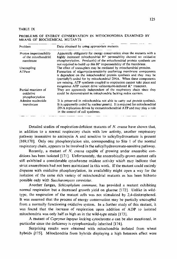
Table IX summarizes the mitochondrial functions that have so far been ex- amined with the help of biochemical mutants.
V. MUTANTS OF OTHER EUCARYOTES DEFICIENT IN ENERGY CONSERVATION IN MITOCHONDRIA
The fungus Neurospora crassa may be an alternative organism to yeast in the genetic approach to bioenergetics. The fungus is easy to culture, several procedures for isolation of intact mitochondria have been worked out and the genetics of the fungus is well known. Normally, it is an obligate aerobe and this has limited its use for preparation of mutants deficient in mitochondrial functions. Thus, a number of respiration-deficient mutants of N. crassa containing diminished amounts of either cytochrome a or of both cytochromes a and b are known [167,168] but mutants that would be entirely devoid of cytochromes or, similar to yeast, o- mutants incapable of mitochondrial protein synthesis, have not yet been prepared since a mutation of this type would apparently be lethal.
125
TABLE IX
PROBLEMS OF ENERGY CONSERVATION IN MITOCHONDRIA EXAMINED BY MEANS OF BIOCHEMICAL MUTANTS
Problem Data obtained by using appropriate mutants
Proton impermeability Apparently obligatory for energy conservation since the mutants with a of the mitochondrial highly increased mitochondrial H ÷ permeability showed no oxidative membrane phosphorylation. Product(s) of the mitochondrial protein synthesis are
not required to build up this H + impermeability of the membrane. Uncoupling The effect of uncouplers may be mediated by mitochondrial proteins. ATPase Formation of oligomycin-sensitivity conferring membrane components
is dependent on the mitochondrial protein synthesis and they may be (partially?) coded for by mitochondrial DNA. When these components are missing, ATP synthesis coupled to respiration cannot take place and exogenous ATP cannot drive valinomycin-induced K ÷ transport.
Partial reactions of They are apparently independent of the respiratory chain since they oxidative could be demonstrated in mitochondria lacking redox carriers. phosphorylation
Adenine nucleotide It is preserved in mitochondria not able to carry out protein synthesis. translocase It is apparently coded by nuclear gene(s). It is required for mitochondrial
DNA replication driven by extramitochondrial ATP and may play a role in the control of cell syntheses.
Detailed studies o f respiration-deficient mutants o f N. crassa have shown that, in addit ion to a normal respiratory chain with low activity, another respiratory pa thway insensitive to antimycin A and sensitive to salicylhydroxamate is present
[169,170]. Only one phosphoryla t ion site, corresponding to Site 1 o f the normal
respiratory chain, appears to be involved in the salicylhydroxamate-sensitive pathway. Recently, a mutant o f N. crassa capable o f growing under anaerobic con-
ditions has been isolated [ 171 ]. Unfortunately, the anaerobically-grown mutant cells still exhibited a considerable cy tochrome oxidase activity which may indicate that
strict anaerobiosis had not been maintained in this work. I f the mutant could entirely
dispense with oxidative phosphorylat ion, its availability might open a way for the isolation o f the same rich variety o f mitochondrial mutants as has been hitherto possible only with Saccharomyces cerevisiae.
Another fungus, Schizophylum commune, has provided a mutant exhibiting normal respiration but a decreased growth yield on glucose [172]. Unlike in wild- type, the respiration o f the mutant cells was not stimulated by 2,4-dinitrophenol. It was assumed that the process o f energy conservation may be partially uncoupled
f rom a normal ly funct ioning oxidative system. In a further study o f this mutant , it was found that the increase o f respiration upon addit ion o f A D P to isolated mi tochondr ia was only half as high as in the wild-type strain [173].
A mutan t o f Coprynus lagopus lacking cy tochrome a can be also mentioned, in particular since the deficiency is cytoplasmically inherited [174].
Surprising results were obtained with mi tochondr ia isolated f rom wheat hybrids [175]. Mi tochondr ia f rom hybrids displaying a high heterosis effect were

126
claimed to exhibit P/O ratios higher than the generally accepted "theoretical" (e.g. 5.8 with a-ketoglutarate, 3.8 with malate and 3.4 with succinate) while mitochondria from parent strains exhibited "normal" P/O ratios. Even mixtures of parental mitochondria exceeded the averages of the parents, i.e. they showed in vitro com- plementation, and approached the activities of hybrid mitochondria. A comple- mentation was observed with ATPase activity as well. Maize hybrids were claimed to behave similarly [176]. Unfortunately, I have been informed that several in- vestigators have not been able to corroborate these unexpected data.
Mitochondria could be a target of genetic defects in some hereditary diseases in mammals. Liver mitochondria from achondroplastic rabbits were found to be morphologically normal but lacking Site 3 phosphorylation [177]. The primary defect in muscular dystrophy seems to reside in a systematic alteration in cellular membranes of which the ionic manifestation is a catastrophic increase in potassium permeability: Liver mitochondria from dystrophic animals were found to be highly permeable for potassium ion (Howland, J., personal communication).
As already mentioned, even those functions that are indispensable for an organism are amenable to genetic studies by means of temperature-sensitive mutants. A temperature-sensitive mutant of a strictly aerobic organism, carrying a lesion in oxidative phosphorylation, would not grow and might even not survive at the restrictive temperature, but would thrive at the permissive temperature at which the genetic lesion would not appear. Therefore, the isolation of temperature-sensitive mutants with modified mitochondria could be tried with any organism, including the classical object of genetic research, Drosophila. A restriction to this approach is posed by the fact that higher organisms are diploid so that only dominant mutations are likely to be detected. This may be a very rare event since loss or modification of mitochondrial functions may be expected to manifest themselves as recessive mu- tations.
VI. MUTANTS OF PROCARYOTES AND THEIR IMPLICATION FOR THE STUDY OF MITOCHONDRIA
Although bacteria have long been the main experimental object of biochemical genetics, until very recently no systematic effort was made to use bacterial mutants in bioenergetics [8]. This may be partly explained by failures to obtain cell-free preparations from bacteria that would exhibit efficient coupling between respiration and phosphorylation and would serve as a starting point for chemical dissection of bacterial oxidative phosphorylation. But even studies of simpler processes, such as glycolysis and energy-dependent membrane transport have not yet fully exploited all the possibilities offered by bacterial mutants. Fortunately, the situation is now changing rapidly and the number of investigators'interested in this kind of bacterial mutants is steadily increasing.
The origin of the use of bacterial mutants in bioenergetics can be traced to studies of Gibson and his associates [178] on biosynthesis of ubiquinone by mutants

127
of Escherichia coli deficient in ubiquinone. Extensive studies of a number of mutants allowed a formulation of the respiratory chain of E. coli in which ubiquinone serves as redox carrier in the linear sequence both on the substrate side and the oxygen side of cytochrome bl (ref. 179). Even though the experimental evidence for such a placement of ubiquinone in the bacterial respiratory chain is not conclusive, it may be of interest for investigators attempting to localize ubiquinone in the mitochondrial respiratory chain where many uncertainities have persisted for a long time. Similarly, a menaquinone-deficient mutant was isolated and used for a tentative formulation of the role of menaquinone in bacterial respiration [180].
The ubiquinone-deficient mutants were selected as cells which, after muta- genesis, were able to grow on glucose but not on non-fermentable substrates. About 10 ~o of mutants unable to grow with succinate as carbon source were affected in the biosynthesis of ubiquinone. As shown in Part II, this selection procedure should also lead to isolation of mutants deficient in oxidative phosphorylation. In fact, a mutant designated uncA- was prepared in this way exhibiting much lower P/O ratios and much lower [Mg2+,Ca2+]-ATPase activity than wild-type bacteria [181]. Similar mutants have also been isolated and studied by other groups [182-185,65]. All investigators concur in the general conclusion that the [Mg2+,Ca2÷]-ATPase is a coupling factor in E. coli similar to FI factor of mammalian mitochondria.
Another mutant, designated uncB-, was also found to be unable to couple oxidation of substrates to the formation of ATP, yet to contain normal amount of [Mg2+,Ca2+]-ATPase [208] (see also refs 186,187). Apparently, another coupling factor has been modified in the uncB- mutant. This points to the multiplicity of coupling factors in bacteria just as in mitochondria.
It was further discovered that, contrary to wild-type bacteria, the energy- linked transhydrogenase could not be driven by ATP in uncA- and uncB- mu- tants [208,188]. However, in another mutant, similar to uncA-, the energy-driven transhydrogenation could be supported by energy derived from electron transport as in wild-type bacteria [189]. The same is true for the uncB- mutant [208]. This shows that there is a "high-energy state" of the bacterial membrane which drives the energy-linked transhydrogenase and this "high-energy state" can normally be maintained either by respiration or by ATP hydrolysis. This is again very similar to what is known in the case of mitochondria.
A further similarity between mitochondrial and bacterial systems is indicated by a study of a mutant of Streptococcus faecalis resistant to dicyclohexylcarbodii- mide [190]. Membrane ATPase, sensitive to dicyclohexylcarbodiimide in wild-type bacterium, was found to be resistant to this drug in the mutant. Soluble ATPase, as well as a protein nectin which is required for binding of the ATPase to the membrane, were found not to be modified in the mutant. Thus, by inference, a membrane component of the ATPase complex must have been affected by the mutation, just as in analogous yeast mutants discussed above.
Of particular interest is the discovery that fl-galactoside transport can be supported either by electron transport or by ATP hydrolysis in wild-type E. coli, but

128
only by electron transport in a mutant lacking [Mg2+,Ca2+]-ATPase [183,191]. It indicates that the "high-energy state" discussed above is also used to drive uptake of metabolites into bacterial cells. A similar conclusion appears to be supported by studies with mutants of E. coli affected in the active transport of amino acids [182, 192,193]. Oxidation of D-lactate was originally hypothesized to play a special role in this active transport but studies with mutants lacking D-lactate dehydrogenase [182,192] do not support the original hypothesis (see also ref. 15). Interesting mutants, designated etc mutants, possess normal electron transport and [Mg2÷,Ca2÷]- ATPase activity and yet have lost the ability to actively transport amino acids [192]. According to growth yield data, these mutants appear not to be able to use energy of substrate oxidation for growth and thus be deficient in oxidative phosphorylation similarly to uncB- mutants. This lends support to a general conclusion that the "high-energy state" of the bacterial membrane represents a common energy pool for transhydrogenase, substrate transport and ATP synthesis. Since other independent studies suggest the possibility that the "high-energy state" involved in fl-galactoside transport may be identical with proton-motive force [194-197], the bacterial studies appear to be generally in favour of Mitchell's [16] chemi-osmotic hypothesis, in particular in its specific form applied to procaryotic cells.
Even though the preparation of bacterial phosphorylating particles that would match in efficiency submitochondrial particles has not yet been achieved, vesicles derived from the bacterial membranes and used hitherto in active transport studies may represent a convenient substitute [193]. The vesicles may closely resemble non-phosphorylating submitochondrial particles capable of "energization" by respiration and may render unique service in defining, in molecular terms, the lesions of various mutants deficient in energy conservation. They also appear to be well suited for reconstitution studies.
vii. PROSPECTS
Biochemical genetics of oxidative phosphorylation is in its early phase of development. In some of its achievements, it has borne out the knowledge obtained in other ways, such as in the case of the participation of the membrane ATPase in ATP synthesis, the requirement of oligomycin-sensitivity conferring components for energy transfer and the independence of energy transfer reactions from electron transport in the respiratory chain. It has helped in the understanding of the genetic control and assembling process of the mitochondrial components, in particular by showing that some components of the energy transfer system are dependent on the mitochondrial genome and on mitochondrial protein synthesis.
The main contributions are so far represented not by what has been resolved but by what has been suggested. A mechanism of adenine nucleotide translocation across the mitochondrial membrane could have been tentatively formulated as- suming conformational changes of the carrier molecule; such a mechanism of carrier

129
function may be of general significance. A support for the existence of uncoupler- sensitive sites in the mitochondrial membrane may be drawn from the study of uncoupler-resistant mutants.
The following puzzling observations of the opl mutant may prove in the future to be of major significance: Owing to the modified adenine nucleotide trans- locator, the rate of adenine nucleotide translocation across the mitochondrial mem- brane of the opt mutant is approximately 2-4 times lower than in wild-type cells [I 56]. It is surprising that such a relatively mild reduction of the translocation rate in opl mitochondria is sufficient to prevent growth on non-fermentable substrates and the utilization of energy of substrate oxidation for cellular syntheses [7,154]. This shows that an appropriately adjusted rate of mitochondrial adenine nucleotide translocation in normal cells may be of crucial importance for triggering extra- mitochondrial syntheses and for cell growth when non-fermentable substrates serve as the energy source. This may be related to the ability of normal mitochondria to maintain a phosphorylation potential in the cytosol as much as fifty times higher than in the mitochondria [198]. The defective translocation machinery of the opl mutant may not sustain such a difference in the two potentials.
Another role of the mitochondrial adenine nucleotide translocation in cell growth has been suggested by experiments in which a combination of the opl mu- tation with the cytoplasmic o- mutation in one cell was attempted. The double mu- tants were not able to grow continually even if fermentable substrate was used as the energy source [199] (see also ref. 153). Accordingly, the cells cannot grow if both respiration and mitochondrial adenine nucleotide translocation are damaged. Apparently, intramitochondrial ATP, furnished either directly by respiration or by fermentation via the intact translocation system, is somehow obligatorily required for cellular growth or multiplication. Apart from serving as fuel cells, mitochondria must have another vital function in the eucaryotic cells. It explains why even the "remnant" mitochondria of cytoplasmic ¢-mutants still contain a functional adenine nucleotide translocation system.
This observation on mutants has been confirmed by experiments on wild-type yeast in which respiration was arrested by antimycin A and adenine nucleotide translocation by bongkrekic acid [66] (Fig. 8).
Future contributions of the mutant studies may be expected to extend along the following lines:
(1) The nature of the "high-energy state". The "high-energy state" has been, respectively, envisaged as a chemical species or group with a "high-energy" bond, as a conformational state of the mitochondrial membrane and as an electrochemical gradient of protons across the membrane [3,4,12l]. The mutants might help to disclose the elusive chemical species in the same way as they did the elusive and labile intermediates of intermediary metabolism. Subtle conformation changes may be ar tificiallyinduced byappropriatemutations. If proton gradient represents a side-path in the mitochondrial energy transformations, mutants may be obtained that are unable to generate a proton gradient and are still capable of oxidative phosphorylation (Fig. 9).

130
ATP
glucose ~-et .hanot -4' C02.
Fig. 8. Sources of intramitochondrial ATP. Intramitochondrial ATP can be furnished either by respiration, which is prevented by antimycin A, or by fermentation via the mitochondrial adenine nucleotide translocation system, which is prevented by bongkrekic acid. When ATP is supplied by neither way, two events take place: The cells are converted into respiration-deficient mutants (if they are "petite-positive", like S. cerevisiae) and their further multiplication is arrested. The effect of antimyein A can be substituted by ~- mutation and the effect of bongkrekic acid by the opl mutation.
(A) Resplratlon 4 - N +gradient q bATP
(B) Respiration ~ ~ -~, X " ATP t-o N + grad ient
Fig. 9. The position of proton gradient with respect to respiration and ATP synthesis according to the chemi-osmotic (A) and chemical (B) hypotheses of oxidative phosphorylation. If, as in B, proton gradient represented a side-path in energy transformation, a mutation could abolish the reaction a, i. e. the generation of proton gradient and still leave oxidative phosphorylation intact. No such mutation could be disclosed if the scheme A were valid.
(2) Mechanism of uncoupling. The study of structural and metabolic changes in uncoupling-sensitive and -resistant mutants may help to decide between alternative hypotheses o f uncoupler action (see p. 121). Uncoupled mutants may be searched for
which may either be naturally "leaky" for protons, as expected according to the
chemi-osmotic hypothesis, or to maintain normal p ro ton impermeability. (3) Organizat ion o f the A T P synthetase. The step-by-step resolution of the
ATPase complex can be viewed as a "classical" task of the biochemical genetics o f mitochondria . A complete ATPase complex may be reconstituted f rom partial components present in different mutants.
(4) Structure of the respiratory chain components with respect to energy conservation. Slight modifications in the structure o f the components in various mutants may not affect their function as redox carriers and yet modify their precise topology in the membrane. This may result in a decrease in phosphoryla t ion efficiency because they would no longer act as t ransmembrane hydrogen and electron

131
carriers (according to the chemi-osmotic hypothesis). Alternatively, the slight struc- tural modifications may prevent the transition of the redox carriers into "high- energy forms" (according to the chemical and conformational hypotheses).
As pointed out above, mutants carrying lesions in mitochondria have proved not to be just servants obediently answering the questions they have been asked. Rather, they have brought about new unexpected scientific problems and new vistas of investigation. There is no reason why it should not be so in the future. Science would lose its unique intellectual attraction if we could foresee its achieve- ments.
ACKNOWLEDGEMENTS
Most of the work of the author, mentioned in this review, was accomplished with his collaborators at the former Department of Biochemistry, Faculty of Science, Komensky University, Bratislava. My thanks are due to all of them as well as to Drs J. Howland (Bowdoin College, Brunswick, Maine, U.S.A.), G. Schatz (Cornell University, Ithaca, N.Y., U.S.A.) and M. Somlo (Centre de genetique mol6culaire du C.N.R.S., Gif-sur-Yvette, France) for critical reading of the manuscript and valuable comments. 1 also thank Dr J. Poggtdy (Psychiatric Hospital, Pezinok) for support and encouragement.
REFERENCES
1 Wagner, R. P. and Mitchell, H. K. (1965) Genetics and Metabolism, 2nd Edn, Wiley, New York 2 Kagawa, Y. (1972) Biochim. Biophys. Acta 265, 297-338 3 Slater, E. C. (1953) Nature 172, 975-978 4 Mitchell, P. (1966) Biol. Rev. 41,445 5 Kov~i6, L. and lstenesov~i, A. (1964) Biochim. Biophys. Acta 82, 162-164 6 Mattoon, J. R. and Sherman, F. (1966) J. Biol. Chem. 241, 4330-4338 7 Kov~t6, L., Lachowicz, T. M. and Slonimski, P. P, (1967) Science 158, 1564-1567 8 Gibson, F. (1971) Search 2, 425-429 9 Levine, R. P. (1968) Science 162, 768-771
10 Pratt, L. H. and Bishop, N. I. (1968) Biochim. Biophys. Acta 153, 664-674 11 Sato, V. L., Levine, R. P. and Neumann, J. (1971) Biochim. Biophys. Acta 253, 437-448 12 Margulis, L. (1970) Origin of Eucaryotic Cells, Yale University Press, New Haven 13 Rabinowitz, M. and Swift, H. (1970) Physiol. Rev. 50, 376-427 14 Ashwell, M. and Work, T. S. (1970) Annu. Rev. Bioehem. 39, 251-290 15 Harold, F. M. (1972) Bacteriol. Rev. 36, 172-230 16 Mitchell, P. (1970) in Organization and Control in Procaryotic and Eucaryotic Cells, pp. 121-
166, Cambridge University Press, Cambridge 17 Harold, F. M. (1970) Advances in Microbial Physiology (Rose, A. H. and Wilkinson, J. F.,
eds), Vol. 4, pp. 45-104, Academic Press, London and New York 18 Kotelnikova, A. V. and Zvjagilskaja, R. A. (1973) Biochemistry of Yeast Mitochondria (in
Russian), Nauka, Moskva 19 Bulder, C. J. E. A. (1964) Antonie van Leewenhoeek 30, 442-454 20 DeDeken, R. H. (1966) J. Gen. Microbiol. 44, 157-165 21 Ephrussi, B. and Slonimski, P. P. (1950) Bioehim, Biophys. Acta 6, 256--267

132
22 Ephrussi, B., Slonimski, P. P., Yotsuyanagi, Y. and Tavlitzki, Y. (1956) C.R. Lab. Carlsberg 26, 87-102
23 Jayaraman, J., Cotman, C., Mahler, H. R. and Sharp, C. W. (1966) Arch. Biochem. Biophys. 116, 224-251
24 K o v ~ , L., Hru~ovsk~t, E. and ~mig~i6, P. (1970) Biochim. Biophys. Acta 205, 520-523 25 Huang, M., Biggs, D. R., Clark-Walker, C. D. and Linnane, A. W. (1966) Biochim. Biophys.
Acta 114, 434-436 26 Clark-Walker, C. D. and Lirmane, A. W. (1967) J. Cell. Biol. 34, 1-14 27 Light, R. J., Lennarz, W. J. and Bloch, K. (1962) J. Biol, Chem. 237, 1793-1800 28 Enebo, L. and Iwamoto, H. (1966) Acta Chem. Scand. 20, 439~43 29 Shafai, T. and Levin, L. M. (1968) Biochim. Biophys. Acta 152, 787-790 30 Wishniewski, B. J., Keith, A. D. and Resnick, M. R. (1970) J. Bacteriol. 101, 160 31 White, G. L. and Hawthorne, J. N. (1970) Biochem. J. 117, 203-213 32 Chang, S. B. and Matson, R. S. (1972) Biochem. Biophys. Res. Commun. 46, 1529-1535 33 Ainsworth, P. J., Tustanoff, E. R. and Ball, A. J. S. (1972) Biochem. Biophys. Res. Commun.
47, 1299 34 Wohlraub, H. and Jacobs, E. E. (1967) Biochem. Biophys. Res. Commun. 28, 991-997 35 Keyhani, E. and Chance, B. (1971) FEBS Lett. 17, 127 36 Light, P. A. (1972) FEBS Lett. 29, 319 37 Lemberg, R. (1961) Adv. Enzymol. 23, 265-321 38 Okuda, S. and Takahashi, H. (1969) J. Bacteriol. 100, 647 39 Ninneman, H. (1972) FEBS Lett. 24, 181-184 40 Light, P. A., Ragan, C. l., Clegg, R. A. and Garland, P. B. (1968) FEBS Lett. 1, 4-8 41 Light, P. A. and Garland, P. B. (1971) Biochem. J. 124, 123-134 42 Ohnishi, T. (1972) FEBS Lett. 24, 305-309 43 Clegg, R. A., Haddock, B. A., Light, P. A., Garland, P. G., Swarm, J. C. and Bray, R. C.
(1969) FEBS Lett. 5, 207. 44 Haddock, B. A. and Garland, P. B. (1971) Biochem. J. 124, 155-170 45 Biggs, D. R. and Linnane, A. W. (1963) Biochim. Biophys. Acta 78, 785-788 46 Luzikov, V. N., Zubatov, A. S. K. and Rainina, E. J. (1972) FEBS Lett. 11,273 47 Luzikov, V. N., Zubatov, A. S., Rainina, E. J. and Bakeeva, L. E. (197l) Biochim. Biophys.
Acta 245, 321-334 48 Barnett, J. A. and Kornberg, H. L. (1960) J. Gen. Microbiol. 23, 65 49 Nossal, P. M. (1951) Ph.D. Thesis, Sheffield,; cf. Krebs, H. A., Gurin, S. and Eggleston, L. V.
(1952) Biochem. J. 51,614 50 Suomalainen, H., Konttinen, K. and Oura, E. (1969) Arch. Mikrobiol. 64, 251-261 51 Hartwell, L. H. and McLaughlin, C. S. (1968) J. Bacteriol. 96, 1664 52 Puglisi, P. P. and Cremona, T. (1971) Biochem. Biophys. Res. Commun. 39, 461-469 53 ~ubik, J., Kolarov, J. and Kov~i~, L. (1974) submitted 54 Ogur, M., St. John, R. and Nagai, S. (1957) Science 125,928 55 Lingens, F. and Oltmanns, O. (1964) Z. Naturforsch. 19b, 1058-1065 56 Mortimer, R. K. and Hawthorne, D. C. (1969) The Yeast (Rose, A. H. and Harrison, J. S.,
eds), Vol. 1, pp. 385-460, Academic Press, New York 57 Fink, G. R. (1970) Methods of Enzymol. 27, 59 58 Sherman, F. (1967) Methods of Enzymol. 10, 610-615 59 ~ubik, J., Kolarov, J. and Lachowicz, T. M. (1972) FEBS Lett. 27, 81-84 60 Sherman, F. (1964) Genetics 49, 39-48 61 Azzi, A., Gherardini, P. and Santato, M. (1971) J. Biol. Chem. 246, 2035-2042 62 Peterajowi, E., Kov~i6, L., Vidov~i, M., ~ubik, J. and Kolarov, J. (1974) submitted 63 Beck-Hansen, N. T. and Rank, G. H. (1972) Can. J. Genet. Cytol. 14, 681-689 64 Gouhier, M. and Mounolou, J. C. (1973) Mol. Gen. Genet. 122, 149-164 65 Kanner, B. I. and Gutnick, D. L. (1972) J. Bacteriol. 111,287-289 66 ~ubik, J., Kolarov, J. and Kov~i~, L. (1972) Biochem. Biophys. Res. Commun. 49, 192-198 67 Bauchop, T. and Elsden, S. R. (1960) J. Gen. Microbiol. 23, 457-469 68 Scholes, P. and Mitchell, P. (1970) J. Bioenergetics 1,309-323 69 Hempfling, W. P. (1970) Biochim. Biophys. Acta 205, 169-182 70 Korman~ikov~i, V., Kowi6, L. and Vidov~i, M. (1969) Biochim. Biophys. Acta 180, 9-17

133
71 Proudlock, J. W., Haslam, J. M. and Linnane, A. W. (1969) Biochem. Biophys. Res. Commun. 33, 590-595
72 Avner, P. A. and Griffiths, D. E. (1973) Eur. J. Biochem. 32, 301-321 73 (1967) Methods in Enzymol. (Estabrook, R. W. and Pullman, M. E., eds), Vol. 10, Academic
Press, New York 74 Mortimer, R. K. and Hawthorne, D. C. (1966) Genetics 53, 165-173 75 Bolotin, M., Coen, D., Deutsch, J., Dujon, B., Netter, P., Petrochilo, E. and Slonimski, P. P.
(1971) Bull. Inst. Pasteur 69, 215-239 76 Wilkie, D. (1972) in Mitochondria/Biomembranes, pp. 85-94, North Holland, Amsterdam 77 Rank, G. H. and Beck-Hansen, N. T. (1972) Genetics 72, 1-15 78 Kleese, R. A., Grotbeck, R. C. and Snyder, J. R. (1972) J. Bacteriol. 112, 1023-1025 79 Lukins, H, B., Tate, J. R., Saunders, G. W. and Linnane, A. W. (1973) Mol. Gen. Genet. 120, 17-25 80 Jakob, H. (1965) Genetics 52, 75-98 81 Sels, A. and Jakob, H. (1967) Biochem. Biophys. Res. Commun. 28, 1453-1459 82 Ephrussi, B., Hottinguer, H, and Chimenez, A. (1949) Ann. Inst. Pasteur 76, 351-367 83 Nagai, S., Yanagishima, N. and Nagai, H. (1961) Bact. Rev. 25, 404-426 84 Kraepelin, G. (1967) Z. Allgem. Mikrobiol. 7, 287-325 85 Borst, P. (1972) Annu. Rev. Biochem. 41,336-376 86 Ku~ela, ~. and Gre~n~i, E. (1969) Experientia 25,776--777 87 Ku~ela, ft., ~mig~i6, P. and Kov/t6, L. (1969) Experientia 25, 1042-1043 88 Schatz, G. and Saltzgaber, J. (1969) Biochem. Biophys. Res. Commun. 37, 996-1007 89 Slonimski, P. P. and Ephrussi, B. (1949) Ann. Inst. Pasteur 77, 47 90 Weiss, H., Sebald, W. and Bticher, T. (1971) Eur. J. Biochem. 22, 19-26 91 Schatz, G., Groot, G. S. P., Mason, T., Rouslin, W., Wharton, D. C. and Saltzgaber, J. (1972)
Fed. Proc. 31, 21-29 92 Weiss, H., Lorenz, B. and Kleinow, W. (1972) FEBS Lett. 25, 49-51 93 Tzagoloff, A., Rubin, M. S. and Sierra, M. F. (1973) Biochim. Biophys. Acta 301, 71-104 94 Mason, T. L. and Schatz, G. (1973) J. Biol. Chem. 248, 1355-1360 95 Weiss, H. (1972) Eur. J. Biochem. 30, 469-478 96 Slonimski, P. P. and Hirsch, H. M. (1952) C.R. Acad. Sci. 235, 741 97 Gregolin, C. and Ghiretti-Magaldi, A. (1961) Biochim. Biophys. Acta 54, 62-66 98 Schatz, G. (1962) Biochim. Biophys. Acta 62, 581-584 99 Sugimura, T., Okabe, K. and Rudney, H. (1964) Biochim. Biophys. Acta 82, 350-354
100 Biggs, D. R., Nakamura, H., Kearney, E. B., Rocca, E. and Singer, T. P. (1970) Arch. Biochem. Biophys. 137, 12
101 Heick, H. M. (1972) Can. J. Biochem. 18, 23-28 102 Kowachevich, R. and Barron, E. S. G. (1964) Arch. Biochem. Biophys. 108, 200--206 103 Perlman, P. S. and Mahler, H. R. (1970) Bioenergetics 1,119-138 104 Galeotti, T., Kov~.~, L. and Hess, B. (1968) Nature 218, 194-196 105 K o v ~ , L. and Weissov~i, K. (1968) Biochim. Biophys. Acta 153, 55-59 106 Schatz, G. (1968) J. Biol. Chem. 243, 2192-2199 107 Tzagoloff, A. and Meagher, P. P. (1971) J. Biol. Chem. 246, 7328-7336 108 Kov~i~, L., Groot, G. S. P. and Racker, E. (1972) Biochim. Biophys. Acta 256, 55-65 109 Kolarov, J., ~ubik, J. and Kov~i~, L. (1972) Biochim. Biophys. Acta 267, 457-464 110 Perkins, M., Haslam, J. M. and Linnane, A. W. (1972) FEBS Letters 25, 271-274 111 Haslam, J. M., Perkins, M. and Lirmane, A. W. (1973) Biochem. J. 134, 935-948 112 Sherman, F. (1963) Genetics 48, 375-385 113 Sherman, F. and Slonimski, P. P. (1964) Biochim. Biophys. Acta 90, 1-15 114 Parker, J. H. and Mattoon, J. R. (1969)J. Bacteriol. 100, 167 115 Lachowicz, T. M., Kotylak, Z., Kolodyfiski, J. and ~niegocka, Z. (1969) Arch. Immunol.
Therap. Exp. 17, 72-85 116 Yarovoy, B. F., Yurchenko, L. V. and Zakharov, I. A. (1970) Genetika 6, 138-144 117 Beck, J. C., Parker, J. H., Balcavage, W. X. and Mattoon, J. R. (1971) Autonomy and Bio-
genesis of Mitochondria and Chloroplasts (Boardman, N. K., Linnane, A. W. a n d Smillie, R. M., eds), pp. 194-204, North Holland, Amsterdam
118 ~ubik, J., Ku~.ela, ~., Kolarov, J., Kov~i(:, L. and Lachowicz, T. M. (1970) Biochim. Biophys. Acta 205, 513-519
134
119 Subik, J. (1970) Thesis, Komensky University, Bratislava 120 Gunge, N. (1966) Jap. J. Genet. 41,215-223 121 Chance, B. and Williams, G. R. (1956) Adv. Enzymol. 17, 65-134 122 MacLennan, D. H., Lenaz, G. and Szarkowska, L. (1966) J. Biol. Chem. 241, 5351-5359 123 Lenaz, G. and MacLennan, D. H. (1966) J. Biol. Chem. 241, 5260-5265 124 Avi-Dor, Y. (1969) Mitochondria: Structure and Function (Ernster, L. and Drahota, Z., eds),
p. 285, Academic Press, New York 125 Penniston, J. T., Green, D. E. and Vande Zande, V. (1966) Arch. Biochem. Biophys. 113,
512-518 126 Subik, J., Kovfi.~, L. and Kolarov, J. (1972) Biochim. Biophys. Acta 283, 146-154 127 Slater, E. C., Berden, H. J. and Wegdam, J. A. (1970) Biochim. Biophys. Acta 223,354-364 128 Chance, B., Wilson, D. F., Button, P. L. and Erecifiska, M. (1970) Proc. Natl. Acad. Sci. U.S.
66, 1175-1182 129 WikstrOm, M. K. F. (1973) Biochim. Biophys. Acta 301,155-193 130 Sato, N., Ohnishi, T. and Chance, B. (1972) Biochim. Biophys. Acta 275,288-297 131 Downie, J. A. and Garland, P. B. (1973) Biochem. J. 134, 1051-1061 132 Groot, G. S. P., Kovfi.~, L. and Schatz, G. (1971) Proc. Natl. Acad. Sci. U.S. 68, 308-311 133 Chen, W. L. and Charalampous, F. C. (1973) Biochim. Biophys. Acta 294, 329-346 134 Goffeau, A., Colson, A. M., Landry, Y. and Foury, F. (1972) Biochem. Biophys. Res. Commun.
48, 1148-1154 135 Avner, P. R. and Griffiths, D. F. (1970) FEBS Lett. 10, 202 136 Lankashire, W. E. and Griffiths, D. E. (1970) FEBS Lett. 17, 209 137 Avner, P. R., Coen, D., Dujon, B. and Slonimski, P. P. (1974) Mol. Gen. Genet., in press 138 Griffiths, D. E., Avner, P. R., kankashire, W. E. and Turner, J. R. (1972) Biochemistry and
Biophysics of Mitochondrial Membranes, pp. 605-621, Academic Press, New York 139 Somlo, M., Avner, P. R., Cosson, J., Dujon, B. and Krupa, M., (I 974) Eur. J. Biochem. 42, 439-445 140 Parker, J. H., Trimble Jr, I. R. and Mattoon, J. R. (1968)Biochem. Biophys. Res. Commun. 33,
590-595 141 Stuart, K. D. (1970) Biochem. Biophys. Res. Commun. 39, 1045 1051 142 Wakabayashi, K. and Gunge, N. (1970) FEBS Lett. 6, 302 143 Wakabayashi, K. (1972) J. Antibiot. 25,475 144 Rank, G. H. and Martin, R. (1972) Can. J. Genet. Cytol. 14, 197-199 145 Mitchell, C. A., Bunn, C. L,, Lukins, H. B. and Lirmane, A. W. (1972) Bioenergetics 4, 363-379 146 Shannon, C., Enns, R., Wheelis, L., Burchiel, K. and Criddle, K. S. (1973) J. Biol. Chem. 248,
3004-3011 147 Weinbach, E. C. and Garbus, J. (1969) Nature 221, 1016-1018 148 Somers, J. M. and Bevan, E. A. (1969) Genet. Res. 13, 1-13 149 Hughes, A. R. and Wilkie, D. (1972) Heredity 28, 117-127 150 Fink, G. R. artd Styles, C. A. (1972) Proc. Natl. Acad. Sci. U.S. 69, 2846-2849 151 Lacroute, F. (1971) J. Bacteriol. 106, 519-522 152 Kovb_6, L. and Hrugovsk~i, E. (1968) Biochim. Biophys. Acta 153, 43-54 153 Beck, J. C., Mattoon, J. R., Hawthorne, D. C. and Sherman, F. (1968) Proc. Natl. Acad. Sci.
U.S. 60, 186-193 154 Somlo, M. (1970) Arch. Biochem. Biophys. 136, 122-133 155 Lachowicz, T. M. (1968) Arch. lmmunol. Therap. Exp. 16, 693-702 156 Kolarov, J., ~;ubik, J. and Kov~t6, L. (1972) Biochim. Biophys. Acta 267, 465-478 157 Haslam, J. M., Spithill, T.W., Linnane, A.W. and Chapell, J. B. (1973) Biochem. J. 134, 949-957 158 Schweizer, E. and Boiling, H. (1970) Proc. Natl. Acad. Sci. U.S. 67, 660-666 159 Thompson, E. D., Start, P. R. and Parks, L. W. (1971) Biochern. Biophys. Res. Commun. 43,
1304-1309 160 Molzahn, S. W. and Woods, R. A. (1972) J. Gem Microbiol. 72, 339-348 161 Karst, F. and Lacroute, F. (1972) 8th Meet. FEBS, Amsterdam, Abstracts of Commun. No. 207 162 Bunn, C. L., Mitchell, C. H., Lukins, H. B. and Linnane, A. W. (1970) Proc. Natl. Acad. Sci.
U.S. 67, 1233-1240 163 Dutton, P. L., Erecifiska, M., Sato, N., Mukai, Y., Pring, M. and Wilson, D. F. (1972) Biochim.
Biophys. Acta 267, 15-24 164 Butow, R. A. and Zeydel, M. (1968) J. Biol. Chem. 243, 2545
135
165 Grimmelikhuijzen, C. J. P. and Slater, E. C. (1973) Biochim. Biophys. Acta 305, 67-79 166 Mounolou, jr. C. (1973) FEBS Lett. 29, 275-279 167 Eakin, R. T. and Mitchell, H. K. (1970) J. Bacteriol. 104, 74-78 168 Betrand, H. and Pittinger, T. H. (1972) Mol. Gen. Genet. 117, 82-90 169 Von Jagow, G., Weiss, H. and Klingenberg, M. (1973) Eur. J. Biochem. 33,140-157 170 Lambowitz, A. M., Smith, E. W. and Slayman, C. W. (1972) J. Biol. Chem. 247, 4859-4865 171 Howell, N., Zuiches, C. A. and Munkres, K. D. (197l) J. Cell Biol. 50, 721-736 172 Hoffman, R. M. and Raper, J. R. (1971) Science 171,418-419 173 Hoffman, R. M. and Raper, J. R. (1972) J. Bacteriol. 110, 780-781 174 Casselton, L. A. and Condit, A. (1973) J. Gen. Microbiol. 72, 521-527 175 Sarkissian, I. V. and Srivastava, H. K. (1969) Proc. Natl. Acad. Sci. U.S. 63, 302-309 176 Sarkissian, 1. V. and Srivastava, H. K. (1967) Genetics 57, 843 177 Bargman, G. J., Mackler, B. and Shepard, T. H. (1972) Arch. Biochem. Biophys. 150, 137 178 Cox, G. B., Gibson, F. and Pittard, J. (1968) J. Bacteriol. 95, 1591-1598 179 Cox, G. B., Newton, N. A., Gibson, F., Snoswell, A. M. and Hamilton, J. A. (1970) Biochem. J.
117, 551-562 180 Newton, N. A., Cox, G. B. and Gibson, F. (1971) Biochim. Biophys. Acta 244, 155-166 181 Butlin, J. D., Cox, G. B. and Gibson, F. (1971) Biochem. J. 124, 75-81 182 Simoni, D. R. and Schallenberger, M. K. (1972) Proc. Natl. Acad. Sci. U.S. 69, 2663-2667 183 Schairer, H. U. and Haddock, B. A. (1972) Biochem. Biophys. Res. Commun. 48, 544-551 184 Turnock, G., Erickson, S. K., Ackrell, B. A. C. and Birch, B. C. (1972) J. Gen. Microbiol. 70,
507-515 185 Evans, D. J. (1969) J. Bacteriol. 100, 914-922 186 Gutnick, D. L., Kanner, B. I. and Postma, P. W. (1972) Biochim. Biophys. Acta 283, 217-222 187 Schairer, H. U. and Gruber, D. (1973) Eur. J. Biochem. 37, 282-286 188 Cox, G. B., Newton, N. A., Butlin, J. D. and Gibson, F. (1971) Biochem. J. 125,489-493 189 Kanner, B. 1. and Gutnick, D. L. (1972) FEBS Lett. 22, 197-199 190 Abrams, A., Smith, J. B. and Baron, C. (1972) J. Biol. Chem. 247, 1484-1488 191 Preziozo, G., Hong, J.-S., Kerwar, G. K. and Kaback, H. R. (1973) Arch. Biochem. Biophys.
154, 575-582 192 Hong, J.-S. and Kaback, H. R. (1972) Proc. Natl. Acad. Sci. U.S. 69, 3336-3340 193 Kaback, H. R. (1972) Biochim. Biophys. Acta 265, 367-416 194 West, I. C. (1970) Biochem. Biophys. Res. Commun. 41,655-66l 195 West, I. C. and Mitchell, P. (1973) Biochem. J. 132, 587-592 196 Kashket, E. R. and Wilson, T. H. (1972) Biochem. Biophys. Res. Commun. 49, 615-620 197 Himata, H., Altendorf, K. and Harold, F. M. (1973) Proc. Natl. Acad. Sci. U.S. 70, 1804-1808 198 Heldt, H. W., Klingenberg, M. and Milovancev, M. (1972) Eur. J. Biochem. 30, 434-440 199 Kovgt~ovfi, V., Irmlerov~i, J. and Kov~i~, L. (1968) Biochim. Biophys. Acta 162, 157-163 200 Laquin, G., Vignais, P. V. and Mattoon, J. R. (1973) FEBS Lett. 35, 198-200 201 Perkins, M., Haslam, J. M., Klyce, H. R. and Linnane, A. W. (1973) FEBS Lett. 36, 137-142 202 Prakash, L. and Sherman, F. (1973) J. Mol. Biol. 79, 65-82 203 Ebner, E., Mason, T. L. and Schatz, G. (1973) J. Biol. Chem. 248, 5369 5378 204 Ebner, E. and Schatz, G. (1973) J. Biol. Chem. 248, 5379-5384 205 Griffiths, D. E. (1972) FEBS Symp. 28, 95-104 206 Lancashire, W. E., Houghton, R. L. and Griffiths, D. E. (1974) Biochem. Soc. Trans., in the press 207 Ebner, E., Mennucci, L. and Schatz, G. (1973) J. Biol. Chem. 248, 5360-5368 208 Butlin, J. D., Cox, G. B. and Gibson, F. (1973) Biochim. Biophys. Acta 292, 366--375


![Biochimica et Biophysica Acta - immed.org considerations/09.07.2017 updates/Membrane... · G.L. Nicolson, M.E. Ash / Biochimica et Biophysica Acta 1859 (2017) 1704–1724 1705 [8]](https://img.dokumen.tips/doc/110x75/5c684f1e09d3f2f5638b5509/biochimica-et-biophysica-acta-immed-considerations09072017-updatesmembrane.jpg)