Embed Size (px)
Citation preview

PATHOPHYSIOLOGY
Basic overview for medical students
1st revised (only electronic) version
MIROSLAV KUBA, ZUZANA KUBOVÁ
Department of Pathophysiology
Charles University - Faculty of Medicine in Hradec Králové
Czech Republic
Hradec Králové, October 2018

2
Preface to the revised version
After 13 years from the 1st edition of our lecture notes a new textbook could be written on the basis of many
scientific novelties - discoveries/hypotheses in Pathophysiology and related fields that appeared in the
meantime. But since our „basic overview” of Pathophysiology is oriented to medical students, we believe
that the main principles of this textbook can stay the same as they are explained in the Introduction to the 1st
issue. It means that we intentionally do not ad too much new information but we have rather revised the text
according to the feedback from side of students. We tried to focus still more to important logic of
relationships between single facts for better understanding of mechanisms of diseases, in particular of those
which can lead to death. We namely believe that despite the modern tendency to introduce clinical guidelines
(which are only mechanically followed by doctors), students should understand well the background of
diseases!!!. It can be very individual and thus, standard using of clinical guidelines can be quite danger in a
distinct proportion of patients. Therefore, we try to instruct students to be enough self-confident about their
ability to recognise important relationships for the correct diagnosis and optimal (individualized) treatment.
This is not the definite version for the second (printed) issue of our Pathophysiology, we will still
continue in the upgrade but now we would like to provide a newer version to our current 3rd year
medical students.
Hradec Králové, October 2018 Miroslav Kuba
Zuzana Kubová

3
Contents
Preface to the revised version .............................................................................................................. 2
1. Organism and environment, adaptation ........................................................................................... 6
2. Health versus disease, Etiology and Pathogenesis, Clinical and biological (somatic) death ........... 8
3. Immunodeficiencies ....................................................................................................................... 11
4. Allergies ......................................................................................................................................... 12
5. Autoimmune disorders, post-transplantation reaction ................................................................... 14
6. Neoplasia, neoplasm-host interaction (effects) .............................................................................. 15
7. Alterations in fluid volume ............................................................................................................ 18
8. Disorders of osmolarity, etiology and pathogenesis of edema ...................................................... 21
9. Hyponatremia, hypernatremia ........................................................................................................ 24
10. Hypokalemia, hyperkalemia ........................................................................................................ 26
11. Importance of Iron, Iodine and other Trace elements .................................................................. 29
12. Disorders of Calcium, Phosphorus and Magnesium metabolism ................................................ 31
13. Osteoporosis, Osteomalacia, Renal Osteodystrophy ................................................................... 36
14. Pathophysiology of Cramps ......................................................................................................... 37
15. Acidosis ........................................................................................................................................ 38
16. Alkalosis....................................................................................................................................... 45
17. Disorders of peripheral circulation .............................................................................................. 48
18. Syncope ........................................................................................................................................ 51
19. Circulatory shock ......................................................................................................................... 53
20. Inflammation ................................................................................................................................ 55
21. Sepsis, Systemic Inflammatory Response Syndrome (SIRS) ...................................................... 58
22. Pathophysiology of pain............................................................................................................... 59
23. Fever, Hyperthermia .................................................................................................................... 62
24. Hypothermia and Reaction of organism to cold .......................................................................... 65
25. Stress, General adaptation syndrome ........................................................................................... 66
26. Psychosomatic diseases ................................................................................................................ 67
27. Anemias – classification .............................................................................................................. 68
28. Anemias – consequences ............................................................................................................. 69
29. Blood transfusion, complications, fetal erythroblastosis ............................................................. 70
30. Leukemias, lymphomas ............................................................................................................... 72
31. Myeloma (monoclonal gammopathy) .......................................................................................... 74
32. Polycythemia ................................................................................................................................ 75
33. Bone marrow depression .............................................................................................................. 76
34. Disorders of hemostasis and coagulation ..................................................................................... 77
35. Thrombophilic states, DIC ........................................................................................................... 81
36. Dyslipoproteinemia ...................................................................................................................... 84

4
37. Endothelial dysfunction, effects of nitric oxide ........................................................................... 86
38. Atherosclerosis ............................................................................................................................. 88
39. Arterial hypertension.................................................................................................................... 90
40. Arterial hypotension ..................................................................................................................... 92
41. Disorders of cardiac rhythm and conduction ............................................................................... 93
42. Ischemic heart disease .................................................................................................................. 95
43. Myocardial infarction and its complications ................................................................................ 97
44. Congenital Heart Defects ............................................................................................................. 99
45. Acquired Valvular Heart Defects ............................................................................................... 100
46. Heart failure ............................................................................................................................... 102
47. Compensatory mechanisms in heart failure ............................................................................... 105
48. Pulmonary circulation, ventilation/perfusion ratio, gas diffusion disorders .............................. 107
49. Obstructive respiratory diseases ................................................................................................. 110
50. Restrictive pulmonary diseases .................................................................................................. 112
51. Pulmonary hypertension, pulmonary edema .............................................................................. 116
52. Adult respiratory distress syndrome - ARDS ............................................................................ 118
53. Respiratory Failure ..................................................................................................................... 119
54. Hypoxia ...................................................................................................................................... 121
55. Hyperoxia, hyperbaria, decompression ...................................................................................... 123
56. Nephritic syndrome .................................................................................................................... 125
57. Nephrotic syndrome ................................................................................................................... 129
58. Pyelonephritis, Kidney Interstitial Processes ............................................................................. 133
59. Hydronephrosis, Nephrolithiasis ................................................................................................ 135
60. Acute and chronic renal failure .................................................................................................. 137
61. Pathophysiology of Hypothalamo-Pituitary system .................................................................. 142
62. Decrease of Anterior Pituitary hormones................................................................................... 143
63. Hyperfunction of anterior pituitary gland .................................................................................. 145
64. Alterations of the Posterior Pituitary gland hormones ............................................................... 146
65. Hyperthyroidism, Hypothyroidism ............................................................................................ 148
66. Adrenal cortex hormones – hypersecretion ............................................................................... 151
67. Adrenal cortex hormones - acute and chronic deficiency .......................................................... 153
68. Adrenal medulla dysfunction ..................................................................................................... 154
69. Diabetes mellitus, etiology and pathogenesis ............................................................................ 155
70. Diabetic coma ............................................................................................................................ 160
71. Diabetes mellitus – complications ............................................................................................. 162
72. Hypoglycemic states .................................................................................................................. 165
73. Pathophysiology of vitamins ...................................................................................................... 166
74. Nausea, Vomiting....................................................................................................................... 170

5
75. Gastric ulcer, duodenal ulcer – pathophysiology ....................................................................... 172
76. Gastritis, postgastrectomy status ................................................................................................ 174
77. Gastro-intestinal bleeding .......................................................................................................... 177
78. Intestinal malabsorption ............................................................................................................. 179
79. Intestinal obstruction (Ileus), irritable colon, meteorism ........................................................... 181
80. Dysfunctions of the colon: diarrhea, constipation, Crohn’s disease, ulcerative ........................ 184
Colitis ............................................................................................................................................... 184
81. Acute and chronic pancreatitis ................................................................................................... 188
82. Cholelithiasis and cholecystitis .................................................................................................. 190
83. Jaundices .................................................................................................................................... 192
84. Liver cirrhosis, Portal hypertension ........................................................................................... 194
85. Hepatic failure, hepatic coma..................................................................................................... 196
86. Starvation, malnutrition ............................................................................................................. 198
87. Obesity ....................................................................................................................................... 202
88. Pathophysiology of the spinal cord ............................................................................................ 204
89. Alterations of peripheral nervous system and motor function ................................................... 208
90. Autonomic nervous system dysfunctions .................................................................................. 213
91. Brain Ischemia, Vascular Brain Disorders ................................................................................. 216
92. Intracranial hypertension............................................................................................................ 218
93. CNS Neurotransmitters changes - Glutamate, GABA, Serotonin ............................................. 221
94. CNS Neurotransmitters changes - Dopamine, Acetylcholine .................................................... 223
95. Epilepsy - etiology and pathogenesis ......................................................................................... 226
90. Demyelination diseases .............................................................................................................. 230
97. Pathophysiology of immature newborns .................................................................................... 233
98. Aging .......................................................................................................................................... 235
99. Pathophysiological view on "alternative/complementary" medicine ........................................ 237
100. Biosignals recording and evaluation. Biofeedback .................................................................. 239

6
1. Organism and environment, adaptation
(M. Kuba)
- continuous interactions between organisms and external environment
- body tries to keep constant internal environment (homeostasis) irrespective of external environment
parameters via adaptation mechanisms (e.g. thermoregulatory)
- any change of external environment requires resetting of regulatory mechanisms to keep homeostasis
- each adaptation mechanism has some capacity (limited)
- failing adaptation → breakdown of homeostasis → dysfunction of the body = disease
Adaptation capacity
- represents adaptability of the body
- can be expressed as a range of external environment parameters within which the particular
organism can keep homeostasis (survive)
- it is !!! very individual!!! - dependent on:
o genetic disposition
o age
o functional reserve of the body systems responsible for the adaptation to a particular change
(dependent on the previous life style - nutrition, physical activity etc.)
- extreme (or too quick) changes of the external environment or failure of adaptation mechanism (due
to failure of some organ) → change in internal environment (broken homeostasis) → disease
- border changes of external environment leading to survival only of individuals with best adaptation
facilities are causing natural selection - Darwin theory (when Medicine interferes in the human
population → delicate ethical problem of genetic information worsening)
- high external temperature requires thermoregulatory response based on peripheral vasodilation →
↓blood pressure (BP) - to keep adequate BP requires ↑ minute volume (MV), people with chronic
heart failure do not have sufficient heart reserve→ COLLAPSE (unconsciousness) (typical for old
people)
Factors influencing organism
- not-changeable
o genetics, gender, ethnic, age
- changeable
o nutrition, life style, personal habits
- factors from environment
o viral and microbial
o chemical (toxins, drugs, allergens)
o physical (irradiation, temperature, sun exposure, altitude - p02)
- psychological factors (family background, stress, religion etc.)
Disease may be dependent:
- exclusively on genetic factors without any possibility to influence its appearance:
o autosomal dominant (Adult polycystic kidney disease, Familial hypercholesterolemia,
Osteogenesis imperfecta, Spherocytosis, von Willebrand's disease)
o autosomal recessive (Color blindness, Cystic fibrosis, Phenylketonuria)
o X - linked recessive (Hemophilia, Bruton-type hypogammaglobulinemia, Duchenne's
dystrophy)

7
- largely dependent on environment or life style
o Colon cancer - low in developing countries (e.g. Africa), high in "westernized" populations
(USA, Europe) - due to different intake of dietary fibers and fat
o Lung cancer - due to smoking (low in Mormons and Seventh day Adventists with code
against use of tobacco – but non-smokers also can have lung cancer!!)
o Skin cancer - due to ultraviolet light
o Malaria - only in countries with mosquitoes transmitting Plasmodium
- dependent on combination of genetic predisposition and factors of environment or life style
influencing manifestation of the disease = most frequent case
o early coronary heart disease - due to predisposition to hypercholesterolemia and
simultaneous high animal fat intake and/or smoking, low physical activity, stress factors etc.
o obesity and insulin non-dependent diabetes mellitus - combination of genetic predisposition
with nutritional habits and low physical activity

8
2. Health versus disease, Etiology and Pathogenesis, Clinical and biological
(somatic) death (M. Kuba)
Concept of normalcy (health):
- all parameters of the human body have a certain amount of interindividual variation and it is
important to know the range of values considered as normal (this can differ among ethnic groups,
sexes and is usually age dependent)
- in normal (standardized resting) conditions a healthy subject should display all measurable
parameters in the normal range, however, any single measurement (laboratory result, observation)
that seems to indicate abnormality must be always judged in the context of the entire individual and
its conditions (e.g. a single reading of elevated blood pressure does not make and individual
hypertensive or single elevated blood glucose does not mean that the person is diabetic)
- the concept of normalcy (especially in the field of mental abilities) may be dependent on cultural
values, which may differ in various populations (e.g. dyslexia - reading disability could not be
considered as a defect of a central nervous system function in a primitive culture (analphabets))
- the WHO definition of health – “Health is a state of complete physical, mental and social well-being
and not merely the absence of disease or infirmity.” - seems to be a very comprehensive definition,
but given the almost general frustration of people at least in some aspects of their life, this definition
does not seem to apply to too large part of the human population (the more intelligent person, the
worse: the intelligent person cannot be happy (in social well-being) for a long time because he/she
will realize very soon some problem he/she has.)
Concept of disease
- can be defined as any situation which changes the internal environment (impaired homeostasis) -
its parameters are not within the normal range
- since formerly there were almost no changes of intrinsic factors measurable in psychiatric diseases, it
would have looked as though the above specified definition of disease did not cover psychic
disorders - however, nowadays it is well evident that at least subtle biochemical (neurotransmitter)
changes are already detectable in many psychic disorders as well
- the subjective feeling of an ill person is that "he/she loses ability to continue in all normal daily
activities"
- in some chronic diseases homeostasis can be kept but the organism has decreased adaptation
capacity - the body can survive in a very limited range of environmental conditions
- health and disease are not strictly separate, there are some border states
Pathological state
- not a fully normal organism - e.g. after amputation of a limb - but having normal homeostasis
Symptoms and signs of diseases
- symptoms - subjective manifestations of a disease (e.g. nausea, headache, fatigue) that cannot be
verified objectively
- signs - objective measurable manifestations (e.g. fever, tachycardia, edema, hypertension)
Phases of diseases
1. latent phase - period between exposure to causal factor(s) and first symptoms/signs (in infectious diseases
it is incubation period) - no manifestations of diseases are recognizable
2. prodromal phase - usually only non-specific symptoms and signs are manifested (e.g. fatigue, fever,
increased FW reading (sedimentation of erythrocytes) that are not sufficient for recognition (diagnosis) of
the disease

9
3. manifest phase including also some specific signs and symptoms (some typical location of pain, typical
efflorescences on the skin, changes in plasma concentrations of minerals and other substances)
4. Reconvalescence - gradual recovery of normal functions of the body
5. Final phase -resolution includes the following possibilities
- "resolution ad integrum" = total recovery of the organism without any consequences or recovery with
some chronic consequences of the disease (e.g. decreased vital capacity after some respiratory
processes)
- "development of chronic variant" of the disease with remissions (improvement of the clinical state) and
exacerbations (repeated worsening of the clinical state)
- death
Etiology - describes causes of diseases
- formerly (on the border of 19. and 20. centuries) monocausal etiology of diseases was typically
recognized - e.g. etiology of tonsillitis is Streptococcal infections
- nowadays mainly polycausal etiology (multifactorial) is stated in majority of diseases - e.g.
appearance of Streptococcal tonsillitis is not dependent on an exposure to Streptococcal infection
only but its development is also dependent on the state of immunity, which is substantially influenced
by nutrition, rest (sleep), physical and psychic exhaustion, exposure to cold (causes peripheral
vasoconstriction and lower input of leukocytes to tonsils)
- usually one etiological factor can be recognized as the main factor and the others as contributing
- it is extremely important (but difficult) to specify correctly the etiology of a disease, otherwise it is
not possible to state proper causal therapy - symptomatic therapy is not so efficient and it is risky
because it can eliminate some important manifestations of the disease (loss of information) - e.g. use
of analgesics may eliminate pain which could signal some critical problems like acute abdominal
events (appendicitis)
- very complex understanding of etiology (“holistic” approach) is necessary (“causes of causes”
should be known) in order to recognize correctly the real background of health problems – providers
of “alternative/complementary” medicine misuse the recent situation in standard (allopathic)
medicine in which many doctors are not able or willing to spend time on enough detailed search for
the etiology including detection of various social frustrations
- a lot of irrational theories unfortunately influence people in their understanding of a backgrounds of
diseases – they are usually used as marketing tricks to force people to spend money for stupid
therapeutical methods or useless dietary supplements
- “idiopathic” (essential) diseases - such disorders in which the real etiology is as yet not known
Pathogenesis
- description of pathogenesis of diseases represents explanation of mechanisms of a disease, its
development, constitution of compensatory mechanisms, and the appearance of complications -
secondary diseases
- good knowledge of pathogenesis enables prevention of complications of a primary disease (e.g.
prevention of hypocalcemia and bleeding disorders in primary biliary problems which lead to
malabsorption of fats and the fat soluble vitamins D and K, and consequently lead to the problems
with Ca and the vitamin K-dependent coagulation factors)
Both etiology and pathogenesis are presently described up to the sub-cellular level. Although this is
necessary for development of new kinds of causal therapy, for a “general-practitioner”, who cannot be
specialized in all diseases, it is more important to achieve competence in the prevention of diseases and the
principles of appropriate therapy, based on understanding of clinical (organs and systems) manifestations.

10
Death
- clinical - represents stop of vital functions (breathing, heart activity); there is possible resuscitation
within about 5 minutes (before some irreversible brain cortical changes) - this interval for
resuscitation can be longer in cases of decreased body temperature causing lower metabolic rate and
postponing irreversible changes due to hypoxia
- biological - means loss of cortical brain functions (can be proved via recording of brain electric
activity or reactions of brain vessels) - proper statement of biological death is very important
especially in cases where organs for transplantation are to be removed from the dead person
Age related diseases
- some diseases or health influencing consequences of some events appear typically at certain ages:
- Birth - 14 years: congenital disorders, allergy, childhood infectious diseases, some tumors (e.g.
Wilm's tumor, leukemia), accidents, early onset Diabetes mellitus
- 15 - 30 years: Allergy (asthma), endocrine disorders, accidents (suicide), venereal diseases
- 30 - 40 years: peptic ulcer, hypertension, breast cancer, alcoholism
- 40 - 60 years: hypertension, heart diseases, myocardial infarction, liver cirrhosis, lung cancer, colon
cancer, breast cancer
- 60 - 80 years: heart failure, cancerogenesis
- 80 - 100 years: cancerogenesis, osteoporosis, infections, heart failure, ictus, accidents (fracture),
Alzheimer disease

11
3. Immunodeficiencies
(M. Kuba)
Inborn (primary) immunodeficiencies
These defects may be in the specific or nonspecific immune mechanisms:
o cell mediated (decreased phagocytes)
o quantitative (leukopenia, agranulocytosis)
o qualitative (syndrome of "lazy leukocytes")
o cell mediated (T-lymphocytes) - ↓ immunol. surveillance (tumors)
o Di George syndrome (thymus aplasia, deficiency of T-lymphocytes - severe infections in
children)
- humoral – antibody (complement) mediated
o C1 angioneurotic edema (C1-INH deficiency)
o C2 opportune infections
o C3 - sepsis
- humoral (B - lymphocytes)
o hypogamaglobulinemia in all classes (IgG, IgM, IgA, IgE) - under 2 g/l =
agammaglobulinemia
o Bruton's - newborn' type (X-linked)
o dysgammaglobulinemia
- combined
Acquired immunodeficiencies
- endogenous - represent complication of some other primary disease influencing immunity (e.g. via
development of hypoproteinemia (e.g. nephrotic syndrome, liver cirrhosis, malabsorption, end-stage
tumorous processes), bone marrow depression, Diabetes mellitus, elderly, disorders of perfusion,
chronic renal insufficiency…)
- exogenous - due to irradiation, cytostatic therapy, malnutrition, stress, exposure to cold,
glucocorticoid therapy,etc.
AIDS
- immunodeficiency caused by virus HIV (formerly HTLV3, LAV)
- transmission via blood transfusions, transplacentar, sexual act, professional - health care staff
- manifestations - fever, enlargement of lymphatic nodes, infections
- characteristic for the transition from asymptomatic HIV infection to AIDS is the continual reduction
in the number of CD4-lymphocytes in the blood while the CD8-lymphocyte count remains
practically constant
- since 1993 there is widely accepted idea that a positive test for HIV-antibodies is specific - i.e.
means HIV- infection
- in untreated patients with CD4 cell counts less than 0.200 x 10⁹ cells/L, the chance of clinical
progression or death over 3 years is approximately 86%
- however, it is not proved so far that HIV positivity automatically means AIDS development - many
HIV positive persons remain healthy and free of any symptoms of AIDS for many years
In all immunodeficiencies there is an increased probability of infectious diseases (epidemies of flu, etc.)
12
4. Allergies
(M. Kuba)
- inappropriate immune reactions to allergens - substances that are indifferent for another organism
I. type - anaphylactic reaction
- antibodies IgE - accumulated in mast cells (mainly in epithelium of respiratory and gastrointestinal
system)
- degranulation of mast cells releases:
o histamine → vasodilation, ↑ permeability → edema, bronchoconstriction, increased mucus
production
o serotonin
o arachidonic acid
- manifestations depend on:
o type of antigen
o individual sensibility - atopic reaction in the case of overproduction of IgE
- forms of allergic reaction
o localized anaphylactic reaction
o generalized anaphylaxis – 1st contact - antibodies sensitize basophils, 2nd contact -
degranulation
Examples: Bronchial asthma, hay fever, most critical is ANAPHYLACTIC SHOCK = circulatory shock with
acute respiratory failure due to bronchoconstriction
II. type - cytotoxic reaction
- circulating antibodies react with antigens on the surface of cells → destruction of cells
Examples: post transfusion reaction, Fetal erythroblastosis
III. type - formation of immune-complexes (activation of complement) - deposits into the vessel wall →
tissue injury
Examples: Glomerulonephritis, serum disease, rheumatic process
IV. type - reaction mediated by cells - delayed type
- appearance of sensitized lymphocytes T (effect of some infections, allergens - e.g. penicillin)
V. type - stimulus hypersensitivity - formation of functionally interfering antibodies → blockage or
activation of target tissues
Examples: Grave's thyreotoxicosis, Myasthenia gravis
VI. type - cytotoxicity mediated by cells dependent on antibodies (ADCC - antibody dependent cellular
cytotoxicity) - mediated by TK lymphocytes
Immunoproliferative states
- lympho- or myeloproliferative states after benign infections (e.g. Infectious mononucleosis)
- can cause chronic lymphatic leukemia or lymphosarcoma
- non-Hodgkin lymphoma, multiple myeloma, Waldenström macroglobulinemia
- accompanied by immunodeficiency

13
Artificial regulation of immune responses
- active and passive immunization
- increase of non-specific immunity
- desensitization
- immunosupression
Immunity in elderly
- thymus deficiency - decrease TH, TS
- B - lymphocytes are hyperactive - increase of para-proteins and autoantibodies
- immunity dependent on the quality of nutrition

14
5. Autoimmune disorders, post-transplantation reaction
(M. Kuba)
Autoimmune disorders
- deficiency of autoimmune tolerance
- probability increases with aging
- primary disorders on the basis of genetic change
- own tissues with changed structure
- can be activated by exogenous factors - bacterial, viral, chemical substances, irradiation
Mechanisms and examples of secondary intolerance:
- change in immune non-accessibility (normally without contact with the immune system)
- eye lens without direct blood supply
- non-transferable membrane - sperms
- Hashimoto thyroiditis
- crossed reactivity
- sensitization by antigen that is very similar to own body structures
- sequestration of clones - appearance of lymphocytes that are not immunologically controlled
- prohibited clones - formed by mutation (intolerant to own tissues)
- autoimmunity on the basis of immunodeficiency (proliferation of surface antigens in lymphocytes
sensitized against own antigens)
- organ specific - Hashimoto thyroiditis, Grave's thyreotoxicosis, m. Bechterev, m. Sjögren, m. Crohn,
Atrophic gastritis, Myasthenia gravis
- organ non-specific - SLE, Polyarteritis nodosa, Scleroderma, Wegener's granulomatosis, Rheumatoid
arthritis
Post-transplantation reactions
- transplanted graft is exposed in the body of a recipient to attack of his HLA immune system (set of
genes coding large number of antigens on the plasmatic membrane) representing immunologic
identity of the individual
- the velocity of the immunological reaction (rejection of the graft) depends on:
o pre-formed antibodies in the body of the recipient - due to former transfusion or
transplantation, pregnancy
o compatibility in the HLA system (mainly HLA2)
o immune suppression of the recipient (necessary use of glucocorticoids, cyclosporin A and
other immunosuppressive agents)
- development of the rejection
o hyperacute - patient has already pre-formed antibodies → microthrombotization, ischemia,
necrosis of the graft - "white graft" (already evident during the transplantation procedure)
o acute – cell-mediated or antibody-mediated
o chronic - based on reactions of vessels
- the graft does not function after rejection - ectomy of graft is necessary
- in contrast to the usually dominant Host versus Graft reaction of organ transplantation, the Graft versus
Host reaction usually is dominant in the cytoablated bone marrow

15
6. Neoplasia, neoplasm-host interaction (effects)
(M. Kuba)
Definition - tumorous process characterized by loss of cell differentiation (loss of normal functions) and loss
of cell growth control (is not coordinated, expansive)
- normal cell has no telomerase, thus the shortening of telomeres allows max. ca 50 reproductions (→
apoptosis)
- tumorous cell with telomerase has unlimited reproduction, is less dependent on oxygen, and has
increased utilization of glucose
Benign tumors
- similar to original tissue, partially differentiated, keeps function
- slow growth, localized, without metastases, rare necrosis
- can cause death just as can malignant tumors, if in some critical location (e.g. intracranial, airways)
or with severe endocrine hyperfunction (e.g. pheochromocytoma)
Malignant tumors
- low differentiation, loss of normal function but possibility of paraneoplastic syndrome - e.g.
secretion of substances imitating various endocrine effects - paraendocrine sy
- quick expansive or infiltrative growth, formation of metastasis, frequent necrosis
Etiology
- genetic mutations either activate proto-oncogens and/or inactivate tumor suppressive genes
- for malignant tumorous process more (at least 6) is necessary
- decreased immunosurveillance (due to chronic catabolic state, hypoproteinemia - possibly caused
also by long-lasting starvation)
Carcinogens - genotoxic, irreversible changes of DNA:
- chemicals - asbestos, benzene, benzpyrene, aniline, some insecticides, fungicides
- food and drugs - smoked foods, aflatoxin, estrogens, cytostatics, too many vitamins or dietary
supplements (taken according to non-adequate recommendations – e.g. folic acid, vit. E, beta
carotene) are newly considered as carcinogenic
- radiation, UV light
- viruses - EBV, herpes virus, retroviruses
- Human papillomavirus (HPV) is one of the most common causes of sexually transmitted infection.
More than 100 different types of HPV exist, most of which are harmless. About 30 types are spread
through sexual contact. Some types of HPV cause genital warts—single or multiple bumps that
appear in the genital areas of men and women including the vagina, cervix, vulva, penis, and rectum.
Some types of HPV can cause cervical cancer. Other types are associated with vulvar cancer, anal
cancer, and cancer of the penis.
Oncogenes and Protooncogenes
An “oncogene” is a gene that contributes to cancer formation when it is mutated to become active
- cellular oncogene c-onc - in the genome of the cancer cell
- viral oncogene v-onc - brought into the cell by an infecting virus
- a protooncogene is the normal gene, before it was mutated
- an oncogene is dominant over a protooncogene

16
- protooncogenes are highly conserved throughout evolution and so are similar in sequence between
different species
Tumor suppressive genes
p53 - causes transient stop of reproductive cell cycle, reparation of DNA and in cases of its failure activates
apoptosis → only healthy cells are reproduced
- activity is changed according to needs
- in case of dysfunction even defective cells can reproduce
Pathogenesis
- expansion of tumorous cells
- formation of metastasis via circulation, lymphatic system, via implantation (during surgery)
o most frequent localization - liver (from abdominal tumors), lungs, brain - distant, bones,
prostate - late metastatic process
- angiogenesis - formation of new capillaries
Tumor markers
- substances released by tumors into blood, cerebrospinal fluid, urine
- relatively specific for particular tumor, thus helpful in diagnostics and monitoring
Examples: antigens, hormones, enzymes (alkaline phosphatase in bone tumors)
Generalized effects of tumorous processes (end stage)
- initial phase usually asymptomatic, thus patients come frequently too late to doctor (in the phase of
metastasis)
Pain - caused by the following mechanisms and forced by stress (decreased threshold):
- pressure of the tumor, destruction of tissues
- ischemia (compression of vessels, embolization by tumorous cells)
- inflammation, necrosis
- iatrogenic effect of therapy
Cachexia (extreme loss of weight) - etiology:
- loss of appetite, problems with nutrition in tumors of GIT, release of TNFα (cachectin)
- nausea, vomiting (either peripheral - direct irritation of stomach, ischemia or central - due to
circulating substances released by tumor or iatrogenic from cytostatic therapy)
- increased metabolism by tumorous process → catabolic state - loss of muscle proteins
- ↑ gluconeogenesis, insulin resistance, ↑ lactic acid
- released toxins from tumorous cells
Bone marrow depression
- due to invasive growth of tumor (metastasis)
- decreased nutrition (catabolic state, hypoproteinemia)
- in cytostatic therapy
Consequences - anemia (with all consequences), leukopenia (with immune effects)
Bleeding disorders
- due to thrombocytopenia, low formation of coagulation factors (hypoproteinemia)
- chronic or acute bleeding
- in some cases increased thrombogenesis can appear - production of coagulation factors by tumors,
obstruction of vessels, immobilization
Immunodeficiency

17
- due to leukopenia, hypoproteinemia (↓ Ig), cytostatic therapy
- causes infections in terminal states of tumorous processes - most frequent cause of death
- causes decreased immunosurveillance (elimination of first existing tumorous cells mainly by TK-
possibility of some other tumorous processes)
Edemas
- compression of vessels, ↓ oncotic pressure, increased permeability (acidosis, tumor substances and
inflammatory mediators)
- elephantiasis - limb edema in infiltration of lymphatic nodes and obstruction of lymphatic system

18
7. Alterations in fluid volume
(Z. Kubova)
Regulation of fluid volume
- depends mainly on “renin-angiotensin-aldosterone system” (RAAS)
- hypovolemia → hypotension → baroreceptors → symphato-adrenal activation → vasoconstriction in
splanchnic area → hypoperfusion of kidney → decrease of pressure in vas afferens → increased
production of renin in juxtaglomerular apparatus → release of aldosterone → reabsorption of sodium
and water in kidney (no change in osmolarity of plasma!)
- Because this system is activated also e.g. in heart failure or atherosclerosis of renal arteries, it can
lead to dysregulation of fluid volume
- ADH system primarily regulates osmotic changes that activate osmoreceptors in the hypothalamus,
however, it also contributes to volume regulation especially in dehydration accompanied by
hyperosmolarity
- natriuretic peptides (NPs) - atrial natriuretic peptide (ANP), B-type natriuretic peptide (BNP), and
C-type natriuretic peptide (CNP). Both ANP and BNP are released in response to myocardial wall
stretch, which is exaggerated especially in heart failure where cardiac filling pressures are
elevated. Their effect includes inhibition of renin and aldosterone (no change in osmolarity!)
Hypovolemia – fluid volume deficit
Iso-osmotic hypovolemia
Causes
- unconsciousness or inability to express thirst
- impaired thirst mechanism (in some CNS disorders, in very old people)
- oral trauma or inability to swallow
- withholding of fluids for therapeutic reasons
- bleeding
- gastrointestinal losses – vomiting, diarrhea (critical mainly in newborns), GIT suction
- burns and wounds (loss of plasma!)
- third space losses – intestinal obstruction = Ileus, Edema, Ascites
- urine losses – diuretic therapy (all diuretics with exception of osmotic ones), adrenal
- insufficiency (Addison’s disease)
Pathogenesis
- severe extracellular deficit: 5% of ECF or more
- decreased ECF - sunken eyes, soft eyeballs, depressed fontanel in infants, increased hematocrit -
viscosity of blood leads to thrombogenesis (mainly in combination with immobilization in old
people)
- decreased salivation and lacrimation
- postural hypotension, decreased vein filling (in unconscious patients the best information about level
of hydration = monitoring of central venous pressure)
- hypotension → activation of sympathetic nervous system → tachycardia, peripheral and splanchnic
vasoconstriction → cold and white periphery, decreased tonus (turgor) of skin, activation of RAAS
→ ↑ aldosterone, low production of urine
- possibility of hypovolemic circulatory shock – in this case aldosterone is not effective due to big
splanchnic vasoconstriction and low perfusion of kidney

19
Hyper-osmotic hypovolemia (loss of hypo-osmotic fluid)
Causes
- Skin losses – sweating - fever, exposure to hot environment, physical activity (loss of 1l/hour), dry
environment (extremely low humidity on board of planes during long flights)
- Gastrointestinal losses – watery diarrhea
- Urine losses - osmotic diuresis (hyperglycemia, osmotic diuretics), central diabetes insipidus,
peripheral diabetes insipidus (hypokalemia, hydronephrosis)
Consequencies
- the same as in iso-osmotic dehydration + hyperosmolar sy → thirst, release of ADH (increased urine
osmolarity), shift of water from ICF to ECF → cell exicosis → manic behavior, increased
temperature, coma, death
Hypo-osmotic hypovolemia (loss of hyper-osmotic fluid)
Causes
- Cerebral salt wasting syndrome (CSWS) - a renal loss of sodium during intracranial disorders
leading to hyponatremia and decrease in extracellular fluid volume
Consequences
- Hypoosmolar sy → water from ECF to ICF - intracellular edema
- Hypovolemia → hypotension - RAAS activation (it is not functioning in kidney disorders) – volume
compensation is prior to osmolarity compensation! – only after volume compensation ADH can
decrease to balance osmolarity
Hypervolemia – fluid volume excess
Iso-osmotic hypervolemia
Causes
- increased dietary intake (does not develop in normal kidney and heart function – fluid is eliminated
via increased glomerular filtration and lower reabsorption - ↓ aldosterone)
- excessive administration of parenteral solutions (monitoring of central venous pressure is
- necessary)
- kidney disorders with glomerular filtration decrease and balanced NaCl and water income
- hyperaldosteronism → isoosmotic hyperhydration + hypokalemia + metabolic alkalosis
o primary hyperaldosteronism: Conn sy, juxtaglomerular hyperplasia - hyperreninism
o secondary hyperaldosteronism:
- congestive heart failure (decreases glomerular filtration, increases aldosterone)
- liver cirrhosis, failure (shift of fluid to ascites and lower degradation of aldosterone)
- Bartter sy - inborn defect of Na+/K+/2Cl- symport → polyuria → hypovolemia → sec.
hyperaldosteronism
- Gitelman sy (inborn defect of Na,Cl symport in distal tubule)
o pseudohyperaldosteronism: Liddle sy (increased resorption of Na in distal tubule at normal
level of aldosterone – the reason is increased number of amiloride ENa channels in
collecting duct)
o increase of glucocorticoids (in physiological amount of glucocorticoids is their MR effect
blocked by 11-beta-hydroxy-steroid dehydrogenase (11BHD) that converts cortisol to
cortisone that does not bind to mineralocorticoid receptors) – Cushing sy, therapy with
glucocorticoids
o licorice excess - glycyrrhizinic acid inhibits 11BHD
o Apparent mineralocorticoid excess (AME) - autosomal recessive disorder - a low-renin low
aldosterone hypertension and hypokalemia caused by inborn deficiency of 11BHD
20
Hypervolemia due to excess of aldosterone or glucocorticoids is partially alleviated by
Phenomena of escape – increased resorption of Na and water in distal parts of nephron is
counterbalanced by pressure diuresis – decreased Na and water resorption in proximal tubule
due to increased flow caused by increased blood pressure)
Signs and symptoms
- clinically significant excess over 5% of ECF
- increased blood pressure (in kidney failure) with all consequences
- pitting edema, puffy eyelids
- pulmonary edema (shortness of breath, rales, dyspnea, cough, venous distension)
- in extreme beer drinkers with normal kidney function only dilated heart ("beer heart" = "Bavarian
heart") is present
Hypo-osmotic hypervolemia – “water poisoning”
Causes
- drinking of pure water after extreme sweating
- drinking of pure water in oliguric kidney disorders
- SIADH - „Syndrome of inappropriate ADH release“ (Schwartz-Bartter syndrome) – increased
release of ADH
o CNS – e.g. meningitis, brain tu, absceses, ischemia
o lung tumor (paraneoplastic syndrome)
Signs and symptoms
- the same as in iso-osmotic hyperhydration + hypo-osmotic syndrome – shift of water from ECF to
ICF – intracellular edema – danger of conus occipitalis (see Q. 8 and 9)
Hyper-osmotic hypervolemia - rare
Causes
- excessive intake of salt in oliguric kidney disorders
Signs and symptoms
- the same as in iso-osmotic hyperhydration + hyper-osmotic sy – cell exicosis

21
8. Disorders of osmolarity, etiology and pathogenesis of edema
(Z. Kubova)
- osmolarity – sum of osmotically active substances in a compartment (osmolarity - mosmol/l x
osmolality - mosmol/kg)
- basic compartments separated from each other
o intracellular (ICC) - main osmolyte – K
o extracelullar (ECC) - main osmolyte - Na
- balance of osmolarity among departments depends on shift of water accoding to osmotic gradient
- effective osmolytes (cause osm. gradient and shoft of water)
o Na – freely across capillary membrane (non-effective o. in ECC, shift between ICC and ECC
is determined by Na/K pump (if it fails - intracel. edema)
o manitol
- non-effective osmolytes
o urea – free movements among all compartments – no osmotic gradient, ev. only delay in shift
– dysequilibrium sy)
o glucose – physiol. circumstances non-effective o. (shoft of g. to cells with help of insulin),
diabetes mellitus – effective o.
- normal range of serum osmolarity is 285-295 mOsm/L
- osmolarity of plasma - 96% Na, 3% glucose and urea, 1% aminoacids and proteins
- calculation: 2 Na (135-145 mmol/l) + urea (2.5-7.1 mmol/l) + glucose (3.9-6.7 mmol/l) = 285-295
mosm/l
- regulation of osmolarity is mainly dependent on antidiuretic hormone -ADH
- ADH is released from the posterior pituitary when osmoreceptors in the hypothalamus are activated by
increasing osmolarity (release start at plasma osmolarity of 280 msomol/l) and eventually also in
hypovolemia (release starts when volume decreases by at least 5%)
- ADH binds to V2 receptors in the collecting duct in the kidney, which leads to reabsorption of water
and decrease of osmolarity and increase of volume of circulating blood, ECF and also ICF, because
decreased osmolarity in the ECF causes shift of water to the ICF (to the compartment with higher
osmolarity)
- any hormonal regulation has some delay in feed-back loops influencing appropriate release of hormone
and hence the regulated parameter (osmolarity in the case of ADH) cannot be immediately set to the
required value, but undergoes some damped oscillations with several overshoots to both sides (beer
drinkers say that "surprisingly after the first beer they only have that real thirst").
Hyperosmolarity
Etiology
1) Hypernatremia
loss of hypoosmolar fluid (relative hypernatremia)
o profound sweating, „ man lost in desert“ (danger of rehydration with pure water → water
intoxication)
o diabetes insipidus
o watery diarrhea
− income of hyperosmolar fluid (absolute hypernatremia)
o drinking of salt water – „lost man in the sea“
o near drawing in sea water
o hypertonic infusion

22
2) Accumulation of other hyperosmolar substances
hyperglycemia – diabetes mellitus
Consequencies: hyperosmolar sy → thirst, cell exicosis, coma
Hypoosmolarity
Etiology
relative hyponatremia - excessive pure water (hypotonic fluid) intake in kidney failure
intake of water after profound sweating (combination of absolute and relative hyponatremia)
SIADH sy (↑ ADH)
absolute hyponatremia (CSWS)
SIADH x CSWS – both can have the same etiology, CSWS dehydration, SIADH – hyperhydration)
Manifestations
- intracellular edema – brain edema – coma - danger of sudden death from conus occipitalis, nausea
- critical is sudden hypoNa in first 3 hr, then adaptation mechanisms start to occur and chronic
hypoNa can be asymptomatic. Persistent adaptation mechanisms can be reason for astrocytes
exicosis in rapid iatrogenic correction of hypoNa, which can lead up to pontine myelinolysis =
osmotic demyelination sy (ODS) – see Q. 9
Edema
Etiology
- intracellular - due to the above specified changes in osmolarity and in ischemia (anaerobic glycolysis
– decreased amount of ATP – insufficiency of Na/K pump – Na gets to cells and brings water)
- extracellular (interstitial)
Extracellular edemas – mechanisms
- increased hydrostatic pressure
o venostasis (generalized - right heart failure, local - thrombosis, thrombophlebitis, vein valves
insufficiency)
o prolonged standing (especially without motion)
o hypervolemia (hyperaldosteronism, oliguric kidney failure, SIADH)
- decreased oncotic pressure (hypoproteinemia, hypoalbuminemia)
o malnutrition, Quashiorkor, malabsorption of proteins
o proteinuria (nephrotic sy)
o liver cirrhosis
o loss of plasma (burns)
- increased permeability of capillaries
o allergic reaction, inflammation, burns
o acidosis!!!
o heat stress
- decreased lymphatic drainage
o lymphadenitis
o tumor infiltration of lymphatic nodes
o loss of lymphatic vessels – postsurgical
o filariosis
- female sexual hormones proportion → sodium retention

23
- premenstrual edema (progesterone partially blocks mineralocorticoid receptors, estrogen partially
blocs ENaC channels – in the end of lutheal phase both hormones decrease, which leads to
abolishing their inhibitory effect and Na a water retention increases)
- myxedema
o hypothyreoidismus (TSH → accumulation of mucopolysacharides in tissues – mainly in
skin and in subcutis – binding of water)
o rarely - Graves Basedow d. (hyperthyroidisms) – activation of lymfocytes - cytokines –
o accumulation of mucopolysacharides, esp. legs – pretibial edema
Tendency to edema formation is dependent on quality of subdermal connective tissues - more frequent in old
people
Generalized massive edema = anasarca
Ascites formation in liver cirrhosis precedes formation of generalized edemas because there is contributing
increased hydrostatic pressure in the portal system.
Differential diagnostics of edemas
- Pitting edema - pressure causes a dent that stays for a while - happens at retention of fluid with low
content of proteins (liver failure, right heart failure)
- Solid edemas - fluid rich on proteins – inflammation, lymphedema
- right heart failure - about symmetric edemas in lower limbs in vertical position (toward evening),
usually accompanied with dyspnea, gastrointestinal problems (nausea, loss of appetite)
- renal edemas - mainly periorbital in the morning (lower GF during night), usually simultaneously
hypertension
- by liver cirrhosis or intra-abdominal tumors - after development of ascites
Ascites
Etiology
- liver cirrhosis (portal hypertension)
- right heart failure (portal hypertension)
- carcinomatosis, TBC, peritonitis
- acute pancreatitis, pancreatic tumors
- renal failure, nephrotic sy
- hypothyroidism - myxedema
- lymphatic obstructions
Pathogenesis (in liver cirrhosis)
- increased hydrostatic pressure in the portal system
- decreased oncotic pressure and changed oncotic gradient (ascitic fluid is relatively rich for proteins)
- secondary hyperaldosteronism (activated RAAS due to loss of fluid to abdominal cavity and
decreased metabolism of aldosterone in failing liver)
- after evacuation of ascites - new-formation!!! (repeated loss of fluid and proteins)
24
9. Hyponatremia, hypernatremia
(Z. Kubova)
- it is important to differentiate whether the change is relative (due to hyperhydration/ dehydration) or
absolute
Hyponatremia < 135 mmol/l
Etiology
- Absolute (depletion) hyponatremia – decrease of Na is higher than decrease of water
o cerebral salt wasting syndrome
- Relative (dilution) hyponatremia – amount of Na is normal but it is diluted by increased amount of
water
o excessive (fast) administration of sodium-free parenteral solution, psychogenic
polydipsia
o before the kidney can react by decrease of ADH
o ingestion of tap water during periods of sodium deficit (sweating)
o kidney failure (decreased glomerular filtration, intake of pure water without intake of
salt)
o increased ADH - SIADH (syndrome of inappropriate ADH secretion)
Pathogenesis
- decreased osmolarity → shift of fluid into cells - intracellular "fingerprint" edema, ev. intracellular
brain edema – in this case especially astrocytes are affected whilst neurons can be spared →
intracranial hypertension → conus occipitalis (tonsillar herniation) – shift of brain through
foramen occipital → cerebellum compresses brainstem – possibility of sudden death from
respiratory arrest (compression of vital centers)
Signs and symptoms
- anorexia, nausea, vomiting, abdominal cramps, diarrhea
- muscular jerks
- headache, mental depression, personality changes, seizures, lethargy, stupor, coma
The first hours of sudden hyponatremia are most critical, later the brain displays some adaptive mechanisms
(activation of intracellular volumoreceptors leads to excretion of Na via Na/K pump, later of K and Ca and
even later of organic osmolytes (some aminoacids - e.g. glutamate and other substances - e.g. myoinositol)
that decrease the effect of hyponatremia (intracellular environment has the same osmolarity as the
extracellular one). Decreased cellular osmolarity can be reason for complications caused by rapid
hyponatremia therapy performed later than in first 24 hours – it can lead to astrocyte exicosis, which disrupts
blood brain barrier, which can lead to demyelination (osmotic demyelination syndrome - pontine
myelinolysis – most frequently pons is affected). Supposed mechanism of demyelination is astrocyte damage
leading to disruption of blood brain barrier and activation of microglia.
Hypernatremia > 145 mmol/l
Etiology
- excessive sodium intake (absolute hypernatremia)
o rapid excessive administration (infusions) of sodium chloride
o excessive oral intake
o near-drowning in salt water
- loss of hypoosmolar fluid (relative – concentration hypernatremia)
o sweating
o diabetes insipidus

25
o osmotic diuresis (diabetes mellitus, osmotic diuretics)
o watery diarrhea
o inability to drink, withholding of water for therapeutic reasons
Signs and symptoms
- thirst
- shift of water from ICF → ECF - exicosis of cells → maniacal behavior, coma, death
- signs as in intracellular dehydration due to hyperosmolarity of ECF (see above)
- fever

26
10. Hypokalemia, hyperkalemia
(Z. Kubova)
- potassium is mainly a weakly-hydrated intracellular ion - it prevents intracellular edema
- normal plasmatic concentration 3.8 - 5.1 mmol/l (ICF ca 160 mmol/l)
- is regulated via aldosterone (↑ aldosterone eliminates K via urine)
- because there is exchange of K+ and H+ between ECF and ICF according to pH, acidosis increases
kalemia and alkalosis decreases kalemia
- K+ also leaves the cells in plasma hyperosmolarity (exicosis of cell means higher intracellular K
concentration)
- insulin and epinephrine increases influx of K+ to cells
Hypokalemia
Etiology
- inability to eat
- GIT losses
o vomiting (losses of K due to RAAS activation by hypovolemia and due to shift of K into
cells because of alkalosis)
o diarrhea (direct loss of K - intestinal fluid contains quite a lot of K, dehydration activates
RAAS system; hypokalemia is slightly counterbalanced by acidosic, but ultimate result is
hypokalemia)
- excessive renal losses
o polyuric phase of renal insufficiency
o osmotic diuresis (DM) – due to increased urine flow
o hyperaldosteronism
o increased glucocorticoids (in physiological amount of glucocorticoids is their MR effect
blocked by 11-beta-hydroxy-steroid dehydrogenase (11BHD) that converts cortisol to
cortisone that does not bind to mineralocorticoid receptors) – Cushing sy, therapy with
glucocorticoids
o Apparent mineralocorticoid excess (AME) - autosomal recessive disorder - a low-renin low
aldosterone hypertension and hypokalemia caused by deficiency of 11BHD
o diuretic therapy (with exception of K sparing diuretics) –
mechanism:
increased income of Na + water to distal nephron → K secretion
loop diuretics also prevent K resorption and decrease medulla osmolarity
hypovolemia due to diuretic effect activates RAAS – aldosterone → K secretion
- shift of K into cells
o alkalosis (H moves out from cells in exchange for K)
o hyperinsulinism (insulinoma or infusions of glucose with insulin)
o Fast cell proliferation (e.g. therapy of pernicious anemia)
o β2-adrenergic agonists – sympathomimetics
Stress
Therapy – e.g. therapy of asthmatic seizure
Consequences of hypokalemia
hyperpolarization of excitable membranes → slowing of nerve conduction and muscle contraction
- skeletal muscle weakness

27
- shortness of breath - shallow breathing, hypoventilation, respiratory distress
- smooth muscle weakness
o obstipation up to paralytic ileus
o postural hypotension
- heart
o delayed ventricular repolarization (early inactivation of voltage gate IKr channels during
repolarization) and delayed duration of action potential (due to delayed repolarization)
o slow ventricular conduction (higher outflow of K from cells – hyperpolarization – resting
membrane potential is more negative)
o shortened refractory period (higher availability of Na quick channels in hyperpolarization +
decreased activity of Na/K pump)
o abnormal pacemaker activity in Purkinje fibers (attributed to increased slope of diastolic
depolarization)
→ danger of ventricular tachyarrhythmia up to ventricular fibrillation and sudden cardiac death
kidney - kaliopenic nephropathy
- resistance to ADH (= nephrogenic diabetes insipidus ) – fall in the number of collecting tubule
aquaporins
metabolic alkalosis
- shift of H+ to cells in exchange for K+
- activation of H/K ATPase in collecting tubule (H+ excretion, K+ + HCO3+ reabsorption)
pancreas
- decreased secretion of insulin (decreased K+ in cells does not provide sufficient depolarization
necessary for Ca influx that initiates insulin secretion)
Hyperkalemia
Etiology
- excessive amount of food containing K is dangerous only in kidney failute
- excessive parenteral administration
- massive tissue trauma (crush sy), burns, incorrect transfusion (hemolysis)
- treatment with cytostatics (massive lysis of cells)
- acute renal failure (hyperkalemia is one of most important indicators for hemodialysis)
- adrenal insufficiency - Addison's disease
- excess of potassium sparing diuretics
- acidosis (distributive hyperkalemia - shift from ICF)
Pathogenesis
- moderate hyperkalemia - partial depolarization of cell membrane – increase of membrane
excitability, but persistent high depolarization inactivates Na channels and results in decreased
membrane excitability!
- heart
o shortened repolarization (increased activity of IKr), shortened AP duration
o mild hyperK - excitability due to depolarization
o severe hyperK – excessive depolarization decreases amount of available Na channels -
decreased excitability and prolonged conduction velocity – bradyarrhytmia, blocs, ev. stop
of heart in diastole

28
Signs and symptoms
- paresthesias, muscle cramps, muscle weakness
- neurologic examination may reveal diminished deep tendon reflexes or decreased motor strength
- nausea, diarrhea, intestinal colic and GIT distress (in more severe hyperkalemia obstipation)
- enlarged peaked T waves in ECG, cardiac arrhythmias up to CARDIAC ARREST in diastole!!!
- cardiac examination may reveal extrasystoles, pauses or bradycardia

29
11. Importance of Iron, Iodine and other Trace elements
(M. Kuba)
Iron
- content in the body - ca 3 - 4 g
- 70% in hemoglobin, 5% in myoglobin, 15-20% deposited in ferritin, hemosiderin, 0. 1% - in
transferrin = transport form
Etiology of deficiency
- low intake (< 6 mg/day)
- decreased absorption - mainly due to defective reduction from Fe3+ to Fe2+ in abdominal hypoacidity
(e.g. in chronic atrophic gastritis)
- higher losses - in women due to intensive menstrual bleeding, pregnancy, chronic occult bleeding to
GIT
Manifestations
- hypochromic (sideropenic) microcytic anemia
- atrophy of mucous membranes in GIT
- gastritis
- trophic changes in skin and nails
- fatigue not only from anemia but also CNS induced, paresthesias
Increased iron content
- due to increased intake and disorder of deposition mechanism - hemosiderosis (deposits in organs -
see hemosiderosis of liver)
Iodine
- recommended daily intake ca 200 μg
- normal source - sea food, water
- in mountains - deficiency leading to "endemic goiter" (compensatory hypertrophy of the thyroid
gland in decreased hormonal production due to iodine deficiency) - more details about
hypothyroidism in "Thyroid gland disorders"
- supplementation of population with iodine in salt or water
Copper
- a component ofa number of enzymes (oxidase cytochrome, amino oxidase, tyrosinase)
- high content in standard nutrition - deficiency does not exist
- 98% bound in ceruloplasmin
- insufficient copper transport-ATPase (and deficit of ceruloplasmin) leads to deposits in tissues =
Wilson's disease
Zinc
- present in enzymes (carbonic anhydrase, carboxypeptidase A and B, alcohol dehydrogenase)
- important for normal conformation of ribosomes, necessary for proteosynthesis
Deficit
- bound to non resorbable parts of nutrients
- losses during sweating
- in liver cirrhosis
- in chronic (repeated) infections
30
Consequences
- slowing of growth
- hypogonadism
- rough skin, disorders of nails and hair
- lethargy
Chromium
- forms complexes with -SH groups of the cell membrane and A chain of insulin
- increases function of insulin
Selenium
- endemic deficiencies - prevention via artificial supplementation of soil with selenium (in
Scandinavia, as a part of carcinogenesis prevention)
- deficiency causes chronic fatigue syndrome
- increased supply recommended to sportsmen and people with increased psychic load
Cobalt
- component of some enzymes and vit. B12
Fluorine
- necessary for a normal quality of hard mineralized tissues
No supplementation of any element is required in the case of a normal nutrition (recommendation of
dietary supplements is unfair commercial activity)!!!

31
12. Disorders of Calcium, Phosphorus and Magnesium metabolism
(M. Kuba)
Calcium
- total Calcium in plasma- 2.25 - 2.75 mmol/l
- free ionized Calcium - ca 50% = 1.2 - 1.5 mmol/l - only free ionized calcium = unbound calcium
ions have physiological functions!!!
- ionization depends on:
o binding of calcium mainly to albumins - hypoproteinemia can mask deficit of calcium, and
after normalization of proteins there may be manifestation of hypocalcemia
o pH of blood - alkalosis increases binding to albumins → ↓ ionized calcium with clinical
manifestations (tetany)
o quick compensation of acidosis → ↓ ionized calcium (must be checked and prevented in
intensive care)
o calcium is also partially binded to calcium phosphate, hydrogen carbonate, citrate, oxalate,
lactate
- concentration of intracellular calcium is 10,000 times lower (regulated by calmodulin) - increase is
harmful - indicates aging of cells, leads to spasm in smooth muscle cells (arterioles - hypertension,
bronchi- bronchospasm)
- intracellular Ca2+ activates calcium sensitive proteases and NO synthase → formation of free oxygen
radicals → oxidative stress - death of cells or genetic mutation
- NMDA receptors (N-methyl-D-aspartate) - their activation leads to increase of intracellular Ca2+
o activated by hypoxia - can be responsible for some hypoxic changes
o probable role in some neurodegenerative diseases
Physiological role of Ca2+
- in basic cellular mechanisms
o influences permeability of membranes - changes membrane potential
o increase of extracellular Ca2+ decreases excitability = stabilization of membranes
o decrease of Ca2+ increases excitability of postsynaptic membrane - at neuro-muscular
junction it leads to tetany (even at normal level of acetylcholine) - in the case of too low Ca2+
there is no tetany but a decrease of contractility due to missing intracellular mechanism for
interaction of the actin-myosin complex
- in heart function
o increases contractility (besides Ca2+ channels there is also output of Ca2+ - in exchange for
Na+, dependent on the Na/K pump - cardiotonics increase intracellular Ca2+ via blocking of
this exchange
o effect on automatic impulse formation and heart conduction system
- influences endocrine and exocrine functions
- enzymatic reactions - as co-factor, catalysator
- influences concentration capacity of kidney – hypercalcemia → polyuria
- important role in hemocoagulation cascade
- morphogenesis of bones and teeth
Regulation of extracellular Ca2+
- recommended daily intake - ca 1 g (only about 30-50% is absorbed)
- elimination via proximal tubule - dependent on Na - diuretics increase loses of Na and Ca
- reabsorption in the distal tubule is dependent on parathormone (PTH)

32
- PTH increases reabsorption of Ca2+ and increases losses of PO43-
- Ca2+ and PO43- - their total amount is constant - when one decreases the other increases to prevent
precipitation of calcium phosphates - deposits in organs
- Effects of parathormone
o increases resorption of Ca2+ from bones (activates osteoclasts)
o increases reabsorption of Ca2+ from distal tubule
o increases absorption of Ca2+ from intestine - indirect effect (works via calcitriol = 1,25
dihydroxycholecalciferol formed from vitamin D3 - its second hydroxylation takes place in
kidney under the influence of PTH)
o facilitates influx of Ca2+ into cells (calcium ionophor)
- Effects of calcitonin
o decreases calcemia via inhibition of osteoclasts
o not important in conditions of normal regulation via PTH
o negative influence in hyperproduction (tumors producing calcitonin)
Hypocalcemia
Etiology
- hypoparathyroidism
o after goitre surgery
o Di George sy (congenital aplasia of the parathyroid gland and thymus)
o autoimmune PTH deficiency
- pseudohypoparathyroidism = Albright disease
o resistance of bones and kidney (distal tubuli) to PTH
- renal diseases
o decreased formation (hydroxylation) of calcitriol
o decreased reabsorption of calcium
o decreased elimination of phosphates → hypocalcemia
- deficiency of vitamin D3
o malabsorption of fats and fat - soluble vitamins (biliary problems, chronic pancreatitis)
o generalized malabsorption - chronic diarrhea, wide-spectrum ATB (dysmicrobia)
- hypomagnesiemia → ↓ PTH → hypocalcemia
- acute pancreatitis - binding of Ca2+ in necrotic lipid tissue - formation of soaps
- osteoblastic metastatic process in bones (incorporation of calcium)
- extreme secretion of calcitonin (medullary carcinoma of thyroid gland)
- substances binding calcium in blood - citrates - large transfusion!!!
- binding of calcium in gut - organic acids in vegetables - Phytic acid in spinach or oxalates
- alkalosis - e.g. respiratory alkalosis in hyperventilation (↓ ionization of Ca)
- hyperphosphatemia with precipitation of calcium phosphates
- in renal failure
- in lysis of cells (cytostatic therapy, crush sy)
- increase of free fatty acids which bind calcium - in stress, alcoholism (in sensitive people), acute
pancreatitis
- in babies - after stop of breastfeeding – cow’s milk contains less calcium than mother’s milk
Clinical manifestations of hypocalcemia
- increased neuro-muscular irritability - TETANY = tonic spasm - no clonic phase as in epileptic jerks
- slow development of tetany - latent phase:

33
- Chvostek's sign – tapping of zygoma activates facial nerve and produces asymmetric motoric activity
in face (blinking, oral jerks)
- Trousseau's sign - tonometer cuff on the arm (100 mm Hg) produces "obstetric's hand" - spasm of
the fingers (N. medianus activity)
- hypocalcemia increases irritability of CNS - leads to centrally induced seizures (jerks)
- decreased contractility of myocardium
- disorders of hemocoagulation (only in very low calcemia)
- chronic hypocalcemia causes hypocalcemic cataract, dermatologic disorders, nail and hair changes
Acute treatment - intravenous calcium (!!!strictly intravenous!!! -otherwise it causes tissue necrosis)
Chronic treatment - vit. D, increased oral intake and causal therapy
Hypercalcemia
Etiology
- hyperparathyroidism - primary (adenoma of parathyroid glands), secondary (compensatory- e.g in
calcitriol deficit = without increase of Ca)
- osteoclastic metastasis or tumors
- intoxication with vitamin D3 (in newborns – mothers overdose)
- milk-alkali sy - enormous intake of milk
- kidney disorders (low degradation of PTH or loss of renal secretion)
- immobilization - release of Ca2+ from bones
- aluminium osteopathy
- familia hypocalciuric hypercalcemia due to low elimination of calcium via kidney
Clinical manifestations of hypercalcemia
- increased muscular contractility
- heart
o extrasystoles, arrythmia – total calciumover 4 mmol/l – arrest of heart in systole
o increased sensitivity to digitalis
- nervous system
o decreased excitability
o changes of consciousness
o psychic changes
- GIT
o nausea, constipation
- kidney
o nephrocalcinosis → kidney failure
o nephrolithiasis
- calcifications
o cornea, conjunctiva
Therapy of hypercalcemia - diuretics or hemodialysis
Phosphorus
Physiological role
- PO43- - intracellular ion (in ECF 0.7 -1.6 mmol/l)

34
- included in nucleic acids
- macroergic substances - ATP, creatine phosphate
- in enzymes
- cAMP, cGMP
- intermediary substances - in glycolysis glc-6-P
- phospholipids in CNS
- pH buffer system
- 80% in hydroxy apatite in bones
- daily intake ca 1.4 g
Regulation of phosphatemia
- PTH - increases elimination via kidney
- calcitriol increases absorption from gut
Hypophosphatemia
Etiology
- redistribution of phosphates into ICF during increased metabolic rate - in case of glucose infusions
with insulin and during realimantation after long starvation
- losses via kidney
o in increased secretion of PTH
o Fanconi sy = "phosphate diabetes" - tubular disorder causing lower reabsorption of glucose,
aminoacids, phosphates and bicarbonates in proximal tubule
o diuretics (carboanhydraze inhibitors decrease phosphate reabsorption in proximal tubule)
o osmotic diuresis - glucosuria, osmotic diuretics
- deficiency of vit. D
- use of antacids containing aluminium hydroxid– Al binds phosphates in the gut, preventing their
absorption
- chronic alcoholism, malabsorption, malnutrition
Manifestations
- muscular dysfunction due to decreased ATP → weakness of respiratory muscles → respiratory
insufficiency
- ↓ 2,3-DPG - malfunction of erythrocytes
- cardiomyopathy
- metabolic alkalosis with exception of Fanconi sy (in this case, loss of bicarbonates causes acidosis)
Hyperphosphatemia
Etiology
- enormous intake in nutrition
- therapeutic use - in laxative substances (against constipation)
- in GF decrease under ca 25 ml/min
- in hypoparathyroidism
- in massive lysis (damage) of cells (cytostatics, crush sy)
Manifestations
- calcifications (in simultaneous hypercalcemia)
- possibility of secondary hypocalcemia

35
Magnesium
Physiological role
- 0.7 mmol/l (plasma), 50% in ICF, 50% in bones
- competition with calcium in absorption from gut - supplementation in deficiency should be separate
from calcium
- competition in plasma protein binding with Ca
Hypomagnesiemia
Etiology
- in hypocalcemia reabsorption of magnesium in kidney decreases
- usually simultaneously with hypocalcemia
- decreased intake via nutrition, malabsorption
- in diuretic therapy
- chronic alcoholism
- intracellular input - in insulin therapy
- acute pancreatitis (formation of soaps)
Manifestations
- CNS and senses
o memory disorders, decreased mental functions
o headache, dizziness
o depression, anxiety
o hallucination, hearing disorders
- cardio-vascular problems
o stenocardia, anginal pain
o tachycardia, extrasystoles
o increased thrombogenesis
- muscular problems - cramps - similar to tetany, paresthesias in limbs
- bronchospasms
- GIT - diffuse abdominal pain, nausea, pylorospasm
Hypermagnesemia
- in increased intake of magnesium or in ↓ of GF under 30 ml/min
- cause decrease of neuro-muscular excitability → hyporeflexia, up to stop of breathing
- nausea and constipation
- very individual symptomatology

36
13. Osteoporosis, Osteomalacia, Renal Osteodystrophy
(M. Kuba)
Osteoporosis
- atrophy of bones without change in proportion of organic/inorganic bone components
- causes pathological fractures, pain
Etiology
- primary insufficiency of osteoblasts (idiopathic) = Osteogenesis imperfecta
- secondary
o postclimacteric - decrease of stimulation effect of sexual hormones in menopause (in women
increasing incidence because of simultaneous missing effect of physical activity in modern
style of life - fitness center twice a week is not sufficient), in men after castration
o senile osteoporosis after about 60 years - irrespective of gender
o immobilization, longer state without gravity effect (space - astronauts)
o iatrogenic affect of glucocorticoid therapy (or thyroid gland hormones) - catabolism of
proteins
o liver disorders or chronic renal insufficiency (uremia)
o myeloma
o decrease of blood perfusion or loss of innervation
Osteomalacia
- insufficient mineralisation of bones - in children = “rachitis”- deformities of bones (genua vara)
- in calcium and phosphate deficiency
- in increased PTH
- in negative balance of Ca2+ (malnutrition, pregnancy, breast feeding etc.)
- important role of vitamin D (routine supplementation in infants - possibility of overdosing -
hypervitaminosis D causes calcifications)
- in Fanconi sy ("phosphate diabetes" = "vitamin D resistant rachitis")
- in chronic hemodialytic programme - intoxication with aluminium
- causes fractures, deformities of bones
Renal osteopathy/osteodystrophy = "uremic bone syndrome"
- osteomalacia due to decreased formation of calcitriol, in disorders of kidney parenchyma
- osteodystrophy due to secondary ↑ PTH (low degradation (metabolism) in kidney and its
compensatory increase in hypocalcemia) and bone buffering of metabolic acidosis
- PTH causes resorption of bones in the form of small cavities (holes) - later filled by granulomatous
tissue (visible in X-ray specific pictures) = "osteosclerosis"
- level of calcemia in chronic renal failure can vary according to prevailing mechanism (hypo-normo-
hypercalcemia); hypercalcemia is also possible in chronic hemodialytic program with “hard water”
used for dialysate preparation)
- pathological fractures, pain
BONE ALKALINE PHOSPHATASE (BAP) has been shown to be a sensitive and reliable INDICATOR of
bone metabolism in which bone degradation or resorption is balanced by BONE FORMATION – BAP
increase in plasma signalises pathological processes in bones including tumors

37
14. Pathophysiology of Cramps
(M. Kuba)
Tetany = tonic cramps - spasm without a relaxing (clonic) phase
Clonic cramps - rhythmic jerks
Tonic-clonic cramps in epileptic seizures
Cramps - causes:
- muscular fatigue - after long lasting physical activity - typically combined with mineral dysbalance
- venous thrombosis, varicose sy
- changes of membrane potential due to mineral disorders - hyperkalemia
- eclampsia (gestosis in pregnancy)
- central (CNS induced) cramps
o in CNS hypoxia
o in meningitis, encephalitis
o tetanus - Clostridium tetani - (typical cramps - opisthotonus, risus sardonicus, trismus) -
hyperpyrexia, respiratory insufficiency, fractures of bones
o Rabies (madness, hydrophobia) - cramps of respiratory muscles, laryngospasm → death
o in phenylketonuria
o hypertensive encephalopathy
o Tay-Sachs disease (amaurosis, dementia)
o febrile cramps - mainly in small children over 39°C, more frequent in boys
o hypoglycemia below 2.2 mmol/l
o intoxication -Pb, CO, alcohol, chloroform)
o idiopathic neurogenic - in emotions, psychic stress
o epilepsy
Hypocalcemic tetany
- most quick development in respiratory alkalosis due to hyperventilation (in emotions, hysteria)
- important to recognize latent phase without developed manifestations - according to positive
Chvostek's and Trousseau's signs
- treatment - 10% Calcium chloratum
Hypomagnesiemic tetany
- similar development like in hypocalcemic tetany
Neurogenic tetany
- subarachnoidal bleeding (opisthotonus)
- intracranial hypertension - central spastic paresis (tetany is not appropriate term)

38
15. Acidosis
(Z. Kubova)
Acid base balance
= homeostasis of H+ ions in body fluids
Acids: Volatile acids - can change to gas - CO2 - excretion by lungs
Non-volatile acids - all other - metabolic elimination or excretion by kidneys
Daily production - 15,000 mmol of volatile acids (CO2) + 100 mmol of non-volatile acids (H+)
The response to an acid-base challenge has three components:
1) Buffering:
ECF - predominantly HCO3-/CO2 buffer system
ICF - the major buffers are proteins and PO4
Buffering reactions are instantaneous and extremely effective.
2) Compensation: Respiratory disorders evoke a compensatory renal response which will tend to correct the
pH back towards normal; metabolic disorders are compensated by respiratory mechanisms.
3) Correction: In metabolic acidosis or alkalosis the kidney can increase net acid or base excretion to correct
the primary abnormality. Respiratory disorders have to be corrected by normalizing lung function and
ventilation.
Timing of ECF pH regulation
- Plasmatic buffers - immediate transient effect (sec)
- Lungs - respiration (min)
- Kidney - urine (24 hours - days)
Role of lungs in acid-base regulation
- high PaCO2 causes hyperventilation
- low PaCO2 causes hypoventilation
Role of kidney in acid-base balance
- reabsorption of HCO3- - proximal tubule
- „new“ HCO3- formation – collecting tubule
- secretion of H+ - collecting tubule
- excretion of H+ - free (very little, but causes pH up to 4), buffered by „titrable“ acids or ammonium
Basic quantities used in ABB assessment:
1) pHa and PaCO2
Interpretation - Acid–base diagram (nomogram) = dependence of pH on PaCO2
2) blood concentration of HCO3-
estimation of Standard base, Base excess, Base deficit
- Standard base = plasmatic HCO3- (22 – 26 mmol/L) when the blood has normal PaCO2 of 40
mmHg
- Base deficit = amount of alkali in mmol required to restore the pH of 1 L of blood in vitro to normal
at PaCO2 of 40 mmHg – can be solved by administration of bicarbonates

39
- Base excess = amount of acids in mmol required to restore the pH of 1 L of blood in vitro to normal
at PaCO2 of 40 mmHg – can be solved by administration of isotonic NaCl solution (physiologic
solution), which increases Cl- in plasma
3) Anion gap
- Sum of cations (Na+ + K+) normally exceeds that of main anions (Cl- + HCO3-) by about 14 mmol/L
(10 to 18 mmol/L) = „anion gap (AG)“, AG = [Na+ + K+] - [Cl- + HCO3-],
- AG conteins unmeasured anions (UA - acids), corrected AG takes into account also albumins and
inorganic phosphates (AG corrected = UA (organic acids) + albumins + inorganic P)
- it is important for differential diagnosis of metabolic acidosis – metabolic acidosis with
- high AG (due to increased amount of acids), hyperchloremic metabolic acidosis with
- normal AG (due to loss of bicarbonates)
4) Strong ion difference (SID)
- is difference between strong positive ions (especially Na, but also K, Mg and Ca) and strong
negative ions (especially Cl, but also UA)
- SID contains HCO3- and Atot (weak acids – albumins and phosphates)
- SID calculation: SID (mmol/l) = HCO3- (mmol/l) + 0,28xAlb (g/l) + 1,8xPi (mmol/l), Pi means
inorganic phosphate
ACIDOSIS
- metabolic
- respiratory
Metabolic acidosis (MA)
= deficit of HCO3-
HCO3- < 22 mmol/L, pH < 7.35
Etiology
Loss of bicarbonates
- through the gut
o diarrhea
o pancreatic fistulae
- through the kidney
o proximal renal tubular acidosis (HCO3- is not reabsorbed sufficiently in proximal tubule –
e.g. Fanconi sy)
Na
K
Ca
Mg
HCO3
Alb SID
AG corig P
Cl
UA
P
Alb
HCO3
Na
K
Ca
Mg
Cl
UA

40
o Acetazolamide therapy (= carbonic anhydrase inhibitor)
Decreased excretion of acids
- kidney failure
- hypoaldosteronism (distal renal tubular acidosis)
Increased production of acids
- ketoacidosis - diabetes mellitus (type 1), starvation
- lactic acidosis
o type A – hypo perfusion of tissues - e.g. severe hypoxia, heart failure, circulatory shock
o type B – biguanides (therapy of DM – increased production of lactate), hepatic failure
(insufficient degradation of lactic acid in liver)
- poisoning by alcohol (changes to acetate)
- poisoning by salicylates
- poisoning by ethylen glycol – changes to glycolic acid
- poisoning by methanol – changes to formic acid
Dilution acidosis
- hyponatremia (hypoosmolar hyperhydration) – pure water after sweating, SIADH
Hyperchloremia
- infusion of physiologic solution
Hyperphosphatemia
- kidney failure
- breakdown of cell)
Classification of MA according to anion gap
1) MA with high anion gap
- arises from ingestion or endogenous generation of acids (usually organic), whose anions are not
routinely measured or by retention of acids due to kidney failure
- Plasma HCO3– is titrated, AG = widened by unmeasured anions
2) Metabolic acidosis with normal anion gap
Hyperchloremic MA
- direct loss of HCO3– - hyperchloremic acidosis
o loss of HCO3– → more Cl– is retained (in renal tubules, to keep electro neutrality) → low
plasma HCO3– is accompanied by hyperchloremia, AG = unaltered
- hyperchloremia (high amount of physiologic solution)
Non-hyperchloremic – dilution MA
- hyponatremia (decrease of Na leads to decrease of both Cl and bicarbonates, AG = unaltered)
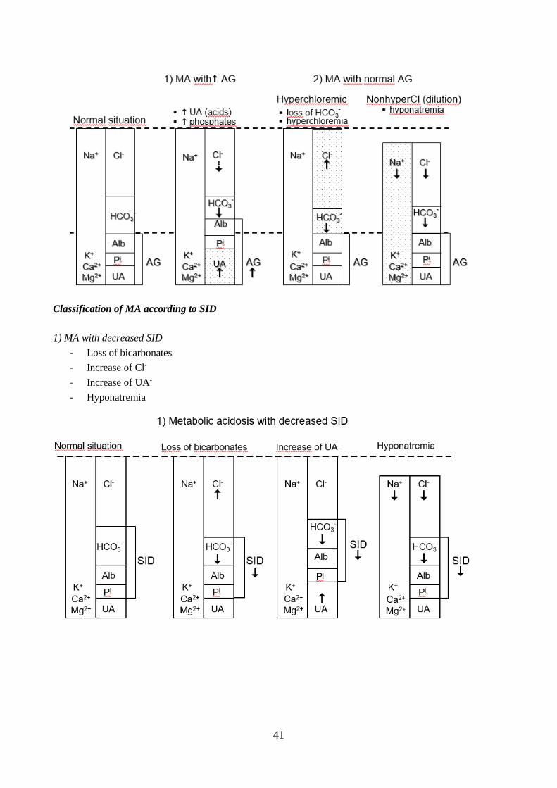
41
Classification of MA according to SID
1) MA with decreased SID
- Loss of bicarbonates
- Increase of Cl-
- Increase of UA-
- Hyponatremia

42
2) MA with normal SID
- hyperphosphatemia
Compensatory response in metabolic acidosis:
- Buffering
o plasma HCO3-
o excess H+ also enters cells - buffering by proteins + phosphates
H+ entry to IC means K+ efflux from IC → hyperkalemia
- Respiratory response – minutes - H+ stimulates central chemoreceptors → hyperventilation →
pCO2
- Renal compensation - slow, but final solution
o H+ is secreted to tubules and excreted as NH4 or titratable acid - H3PO4
o new HCO3– formation in collecting tubule
- Bone buffering – mainly in chronic kidney failure
o breakdown of hydroxyapatite crystals – release of calcium carbonate → release of Ca from
bones – osteomalacia (non-dependent on parathormone)
Pathogenesis and symptomps
- Vague signs up to level of serum HCO3 – below 15 mmol/L
- Hyperventilation - Kussmaul's breathing - direct effect of H+ on respiratory center in oblongate
medulla
- Cardiovascular dysfunction
o - negative effect on inotropic function
o + decreased sensitivity to catechol amines
o + peripheral vasodilation and increased permeability of capillaries (due to lactacidosis)
- Neurologic - less pronounced than in respiratory acidosis (lipid-soluble CO2 enters via blood brain
barrier more rapidly than water soluble HCO3-), depression of CNS - lethargy up to coma
- Metabolic - glycolysis is inhibited by acidosis (↓ phosphofructokinase, a rate-limiting enzyme of
glycolysis), hepatic gluconeogenesis from lactate is inhibited = responsible for perpetuating and
worsening lactic acidosis

43
- DECOMPENSATED SEVERE ACIDOSIS CAUSES VASODILATION OF PERIPHERAL
VESSELS AND THEIR INCREASED PERMEABILITY → HYPOTENSION AND EDEMA
FORMATION = DISTRIBUTIVE CIRCULATORY SHOCK
- THE SAME MECHANISM CAUSES BRAIN EDEMA → INTRACRANIAL HYPERTENSION
→ "CONUS OCCIPITALIS" → DEATH
Treatment
- Sodium bicarbonate (NaHCO3) – suitable mainly in bicarbonate losses
- the HCO3- deficit can be calculated by using the following equation: HCO3
- deficit = deficit/L
(desired serum HCO3- - measured HCO3
-) × 0.5 × body weight (volume of distribution for HCO3-)
- to prevevent hypernatremia, NaHCO3 should be added to 5% dextrose in water or to 0.25 NaCl (it
dilutes Na)
- Lactacidosis – lactate can be removed by hemodialysis
Respiratory Acidosis
Primary rise in pCO2 (hypercapnia) results in pH decrease
PaCO2 > 45 mmHg, pH < 7.35, HCO3- slightly rises (but it cannot be used for buffering)
primary cause = alveolar hypoventilation: causes first hypoxaemia (low pO2 - because of lower diffusion
coeff. of O2 ) → anaerobic metabolism → lactic acid - metabolic acidosis!!!, only later retention of CO2
= respiratory acidosis
- acute - before visible effect of renal compensation
- chronic - with renal compensation
Etiology
- Acute upper airway obstruction
o aspiration of foreign body or vomiting
o laryngospasm or laryngeal edema
- Inhibition of the brain stem respiratory center
o drugs: opiates, sedatives
o oxygen therapy in chronic hypercapnia
o sleep apnea
- Disorders of respiratory muscles + chest wall
o neuromuscular disease – e.g. myasthenia gravis, amyotrophic lateral sclerosis
o chest wall injury
- Disorders of gas exchange
o pulmonary diseases when causing hypercapnia
Compensatory mechanisms
Acute respiratory acidosis: - only cellular buffering! - not sufficient!
(ECF buffering is not active because increased H2CO3 is a member of the main buffer system)
Chronic respiratory acidosis - well compensated - nearly normal pH
kidneys increase excretion of H+ + resorption of HCO3-
Clinical features
clinical signs are more attributable to hypoxemia
chronic state is better tolerated than the acute one
similar to metabolic acidosis

44
- high pCO2 - brain vessel dilatation, vascular congestion → intracranial hypertension
- pCO2 ˃ 70 - direct depression of respiratory center
- chronic high pCO2 - respiratory response to pCO2 increments becomes progressively lost and
ventilation is dependent on hypoxic drive
Treatment
- restoration of effective ventilation
- oxygenotherapy - safe in acute acidosis patients, dangerous in chronic (removal of hypoxic drive for
ventilation)
- treatment of underlying course

45
16. Alkalosis
(Z. Kubová)
- metabolic
- respiratory
Metabolic alkalosis
= HCO3- excess
ECF HCO3- 26 mmol/L, pH 7,45
Rare, since renal bicarbonate excretion is normally very efficient
Factors stimulating HCO3- reabsorption despite of presence of alkalosis:
- EC volume depletion
- K+ deficiency (e.g. mineralocorticoid excess, thiazide and loop diuretics)
Causes:
- Hypokalemic alkalosis
o Mineralocorticoid excess (Conn sy, Cushing sy)
o Bartter, Gitleman and Liddle sy
- Hypochloremic alkalosis
o Loss of gastric juice (vomiting, nasogastric sucction)
o diuretics (loop and thiazide – ↑ excretion of Cl)
o inborn chloridorhea – disorder of chloride-bicarbonate antitransport in ileum - excretion
of Cl + excretion of bicarbonate
- Administration of Alkali
o Milk-alkali sy (antacids, milk)
o high intake of bicarbonate
- Hypoalbuminemia (Kwashiorkor, nephrotic syndrome, liver insufficiency) – proteins are weak acids
- Hypernatremia – concentration alkalosis
Compensation
- IC buffering - H exits cells, K moves in → hypokalemia
- Respiratory - pH is detected by central chemoreceptors → hypoventilation
- (limited by O2 need)
- renal excretion of base excess (difficult in hypochloremic alkalosis!)
Pathogenesis
- Hypokalemic alkalosis
o hyperaldosteronism causes hypokalemia + increases activity of proton pump in intercallated
cells of collecting tubule – acidotic urine
o hypokalemia leads to shift of K+ from cells in exchange for H + and activation of H/K
ATPase in intercallated cells of collecting tubule - K + resorption, H + excretion
- Hypochloremic alkalosis - e.g. vomiting - loss of HCl + water
o Cl- depletion prevents HCO3- excretion - maintains alkalosis!!
o volume depletion stimulates RAAS (ECF volume protection is prior to pH protection!)
o aldosterone deepens alkalosis (activation of proton pump → acidotic urine!) and causes
hypokalemia

46
Clinical features
- cardiac dysrhythmias – in severe alkalosis 7.6 (due to hypokalemia)
- hyperexcitability of nervous system – tetany (directly ↑ neuromuscular irritability), + less Ca+ is
ionized - it binds to albumins
Treatment
- Chloride responsive metabolic alkalosis - usually connected with ECF volume contraction
(Vomiting, Nasogastric suction, Diuretics) - supply of Cl- enables HCO3- excretion, solved by
replacement of volume deficit (infusion of physiological solution) + KCl
- Chloride-resistant metabolic alkalosis - usually not connected with volume contraction -
mineralocorticoid excess (leading to losses of H+) – Spironolactone (antagonist of aldosterone)
Respiratory alkalosis
= HCO3- deficit due to decrease of pCO2 (hypokapnia) results in pH increase
pCO2 < 35 mmHg, pH > 7.45
Etiology:
- Central stimulation of respiration
o psychogenic hyperventilation caused by emotional stress
o hypermetabolic states – thyreotoxicosis, fever – eg. Gram-negative sepsis
o voluntary hyperventilation (e.g. inflating balls)
o head trauma, brain tu
- Hypoxia at healthy lungs (high altitude, exercise – accompanied by metabolic acidosis
- from lactic acid)
- Excessive mechanical ventilation
Compensation:
Acute phase
ICF buffering - H+ is released from tissue cells
little ECF buffering
Chronic phase
Renal tubular reabsorption and production of HCO3- is inhibited - this develops in about 24
hours after onset
Clinical features
- dizziness
- circumoral paresthesia
- tetany in severe alkalosis (Ca binds more to plasma proteins)
- inability to concentrate, tiredness, palpitations, anxiety
Treatment
- the only possibility - treatment of underlying cause
- in artificial ventilation - adequate settings are necessary
- in emotional attack - plastic bag
Mixed pH dysbalances
Two or more primary disorders can coexist, they occur in complex medical problems
A) With additive effect on pH
metabolic acidosis + respiratory acidosis

47
metabolic alkalosis + respiratory alkalosis
B) Without additive effect on pH (resulting pH can be about normal)
metabolic acidosis + respiratory alkalosis
metabolic alkalosis + respiratory acidosis
Examples of mixed pH disorders
- global respiratory failure and heart failure → respiratory + metabolic acidosis
- septic shock → respiratory alkalosis (due to hyperventilation in extreme fever) + metabolic acidosis
(due to hypoperfusion in periphery)
- chronic obstructive lung disease + vomiting → respiratory acidosis + metabolic alkalosis
- emotional stress + vomiting → respiratory + metabolic alkalosis

48
17. Disorders of peripheral circulation
(Z. Kubova)
- blood flow depends (according to Poiseuille's law) on the fourth power of the vessel radius - any
change of radius has significant effect on the blood flow and blood pressure
- main factors influencing peripheral blood flow: tonus of the sympathetic system (level of
catecholamines) together with α and β-adrenergic receptor distribution, intracellular (smooth muscle
cell) concentration of Ca2+ ions, endothelial NO and endothelin formation (see separate chapter),
effects of vasoactive substances (e.g. inflammatory mediators)
- effect of blood viscosity
- "critical closing pressure" = pressure at which blood vessels collapse and the blood flow in a
particular organ stops
Peripheral blood flow alterations
- increase of blood flow = hyperemia
- decrease of blood flow – hypoperfusion, ischemia
o systemic - decrease of circulating volume, pressure
o local - vessel obstruction (structural changes – atherosclerosis, inflammation, thrombus,
embolus), vasospasm
Hyperemia
- active hyperemia - ↑ blood influx - effect of vasodilators (histamine, bradykinin, PGE, NO, CO2)
o reactive – after transient vessel occlusion
o functional – necessary for increased function (thermoregulation –hypothalamus, muscles in
exertion - beta2 effect of adrenaline)
- passive hyperemia – blood stagnation – congestion (e.g. venous insufficiency (legs), generalized –
right heart failure)
DISEASES OF PERIPHERAL ARTERIES
Vasospasm
- Raynaud phenomenon (syndrome) – 4-times more frequent in women
o attacks of vasospasm (mainly in fingers of hands)
o usually reaction to cold, high-frequency vibrations - vibrating tools (↑ tonus of sympathetic
nervous system, ↑ number of α receptors) → whitening of fingers (nose), pain (later reactive
hyperemia - blush)
o frequently accompanies autoimmune diseases – SLE, scleroderma
Acute obturation of artery → ischemia, infarction (thrombus or embolus usually from heart)
Chronic obturation of arteries
- Arteriosclerosis
- Diabetic macro- and microangiopathy - acceleration of atherosclerosis, ↑ protein glycation
- Thrombangitis obliterans (Buerger‘s disease) – mainly in 20-40 years of age
Etiology:
o not clear etiology but exposure to tobacco is essential for both initiation and progression of
the disease
o in some cases inflammation of the arterial wall + event. thrombi (possible reaction to
circulating immune complexes)
o vasospasm due to increased sympathetic tonus
Clinical manifestations:

49
o smaller arteries – esp. plantar and leg (calf)
o early phase – intermittent claudicatio (limp) because of ischemic muscle pain, weakness, ↑
sensitivity to cold
o later ulceration, gangrene
Thrombosis
- arterial or venous occlusion, coronary arteries → obturation of vessels, possibility of embolisation
- Etiology - Virchow‘s triad
1) stasis or turbulence of blood
2) endothelial injury
3) increased blood coagulability
o hemoconcentration
o contraceptives (↑ hepatic production of coagulation f.)
o nephrotic sy
o cancer (e.g. pancreatic Ca)
o DIC
o Leiden mutation
o venous thrombosis – phlebothrombosis superficial system – does not result in
pulmonary emboli, deep system – (deep vein thrombosis) - can result in pulmonary
emboli!!!
Leiden mutation
- most common inherited predisposition to thrombosis
- 5 % of population
- mutation of f. V, which makes f. V resistant to degradation by activated protein C
Embolism
- 99% - thrombi = thrombemboli
- systemic or pulmonary circulation
- rare – fat (mechanical + biochemical consequences)
- bubbles of air or nitrogen (Keson disease)
- atherosclerotic debris (cholesterol emboli)
- tu fragments
- amniotic fluid
development of infarction depends on:
- duration of occlusion
- velocity of obturation
- properties of vessel system - existence of collaterals, double vessel system – functional and nutritive
(lungs, liver), terminal artery → infarction
- sensitivity of the tissue to hypoxia (neurons -3 – 4 min, cardiomyocyte - 20 min)
- oxygen content of blood
Infarction
- anaerobic glycolysis – lactic acid - acidosis
- shortage of ATP → disorder of active ion transmission → intracellular edema, activation of enzymes
– damage to cells

50
Disseminated intravascular coagulation (DIC)
- Abnormal activation of tissue factor (TF)
o polytrauma, surgery, delivery (amnial fluid)
o sepsis, shock (release of TF)
o production of TF by tumor cells
- Consequence:
o trombi → ischemia, infarction
o consumption of coagulation factors + platelets → bleeding
DISEASES OF PERIPHERAL VEINS
Venous thrombosis - phlebothrombosis
1) superficial system
o red strips, inflammation, ev. fever
o does not result in pulmonary emboli
2) deep system – (deep vein thrombosis)
o peripheral edema, Hommans sign
o can result in pulmonary emboli!!
Varicous veins
- primary – idiopathic – weakness of vessel wall
- secondary
o valves insufficiency
o pathology of deep system
Chronic vein insufficiency
- reflux of blood from deep to superficial system
- transfer of high pressure to superficial system and microcirculation → ↑ of capillary permeability
→ leakage of fluid – edemas, disorders of skin nutrition, pigmentations (hemosiderin), ulcers

51
18. Syncope
(Z. Kubová)
- is a transitory loss of consciousness caused by hypoperfusion of brain resulting from a sudden
decrease of blood pressure
- is accompanied by loss of postural tone (fall)
- usually happens in standing or sitting position
- usually spontaneously fades away within few min
- Pre-syncope = incomplete syncope - dizziness without unconsciousness
- Untrue syncope – e.g. in epilepsy, metabolic coma (hypoglycemia) or so called psychogenic
unconsciousness (post-traumatic loss of consciousness)
- Note that European pathophysiology differentiates "collapse" (peripheral syncope), caused by
decreased preload, and "syncope" (central syncope), caused by transient decrease of the heart MV. In
contrast, English/American textbooks use the term “syncope” to denote any short unconsciousness
due to hypoperfusion of the brain in decreased blood pressure.
Classification is non-uniform
According to mechanism
- Cardiac syncope – arhytmias, structural changes in heart
- Cerebrovascular syncope – problem is in brain vessels
- Neurogenic syncope – dysregulation of ANS – decreased tone of sympathetic NS and/or increased
tone of parasympathetic NS (esp. of vagus nerve)
- Syncope from insufficient return of blood to heart – originally called collapse
Central syncope
Cardiac syncope
- Arrhythmias
o Bradycardia
o Paroxysmal supraventricular and ventricular tachyarrhythmia
o Dysfunction of SA node, AV blocks
o Disorder of cardio stimulator
- Structural changes in heart
o Physical effort in aortal stenosis
o Myocardial infarction
o Pericardial diseases, heart tamponade
o Cardiomyopathy
- agents that decrease cardiac output (eg. beta-blockers, digitalis, antiarrhythmics)
Cerebrovascular syncope
Diseases of arteries that bring blood to brain or arteries in brain mainly due to atherosclerosis
- Stenosis of carotic a.
- Vertebral or basilar arteries insufficiency
- Steal syndrome – especially subclavian steal sy – decreased flow of blood through subclavian a. to
arm leading to reverse flow in vertebral arteries

52
Neurogenic syncope
- Reflexive – nerve mediated - hyperactivity of vagus nerve causing sudden decrease of heart activity
o Artificially – oculo-cardial reflex, sino-carotic reflex
o Sy of carotic sinus
o Strike on the solar plexus
- Vasovagal syncope – disorder of ANS regulation – besides activation of parasympathetic NS (→
decrease of heart activity) also a decrease of sympathetic activity occurs (→ vasodilation) – up to
40% of syncopes (this time is something between central and peripheral type of syncope)
o Long standing in hot environment
o Pain, emotional excitement
Peripheral syncope (Collapse)
- transient decrease of venous blood return (decreased preload) due to peripheral or increased
intrathoracic pressure → decreased cardiac output, hypotension
- predisposed are old people, dehydrated subjects, patients treated with antihypertensives
Causes:
- hot environment - thermoregulatory reaction leads to vasodilation
- orthostatic (postural) collapse caused by change of body position from laying (sitting) to upright -
more frequent in old people due to delayed (decreased) orthostatic reflex keeping roughly constant
blood pressure at the level of the brain during changes of body position (senile degeneration of the
vegetative nervous system, decreased baroreceptor responsiveness) and in diabetics or uremia due to
peripheral polyneuropathy involving also the sympathetic system
- long standing
- ↑ intrathoracic or intra-abdominal pressure - Valsalva maneuver, coughing, difficult defecation (in
constipation), micturition (hypertrophy of prostate)
- hypoactivity of the sympathetic nervous system in peripheral polyneuropathies – e.g. diabetes
mellitus
- agents that reduce blood pressure (eg, antihypertensive drugs, diuretics, nitrates)
Causes of any short unconsciousness should be verified. It can indicate severe heart or circulatory problems.
However, it can happen also in young and completely healthy subjects (dehydration, heat stress, emotions
etc.)

53
19. Circulatory shock
Z. Kubová
Circulatory shock is a failure of the circulatory system (longer than syncope or collapse) with decreased
peripheral perfusion and inadequate oxygenation of organs and tissues (usually not spontaneously
recovering). It can occur in the course of many life-threatening, traumatic or disease states.
Etiology - types:
Hypovolemic shock - volume - low venous blood return - low „preload“
- bleeding - loss of more than 20 – 30% of blood
- burns, loss of isotonic fluid
- dehydration = hemoconcentration (↑ Ht) - ↑ viscosity – worsens
- capillary leakage sy (shift of fluid to edemas)
Cardiogenic shock – is due to any critical decrease of MV
- infarction (in case of large involvement > 40% of LV myocardium), cardiomyopathy
- rhythm changes – blocs, severe bradycardia, tachycardia
- mechanical – papillary muscle defects, ventricular aneurysm, ventricular septal defect
- obstructive shock - pneumothorax, heart tamponade
Distribution shock - normal volume, severe generalized vasodilation
- Neurogenic shock – purely distribution shock
o transversal lesion of spinal cord and sympathetic trunk – loss of sympathetic innervation
(severity dependent on the level of transection)
- Anaphylactic shock – distribution shock combined with hypovolemic shock
o Antigen antibody complex - activation of complement, kinins, histamine → vasodilation,
increased permeability and bronchoconstriction
- Septic shock – distribution shock combined with hypovolemic shock
o endotoxin (released from Gram- bacteria by macrophages, complement) → activation of
interleukins 1,6,12 and TNF → NO → vasodilation, pyrogenic effect, activation of
complement, prostaglandins, leukotrienes, inflammatory mediators (histamine, bradykinin) -
↑ permeability → capillary leakage sy, ARDS, platelet activation f. → DIC
Pathogenesis - phases of shock
development of severe hypotension due to a specific condition
1) compensated (non-progressive) phase
2) decompensated (progressive) phase
3) irreversible (refractory) phase
1) compensated (non-progressive phase) – leads to activation of the following compensatory mechanisms
- hypotension activates baroreceptors, chemoreceptors → activation of sympatho-adrenal axis
(endogenous stress reaction) → positive effect on heart activity + vasoconstriction in periphery
and splanchnic area (centralization of circulation) + respiratory effect (bronchodilation) – this tries
to improve oxygenation of vital organs
- activation of RAAS due to renal hypo perfusion + increased ADH tries to increase circulating
volume, however strong splanchnic vasoconstriction might prevent their effect
54
These mechanisms can be effective in hypovolemic and distributive shock (via compensation of volume or
blood pressure) but they are disadvantageous in cardiogenic shock where increased afterload (blood
pressure) and preload (circulating volume) lead to earlier heart failure (because of ↑ demands on
myocardium) – thus, it is necessary to block these compensatory systems!!!
2) decompensated (progressive) phase
- prolonged centralization of circulation → hypo perfusion of peripheral tissues and splanchnic area
- peripheral hypoxia → anaerobic metabolism → lactic acidosis (+ accumulated CO2)
- splanchnic area hypo perfusion → ischemic damage of cells e.g.:
o proximal tubule → acute tubular necrosis (can lead to severe polyuria after eventual
recovery from shock)
o necrosis of the wall of intestines → escape of bacteria → peritonitis
o pancreas – production of myocardium inhibiting factor (MIF)
- buildup of lactacidosis → forced vasodilation (now prevailing over the former tendency to
vasoconstriction) → release of substances from necrotic cells - proteolytic enzymes, myocardium
inhibiting factor, tissue thromboplastin → disseminated intravascular coagulation (microthrombi),
reperfusion tissue damage – ROS production (in the case of at least partial reperfusion)
- hypoxia, lactacidosis and damage to endothelium → generalized edema formation (including
brain and pulmonary edema – adult respiratory distress sy) → further decrease of circulating
volume – total failure of circulation - transition to
3) irreversible phase of shock (eventually provided fluid shifts to edemas and does not stay any more in
circulation!)
- multiple organ damage sy (MODS) results from microthrombi and necrosis of cells (decreased
ATP production → decreased activity of Na/K pump → intracellular Na + water = cellular edema
+ release of lysosomal enzymes, production of ROS) – most early changes in kidney – “shock
kidney” = tubular disorder with polyuria, in lungs ARDS
Possible prevention of irreversible shock changes = early volume replacement with volume-expanders:
- hypertonic solutions (3- 7.5% NaCl) → shift of water from ICF to ECF, increases ADH production –
relatively short term effect
- colloid solutions (albumin, artificial albumins, dextran, hydroxyethyl starch) – danger – decrease of
GF
- advantageous combination of both (7% NaCl + 6% dextran)

55
20. Inflammation
(M. Kuba)
Inflammation is reaction of living tissues to all kinds of injury. Despite undesirable effects (pain, edema, and
crippling effects) it is a beneficial reaction trying to eliminate causes (e.g. infection, exposure to poison)
with local processes avoiding a spread of the harmful agents. The failure of a local anti-infectious
inflammatory reaction (due to low immunity or very virulent infection) generalized sepsis develops.
Agents causing inflammation
- exogenous - e.g. trauma, surgery, infection, chemicals, extremes of heat and cold, irradiation
- endogenous - e.g. ischemia, immune responses
Manifestations (according to Celsus and Galen)
Local:
- rubor (redness)
o caused by active hyperemia following momentary vasoconstriction due to stress reaction
(catecholamines) related e.g to pain (injury) - dependent on vasoactive inflammatory
mediators, in the developed (end stage) inflammation hypo perfusion of the inflamed
area can appear due to micro-thrombotisation of vessels
o hyperemia increases delivery (supply) of cells (leukocytes) to the place of inflammation
- calor (heat)
o ↑ temperature of the inflamed area due to hyperemia and increased metabolism
- tumor (edema)
o develops because of increased hydrostatic pressure and increased permeability of vessels
(caused by inflammatory mediators -see below); besides transudation into the interstitium,
active exudative (secretion) processes also take place (peritoneal cavity, pericardium, pleural
cavity)
o edema decreases the concentration of toxins, which can prevent necrosis of cells, edema
helps also to local leucocyte reactions
- dolor - caused by:
o irritation of free neural endings by inflam. mediators - mainly prostaglandins, substance P,
bradykinin
o compression of nerves by edema
o developing acidosis in the case of thrombotisation of vessels (in developed, chronic inflam.
process)
o pain is an important informative factor (especially in the case of hidden inflammatory
processes – such as intra-abdominal inflammation - acute appendicitis etc.), it is
recommended to eliminate pain (to give analgesics) only in cases where the cause of pain is
well known
o pain also prevents damage by overloading (using) the inflamed part of the body
- functio laesa (malfunctioning) - caused by pain and edema - disabling mainly motion - prevents
overload of the involved parts of the body
General manifestations:
- fever - caused by activation by pyrogens (IL-1, IL-6, TNF ...) of the thermoregulatory center
(hypothalamus)
- leukocytosis - increased number of leukocytes released from bone marrow (over 10.000/mm3),
enormous increase in some childhood viral infections

56
- changes in the differential count: bacterial infections - ↑ neutrophils, viral infections - ↑
lymphocytes, allergies and parasites -↑ eosinophils
- decreased Hynek's number (number of segments in neutrophils) = "shift to the left" in prolonged
(large) inflammations, extreme "shift to the left" with appearance of non-mature leukocytes in the
peripheral blood = "leukemoid reaction"
- leukocytosis is a very non-specific but important sign (e.g. in suspected appendicitis)
- increased FW (sedimentation of erythrocytes) - non-specific sign pointing to inflammation
- lymphadenitis - enlargement of lymphatic nodes
Acute inflammation
- reaction of vessels
o active hyperemia, tendency to stasis of blood
o increased permeability for both plasma and cells
- reaction of cells
o attraction of leukocytes to the place of inflammation - opsonisation (IgG, C3b fragment of
complement)
o margination of leukocytes and emigration (via diapedesis), chemotaxis, phagocytosis
(destruction of pathogens - pH, enzymes - e.g myeloperoxidasis)
Neutrophils (polymorphonuclears)
- the first cells at the place of inflammation (within 90 min. after onset) with maximum phagocytic
capacity in acute inflammations (60 - 70% of all leukocytes)
- survival - only about 10 hours (must be steadily replaced from bone marrow 1 - 2 days)
- dominate in bacterial infections
Monocytes (macrophages, histiocytes))
- after several days but dominate in chronic inflammations
- formation of lysosomal enzymes
Lymphocytes
- mainly in viral inflammations
Basophils
- in stress reactions
- production of heparin, histamine
Eosinophils
- allergies, parasites, reaction to chemical injury
Inflammatory mediators
- Vasoactive (dilating)
o histamine (increases permeability - causes edemas - mainly in allergic reactions)
o PGE (can be blocked by inhibition of cyclooxygenasis by non-steroid anti-inflammatory
drugs - e.g. Acetylosalicylic acid; availability of Arachidonic acid (the substrate) is
blocked by glucocorticoids)
o bradykinin
o leukotrienes
o platelet activating factor (PAF)
- Pain mediators
o prostaglandins (PG), substance P, bradykinin
- Edogenous pyrogens

57
o IL-1, TNF
Types of inflammation according to the type of exudate:
- erythema - no exudate, only redness (exposure to sunshine, light frost-bite)
- serous - watery character of exudate, low content of proteins, catarrhal inflam. with no complications
- e.g. "hay fever"
- fibrinous - high content of fibrin - leads to formation of adhesions - prevent spread of inflammation
(helpful e.g. in peritoneal cavity) but can cause complications - constrictive pericarditis
- membranous - e.g. on the surface of trachea in Diphtheria or in Membranous enterocolitis - causes
obstruction
- purulent - includes pus (tissue debris, necrotic leukocytes)
- abscesses - encapsulated purulent exudate with pyogenic membrane
- sanguineous (hemorrhagic) - including blood (damage of blood vessels, diapedesis of erythrocytes)
Resolution of acute inflammation
1. it can undergo full resolution (healing "ad integrum")
2. it can progress - suppurative process
3. can proceed to the chronic phase
Chronic inflammation
- either primary chronic character or transfer to chronicity
- due to long lasting exposure to some pathologic agent (smoking - chronic bronchitis)
- autoimmune diseases (Rheumatoid arthritis)
Influencing factors
- early application of cold can limit or postpone development of inflammation (via vasoconstrictive -
anti-edematous effect)
- application of warm can accelerate development of inflammation
- application of glucocorticoids - most potent anti-inflammatory effect (detailed description of
glucocorticoid effects - see stress - general adaptation sy)
Complications of inflammation
- bacteriemia (can make colonies - e.g. infective endocarditis) - typically asymptomatic - without
manifestations of inflammation and activation of the immune system
- pyemia -spread of infected thrombi - formation of abscesses in obstructed small vessels

58
21. Sepsis, Systemic Inflammatory Response Syndrome (SIRS)
(M. Kuba)
Sepsis
- bacterial growth in blood when immune system was not able to liquidate the infection
- systemic inflammatory response to infection (leukocytosis, fever), release of endo- and exotoxins
- danger of SEPTIC SCHOCK (systemic reaction of circulation - vasodilation, increased permeability
→ hypotension, edemas)
Causes - peritonitis, criminal abortions, infections of urinary system, phlegmones, etc.
Manifestations
- intermittent fever, increased metabolism with higher oxygen consumption - hyperventilation
(respiratory alkalosis can develop)
- tachycardia - heart overload
- fatigue, lethargy
- leukocytosis - possible "leukemoid reaction", splenomegaly
- catabolic state, proteinuria
- dehydration, hypovolemia, hypotension → centralization of circulation → failure of
thermoregulation
- peripheral hypoxia → anaerobic metabolism → lactic acidosis starts to dominate (over the former
respiratory alkalosis) → increased formation of edemas including brain edema → coma, death
- possible DIC (disseminated intravascular coagulation) with subsequent consumption coagulopathy
(see Bleeding disorders)
- multiorgan dysfunction (ARDS, shock kidney – oliguria, proteinuria, CNS failure)
- hyperglycemia in diabetics
SIRS = Systemic Inflammatory Response Syndrome – related to sepsis, causes circulatory shock, MODS
(kidney, lungs involvement), criteria: heart rate, tachypnea, leukocytosis

59
22. Pathophysiology of pain
(Z. Kubová)
Definitions of pain and its significance:
Pain = unpleasant sensory and emotional experience connected with real or potential tissue damage, or
described in these terms (International Association for the Study of Pain -IASP, 1990)
- Causes reactions directed to removal of pain stimuli (unconscious – reflexes)
- Represents probably the most common symptom of a disease that motivates a person to seek
professional help
- Pain is of a great informative value for a doctor, thus it should not be eliminated totally before
recognition of its causes (critical especially in hidden processes - e.g. abdominal).
- In some cases pain helps to prevent overloading of parts of the body with pathological processes
(e.g. arthralgias, lumbago, inflammatory processes in limbs) and contributes to recovery.
Nociception
- a separate sensory quality - separate nociceptive system
- over-excitation of some other sensory systems (via pressure, cold, heat receptors)
Nociceptors (= nocisensors) = pain receptors
- free nerve endings A and C
- uneven distribution in skin, muscles, joints
The way of nociceptor stimulation
- after initial damage/irritation of tissue (by chemicals, heat/frost, mechanical insults) → release of
chemical substances
- direct stimulation of nociceptors (opening of Na/Ca channels or closure of K channels → membrane
depolarization) or sensitization of nociceptors
Origin of endogenous chemicals
- cell destruction → H+, K+, ATP release → direct stimulation of nociceptors
- inflammation – bradykinin, histamine, serotonin → direct stimulation of nociceptors
- non-damaging mechanical + thermal stimuli can cause “neurogenic inflammation” (mechanical and
thermal information enters spinal cord via dorsal root and this information is mediated backwards to
afferent terminals → release of Substance P, neurokins and CGRP (calcitonin gene related peptide)
→ vasodilation, leukocyte adhesion, release of inflammatory mediators that stimulate nociceptors)
Sensitization of nociceptors
- physiologic response to painful stimuli leading up to hyperalgesia
- Peripheral - ↓ of threshold + activation of surrounding nociceptors
- Central – in spinal cord
Transmission of pain information
- C and A fibers enter the spinal medulla via dorsal root and synapse together with afferent fibers
from mechanoreceptors on the second neuron of the pain pathway (neurotransmitter = glutamate,
Substance P enhances the transmission). Information brought from mechanoreceptors can decrease
the pain transmission (according to gate theory)
- The second neuron of the pathway relays in the thalamus to continue to:
o The primary somesthetic cortex (recognizes location and intensity of the stimulus)
o Association somesthetic cortex
o Limbic cortex (responsible for emotional experience and memory)
o Hypothalamus (responsible for vegetative accompanying symptoms)

60
o Periaquaeductal gray (endogenous anesthetic center) that sends backward information to
dorsal horn neurons to influence the pain transmission (by release of encephalins, the release
of Substance P is decreased)
Types of pain
1) Somatic pain
- Superficial somatic pain - receptors located in skin
o rapid (first) pain - myelinated A fibers
‒ very acute, well localized, rapidly diminishing
o slow (second) pain - appears later, lasts longer, is not precisely localized
- Deep somatic pain
o originates in muscles, fascias, joints, periosteum
o more similar to visceral pain
o cannot be localized
o often is accompanied by nausea, sweating and other vegetative symptoms
2) Visceral pain - originates in internal organs in chest and abdomen
- cause = ischemia (lactic acidosis), chemical substances, spasm or over distension of hollow organs
- is diffuse
- is accompanied by vegetative symptoms, also by reflex spasm of muscles
Acute pain
Etiology:
- inflammation (due to compressive effect of edema, release of pain mediators, developing acidosis)
- colicky pain (due to intensive periodic peristalsis in obstructed and distended intestine, cystic duct,
ureter)
- acute ischemia - IM
- traumatic pain, burns, post-operative pain
- live birth
The organism reacts with the sympathetic pattern of response (stress response) - tachycardia, heart volume,
blood pressure, hyperventilation + anxiety
Chronic pain
No sympathetic pattern of reaction, but vegetative + psychosocial changes (= algogenic psychosyndrome) -
sleep disorders, appetite, pain tolerance, depression, hostility, hypochondria, abnormal pain behavior
Specific types of pain
- Projected pain - originates elsewhere than is produced (e.g. pain in limb caused by compression of
spinal nerve)
- Referred pain - Head zones – some organs have connection to some parts of skin
- Phantom pain - pain in non-existing limb (after amputation)
- Neurogenic pain (neuralgia) - persisting nerve stimulation
- Causalgia - after devastating injury (shooting) accompanied by vessel reactions + trophic changes of
skin
- Thalamic pain - arises in thalamic nuclei
- Hypoalgesia, analgesia - inborn decrease or total loss of pain sensation - danger!!! leads to injuries,
ischemic necrosis etc.
- Allodynia - normal mechanical and thermal stimuli are perceived as painful

61
- Migraine - headaches with a neuro-vascular background, in women 3x more frequent than in men,
inborn „readiness“ of brain
Pathogenesis:
o vascular theory) explains migraine as a reactive hyperemia in response to vasoconstriction-
o induced ischemia during aura
o „something“ (stress, noise, hormonal changes, traveling, food skipping, sleep deprivation)
o activates „migraine centers“ in pons, periaqueductal grey, locus coeruleus (↑ adrenaline),
raphe
o nucleus (serotonin) → vasoconstriction → aura → stimulation of trigeminal nucleus →
o neurogenic inflammation, release of mediators → pain
o vasodilation of brain and meningeal arteries → pain
o connection between „migraine centers“ and hypothalamus and centers of reflexes in pons →
o vegetative symptoms – nausea, vomiting, photo- and phonophobia

62
23. Fever, Hyperthermia
(M. Kuba)
Thermoregulation of the human body
- constant body temperature is maintained throughout a wide range of temperature in the environment,
provided there is normal function of body systems
- constant temperature is necessary for normal enzymatic reactions and metabolism
Body temperature
- differs in various locations:
- axillary 36.5°C
- oral 37.0°C
- rectal, vaginal (closest to body core temperature) 37.5°C
also ear-drum infra-red measurement)
- circadian variations – minimum temp. in the morning, max. in the late afternoon (accord. to
metabolism - dependent on the level of glucocorticoids)
- influence of menstrual cycle, physical load
- basal temperature is measured immediately after waking-up
- comfortable temperature of the environment (no sweating, no thermogenesis) - 20 - 21°C in a
dressed person, 28-30°C in a naked person
Thermoregulation
- thermoregulatory center (TRC) in hypothalamus reacts to the temperature of perfused blood and to
signals from central and peripheral thermoreceptors
- increased activity of TRC causes vasoconstriction in periphery - ↓ release of heat
- sweating - evaporation (insensible perspiration) of water from skin → increased losses of heat - 1g =
0.58 kCal (up to 75% - dependent on air humidity), loss of water - up to 1l/hour (however, all of this
volume cannot be evaporated)
- increased peripheral perfusion (via arterio-venous shunt closure)→ increased losses of heat via
irradiation into environment - in the resting condition this represents the main loss of heat (45%)
- via respiratory system - loss of only 10% of heat
- increased glucocorticoids activate metabolic rate (production of heat in liver)
- thyroid gland hormones increase metabolism and temperature
- influence of nutrition – specific dynamic effect in metabolism of proteins – 15% released as a heat,
highest energy content in fats
- muscle shivering, chattering of the teeth (thermogenesis)
- brown fat in newborns - source of metabolic heat
- physical activity leads to high production of heat (mainly in non-effective motions in non-trained
people)
- people with low cardio-vascular capacity have low capacity for thermoregulation - during
vasodilation in the periphery they cannot sufficiently increase cardiac output and the resulting
hypotension leads to syncope (collapse) (short unconsciousness due to hypo perfusion of the brain) -
critical especially with simultaneous dehydration - thus old people should not be exposed to hot
environment
63
Fever (in contrast to hyperthermia, this is a defense reaction of the body)
- increase of axillary temperature over 37.2°C, critical over ca 40°C
- non-specific information about either an inflammatory or a stress reaction (when in basal conditions,
without physical activity or activation of metabolism)
- it is intentional resetting of TRC in hypothalamus - reaction to pyrogens probably through the action
of prostaglandin E
Meaning of fever
- fever is a beneficial reaction because of liquidation of thermo-sensitive bacteria and viruses
- particularly in some viruses, it is the only possible reaction of the body for elimination of the
infection (when viruses do not activate immune reaction) - thus it is not good when in any fever
antipyretics are immediately applied (should be used only in temperatures over 39°C, mainly in
small children at risk of febrile cramps)
- fever increases production of antibodies and phagocytosis
Etiology
- exogenous pyrogens - infectious bodies (bacteria, viruses....), toxins, drugs
- endogenous pyrogens - inflammatory mediators, tumorous cells (released substances), injured
tissues, substances from immune reactions (PGE2, IL-1, IL-6, TNF ...)
Phases of fever
- increase of temperature (incrementi) - characterized by vasoconstriction accompanied by shivering,
feeling of cold, increased basal metabolism
- plateau (acme) - achieved temperature set by TRC
- decrease of temperature (decrementi) - elimination of heat from the body - vasodilation, sweating,
feeling of heat
Patterns of fever
- intermittent fever - temperature comes to norm at last once in 24 hours
- remittent fever - oscillating increased temperature
- sustained or continuous - without variations
- recurrent (relapsing) - several days lasting periods with one or more days of normal temperature -
e.g. in malaria
Differences in max. temperature according to age - highest fever children up to about 6 years, old people -
even low fever can point to a severe process
Consequences (manifestations) of fever
- heart rate increases by about 15 beats/min with temperature increase of 1°C - can be critical in old
people with compensated heart failure (high mortality of old people in epidemic influenza)
- very demanding of energy supply (it is necessary to ensure good nutrition in long-lasting febrile
diseases to prevent catabolic state, despite low appetite (probably due to TNF release)
- quick development of dehydration - important supply of fluids - mainly in old people without
expressed thirst
- constipation
- hyperventilation due to increased basal metabolism
- catabolism of proteins, hyperglycemia
- CNS hyperactivity (hallucinations) up to depression, sy of restless legs

64
Hyperthermia
- failure of thermoregulation
- heat stroke - core temperature over 40°C - unconsciousness
Etiology
- in sunburn
- prolonged exertion in hot environment
- dehydration, heart failure
- drugs (increasing muscle tone and metabolism or influencing thermoregulation)
- peripheral heat dissipation can be impaired by antihistamines, atropine, trycyclic antidepressants,
sympathomimetics - producing vasoconstriction
- hypersensitivity reaction to drugs
- neuroleptic malignant sy with tachycardia (30% mortality)
- malignant hyperthermia - autosomal-dominant disorder with uncontrolled skeletal muscle
contractions (potentially fatal - temperature up to 43°C with rise of 1°C per 5 minutes) - caused by
abnormal release of intracellular Ca from mitochondria and sarcoplasmatic reticulum - triggered by
stress or general anesthesia
Manifestations (besides those described in fever)
- in extreme exogenous temperature danger of heat collapse (syncope) due to vasodilation (in people
with low cardiovascular reserve)
- can cause pulmonary edema

65
24. Hypothermia and Reaction of organism to cold
(M. Kuba)
Reactions in exposure to cold
- sudden exposure to cold causes first vasoconstriction (stress reaction - effect of catecholamines)
- signals from peripheral thermoreceptors relayed via TRC cause peripheral vasodilation to try to
maintain the temperature of peripheral tissues → redness represents active hyperemia
- when blood temperature decreases, TRC activates peripheral vasoconstriction - preference of
constant body core temperature - hypo perfusion in periphery leads to necrotic changes (in longer
exposure to extreme cold, especially mainly in combination with high humidity) - "frost bite" (even
in temperatures above zero)
- influence of alcohol – causes peripheral vasodilation → subjective feeling of warm, prevention of
frost bite but increased loss of heat leads to hypothermia of body core → death - drunk people can
die earlier in cold environment
Hypothermia
- core temperature below 35°C
Etiology
- low external temperature (with high humidity) + physical exhaustion
- sepsis
- intoxication - alcohol, barbiturates
- low basal metabolism (hypothyroidism)
- neurological disorders
Pathogenesis
- infectious diseases are more frequent after exposure to cold (tonsillitis, influenza, pneumonia etc.)
because of impaired balance between pathogens and local immune reactivity - due to
vasoconstriction (reaction to cold), there is lower supply of leukocytes, antibodies
- below 30°C loss of thermogenesis, ↓ HR, ↓ respiration, muscle rigidity, stupor, loss of reflexes,
analgesia, hypoglycemia
- below 25°C cessation of breathing, cardiac arrest or ventricular fibrillation
Use in clinical medicine - hypothermia decreases metabolism and consumption of oxygen - thus it is used in
surgery for prevention of irreversible tissue changes during ischemia

66
25. Stress, General adaptation syndrome
(M. Kuba)
Stress can be anything that can alter the homeostatic processes. Stress may contribute directly to the
development of disease.
Response (set of reactions) to stress was termed as General Adaptation Syndrome – GAS (by Hans Selye in
1946) - enables the organism to resist to the stressor in the best possible way by enhancing the functions
of the system.
The events or environmental agents responsible for initiating the stress response are called “stressors”.
- stressors (exogenous, endogenous) can be physical, chemical, psychological or sociologic (extreme
temperature, hunger, thirst, fatigue, hypoxia, etc)
- in modern society the most frequent is psychic stress
- the majority of signs and symptoms of the stress reaction are common for all stressors, however,
some can be specific
- the ability of the same stressor to produce different responses in various individuals indicates
different adaptive capacity influenced by conditioning factors (genetic predisposition, age, sex,
dietary factors, sleep-wake-cycles, drugs etc.), and also the dynamics of the stressor are important
for possible adaptation
Stages of General adaptation syndrome (GAS)
1. The alarm stage - characterized by immediate release of catecholamines and their related effects - ↑ heart
MV (via tachycardia, increased contractility), ↑ BP (vasoconstriction in periphery but not in muscles!!!
(be aware of adrenergic receptor distribution), ↑ glycemia, ↑ lipoproteinemia (including LDL) etc.
2. The stage of resistance - characterized mainly by release of glucocorticoids after activation of the
hypothalamic-pituitary-adrenal gland axis → increased glycemia, and anti-inflammatory effects, and
decreased of all immune reactions with the exception of the number of circulating polymorphonuclears
(neutrophils) - it is increased (prevents bacteriemia and sepsis when pathogens enter into circulation)
- this phase can be quite long (up to several weeks- dependent on capacity for glucocorticoid production
and sources of energy for gluconeogenesis)
3. The stage of exhaustion - the necessary level of glycemia is not ensured, hypotension, shock - it leads to
malfunctioning of all systems, breakdown of homeostasis and death
Stress response - General adaptation syndrome has been termed "the fight-or-flight-response" because in
animals it represents a preparation for intensive physical activity with possible injuries. From the above
specified summary of changes during GAS, it is evident that all are oriented toward better perfusion of
muscles, increased supply of oxygen and energy (glucose) for physical activity.
The phylogenetically very old set of reactions has persisted with little change into the modern human society,
where the most frequent psychic stress is not accompanied with physical activity (should not be - in
civilised population). Thus a lot of changes can have harmful effects leading to development of
psychosomatic diseases (hypertension, atherosclerosis, secondary DM, stress peptic ulcers etc. - see
appropriate chapters).

67
26. Psychosomatic diseases
(M. Kuba)
- civilization diseases - after prolonged or repeated stress
Examples
- cardiovascular disorders
o hypertension - probably due to resetting of the vasomotor center during increased BP in
stress
o atherosclerosis, ischemic heart disease - accelerated by increased lipoproteinemia and BP
o more frequent in personality type A (according to typology by Friedman and Rosenman,
1974) - responsible, ambitious men (not so pronounced in women up to menopause)
- gastrointestinal diseases
o stress peptic ulcer - mainly duodenal - caused by:
- increased acidity due to activation of vagus nervevand increased level of
glucocorticoids
- low mucus production due to gastric ischemia in splanchnic vasoconstriction
o Crohn's disease - genetic predisposition, psychic stress
o irritable colon
o psychogenic nausea, vomiting
- endocrine and metabolic disorders
o secondary diabetes mellitus
o hyper/hypothyroidism
o obesity
o hyperthermia
- respiratory system
o bronchial asthma
o
- immune system
o combined immunodeficiency - higher incidence of infections, tumors (due to decreased
immunological surveillance
o interaction between progression/regression of tumorous process and stress (behavioral access
to treatment)
- sexual dysfunction and infertility
o erectile dysfunction, decreased spermatogenesis
o functional infertility in women due to imbalance in sexual hormones (hypothalamo-pituitary
dysregulation) - usually disappears after adoption of a child (eliminates stress from
infertility)
- lumbago, stiffness of neck - increased tonus of paravertebral muscles (increased level of gamma
innervation)
- dermatological problems - psycho-dermatosis
- psychogenic disorders in nutrition
o bulimia nervosa
o mental anorexia

68
27. Anemias – classification
(M. Kuba)
Etiology
Decreased/defective production of erythrocytes
- Altered hemoglobin synthesis
o iron deficiency (hypochromic microcytic anemia) – dominant factor (in 25 – 30% of
population) either low intake or problems with absorption (gastric hypoacidity) or prolonged
iron loss (bleeding)
o Thalassemia - β-thalassemia most common in Mediterranean populations (Greece, Italy)
o anemia of chronic inflammation (chronic diseases) - deficiency of erythropoietin (renal
failure) and/or resistance to erythropoietin, catabolic state, decrease of Fe
- Altered DNA synthesis (deficit of substrate)
o Pernicious anemia (megaloblastic) - decreased B12, folate or gastric "intrinsic factor"
- Stem cell dysfunction
o Aplastic anemia
o Myeloproliferative leukemia
o Pure red cell aplasia
o hypothyroidism
- Bone marrow infiltration
o Carcinoma
o Lymphoma, leukemic
- Anemia of chronic disease (catabolic state)
Increased erythrocyte loss/destruction
- Blood loss
o acute hemorrhage, trauma (volume of plasma is replaced much quicker than erythrocytes - 3-
4 weeks)
o chronic bleeding - GIT (occult = hidden), menorrhagia
- Hemolysis (intracorpuscular defect)
o membrane - Hereditary spherocytosis (deficiency of ankyrin, a protein in the membrane of
the red cell)
o Hemoglobin - Sickle sell trait/disease
o hereditary pyropoikilocytosis - a rare severe form of congenital hemolytic anemia mostly
observed in people of African descent. It is clinically similar to, and now considered a
subtype of, homozygous hereditary elliptocytosis. Hereditary pyropoikilocytosis is an
autosomal recessive disorder that produces a molecular defect in spectrin and a partial
spectrin deficiency. It manifests as a severe hemolytic anemia with thermal instability of the
red blood cells. The severe anemia can result in growth retardation and early gallbladder
disease.
o Glycolysis - pyruvate kinase
o Oxidation - G6PD deficiency = favism
- Hemolysis (extracorpuscular defect)
o immune mechanisms - warm/cold antibodies, transfusion reaction, fetal erythroblastosis
o infection - Malaria (so far causes death of millions of people in Africa), Clostridia
o trauma to erythrocytes - artificial heart valves, hemolytic-uremic sy, DIC, hemodialysis,
venoms,
o splenic sequestration - hypersplenism (capture of erythrocytes)

69
28. Anemias – consequences
(M. Kuba)
Pathogenesis
- impaired oxygen transport - in slow development up to 50% loss of erythrocytes need not have
clinical manifestation (in resting conditions)
- normal p02!! – sympathetic system activation works only via central chemoreceptors (↓ pH)!!
- tissue hypoxia → anaerobic metabol. → lactic acidosis → vasodilation → edemas → circulatory
shock
- in physical activity - decompensation - brain hypoxia → transient unconsciousness
- hyperventilation, dyspnea
- fatigue, weakness, night cramps, loss of concentration, sleepiness
- angina pectoris (relative insufficiency of coronary arteries)
- tachycardia, palpitations - activation of sympathetic system → high-output heart failure (high but
insufficient MV)
- systolic murmur - from decreased blood viscosity and increased MV
- increased 2,3-DPG - decreases hemoglobin affinity to oxygen → better oxygenation of tissues in low
partial pressure of O2
- increased permeability of glomeruli - proteinuria
- GIT - dyspepsia (loss of appetite)
- jaundice in hemolytic anemias
- pallor of the skin, mucous membranes, conjunctiva, nail beds
- increased levels of erythropoietin (when it is not primary deficiency)

70
29. Blood transfusion, complications, fetal erythroblastosis
(M. Kuba)
Since there are relatively frequent complications (mainly transmission of infections such as Hepatitis C or
HIV - despite careful screening of donors), indications for transfusions are quite restricted. Family
members or friends can be used for a "directed donation" or in planned surgery autologous (own blood)
transfusion is performed. Strict refusal of transfusion in some religions - 7th day Adventists
(controversial with the Hippocratic Oath when it is impossible to earn obligatory patient’s consent)
- If Hb < 100 g/1l transfusion is rarely indicated
- If Hb < 70 g/1l transfusion is usually necessary (e.g. for successful breast feeding)
- many laboratory tests are performed to exclude transmission of health problems
- two cross-matching tests are used (both reaction against donor's erythrocytes and against recipient's
erythrocytes) to verify compatibility (in AB0 and Rh systems)
- universal donorship - group 0- is used only in extreme emergency (or large catastrophes)
- "biologic test" - after about 10 ml (and later still 20 ml) doses, the recipient is observed in, order to
detect any signs of incompatibility which may become apparent within about 15 minutes (see below)
- normal velocity - ca 80 drops/min
- massive transfusion - replacement of a patient's total blood volume in less than 24 hours
Complications
- pyretic reaction (pyrogens in the blood conserve)
- hemolytic reaction (in cases of incompatibility of blood, inadequate temperature of the conserve,
sepsis etc.) - causes pain in the lumbal area, oliguria or anuria (due to
microthrombotisation/embolisation in glomeruli), hemoglobinuria, tachycardia, hypotension,
dyspnea, development of icterus, hives and itching, nausea → stop of transfusion and hemodialysis
- circulatory reaction - due to overload of left ventricle in old people or premature newborns by too
rapid transfusion - hypotension, increased central venous pressure
- allergic reaction - urticaria, itching, edemas, dyspnea - up to asthma and anaphylactic shock
- septic reaction - infected conserve - high fever
- toxic reaction - too massive transfusion (or low metabolic/detoxification function of the liver),
citrates bind Ca → tetany, hypotension
- embolism - air - in pulmonary circulation, microthrombi - mainly in glomeruli
- hemosiderosis - in repeated transfusions
- rarely ARDS or dilution thrombocytopenia (thrombocytes survive in blood conserve only for a few
days)
Transmitted infections:
- Hepatitis C (ca 1 in 10,000 transfusions), HIV, CMV, bacterial infections, malaria
Fetal erythroblastosis
- hemolytic disease of newborns
- manifestation of Rh incompatibility between Rh- mother and Rh+ fetus

71
- it develops only after prior immunization of the mother:
o after Rh+ blood transfusion
o after previous baby deliveries, abortions (input of fetal blood into circulation)
o after amniocentesis
o even during pregnancy - transmission of fetal erythrocytes via placental barrier is possible
e.g. in transplacental fetal bleeding
- the severity of the disorder increases in repeated pregnancies (1st one usually without problems)
- prevention - to all Rh- mothers are given anti-D (RHEGA) antibodies immediately after baby
delivery (within 72 hours) - it eliminates Rh+ erythrocytes in circulation of mother - prevents
immunization
- in the case of immunization anti-D antibodies transmit via placenta to the fetal circulation and cause
hemolysis → release of immature erythroblasts from bone marrow of the fetus
- cause deficiency of foetal liver (hepatosplenomegaly) → hypoproteinemia → edemas = "hydrops
fetalis" → death
- the increased level of bilirubin from hemolysis can give rise to deposits in the basal ganglia = "kern
icterus" - causes CNS dysfunction
- normal jaundice of newborns - bilirubin up to 12mg/100 ml (due to destruction of high number of
erythrocytes with HbF)
- immature newborns > 15 mg/100 ml - starts to be risky - phototherapy is used- illumination with
blue light (wavelengths 420-470 nm) - in subepidermal capillaries the non-conjugated bilirubin is
transformed to an isoform that is better water-soluble (better eliminated from blood by immature
liver) and less toxic
- exchange transfusion of blood in the worst cases (deposits to basal ganglia start at concentrations of
25 mg/100 ml)
- in critical situations even intrauterine transfusion is used
- severe Erythroblastosis fetalis causes death, mental retardation, athetosis
- in simultaneous AB0 incompatibility anti-A or anti-B antibodies can liquidate fetal erythrocytes
before Rh immunization and Erythroblastosis foetalis need not develop
- individual differences in Rh immunization

72
30. Leukemias, lymphomas
(M. Kuba)
Leukemias - malignant neoplasms of the hematopoietic stem cells, diffuse replacement of bone marrow with
immature neoplastic cells distributed usually also in blood
- leading cause of death in children aged 3 to 14
- classified according to the predominant cell type - lymphocytic or myelocytic and whether the
condition is acute or chronic
Etiology
- not known in all cases
- high incidence after irradiation (Hiroshima, Nagasaki - atomic blasts, Tchernobyl - nuclear power-
station catastrophe)
- exposure to benzene and some other chemicals and drugs - e.g. ATB chloramphenicol
- after chemotherapy (cytostatics) of some other primary tumor
- in association with some chromosomal abnormalities - Down's sy, Klinefelter's sy etc.
- very frequent in chromosomal aberrations - usually translocations - e.g. Philadelphia chromosome
(translocation from chromosome 22 to 9) - in 90% of people with chronic myeloid leukemia (CML)
- viral etiology - suspected e.g. EBV, CMV
Pathogenesis and clinical manifestations
- some characteristics are quite specific for particular type of leukemia, but the majority of them are
non-specific - such as anemia and thrombocytopenia due to bone marrow depression (infiltration)
Acute leukemias - more frequent in children and adolescents
- sudden onset with critical decrease of immunity and bone marrow depression
- pancytopenia, lymphadenopathy, splenomegaly, hepatomegaly
- fatigue, pallor, weight loss, repeated infections, bleeding disorders, bone tenderness and pain, bone
fractures
- CNS involvement - headache, nausea, vomiting, cranial nerve palsies
- leukostasis - number of blasts in peripheral blood over 100,000/ml - circulatory problems -
leukapheresis (removal of blasts from blood) is necessary (also aleukemic phases exist)
- in chemotherapy "tumor lysis syndrome" appears = hyperkalemia, hyperphosphatemia,
hyperuricemia, hypocalcemia, hypomagnesiemia and acidosis develops
Chronic leukemias - mainly in adults
- because of insidious onset may only be discovered during a routine medical examination
- bleeding disorders, anemia
- in CML blast crisis is critical
- in CLL "Hairy cell leukemia" represents rare malignant variant
Myeloproliferative disorders - group of conditions that cause an overproduction of blood cells – platelets,
white blood cells, and red blood cells – in the bone marrow.
- Polycythemia vera – overproduction of red blood cells by the bone marrow

73
- Essential thrombocytosis – overproduction of platelets
- Primary or idiopathic myelofibrosis (myelosclerosis) – overproduction of collagen or fibrous tissue
in the bone marrow; impairs bone marrow's ability to produce blood cells
- Chronic myelogenous leukemia (CML)
Lymphoproliferative disorders - represent a heterogeneous group of expanding, monoclonal or oligoclonal,
lymphoid neoplasms that occur in the setting of immune dysfunction.
Lymphomas
- malignant neoplasms of cells native to lymphoid tissue (lymphocytes, histiocytes)
Hodgkin lymphoma
- belongs among the most curable neoplasms
- high probability of viral etiology
- lymph distribution → painless lymhadenopathy (adenomegaly)
- cell-mediated immunodeficiency
- fatigue, loss of weight
- "febris undulans", night sweating
Non-Hodgkin lymphomas
- highly malignant
- unknown etiology - EBV?? Burkitt lymphoma, in acquired immunodeficiencies
- hypogammaglobulinemia - infections
- hematological spread
- infiltration of organs with malfunctioning
In the end stage of leukemias and lymphomas as well as in the end stage of any malignant tumorous
processes patients suffer from the following complications (leading to death):
- catabolic state with hypoproteinemia and edemas
- cachexia caused by extreme metabolic rate of tumours, by low appetite (due to nausea-
chemotherapy, intrabdominal expansions - adenomegaly), by malabsorption (GIT dysfunction)
- pains - compressions, expansion of GIT organs - increased capsule tension, acidosis, pain mediated
substances,
- anemia, bleeding disorders
- immunodeficiences with severe infections

74
31. Myeloma (monoclonal gammopathy)
(M. Kuba)
Multiple myeloma
- abnormal clone of plasma cells - mainly IgG and IgA type
Etiology
- generally unknown
- a familial predisposition to myeloma exists
- a chromosomal translocation between the immunoglobulin heavy chain gene is frequently observed
- monoclonal gammapathy of undetermined significance (MGUS) increases the risk of developing
multiple myeloma, almost all cases of multiple myeloma are preceded by MGUS.
- risk factors: drinking alcohol, obesity
Pathogenesis
- proliferation of plasma cells erode the hard bone → pathologic fractures, hypercalcemia
- production of paraproteins - can cause hyperviscosity and form amyloid (heart failure, neuropathy)
- Bence Jones proteins of low molecular weight can be filtered into urine - can be toxic for tubular
system and can cause tubular failure
- abnormal antibodies can cause kidney problems
- some malignant cells can form plasmocytomas making spinal cord compressions
- often, no symptoms are noticed initially, when advanced, bone pain, bleeding
- anemia - normocytic and normochromic, results from the replacement of normal bone marrow by
infiltrating tumor cells and inhibition of normal red blood cell by cytokines.
- complications may include amyloidosis
- the plasma cells can also form a mass in the bone marrow or soft tissue
- when only one mass is present, it is known as a plasmacytoma while more than one is known as
multiple myeloma
- high risk of opportunistic infections (e.g., pneumonia)
- weight loss
Diagnosis
- blood or urine tests finding abnormal antibodies
- bone marrow biopsy finding cancerous plasma cells
- imaging methods finding bone lesions
- common finding is high blood calcium levels

75
32. Polycythemia
(M. Kuba)
= erythrocytosis, polyglobulia
Polycythemia vera = primary polycythemia
- recognized as benign hemoblastosis
- chronic increase of erythrocytes due to proliferation of erythroblasts - up to 10.1012/l, Ht = ca 60%
- decreased Erythropoietin!!!
- normal character of erythrocytes
- possible shift to Acute myeloid leukemia
Secondary polycytemia
- due to increased Erythropoietin - compensatory mechanism in hypoxia (in high altitude, in some
heart defects - mainly cyanotic, in chronic pulmonary disorders, chronic CO poisoning, kidney
disorders with increased production of Erythropoietin (e.g. in 10% of Grawitz tumors)
- modern kind of doping in sportsmen (instead of long- lasting intensive training in high altitude) -
either autologous transfusions (before the important race) or chronic use of Erythropoietin synthetic
derivates
- increase of Ery is not so high as in primary polycythemia
Pathogenesis (consequences)
- increases viscosity of blood = increased load for heart → hypertrophy → failure
- hypertension (increased peripheral resistance)
- stasis of blood in periphery - thrombogenesis , embolism, infarctions
- decreased perfusion of fine capillary nets - e.g. in hypothalamo-pituitary portal system →
endocrinopathies, in Basal ganglia - Parkinsonism, in kidney - decreased GF, low perfusion of
coronary arteries
- hepatomegaly, splenomegaly
- plethoric color - up to cyanosis in periphery (slow blood flow, reduced Hb over 50 g/1l of blood)
- osteoporosis
- critical in combination of intensive physical activity - in sportsmen causes acute heart failure
- there is tendency to stop participation in endurance sports with Ht over 50%
Relative polycythemia (increased hematocrit with normal absolute number of Ery - in severe dehydration) -
risk of thrombogenesis not only due to increased viscosity but also supported by simultaneous low heart
MV
Principles of therapy (in primary polycytemia) - repeated venipuncture, radioactive phosphorus, cytostatics

76
33. Bone marrow depression
(M. Kuba)
Disorders of the hematopoietic stem cells involve either one cell line or all of the myeloid cell lines (ie,
erythroid for red cells, myeloid for white blood cells, megakaryocytic for platelets). The lymphoid cells,
which primarily are involved in lymphoproliferative disorders, usually are spared.
Etiology
- in 50% idiopathic (causes not known)
- primary aplasia of bone marrow - genetic predisposition - distinct chromosomal aberrations
- secondary:
o irradiation
o chemical substances - carcinogens (derivatives of benzene, aniline dyes...)
o drugs - cytostatics, ATB (chloramphenicol)
o endogenous - urea (kidney failure uremia), ammonium (liver failure)
o biological factors- viruses (oncorna-viruses, CMV, EBV - high asymptomatic incidence in
population)
o infiltration of bone marrow by tumorous, metastatic processes
o low activity of bone marrow in chronic inflammatory processes (chronic diseases) due to
catabolic state, hypoproteinemia (low supply of substrate -aminoacids to bone marrow)
o similarly decreased activity of bone marrow in malnutrition, general malabsorption (and
especially malabsorption of proteins), liver failure, loss of proteins (nephrotic sy, plasma
losses), Cushing's sy, hypothyroidism
Pathogenesis
- Morbidity and mortality from pancytopenia are caused by low levels of mature blood cells. Severe
anemia can cause high-output cardiac failure and fatigue. Neutropenia can predispose individuals to
bacterial and fungal infections. Thrombocytopenia can cause spontaneous bleeding and hemorrhage.
Details are in the particular chapters to these topics
- The severity and extent of cytopenia determine prognosis. Severe pancytopenia is a medical
emergency, requiring rapid institution of definitive therapy (ie, early determination of supportive
care and bone marrow transplant candidates).
Principles of therapy
- treatment of primary diseases
- realimentation, stimulation of bone marrow, blood transfusions, bone marrow transplantation
Bone marrow transplantation (BMT) candidates are patients who are younger than 55 years who have severe
disease and a matched related donor. With current BMT regimens, most patients with severe aplastic
anemia have a 60-70% long-term survival rate. Those with a matched sibling are excellent candidates for
hematopoietic stem cell transplantation. Consider saving cord blood from healthy siblings when
identified. (Cord blood saving can be sometimes offered only as a business activity.)

77
34. Disorders of hemostasis and coagulation
(M. Kuba)
4 phases of hemostasis:
1. reaction of the vessel wall to injury - vasoconstriction
2. reaction of thrombocytes - primary thrombus formation
3. hemocoagulation - fibrin fiber formation - fixation of the thrombus
4. fibrinolysis - dissolution of the thrombus
Some physiological notes
1. phase - Vasoconstriction
- in the first instance reflexive (short duration), then humoral (serotonin, thromboxane A2 - from
thrombocytes)
- can stop bleeding from small vessels
2. phase - Primary thrombus formation
- adhesion of thrombocytes to injured endothelium (normal intact endothelium, due to its high
negative charge, presents non-wettable surface which resists adhesion) – subendothelial collagen
- reversible aggregation of platelets
3. phase – Hemocoagulation – although a new very complex concept of pro-coagulation and anti-coagulation
activities was introduced, the standard below specified scheme is basically applicable:
- via intrinsic system activated by contact with wettable surface (subendothelial collagen fibres) fXII
→ XIIa → XIa → IXa → VIIIa + Platelet lipoprotein+Ca2+ → Xa
- via extrinsic system activated by release of "tissue thromboplastin" (from injured tissues) → VII+
Platelet lipoprotein+Ca2+ → Xa
- joint part of hemocoagulation cascade: Xa+Platelet phospholipid + V →Prothrombin/Thrombin →
Fibrinogen/Fibrin monomer - XIII → Fibrin monomer/Fibrin polymer - stabilizes primary thrombus
- retraction of coagulum under the influence of thrombosthenin (from platelets) decreases size of the
thrombus (release of serum = plasma minus proteins) - enables at least partial blood flow in the
injured vessel
- vitamin K dependent coagulation factors formed in liver: II, VII, IX, X
- inhibitors of coagulation: antithrombin III - supported by heparin; protein C - supported by protein S
4. phase - Fibrinolysis (after reparation of the vessel wall - ca 5 - 7 days)
- XII a + activators derived from tissue or bacteria → plasminogen/plasmin → fibrin in clot/fibrin
degradation products
Vasculopathies - abnormalities of the vessel wall (disorders in the 1. phase)
- primary - congenital (very rare)
o m. Rendü-Osler - (autosomal dominant) = "teleangiectasia hereditaria hemorrhagica" =
anomaly of muscle and connective tissue layers of the vessel wall
o m. Ehlers-Danlos (autosom. dom.) - abnormal fragility due to abnormality of collagen fibers

78
- acquired
o avitaminosis C - low mechanical resistance of vessels (abnormal synthesis of collagen
fibers) - in adults - scurvy (gingival bleeding), in children - scorbut → bleeding under periost
(m. Möller-Barlow)
o Purpura senilis - senile atrophy of vessels - formation of hematomas
o diabetic angiopathy
o m. Henoch Schönlein - "chronic anaphylactoid purpura" = toxic capilaritis - allergic reaction
to infections, drugs (ATB) - ↑ permeability (purpura = reddish colour of the skin because of
multiple petechias (small - point-like haemorrhages)
o thrombotic microangiopathies, thrombotic thrombocytopenic purpura (TTP)
o hemolytic-uremic sy
o Goodpasture sy (antibodies against basal membrane - pulmonary bleeding,
glomerulonephritis)
o Purpura fulminans - effect of bacterial endo- and exotoxins (typhus)
o snake venom
Abnormalities of thrombocytes (disorders of the 2nd phase of hemostasis)
- quantitative - thrombocytopenia
o central - amegakaryocytic
o disorders of maturation in irradiation, drugs, tumor infiltration of bone marrow
o peripheral - increased damage - non-immune origin (DIC), autoimmune- idiopathic
thrombocytopenic purpura - m. maculosus Werlhoffi, therapeutical - ATB, antiepileptics,
analgesics, diuretics
o hypersplenism (m. Gaucher, m. Banti)
o hyperutilisation - artificial valves, massive transfusions
- qualitative - thrombocytopathy - thrombasthenia
o Thrombasthenia Glanzmann-Naegeli (autosomal. recesive) - abnormal aggregation and
retraction of the coagulum
o Wiskott-Aldrich sy - abnormal aggregation
o acquired - decrease of platelet f.3 and aggregation abnormality - in therapy of Acetylsalicylic
acid, Dipiridamol + other non-steroid antiinflam. drugs (used therapeutically as prevention
of spontaneous thrombogenesis - after IM, prevention of ictus)
Disorders of coagulation factors - hemocoagulation
- inborn
o Hemophilia A (most frequent and severe - deficiency of f. VIII - gonosomal recessive ), B (f.
IX), C (f. XI)
• Hemophilia A - transmitted via women, clinical manifestation only in man (homozygous
women are aborted), severe bleeding - typically to joints - hemarthros - immobilization
• difficult treatment - direct transfusions (f. VIII is very thermosensitive) or Desmopressin
Acetate (DDAVP)
- possible but rare deficiencies of some other factors
- von Willebrand disease - deficiency of the von Willebrand factor, which is a large multimeric
glycoprotein that functions as the carrier protein for f. VIII. This factor is also required for normal
platelet adhesion. In the second phase of hemostasis, von Willebrand factor (vWF) binds on
platelets to its specific receptor glycoprotein Ib and acts as an adhesive bridge between the
platelets and damaged subendothelium at the site of vascular injury, its defficiency means severe

79
insufficiency of primary thrombus formation. In the third phase of hemostasis it protects f. VIII
from degradation and delivers it to the site of injury (less significant disorder). Von Willebrand
disease is the most frequent primary (congenital) bleeding disorder. Is is recognizable via
prolonged Duke's test of bleeding (primary thrombus formation is normal in Hemophilia A).
- Principles of therapy: Minor bleeding problems, such as bruising or a brief nosebleed, may not
require specific treatment. In more serious the goal of therapy is to correct the defect in platelet
adhesiveness (by raising the level of effective vWF) and the defect in blood coagulation (by
raising the f. VIII level). In recent years, desmopressin (1-deamine-8-D-arginine vasopressin,
DDAVP) has become a mainstay of therapy for most patients with mild vWD. At appropriate
doses, DDAVP causes a 2- to 5-fold increase in plasma vWF and f. VIII concentrations in
individuals who are healthy and patients who are responsive. In general, a patient's responsiveness
to DDAVP prior to its use for these purposes can be determined.
- acquired disorders of hemocoagulation
o anticoagulation activities in Rheumatoid arthritis, after baby delivery, Multiple myeloma
o vit. K. deficiency - in malabsorption of fats, chronic use of broad-spectrum ATB (↓
intestinal flora), liver failure
o severe hypocalcemia
o after DIC (disseminated intravascular coagulation - see thrombosis) = phase of
"consumptional coagulopathy" - missing consumed factors - decreased coagulation
(formerly quite a frequent problem during baby delivery - abruption of placenta,
embolisation by amniotic fluid → DIC → disorder of coagulation →severe bleeding of
mother → death (nowadays solved through supply of missing factors- mainly
fibrinogen)
Basic tests for examination of hemostatic functions (Not all these test are so far clinically used but their
demonstration is useful for educational purposes.)
- Rumpel-Leede's test: max. 10 petechias on 16 cm2
- Bleeding time (Duke): 2-5 min.
- Thrombocytes: 150-300 x 106/l
- Prothrombin time (Quick's test) – norm = 12 - 16 s, 70-110 % from the calibration curve, INR
(International Normalized Ratio) was created by the World Health Organization because
Prothrombin time results vary depending on the thromboplastin reagent used.
- Via extrinsic pathway the prothrombin activator is created - velocity of fibrin formation depends
on the concentration of factors V, VII, X, prothrombin and fibrinogen.
Therapeutic ranges for Warfarin therapy (long-lasting decrease of coagulation activity via inhibition of
vitamin K dependent coagulation factors II, VII, IX, X):
Indication INR value
Prevention of venous thrombosis 2.0 – 3.0
Treatment of venous thrombosis 2.0 – 3.0
Treatment of pulmonary embolism 2.0 – 3.0
Prevention of systemic embolism 2.0 – 3.0
Mechanical prosthetic heart valves 2.5 – 3.5
Prevention of recurrent myocardial infarction 2.5 – 3.5
- Clot retraction test: decreased in thrombocytopenia and thrombasthenia

80
- Prothrombin consumption test (assessment of the prothrombin 2 hours after blood clotting): norm.
over 45 sec. (pathol. in case of delayed prothrombin activator formation (= "blood thromboplastin" -
according to the clinical terminology)
- Blood clotting - Lee-White: 5 - 10 min. (testing of the intrinsic coagulation pathway - activated by
contact of blood with subendothelial tissue (e.g. collagen)
- Thromboplastin generation test (Activated Partial Thromboplastin Time - APTT): tests the
formation of prothrombin activator via intrinsic pathway - the most sensitive test for detection of
hemophilias and their discrimination (modification of APTT)
Differentiation of bleeding disorders with the use of basic tests
Laboratory test Vaskulopathy Trombocytopenia Trombastenia Hemophilia
A, B
deficit
of fact.
II, V,
VII, X
von Willebrand
disease DIC
Rumpel–Leed
test A A A N N N N
Bleeding time
(Duke test) ↑ ↑ ↑ N N ↑ ↑
Trombo count N ↓ N nebo ↓ N N N ↓
Prothr. time
(Quick test, INR) N N N N ↑ N ↑
Clot retraction
test N ↓ ↓ N N N –
Prothromb.
consumption test N ↓ ↓ ↓ ↓ ↓ ↑
Clotting time –
Lee–White N N N nebo ↑ ↑ ↑ ↑ ↑
APTT N N N A A N N
N - normal result; A – abnormal; ↑ - increased; ↓ - decreased
81
35. Thrombophilic states, DIC
(Z. Kubova)
THROMBOPHILIC STATES
Spontaneous primary thrombogenesis represents dysbalance between the system of hemocoagulation and
fibrinolytic system that develops when some factors from these systems are missing (important is e.g.
proportion of fibrinogen and plasminogen).
Secondary thrombosis can be a result of changed rheologic parameters (parameters of the blood flow) in the
peripheral circulation causing stasis of blood and predominance of some pro-coagulation factors.
According to Virchow is increased risk of thrombosis in any of the three situations (Virchow‘s triad)
1) stasis or turbulence of blood
2) endothelial injury
3) increased blood coagulability
Thrombophilia - primary increase of blood coagulability
- deficiency of antithrombin, protein C and protein S
- all these deficiencies are responsible only for 10% of inherited thrombophilic states
- Leiden mutation
o only in 1993 did Dahlbäck et al. describe resistance to the anticoagulation effect of activated
protein C (APC) (inactivates Va and VIIIa)
o resistance to APC has high prevalence in population - responsible for up to 64% of venous
thrombosis - it is of autosomal dominant heredity
o the changed gene for f. V which is responsible for resistance to APC was named as factor V
Leiden (place of detection)
o in heterozygous subjects with resistance to APC the probability of thrombosis increases 5-10
times, in homozygous individuals up to 80 times
o resistance to APC can be induced by oral contraceptives - the probability of thrombosis is
increased about 4 times and in women with factor V Leiden 35 times
o over 50% of pregnant women with resistance to APC had some thrombotic complication
(e.g. thrombotisation of placenta), presenting an increaseed risk of abortion
- in ca 30% of thrombophilias the causative factors remain unknown
Etiology of secondary thrombogenesis
- polycythemia - increased viscosity → slowing of the blood flow
- dehydration - increased viscosity (sy of "economic class" - dehydration + immobilization during
long-lasting flights)
- immobilization (confined to bed due to some disease or working in a fixed standing position - stasis
of blood in lower limbs)
- varices
- elderly - usually with heart failure - low MV and slow blood flow in periphery
- use of contraceptives (development of resistance to APC)
- combination of these factors increases risk (old woman immobilized in hospital, dehydrated, with
varices, chronic respiratory insufficiency (compensatory polycythemia) and heart failure = almost
sure thrombogenesis - necessary daily ultrasound exam., lower limb bandage, chronic use of
anticoagulation therapy - warfarin)

82
- the concept was described by Virchow (additional factors):
- injured t. intima (endothelium) - atherosclerosis, inflammation (phlebitis), ischemic injury - access to
subendothelial collagen activates adhesion and aggregation of platelets
- effect of slow or turbulent blood flow - in atrial fibrillation, aneurysmas
- thrombogenic factors- carcinomas, postsurgical status, smoking, large burns
Development of thrombi
- embolisation
- dissolution of thrombus (fibrinolysis) - spontaneous or therapeutical – streptokinase (kabikinase) -
mainly in coronary and brain arteries
- organization of thrombus - fibrotisation → definite obstruction
- recanalisation
- arterial thrombi → infarctions
- venous thrombi → venostasis → thrombophlebitis, phlebothrombosis → embolisation to lungs
Treatment for deep venous thrombosis includes therapy with heparin and warfarin. Warfarin is usually given
preventatively after a first clotting episode in patients identified to have this disorder.
- TFPI - tissue factor pathway inhibitor (lipoprotein-associated novel coagulation inhibitor)
DISSEMINATED INTRAVASCULAR COAGULATION (DIC)
- is pathologically activated hemocoagulation in vessels leading to formation of microthrombi in
peripheral circulation and gradual consumption of coagulation factors and bleeding ("consumptional
coagulopathy")
Classification
- Rapidly evolving DIC
- Slowly evolving DIC
Etiology – usually abnormal release of tissue factor (TF)
- Rapidly evolving DIC
o sepsis (more frequently gram-negative bacteria)
o trauma (surgery, burns, fat emboli)
o obstetric conditions - amniotic fluid embolism, abruptio placentae, eclampsia, retained
placenta, septic abortion)
o shock - hemorrhagic, septic, cardiogenic, septic
o myocardial infarction
o acute pancreatitis
- Slowly evolving DIC
o malignancies (acute leukemia, carcinomas, sarcomas, pheochromocytoma)
o aortic aneurysms or cavernous hemangiomas associated with vessel wall damage
Pathogenesis of DIC (manifestations)
- The main problem is a release of tissue Factor (TF) and activation of outer coagulation pathway
- microthrombi – local ischemia up to infarctions – frequent especially in places with delicate
capillary network – e.g. in hypothalamo-pituitary system

83
o acrocyanosis, gangrene
o altered consciousness
o endocrine disorders - dysfunction
- widespread hemorrhage as a consequence of "consumptional coagulopathy"
o subarachnoid hemorrhage
o hematuria, oliguria, renal failure
- Rapidly evolving DIC – main problem is bleeding
- Slowly evolving DIC – main problem is thrombosis and embolisation
Principles of therapy:
- Acute rapid DIC - replacement therapy of the coagulation factors is achieved by transfusion of fresh
frozen plasma. Cryoprecipitates may also be used if fibrinogen is significantly low.
- Slowly progressive DIC – Heparin (a medication used to prevent thrombosis), is sometimes used in
combination with replacement therapy.

84
36. Dyslipoproteinemia
(Z. Kubová)
- according to recent theories of the pathogenesis of atherosclerosis it is one of the main etiological
factors
- is imbalance between “bad” and “good” cholesterol
- LDLs (low density lipoproteins - "bad cholesterol") - arise by degradation of IDL in liver and
transport cholesterol to peripheral tissues. They bind to “normal” LDL receptors localized on surface
of all cells, esp. on hepatocytes and cells with steroidogenesis, income of LDLs is regulated by
intracellular amount of cholesterol. Modified (e.g. glycated, oxidized) or abundant LDL bind to
scavenger receptors (endothelial cells and macrophage – e.g. LOX1, SRA, CD36) that have no
feedback regulation.
- HDLs (high density lipoproteins - "good cholesterol") - take cholesterol from peripheral tissues,
vessel wall and other LPs and transport cholesterol to liver (reverse transport)
- more important than absolute level of LDL is its proportion to HDL
Etiology
Inborn dyslipoproteinemia
- among all possible types of dyslipoproteinemias, "Familial hypercholesterolemia" (type IIA)
represents the main risk - is characterized by elevated LDL (defect of gene for “normal” LDL
receptors, LDL are cleared only by scavenger receptors, which is not fast enough) and normal
triglycerides. The patients display tendon xantomatose, xanthelasma palpebrarum, arcus lipoides
corneae and very early and distinct atherosclerosis. In homozygous patients with familial
cholesterolemia it is necessary to carry out regular plasmapheresis (removal of cholesterol from
plasma) to prevent very early death due to complications of atherosclerosis.
- Familiar defect of B100 apolipoprotein - mutation of apolipoprotein B-100 - ability of LDL to
bind to LDL receptor – LDLs are not sufficiently cleared from blood
- Familiar dysbetalipoproteinemia - defect of apolipoprotein E – decreased binding of chylomicrone
remnants and IDL in liver - accumulation of IDLs, triglycerids and cholesterol, not LDL
- Symptoms: orange-yellow discoloration of the palmar creases, tendon and tuberoeruptive xantomas
- Familiar combined hyperlipoproteinemia - most frequent inborn disorder (frequency 1:50), caused
by ↑ synthesis of VLDL in liver, either with or without impaired clearance of TG-rich LPs from
plasma. It is usually accompanied by central type obesity, hypertension, hyperinzulinemia and
worsened glucose tolerance (= Reaven syndrom, metabolic syndrom X),
Secondary dyslipoproteinemia accompanies
- hypothyreosis (decreased activity of LDL receptors)
- Cushing sy, stress (increase lipolysis)
- nephrotic syndrome (increased LP production in liver)
- Diabetes mellitus (type 1 – decreased activity of lipoprotein lipase and increased activity of hormone
sensitive lipase, type 2 – increased production of LPs in liver in excess of glucose that cannot be
changed to glycogen)
- obesity (high income of lipids)
- smoking and physical inactivity (decrease HDL)

85
Principles of therapy
Diet
- income of animal lipids
- omega 3 FA
- moderate alcohol intake (up to about 20 g of pure alcohol/day - sex and age dependent - highest
tolerance is in older men) increases HDL levels
- there is some evidence that soluble fiber may reduce the absorption of cholesterol from the gut into
the bloodstream (dietary fiber is a collective term for a variety of plant substances that are resistant
to digestion by human gastrointestinal enzymes. They can be classified into two groups depending
on their solubility in water. It has been suggested that soluble fibers such as oats, psyllium, pectin
and guar gum lower total and LDL cholesterol.)
- suspected beneficial effect of endogenous antioxidants - limit oxidation of LDL cholesterol (vit. E,
beta-carotene)
- among all possible exogenous antioxidants particularly vit. C and flavonoids seem to be beneficial
(red wine and tea are high in flavonoids and in some studies the low incidence of atherosclerosis in
France and Greece is attributed to the high consumption of red wine in these countries - however,
other factors such as genetics and style of life can play a role in epidemiological studies of this kind)
- Medicaments
- fibrates (e.g. Clofibrate) - increase HDL, decrease TG by decreased production of TG in liver and by
activating lipoprotein lipase
- it is possible to reduce synthesis of endogenous cholesterol (more significant than decreased intake
of exogenous cholesterol in nutrition) in liver by statins (e.g. lovastatin, pravastatin) - tendency to
achieve level of total cholesterol < 4.5 mmol/l
- cholesterolemia can be decreased also via binding of bile acids in the gut (e.g. with anionexchangers
- cholestyramine) and their increased elimination (their new formation from cholesterol decreases
cholesterolemia)

86
37. Endothelial dysfunction, effects of nitric oxide
(Z. Kubova)
Endothelial dysfunction is localized (rarely generalized) involvement of endothelium causing increased
permeability of the wall (occurrence of “leaky junctions”) and imbalance between vasoactive and
hemocoagulation (pro- /anti-) factors. It leads to atherogenic, vasoconstrictive and thrombogenic effects.
Endothelial dysfunction = first significant stage of atherosclerosis.
- normal endothelium produces and degrades a lot of vasoactive substances
- the most important vasodilating agent is NO (nitric oxide)
- also prostacyclin PGI2 and prostaglandin E2 represent potent vasodilators
- the most potent vasoconstrictive substance from endothelium is endothelin 1 (ET-1)
Nitric oxide - NO
- synthetized by endothelial cells
- arises from L-arginin through activity of endothelial nitric oxid synthase (eNOS) and diffuses to
blood and muscle cells in vessel wall, has very short biologic half-time - 3-5 s
- main stimulus in vivo = shear stress (higher shear stress – higher NO production)
- further stimuli – estrogens, bradykinin, histamine
- when NO, the signaling molecule, enters a smooth muscle cell, it activates the enzyme guanylate
cyclase, which produces cyclic GMP, the second messenger. It is actually cyclic GMP which brings
about the relaxation and dilatation of the blood vessels. After fulfilling its task, cGMP changes to
GMP due to activity of phosphodiesterase 5. Understanding of the function of NO led also to
development of Viagra (Sildenafil citrate) and Cialis (tadalafil). Sildenafil and Tadalafil mediate
their effects by inhibiting phosphodiesterase 5, thereby increasing cyclic GMP. The increased levels
of cyclic GMP cause relaxation of smooth muscles in penile vessels and this leads to an erection.
- NO helps to prevent atherosclerosis by preventing platelets and leukocytes from sticking to the
vessel wall. It suppresses abnormal growth of smooth muscle cells which causes thickening of the
vessels.
- NO also reduces the production of free radicals that contribute to the aging of vessels.
Endothelin (increased levels in endothelial dysfunction)
- ET-1 release is stimulated by angiotensin II (AII), antidiuretic hormone (ADH), thrombin, cytokines,
reactive oxygen species, and shearing forces acting on the vascular endothelium.
- ET-1 release is inhibited by nitric oxide as well as by prostacyclin
- Futher effects of ET-1:
o stimulates aldosterone secretion, produces positive inotropy and chronotropy in the heart
o decreases renal blood flow and GF, and releases atrial natriuretic peptide (ANP).
o ET-1 has been implicated in the pathogenesis of hypertension, vasospasm, atherosclerosis
and heart failure
Factors inducing endothelial dysfunction
- hypertension - mechanical damage of endothelium and decreased production and increased
degradation of NO
- oxidative stress – intravascular production of ROS e.g. due to hyperhomocysteinemia (either inborn
defect of enzymes necessary for methionin or cystein production or hypovitaminose of B6 or B12
vitamins) - prevention - enzymes - superoxide dismutase (SOD), catalase (CAT), glutathione
peroxidase and oxygen free-radicals scavengers - vit. C, E, beta-carotene etc.

87
- intracellular influx of calcium - prevention - Ca channels blockers
- hemodynamic forces - low shear stress (below 4 dyne/cm2) that is present in turbulent or oscillatory
blood flow (e.g. outer walls of arterial bifurcation) causes endothelial dysfunction
- with release of vasoconstrictive agents - this stimulates atherogenesis (shear stress over 15 dyne/ cm2
induces an atheroprotective profile)
Endothelial dysfunction consequences
- formation of „leaky junctions“ - increased permeability for LDL
- proinfammatory state
- adhesion and migration of monocytes
- decrease of NO and increase of vasoconstricting factors
- prothrombotic state

88
38. Atherosclerosis
(Z. Kubova)
Atherosclerosis is a type of arteriosclerosis (hardening of the arteries). It represents degenerative disease of
arteries with formation of fibro-fatty lesions (plaques) in the intimal lining that change physical
parameters of the large and medium-sized arteries and predispose them to aggregation of platelets -
thrombus formation.
Atherosclerotic lesions can be characterized by:
1. accumulation of intracellular and extracellular lipids
2. proliferation of vascular smooth muscle cells
3. formation of large amounts of scar tissue and connective tissue proteins
Historical hypotheses of atherogenesis:
1852 - Thrombogenic hypothesis by Rokitansky: Repeated depositions of blood elements (fibrin, platelets,
etc.) on the endothelium results in formation of microthrombi; old deposits degenerate as new
material continues to accumulate and platelets release a factor that stimulates smooth muscle
proliferation.
1862 - Lipid infiltration by Virchow: Circulating lipids enter arterial endothelium and accumulate around the
smooth muscle; endothelial permeability is adversely affected, allowing continued lipid filtration.
These historical opinions fit well to the much newer theories:
Endothelial dysfunction: Arterial endothelium is injured by altered hemodynamic forces (hypertension, low
shear stress), harmful chemicals (nicotine, CO in smokers), bacterial infection (Chlamydia,
cytomegalovirus), hyperlipidemia; endothelial membrane is disrupted, permeability is altered and
lipids enter the intimal layer.
Lipogenic hypothesis: Elevated levels of low density lipoproteins (LDLs), decreased levels of high density
lipoproteins (HDLs) result in migration and accumulation of LDLs and cholesterol in the tunica
intima and tunica media.
Predisposing/contributing factors:
- increasing age
- male gender - women are protected by estrogens (until menopause, then they also have acceleration)
which increase formation of NO
- genetic (familial dyslipoproteinemias, especially familial hypercholesterolemia (see Q 30a),
hyperhomocysteinemia - not proved sufficiently so far, mechanism is not well known - hypotheses
about increased oxidative stress – endothelial dysfunction and increased oxidation of LDL)
endocrine and metabolic
- diabetes mellitus - lipoprotein glycation, ↑ oxidative stress, ↑ permeability of endothelial
- cells for LDLs and monocytes, ↓ production of NO (for detail see Q 66)
- obesity (increased intake of fat)
- stress (increased lipolysis)
- personality type A (see Q. 25 - Stress)
- mechanical factors - hypertension, turbulent flow (low shear stress in bifurcation of arteries)
- toxic (nicotine and CO in smokers)
- infections (Chlamydia ??)
- endothelial dysfunction (see Q. 37)
- sedentary lifestyle

89
Pathogenesis
- predisposing factors lead to endothelial dysfunction (lower mechanical resistance, presence of leaky
junctions - increased permeability for LDLs, decreased NO production)
- abundant LDL enter through leaky junctions or via LOX receptors and are oxidised by ROS and/or
- oxidised LDL or glycated LDD enter via LOX (scavenger receptors)
- activation of endothelial cell → decrease of NO production, expression of VCAM (vascular cell
adhesive molecules)
- and ICAM (intercellular adhesion molecules), release of chemotactic factors for monocytes
- Monocytes bind to VCAM and ICAM, enter to t. intima (diapedesis), change to macrophages,
produce further chemotactic factors, express LOX and CD4 and digest oxLDL → foam cells
- development of chronic aseptic inflammation, involvement also of lymphocytes – production of
- cytokines → proliferation of smooth muscle cells of tunica media - shift to sub endothelial space,
some smooth muscle cells change also to foamy cells, some to fibrose cells
- oxidized LDL are toxic → damage to foam cell → release of foam cell content → core of
- atherosclerotic plague contains lipids and cholesterol crystals and it is covered by fibrose cap and
- modified endothelium prone to thrombosis on damaged endothelium
- atherosclerotic plague increases during the life and decreases diameter of arteries
Development during the life
- early changes are detectable already un childhood (isolated foam cells or fatty streaks)
- atheroma appears usually from third decade
Predilection places
- coronary arteries
- carotids, cerebral artery
- visceral branches of abdominal aorta, ascendant aorta
- iliac arteries
- arteries in arms are affected only very rarely
Consequences
- hemodynamic
o stenosis (predominance of vasoconstrictive agents)
o lower perfusion, ischemia, infarctions
o aneurysms
o rupture of plates, internal bleeding → stenosis
- thrombogenic
Possible prevention/therapy
- elimination of risk factors
- pharmacotherapy – hypolipidemics: fibrates, statins (↓ LDL), supplementation by estrogens in
menopause
- diet rich for L arginine → increased availability of NO, vit. B6, B12, folic acid → ↓ homocysteine
(?? - not sufficiently proved)

90
39. Arterial hypertension
(M. Kuba)
Changes of blood pressure in the systemic circulation can be understood through a “water-pipe analogy for
Ohm's Law”: If we have a water pump that exerts pressure (voltage) to push water around a "circuit"
(current) through a restriction (resistance), we can model how the three variables interrelate. If the
resistance to water flow stays the same and the pump pressure increases, the flow rate must also increase
etc.
Decisive factors for systolic blood pressure: force and velocity of left ventricle contraction, ejection volume,
elasticity (compliance) of aorta and capacity of large arteries
Decisive factors for diastolic blood pressure: peripheral resistance (given by tonus of smooth muscle in the
vessel wall, volume of circulating blood and its viscosity (hematocrit)
Level of perfusion in the periphery depends on the mean blood pressure = diastolic BP + 1/3 of the systolic-
diastolic BP difference (systole duration is about 1/3 of diastole duration).
Regulation of blood pressure is both vasomotor (vasomotor center in the brain stem - can be reset to
increased BP during stress) and volumic (dependent mainly on GF in kidney and the RAAS, partially
influenced by atrial natriuretic peptide).
Former criteria for hypertension - diastolic BP > 90 mm Hg, systolic BP > 140 mm Hg
Criteria recommended by "The 7th report of the Joint National Committee on prevention, detection,
evaluation and treatment of high blood pressure" (USA, 2003) - diastolic pressure should not be over 75
mm Hg, nor systolic pressure over 115 mm Hg, irrespective of age!!!! (This does not seem to be
appropriate in many older people who suffer from brain hypoperfusion in this low level of BP) - above
these values there is increasing risk of cardiovascular disorders
Measurement of BP should be done in the horizontal position of the body, on all limbs and in resting
conditions (not stressful - difficult in some patients coming to doctor!!!!).
Etiology of arterial hypertension
Primary (essential) - in 90 - 95% of patients:
- without known mechanism, probably due to primary peripheral vasoconstriction of not fully
understood non- uniform origin - there are various hypotheses - the majority of them based on some
genetic predisposition eg.:
- change in metabolism of catecholamines (increased production or increased sensitivity of receptors)
- decreased activity of Na/K ATPases → increased intracellular Na and Ca → increased contractility
of vessels
- alcohol (intensive drinking over 20 g/day)
- suspected enzymatic defects
Secondary - 5- 10% (caused by a primary disorder of some other organ or system, which changes the
volume, peripheral resistance or elasticity of large arteries):
- renal failure - decreased glomerular filtration leads to increase of circulating volume, worsened by
high intake of salt - inability to eliminate sodium leads to accumulation of water - in critical oliguria
(anuria) dialysis is necessary
- reno-vascular -due to arteriosclerosis of the renal artery (vas afferens) there is decreasing perfusion
of the kidney - this is recognized as volume deficit and the juxtaglomerular apparatus activates
RAAS → enormous increase of BP - formerly it was necessary to carry out nephrectomy (in cases of

91
a single-sided process), but now - ACE inhibitors or angiotensin receptors (AT1) blockers,
angioplasty
- endocrine:
o hyperaldosteronism - primary (Conn's sy), secondary hyperaldosteronism (increased
secretion, decreased degradation in liver failure), Cushing's sy - not only the
mineralocorticoid effect but also permissive effect on catecholamines increases BP)
o pheochromocytoma (Tu of adrenal medulla - vanillylmandelic acid in urine)
o hemangiopericytoma (Tu from juxtaglomerular cells)
o DM (hyperinsulinemia) - macro-and microangiopathies, retention of Na + water, increased
sympathetic tonus
o hyperthyroidism - hyperkinetic circulation
- cardiovascular problems:
o coarctation of aorta (increased BP only in the upper part of body- necessary to measure BP
in lower limbs in young people)
o aortic sclerosis - selective systolic hypertension named "elasticity hypertension"
o aortic valvular insufficiency - large systol-diastol. pressure amplitude
- pathology of CNS:
o intracranial hypertension (of any origin) - brain ischemia activates vasomotor center
o encephalitis
- in pregnancy - gestosis=eclampsia (hypertension, proteinuria, edemas), in hypoperfusion of placenta
or caval compression → activ. of RAAS
- increased peripheral resistance
o stress (personality type A)
o increased viscosity of blood (polycythemia)
- side effect of some drugs (activ. RAAS, vasoconstrictive effects, cumulation of Na)- contraceptives,
inhibitors of mono-amino-oxidase
Phases of hypertension
1. increased BP without any subjective problems and organ changes
2. presence of some organ changes (due to hypertension) - left ventricular hypertrophy, changes in retinal
vessels (in hypertonic patients regular ophthalmoscopy can inform about progression - effect of
treatment), glomerular pathology - nephrosclerosis (with proteinuria)
3. organ changes with failures of organs - left ventricular failure, hypertensive encephalopathy, intracerebral
bleedings (stroke), retinal hemorrhage, kidney failure, aortic aneurysm
Malignant hypertension = diastolic BP > 140 mm Hg - quick development of fibrinoid necrosis of cerebral,
heart and especially kidney vessels
Hypertonic crisis - sudden high increase of BP (intracerebral hemorrhage)
Treatment - should be strictly causal (not symptomatic) - according to the etiology either volume
(recommended thiazide diuretics), peripheral resistance, or heart activity (via beta-blockers) should be
normalised
- in primary hypertension calcium channel blockers or ACE inhibitors are mostly used
- important!!! - in many patients (especially younger men) elevated blood pressure is induced only
by stressful life - appropriate psychopharmacs or alpha-sympatholytics should be tried

92
40. Arterial hypotension
(M. Kuba)
Level of blood pressure that is insufficient for adequate perfusion of organs (especially brain) - large inter-
subject differences - usually it is below 100 mm Hg of systolic pressure, however, more decisive for
perfusion is the mean pressure
- the brain has an autoregulatory mechanism for ensuring roughly constant perfusion irrespective of
actual blood pressure - in the brain, perfusion remains about normal throughout the range ca 70 - 170
mm Hg of the mean arterial pressure
- however, in many situations mean blood pressure decreases below the critical level and
hypoperfused brain displays dysfunctions up to unconsciousness
Etiology of hypotension
- acute hypotension
o syncope, collapse, critical in circulatory shock, light acute dehydration (e.g. in sweating)
- chronic
o in heart failure
o decreased sympathetic tonus
o Addison's disease (hypocorticalism of adrenal gland - dehydration with loss of permissive
effect of glucocorticoids on catecholamines)
o chronic dehydration (Diabetes insipidus, tubular failure)
o side effect in some drugs, overdosing of antihypertonic drugs
o in some people there is primary hypotension as a part of their body characteristics - it is
mainly in young asthenic women
o in very old people not only due to heart failure but also as a part of general senile atrophy of
the nervous system - loss of sympathetic tonus and vasomotor activity - supported quite
commonly by dehydration – orthostatic hypotension - frequently causes orthostatic collapse
Pathogenesis (manifestations)
- first manifestations are usually from hypoperfusion of brain: fatigue, weakness, sleepiness, dizziness,
Parkinsonism in older people
- GIT symptomatology: loss of appetite, nausea, malabsorption - leads to asthenic habitus - still
supports hypotonic factors
- low GF → development of periorbital edema during night
- low thermoregulation capacity (in cold - low perfusion of periphery, in heat - vasodilation intensifies
hypotension)
- hypotensive symptomatology disables these people in many activities, they are very sensitive to
factors causing vasodilation → worsening of hypotension up to collapse
Principles of therapy
- according to primary causes - supply of fluid volume, increase of heart MV
- main problem - peripheral vasodilation - treatment is difficult because of the significant unpleasant
side effects of sympathomimetics (e.g. tachycardia - palpitation, dry mouth, sweating)

93
41. Disorders of cardiac rhythm and conduction
(M. Kuba)
Etiology
A variety of underlying factors can lead to disorders of cardiac rhythm and conduction:
- cardiac conditions, including coronary artery disease, cardiomyopathy, mitral valve prolapse, etc.
- abnormal levels of electrolytes in the blood (decreased potassium and/or magnesium are the most
common associated abnormalities of electrolytes - both may be caused by the use of diuretics among
other reasons)
- congenital (familial) causes of ventricular arrhythmias
- changed activity of vegetative nerves
- increased thyroid hormones
- toxins, including alcohol.
- stimulants (e.g. Caffeine, Nicotine, Cocaine)
- infection, inflammation or degeneration of the heart muscle
- they are often worse with lack of sleep or stress.
Consequences (manifestations) of hemodynamically significant arrhythmias:
Chest pain, shortness of breath, lightheadedness, dizziness, fainting (syncope) or near fainting
Disorders of impulse formation
Sinus bradycardia - HR below 60, decreased MV, BP (caused by hyperkalemia, vagal activity)
Sinus tachycardia - HR 100-150, decreased filling time, decreased BP, increased myocardial demands
(caused by catecholamines, Ca influx, fever, early failure)
Atrial extrasystoles - P waves of changed morphology and deviant PQ int., influence on filling of ventricles,
(hypoxia, elevated preload, cell membrane disorder, hypercalcemia)
Atrial flutter - F waves up to ca 360, decreased ventricular filling, ↓ MV (causes as for trial extrasystoles,
aging)
Atrial fibrillation - f waves > 360, decreased ventricular filling (causes as for extrasystoles)
Junctional rhythm - P absent or independent, HR up to 60, regular, decreased MV (sinus bradycardia or
standstill or block)
Junctional extrasystoles - premature QRS without P waves, decreased MV (hyperkalemia over 5.4 mmol/l,
hypercalcemia, hypoxia, elevated preload)
Idioventricular rhythm - P absent or independent, QRS > 0.11, HR 20 - 40, severe decrease of MV (sinus,
atrial and junctional bradycardia, standstill or block)
Ventricular bradycardia - as above, HR 20-60
Ventricular extrasystoles - premature QRS without P, > 0.11 s, decreased MV (aging, anesthesia, hypoxia,
hyperkalemia, hypercalcemia)
Ventricular tachycardia - P absent or independent, QRS > 0.11 s, HR > 100, severe decrease of MV (aging,
anesthesia, hypoxia, hypercalcemia)
Ventricular fibrillation (VF) - P absent, QRS frequency over 300 - zero MV - as in ventricular standstill
VF occurs in a variety of clinical situations (antiarrhythmic drug administration, hypoxia, atrial
fibrillation, very rapid ventricular rates in the preexcitation syndrome, electrical shock administered
during cardioversion or caused by accidental contact with improperly grounded equipment, competitive
ventricular pacing to terminate ventricular tachycardia)
but is most often associated with coronary artery disease and as a terminal event. VF may be due to acute
myocardial infarction or ischemia, or it may occur in the setting of chronic infarct scar. Intracellular

94
calcium accumulation, the action of free radicals, metabolic alterations, and autonomic modulation are
some important influences on the development of VF during ischemia. Thrombolytic agents reduce the
incidence of ventricular arrhythmias and inducible ventricular tachycardia after myocardial infarction.
Disorders of impulse conduction
Sinus block - occasionally absent P and QRS, decrease of MV (local hypoxia, scarring of conduction
pathways, electrolyte imbalances, increased atrial preload)
First-degree A-V block - PQ int. > 0.2 s, no functional consequences (hyperkalemia/hypokalemia,
endocarditis with abscesses, aging)
Second-degree A-V block - Mobitz I - Wenckebach's periods - progressive prolongation of PQ int. until one
QRS is dropped - decrease of MV (hypokalemia, digoxin toxicity, beta-blockade, diabetes)
Second degree A-V block - Mobitz II - constantly prolonged PQ int. with some QRS missing (regular
pattern), decreased MV, danger of development of Third-degree block (hypokalemia, faulty cell
metabolism below the A-V node, antiarrhythmics, cyclic antidepressants, hypoxia, diabetes)
Third stage A-V block - P waves independent of QRS, MV dependent on the frequency of idioventricular
rhythm - can be critically low - in some cases asystolia with immediate requirment for pacemaker
(causes as above)
Preexcitation - sy Wolff-Parkinson-White - shorter PQ int. < 0.12, presence of delta wave at the beginning
of QRS - depolarization of ventricles before completion of atrial systole - can decrease filling of
ventricles and lower MV - not significant in resting conditions (caused by congenital existence of bundle
of Kent and fibers of Maheim bypassing the A-V node, thus causing earlier depolarization of ventricles -
danger of ventricular tachycardias)
His bundle blocks - in all of them there is no normal co-ordination of ventricle contraction,
hemodynamically most significant in LBBB (causes - faulty cell metabolism, scar after infarction)
- RBBB (right branch His bundle block)
- LBBB (left) - less frequent
- LAH - left anterior hemiblock
All cases of dysrhythmias and conduction problems are demonstrated in practical classes.

95
42. Ischemic heart disease
(M. Kuba)
Ischemic (coronary) heart disease represents insufficient perfusion (oxygenation) of myocardium via
coronary arteries. The insufficiency of coronary arteries can be absolute (decreased volume of perfused
blood) - most frequently due to atherosclerotic process or it can be relative (normal or even increased but
still inadequate volume of perfused blood relative to the myocardium demands for oxygen) - typically in
myocardial hypertrophy or anemia. Because of quite characteristic pains from myocardial hypoxia in the
majority of patients (behind the sternum, irradiating to neck, arms under scapula), it is named as "Angina
pectoris".
Etiology
- Absolute insufficiency of coronary arteries
o atherosclerosis (with all predisposing factors described in the chapter concerning
atherosclerosis) - coronary arteries represent one of the most frequent localizations of this
process
o vasospasm (predominates in countries with low incidence of atherosclerosis - e.g. Japan) -
caused by catecholamines (??- dominating beta 2 receptors should cause vasodilation),
histamine, serotonin, endothelin, thromboxane A2 (from platelets), intracellular influx of
calcium)
o embolization
o congenital abnormality of coronary arteries (only 1 - 2% in population)
o systemic causes - hypotension, hypovolemia, aortic valve deficiency (lower filling of
coronary arteries)
- Relative insufficiency of coronary arteries
o myocardial hypertrophy
o anemia, hypoxemia of another origin (respiratory insufficiency, low pO2)
o polyglobulia (can decrease blood flow due to high viscosity)
o significant tachycardia (decreasing coronary filling time due to shortening of diastole - the
only period of coronary perfusion) - in physical activity, fever, hyperthyroidism, when
myocardial demands are increased
Types of Angina pectoris
- Stable angina pectoris - classic, exertional angina (related to physical activity or emotional stress), usually
predictable, caused by atherosclerotic process, pain is relieved by rest and nitrates - in unfavorable case
→ infarction
- Unstable angina - (preinfarction, crescendo angina) - advanced ischemia, unpredictable, also at rest,
ischemia caused by combination of atherosclerotic changes and vasospasm
- Prinzmetal angina (variant) - transmural ischemia, unpredictable, exclusively at rest caused by vasospasm
(even without atherosclerosis), frequently in night - in REM sleep, explained by hyperactivity of
sympathetic nervous system (??- beta 2 receptors?), influx of calcium, prostaglandin I2 or thromboxane
A2
Diagnostics
- ECG - can be normal in the absence of pain, should be performed during an attack of angina or during
exertion (bicycle ergometry) - includes transient ST segment depression and T wave inversions
(subendocardial ischemia) or ST elevation can be present in transmural angina (in Prinzmetal's angina)
- radioisotope imaging with Thallium-201 - ischemic area is displayed as a "cold spot" in the scan
- coronary angiography - the most precise examination prior to by-pass surgery, percutaneous transluminal
coronary angioplasty (PTCA - balloon dilation catheter is used) or use of stents

96
Pathogenesis
- hypoxia develops within 10 seconds of decreased perfusion
- cardiac cells remain viable for approx. 20 minutes under ischemic conditions - then → irreversible
changes - myocardial infarction
- pain is transient - lasts ca 3-5 minutes, temporary ischemia has only reversible effects (character of
pain is sometimes mistaken for indigestion)
- decreased contractility causes ↓ MV, ↓ BP → activation of sympathetic system → tachycardia
- use of nitrates reduces coronary spasm, peripheral vasodilation decreases afterload and preload →
decreased oxygen demands
- β-blockers decrease contractility and HR = decrease of oxygen consumption, prolonged diastole
helps filling of coronary arteries
- Calcium channel blockers – decrease vasospasm and contractility (↓ oxygen consumption)

97
43. Myocardial infarction and its complications
(M. Kuba)
Myocardial infarction has been the most frequent cause of death in developed countries - nowadays its
incidence is decreasing thanks to modern diagnostics of ischemic heart disease and its preventive
treatment (surgery).
Etiology
- necrosis of myocardium develops after about 20 min. of ischemia due to obstruction of some larger
branch of coronary artery (much more frequently the left one)
- obstruction is caused by atherosclerotic process and its complications: hemorrhage into plaque,
embolism caused by thrombi or atheromatous material or vasospasm
- thrombus formation seems to be most frequent cause of occlusion
- fissuring and rupturing of atherosclerotic plaques stimulate platelet aggregation and increased
vasomotor tone
- serotonin, thromboxane A2, ADP, ATP, platelet activating factor (PAF) contribute to vasospasm
Pathogenesis
- myocardial oxygen reserves are used within about 8 seconds
- anaerobic glycolysis does not provide sufficient energy
- accumulation of H+ and lactic acid, low pH buffering capacity and high sensitivity of myocardium to
low pH
- suppressed conduction, contractility → heart failure – cardiogenic circulatory shock
- lysosomal enzyme activation - damaging effects
- electrolyte disturbances - loss of K, Ca and Mg from cells
- release of catecholamines - dysrhythmia, hyperlipidemia, hyperglycemia
- after 20 min. tissue necrosis develops - however, these changes are recognizable (e.g. in ECG) only
after 6 - 12 hours after onset of the infarction
- for early diagnostics myoglobin (non-specific) or enzymes released by myocardial cells are detected
in plasma - creatine kinase (CK), lactic dehydrogenase (LDH) - most specific for myocardial
infarction is isoenzyme CK-MB
- still more specific as a marker of myocardial injury are monoclonal antibodies against troponin I
- attempts for reperfusion should be performed within 20 min. after onset of infarction - later
reperfusion causes "reperfusion injury" of the myocardium via generation of oxygen free-radicals
(reactive oxygen species - ROS)
- during the ischemic phase high levels of hypoxanthine accumulate in the myocardium - after
reperfusion the hypoxanthine is transformed under the increased oxygen supply to uric acid, with
simultaneous production of O2-● and OH● causing myocardium injury
- effect of antioxidants in acute myocardial infarction has been intensively studied without convincing
results
- "pre-conditioning" - transient episodes of ischemia prior to infarction seem to have a beneficial
effect on infarction development - decrease of myocardial injury - it is based not only on the
formation of anastomoses (better outcome of infarction in people who suffer from angina pectoris
compared to those who have had no apparent coronary insufficiency), but also thanks to induction of
enzymes which help to protect myocardium during prolonged ischemia - e.g. protein kinase C (PKC)
- its activation seems to be positively influenced by alcohol intake several minutes before ischemia

98
- ischemic myocardium releases also kinins that seem to have cardioprotective action (resembling
effect of ACE inhibitors)
- post-infarct scar (according to its size) can significantly decrease contraction and its co-ordination -
leads to distinct decrease of MV
ECG changes in myocardial infarction
- 12-lead ECG helps to localize the affected area (anterior, inferior-diaphragmatic, basal, lateral,
posterior, transmural, subendocardial, epicardial etc.)
- acute infarction- Pardee's wave (elevation of ST segment)
- large scar after transmural infarction produces pathological Q after 2-3 weeks, small subendocardial
infarction is "non-Q wave infarction"
- ST segment and T wave changes are reversible, pathological Q as a sign of a scar persist
Principals of treatment
- dissolution of thrombi by thrombolytic agents- e.g. streptokinase or plasminogen or PTCA
application (recanalization with a special catheter or stent), acetylsalicylic acid (prevention of
trombotization)
- oxygen supply
- pain relief - morphine
- prevention of arrhythmias (atropine or β-blockers)
- β-blockers are recommended to reduce infarction size (decreased consumption of oxygen)
- attempts to use stem cells for replacement of the infarction area
Complications of myocardial infarction
- Dysrhythmias - (due to hypoxia, vegetative nervous system imbalance, lactic acidosis, electrolyte
abnormalities, drug toxicity):
o ventricular fibrillation - frequent cause of death
o atrial fibrillation - no critical hemodynamic effect
o bradycardia - can cause critical decrease of MV
- left ventricular failure with pulmonary edema
- cardiogenic shock - if 40% or more of the left ventricle myocardium is infarcted - causes death
within first minutes
- pericarditis - after about 2-3 days
- Dressler's sy - after 1 week - delayed immunologically caused pericarditis - pain, fever, pleural
effusion, friction rub, arthralgias
- rupture of heart structures - wall rupture after aneurysm formation → hemopericardium, cardiac
tamponade, septal rupture, rupture of papillary muscles
- systemic thromboembolism - long prophylactic anticoagulation therapy is necessary

99
44. Congenital Heart Defects
(M. Kuba)
- incidence - in about 1% of newborns (without abortions)
- about 1/3 of them die during the first year of life
- improved intrauterine diagnostics (ultrasound) lead to decrease of deaths
Etiology
environmental and genetic risk factors:
- intrauterine viral infection (mainly rubella)
- diabetes mellitus, phenylketonuria, maternal alcoholism, hypercalcemia
- drugs - thalidomide, lithium, dilantin
- increased maternal age
- antepartum bleeding, prematurity
Genetic:
- Trisomy 18 - 90% of defects: ventricular septal defect, patent ductus arteriosus Botali, patent
foramen ovale, bicuspid aorta, dextrocardia
- Trisomy 13 - 15, 80% of defects: ventricular septal defect, atrial septal defect, patent ductus
arteriosus, anomalous pulmonary venous connection, bicuspid aorta
- Down sy - trisomy 21, 12 - 44% of defects: endocardial cushion defect, ventricular septal defect,
patent ductus arteriosus, atrial septal defect, transposition of great vessels, tetralogy of Fallot,
persistent truncus arteriosus, coarctation of aorta
- Turner sy - 20 - 40% of defects: coarctation of aorta, pulmonary stenosis, aortic stenosis, patent
ductus arteriosus, septal defects
Incidence of congenital heart defects (in percentage of all cases)
- Ventricular septal defect - 25 - 30% - blood is usually shunted from left to right
- Atrial septal defect - 6 - 8% - blood is shunted from left to right
- Patent ductus arteriosus (Botali) - 6 - 8% - blood from aorta is shunted back to the pulmonary artery
- Coarctation of aorta - 5 - 7% - enormously increased afterload → left ventricular failure
- Tetralogy of Fallot - 5 - 7% - ventricular septal defect + dextroposition of aorta, right ventricular
outflow obstruction and right ventricular hypertrophy, blood is shunted from right to left
- Pulmonic valve stenosis - 5 - 7% - decreased pulmonary blood flow + right ventricular hypertrophy
- aortic valve stenosis - 4 - 7% - increased afterload, left ventricular failure
- transposition of great vessels - 3 - 5% - life-incompatible when simultaneously ventricular septal
defect is not present (as in tetralogy of Fallot)
Classification
- Acyanotic - with no shunting or with shunting from left to right
- Cyanotic - with significant shunting from right to left
Shunting can differ according to actual pressure proportion between the left and right heart (dependent on an
outflow obstruction)
- normal - increased -decreased pulmonary flow
- increased pulmonary flow → pulmonary hypertension and possibility of Eisenmenger sy (change in
shunting to right → left in ventricular septal defect = cyanosis

100
45. Acquired Valvular Heart Defects
(M. Kuba)
Etiology of acquired heart valve disorders
- rheumatic fever - delayed immune response to Streptococcus A (β-hemolytic) - cross autoimmune
reaction (both humoral and cell-mediated) primary against Streptococcal membrane antigens, 10%
of people after rheumatic fever display involvement of heart valves, prevention - treatment of
pharyngeal Streptococcal infections (tonsillitis) with ATB by the 9th day from the onset at latest
o acute form - pancarditis, polyarteritis, involvement of joints, CNS, subfebrilias 7-14 days
after Streptococcal infection, most susceptible are children between ages 5-15 years
o chronic form - valvular disorders (even many years after acute attack), almost exclusively in
the left heart, fibro-hyaline thickening + commissural adhesions - insufficiency and/or
stenosis (quite frequently combined)
o skin infections cannot cause rheumatic fever but can cause glomerulonephritis
- infectious (bacterial) endocarditis - insufficiency of left ventricle valves, predisposition - formerly
existing primary involvement of valves, endothelial damage
o infection - bacteriemia (via hemodialysis, surgery, urinary infections, teeth infection, skin
infection, intravenous drug abuse)
o 60% Streptococcal - on formerly involved valves
o 20% Staphylococcus - also normal valves, very lethal - up to 70%
o colonization on endocardial surface - growth of vegetation - destruction of valve - acute →
death, chronic →adaptation (hypertrophy)
o release of vegetations = septicemia (central pyemia) → abscesses
- immune reaction - immunocomplexes (Systemic lupus ...)
- mechanical injury - catheterization, turbulent flow in primary disorder
- hypoxia, stress, non-stable angina pectoris, high altitude, exposure to cold
- non-bacterial thrombotic endocarditis
- prolapsus of mitral valve
Pathogenesis - manifestations
- valvular stenosis
o pressure overload = increased afterload → concentric myocardial hypertrophy (with relative
insufficiency of coronary arteries)
o long-lasting compensation is possible, late decompensation
- valvular insufficiency
o volume overload = increased preload → excentric hypertrophy
o short compensation → dilation (insufficient overlapping of myosin and actin disables
contraction), decompensation
Mitral stenosis
- stasis of blood in lungs →pulmonary hypertension (post-capillary) with pulmonary edema
- right ventricular failure (with all consequences)
Mitral insufficiency
- volume overload of the left ventricle and atrium - dyspnea, hemoptysis - pulmonary post-capillary
hypertension not so frequent, left heart failure - decreased MV → fatigue, weakness

101
Aortic stenosis
- increased afterload → left ventricular failure, fatigue, dyspnea on exertion (pulmonary edema),
angina pectoris (hypertrophy - relative insuff. of coronary arteries, their filling is worsened due to
Venturi effect (increased velocity of blood → suction effect at coronary orifices), syncopes -
especially on exertion
Aortic regurgitation
- volume overload of the left ventricle → left heart failure, dyspnea, syncope, angina, large pulse
amplitude
- worsening by hypervolemia – dangerous large infusions before ultrasound examination of the heart!!
Tricuspid regurgitation - almost exclusively congenital disorder
- right heart failure, peripheral edema, atrial fibrillation, dyspnea

102
46. Heart failure
(M. Kuba)
Heart failure is the condition in which the heart as a pump is unable to meet the metabolic requirements of
the tissues for blood. The failure is defined relative to the metabolic requirement. so it is not possible to
define a particular critical MV which is insufficient.
Myocardial failure refers specifically to abnormalities in myocardial function - circulatory compensatory
mechanisms can delay progression to failure of the heart as a pump.
Circulatory failure refers to the inability of the cardiovascular system to perfuse the tissues adequately
(includes alteration in blood volume, vascular tone).
- the most frequent disease requiring health care in patients over 65 years, 50% mortality, high cost of
treatment
- Classification: acute x chronic; systolic x diastolic
Etiology
- Myocardial disease
o Cardiomyopathies
o Myocarditis
o Coronary insufficiency - Myocardial infarction
- Valvular heart disease
o Stenotic valvular disease
o Regurgitation valvular disease
- Congenital heart defects
- Pericardial disorders
o Constrictive pericarditis
o Cardiac tamponade (hemopericardium)
o Congenital disorders or neoplastic disease
- Increased pressure work
o Systemic hypertension
o Pulmonary hypertension (e.g. emphysema, embolism)
o Polycythemia
- Increased volume work
o Arteriovenous shunt
o Hypervolemia
- Increased perfusion work
o Thyreotoxicosis
o Pheochromocytoma
o Anemia
Left heart failure - pathogenesis
- low cardiac output (insufficient for actual body oxygen demands) causes general hypoxia which
influences function of all organs
- activation of the sympathetic system causes centralization of the circulation which keeps vital organs
better perfused compared to peripheral tissues and the splanchnic area

103
- due to peripheral hypoxia and anaerobic metabolism, lactic acidosis can develop, compensation is
difficult despite hyperventilation (low perfusion ), and kidney compensation also fails because of
kidney ischemia
- acidosis leads to vasodilation, increased permeability and edemas formation - these mechanisms
subsequently cause circulatory failure
- hypoperfusion of kidney → cummulation of fluid during day and higher production of urine in night
(increased kidney perfusion at rest) = nocturia
- chronic hypoxia increases erythropoiesis with increased viscosity of blood contributing to left heart
failure via increased afterload and worse perfusion of coronary arteries
- manifestations are the same as in any other primary disorder causing peripheral hypoxia with lactic
acidosis
- stasis of blood in pulmonary veins and capillaries (postcapillary hypertension) increases hydrostatic
pressure over 25 mm Hg (oncotic pressure) and leads to pulmonary edema (= congestive heart
failure)
- pulmonary edema causes respiratory insufficiency which contributes to general hypoxia and lactic
acidosis
- slowing of blood flow in periphery (with simultaneously increased viscosity) predisposes these
patients to thrombogenesis and embolism
- increased volume overload of atria activates release of natriuretic peptide which should decrease
volume of circulating blood - however, its activity is lower compared to effect of RAAS system (see
below) - attempts to use natriuretic peptide in a higher dose as a drug in heart failure
Principles of treatment of congestive heart failure
- restriction of physical activity
- instead of bed a chair is preferable - there is lower hydrostatic pressure in pulmonary circulation in
vertical position = preventive factor of lung edema
- restriction of sodium intake → lower circulatory volume (preload)
- digitalis glycoside - increased contractility
- inotropic agents - increases MV
- diuretics - decrease of circulatory volume (preload) - prevention of lung edema (instead of formerly
used venipuncture)
- vasodilators - decrease afterload (block effect of catecholamines and angiotensin)
- important monitoring of the hydrostatic pressure in pulmonary capillaries = "wedge pressure" via
Swan-Ganz catheter (should not exceed oncotic pressure - normally up to 20 mm Hg)
- consider intraaortic baloon pump, left ventricular assist device or heart transplantation
- artificial heart (various prototypes - all have the same problem - to supply adequate energy it must
be too heavy - limits mobility)
Right heart failure - pathogenesis
- function of the right ventricle is mainly dependent on the blood pressure in the pulmonary
precapillary circulation
- in the case of right ventricle failure - stasis of blood in the venous system causes:
- peripheral edema (due to increased hydrostatic pressure and later also due to hypoperfusion and
acidosis) worsening towards evening (particularly in the lower limbs in the upright position)
- hepatic angina with venostatic induration of the liver and subsequent centrilobular cirrhosis with
liver failure and ascites

104
- splenomegaly causes anemia
- venostasis in GIT - edema, malfunctioning (malabsorption causes decreasing body weight in chronic
heart failure)
- stasis of blood in the brain - edema
105
47. Compensatory mechanisms in heart failure
(M. Kuba)
Myocardial hypertrophy (concentric)
- in the case of prolonged increased afterload
o aortic stenosis
o systemic hypertension (pulmonary hypertension for the right ventricle)
- myocardium cannot increase number of cardiomyocytes - it can only hypertrophy (enlargement with
increase of myofibrils, mitochondria)
- hypertrophy compensates the afterload for a long time but requires more energy - possible hypoxia
(not adequate growth of capillaries - relative insufficiency of coronary arteries) → decreased ATP
production → decreased myocardial contractility → decreased ejection fraction → increased
endsystolic ventricular volume → dilation = non-sufficient overlapping of actin and myosin
filaments for contraction = decompensation - heart failure
- in the right ventricle there are very limited possibilities of hypertrophy - quick decompensation
Dilation of the heart (excentric hypertrophy - according to classification of Pathologists)
- reaction to increased preload = increased filling of ventricles
- e.g regurgitation of blood in aortic valve insufficiency
- according to Starling's law, the higher the filling of ventricles, the better is their contractility,
however this is not the case for extreme situation where overlapping of actin and myosin filaments is
insufficient for normal contraction (normal length of sarcomeres is 0.5 - 2.2 μm - at both extremes
contractility decreases)
- volume overload leads to a quick dilation and failure of the heart
Peripheral compensatory mechanisms
Release of natriuretic peptides
The natriuretic peptides are hormones released mainly from the cardiac atria in response to increased wall
tension, and involved in volume homeostasis and cardiovascular remodeling. So far, Atrial
Natriuretic Peptide (ANP), Brain Natriuretic Peptide (BNP) and C-type Natriuretic Peptide (CNP)
have been identified.
The levels of B-type natriuretic peptide are elevated in patients with left ventricular dysfunction and they
correlate with both the severity of symptoms and the prognosis. Observational studies have suggested
that rapid measurement of B-type natriuretic peptide in the emergency department may be useful in
establishing or ruling out the diagnosis of heart failure in patients with acute dyspnea. Furthermore,
plasma peptide levels predicted the risk of death and cardiovascular events after adjustment for
traditional risk factors.
Heart failure → decreased MV →
- activation of sympathetic system → increased HR+ increased contractility + increased peripheral
resistance → increased MV + increased BP = compensated heart failure
- activation of RAAS (due to hypo perfusion of kidney) → increased circulatory volume + peripheral
resistance - up to some level it can help in compensation
!!!However!!!
- activation of sympathetic system:
o decreases diastolic time → worse coronary perfusion

106
o increases afterload → increases pressure work and consumption of oxygen (also increased
contractility requires more oxygen)
o decreased perfusion of kidney (due to hypo perfusion in the splanchnic area) moreover
activates RAAS
- activation of RAAS:
o increases preload - leads to earlier dilation because of decreased contractility (if not primary
then secondary because of hypoxia)
o contributes to pulmonary edema formation due to the left ventricle failure
Both mechanisms contribute to (accelerate) heart failure, when primary is myocardial hypoxia (coronary
insufficiency) - in such a case the main task in treatment is to decrease heart work - consumption of
oxygen via blocking of these compensatory systems
In the case of some cardiomyopathies the specified compensatory mechanisms can be more beneficial
There is production of vasodilating substances in tissues as a response to extreme hypo perfusion - this can
partially help

107
48. Pulmonary circulation, ventilation/perfusion ratio, gas diffusion disorders
(Z. Kubova)
Dual circulation - nutritive and functional
- Bronchial circulation - oxygenated blood from systematic circulation for metabolic needs
from thoracic aorta
larger veins → azygos system → sup vena cava - right heart
small veins → pulmonary veins (shunt - 2-3% of cardiac output)
- Pulmonary circulation
From right ventricle to left atrium - gas exchange
Characteristics of pulmonary circulation
- Low pressure system
- systolic pulmonary arterial pressure at rest <30 mmHg or the mean pressure < 20 mmHg = lower
than colloid osmotic pressure (25 mmHg)
- Great distensibility, low resistance
- relatively low workload for right heart
- possibility to increase blood flow during exercise without significant increase of pulmonary blood
pressure
Regulation of pulmonary flow - differences from the systematic circulation:
- sympathetic nervous system – minimal influence
- ↑ of blood pressure in pulmonary capillaries → precapillary vasoconstriction (= prevention of
edema)
- hypoxia - local (e.g. atelectasis), generalized (obstruction, restriction diseases, high attitude) →
precapillary vasoconstriction !!! (to maintain normal ventilation/perfusion ratio)
Ventilation/perfusion (V/Q) ratio
V = alveolar ventilation/min, Q = pulmonary blood flow (cardiac output)/min
optimal V/Q relationship is necessary for optimal gas exchange
- average V/Q = 4/5 = 0,8
Ventilation without perfusion = dead space
Perfusion without ventilation = shunt
Local regulation of V/Q imbalances
1) Reduction of blood flow → broncho-constriction (= air redistribution)
2) Obstructed bronchioles - hypoxia + accumulation of CO2 → vasoconstriction (= blood redistribution)
General hypoxia (high altitude, pulmonary diseases) - whole lung precapillary vasoconstriction
(disadvantageous)
prolonged vasoconstriction → precapillary pulmonary hypertension (PH)
Pulmonary perfusion depends on gravity
- smallest in apical zone
- largest in basal zone (blood amount = 8x larger than in apex)
- apex - better ventilation than perfusion

108
- basal part - better perfusion than ventilation (pulmonary edema starts at base of lungs)
Gas diffusion depends on:
- partial pressure of gas in alveoli
- pressure gradient
- diffusion coefficient (CO2 - 20x easier transport)!!!
- extent of alveolo-capillary area
- length of diffusion pathway - thickness of alveolo-capillary membrane
Values of partial pressure of gases
pO2 (mm Hg) CO2 (mm Hg)
Air in alveoli 100 40
Arterial blood 95 40
Venous blood 40 46
Diffusion lung dysfunction
Etiology
a) Restriction of alveolo-capillary membrane extent
- atelectasis
- pneumothorax
- emphysema (rupture of alveoli)
- pneumonectomy
b) Restriction of capillary bed
• successive pulmonary embolism
• emphysema
c) Increased thickness of alveolo-capillary membrane (longer diffusion pathway)
- atypical pneumonia (inflammatory process in interstitium)
- increased thickness of membrane by accumulation of fibrous tissue
o primary idiopathic fibrosis
o fibrosis due to pneumoconiosis
o irradiation injury
o sarcoidosis
- noxious inhalation of nitrogen oxides (silo filler), chlorine, sulphur oxides, metal fumes
o mucoviscidosis
- increased thickness of capillary wall
All these reasons also decrease complience
d) Decreased intra-alvoelar space
- intraalveolar edema or exudate (left heart failure, bacterial pneumonia, ARDS)
Signs of inadequate gas exchange
- Dyspnea
- Cyanosis

109
= bluish coloration of skin resulting from in absolute amount of reduced Hb
= detectable when amount of reduced Hb is 50 g/l or more (normal is 25 g/dl)
Central cyanosis - insufficient oxygenation of Hb in lungs, apparent in face, lips, earlobes

110
49. Obstructive respiratory diseases
(Z. Kubova)
main pathological change = resistance against flow of air
→ difficulties during expiration
→ air trapping → change in composition of alveolar air → hypoxemia and event.
hypercapnia + ↑ of residual volume → ↓ respiratory excursions → insufficient ventilation
Bronchial asthma
Obstruction of medium-sized bronchi and bronchioles down to 1 mm diameter
Allergic asthma (= extrinsic a.) - predominantly in children
- results from sensitization to allergen, IgE AB complexes bind to mast cells
Idiopathic asthma (intrinsic a.) - after 40
- usually after infections
Acute asthma
exposure to allergen → IgE stimulation → mast cell degranulation → release of histamine
+ leukotriens (slow reacting substance) → mucus secretion, inflammation + bronchospasm → airway
narrowing
Chronic asthma
- muscle thickening
- new vessel formation
- epithelium thickening + deposition of collagen beneath epithelium → further narrowing of airways
between acute seizures (remodelation)
Although asthma begins as an allergic response, in time attacks can be triggered by nonspecific factors such
as cold air, exercise, and tobacco smoke
Asthma attack lasting for days = “status asthmaticus”
Principles of therapy:
Medications used for asthma are generally divided into 2 categories, quick relief and long-term control.
These medications include short-acting beta2-agonists, anticholinergics and systemic corticosteroids. Long-
term control medications include inhaled corticosteroids, long-acting beta-agonists and leukotriene
antagonists.
Chronic bronchitis
= clinically characterized by coughing for at least 3 months in a year in at least 2 successive years
Smoking, air pollution → ciliary and phagocytic activity of epithelium → bronchi wall inflammation
- inflammatory infiltration of wall, edema
- inflammatory exudate
- small bronchioles - destruction of walls – event. bronchiectasias
- hypertrophy of mucous glands - production of mucus → mucus accumulation →
- obstruction , cough + expectoration

111
- obstruction → expiratory dyspnea → ↑ of residual volume → hypoventilation → hypoxia,
- hypercapnia → precapillary vasoconstriction – pulmonary hypertension, event. cor
- pulmonale
- hypoxia → ↑ production of erythropoietin → polycythemia + cyanosis („blue bloaters“)
- chronic bronchitis can also lead to emphysema (proteolytic enzymes + pressure or trapped
- air)
Chronic obstructive pulmonary disease (COPD) is the fourth leading cause of death in many countries.
Patients typically have symptoms of both chronic bronchitis and emphysema, but the classic triad also
includes asthma. Most of the time COPD is secondary to tobacco abuse, although cystic fibrosis, alpha-1
antitrypsin deficiency, bronchiectasis, and some rare forms of bullous lung diseases may be causes as well.

112
50. Restrictive pulmonary diseases
(Z. Kubova)
= reduction of vital capacity
Causes:
1) non-pulmonary deficits (chest injuries, respiratory muscle weakness, paralysis)
2) pleural disorders (inflammations, pain, pneumothorax)
3) pulmonary deficit
- alveolo-capilar area restriction (pneumothorax, atelectasis, RDS, Emphysema)
- alveoli filled by fluid (pulmonary edema, pneumonia, ARDS)
- lung stiffness – fibroses, atypical pneumonias, after ARDS
Emphysema
= dilation of the alveolar spaces and destruction of the alveolar walls
Bullae represent restriction of alveolo-capillary membrane + capillary bed
+ ↑ residual volume → decrease of vital capacity (results in restrictive pulmonary disease!)
Primary (idiopathic) emphysema
= panlobular
- less common
- both central and peripheral parts involved
Pathogenesis
Inherited absence of Alpha1-anti protease (homozygotes) decrease protection of lung against proteases
(destroy elastin + collagen fibers) produced by bacteria, neutrophils, monocytes,
macrophages
Emphysema accompanying chronic bronchitis (most common form, has obstructive etiology)
= centrilobar emphysema
- increased intra-alveolar pressure (air trapping)
- inflammation → production of proteases + oxidants → inhibition of Alpha1-anti protease
- frequent in people with low production of Alpha1-anti protease ( heterozygotes - cca 10%
of population)
Senile emphysema
- deterioration of elastic + reticular lung fibers due to aging - usually does not significantly decrease lung
function
Pathogenesis of emphysema
pressure of air trapped in enlarged spaces + proteolytic enzymes → bullae
obstructive cause (when resulting from chronic bronchitis)
restriction result - bullae residual volume (decreased vital capacity) = mixed disorder!!!
- alveolo-capillary septa reduction → pulmonary capillary bed reduction

113
- blood flows through remaining capillaries → hypertension (tendency to edema) → protective
vasoconstriction ( edema development possibility) → precapillary hypertension → Cor pulmonale
- ↑ residual volume → vital capacity → dyspnea, especially on exertion
barrel shape thorax in inspiratory position
- cyanosis is relatively rare (dyspnea is usually only on exertion) - „pink puffers“
- superficial bullae may rupture to pleural cavity → pneumothorax
Atelectasis
Alveoli are collapsed, out of ventilation - ↓ of VC and of residual volume (RV)
- Absorption atelectasis - distal to an obstruction (intrinsic - bronchial) or extrinsic - tumor,
- lymphatic node, aneurysm
- Compression atelectasis - compression of a larger part of lung causing its collapse
Pneumonia
= Acute inflammation of lung parenchyma
Bacterial pneumonias
- Streptococcus pneumoniae (75%)
- intra-alveolar suppurative exudate with consolidation
- lobar
- lobular = bronchopneumonia - infectious areas (3-4 cm) surround and involve bronchus
Atypical pneumonia
Alveolar spaces are free; inflammation is in interstitial space – thickening of alvoelo-capillary membrane
(longer pathway for gases) and decreased compliance of lungs
Etiology:
- Bacteria: Mycoplasma pneumoniae - most common
- Legionella sp. (Legionnaire's disease)
- Francisella tularensis (tularemia)
- Bacillus anthracis (anthrax)
- Chlamydia psittaci (psittacosis)
- Coxiella burnetii (Q fever)
- Viruses: Influenza, Parainfluenza, Adenovirus
- Fungi
Non-infectious pneumonias
Chemical - aspiration pneumonia
- aspiration of acidic stomach content (anesthesia), very fast onset
- massive - death from obstruction
- less massive - dyspnea, tachypnea, fever
- aspiration of non-acidic matter (food) - obstruction

114
Pulmonary fibrosis
- pneumoconiosis
- status after viral pneumonias
- mucoviscidosis
- idiopathic pulmonary fibrosis (Haman Rich sy)
Fibrosis increases stiffness of the lung tissue – decreased compliance – decrease vital capacity
Idiopathic pulmonary fibrosis (Haman Rich sy)
- unusual interstitial pneumonia with rapid + often fatal course
- Mortality 62% , mean survival of 1-2 months
Pneumoconioses
- = diseases caused by the inhalation of certain inorganic or organic dusts
- some of the dusts, when inhaled, produce a fibrous tissue reaction and the lungs become less
distensible
- the dust inhalation is usually related to certain professions
Etiology
- Inorganic dusts:
o Silica – silicosis (grinders, sandblasters)
o silica particles destroy macrophages by which they are phagocyted, which results in
o formation of fibrotic nodules
o Coal - black lung – coal miner's pneumoconiosis
o Iron - siderosis
o Asbestos - asbestosis
- Organic dusts
o Sugar cane - bagassosis
o Cotton - byssinosis
o Moldy hay - farmer's lung
o Maple bark
Organic material can have unusual antigenic effect → allergic reaction
Symptoms
No symptoms (early stages) to progressive dyspnea on exertion
Pathogenesis
- extensive fibrosis means lung stiffening - loss of lung elasticity → restriction of lung volumes
- dyspnea reflects the poor compliance and results in a concomitant ↑ of the work of breathing
- hypoxemia, pulmonary hypertension and cor pulmonale may develop
Pneumothorax
Presence of air in pleural space → partial or complete collapse of the lungs
break in parietal pleura (trauma) or in visceral pleura (subpleural air pocket or necrosis rupture)

115
Types of pneumothorax
1) Open pneumothorax
2) Tension pneumothorax - one-way valve
most dangerous
intrapleural pressure gradually increases
- compression of the other lung
- compression of venous return → blood pooling in capacitance veins
→ cardiovascular collapse and shock

116
51. Pulmonary hypertension, pulmonary edema
(Z. Kubova)
Classification:
1) Precapillary PH = arterial PH
2) Postcapillary PH = venous PH
Precapillary PH = arterial PH
= systolic pulmonary arterial pressure at rest > 30 mmHg or the mean pressure > 20 mmHg
Etiology
- Primary pulmonary hypertension - idiopathic, probably caused by problem in pulmonary vasculature
itself - likely deficient release of vasodilator mediators (NO) from endothelium + uncontrolled
proliferation leading to remodeling of vessel wall + thrombi formation
- Drug- and toxin-induced hypertension
o methamphetamines or cocaine abuse
o toxic pyrrolizidine alkaloids
o appetite decreasing drugs (Aminorex, Dexfenflurazin)
- Left to right shunts - flow through pulmonary vascular bed → precapillary PH
(hyperkinetic)
- Long-term hypoxia (e.g. in high attitude, respiratory diseases)
o Emphysema - alveolar wall destruction - bullae vascular bed reduction pressure in
remaining capillary bed + hypoxia precapillary vasoconstriction + reflexive
vasoconstriction to prevent edema
- Emboli (frequently from deep lower limb veins - phlebothrombosis)
o small emboli (successive embolization) - only local inflammation, pain, dyspnea (↓ of
alveolo-capillary membrane extent)
o massive embolization - large embolus → dyspnea, tachypnea, tachycardia, vascular bed
reduction precapillary vasoconstriction in remaining circulation + reflexive
bronchoconstriction (to maintain normal V/Q ratio) → hypoxia → strengthening of
precapillary vasoconstriction (to maintain ventilation/perfusion ratio), death results from
acute right heart failure
o pulmonary infarction is rare because of existence of dual circulation → necrosis,
hemoptysis
Precapillary PH consequences
This type of PH does not lead to edema!!! In the case of increasing pressure, vasoconstriction at the
arteriole level keeps roughly normal perfusion and hydrostatic pressure at the capillary level!
However, it represents ↑ resistance – increased afterload for the right ventricle!!!
→ blood accumulates in front of pulmonary artery → pulmonary valve dilatation – insufficiency → COR
PULMONALE (= right ventricle hypertrophy and dilatation) → right heart failure
Postcapillary PH - pulmonary venous hypertension
Etiology
- left heart failure
- mitral stenosis

117
- mitral insufficiency
Pathogenesis
- stasis of blood in pulmonary veins → pressure in capillaries → PULMONARY EDEMA!!! (there
is no protective mechanism against edema formation here – no available vasoconstriction in the
venous system)
Cor pulmonale
= hypertrophy and dilation of right ventricle resulting from lung disorder
Causes:
- All reasons for precapillary pulmonary hypertesion
Pahogenesis
- Right ventricle dilatation → right atrium dilatation → amount of blood in neck veins → blood
accumulation in systematic vessels → venous return to heart → MV → peripheral
vasoconstriction with peripheral cyanosis
- right heart failure signs = lower limb edemas, hepato-splenomegaly with consequences
Pulmonary edema
= excessive accumulation of fluid in interstitial spaces and in alveoli - decreases diffusion of gases →
hypoxemia
Causes
- increased hydrostatic pressure in pulmonary capillaries
o mitral stenosis
o mitral insufficiency
o left heart failure
starts in lung bases (highest hydrostatic pressure)
- decreased oncotic pressure in capillaries
o nephrotic sy - loss of proteins
o liver insufficiency
o damage to capillary wall - permeability – circulatory shock, ARDS
Signs of pulmonary edema
- Interstitial edema: tachypnea, dyspnea, orthopnea, cyanosis
- Intralveolar edema: + bubble sounds, cough, pink sputum

118
52. Adult respiratory distress syndrome - ARDS
(Z. Kubova)
ARDS = pulmonary edema leading to acute respiratory failure
mortality = 50% within 48 hours of onset if not promptly diagnosed and treated
Etiology
Damaging factors come via blood flow or by airways:
- trauma, with or without pulmonary contusion (most common cause)
- trauma-related factors - fat emboli
- sepsis
- anaphylaxis
- aspiration of gastric contents
- diffuse pneumonia, especially viral pneumonia
- acute pancreatitis
- drug overdose - heroin, aspirin
- inhalation of noxious gases - nitrogen dioxide, ammonia, chlorine
Pathogenesis
- ARDS is associated with diffuse alveolar and lung capillary endothelial injury
- neutrophils – play key role in endothelial and epithelial injury - release of proteinases (collagenase,
elastase) and free oxygen radicals damage to endothelial and epithelial cells
- other components that take part in endothelial end epithelial damage are cytokines (TNF and
interleukins – especially in sepsis), prostaglandins, leucotrienes, activation of complement,
activation of vasoactive mediators (histamine, bradykinin) and activation of coagulation factors
- damage to endothelium - increased capillary permeability and the influx of protein-rich fluid into the
interstitial spaces and alveoli pulmonary edema
- activation of coagulation obstruction of vessels by thrombi increase of vascular resistance
pulmonary hypertension increase of capillary hydrostatic pressure increase of pulmonary
edema
- damage to alveolar epithelium
o type I cells (90% of epithelium) are damaged early increased income of fluid into the
alveoli
o inactivation or change of protein components ratio of surfactant – early sign alveolar
collapse – microatelectases
o type II cells (production of surfactant) are damaged later decreased surfactant production
- event. recovery – organization of edema pulmonary fibrosis
= different from RDS – respiratory distress syndrome in premature newborns – shortage of surfactant in
immature lungs

119
53. Respiratory Failure
(Z. Kubova)
Acute x chronic
Chronic respiratory failure
= long lasting impairment of respiratory functions
= combination of disease + compensatory mechanisms
blood gases - mildly abnormal at rest, but markedly abnormal during exertion
Acute respiratory failure
Arterial blood gases definition:
pO2 of 50 - 60 mmHg or less
with or without pCO2 of 50 mmHg or more
Classification of respiratory failure
1) Hypoxemic respiratory failure = hypoxemia + normo/hypocapnia
2) Ventilatory (hypercapnic) failure = hypoxemia + hypercapnia
Partial (hypoxemic) respiratory failure
characterized by hypoxia + normocapnia or hypocapnia
Etiology
- decreased amount of O2 in air – high altitude
- less severe respiratory diseases
Global (hypercapnic) respiratory failure
= characterized by hypoxemia + hypercapnia
Etiology
- extrinsic causes = “pure” hypoventilation e.g. chest injury, respiratory center depression
- severe respiratory disease
- massive pulmonary embolization (= “dead space” diseases – no perfusion)
Clinical symptoms of respiratory failure
Combination of underlying cause + hypoxemia + event. hypercapnia
- Hypoxia: (see also Chapter 54)
o CNS – agitation, restlessness, headache, confusion, tiredness
o tachycardia
o vasoconstriction in pulmonary vessel bed
o tissues - anaerobic metabolism – lactacidosis (with consequences to peripheral circulation)
- Hypercapnia
o always accompanied by hypoxia mixture of symptoms
o stimulation of respiratory center up to level of 70 mmHg, further ↑ respiratory center
depression
o adaptation to hypercapnia (central chemoreceptors adapt to long-termed hypercapnia)
o depression of CNS - confusion to coma, severe condition = “CO2 narcosis”

120
o respiratory acidosis - severe hypercapnia - systematic vasodilation – in brain contributes to
intracranial hypertension, in body - generalized edema - hypotension - circulatory shock!!!

121
54. Hypoxia
(Z. Kubova)
= shortage of O2 in peripheral tissues
(Hypoxemia = decrease of O2 in arterial blood, Asphyxia = shortage of O2 and accumulation
of CO2)
1) Hypoxic hypoxia
is characterized by ↓ pO2 in arterial blood
Etiology
- ↓ atmospheric partial pressure of O2
- respiratory diseases
Pathogenesis
- acute -↓ pO2 activation of peripheral chemoreceptors (glomus aorticum and caroticum)
o stimulation of respiratory and cardiovascular centers hyperventilation and activation of
sympathetic NS - ↑ heart frequency + systolic blood pressure; accumulation of 2,3- DPG in
red blood cells ( ↓ of blood affinity to O2)
o in case of high altitude (healthy lungs) hyperventilation leads to hypocapnia and respiratory
o alkalosis hyperventilation restriction increase of HCO3 excretion by kidney =
o ventilatory acclimatization (takes about 4 days) = height acclimatization
- chronic increase of erythropoietin - compensatory polycythemia (in about 14 days) – this can even
worsen peripheral oxygenation due to decreased perfusion (increased viscosity of blood)
Acute mountain sickness can occur after fast ascent to height above 2500 m
- first 12 – 24 hours asymptomatic, then headaches, dyspnea, fatigue
- does not depend on physical condition
- danger of pulmonary and/or brain edema
High altitude pulmonary edema (HAPE): hypoxic vasoconstriction precapillary hypertension failure
of some precapillary sphincters hydrostatic edema, which is supported by water retention (RAAS)
High altitude cerebral edema (HACE): Reason is unclear. At present, the leading theory is that HACE is a
vasogenic edema - a disruption of the blood–brain barrier, supporting factors are hypoxia-induced
vasodilation and increased capillary permeability.
2) Anemic hypoxia
is characterized by ↓ capacity of oxygen transporters, pO2 stays normal!
Etiology
- anemia
- CO poisoning
Pathogenesis:
- normal pO2 – no stimulation of peripheral chemoreceptors
- hyperventilation occurs if acidosis or hypercapnia develop (stimulation of central
- chemoreceptors)
- fastening and increase of circulation
o arterial vasodilation – decreased resistance – reduced afterload – increase of stroke volume

122
o decreased blood viscosity (up to 1,5x lower) - increase of blood return – increase of preload
– increase of heart output
3) Stagnation hypoxia
- arterial blood pO2 = normal ¨
- distinct A-V difference = all oxygen is consumed
Etiology
- heart failure, emboli, thrombosis
4) Histotoxic hypoxia
is characterized by inability of tissues to use oxygen
Etiology
- poisoning (e.g. H2S, potassium cyanide - KCN), radiation
Pathogenesis
Deficit/inhibition of tissue respiratory mechanisms (cytochrome oxidase) high oxygen
content in venous blood (low AV difference) + large increase in lactic acid production –
metabolic acidosis. The tissues most dependent on oxidative phosphorylation (heart and
brain) are the most severely and quickly affected. Inhibition of the respiratory center
respiratory depression, respiratory failure, coma, and death. Myocardial depression causes
further hypoxia with decreased cardiac output and shock.
Clinical signs of hypoxia
- ↑ of heart output (tachycardia) – via activation of sympathetic system (endogenous stress)
- ↑ of pulmonary ventilation (hyperventilation, dyspnea)
- paleness of skin and mucous membranes (in anemia or ischemia)
- cyanosis (in case of ↑ of deoxygenated Hb)
- ↓ of skin temperature (in peripheral ischemia due to sympathetic activation)
- ↑ fatigue
- ↓ physical performance
- ↓ mental performance
- pain (acidosis)
123
55. Hyperoxia, hyperbaria, decompression
(Z. Kubova)
Normobaric oxygen therapy = breathing of high concentration O2 - up to100% - increases
Hb saturation, PaO2 and amount of free O2 dissolved in blood (100% oxygen – increase of
PaO2 up to 600 mm Hg, which represents increase of dissolved oxygen by 20ml/L in arterial
blood)
= effective in disorders accompanied by ↓ arterial pO2 (high altitude, respiratory diseases)
but not in those related to shunts (inborn right to left heart shunts)
Hyperbaric oxygen therapy (HBOT) (in hyperbaric chamber)
- is a mode of treatment in which a patient breathes 100% oxygen at pressures greater than normal
atmospheric (sea level) pressure, which leads to increase of PaO2 and amount of free O2 dissolved in
blood much higher than in normobaric oxygen therapy (PaO2 can increase up to 2250 mm Hg)
- HBOT involves the systemic delivery of oxygen at two to three times greater than atmospheric
pressure. This procedure can be used only temporarily because of toxic effect of oxygen.
- mild problems associated with HBOT include claustrophobia, fatigue and headache.
- More serious complications include myopia that can last for weeks or months, sinus damage,
ruptured middle ear and lung damage. Oxygen toxicity leads to convulsions, fluid in the lungs and
even respiratory failure can occur. Pregnant women should not be treated with HBOT.
HBOT is effective in the following indications:
- acute carbon monoxide poisoning
- acute cyanide poisoning (after antidote administration)
- acute traumatic peripheral ischemic wounds (including crush injury)
- arterial gas embolism
- infections caused by anaerobic organisms
- clostridal myonecrosis (complicated by gas gangrene)
- compromised skin grafts and flaps
- decompression sickness
- hemorrhagic cystitis (resulting from chemolytic response or graft-versus-host disease
- necrotizing soft tissue infections (including Meleny’s ulcer)
- osteomyelitis, refractory (unresponsive to conventional medical and surgical interventions)
Toxicity of O2
Depends on pO2 and on duration of exposure
- depression of breathing (in cases where the shortage of O2 is the only stimulus for breathing – e.g in
chronic bronchitis, when the central chemoreceptors are adapted to hypercapnia)
- lungs – change of capillary permeability and ↓ surfactant production - pulmonary edema,
- atelectasis
- tissues – vasoconstriction, ↑ production of free oxygen radicals, cramps
- premature newborns – retina neovascularisation, induction of fibrotic process that leads to
- detachment of the retina

124
Diving - ↑ pressure = hyperbaric condition
40 m → loss of judgment, drowsiness
80 m → nitrogen narcosis
Caisson disease = decompression disease
- Every 10 m = +100 kPa – N dissolves esp. in fat
- Rapid loom – N bubbles (other gases metabolize more quickly) → joint pains, neurologic signs,
embolization

125
56. Nephritic syndrome
(Z. Kubová)
is a clinical complex characterized by
- hematuria with red cell casts
- mild proteinuria
- oliguria
- azotemia
- hypertension
General etiology
Nephritic syndrome results from a variety of glomerular disorders (glomerulonephritis –
GN/glomerulopathies) in which the glomerular injury is mainly caused by activated immune mechanisms
Acute nephritic syndrome
Three main forms:
1) immune complex initiated (90%)
- injury results from deposition of soluble circulating antigen-antibody complexes trapped in
glomeruli during filtration
- possible antigens:
o endogenous - aberrant IgA1, systemic lupus, tu (e.g. pulmonary carcinoma)
o exogenous - hemolytic Streptococcus, hepatitis B, C, some viruses
- immune complexes → inflammation (by activation of complement and attracting inflammatory cells
(lympho, macrophages and neutrophils) + activation of platelets and mesangial cells
2) antirenal
- injury is caused by antibodies (autoimmune or cross reaction) reacting with structures situated in
glomerulus – immune complexes arise in situ
o antigens
- insoluble fixed (intrinsic) antigens – e.g. basal membrane – e.g. Goodpasture's syndrome
- „planted“ antigens – cationic molecules (e.g. bacterial products) binding to glomerular
capillary anionic sites
- injury is caused by AB strengthening complement – C3 GN
3) GN associated with ANCA antibodies
- pauci-immune GN - no immune complexes
- ANCA – Anti Neutrophil Cytoplasmic Antibodies - AB against cytoplasmic antigens of neutrophils
- AB effect - degranulation of neutrophils – damage by proteinase, myeloperoxidases and elastase
Resulting injury:
- damage to glomerulo-capillary membrane →↑ permeability → mild proteinuria + hematuria (cola
like urine)

126
- proliferation of mesangium and/or proliferation of extracellular matrix and/or intra-capillary
trombotisation → ↓ glomerular filtration → oliguria → retention of water (→ edemas,
hypertension) + retention of nitrogen wastes (→ azotemia)
Time course of GN
- Acute GN
o sudden onset, might progress to acute kidney failure, ev. recovery within weeks
- Rapidly progressive GN
o progressive loss of kidney functions within weeks to months
- Chronic GN
o years
Acute glomerulonephritis
- Sudden onset of nephritic sy
- Most frequent reason is postinfectious GN
Post-infectious glomerulonephritis
usually occurs in children
- after A hemolytic streptococcus infection
o most frequent in children, esp. in developing countries
o pathogenesis not definitively solved – 2 theories
1. streptococcal neuramidase alters Ig – reacts than with BM (cross reaction)
2. and/or streptococcal neuramidase alters Ig – altered Ig/anti Ig complexes are trapped in
glomeruli
- less common – other infections (bacterial endocarditis, pneumonia, hepatitis B, C, varicella,
o mumps, toxoplasmosis, malaria)
- results in diffuse proliferative GN
Rapidly progressive glomerulonephritis (RPGN) - crescentic
- principal histologic finding - extensive crescents (presence of 2 or more layers of cells in the
Bowman space) obliterating Bowman‘s space
- rapid loss of renal function (usually a 50% decline in the GFR within 3 months
- Classification
o Type I - linear deposits of IgG - anti–glomerular basement membrane (GBM) disease,
Goodpasture sy
o Type II - immune-complexes mediated – Postinfectious GN, mesangial proliferative GN
(Henoch-Schönlein purpura, IgA nephropathy), membranoproliferative GN (Systemic lupus
erythematodes – SLE, C3 nephritis)
o Type III - with few or no immune deposits - pauci-immune ANCA associated GN
All these diseases can cause acute GN!
Goodpasture sy
- autoimmune disorder that typically causes kidney damage and/or diffuse alveolar hemorrhage
- more young men

127
- genetic predisposition (specific HLA type) + smoking or some viral respiratory infections →AB
against glomerular or alveolar basement membrane (against part of collagen IV) → injuries
- most frequent reason for Rapidly progressive GN - formation of crescent shaped structures
obliterating Bowman‘s space - rapid decrease of GF of at least 50% over a short period, from a few
days to 3 months
- Pathological anatomy: most glomeruli are partly destroyed, focal and segmental extra-
- capillary proliferation, activation of neutrophils and monocytes (macrophages) –
- hypercellularity and formation of crescent shaped structures obliterating Bowman‘s space
Mesangial proliferative IgA nephropathy - Berger disease
- most common glomerulonephritis throughout the world
- the reason of presence of aberrant IgA1 (galactose deficient) is not definitely clear – in some cases
there is a genetic predisposition or association with respiratory or GIT syndromes
- aberrant IgA1 forms large complexes with IgG or IgA1 in circulation which are trapped in glomeruli
inducing glomerular injury
- relatively good prognosis - some patients have only one hematuric episode, some others exhibit slow
progression with recurrent hematuria
- similar nephritis occurs also in Henoch-Schönlein purpura , which is considered to be a systemic
form of IgA nephropathy
Henoch-Schönlein purpura nephritis (HSPN)
- general vasculitis (skin, joints, GIT, kidney), in children, often after some respiratory disease
- HSPN and IgA nephropathy (IgAN) are related
- Similar histologic features and IgA abnormalities
- causes Rapidly progressive GN - formation of crescent shaped structures obliterating Bowman‘s
space
Membrano-proliferative GN (MPGN)
Three characteristic histopathologic findings:
- Proliferation of mesangial and endothelial cells and expansion of the mesangial matrix
- Thickening of the peripheral capillary walls by subendothelial immune deposits
- Mesangial interposition into the capillary wall
Classification
- Immune complex-mediated MPGN
o Deposits of immune complexes in subendothelial space
o Immune complexes arise in AI diseases – e.g. SLE and rheumatoid arthritis
- Complement-mediated MPGN
o Associated with antibodies against Complement components
o e.g. C3 nephritic factor against C3 convertase – makes it constantly active – formation of
MAC – damage to GBM
Systemic lupus erythematosus (SLE)
- autoimmune disease – auto antibodies against DNA, histones and nucleosomes that are part of
neutrophil extracellular traps that are in genetically predisposed subjects improperly degraded.
Immune complexes are deposited to tissues and cause damaging inflammation.

128
- many tissues, including kidney are afected
- idiopathic, non-curable
- characteristic „butterfly“ rush
- nephritic sy, sometimes nephrotic sy
Pauci-immune GN - ANCA associated GN
- no immune complexes
- ANCA – Anti Neutrophil Cytoplasmic Antibodies - AB against cytoplasmic antigens of neutrophils
- AB effect - degranulation of neutrophils – damage by proteinase, myeloperoxidases and elastase
- Is related to small vessel vasculitis
- may be renal-limited or part of a systemic disease, like Granulomatosis with polyangiitis (previously
known as Wegener granulomatosis)
Chronic glomerulonephritis
- slowly progressive - years
- 50% of people have underlying glomerulopathy, although usually asymptomatic
o (some glomeruli are affected, some not – these take over the function of those damaged -
therefore can be unnoticed in most people)
- event. can proceed to chronic renal failure
Alport's syndrome (hereditary nephritis)
- hereditary glomerulopathy (1 in 5000)
- genetic mutation of type IV collagen (main constituent of GBM, inner ear and eye)
- defect of collagen IV → increased permeability + gradual scarring → nephritic sy → kidney failure
+ deafness and eye abnormalities
- disease starts in the age of about 5 (hematuria), kidney failure in the age of about 16

129
57. Nephrotic syndrome
(Z. Kubová)
Is a sy characterized by
- loss of proteins ( 3.5 g/day, can be 35 g/day)
- hypoproteinemia
- edemas
caused by processes leading to ↑ glomerular permeability for plasma proteins
General reasons for proteinuria
Proteinuria = more than 0.2 g of proteins/ 24 hod in urine
- Functional proteinuria - ortostatic, after excessive exertion – not caused by damage to glomerulo-
capillary m. but by increased hydrostatic pressure in capillaries - non-selective proteinuria
- Glomerular proteinuria
o selective: damage to glycocalyx (endothelial surface layer but no to basement membrane)
o non-selective: damage to basement membrane and podocytes and slit membranes
- Tubular proteinuria
o normal glomeruli but affected tubules – decresed reabsorption of small proteins
Etiopathogenesis of nephrotic sy
The glomerular structural changes that may cause proteinuria are due to damage to the negative charge of
endothelial surface, the glomerular basement membrane, or the podocytes. One or more of these mechanisms
may be seen in any one type of nephrotic syndrome
The damage results from:
- not known reasons - Idiopathic nephrotic sy – 90% in children
- defect of heparan sulfate – loss of negative charge – Diabetes mellitus
- defect in genes for protein of the filtration slit (nephrin) or protein of podocytes
- (podocin) – Congenital nephrotic sy
- cytokine injury – Minimal change disease, primary focal glomerulosclerosis
- deposition diseases – deposits of abnormal circulating proteins – e.g. in Amyloidosis, Light chain
deposition disease
- hyperfitration - Hypertension, Diabetes mellitus
- immune complex mediated injury (complexes are located in subepithelial spaces +
- gradual GBM thickening – change in permeability + less pronounced mesangial
o proliferation and restriction of GF) – Membranous nephropathy
Pathogenesis:
- Loss of negative charge – selective proteinuria (albumins)
- Damage of epithelium → retraction (fusion) of foot processes → decrease of filtration area →
efferent arteriole vasoconstriction → ↑ filtration pressure, which together with defective epithelium
(larger pores) leads to massive non-selective proteinuria
- Severe proteinuria (daily loss 3.5 g) - loss of proteins > daily production by liver →
hypoproteinemia (albumins 3 g/100ml) leads to:

130
o plasma oncotic pressure → generalized edema → activation of RAAS (water retention);
edema results also from increased reabsorption of Na in collecting tubule probabply due to
direct effect of proteinuria - higher activity of Na/K pump on basolateral side causing
higher activity of “amiloride” channels independent on aldosterone, which can even
precede distinct decrease of oncotic pressure
o loss of plasmatic binding proteins → e.g hypocalcemia (total calcium, free Ca level is not
affected)
o loss of immunoglobulins → resistance to infections
o loss of anti-coagulation factors (they are smaller than coagulation factors)
→ hypercoagulability - thrombosis
o compensatory synthesis of proteins + lipoproteins by liver → hyperlipidemia (cholesterol
300 mg/dl, triglycerides) + lipiduria → risk of atherosclerosis
Clinical picture
- loss of appetite, a generally sick feeling, puffy eyelids, abdominal pain, wasting of muscles,
- edemas (in hypoalbuminemia - albumin less than 3g/100ml) - first peri-orbital, then generalized
(anasarca) – ascites, fluid in pleural space → shortness of breath
- typically - moving edemas - eyelids in the morning, ankles after walking
- blood pressure - children - low blood pressure, orthostatic hypotension, adults - no specific blood
pressure status, can be even hypertension
- urine output may be increased (polyuria) or decreased
Diseases connected with nephrotic syndrome
- Primary causes (in approximate order of frequency)
o Idiopathic
o Minimal-change nephropathy
o Focal glomerulosclerosis
o Membranous nephropathy
o Hereditary nephropathies
- Secondary causes (in approximate order of frequency)
o Diabetes mellitus
o Lupus erythematosus
o Viral infections (e.g., hepatitis B, hepatitis C, human immunodeficiency virus [HIV] )
o Amyloidosis and paraproteinemias
o Preeclampsia
Minimal change disease (lipoid nephrosis)
relatively benign, well responding to corticoids
Etiology
- most frequent in children - often connected with respiratory infection or routine immunization
- in adults – Hodgkin disease, leukemia
Pathogenesis
- mechanism not known completely
- T cell release of cytokins → epithelial foot processes injury → defect in charge barrier
+ eventuel detachment of epithelial cells → massive proteinuria (90% = albumins)
131
Focal segmental glomerulosclerosis
steroid-resistant nephrotic syndrome
Pathological anatomy
- only some glomeruli (= focal), only their tufts (segmental)
- collagen depositions + hyaline mass (=“sclerosis”)
Etiology
- primary - idiopathic
- secondary – HIV, heroin, analgesics, anabolics
Pathogenesis
- mechanism similar to min. ch. disease, but is more severe - distortion of the foot processes of
podocytes → podocyte loss + progressive of extracellular matrix + obliteration of glomerular
capillaries
- frequently progresses to end-stage kidney disease
Membranous nephropathy
Pathological anatomy - diffuse thickening of GBM and subepithelially located immunocomplexes
Etiology
- 2/3 idiopathic
- 1/3 secondary
o autoimmune d. - systemic lupus erythematosus (SLE), Graves' disease
o infections - hepatitis B, malaria
o drugs – penicillamine
o tumors - bronchial and colorectal Ca
Pathogenesis
- immune complexes deposition in the sub-epithelial space
- C5-C9 attack followed by epithelial and mesangial cell release of proteases → epithelial detachment
and BM damage → proteinuria + thickening of part of BM
Diabetic nephropathy
main complication of diabetes mellitus (appears in 30% patients with DM I, also in DM II)
3 glomerular sy
1. Non-nephrotic proteinuria - microalbuminuria results from loss of negative charge
(decrease of heparan sulphate – principle component responsible for negative charge)
2. Nephrotic sy - loss of negative charge + increase of BM pore size due to:
o increase of intraglomerular pressure (glycosuria - ↑ GF – hyperfiltration)
o imbalance between several growth factors
o non-enzymatic glycation of basal membrane constituents
3. Chronic renal failure
o deposition of glucose to extracellular space → thickening of BM + diffuse proliferation
ofmesangial cells + event. nodular deposition of hyaline to mesangium (= Kimmelstiel-Wilson -
glomerulosclerosis)
o mesangial cells gradually infringe on capillary lumen (→↓ GF) up to event. complete obliteration
→ renal insufficiency

132
Hypertensive glomerular disease
- hypertension can be viewed as both a cause and an effect of kidney disease
- hypertension → hyperperfusion and hyperfiltration + glomerular changes → proteinuria
Amyloidosis
- group of diseases – some inherited, some acquired, most frequent is AL amyloidosis (primary
amyloidosis) – plasmatic cells produce abnormal Ig light chain proteins (predominantly kappa
chains)
- folding of proteins → insoluble proteins (amyloid)
- deposition of amyloid within the extracellular space in tissues – also in glomeruli
- proteinuria + ev. reduction of GF – nephrotic/nephritic sy

133
58. Pyelonephritis, Kidney Interstitial Processes
(Z. Kubová)
Tubulo-interstitial nephritis
Affects tubules and/or interstitium
- Bacterial tubulo-interstitial nephritis (Pyelonephritis) – affects predominantly tubules
- Abacterial tubulo-interstitial nephritis – affects predominantly interstitium – other origin than
infection (usually allergic reaction)
Pyelonephritis
- is most common renal disease
Etiology:
- bacterial infection of one or both kidneys caused by Escherichia coli in 90% of patients
- has usually ascendant origin (more frequent in women) but can be also blood born (esp. in
- case of obstruction)
- is more frequent during pregnancy, in patients with diabetes mellitus, vesico-ureteral reflux
- and hydronephrosis
Pathogenesis
- early stages - diminished tubular reabsorption of Na → decreased concentration ability
- of kidney → polyuria
- can lead to interstitial suppurative inflammation + tubular necrosis
Diagnostics:
- microscopic analysis of urine – leucocytes, leucocyte casts in upper renal infection
- bacterial culture
Acute pyelonephritis
- sudden onset with chills, fever, pain in the lower part of the back
- nausea, and vomiting
- in 1/3 of patients + symptoms of lower urinary tract infection (frequent + painful urination), pyuria
- acute form is usually mild, but can lead to damage of interstitium and renal failure
Chronic pyelonephritis
- slow, longtime deterioration of renal function - can be asymptomatic for a long time
- infection gradually destroys tubulo-interstitium → scars - loss of functional parenchyma
- affects tubules (glomeruli usually are not affected!)
- loss of tubular functions and concentration capacity
- polyuria, nocturia, event. mild proteinuria
Interstitial nephritis
- is an inflammation of the spaces between tubules and event. of tubules
Etiology:
- allergic reaction to drugs (acute interstitial allergic nephritis)

134
- side-effect of medicaments (ATB – penicillin, ampicillin, NSAIDs, furosemide, thiazide
- antidiuretics, allopurinol and others)
- autoimmune disorders such as systemic lupus erythematosus
- idiophatic
Patogenesis:
- the drug may get trapped in the interstitium where it acts as a “planted antigen” and generates
immunogenic reaction or it can form a complex with other circulating or tubular proteins thereby
becoming antigenic or it can get metabolized, usually in the liver, and the resultant product acts as an
antigen causing allergic reaction
- release of inflammatory mediators → interstitial edema → decrease of medullar osmolality (decrease
of adequate gradient for water reabsorption in collecting duct)→ decrease of concentration ability→
polyuria
- release of cytokines → interstitial and ev. tubular damage → reduction of kidney function, ranging
from mild dysfunction to acute kidney failure (due to decreased glomerular filtration - because of
tubular blockage by cellular debris or in the case of severe dehydration) or to chronic kidney
insufficiency due to kidney fibrosis arising when nephritis becomes chronic in chronic tubule-
interstitial nephritis an injury to tubular cells and presence of inflammatory
- cells lead to release of various growth factors, which causes “epithelial-mesenchymal transition” -
the tubular cells undergo transition to fibroblast like cells
Clinical picture:
- signs of acute allergic reaction – rush, eosinophilia, presence of eosinophils in urine, fever - -
polyuria, nocturia
- ev. signs of acute kidney failure
Analgesic nephropathy (phenacetin kidney)
- damage to kidney caused by aspirin and analgesic mixed drugs containing phenacetin and
paracetamol
- phenacetin/paracetamol metabolize to reactive intermediates that accumulate in the renal medulla ,
esp. in papillary tips at high concentrations → damage to cells lining the ducts by oxidation
- aspirin depletes glutathione that would detoxify the reactive intermediates and reduces renal blood
flow by inhibiting prostaglandins
- the specific kidney injuries induced by analgesics are papillary necrosis and chronic interstitial
nephritis, which can lead to progressive chronic renal failure

135
59. Hydronephrosis, Nephrolithiasis
(Z. Kubová)
= chronic progressive distention and dilation of the renal pelvis and calyces, usually caused
by obstruction of the free flow of urine from the kidney. The proces is usually unilateral.
If not treated, it leads to progressive atrophy of the kidney.
Causes
- primary (inborn) - e.g. compression of ureter by fibrous bands, an abnormally located artery or vein,
too high insertion of ureter into renal pelvis
- vesicoureteral reflux - in either primary or secondary insufficiency of vesical valves (retrograde
increase of urine pressure up to the kidney pelvis, calyces and tubular system)
- secondary – slowly growing tumor
Pathogenesis
• urine stasis
- infection - pyelonephritis
- calculi formation
- backward pressure – urine formation disorders
o first the parts of nephrons most adjacent to pelvis (Henle loop, collecting tubule) are
affected: retrograde pressure damage to collecting tubules → loss of sensitivity to ADH and
ischemia of Henle loop (due to compression from pelvis) → decrease of medulla osmolarity
→ loss of concentration ability and tendency to polyuria → further progress of pelvic
dilation → loss of renal tissue → loss of functional area
o cortex atrophy → ↓GF → kidney insufficiency (if only one kidney is affected, which is in
most cases, the patient does not display signs of renal insufficiency)
Clinical picture
• acute obstruction (urinary stones) - leads to renal colic (in repeated problems - surgical treatment or
ultrasound lithotripsy) – the picture of hydronephrosis does not develop!
• inborn hydronephrosis (in majority of cases only unilateral) can be latent for a long time (if no
ultrasound intrauterine or perinatal examination is not performed) with either no symptoms or
attacks of dull, aching discomfort in flank and repeated urinary tract infections (mainly in
vesicoureteral reflux)
• after some time (usually several years) tendency to polyuria (due to the above specified tubular
failure) can develop (becomes evident if the obstacle causing hydronephrosis is removed and might
require unilateral nephrectomy (to prevent chronic critical dehydration)) or it can cause atrophy of
the renal cortex with unilateral glomerular kidney failure
UROLITHIASIS
= presence of a solid piece of material (kidney stone) in the urinary tract
1 – 2 % of population, more men
Etiology
• concentration of stone-forming substance
- metabolic dosorders (oxaluria, calciuria, gout, cystinuria)

136
- relative of concentration in chronic dehydration
• pH of urine:
- acid pH – urate stones
- alkaline pH (due to some bacteria that break urea) – formation of triple phosphate (struvite)
• urine retention (hydronephrosis)
• presence of crystallization nuclei – bacteria, necrotic cells
Types of stones
• calcium oxalate stones
- 3/4 of cases, rtg positive
- Primary oxaluria – inborn enzymatic defect – excessive formation of oxalate in liver due to
shortage of enzymes neccessary fot detoxification of glyoxalate in liver perisomes
- Secondary hyperoxaluria - ↑ absorption of oxalate in intestine (in shortage of Ca and Mg which
normally form inabsorbable compounds in intestine or in malabsorption of FA which bind Ca in
intestine)
- Calciuria – too much Ca in urine (e.g. in renal tubular acidosis (breakdown of bones due to
buffering))
• triple phosphate - struvite (magnezium-ammonium phosphate) stones
- rtg +
- in alcalic pH of urine – esp. due to urea breaking bacteria (e.g.. Proteus vulgaris, H.
influenzae, Staphilococcus aureus)
• urate stones
- in acidotic pH of urine, gout
- rtg negative
• cystine stones (rtg +), xanthine stones (rtg -) - rare
Symptoms
• urine retention
• mucose erosion infections, hematuria
• renal colicy pain when the stones gets to hollow system

137
60. Acute and chronic renal failure
(Z. Kubová)
Although it is not usually explicitly specified, it should be differentiated whether the failure concerns
glomerular or tubular functions (or both). Commonly it is understood to be loss of the kidney's ability to
clear blood of toxic substances leading to accumulation of nitrogenous wastes in blood (due to decreased
GF).
Both glomerular and tubular functions can be involved due to primary renal, pre-renal or post-renal
diseases (pathological processes).
Acute
- loss of glomerular functions with preserved tubular functions = oliguric/anuric
- loss of tubular functions – acute tubular necrosis (ATN) = starts as non-oliguric (polyuric)
- problem, which later on also changes to oliguric failure
Chronic
- loss of entire nephrons - enlargement + hyperfunction of remaining nephrons → gradual insufficiency
up to the end-stage disease
Oliguric/anuric acute renal failure
General features:
Decreased GF leads to oliguria (500 ml/day) - anuria (150 ml/day), which causes:
- hyperhydration → hypertension, edemas
- azotemia - progressive ↑ in blood concentration of creatinine + urea (= BUN = blood urea nitrogen)
+ uric acid; increased urea → ↑ permeability of hematoencephalic barrier → danger of brain edema
- metabolic imbalances: metabolic acidosis, hyperkalemia (danger of bradyarrhythmia up to stop of
heart in diastole), hyperphosphatemia, natremia is variable depending on fluid and salt intake
PRERENAL kidney failure
Etiology
- hypovolemia - hemorrhage, dehydration, excessive loss of gastrointestinal tract fluids, excessive loss
of fluid due to burn injury
- circulatory shock
- heart failure
- decreased renal perfusion due to vasoactive mediators (e.g. hepatorenal sy)
- Hepatorenal sy = complication of advanced liver disease, pathogenesis is not completely clear - may
be related to renal arterial vasoconstriction caused by catecholamines (activation of the sympathetic
system) after primary hypotension due to portal hypertension, which increases shear stress in GIT
vessels (this increases NO production) and due to domination of vasodilating substances in failing
liver with cirrhosis.
- atherosclerosis of renal artery or embolization to renal artery
- drugs, diagnostic agents
Pathogenesis
Essential = reduction of blood flow to kidneys (failure of renal autoregulation) →
inadequate GF → oliguria + azotemia
- oliguria first does not affect tubular functions but long-termed and /or severe drop of kidney perfusion
→ ischemia of tubular epithelial cells (most vulnerable) → acute tubular necrosis (ATN - see below)

138
INTRARENAL kidney failure
- acute renal disease - glomerular disorder (acute glomerulonephritis, acute vasculitis) → ↓ permeability
of glomerular membrane → ↓ GF
- intratubular obstruction - hemoglobinuria, myoglobinuria, myeloma light chains, or uric acid casts →
↑ of tubular pressure → ↑ pressure in Bowmann’s space → ↓GF
Acute tubular necrosis (ATN) = kidney disorder involving damage to the renal tubule cells resulting in
acute kidney failure
Etiology
- ischemia of the kidneys (lack of oxygen to tissues)
- shock or severe hypotension (longer than 30 minutes)
- exposure to nephrotoxic agents
o aminoglycoside ATB,
o antifungal agents - amphotericin
o medications against rejection of transplanted organs (cyclosporine)
o poisoning by mercury, lead …
Pathogenesis of ATN
- shock, hypotension → vasoconstriction of renal artery → ↓GF + ischemia of proximal tubular
cells → tubular necrosis
- toxic ATN → direct tubular cell damage
- tubular cell necrosis → loss of concentration + acidification ability → polyuria + acidosis
- decrease of Na reabsorption → more Na comes to macula densa → vas afferens vasoconstriction
→ ↓GF
- tubular obstruction (detached epithelial cells) → ↑ of tubular pressure → ↑ pressure in
Bowmann’s space → ↓GF
POSTRENAL kidney failure - essential problem = urine flow obstruction
- bilateral ureteral obstruction
- bladder outlet obstruction - stones, compressions by tumor or swollen tissues
Pathogenesis
- back pressure causes urine accumulation →↑ intratubular pressure → ↑ pressure in Bowmans
capsule → GF → (later) compression of renal tissue → ischemia → loss of tubular
concentration ability
- after removal of obstruction GF returns to normal, but tubules do not concentrate properly →
polyuria (= post-obstructive diuresis) - danger of dehydration
Acute renal failure - development
1) acute phase – oliguric/anuric either dialysis necessary or recovery of glomerular function - but tubules still
do not function properly → polyuria (about 2 weeks)
2) either complete recovery or change to progressive disease with gradual loss of nephrons leading to gradual
oliguria
Treatment of acute kidney failure
- restriction of water intake to the volume actually lost from the body - person's weight is measured
every day to monitor fluid intake
- restriction of all substances that are eliminated through the kidneys (e.g. digoxin and some
antibiotics must be strictly limited)

139
- prevention of blood phosphorus level from rising too high - antacids that contain Al bind Ph in
intestines
- treatment of high blood level of K
- dialysis
Dialysis
Indications for dialysis
absolutely urgent:
hyperkalemia 6.0 mmol/L (norm = 3,8–5,4 mmol/L)
non-manageable hyperhydratation with heart failure
severe metabolic acidosis
manifest uremia (pericarditis, neuropathy)
Necessary:
oliguria or anuria longer than 3 days
plasmatic BUN 30 mmol/L (norm = 1,7–8,3 mmol/L)
creatinine 500 mol/L (norm = 44–110 mol/L)
Types of dialysis
Hemodialysis – most frequently used
- blood is removed from the body and circulated through a machine called a dialyzer that filters the
blood. Inside the dialyzer, an artificial membrane separates the blood from a fluid (the dialysate)
that's similar to normal body fluids. Fluid, waste products, and toxic substances in the blood filter
through the membrane into the dialysate. The purified blood is returned to the person's body
Possible complications of hemodialysis
- life-threatening allergic reaction (anaphylaxis) - allergy to a substance in the machine
- fever
- possibility of hepatitis B transmission
- low blood pressure - removal of too much fluid
- air embolus - air entering blood in the machine
- bleeding in the intestine, brain, eyes, or abdomen - heparin being used to prevent clotting in the
machine
- "disequilibrium syndrome" – too fast changes in plasma ion concentrations (especially urea) can
lead to brain edema
- aluminium intoxication - encephalopathy (in cases of high aluminium content in dialysate)
Peritoneal dialysis, the peritoneum is used as a filter - a catheter is inserted through a small incision in the
abdominal wall into the peritoneal space. The dialysate drains by gravity or is pumped through the
catheter and left in the space long enough to allow waste products from the bloodstream to filter through
the peritoneum into the dialysate. Then the dialysate is drained out, discarded, and replaced.
Automated cycler intermittent peritoneal dialysis can be performed at home, eliminating the need for
constant nursing attention. A timed device automatically pumps fluid into and drains it from the
peritoneal cavity. Usually, people set the cycler at bedtime so the dialysis takes place while they're
sleeping. These treatments need to be performed 6 or 7 nights a week
Chronic renal failure (CHRF)
= slowly progressive decline in kidney function leading to azotemia
= often progressive, event. resulting in end-stage renal failure
(homeostatic mechanisms are insufficient to preserve life)

140
Etiology
- glomerulonephritis
- diabetes mellitus
- arterial hypertension (mainly malignant)
- kidney abnormalities, such as polycystic kidney disease
- autoimmune disorders, such as SLE
Pathogenesis = different from acute renal failure
- irreversible loss of some nephrons → greater functional burden for remaining nephrons →
filtration pressure → hyperfiltration in individual nephrons → ↓ reabsorption of solutes→ decrease
of concentration ability → polyuria, isostenuria → ev. hypovolemia
- hyperfiltration also leads to increased production of mesangial matrix → glomerular sclerosis →
nephron destruction → greater functional burden for remaining nephrons – loss of further nephrons
Development of chronic renal failure:
- Diminished renal reserve - GF = about 50% of normal
o usually asymptomatic (latent) - but with little functional reserve any additional problem
(infection, obstruction) may → manifest uremia
- Renal insufficiency - GF = 20 - 50% of normal
o adaptive changes in remaining nephrons, early symptom - isostenuria, polyuria, nocturia
- Renal failure - GF < 20% of normal
- fluid retention, edema, metabolic acidosis, hyperphosphatemia, hypercalcemia/hypocalcemia (stage
dependent), uremia, necessity of dialysis, transplantation
- End-stage renal disease - GF < 5% - reduction of renal capillaries, scarring in glomeruli,
o tubular atrophy + fibrosis
Clinical manifestations of CHRF - clinical picture of uremic syndrome
Uremia
- characterized by accumulation of toxic metabolic wastes in blood (urea, uric acid, creatinine,
phenols etc.) and by water and mineral imbalance (dependent on nutrition and drinking - important
coordinated behavior of patients)
- GIT - anorexia, nausea, vomiting - due to accumulation of toxic waste products
- skin – yellow-brown, event. with „uremic frost“ (urea), pruritus (= itching)
- central nervous system – direct effect of uremic toxins + increase of permeability of blood-
- brain barrier (interstitial brain edema) - confusion, apathy, convulsions, event. coma
= uremic encephalopathy
- peripheral nervous system – peripheral polyneuropathy → paresthesias, motoric disorders,
vegetative dysfunctions (e.g. orthostatic hypotension, collapses)
- water retention
- metabolic acidosis
- renal osteodystrophy (low calcitriol formation + low P excretion, secondary hyperparathyroidism +
bone buffering of metabolic acidosis)
- bone marrow depression (due to toxicity of metabolites) → anemia ( + erythropoietin + decreased
life span of ery), thrombocytopenia (cause bleeding), leukopenia (infections)
- cardiovascular system
o systematic hypertension (kidney is unable to excrete water + salt)
o coronary ischemia (effect of hypertension + hyperlipidemia)

141
o pericarditis - relatively often - indication to start with dialysis
Renal changes in CHRF
When some nephrons are destroyed, those remaining ones adapt to improve kidney function
1) Glomerular
Glomerular hyperfiltration = markedly afferent arteriole tone (→ vasodilation)
+ little efferent arteriole tone → GF
with high number of destroyed glomeruli kidney becomes insufficient
2) Tubular
- Isostenuria - urine of relatively invariable osmolarity (300 mOsm/kg or 1,010 specific gravity =
isotonic with plasma) - decreased ability to dilute or concentrate (decreased solute excretion from
affected nephrons combines with higher solute excretion from surviving nephrons)
Ion changes:
- Sodium – plasma concentration
o may be relatively normal - ↑ Na excretion in remaining nephrons
o may be ↑ - insufficient excretion – edemas…
o may be = salt losing nephropathy - usually in CHRI after hydronephrosis, pyelonephritis
danger of dehydration esp. in salt restriction diet
- Potassium - despite a general tendency to hyperkalemia, due to ↑ secretory capacity of surviving
nephrons (number of K pump) in some patients the K balance is maintained till the end-stage
- Phosphate
o mild CHRI - plasma P or normal - due PTH (due to D vit)
o severe CHRI - P exceeds renal excretory capacity → of plasma P
- Calcium
o ↑ PTH (due to D vit - intestinal Ca absorption) → of plasma Ca
o severe CHRI – ↓ Ca - hyperphosphatemia → ↓ Ca (PxCa in plasma = constant) +
o development of skeletal resistance to PTH
Acid base balance
- mild CHRI - ammonia production → H+ excretion → H+ in ECF → ECF HCO3- consumption
- severe CHRI - metabolic acidosis worsens:
- HCO3- in ECF falls
- H+ is buffered by hydroxyapatite of bone – this helps to keep HCO3- but at expense of dissolution of
bone minerals - contributes to renal osteodystrophy
142
61. Pathophysiology of Hypothalamo-Pituitary system
(M. Kuba)
Principles of hormonal action
- endocrine - produced substances (hormones) are distributed via blood to the target cells
- paracrine - hormones influence directly surrounding cells
- autocrine - hormones regulate directly the cells which produced them
Endocrine regulation
- center for endocrine regulations - Hypothalamus - its activity is dependent on signals form the
periphery (feedback loops) and from CNS: psychic state can cause dysbalance - psychosomatic
disorders and psychotherapy (relaxation of stressed people) can improve/eliminate endocrine
disorders
- liberins and statins regulate anterior pituitary - trophic hormones influence peripheral glands
- Endocrine glands are regulated via feedback loops - ultra-short (to its own production level - e.g.
trophic hormones to pituitary), short (over two levels - e.g trophic hormones from pituitary influence
hypothalamus), long (e.g. hormone from the peripheral endocrine gland act up to hypothalamus)
Endocrine disorders
- hypo/hyperproduction of a hormone
- disorder at the level of receptors on the target organs (tissues) - deficient, insensitive (=
pseudoendocrinopathy), blocked or activated by specific antibodies
- disorder at the level of intracellular "second messenger" (mostly cAMP, in some endocrine actions
not known - e.g . for insulin, growth hormone)
- disorder at the level of intracellular receptor protein
- paraneoplastic disorders- hormone production outside endocrine gland - e.g. by tumours
- Hypothalamo- pituitary portal system - fine capillary nets sensitive to changes of blood pressure →
hypoperfusion → endocrine disorders - e.g Sheehan sy (postpartum severe hypotension with
irreversible severe endocrine changes)
- hypoperfusion also in increased viscosity of blood (polycythemia) or DIC
- post-traumatic decrease/increase of hypothalamic/pituitary hormonal production
- precocious puberty - idiopathic or in case of hypothalamic, adrenal or gonadal tumours

143
62. Decrease of Anterior Pituitary hormones
(M. Kuba)
Hypopituitarism
Etiology
- non-secreting adenoma
o compression of hypothalamus →diabetes insipidus (↓ ADH)
o expansion of Sella turcica, suprasellar extension → compression of optic chiasm or optic
nerves - typical visual field defects - bitemporal hemianopsia (loss of lateral vertical half-
fields in the visual field) = compression of the central crossing fibres of visual nerves,
binasal hemianopsia = compression of the lateral (non- crossing) fibers
o hydrocephalus - blocked outflow from the third ventricle
o compression of ventricles - headache
o cerebrospinal fluid rhinorrhea
o may invade into paranasal sinuses, cavernous sinus
- craniopharyngeoma - suprasellar expansion
- suprasellar chordoma
- histiocytosis X (Hand-Schüller-Christian disease)
- ischemic necrosis - within Sheehan sy - stop of lactation and amenorrhea
- chronic inflammatory lesions - tuberculosis, syphilis, sarcoidosis
- infiltrative processes - amyloidosis, hemochromatosis, mucopolysaccharidoses
- congenital pituitary dwarfism - Laron type (normal proportions and intelligence but delayed sexual
development), Fröhlich type (obese with stunted growth, mental retardation and abnormal sexual
development)
- hypothalamic etiology for pituitary deficiency - with simultaneous decrease of ADH (Diabetes
insipidus)
Growth hormone (GH) deficiency → short stature
- Growth hormone influences bone growth indirectly - via activation of IGF production in liver (IGF =
insulin-like growth factors = "somatomedins"; two kinds - IGF-I (more important) and IGF-II -
similar to proinsulin - explains partial cross effects of insulin and GH)
- regulation via somatoliberin and somatostatin from hypothalamus
- somatostatin released also in delta cells of Langerhans islets
- 70% of GH is produced during the night (sleep stages 3,4) - mainly in children
- secretion stimulation: hypoglycemia, fasting, ↑ aminoacids (mainly arginine), stress condition, heavy
exercise
- secretion inhibition: ↑ glycemia, free fatty acids, cortisol, obesity, severe emotional deprivation in
children
Causes of short stature:
Variants of norm
- genetic short stature
- constitutional short stature
Endocrine disorders
- deficit of somatoliberin

144
- growth hormone deficiency - primary, secondary (hypothalamic-pituitary tumors, radiation, head
injuries, brain infections, hydrocephalus, production of biologically inactive growth hormone)
- Laron type dwarfism - normal (elevated) GH but missing IGF receptors
- Hypothyroidism
- Diabetes mellitus (not treated)
- Glucocorticoid excess (Cushing's sy, exogenous glucocorticoids)
- chronic illness malnutrition
- malabsorption sy
- Turner's sy (pubertas precox)
Pathogenesis
- congenital deficiency of GH - normal parameters of body at birth but decreased growth rate, obvious
by the end of the second year - normal intelligence, obesity, immature facial features, delayed
puberty, microcephalus in males, hypoglycemia
- acquired GH deficiency appears in later childhood (most frequently due to hypothalamo-pituitary
tumors or irradiation for therapeutical reasons)
- prevention of short stature - replacement therapy- formerly GH from cadavers, now prepared via
recombinant DNA (rDNA) technology
Other hormone deficiency
- either panhypopituitarism (more frequent) or single hormone deficiency
- ↓ TSH - hypothyroidism
- ↓ FSH, LH - hypo-gonadism, amenorrhea, infertility
- ↓ ACTH - Addison's disease - central type without hyperpigmentation (different from the peripheral
- adrenal cortex insufficiency)
- ↓ prolactin - disorder in milk production
- ↓ MSH - hypo-pigmentation

145
63. Hyperfunction of anterior pituitary gland
(M. Kuba)
- primary hyperpituitarism mainly caused by small adenomas with excessive hormone secretion:
o 30% produce prolactin → galacmm Hghea, gynecomastia
o 25% GH → gigantism (children) or acromegaly (adult)
o 10% ACTH → Cushing's sy (see adrenal cortex disorders)
o 5% TSH → hyperthyroidism - hyperfunctioning goiter
o < 5% LH, FSH -amenorrhea, impotence, infertility
o 30% of adenomas have no excessive hormone secretion
- secondary hyperprolactinemia - dopamine receptor antagonists (chlorpromazine, fluphenazine,
haloperidol etc.), exogenous estrogens, some antihypertensives (methyldopa, reserpine, verapamil),
primary hypothyroidism, liver cirrhosis, chronic liver failure, stress
Effects of GH excess (in adulthood)
- general - fatigue, increased sweating, heat intolerance, weight gain, glucose intolerance - blockage of
the insulin effect up to exhaustion of β-cells of Langerhans islets → secondary diabetes mellitus
- skin and subcutaneous tissue - enlarging hands, feet, coarsening facial structures (acromegaly), oily
skin, hypertrichosis
- head - headache, parotid enlargement
- eyes - decreased visual acuity, visual field defects
- ears - enlarged
- throat, face - increased tongue size, voice change, prognathism, obstructive sleep apnea due to
visceromegaly
- cardiovascular system - hypertension (retention of Na and water), left ventricular hypertrophy
- genitourinary system - urolithiasis, decreased libido, impotence, oligomenorrhea, infertility
- neurologic system - paresthesias, hypersomnolence, carpal tunnel sy
- muscles - weakness, proximal myopathy
- skeletal system - joint pains, increased calcitriol due to increased hydroxylase - increased bone
density
Giantism
- GH excess occurring before puberty - before fusion of the epiphyses of the long bones (GH
stimulates differentiation of prechondrocytes into early chondrocytes which then secrete IGF-I. IGF-
I stimulates clonal expansion and maturation of chondrocytes.)
Tall stature
- constitutional
- genetic or chromosomal disorders - e.g. XXY (Klinefelter's sy) or Marfan's sy
- therapy - use of estrogens (girls) or testosterone (boys) can make early epiphyseal closure (about
- 3-4 years before expected epiphyseal fusion)
Somatomedins, or insulin-like growth factors (IGFs), comprise a family of peptides that play important
roles in mammalian growth and development. IGF1 mediates many of the growth-promoting effects of
growth hormone. Early studies showed that growth hormone did not directly stimulate the incorporation of
sulfate into cartilage, but rather acted through a serum factor, termed 'sulfation factor,' which later became
known as 'somatomedin'. Three main somatomedins have been characterized: somatomedin C (IGF1),
somatomedin A (IGF2) and somatomedin B.

146
64. Alterations of the Posterior Pituitary gland hormones
(M. Kuba)
Antidiuretic hormone (ADH = vasopressin)
- produced in Paraventricular and Supraoptic nuclei of hypothalamus, stored in posterior pituitary
- ADH binds to receptors in the distal or collecting tubules of the kidney and promotes reabsorption of
water back into the circulation. In the absence of antidiuretic hormone, the kidney tubules are
virtually impermeable to water, and it flows out as urine. ADH stimulates water reabsorption by
stimulating insertion of "water channels" or aquaporins into the membranes of kidney tubules. These
channels transport solute-free water through tubular cells and back into blood, leading to a decrease
in plasma osmolarity and an increase osmolarity of urine.
- Effects on the vascular system - in many species, high concentrations of antidiuretic hormone cause
widespread constriction of arterioles, which leads to increased arterial pressure. It was for this effect
that the name vasopressin was coined. In healthy humans, antidiuretic hormone has minimal
pressure effects.
Diabetes insipidus
Etiology
Central (decrease of ADH production):
- hereditary - autosomal dominant
- acquired
o idiopathic
o traumatic or postsurgical
o neoplastic disease (craniopharyngeoma, lymphoma, meningioma, metastatic carcinoma)
o ischemic or hypoxic - Sheehan's sy, aneurysms, cardiopulmonary arrest, circulatory shock,
thrombosis, brain death
o granulomatous disease (sarcoidosis, histiocytosis X)
o infections (viral encephalitis, bacterial meningitis)
o autoimmune disorder
Nephrogenic (insensitivity of tubular system to ADH):
- hereditary - familial (X-linked)
- acquired
o hypokalemia (loss of concentration capacity of the tubular system)
o hypercalcemia
o postrenal obstruction (hydronephrosis - mechanical effect on tubules)
o drugs - e.g. lithium, methoxyfluran
o sickle cell trait or disease
o amyloidosis
o pregnancy
Pathogenesis
- total inability to concentrate urine - comes from polyuria causing washout of the renal medullary
concentration gradient
- polyuria - loss of volume up to 12 l/day - low urine specific gravity - 1.00 - 1.005
- hyperosmolarity of plasma → thirst - polydipsia

147
- treatment via supply of synthetic ADH = desmopressin
- must be distinguished from other polyuric states - diabetes mellitus, osmotic diuresis, primary
polydipsia - possible differentiation via restriction of water - in diabetes mellitus there is no decrease
of urine volume
- psychogenic diabetes insipidus - polydipsia with urine production over 18 l/day
SIADH (syndrome of inappropriate (↑) ADH release)
Etiology
- secreting tumors
o bronchial carcinoma (small-cell type mainly)
o other carcinomas (duodenum, pancreas, bladder, ureter, prostate)
o leukemia, lymphoma
o thymoma, sarcoma
- CNS disorders
o infections
o mass lesions (tumors, abscess, hematoma, trauma)
o hydrocephalus
o delirium tremens, acute psychosis
o -stress, pain
- pulmonary disorders
o infections - tuberculosis, pneumonia
o acute respiratory failure
- drugs
o vasopressine analogue - desmopressin acetate, chlorpromazine, clofibrate, carbamazepine,
cytostatics, tricyclic antidepressants
- idiopathic
Pathogenesis - manifestations
- hyperhydration - hypertension, heart failure
- hypoosmolarity of ECF (consequences - see osmolarity changes)
Oxytocin
- in women:
o helps ejection of milk (contraction of myofibroepithelial cells in breast)
o starts contractions of uterus at the beginning of physiological baby delivery -
mechanoreceptors in the cervix of uterus react to distension by fetal head (in the case of
normal position and normal mature size - does not work normally in the opposite pelvic
position or hypotrophic fetus)
- in men:
o facilitates transport (ejaculation) of sperm
o supports libido and normal sexual behavior

148
65. Hyperthyroidism, Hypothyroidism
(M. Kuba)
- iodination of thyreoglobulin and formation of physiologically active T3 (triiodothyronine - much
higher physiological effects compared to T4) and T4 (tetraiodothyronine) is dependent on normal
levels of iodine in blood
- storage in colloid form
- formation of thyreoliberin and TSH via negative feedback loops - dependent on levels of T3 and T4
(more important for precise regulation because of shorter "life time" in circulation)
Physiological effects of thyroid hormones
- metabolic
o general calorigenic effect in all tissues - increase metabolic rate via activation of oxidative
processes (exceptions - adult brain, testes, uterus)
o stimulate lipolysis (adipose tissue)
o catabolism of proteins - increased protein breakdown (muscles)
o stimulate formation of LDL receptors → decreased cholesterolemia
- heart
o positive chronotropic effect - increases number and affinity of beta-adrenergic receptors
(acceleration of heart - increased MV for better oxygen supply in the increased metabolic
rate)
o positive inotropic effect (via permissive effect on catecholamines) - in deficiency causes
decrease of MV
- bone - promote normal growth and skeletal development
- CNS - promote normal brain development in childhood -(deficit = cretinism)
- gut - increase of carbohydrate absorption + increase of digestion processes = controversial to
accelerated peristaltics causing diarrhea with malabsorption
- bone marrow - activates erythropoiesis - deficiency leads to macrocytic anemia
- increase of neuro-muscular irritability (shortening of reflexes)
Hyperthyroidism
Etiology and pathogenetic mechanisms
- Grave's (Basedow) disease - overproduction via receptor-stimulating antibodies (TSH-R[stim] -
autoimmune disorder - stimulating antibodies are formed probably due to primary defect in
suppressor T lymphocytes (Ts) allowing helper lymphocytes (Th) to stimulate B lymphocytes for
antibody formation), there are some other autoimmune processes associated with Grave's disease
- toxic multinodular goiter - autonomous hyperfunction
- follicular adenoma - autonomous hyperfunction
- pituitary adenoma - rare TSH hypersecretion
- pituitary insensitivity - rare resistance to thyroid hormones → dysregulation
- hypothalamic involvement with increased thyreoliberin (TRH) formation (rare)
- germ cell tumours - choriocarcinoma, hydatidiform mole
- struma ovarii - ovarian teratoma
- metastatic follicular thyroid carcinoma

149
- iatrogenic - overdosing of T3 therapy or use of thyroid gland hormones in pills against obesity
- thyroid gland destruction with transient release of stored hormones - Hashimoto's thyroiditis,
lymphocytic thyroiditis, granulomatous thyroiditis
Consequences - clinical manifestations
Symptoms:
- alertness, emotional lability, irritability
- poor concentration
- muscular weakness (breakdown of proteins), fatigability
- voracious appetite but weight loss
- diarrhea due to increased peristalsis of the gut (decreases absorption and enhances catabolic state
with weight loss)
- heat intolerance
Signs:
- hyperkinesia, rapid speech
- muscle weakness (proximal - quadriceps), fine tremor
- fine, moist skin; fine, abundant hair; onycholysis
- lid lag, stare, proptosis (exophthalmos), chemosis, periorbital edema, pretibial edema (fibroblasts +
hyaluronic acid)
- tachycardia, accentuated first heart sound, atrial fibrillation, increased pressure amplitude,
hyperkinetic circulation ↑ MV (can lead to “high output” heart failure)
Laboratory findings:
- elevated serum T3, T4 , suppressed serum TSH (in case of peripheral hyperthyroidism), increased
radioiodine uptake by thyroid gland (in scan)
- increased basal metabolic rate
- decreased cholesterol level
Hypothyroidism
Etiology and pathogenic mechanisms
- Congenital
o aplasia or hypoplasia of thyroid gland
o defects in hormone biosynthesis or action
- Acquired
o Hashimoto's thyroiditis - autoimmune destruction (in the 1st phase ↑ T3, T4)
o severe iodine deficiency - diminished hormone synthesis, endemic goiter (compensatory
hyperplasia in regions with low availability of iodine - in central continental mountains with
low proportion of sea food in nutrition and low content of iodine in superficial sources of
potable water), accompanied by low IQ - prevention - iodination of salt or water
o lymphocytic thyreoiditis - diminished hormone synthesis
o thyroid ablation - non adequate surgery or radiation treatment of hyperthyroidism
o external radiation therapy of head and neck cancer

150
- Drugs
o iodine - inorganic, organic (amiodarone)
o lithium
o hypofunctional goiter - inducing substances in nutrition - e.g. cabbage
- hypopituitarism - deficient TSH secretion
- hypothalamic disease - deficient thyreoliberin secretion
Consequences - clinical manifestations
Symptoms
- slow thinking, lethargy, decreased vigor (dementia -cretinism - in case of childhood deficit)
- loss of appetite, diminished food intake but weight gain
- constipation (due to decreased peristalsis of gut - increases absorption of nutrients - contributes to
weight gain)
- cold intolerance
- diminished libido, menorrhagia
Signs
- dry skin, thickened hair, hair loss, broken nails
- round puffy face, slow speech, hoarseness
- hypokinesia, generalized muscle weakness, slower tendon reflexes
- periorbital edema, ankle edema, ascites (decreased plasma proteins), pericardial effusion
- myxedema = subdermal accumulation of mucopolysacharids (hyaluronic acid) = non-pitting edema
- faint cardiac impulse, indistinct heart sounds, cardiac enlargement, bradycardia, increased circulation
time, ↓MV, hypotension
- mental clouding, depression
Laboratory findings
- decreased serum T3, T4 , increased serum TSH (in case of peripheral disorder)
- decreased radioiodine uptake
- decreased basal metabolic rate
- macrocytic anemia
- elevated cholesterol level - atherosclerosis
Goiter
- eufunctional = normal thyroid function, compensatory hyperplasia of thyroid gland (e.g. in non-
severe iodine deficiency)
- hyperfunctional goiter - e.g. in increased levels of TSH
- hypofunctional goiter - compensatory hyperplasia with insufficient function - e.g. in severe
deficiency of iodine = endemic goiter

151
66. Adrenal cortex hormones – hypersecretion
(M. Kuba)
- Zona glomerulosa - aldosterone
- Zona fasciculata - cortisol, sex hormones
- Zona reticularis - sex hormones, (cortisol?)
Steroid hormones all have similar structure - formed from cholesterol under actions of various enzymes -
their different activity can cause changes in hormonal production
Actions of cortisol
- Metabolic
o increase glycemia via gluconeogenesis, decreases utilization of glucose by the tissues
o increases breakdown of proteins, increases plasma protein levels (acute effect)
o increases mobilization of fatty acids, increases utilization of fatty acids
- Antiinflammatory
o stabilizes lysosomal membranes of the inflammatory cells - prevents release of inflammatory
mediators
o inhibits phospholipase2 – decreases Pg
o decreases capillary permeability and prevents edema
o suppresses all immune responses with exception of increased number of circulating
polymorphonuclears (neutrophils) - prevents bacteriemia (sepsis) via increased
phagocytosing capacity
o reduces fever
o inhibits fibroblast activity (decreases formation of scars)
- Psychic effect - contributes to emotional stability
- Permissive effect on catecholamines (contributes to normal vascular tonus and blood pressure)
Distinct circadian rhythm in release of glucocorticoids - with maximum in the afternoon
Overproduction of glucocorticoids - Cushing's syndrome or Cushing's disease (used as synonym in English
textbooks)
- central cause - increased production of ACTH in pituitary (or corticoliberin in hypothalamus)
- peripheral cause - adrenal adenomas, cancer, ectopic overproduction of ACTH (mainly from a
small cell lung carcinoma),
- iatrogenic hypercortisolism (therapeutic use of glucocorticoids)
Clinical manifestations
- central obesity (80% of patients) - fat trunk and abdomen, thick neck, "moon" face, thin extremities
(due to atrophy of muscles - breakdown of proteins)
- striae
- steroid myopathy (muscle wasting and weakness)
- osteoporosis (pathological spontaneous fractures)
- easy bruising, delayed healing
- steroid encephalopathy (psychiatric effects)
- growth retardation (in childhood)

152
- androgen excess in female - virilism, acne, menstrual irregularity, infertility, in childhood -
adrenogenital sy, pubertas praecox
- increased mineralocorticoid effect (hypertension, hypokalemia)
- steroid (secondary) Diabetes mellitus (exhaustion of the Langerhans islets)
Mineralocorticoids - Aldosterone
- retention of sodium and water (hypervolemia, hypertension), increased release of potassium under the
influence of aldosterone → hypokalemia
Hyperaldosteronism
- primary = Conn's syndrome
o adenoma, secreting adrenocortical carcinoma
o bilateral hyperplasia of zona glomerulosa
o idiopathic
- secondary - increased production
o renal ischemia (renal artery stenosis)
o decreased intravascular volume or congestive heart failure (left ventricle)
o juxtaglomerular cell hyperplasia (Bartter's sy)
o diuretic ingestion (pseudo-Bartter's sy)
o oral contraceptives
o renin-secreting tumors
- secondary - decreased degradation (metabolism) of aldosterone in liver
o liver failure - cirrhosis (hyperaldosteronism contributes to ascites formation)
Clinical manifestations – in primary disorder (not in compensatory secondary cases)
- increased volume of circulating fluid → hypertension, edemas (pulmonary edema in failing left
ventricle)
- mineral imbalance - hypokalemia
Androgens (testosterone, dihydrotestosterone (DHT), androstenedione, dehydroepiandrosterone (DHEA)) -
enhance muscle mass, stimulate cell growth, and aid in the development of the secondary sexual
characteristics - in hyperfunction it leads to adrenogenital sy, increased muscle mass, virilisation and
decreased fertility in women

153
67. Adrenal cortex hormones - acute and chronic deficiency
(M. Kuba)
Deficiency usually concerns both glucocorticoids and mineralocorticoids and has prevailing manifestations
of glucocorticoid deficit = Addison disease.
Etiology
- primary
o autoimmune (about 80% of primary cases)
o Tuberculosis
o adrenal hemorrhage and infarction = Waterhouse-Friedrichsen sy
o metastatic and lymphomatous replacement
o AIDS related, CMV adrenalitis
o amyloidosis, sarcoidosis, hemochromatosis
o radiation therapy
o surgical adrenalectomy
o congenital defects due to enzymatic deficit - Congenital adrenal hyperplasia (CAH) -
autosomal recessive diseases resulting from defects in steps of the synthesis of cortisol. All
of the forms of CAH involve excessive or defective production of sex steroids and can
impair development of primary or secondary sex characteristics in affected children and
adults. Many also involve excessive or defective production of mineralocorticoids, which
can cause hypertension or salt wasting. The most common type of CAH is due to deficiency
of 21-hydroxylase.
o enzyme inhibitors, various cytostatics (Mitotane)
- secondary
o chronic exogenous glucocorticoid therapy
o pituitary tumors (hyposecreting)
o hypothalamic tumors
Clinical manifestations and pathophysiologic mechanisms of Addison disease
- includes deficiency of all adrenal gland hormones - destruction of parenchyma
- weakness and easy fatigability worsening towards evening and in stress situations (due to
hypoglycemia, decreased perfusion of tissues in hypotension, decreased metabolism of proteins)
- hypotension (ECF decrease, missing permissive effect ont catecholamines) - can lead up to
Addisonian crisis = circulatory shock (combined hypovolemic and distributive) with severe
hypoglycemia, hyperkalemia (cardiac arrest) - develops e.g. in massive bleeding to adrenal cortex
- hypoglycemia - decreased gluconeogenesis, decreased glycogen storage, increased insulin sensitivity
- leads to fatigue, mental confusion, apathy, psychosis
- mineral dysbalance - hyperkalemia
- gastrointestinal disturbances - anorexia, nausea, vomiting, diarrhea, abdominal pain (probably due to
decreased perfusion, hypoglycemia and mineral changes)
- hyperpigmentation - only in cases with increased ACTH (primary deficiency of adrenal gland) -
increased ACTH is accompanied with increased secretion of beta-lipoprotein and MSH - both induce
pigment changes of epithelial cells
Glucocorticoid therapy must only be discontinued by gradual reduction of the dosage - several weeks of
gradual decrease of the cortisol doses to prevent deficiency of own production due to its suppression
during therapy (negative feedback to the hypothalamus). Sudden decrease could cause even Addisonian
crisis.

154
68. Adrenal medulla dysfunction
(M. Kuba)
Catecholamines
- production is dependent on sympathetic tonus
- common stimuli for secretion of adrenomedullary hormones include e.g. exercise, hypoglycemia,
hemorrhage and emotional distress
- physiologic effects are dependent on distribution of adrenergic receptors in tissues (α, β1, β2,…)
Major effects mediated by adrenaline and noradrenaline are:
- increased rate and force of contraction of the heart muscle - predominantly an effect of adrenaline
acting through beta receptors (β1)
- constriction of peripheral (not muscular) blood vessels - mainly noradrenaline causes widespread
peripheral vasoconstriction, resulting in increased resistance and hence arterial blood pressure
increase (α1)
- dilation of bronchioles - helps in pulmonary ventilation (β2)
- stimulation of lipolysis in fat cells - provides fatty acids for energy production in many tissues and
aids in conservation of dwindling reserves of blood glucose
- increased metabolic rate - oxygen consumption and heat production increase in response to
adrenaline, breakdown of glycogen in skeletal muscle to provide glucose for energy production
- dilation of the pupils - particularly important in dangerous situations under conditions of low
ambient light
- inhibition of certain "non-essential" processes in a stress situation - an example is inhibition of
gastrointestinal secretion and motor activity.
Hypoproduction of catecholamines
- primary selective hypofunction of the adrenal medulla is virtually unknown
- can accompany Addison's disease in cases of extensive destruction of adrenal parenchyme
- senile atrophy of the adrenal gland can partially decrease capacity for catecholamine production
- consequences - according to the above specified effects
Hyperproduction of catecholamines - Pheochromocytoma
The tumour can exist sporadically or in conjunction with other endocrine tumours in the multiple endocrine
neoplasm (MEN) series.
- MEN1 is a familial tumor syndrome in which persons develop tumors of the parathyroid glands,
the enteropancreatic neuroendocrine system, the anterior pituitary gland, and the skin. The most
common endocrine tumors are parathyroid tumors that cause hyperparathyroidism and
hypercalcemia. Other tumors include gastrinomas, insulinomas, prolactinomas, and carcinoid
tumors.
- MEN 2 is a rare familial cancer syndrome caused by mutations in the RET proto-oncogene.
Inherited as an autosomal dominant disorder, MEN 2 has 3 distinct subtypes, including MEN 2A,
MEN 2B, and familial medullary thyroid carcinoma-only (FMTC-only). The subtypes are defined
by the combination of tissues affected.
Pheochromocytoma
- 90% from adrenal medulla, 10% from paravertebral sympathetic ganglia
- constant or paroxysmal hypertension with tachycardia, sweating, headache, chest pain
- abdominal pain, nausea, vomiting (splanchnic hypoperfusion)
- anxiety, exhaustion, tremor
- visual disturbances
- symptoms between paroxysms - increased sweating, cold hands and feet, weight loss, constipation
- important is early adrenalectomy - before fixation of the extreme hypertension
Neuroblastoma - malignant adrenal tumor usually without production of catecholamines
155
69. Diabetes mellitus, etiology and pathogenesis
(Z. Kubova)
Diabetes mellitus (DM) represents a chronic disorder of insulin production or its actions leading to
consequences in metabolism of carbohydrates, proteins and lipids and subsequently to changes in water,
mineral and pH balance with a lot of secondary complications.
Insulin
- polypeptide formed from two chains linked by two disulphide bridges - created from pro-insulin
after separation from connecting peptide (C peptide), measurement of released free C peptide can be
used to determine how much insulin the patient's pancreas is still producing
- produced by β-cells of Langerhans islets - exocytosis into ECF → blood
- for release of insulin a depolarisation of β cells is necessary (G → ATP → closure of K channels →
depolarisation), which causes Ca influx responsible for insulin release
- basal secretion – pulsatile – inhibits hyperproduction of glucose by liver
- release stimulated mainly postprandially by increased glycemia (over 4 mmol/l), incretins (glucagon-
like peptide-1 and gastric inhibitory peptide activated by income of glucose to small intestine),
gastrin, secretin, cholecystokinin, STH (in order to increase intracellular glucose for growth
support), glucocorticoids (via increasing glycemia), sulphonylurea derivates (peroral antidiabetics)
- insulin antagonists - glucagon, catecholamines, growth hormone, glucocorticoids
- secretion of insulin is inhibited by increased insulinemia via negative feedback loop and
somatostatin
- hyposecretion in hypokalemia (in hyperaldosteronism pathological Glucose tolerance curve - see
below), hypersecretion in hyperkalemia
- degradation of insulin mainly in liver - ca 80% (in one perfusion- decrease to about 50%)
- insulin dependent tissues – muscle cells (esp. red fibers) and adipose tissue
- insulin independent (non-sensitive) tissues - CNS, erythrocytes
Effects
Carbohydrate metabolism
- increases glucose transport into skeletal muscles and adipose tissues via GLUT 4 (together with
potassium - should be included in glucose infusions) → decreased glycaemia
- increases glycogen synthesis in liver and muscle cells
- decreases gluconeogenesis in liver
- increases glycolysis
o in muscles - source of energy
o in adipocytes - source of glycerol
Lipid metabolism
- promotes synthesis of fatty acids and triglycerides in liver (especially in excess of glucose)
- increases fatty acid transport into adipose tissues (activates lipoprotein lipase in capillaries)
- blocks lipolysis in adipose tissue (inhibition of hormone sensitive lipase) - no ketone formation in
DM II
Protein metabolism
- increases active transport of amino acids into cells - increases protein synthesis – proteoanabolic
effect
Insulin-like activity
- proinsulin

156
- somatomedins (= IGF insulin-like growth factors) - substances (polypeptides) formed in liver under
influence of growth hormone
- incretins
Glycaemia
- kept in the range ca 3.9 - 6.1 mmol/l (fasting value)
- over about 10 mmol/l (renal threshold) - glycosuria (interindividually variable) - due to limited
capacity of glucose reabsorption from primary urine
- Glucose Tolerance Test - evaluation of glycaemia increase after oral intake of glucose - maximum
after ca 60 min. - normal value not more than 7.8 mmol/l, impaired tolerance up to 11.1 mmol/l (in
capillary whole blood) - over this value = Diabetes mellitus
- fasting and long-lasting exertion influences glycaemia according to liver function (glycogen
storage), glucocorticoid levels and fat deposits
Diabetes Mellitus (DM)
- increasing number of diabetics due to survival and reproduction (increased genetic endowment) and
due to increasing effect of environmental factors (obesity, sedentary life style)
Type I - Insulin-dependent diabetes mellitus (IDDM) = Juvenile onset DM - 10-20%
- total deficit of insulin
- starts in childhood or early adulthood
- tendency to ketoacidosis
Classification
- 1A – Autoimmune DM
o Severe – „classic“ Type 1 DM
o Moderate – LADA
- 1B – Non-autoimmune DM
o Idiopathic
o Inborn mutations of genes responsible for proper insulin production and release
- Severe – e.g. Wolfram sy
- Mild – MODY (Maturity onset diabetes of young)
Etiology
- Genetic predisposition to autoimmune defect is connected with expression of certain HLA antigens
of class II – DR3 (DRB1*03), DR4 (DRB1*04) and DQ (DQA1 and DQB1) - 95% of AI DM
patients
- Environmental factors
o viral infection – mainly Enteroviruses (esp. Coxsackie virus B) but eventually also
cytomegalic virus, Epstein-Barr virus, mumps, varicella
- autoantigen – e.g. Pre-proinsulin or GAD (glutamate decarboxylase) – share some
epitopes with viruses
- or mild destruction of β cells produces autoantigens
o Substances in soil, water – e.g. Colloid amorphous silica with Zn – autoantigen - Zn
transporter
Pathogenesis
- insulin is the only hormone decreasing glycaemia
- deficiency of glucose in cells - activates catabolic mechanisms (as in fasting)
- main source of energy are lipids

157
Lack of insulin leads to:
- decreased input of glucose to skeletal muscles and adipose tissue + glycogenolysis → hyperglycemia
→ glycosuria, osmotic diuresis, polyuria
- hyperglycemia → glycation of proteins and glycation of Hb - increases affinity of Hb for oxygen
(interference with 2,3-DPG) → hypoxia (level of Hb glycation represents a criterion for evaluation
of DM compensation)
- lipolysis (loss of inhibitory effect on hormone sensitive lipase) → loss of weight,
hyperlipoproteinemia (also due to decreased activity of lipoprotein lipase)
- gluconeogenesis from glucoplastic amino acids and glycerol
- ketoacidosis (β-hydroxybutyrate, acetoacetic acid and acetone that arise from AcetylCoA which
cannot enter Krebs cycle since glycerol and glukoplastic amino acids are used for glucose
production)
- proteo-catabolism - increased plasma amino acids
- hypernatremia (diabetic osmotic polyuria – losses of water are higher than losses of Na)
Initial symptoms:
- acidotic Kussmaul's breathing
- polyuria, nocturia
- excessive thirst
- fatigue (glucose is not being converted into energy)
- loss of weight
- ↑appetite
- orange skin color due to increased beta-carotene (decreased transformation to vitamin A in insulin
deficiency)
Therapy – insulin, Gliflozins – inhibitors of glucose resorption in kidney – glycosuria (universal – DM 1 and
2)
Type II - Non-insulin-dependent DM (NIDDM) = Adult onset DM - 80%
1. Nonobese NIDDM
2. Obese NIDDM (60 - 90%)
- at least partial production of insulin (hyperinsulinemia is quite common)
- not ketosis prone (insulin prevents lipolysis)
Etiology
- non-autoimmune
- insulin resistance
- exogenous factors are more important than inherited ones
Classification of insulin resistance
- Primary
o Prereceptor type – abnormal insulin
o Receptor type
- only about 1-2%
- inborn defect of gene for insulin receptor (chromosome 19)
- several types according to severity – most severe – Donohue disease –
hyperinsulinism, insulin resistance and further malformations
o Postreceptor type – intracellular signaling

158
- Secondary
o metabolic or hormonal changes:
- stress, obesity
- puberty, gravidity, older people
Mechanisms of development of insulin resistance due to secondary factors
Obesity
- high intake of lipids → more FFA in liver → ↓ insulin effect → higher release of glu →
hyperglycemia
- more FA in plasma →
o ↓ of glucose uptake and utilization in muscles (Randle phenomenon)
o activation of IC cascade that leads to insulin resistance (lipotoxicity) → hyperinsulinemia +
hyperglycemia
- visceral fat – release of TNF + ↑ cytokines → insulin resistance
Stress – glucocorticoids amount and activity of GLUT4
Smoking, low physical activity
Metabolic sy X = sy of insulin resistance = Reaven sy is characterized by a group of metabolic risk factors
in one person. They include:
- Central obesity (excessive fat tissue in and around the abdomen)
- Atherogenic dyslipidemia (mainly high triglycerides and low HDL cholesterol — that
- foster plaque buildups in artery walls)
- Insulin resistance or glucose intolerance
- Prothrombotic state (e.g., high fibrinogen or plasminogen activator inhibitor in the
- blood)
- Increased blood pressure
- Proinflammatory state (e.g., elevated high-sensitivity C-reactive protein in the blood)
- The underlying causes of this syndrome are overweight/obesity, physical inactivity and
o genetic factors. People with the metabolic syndrome are at increased risk of coronary
o heart disease, other diseases related to plaque buildups in artery walls (e.g., stroke and
o peripheral vascular disease) and type II diabetes
Pathogenesis
hyperglycemia – the same effect as in DM type I
hyperinsulinemia
- ↑ glycogen synthesis
- ↑ synthesis of fatty acids and cholestorol in liver (at excess of glycogen is its further
- formation inhibited and glucose is source of AcetylCoA for FA production; insulin ↑
- NADPH necessary for triglycerid synthesis (stimulates Wartburg-Dickens pentose cycle)
- and ↑ synthesis of cholesterol (activates HMG reductase) → hyperlipidemia
- ↑ activity of lipoprotein lipase (LPL) in adipose and muscle tissue → ↑ input of FA to adipose and
muscle cells
- ↓ lipomobilisation (inhibition of hormon senzitive lipase) → obesity
- hyperlipoproteinemia due to increased lipoprotein production in liver (when the glycogen
- amount reaches its limit, the rest of glucose is changed to lipids – see details above)
Therapy
- Diet and physical activity - decrease of weight
- Peroral antidiabetics

159
o Sulphonyl urea derivates → ↓ glycaemia via ↑ insulin secretion, danger of hypoglycemia
o Biguanids → ↓ output of glucose from liver and ↑ peripheral utilization if glucose - danger
of lactacidosis (glu → lactic acid)
o α-glukosidase inhibitors → ↓ intestine hydrolysation of disaccharides and oligosaccharides
o Gliptins - inhibit enzyme degrading incretin hormones
MODY diabetes: Maturity Onset Diabetes of the Young (MODY)
Etiology
- strongly genetically inherited in an autosomal dominant manner
- caused by mutations in glucokinase gene (glucokinase MODY) or by mutations in
- transcription factors (transcription f. MODY)
Pathogenesis
- mild impairment of insulin secretion (no problem with insulin sensitivity !!) → mild, non-
progressive hyperglycemia
- treated with diet
Secondary DM
- pancreatic disease - chronic pancreatitis, carcinoma, pancreatectomy, hemochromatosis "bronze
diabetes"
- hormonal
o Cushing sy - "steroid diabetes" (increased gluconeogenesis and ev. exhaustion of β-cells, ↓
amount and activity of GLUT4)
o acromegaly - ↑ growth hormone → insulin resistance
o hyperaldosteronism - Conn sy – via hypokalemia (insufficient amount of K prevents normal
insulin release)
o feochomocytoma – epinephrin inhibits insulin secretion and ↑ glycogenolysis and
gluconeogenesis
o hyperthyroidism (via ↑ G output to blood from liver and ↓ output of insulin from pancreas
and ↑ clearance of insulin)
o excess of glucagon
o gestational DM - glucose intolerance developed during pregnancy - in all pregnant women -
↓ sensitivity to insulin (influence of pregnancy hormones) →↑insulin
o production, in some of them the insulin production capacity is insufficient → DM
o perinatal complications (large fetus, fetal hyperinsulinemia - danger of hypoglycemia), risk
of DM in the mother after 5-10 years or in next pregnancy
o drug or chemical induced insulin receptor abnormalities – e.g. Alpha-blockers, Beta-
blockers, Thiazides, Furosemide, estrogen-containing products (such as oral contraceptives)

160
70. Diabetic coma
(Z. Kubova)
Ketoacidotic coma
- not compenased DM type I
- pH 7.3 - 7; hyperglycemia 16 - 60 mmol/l
1) Hyperglycemia → glycosuria (osmotic diuresis) → polyuria → dehydration + serum
hyperosmolarity → cell exicosis (brain – mental disorders up to coma)
2) Accumulation of ketoacids - ketonemia
- ketonuria – presence of ketone bodies in urine
- nausea, vomiting (direct effect of ketone bodies) → water loss
- acidosis:
→ respiratory compensation → acidotic Kussmaul breathing
→ peripheral vasodilation → hypotension (is stronger than
sympatoadrenal activation) → flushed appearance and hot dry skin
→↑ capillary permeability → tendency to edemas – possibility of brain edema
→ K leaves cells in exchange for H, but is lost by osmotic
diuresis (K depletion despite rel. normal kalemia)
- compensation of acidosis must be careful - not to switch to respiratory alkalosis due to
persisting Kusmaul's hyperventilation
Hyperglycemic hyperosmolar coma
- not compensated diabetes (mainly in type II and older patients)
- hyperglycemia 33 - 90 mmol/l
- no signs of ketoacidosis
- extreme glycemia causes polyuria (glucose in urine induces osmotic diuresis) with large loss of
water → dehydration (hypovolemic circulatory shock), hyperosmolarity of ECF → exicosis (drying)
of cells including CNS → coma
- simultaneous depletion of Na and K, however, there is usually no hypokalemia because of a shift of
K from ICF to ECF (low input of K into cells due to missing insulin)
- activation of sympathetic system causes tachycardia and decreased perfusion in periphery - cold skin
but dry skin (dehydration)
- extreme thirst
- hyperthermia
- neurologic manifestations - positive Babinski's sign, paresis or paralysis, sensory impairment,
seizures
- signs and symptoms are masked by diuretic therapy (quite common in old people), decreased
expression of thirst and psychic alteration (also frequently seen in old patients)
Lactoacidotic coma
- can be a consequence of high doses of biguanide peroral antidiabetics (was very often when first
biguanides (Phenformin, Buformin) were used, Metformin is much safer) – biguanides prevent
change of pyruvate to oxalacetate and Acetyl-CoA and so its entrance to TCA cycle, which leads to
change of pyruvate to lactic acid
- peripheral hypo perfusion (diabetic micro/macro-angiopathies) or heart failure increasing anaerobic
metabolism and event. physical activity can contribute
- low ability to compensate via hyperventilation in old people as well as through decreased kidney
function

161
- lactic acid over 8 mmol/l (can be decreased by hemodialysis)
- mechanism of coma development as in ketoacidosis - via increased vessel permeability and brain
edema formation
Hypoglycemic coma
- see Q. 72
Important differentiation of diabetic (hyperglycemic) and hypoglycemic comas - may not be clear outside
hospital (despite some signs like hyperreflexia in severe hypoglycemia - irritation of the CNS) - anyhow, it is
recommended to give injection of 40% glucose (improves hypoglycemia, does not increase significantly
hyperglycemia)

162
71. Diabetes mellitus – complications
(Z. Kubova)
Specific – occurring only in diabetes mellitus
- Diabetic microangiopathy
- Diabetic neuropathy
- Diabetic glomerulopathy
Non-specific
- immune compromising
- macroangiopathy – accelerated atherosclerosis - ischemic heart disease, ischemic disease of legs
Etiology of complications
hyperglycemia → activation of polyol pathway, activation of Proteinkinase C and glycation of proteins
Polyol pathway (glu excess (not used for energy) → sorbitol → fructose) - in cells with glucose input not
dependent on insulin (nerves, eye lens, kidney, wessels) hyperglycemia - ↑ activity of polyol pathway.
- sorbitol + fructose → ↑ IC osmolarity → shift of water from ECT
- sorbitol glycates proteins → AGEs (advances glycation endproducts)
- sorbitol damages ion pumps → damage to Schwan cells and pericytes in retinal capillaries
Protein kinase C (multifunctional phosphorylating isoenzymes, key activator is diacylglycerol, the formation
of which is induced by glucose)
- in vessels → ↑ basement matrix protein formation → microangiopathy, ↑ permeability →
- atherosclerosis
- in glomerulus → ↑ growth factor production → ↑ mesangial proliferation
Glycation of proteins (non-enzymatic reaction between ketone group of glucose molecule or aldehydes and
amino groups of proteins) → AGEs formation - irreversible, potentially pathogenic:
- accumulate on vascular wall collagen and basement membranes
- produce cross-linking of proteins → rigidity and the loss of elasticity of arterial walls, thickening of
capillary walls → microangiopathy
- in endothelium ↑ permeability, expression of VCAM (vascular cellular adhesion molecules)
- adhesion of monocytes, and ↓ NO production – promotes atherosclerosis
- in subendothelium trap plasma proteins such as albumin (→ microangiopathy) , LDL (→
atherosclerosis) and IgG (→ inflammmation)
- glycated LDLs – easily oxidised, bind to scavenger receptors
- decrease type IV collagen and heparan sulfate proteoglycan → loss of negative charge in glomerular
capillaries → proteinuria
- in mesangium → proliferation → glomerulosclerosis
- glycation of Hb → ↑ affinity of Hb to oxygen → hypoxia
Microangiopathies:
Diabetic retinopathy
- damage to pericytes covering endothelium → microaneurysmas, event. bleeding to retina, capillary
permeability → protein and lipid leakage
- proliferative processes – abnormal vessels → hemorrhages → visual problems up to blindness

163
Diabetic nephropathy
main complication of DM (30% patients with DM)
3 glomerular sy
1) non-nephrotic proteinuria - microalbuminuria
2) nephrotic sy
3) glomerulosclerosis proceeding to renal insufficiency
- Microalbuminuria results from loss of negative charge (decrease of heparan sulphate –principle
component responsible for neg. charge)
- Nephrotic sy – loss of negative charge + increase of BM pore size due to: increase of
- intraglomerular pressure (glycosuria - ↑ GF – hyperfiltration), imbalance between several growth
factors and non-enzymatic glycation of BM constituents
- Glomerulosclerosis - deposition of glucose to extracel. space → thickening of BM + diffuse
proliferation of mesangial cells + event. nodular deposition of hyaline to mesangium (=
Kimmelstiel-Wilson sy). Mesangial cells gradually infringe on capillary lumen (↓ GF) up to event.
complete obliteration → renal insufficiency
Diabetic neuropathy - microangiopathy of vasa nervorum and damage of nerves by hyperglycemia induced
changes (activation of polyol pathway)
- Distal neuropathy - symmetric loss of skin sensitivity → diabetic foot, motor problems
- Vegetative nerve dysfunction - heart arrhythmias, postural hypotension (collapses), impotention,
urine incontinentia, GIT – stomach dystonias, diarrheas, anhidrosis or profuse sweating
Macroangiopathies
- 3/4 of diabetic patients die because of some vascular complication - myocardial infarction is 3 times
more frequent than in the rest of population
Acceleration of atherosclerosis:
- Dyslipoproteinemia - ↑of VLDL, cholesterol, LDL and ↓of HDL
o DM Type I – ↑ lipolysis (↓ inhibition of hormone sensitive lipase) and ↓ breakdown of
lipoproteins in peripheral capillaries (↓ activity of lipoprotein lipase)
o DM type II – hyperinsulinemia promotes synthesis of fatty acids and triglycerides in liver
(especially in excess of glucose when amount of glycogen exceeds its limit)
- AGEs
o Extracellular matrix changes
- Enhanced synthesis of extracellular matrix components
- Trapping of LDL in the subendothelium
- Glycosylated subendothelial matrix quenches nitric oxide
o Lipoprotein modifications
- Glycation of LDL – glycated LDLs oxidize easily and bind to scavenger receptors
o Endothelial dysfunction
o Increased permeability of endothelium
o Increased expression of vascular adhesion molecules
Manifestations
o hypertension, coronary heart disease → myocardial infarction
o diabetic leg (gangrene due to hypoperfusion)

164
Immunodeficiency
- glycation of immunoglobulins
- decreased chemotaxis of neutrophils, activity of complement
- infections (e.g. renal - pyelonephritis, skin infections, vulvovaginitis, pruritus vulvae), risk of sepsis

165
72. Hypoglycemic states
(Z. Kubova)
Etiology
- nutrition disorders
o severe fasting, mental anorexia
o exercise combined with fasting
- ↓ glucose production
o shortage of contraregulatory hormones
- hypopituitarism
- renal cortex insufficiency – Addison disease
- glucagon deficit
- hypothyreosis – rarely
- serious liver diseases - ↓ amount of glycogen, ↓ gluconeogenesis
- alcohol excess in undernourished people or after excessive exercise – inhibition of gluconeogenesis
via increased amount of NADH (lactic acid cannot be oxidized to pyruvate)
- enzymopathies
o hereditary intolerance of fructose (missing of aldolase B that breaks fructose →
acummulation of Fructose-1-phosphate, trapping phosphate → shortage of phosphate
o necessary for glycogen phosphorylase → ↓ breakdown of glycogen
o glycogen storage disorders causing insufficient glycogenolysis and accumulation of
glycogen in liver and muscles (von Gierke disease, Cori disease)
- ↑ glucose disposal - hyperinsulinemia
o congenital hyperinsulinemia (failure to reduce pancreatic insulin secretion in the presence of
hypoglycemia)
o in infants of diabetic mothers, due to insulin hypersecretion as a response to hyperglycemia
o dumping sy
o alcohol intake - alcohol → massive redistribution of pancreatic blood flow from the exocrine
into the endocrine part → ↑ late phase insulin secretion
o overdose of insulin in DM patients
o therapy with sulphonylurea derivates – via ↑ insulin secretion
o insulinoma – nesidioma – tu from cells of Langerhans islets
Signs and symptoms of hypoglycemia (insulin reaction)
- blood glucose below ca 2.5 mmol/l
- sudden onset
- impaired cerebral function - headache, slurred speech, vision impairment, impaired motor function,
emotional changes, seizures, hypoglycemic coma
- autonomic nervous system responses - hunger, anxiety, hypotension, sweating, skin vasoconstriction
(pale, cool and sweat skin), tachycardia (due to sympathetic activation)

166
73. Pathophysiology of vitamins
(M. Kuba)
- Kazimierz Funk, Polish biochemists introduced in 1912 the term "vitamines" = vital amines (no
longer valid - not all are amines)
- first discovered vitamin A in 1913 (synthesized in 1947)
WATER-SOLUBLE VITAMINS
Vitamin C - ascorbic acid
- important for synthesis of collagen (changes proline → hydroxyproline, lysine → hydroxylysin)
- hydroxylation of cholesterol (bile acids)
- formation of noradrenaline
- release of Fe from ferritin and promotes its resorption from the gut (contributes to change of Fe3+ to
Fe2+)
- transformation of folic acid into FH4
- effect on immunoglobulins IgA, IgM, C3 complement
- antioxidative effect - prevents oxidative stress
- prevents scurvy (scorbut) - bleeding disorder because of vasculopathy - due to deficit in collagen
maturation (mainly gingival bleedings)
- in childhood - m. Möller Barlow - bleeding under periost
- sources: fruits, vegetables
- recommended dose - 60 mg/day, not cumulated, eliminated via urine, no hypervitaminosis
- hypovitaminosis - frequent in bad quality nutrition
Vitamin B1 - thiamin (aneurin) (isolated 1926)
- important for the function of carboxylases (Krebs cycle, glycolysis)
- influences metabolism of nerves and muscles (myocardium)
- severe hypovitaminosis causes "Beriberi"
dry Beriberi
- neuritis - involved are spinal cord, peripheral nerves (distal motoneurones, sensory nerves -
symptomatology similar to Multiple Sclerosis)
- quite frequent neuropathies similar to Beriberi in chronic alcoholism (probably due to malnutrition)
- CNS involvement - Wernicke-Korsakoff sy (encephalopathy)
wet Beriberi - cardiomyopathy (decreased contractility → dilation), vasodilation, edemas
- Beriberi appears in Asian countries with monotonous nutrition - polished rice
- sources: non-polished corn, liver, dark bread
- recommended dose - 1.5 mg/day (in alcoholics 50 mg or more)
Vitamin B2 - riboflavin (isolated 1932)
- influences metabolism (cofactor of enzymes)
- deficiency causes inflammations of mucous membranes - "anguli infectiosi" - quite specific sign,
stomatitis,
- sources: milk, yeast, liver
- dose - 1.5 mg/day

167
Vitamin B6 = pyridoxin
- transmits amine group - NH2 and carboxyl group -COOH - influences metabolism of amino acids,
increases heme synthesis via production of the rate-limiting enzyme ALA synthase (delta-
aminolevulinic acid synthase - ALA synthase reaction occurs in the mitochondria)
- deficit causes jerks, irritability, nausea, vomiting, diarrhea, anemia
- sources: corn, liver, yeast
Vitamin B12 - (cyano)cobalamin
- gastric "intrinsic factor" is necessary for its resorption (formed by parietal cells - deficiency e.g. in
atrophic gastritis, after gastrectomy)
- important for synthesis of nucleic acids - in erythropoiesis, metabolism of CNS (transmethylation of
neuroactive substances - myelin, neurotransmitters is impaired in vitamin B12 deficiency -
"hypomethylation hypothesis")
- deficit - pernicious (macrocytic) anemia, neuropathy - "sy of socks and gloves"
- sources: liver, meat, eggs, milk
- long deposits
Vitamin PP = niacin = "pellagra preventive" =PP = vitamin B3
- part of NADP, important in metabolism of carbohydrates
- partially synthesized in the body from Tryptophan
- deficit - pellagra = dermatitis, dementia, diarrhea
- sources: meat, liver, yeast
Vitamin FF (Filtration Factor - captured on the filtration paper) = panthothenic acid = vit. B5
- protects acetylating - metabolism, part of CoA
- supports epithelisation
- deficit - sy of feet burning = neuropathy, dermatitis, alopecia, enteritis
- source: liver, eggs, yeast
Vitamin H = biotin = vitamin B7
- transmits carboxyl group - COOH
- produced by intestinal bacteria - blocked by avidin from raw egg white matter
- deficit leads to dermatitis, enteritis
- source: liver, vegetables yeast
Folic acid = folacin = vitamin B9
- transmits methyl groups
- important for erythropoiesis in connection with B12
- deficit - macrocytic anemia, leucopenia, CNS disorders (similar effects like B12 vit.)
- source- vegetables, formed by intestinal bacteria
- needs to be supplemented because of the increased need during pregnancy and breast feeding
- since folate and vitamin B(12) intake and status are mostly insufficient in elderly subjects, a
supplementation can generally be recommended
Vitamin P = bioflavonoids
- e.g. rutin - influences quality of vessel walls
- and others - PABA, choline, etc.
168
Water soluble vitamins are not cumulated in the body (quickly eliminated via urine), thus there is almost no
danger of a toxic effect in overdosing.
LIPID-SOLUBLE VITAMINS
- resorption from the gut is dependent on absorption of fats - deficit in malabsorption of fats (bile
disorders, pancreatic disorders, chronic diarrhea etc.)
- compared to water-soluble vitamins, lipid-soluble ones are stored in fatty tissues and
hypovitaminosis develops only after longer deficit
- there is danger of overdosing (cumulation) with toxic effects or increased cancerogenesis – in
general, some of widely used dietary supplements (under the pressure of non-correct advertisements
– marketing tricks) seem to be risky (increased cancerogenesis)
Vitamin A = retinol
- first known vitamin (discovered in 1831)
- provitamin A = beta-carotenes
- 5-10 month reserve exists in the body
- main functions: 11-cis-retinal isomer is the chromophore of rhodopsin (retinal pigment), important
for normal quality of epithelia/mucous membranes, antioxidant activity (oxygen free-radical
scavanger)
- it was considered that it prevents lung carcinoma - 20 mg of carotenes were recommended daily to
smokers – however, increased risk of prostate carcinoma was reported
- hypovitaminosis manifestations: nyctalopia - decreased vision in dim conditions, decreased quality
of epithelia and skin - inflammations, xerophthalmia, keratomalacia (corneal changes - frequent
cause of blindness in undeveloped countries with low quality of nutrition), acne
- Vit. A = potential teratogen!!! in hyperdoses
- recommended dose - retinol 1 mg/day or beta-carotenes 6 mg/day
- hypervitaminosis - toxicity of vit. A, liver steatosis, hepatomegaly
- sources: carrot, liver, milk, eggs, yellow vegetables
Vitamin D (D2 - ergocalciferol, D3 - cholecalciferol)
- via two hydroxylations (first one in liver, second one in kidney under the influence of parathormone)
is converted to calcitriol (1,25 dihydroxycholecalciferol), essential for absorption of Ca from the gut
- D2 - plant cources
- D3 - fish liver, milk, yeast, formed in skin under UV light
- hypovitaminosis or liver and kidney disorders leads to rachitis in children = severe ossification
disorder leading to skeletal anomalies
- osteomalctia in adults - decreased inorganic components of bones → pathological deformits,
fractures
- calcemia may not be decreased - parathormone causes resorption of calcium from bones
- hypervitaminosis - in overdoses of D3 in newborns - calcifications - mainly in kidney
- recommended 0.01 mg/day, reserves in the body for 2-4 months
- besides the calcium metabolism related problems, currently a lot of CNS problems is reported in vit.
D deficiency – e.g. Autism, Alzheimer's disease, Parkinson's disease, multiple sclerosis, epilepsy,
schizophrenia
Vitamin K (quinones)
- necessary for formation of vit. K dependent coagulation factors in liver - II,VII,IX,X (via gamma-
carboxylation of glutamic acid)

169
- deficit, or reversible inhibition via coumarin preparations (Warfarine), leads to bleeding disorders
(tested with the use of Prothrombin time = Quick test, INR)
- sources: vegetables
- recommended daily intake - 0.08 mg
Vitamin E = tocopherol
- plays role in metabolism of muscles, probably important in pregnancy, may contribute to maturation
of sperm
- antioxidation effect - largest among vitamins
- effects not fully clarified yet
- sources: eggs, milk, vegetables
- dose- about 10 mg/day
Vitamin F - essential fatty acids (arachidonic acid, linolenic acid, ...)
- formation of prostaglandins
- added to cosmetics

170
74. Nausea, Vomiting
(M. Kuba)
Nausea is a subjective unpleasant sensation due to stimulation of the medullary (brain stem) vomiting center.
Nausea is usually preceded by anorexia (lack of a desire to eat despite physiologic stimuli that would
normally produce hunger), and some stimuli (e.g. foods and drugs) that cause anorexia in small doses usually
produce nausea or vomiting when given in larger doses. Nausea is frequently accompanied by autonomic
responses such as salivation and peripheral vasoconstriction with pallor, sweating and tachycardia. Nausea
may function as an early warning signal of pathology (not only GIT but also cardiovascular, pulmonary,
hepatic, renal and CNS!!! - see below).
Vomiting is a basic physiologic protective mechanism that limits the possibility of damage from ingested
noxious agents by emptying the contents of the stomach and upper part of small intestine. However, it can
also represent a total-body response to a lot of factors influencing either functional state of the stomach or
directly affecting vomiting center or chemoreceptor trigger zone in the brain stem (important diagnostic
information).
- chemoreceptor trigger zone (area postrema, floor of the fourth ventricle) - reacts to input of
emetogenic (emesis = vomiting) substances from blood or cerebrospinal fluid
- vomiting center (dorsal part of of the reticular formation) - integrates emetogenic signals from
chemoreceptor zone, vestibular system, hypothalamus, thalamus, cortical structures (psychic stimuli
- emotions, stress) and also peripheral signals from GIT (stomach, upper intestine - via n. vagus and
splanchnic nerves)
- efferent signals activating vomiting - via cranial nerves VII, IX, X, XII and spinal cord (diaphragm,
abdominal wall)
- neuromediators of vomiting - dopamine, serotonin
Etiology
Nausea and/or vomiting is activated either through peripheral stimuli coming from GIT - this kind of
"peripheral" vomiting has a protective character or via direct influences on the centers within CNS
(chemoreceptor zone, vomiting center) - this kind of "central" vomiting represents usually complication of
some other primary pathological process but has warning (signaling) character important for early
diagnostics or intervention in case of pathological CNS processes.
Peripheral vomiting
- mechanical stimuli
o distension (not normal emptying) of the stomach - overeating, upper ileus (see below)
o distension of the duodenum (upper intestine) - dumping sy (see below), ileus
o mechanical influence to the solar plexus (strike to the abdomen)
o 3rd trimester of pregnancy - enlarged uterus compresses stomach
o renal-gastric reflex - nausea up to vomiting in renal colic (obstruction of the ureter by a
stone)
- chemical stimuli
o prevention of toxin absorption - however, some toxins are not recognized (not emetogenic) -
e.g. Phalloidin from the toxic mushroom Amanita phalloides (hepatototoxic), in such cases
vomiting must be evoked (e.g. by oral intake of hypertonic solution of NaCl) or gastric
suction must be used
o irritation by blood in massive bleeding to esophagus or stomach (esophageal varices in portal
hypertension, peptic ulcer) - vomiting of blood = hematemesis
- ischemia/hypoxia of the stomach (digestion is energetically demanding)
o extreme physical activity (redistribution of blood to muscles)

171
o stress or circulatory shock, heart failure - centralization of circulation (vasoconstriction in
the splanchnic area via sympathetic activity)
o respiratory failure - e.g. pneumonia in children
o drugs causing gastric vasoconstriction (also caffeine)
- signals from inflammations in GIT and urinary system
Central vomiting
- chemical stimuli
o 1st trimester of pregnancy - hormonal changes (estrogenes) - already from the 2nd week of
pregnancy
o antibiotics, cytostatics, analgetics
o toxins of some bacteria
o uremia, hepatic encephalopathy
o alcohol (high plasma levels + acetaldehyde)
- physical stimuli
o intracranial hypertension (brain edema, hematomas, tumors, hydrocephalus) - leads to
herniation (passage) of the brain through Foramen occipitale magnum = conus occipitalis -
compression of the brain stem - activates vomiting center = warning signal (develops
quickly to death)
o brain trauma
o commotion or contusion of the brain - extension of the brain stem activates vomiting center
o meningeal irritation - bleeding, inflammation
- increased activation of the stato-kinetic apparatus
o sea sickness, carousel, bus - combined with opto-kinetic nystagmus (repeated changes of
visual fixation in viewing through side window) - more frequent in children (sedatives help)
- emotional reactions
o fear, stress (observation of bloody scenes) particularly in the case of a previously
experienced bad smell (from bad meal) which was followed by vomiting (retained in
memory for a long time)
- bad smell
- psychic disorders - e.g. in Anorexia mentalis
Pathogenesis and clinical manifestations
- dehydration in prolonged (repeated) vomiting - requires infusion rehydration in babies within 24
hours after onset
- development of metabolic alkalosis (loss of HCl) with subsequent hypoventilation leading to lower
oxygenation of blood
- mineral dysbalance - mainly Cl losses
- esophagitis, erosions - Mallory-Weiss sy
- risk of aspiration of vomitus during unconsciousness - in central vomiting due to CNS disorders
(intracranial hypertension), alcohol intoxication, total anesthesia

172
75. Gastric ulcer, duodenal ulcer – pathophysiology
(Z. Kubová)
Etiology
- multifactorial, not fully understood
- dysbalance between protective mechanisms and aggressive factors towards gastric wall
- protective factors
o mucin
o bicarbonates in mucus gel
o prostaglandins E2 - inhibit secretion of HCl, induce vasodilation of submucosal vessels (this
increases elimination of H+ and oxygen supply), display cytoprotective effect via increased
secretion of mucin and bicarbonates and increase epithelium regeneration
o inhibitors of PG E2 (non-steroid antiinflammatory drugs inhibit cyclooxygenase;
glucocorticoids - block phospholipase A2) can activate ulcer formation
- aggressive factors - intrinsic
o genetic predisposition - familial, more frequently blood group 0
o stress - frequent psychosomatic disease (catecholamines cause splanchnic vasoconstriction,
glucocorticoids increase acidity of the gastric fluid and decrease production of PG E2)
o Cushing sy
o Helicobacter pylori - in 95% of patients with duodenal ulcer, but also in asymptomatic
people, eradication of Helicobacter (combination of ATB) helps in many cases.
Helicobacter:
- produces urease - converts urea to ammonia →↑ local pH → bacteria survival
and ↑ gastrin production →↑ HCl production
- produces protease – breaks down glycoproteins of the mucus
- produces platelet activating factor → trombotic occlusion of superficial
capillaries
- produces cytotoxins + chemokins → local inflammation → chronic active
gastritis → weakening the gastric-mucosal barrier → back-diffusion of H+
resulting in further tissue injury
o chronic venostasis in right heart failure (with simultaneous high acidity) or portal
hypertension
o increased HCl production - gastrin (gastrinoma - Zollinger-Ellison sy), acetylcholine (M1
receptors), histamine (H2 receptors)
- aggressive factors - extrinsic
o nutrition (caffeine, large portions of meat, alcohol, sugar, spicy meals) - worsen protective
layer of mucin
o smoking - causes vasoconstriction, decreases production of mucin
o acetylsalicylic acid and non-steroid anti-inflammatory drugs - inhibit formation of PG E2 and
decrease protective mucin layer
o treatment with glucocorticoids
Acute stress ulcers
- both gastric and duodenal
- quick formation (in several hours)
- spontaneous recovery after elimination of the stress factors
- tendency to perforation
- main role - disorder of microcirculation - in shock, burns, trauma, surgery

173
PEPTIC ULCERS = ULCER DISEASE
- chronic disorder, relapses
- slow development
- seasonal appearance - spring, autumn
- mainly around 35 years of age
- typical dysbalance between protective mechanisms and acidity
- dominant pain related to meals
- different symptomatology in gastric and duodenal ulcers, rarely are both combined
Gastric ulcers
- less frequent (4-5 times compared to duodenal)
- not distinct sex differences
- danger of tumorous process
- multiple ulcers mainly in gastrinoma, glucocorticoids and increased HCl secretion
Symptoms: pain usually immediately after meal, periodic character of relapses, dyspepsia, loss of body
weight, massive bleedings - hematemesis or melena (dark stool)
Duodenal ulcers
- more frequent
- benign (never cause tumorous process)
- dominant etiological factor - stress, frequently also decreased production of pancreatic fluid
(decreased neutralization of acidity in duodenum)
Symptoms: pain in fasting or 2 or more hours after meal, in night, meal (mainly milk) decrease pain,
tendency to increase of body weight (frequent eating), small chronic bleeding - occult (= hidden, not
evident, detectable via chemical tests of feces) leading to anemia
Complications of ulcers
- bleeding
- perforation - causes peritonitis
- penetration - into some close organs - colon transversum, pancreas, liver, gallbladder
- ulcerative deformity - pylorostenosis
- gastric adenocarcinoma in 10% of patients with gastric ulcers
Principles of therapy
- change of nutritional habits - regular nutrition, avoid coffee, smoking, alcohol,
- antacids
- inhibitors of the proton pump H+/K+ ATPase (e.g. omeprazole)
- antagonists of M1 and H2 receptors
- antibiotics in Helicobacter pylori
- surgical treatment - vagotomy (decreases acidity - can cause diarrhea due to the reduced ability of
the stomach to kill ingested bacteria), relapses in case that hyperacidity is not the only etiological
factor
- gastric resections - Billroth I, II, III (partial and total gastrectomy)

174
76. Gastritis, postgastrectomy status
(Z. Kubová)
Gastritis
- inflammation of the gastric wall causing erosions (ulcer is a deeper involvement including lamina
muscularis mucosae)
ACUTE GASTRITIS
Acute erosive gastritis
- involvement concerns whole stomach
- mucous membrane is hyperemic, edematous with small multiple erosions and sticky bloody mucin
Etiology
- drugs - aspirin (mainly with no drink or meal), NSAID
- alcoholism, smoking
- spicy meals (in sensitive people - not harmful in adapted subjects)
- acute infections (from meals), candidiasis
- stress
Pathogenesis
- usually quickly healed - in the case of infection simple chemotherapy is sufficient
- appropriate diet in people sensitive to some agents in nutrition (drugs) prevents relapses
- no tendency to chronic process (lasts about 1-3 days only)
Acute corrosive gastritis
- damage of stomach wall by acids or lye → first to erythema, edema, later to necrosis → wall
penetration → peritonitis
- Symptoms: Strong pain, hematemesis, vomiting of necrotic tissue
- Therapy: Immediate drinking of milk or water, parenteral nutrition, Antibiotics
CHRONIC GASTRITIS
Hypertrophic gastritis
- Zollinger Ellison sy - ↑ gastrin → mucosal proliferation, multiple ulcers formation
- Ménétrier's disease
o idiopathic
o folds in the stomach become enlarged and swollen
o often protein loss into stomach
Atrophic gastritis
- A - fundal = autoimmune gastritis (antibodies against parietal cells)
- B - anthral - chronic inflammation due to Helicobacter pylori - can cause ulcers
- Pathogenesis:
o hypoacidity - causes repeated gastrointestinal infections, malabsorption of proteins
o pernicious anemia - missing intrinsic factor
o also hypochromic microcytic anemia can develop because of inadequate reduction of Fe3+ to
Fe2+ (the only absorbable form) due to low acidity
POSTGASTRECTOMY STATUS
- Vagotomy
- Gastric resections:

175
o Billroth I - Removal of lower portion of stomach (pylorus) with end to end anastomosis of
the remaining stomach with the duodenum.
o Billroth II - gastric stump connected to the jejunum
o Billroth III - Removing the entire stomach is done only for resistant Zollinger-Ellison
syndrome or extensive cancers.
The stomach serves as the receptive and storage site of ingested food and releases its contents downstream
into the duodenum in a controlled fashion. The capacity of the stomach in adults is approximately 1.5-2
liters. Alterations in gastric anatomy after surgery or interference in its extrinsic innervation (vagotomy) may
have profound effects on gastric emptying. These effects have been termed postgastrectomy syndromes:
small capacity, dumping syndromes, bile gastritis, afferent loop syndrome, efferent loop syndrome,
anemia, and metabolic bone disease.
Dumping syndrome
- characteristic alimentary and systemic manifestations
- the most common and often disabling postprandial syndrome observed after definitive surgery for
peptic ulcer disease
- removal or bypass of pylorus →↑rate of gastric emptying
- gastro-jejunostomy → loss of duodenal feedback inhibition of gastric emptying → rapid delivery of
large amounts of food into small intestine
- surgery alters gastric mucosal function →↓acid and enzymatic secretions
- hormonal secretions that sustain the gastric phase of digestion are affected
- dumping syndrome can be separated into early and late forms depending on the occurrence of
symptoms in relation to the time elapsed after a meal
Early dumping syndrome
- symptoms of early postprandial dumping syndrome appear 30 - 60 min after meal
- accelerated gastric emptying of hyperosmolar contents into the small bowel leads to fluid shifts from
the intravascular compartment into the bowel lumen → rapid small bowel distention and an increase
in the frequency of bowel contractions
- bowel distention causes crampy abdominal pain, vomiting, bloating and diarrhea
- intravascular volume contraction due to osmotic fluid shifts is perhaps responsible for the vasomotor
symptoms such as tachycardia, sweating and lightheadedness (even hypovolemic circulatory shock
could develop in some cases)
Late dumping syndrome (late postprandial syndrome)
- occurs 1 - 3 hours after a meal
- rapid delivery of high amount of carbohydrates to the small intestine → rapid absorption of glucose
→ hyperinsulinemic response → subsequent hypoglycemia (effect of enhanced insulin release after
an enteral glucose load as compared to intravenous glucose - incretin effect – based on increased
release of glucose-dependent insulinotropic peptide and glucagonlike peptide-1 (GLP-1)
- the reason why some patients remain asymptomatic after gastric surgery, while others develop
severe symptoms, remains elusive
Bile Gastritis
- Reflux of bile and alkaline duodenal secretion → damage of gastric mucosal barrier → inflammatory
reaction
176
Afferent loop syndrome
- complete or partial mechanical obstruction of afferent loop → ↑ intraluminal pressure and distension
due to accumulation of enteric secretions
- acute → wall ischemia, gangrene, perforation and peritonitis, event. acute pancreatitis
- chronic → obstructive jaundice, ascending cholangitis, pancreatitis, bacterial overgrowth in the
afferent loop → steatorrhea, malnutrition, vitamin B-12 and iron deficiency
Efferent loop syndrome
obstruction of the efferent jejunal loop after gastrectomy
- acute – soon after operation – e.g. invagination at the sutura line → high ileus
- chronic – scarred stenosis or hernias of the loop
Symptoms
- abdominal cramps, vomiting
Malabsorption
- deficit of HCl causes decreased reduction of Fe3+ to Fe 2+, hence lower absorption of iron, and
microcytic, hypochromic anemia
- loss of parietal cells leads to deficit of intrinsic factor → pernicious anemia
- malabsorption of proteins (due to hypoacidity) → diarrhea, flatulence, loss of weight, protein
catabolism
- decreased absorption of Ca (rapid food transit allowing less time for absorption)
→ increase of parathormone → osteomalacia
Recommendation for partial prevention of the above specified postgastrectomy syndromes: frequent eating
in very small portions and with low concentration of carbohydrates in the diet.

177
77. Gastro-intestinal bleeding
(Z. Kubová)
Gastrointestinal bleeding refers to any bleeding that originates in the gastrointestinal tract, from the mouth to
the large bowel. The degree of bleeding can range from nearly undetectable to acute, massive, life-
threatening bleeding. Bleeding may originate from any site along the gastrointestinal tract, but is often
divided into:
- upper GI bleeding (considered any source located between the mouth and duodenojejunal junction) –
represents about 90% of all cases
- lower GI bleeding (from the duodenojejunal junction to the anus, small and large bowel included) –
about 10%
Etiology
- main causes of gastrointestinal bleeding include: hemorrhoids, duodenal ulcer, gastric (stomach)
ulcer, bleeding diverticulum, ulcerative colitis, Crohn’s disease, esophageal varices, arterio-venous
malformations, Mallory-Weiss tear (tear in esophagus after vomiting), esophagitis, dysentery
(bloody, infectious diarrhea), ischemic bowel, colon cancer, intestinal polyps, celiac sprue,
radiation injury to the bowel, portal hypertensive gastropathy, stomach cancer, intestinal vasculitis,
small intestinal cancer, Meckel’s diverticulum, aorto-enteric fistula, cow’s milk allergy, ileus, anal
fissure
- acute massive bleeding leads to loss of critical volume of blood (20%) within max. several hours
→ causes hypovolemic circulatory schock – must be early recognized (according to shock
symptomatology) despite its hidden character (with exception of visible hematemesis)
- occult (hidden) chronic small bleeding (without macroscopic evidence) can be clinically silent for
a long time – manifested sometimes only after development of sideropenic anemia (due to chronic
losses of iron) - quite common in early stage of colon carcinoma – important screening!!!
(biochemical tests for presence of hemoglobin in stool)
Signs and symptoms
- hematemesis
o non-digested fresh (light colored) blood – from esophageal varices, Mallory-Weiss tears
after prolonged vomiting, acute perforation of a gastric ulcer
o digested blood by HCl – coffee like color – peptic ulcers, gastric carcinoma
o differential diagnosis from “hemoptysis” – expectorated blood from lungs or from epistaxis
(bleeding from nose) – swallowed during the night and vomited in the morning
- melena (stools stained black by digested blood pigment or dark blood products)
o tarry, and foul-smelling stools – caused by bleeding from esophagus, stomach, duodenum
(beginning of jejunum)
o to be visible at least 50-100 ml of blood must contaminate stools
o lasts usually longer than 3 days
o important differentiation of artificial staining of stools with medicinal charcoal, blueberries,
etc.
- intestinal hemorrhage (enterorrhagia) – fresh blood in stools – from jejunum to the anus
o when the blood is only on the surface, this indicates a more distal source of bleeding
(hemorrhoids)
o massive – diverticula, polyps
o occult – Meckel’s diverticulum, some tumors – proved via biochemical tests of stool

178
Pathogenesis
- acute massive bleeding
o increased HR, pallor, sweating (stress activation of the sympathetic system)
o decreased pulse amplitude, BP (↓ MV)
o development of hypovolemic shock → decreased diuresis (↓ GF), abdominal pain (from
hypoxia, infarctions and acidosis)
- occult chronic bleeding
o developing fatigue – mainly on exertion (up to unconsciousness) signalises anemia
(microcytic hypochromic)

179
78. Intestinal malabsorption
(Z. Kubová)
- malabsorption is the inadequate absorption of nutrients from the small intestine
- maldigestion is failure of the chemical process of digestion that take place in the intestinal lumen or
at the brush border of the intestinal mucosa
- frequently maldigestion and malabsorption are interrelated or occur together, making classification
difficult – generally maldigestion is caused by deficiencies of enzymes, and inadequate secretion of
bile; malabsorption is the result of mucosal disruption (intestinal disease, vascular disorders,
intestinal resection)
Etiology of malabsorption/maldigestion
Gastric disorders
- total gastrectomy or Billroth II gastrectomy - decreased digestive functions and postgastrectomy
syndromes leading to chronic diarrhea with malabsortpion
- vagotomy - hypoacidity + motility disorders
- chronic atrophic gastritis with decreased acidity and pepsin production
- Zollinger Elison sy – overacidity of intestinal environment → inactivation of pancreatic enzymes
and precipitation of bile acids
- Menetrier disease – increased release of proteins to GIT
Pancreatic disorders
- chronic pancreatitis with decreased exocrine functions
- cystic fibrosis
- pancreatic cancer
- pancreatic resection
Hepatobiliary diseases
- cirrhosis and hepatitis
- biliary tract obstruction (gallstones)
- biliary fistula
Small intestine disorders
- primary selective disorders - congenital deficit of enzymes or defect of transporters
o defect of lactase, maltase, saccharase → osmotic diarrhea → malabsorption
o defect of enteropeptidases - disorder in breakdown of proteins
o abetalipoproteinemia – enterocytes do not produce proteins necessary for chylomicron
formation
o Congenital chloride diarrhea – disorder of chloride-bicarbonate transporter - ↓ resorption of
Cl, ↓ resorption of Na (and water) and retention of bicarbonates → diarrhea, hypo-chloremia
and metabolic alkalosis
o vitamin B12 malabsorption
o Cystinuria - disorder of transport for cystine and basic aminoacids in intestine and in
o kidney (stones)
o Hartnup disease – disorder of transport of neutral aminoacids in renal tubules and small
intestine, leads to shortage of aminoacids, damage of CNS with metabolic intestinal products
of aminoacids (e.g. indican and indol) and decreased synthesis of niacin (result of
malabsorption of trypthophan) – skin pelagroid changes

180
o selective malabsorption of trypthophan (blue diaper sy) - tryptophan oxidises to indican
- primary nonselective intestinal disorders
o Gluten sensitive enteropathy (Celiac disease) - enteropathy caused by inborn allergy to
o cereal protein gluten (mixture of proteins in wheat, rye, barley), develops after gluten
o exposition – gluten binds to abnormal receptors, is cytotoxic → chronic inflammation,
o atrophy (flattening) of villi and malabsorption of all nutrients, is worst in proximal
o jejunum, extends distally. A gluten-free diet allows recovery of normal functions
o Tropical sprue - suspected infectious background - endemic appearance in South-East Asia,
similar histological changes as in celiac disease - shortened villi
- secondary intestinal disorders
o massive resection of the gut (e.g. in Crohn's disease)
o bacterial or parasitic infections causing chronic diarrhea with malabsorption
o increased motility of the gut (vagal hyperactivity, hyperthyroidism, drugs) - shortened time
for absorption, diarrhea
o changed pH in the intestine due to hyperacidity - Zollinger-Ellison sy
o bacterial overgrowth from stasis in afferent loop after Billroth II
o intestinal ischemia - mesenteric atherosclerosis, thrombosis, heart failure
o lymphatic obstruction - e.g. intestinal lipodystrophy - Whipple's disease (caused by gram-
positive bacterium Tropheryma Whippelii), tumorous obstruction of lymphatic vessels/nodes
o systemic diseases - amyloidosis, sarcoidosis, scleroderma
o Crohn's disease, lymphoma
o exudative enteropathy - disorders characterized by increased release of proteins into GIT
Large intestine disorders
- postresection states (tumors, Crohn's disease) - danger of dehydration (watery diarrhea)
- dysmicrobia causing diarrhea - after wide-spectrum ATB
- ulcerative colitis
- increased/accelerated peristaltic of the gut = quicker passage = shorter absorption time – e.g. in
vagotony – special kind of stress/anxiety, hyperthyroidism
Pathogenesis of malabsorption/maldigestion
- decreased intake of energy - despite tendency to hyperphagia leads to loss of weight, catabolic state,
anemia, fatigue
- malabsorption of proteins - hypoproteinemia, hypoalbuminemia - edemas, hypoglobulinemia -
immunodeficiency, bleeding disorders, wasting of muscles
- lipid malabsorption - hypovitaminoses A,D,E,K with all consequences (prevention should be done
with supplementation)
- decreased vit. B12, folate, iron absorption (anemia)
- mineral dysbalance - especially hypokalemia - heart tachyarrhythmias, obstipation, muscle weakness
- changes in gas resorption - meteorism (flatulence) - painful distension of the gut can even imitate
acute abdominal events of myocardial infarction
- bulky, foul-smelling stools

181
79. Intestinal obstruction (Ileus), irritable colon, meteorism
(Z. Kubová)
Ileus
Sudden stop of intestinal passage - total obstruction of the gut
Classification - upper, lower; acute, subacute, "chronic"
Etiology
mechanical
- simple obstruction
o intraluminal - large non-absorbable particles of nutrition, biliary - large gallstone enters the
gut via fistula caused by decubitus in the gallbladder, parasites
o intramural - carcinoma, scar strictures (Crohn's disease, ulcers, diverticula)
o extramural - tumors of the surrounding organs (e.g. gynecological)
- strangulation - mechanical closure of the intestinal lumen + compression of nutritional vessels
o volvulus - torsion of bowel around its mesentery
o invagination (intussusception) - proximal part is invaginated to the distal part due
o to dyscoordination of peristalsis
o herniation of the gut (e.g. through the inguinal ring)
vascular
- embolism, thrombosis - infarction of the gut - loss of peristalsis
neurogenic (paralytic)
- after surgery - dysbalance in the tonus of sympathetic innervation or due to pre-
medication/anesthesia
- Multiple sclerosis
- hypokalemia
- reflexive - CNS trauma, renal colic, severe retroperitoneal inflammation
- toxic paralytic states- sepsis, uremia, intoxications
- parasympatolytics overdose
spastic
- irritable intestinal content (toxins)
- 3rd stage of syphilis, Tabes dorsalis
Pathogenesis
Intra-abdominal changes
- increased peristalsis (in non-paralytic ileus) tries to overcome the obstruction - produces very
intensive colicky pains (repeated at regular intervals) - later, peristaltic activity stops due to
exhaustion
- acummulation of the intestinal content in front of the obstruction causes enormous distension of
the intestine
- compression of vessels leads to decreased resorption of gases and fluids - visible "air-fluid levels"
in X-ray examination

182
- increased pressure in capillaries → transudation into lumen and into the intestinal wall - edema of
the wall
- transudation into the peritoneal cavity → ascites
- compression of arteries - intestinal ischemia → increases permeability - toxins and bacteria into
peritoneal cavity → peritonitis
- necrosis and perforation of the intestine
- in the upper ileus - distention limits diaphragmatic movement → respiratory insufficiency, risk of
pneumonia in the most poorly ventilated parts of lungs
Metabolic consequences
- dehydration - losses of fluid from the circulation into intestinal lumen, peritoneal cavity, losses due
to vomiting (earlier onset in the upper ileus)
- metabolic alkalosis in the upper ileus due to early intensive vomiting
- lower ileus and prolonged ileus - tendency to acidosis - no reabsorption of bicarbonates from
pancreatic secretion and bile, metabolic ketoacidosis caused by starvation
Circulatory consequences
- hypovolemia
- development of hypovolemic and distributive shock (septic in peritonitis) - reaction of the
sympathetic system tries to centralise the circulation - still worse perfusion of GIT
- hemoconcentration - increased viscosity - contributes to decreased perfusion of periphery
Clinical symptoms
- tachycardia, pallor, sweating - activation of sympathetic system
- abdominal pain - later peritoneal signs develop and fever appears
- no colicky pain in paralytic ileus
- vomiting and defecation stops according to the level of obstruction
- "miserere" - vomiting of intestinal content - in prolonged low ileus
- enlarged abdomen - first with various auscultable sounds, later silent
Principles of therapy
- in obstruction - surgery as soon as possible (within 24 hours)
- prevention of metabolic changes
- in paralytic ileus - parasympathomimetics
Irritable colon
- irritable bowel syndrome (IBS) is a functional GI disorder characterized by abdominal pain and
altered bowel habits in the absence of specific and unique organic pathology
- criteria for the diagnosis of IBS require that patients must have the following continuous or recurrent
symptoms:
- abdominal pain or discomfort relieved by defecation, associated with a change in stool frequency
and stool consistency
Etiology
- causes remain poorly defined

183
- hypothesized are distinct aberrations in small and large bowel motility, visceral hyperalgesia and
psychopathology (patients with psychological disturbances relate more frequent illness than control
populations - higher incidence of panic disorder, major depression, anxiety disorder and
hypochondriasis)
- intestinal dysmicrobia
- recently, even microscopic inflammation has been documented in some patients
Clinical manifestations
- diarrhea is described as small volumes of loose stool, with evacuation preceded by urgency or
frequent defecation - postprandial urgency is common
- constipation variably results in complaints of hard stools of narrow caliber, painful or infrequent
defecation, and intractability to laxatives
- alternation of both is common - characteristically, one feature predominates in a single patient, but
significant variability exists among patients
Principles of treatment
- to acknowledge stressors and to use avoidance techniques
- improvement in GI symptoms with successful treatment of psychiatric comorbidities
- fiber supplementation may improve symptoms of constipation and diarrhea (individually variable -
data regarding the effectiveness of fiber are controversial because 40-70% of patients improve with
placebo)
- caffeine avoidance may limit anxiety and symptom exacerbation
- restore a healthy balance of bacteria in the gut - Lactobacillus acidophillus and Bifidobacterium
bifidum can help to restore healthy balance of bacteria in the gut
Meteorism and Flatulence
Flatulence is the state of having excessive stomach or intestinal gas. This can result in uncomfortable
feelings of bloating, as well as increased belching or passing of gas from the rectum.
Most people produce about 1-3 pints a day and pass gas about 14 times a day. Flatulence itself, although not
life threatening, can definitely cause social embarrassment.
Etiology
- body does not digest and absorb some carbohydrates (for example, the sugar, starches, and fiber) in
the small intestine because of a shortage or absence of enzymes
- undigested food then passes from the small intestine into the large intestine, where normal,
saprophytic bacteria break down the food, producing hydrogen, carbon dioxide, and, in about a third
of all people, methane
- lactase deficiency or some other malabsorption syndromes
- swallowed air (aerophagia) - this can occur with improper swallowing while eating or unconscious
swallowing of air, activities that cause swallowing of air include rapid drinking, chewing gum, use
of tobacco products, drinking carbonated beverages, hyperventilation in anxious people
Consequences
- abdominal pain and discomfort - when pain is on the left side of the colon, it can be confused with
heart disease, on the right side of the colon, it may mimic gallstones or appendicitis

184
80. Dysfunctions of the colon: diarrhea, constipation, Crohn’s disease, ulcerative
Colitis
(Z. Kubová)
DIARRHEA
Specification is sometimes difficult, what is recognized by a patient as a diarrhea depends on the accustomed
defecation regime (temporary change of nutrition can cause differences)
Etiology
Osmotic
- disaccharidase deficiencies (e.g. lactase)
- glucose, galactose or fructose malabsorption
- mannitol, sorbitol ingestion
- lactulose therapy (for prevention of hepatic/porto-systemic encephalopathy – to decrease production
of ammonium by bacteria in the gut via decreased pH, decreases also absorption of ammonium from
the gut)
- some salts (e.g. magnesium sulfate)
- some antacids (Maalox)
- generalized malabsorption
Secretory
- enterotoxins (Vibrio cholerae – opening of Cl channels – secretion of Cl together with Na and water,
loss of fluid up to 1 L/hr)
- congenital chloridorrhea
- tumor products (e.g. vasoactive intestinal peptide – VIP, serotonin)
- laxatives (castor oil (ricinoleic acid), senna, aloe)
- congenital defects
- drugs (e.g. some types of asthma medications, antidepressants, cardiac drugs)
- certain metals, organic toxins, and plant products (e.g. arsenic, insecticides, mushroom toxins,
caffeine)
Inflammatory and infectious
- acute infections – Shigellosis, Salmonellosis, traveler diarrhea (infection of travelers by flora which
is saprophytic for local population) - exudation of serum and blood into the lumen + widespread
destruction of absorptive epithelium
- Inflammatory bowel disease (IBD) - Crohn disease, Ulcerative colitis – proinflammatory cytokines
(TNFα, INFγ and IL-1β) down regulate transporters for Na resorption –
- decreased resorption of water
Malabsorption
- pancreatic enzyme deficiency or inactivation – eg. by hyperacidity in the duodenum
- defective fat solubilisation – defective bile production (cholecystolithiasis)
- ingestion of nutrient binding substances
- bacterial overgrowth
- loss of enterocytes (irradiation, infection, ischemia)
- lymphatic obstruction (lymphoma, tuberculosis, tumors)

185
Motility disorders
- chronic stress (anxiety) increases motility via n. vagus activation (very frequent problem – e.g. in
“exam fever”)
- in diabetes mellitus - in early stages (constipation prevails later due to polyneuropathy) – activated
mainly as a result of hypoglycemic phases (irritates nervous system)
- hyperthyroidism
- irritable colon – fixed hypermotility reaction to either psychic stimuli (psychosomatic disorder) or
some irritating substances in the intestinal content (individual variability)
Pathogenesis – consequences
- dehydration – important proportion of the fluid lost relative to the amount of ECF – critical in
newborns in case of GIT infections (much more frequent in newborns without breast feeding which
substantially increases immunity)
- catabolic state due to loss of nutrients (energy) and breakdown of own lipids, proteins
- lipolysis → ketoacidosis – Kussmaul's breathing
- metabolic acidosis (loss of bicarbonates)
- mineral dysbalance – loss of Na, K – in chronic diarrhea critical hypokalemia can develop
CONSTIPATION
Constipation is passage of small amounts of hard, dry stool, usually fewer than three times a week. People
who are constipated may have difficult and painful defecation. Many people think they are constipated even
when their stool is regular but not every day. However, there is no correct number of daily or weekly bowel
movements. Normal may be three times a day or three times a week depending on the person. Also, some
people naturally have firmer stools than others.
The hard and dry stools of constipation occur when the colon absorbs too much water or if the colon's muscle
contractions are slow or sluggish, causing the stool to move through the colon too slowly.
Common causes of constipation are:
- endocrine disorders – hypothyroidism
- neurogenic – spinal cord injuries, Parkinsonism, myenteric plexus hypoactivity (various neuropathies
– e.g. diabetic, Multiple sclerosis)
- hypokalemia (low motility of the gut)
- longer dehydration
- mechanical stenosis – hernias
- painful defecation – people try to avoid/postpone (anal fissures, hemorrhoids, perianal abscesses)
- some drugs – antacids (that contain aluminum and calcium), antidepressants, anti-hypertonic drugs
(calcium channel blockers), opioids, parasympatholytics
- not enough fiber in the diet (too much absorbable nutrition, low content of the colon)
- lack of exercise – sitting position decreasse motility of the gut
- changes in life style - such as pregnancy, older age, and travel (decrease of motility)
- abuse of laxatives – addiction (habit-forming) leads to decreasing spontaneous defecation
- specific diseases such as stroke (very common)
- systemic disorders (amyloidosis, lupus, scleroderma)
- chronic idiopathic constipation

186
Consequences
- difficult defecation leads to hemorrhoids, anal fissures, diverticulosis
- increased intra-thoracic pressure (Valsalva's maneuver) decreases pre-load of the heart and in older
people it can cause syncope
- prolonged time for absorption of potentially toxic substances (products of putrefactive processes)
can lead to encephalopathy in people with lower detoxification function of liver (cirrhosis)
- longer contact of intestine with potential cancerogens causes increased incidence of colon cancer,
also mechanical influences contribute
CROHN'S DISEASE
- causes inflammation in the small intestine
- usually occurs in the lower part of the small intestine (ileum), but it can affect any part of the
digestive tract, from the mouth to the anus
- the inflammation extends deep into the lining of the affected organ, it can cause pain and diarrhea,
bleeding, weight loss and fever may also occur
Etiology and pathogenesis
- cause is unknown
- no convincing evidence that the disease is caused by infection (Crohn's disease is not contagious)
- inherited susceptibility – mutation of NOD2 gene causes defect in the innate immune response to
intestinal bacteria (normally an inflammatory response to bacteria is suppressed),
- abnormall activation of immune reaction leads to increase of CD4 TH1 subset of T lymfo – TH1
cytokines (IL12,TNF) → inflammation
- and/or an infection by certain bacteria (e.g. mycobacterium) may trigger abnormal activation of the
intestinal immune system (esp. T cells)
Complications of Crohn's disease
- blockage of the intestine (due to thickening of the intestinal wall with swelling and scar tissue,
narrowing the passage)
- ulcers with fistulas to the surrounding tissues such as the bladder, vagina, or skin (most frequently
around the anus and rectum
- nutritional complications -deficiencies of proteins, calories, and vitamins may be caused by diarrhea
or malabsorption
- complications associated with Crohn's disease - arthritis, aphthous ulceration, uveitis, stomatitis,
- kidney stones, gallstones, cancer of colon
Principles of treatment
- cells affected by Crohn's disease contain TNF-alpha - it may be responsible for the inflammation and
so anti-TNF drugs seem particularly helpful
- Interleukin 10 suppresses inflammation
- antibiotics are used to treat the bacterial infections that often accompany Crohn's disease
- corticosteroids
- immunosuppressive drugs

187
- surgery - to remove part of the intestine can help in some complications but cannot cure Crohn’s
disease
ULCERATIVE COLITIS
- similar predisposing factors as in Crohn’s disease, increased CD4 TH2 subset of T lymfo (activates
humoral immunity), TH2 cytokines – e.g. Interleukin 13
- alternative theory of hydrogen sulfide toxicity (abundance of sulfate reducing bacteria)
- localization only in colon, rectum
- similar symptomatology with different frequency of particular problems (more frequent bleeding,
less diarrhea, abscesses and fistulas, pain is uncommon but may occur)
DIVERTICULOSIS AND DIVERTICULITIS
- Diverticular disease is common in developed or industrialized countries where low-fiber diets are
common. The disease is rare in countries of Asia and Africa, where people eat high-fiber vegetable
diets. Constipation increasing pressure in the colon might cause the weak spots in the colon to bulge
out and become diverticula.
- Most people with diverticulosis do not have any discomfort or symptoms, symptoms may include
mild cramps, bloating, and constipation.
- In diverticulitis (inflammation in diverticuli) is abdominal pain, tenderness around the left side of the
lower abdomen; fever, nausea, vomiting, chills, cramping, and constipation may also occur.
- Complications: bleeding, infections, perforations or tears, or blockages, fistulas

188
81. Acute and chronic pancreatitis
(M. Kuba)
ACUTE PANCREATITIS
- acute inflammation with destructive autodigestion of the pancreas and peripancreatic tissues due to
escape of activated proteolytic enzymes from the ducts (can cause death within 2 days mainly from
hemorrhage, circulatory shock and/or sepsis)
Etiology
- two major factors: biliary disease and alcohol abuse
o 40 - 80% of patients without alcoholic etiology (differences among countries) have small
gallstones → reflux of bile probably via obstruction at the ampulla of Vater
o up to 70% caused by alcohol abuse - (after heavy drinking) - mechanism is not clear, it can
be direct toxic effect on pancreatic acinar cells or inflammation of sphincter of Oddi with
spasm (quite frequently in combination with overeating - especially fatty meals)
- minor factors: trauma, ampullary tumors, viral infection (mumpsvirus, Cocksackie B-hepatitis),
drug-induced (e.g. azathioprine, valproic acid, corticosteroids, aspirin?), hyperparathyroidism
(precipitation of calcium in the pancreatic duct or activation of trypsinogen), hyperlipidemia
(especially with chylomicrons), hypothermia, shock, infarction, radiation therapy, duodeno-
pancreatic reflux (in the case of some duodenal obstruction - activated enzymes come back into the
pancreatic duct)
- 25% idiopathic (can be due to occult biliary microlithiasis, rarely familia)
Pathogenesis and clinical manifestations
- pathologic changes result from the action of activated pancreatic enzymes
- swelling of the gland, necrosis, abscess formation, hemorrhage, ascites (includes blood, fat globules,
amylase and other enzymes)
- fat necrosis around pancreas with soap formation (leads to hypocalcemia)
- activation of kinins, complement, coagulation factors, plasmin → thrombosis, hemorrhage
- vasoactive substances → vasodilation, increased permeability → edemas, decreased blood pressure
- cardiodepressive effects
- phospholipases interfere with surfactant → ARDS
- extreme pain (distended ductules, edema, hemorrhage - stretching of the pancreatic capsule)
- peritonitis
- nausea, vomiting
- paralytic ileus (due to hypokalemia)
- fever (endogenous pyrogens, infectious complications)
- circulatory shock - hypovolemia (hemorrhage mainly into the retroperitoneal space, accumulation of
fluid in the gut) and vasodilation (kinins) - with all possible consequences
- hyperamylasemia (10 - 20 times over the norm) and hyperlipemia - cardinal laboratory findings (but
need not be expressed in some patients)
- activation of DIC
- hypoglycemia/hyperglycemia
- jaundice (in about one fifth of patients) - compression of the bile duct by pancreatic pseudocysts

189
- acidosis - first lactic (due to schock), later ketoacidosis due to starving and developing DM
- hyperkalemia (release from necrotic cells) develops into hypokalemia after rehydration and
compensation of acidosis
CHRONIC PANCREATITIS
Etiology
- obstructive (tumor, post-inflammatory strictures)
- calcificatory - 70 - 80% of chronic pancreatitis (frequently in chronic alcoholism) - precipitation of
proteins and calcium - poorer enzyme release → exocrine defect (endocrine function usually normal)
- cystic fibrosis (mucoviscidosis) - hyperviscosity of secretion (also in lungs - infections, fibrosis; in
the gut - after birth -meconium ileus; sweat glands - high concentration of salt) - stasis predisposes to
pancreatitis
- alcoholism leads to decreased function of the pancreas
- familial hereditary (induction of cytochrome P450 → lipid peroxidation by oxygen free-radicals)
- idiopathic
Consequences
- leads first to exocrine, later also endocrine dysfunction
- chronic malabsorption (weight loss, deficiency of fat soluble vitamins with consequences)
- diarrhea, pain (intermittent)
- secondary diabetes mellitus
- vit. B12 deficiency due to decreased degradation of B12 binding protein
- thrombosis of the portal vein
- obstructive jaundice

190
82. Cholelithiasis and cholecystitis
(M. Kuba)
Cholelithiasis
- much more frequent in developed countries - 10-20% of population
- cholesterol stones are more common than pigmented (bilirubin)
- risk factors for cholesterol stones: obesity, middle age, female gender, gall-bladder, pancreatic or
ileal disease, familial incidence
- pigmented stones in older age appear in cirrhosis
Etiology
- cholesterol gallstones are formed from cholesterol crystals which develop in hyper-saturated bile
with cholesterol
- causes of hyper-saturation: enzymatic defect in cholesterol secretion (especially in obese people),
female gender (2x more frequent), decreased production of bile acids promoting cholesterol
insolubility, decreased resorption of cholesterol in the ileum-decreases bile acid pool (e.g. after
resection of the gut), hyperlipoproteinemia, drugs and hormones (estrogens in gravidity), high
content of cholesterol in nutrition, DM, liver cirrhosis, also in low stimulation of secretion -
starvation or in parenteral nutrition
- stability of cholesterol in bile is also influenced by pH - decreased pH (e.g. in inflammation -
cholecystitis) activates formation of crystals (microstones)
- pigmented stones associated with biliary infection of increased unconjugated bilirubin in bile
Clinical manifestations and complications
- stones can be "silent" or can obstruct cystic or common duct and cause colicky pain (especially after
a fatty meal) radiating from the right subcostal area under the right scapula
- intolerance of some meals - fats and cabbage
- stones in gallbladder evoke cholecystitis
- cholangitis (dilation of the intrahepatic bile canaliculi)
- posthepatic obstructive jaundice
- pancreatitis
- large stones can produce decubitus in the gallbladder - communication with the duodenum (fistula) -
stone can cause obstruction of the gut - biliary ileus
Principles of treatment
- either standard laparoscopic cholecystectomy or microsurgery (laparoscopic cholecystectomy) with
stone removal (usually relapses)
- dissolution of stones - e.g with the use of chenodeoxycholic acid - a lot of toxic side effects, relapses
- extracorporeal shock wave-lithotripsy - if small particles are not eliminated via cystic duct, they can
again increase size
Cholecystitis
- Cholecystitis is an inflammation of the gallbladder caused by obstruction of the cystic duct. A
gallstone usually causes the obstruction (calculous cholecystitis). The inflammation may be sterile or
bacterial. The obstruction may be acalculous or caused by sludge.

191
- Bacterial infection is thought to be a consequence, not a cause, of cholecystitis. Approximately 75%
of bile cultures are positive. The most common organisms are ESCHERICHIA COLI,
KLEBSIELLA species, and enterococci. Common bile duct stones (choledocholithiasis, 10%) are
either secondary (from the gallbladder) or primary (formed in bile ducts).
- after surgery, traumas, severe burns, shock status, complicated baby delivery,dehydration, infections
- Associated symptoms of nausea, vomiting, or referred pain were only present in distention of the
common bile duct but not the gallbladder.
- Persistence of biliary obstruction leads to cholecystitis and persistent right upper quadrant pain.
Character of pain is similar to gallbladder colic except that it is prolonged and lasts hours or days.
Nausea, vomiting, and low-grade fever are associated more commonly with cholecystitis.
- Indigestion, belching, bloating, and fatty food intolerance are thought to be typical symptoms of
gallstones; however, these symptoms are just as common in people without gallstones and frequently
are not cured by cholecystectomy.
- acute or chronic process
- distended gallbladder is ischemic - necrosis and perforation is possible - intense pain similar to that
caused by gallstones can also imitate acute pancreatitis, myocardial infarction, acute pyelonephritis -
cholangiography or radioactive scan can confirm a diagnosis
- fever, leukocytosis, increased bilirubin, amylases
- cholecystectomy is usually necessary
- chronic process can be asymptomatic

192
83. Jaundices
(M. Kuba)
- daily production of bilirubin is about 300 mg - 80% from hemoglobin: destroyed old red blood cells
→ heme → biliverdin → unconjugated bilirubin (lipid soluble) + albumin = soluble in blood +
glucuronic acid - soluble in water, eliminated with bile to stools → bacterial action → urobilinogen
→ urine
- Icterus - yellowish color of tissues caused by increased plasmatic level of bilirubin (either
conjugated or unconjugated) - norm is below 20μmol/l - affinity for collagen fibers - causes yellow
color mainly of skin, sclera, mouth epithelium
- false icterus - caused by increased level of carotenes in blood - mucous membranes and sclera have
normal color
Classification of icterus
Prehepatic - excessive red blood cell destruction (see Chapter 23, 24)
- hemolytic blood transfusion reaction
- hereditary disorders of the red blood cell
o Sickle cell anemia
o Thalassemia
o Spherocytosis
- acquired hemolytic disorders:
o hemolytic disease of the newborn (premature birth, fetal erythroblastosis)
o autoimmune hemolytic anemias
o toxic - chemicals, drugs (snake venom)
o infectious - malaria
o mechanical (artificial valves, prolonged marching)
o large hematomas, infarctions
Characteristics - increased unconjugated bilirubin, conjugated bilirubin is normal, increased urobilinogen in
urine, no bilirubin in urine, normal or darker color of stool, normal liver tests, decreased hematocrit
Intrahepatic - dysfunction of hepatocytes, decreased conjugation and excretion into bile canaliculi
- Gilbert's disease - inborn decreased bilirubin uptake by the liver and decreased conjugation
- Crigler-Najjar sy - enzymatic defect of bilirubin conjugation
- Decreased conjugation of bilirubin - hepatocellular damage
- hepatitis, cirrhosis, cancer of the liver
- drug induced intrahepatic cholestasis
Characteristics - increased unconjugated bilirubin (also conjugated in the case of some remaining liver
function and intrahepatic obstruction due to inflammatory edema), bilirubin in urine, normal color of
stool, pathologic liver tests
Posthepatic icterus (cholestatic) - obstruction of bile flow
- structural disorders of the bile duct
- cholelithiasis
- congenital atresia of the extrahepatic bile ducts

193
- bile duct obstruction caused by tumors
- some pharmacs inhibit membrane transport
- primary biliary cirrhosis - immune reaction against bile canaliculi epithelium - deficient passage
- pancreatic and liver cysts, pseudocysts
- parasites
Characteristics - increased conjugated bilirubin only, dark urine (bilirubin), no urobilinogen in urine, pale
color of stool, liver tests normal or pathological (in the case of activation of biliary cirrhosis),
steatorrhea, deficit of fat soluble vitamins, increased bile acids in plasma - inhibit acetylcholinesterase,
increased nerve irritability, irritation of free neural endings → intense itching, increased lipids and
cholesterol → xanthelasma
JAUNDICE OF NEWBORNS
- higher level of unconjugated bilirubin (hemolysis - HbF → HbA) - immature conjugation
mechanisms → benign transient (physiological) icterus of newborns
- penetration into CNS due to immature hemato-encephalic barrier, deposits in basal ganglia (rich in
lipids) > 25 mg/100 ml → kern icterus (apathy, hypotonicity, encephalopathy, rigidity, chorea,
deafness) - appears in fetal erythroblastosis
- blue light changes bilirubin to isomer which is more water soluble - can be better eliminated
- exchange blood transfusion in the worst cases
194
84. Liver cirrhosis, Portal hypertension
(M. Kuba)
Liver cirrhosis is represented by a large diffuse nodular scarring process causing liver failure and portal
hypertension.
- micronodular (< 3 mm), macronodular - is not influenced by etiology
- acute - active, or chronic - passive (in right heart failure, in former alcoholics after they stop
drinking)
Etiology
alcoholic cirrhosis - Laennec cirrhosis (fatty cirrhosis), 50% - toxic effects of chronic excessive alcohol
intake (toxicity of acetylaldehyde formed by alcohol metabolism - damages hepatocytes, disorganization of
the lipid portion of cell membranes) - causes fatty liver, inflammation, derangement of the lobular
architecture - necrosis, fibrosis (cirrhosis)
- lower resistance to alcohol in women (positive effects of alcohol max. up to 20 g of pure alcohol
per day)
- cumulative effect, not present in beer drinkers – wine (formerly mainly wine producers in France),
spirits (mainly vodka in Russia)
postnecrotic cirrhosis, 20 - 40% - after viral hepatitis, drugs, toxins (tetrachlomethane), autoimmune
destruction - replacement of necrotic tissue with fibrous cirrhotic tissue
biliary cirrhosis
- primary biliary cirrhosis (unknown, possibly autoimmune mechanism - scarring of lobular bile
ducts)
- secondary biliary cirrhosis - obstruction by neoplasms, strictures, gallstones - causes inflammation
and scarring
metabolic cirrhosis
- Wilson's disease (abnormal storage of copper due to its decreased excretion - autosomal recesive
heredity, liver involvement pronounced in childhood, also CNS disorders - extrapyramidal signs,
dementia)
- Hemochromatosis (increased deposits of iron - autosomal recessive disease, increased absorption,
much more frequent in men - toxic effect of iron - formation of agressive free radicals, compensatory
mechanisms exhausted after 40 years of age, liver cirrhosis, heart failure, pancreas - bronze diabetes,
treatment - regular venipuncture), galactosemia, glycogenosis - IV. type, deficit of α1 antitrypsin
venostatic cirrhosis
- in right heart failure, stasis of blood in liver, leads to hypoxia - centrilobular!!!, venostatic induration
of liver develops into cirrhosis
idiopathic cirrhosis - 10 -15%
Pathogenesis
- toxic effect (acetaldehyde, copper, viruses), deficiency of ATP, increased oxygen free-radicals, low
anti-oxidative capacity (gluthatione peroxidase, superoxide-dismutase etc.)
- injured hepatocytes release cytokines, activation of Kupffer's cells, fibroblasts and Ito's cells →
production of collagen
Clinical manifestations and complications of liver cirrhosis
- portal hypertension - due to fibrosis and scarring- lower blood flow in the portal system

195
o ascites - increased hydrostatic pressure (over 18 mm Hg = 2.4 kPa) ↑ filtration, ↓ oncotic
pressure due to hypoalbuminemia in failing liver, secondary hyperaldosteronism (low
degradation of aldosterone in liver and activation of the renin, angiotensin aldosterone system
due to large volume shift to peritoneal cavity), complete evacuation not recommended - leads to
renewed formation and further loss of proteins to the ascitic fluid, danger of bacterial peritonitis
- edemas - due to hypoalbuminemia
- splenomegaly - leads to anemia, thrombocytopenia, leukopenia
- varices - esophageal (most critical, cause massive bleeding), hemorrhoids, superficial abdominal
veins (caput medusae) - these spontaneous porto-systemic shunts induce liver encephalopathy (see
below)
- low immunity - porto-systemic shunts cause loss of phagocytosing capacity of liver (50% of the total
reticuloendothelial system)
- malabsorption - due to venostasis in GIT
- bleeding disorders- thrombocytopenia, decreased production of coagulation factors in liver
- icterus - in larger involvement of liver - indicator of liver failure
Other symptoms of failing liver are described in the next chapter.
Prevention of bleeding from esophageal varices (and large ascites formation)
- TIPS = transjugular intrahepatic porto-systemic shunt - represents large artificial shunting of blood
to systemic circulation (via spiral stent ensuring communication) - can lead to development of porto-
systemic encephalopathy (similar symptoms as in liver encephalopathy - see below) - in such cases
the diameter of the stent should be decreased to limit the volume of shunted blood going directly to
the brain without undergoing residual detoxification in the liver
Causes of portal hypertension
- prehepatic - thrombosis of the portal vein or v. lienalis (trauma, thrombophlebitis, compressive
tumors, hypercoagulability)
- intrahepatic - compression of interlobular branches of the portal vein, congenital periportal fibrosis,
periportal infiltration in myeloproliferative states, sinusoidal portal hypertension in all forms of
cirrhosis
- posthepatic - thrombosis of v. hepatica, compression by tumors (Grawitz, hepatocellular carcinoma),
venostasis (right heart failure)

196
85. Hepatic failure, hepatic coma
(M. Kuba)
Since there is a high reserve in functional capacity of the liver, about 90% of the parenchyme must be
damaged in order to lead to failure of metabolic, detoxication, proteosynthetic, immunologic and other
functions.
Acute liver failure
- poisoning - organic solvents, phalloidin (mushroom Amanita phalloides), taxin (from bush Taxus
baccata), paracetamol (acetaminophen)
- fulminant hepatitis
- Reye sy (virosis + acetylsalycilic acid)
Chronic liver failure
- viral hepatitis
- alcoholic cirrhosis
- drugs, venostasis, metabolic cirrhosis, cholestasis, mucoviscidosis, tumors
Symptoms
- cholestasis - fat malabsorption, hypovitaminosis K, icterus
- hypoproteinemia
- bleeding disorders
- fetor hepaticus (breath smells like a freshly open corpse - caused by methylmercaptan from gut)
- spider nevi, palmar erythema (unmetabolized vasoactive substances)
- hepatorenal sy (either due to primary predominance of vasoconstrictive substances or secondary due
to activation of sympathetic system - because of general vasodilation caused by unmetabolized
vasoactive substances, there is low perfusion of kidney leading to critical decrease of glomerular
filtration) - elevated serum creatinine, azotemia
- gynecomastia (unmetabolized female sexual hormones)
- loss of sexual hair, testicular atrophy (dysbalance in sexual hormones)
- menstrual dysfunction (hyperestrogenemia)
- liver "flap" = flapping tremor - non-specific hand tremor
- bleeding disorders (decreased prothrombin and other coagulation factors and thrombocytopenia due
to hypersplenism in portal hypertension))
- edemas
- anemia
- hypoglycemia - impaired gluconeogenesis
- hyperammonemia - decreased ability to convert ammonia to urea
- nausea, vomiting, anorexia, loss of body weight
Hepatic/porto-systemic encephalopathy
- set of psychiatric and neurologic symptoms appearing in acute or chronic liver failure - usually
reversible functional abnormalities without morphological changes - however when it reaches the
stage of coma it can be irreversible and cause death

197
- main pathogenetic factors are probably increased ammonemia, dysbalance among plasmatic levels of
aromatic and branched aminoacids, changes in neurotransmitters
- it is caused by failure of liver detoxication and metabolic functions
- important is production of potential toxins (especially ammonia produced by bacterial action onto
proteins in the gut, phenols, amines, bacterial toxins) - disadvantageous is a high content on proteins
(mainly animal proteins) in nutrition - can be positively influenced by low-protein diet, preferably
plant proteins (but positive nitrogen balance must be ensured!!!), wide spectrum antibiotics to
eliminate bacterial flora in the gut, and by administration of lactulose (a synthetic disaccharid) which
decreases intestinal pH (thus trapping ammonia as ammonium ion and reducing its absorption), and
influences the bacterial flora
- increased production of ammonia and worsening of encephalopathy appears after bleeding from
esophageal varices - blood = source of proteins for ammonia formation
- insufficient hepatocytes do not have normal detoxication capacity
- contributing factor is shunting of blood, by-passing the liver and arriving directly at the brain with
potential toxins
Pathogenetic mechanisms of hepatic encephalopathy
- high ammonia levels → increased formation of glutamine with consumption of glutamate
(deficiency decreases excitability of the brain) and ↓α-oxoglutaric acid (deficiency leads to low
production of ATP in the brain)
- increased ratio of aromatic/branched aminoacids (due to hyperinsulinemia - causes increased input
of branched aminoacids into muscles)
- increased phenylalanine → production of false neurotransmitter phenyletanolamine
- increased tryptophan blocs conversion of tyrosine to DOPA → instead of noradrenaline octopamine
is produced (false neurotransmitter) - besides CNS disorders it causes also hyperkinetic circulation
and can contribute to hepato-renal syndrome
- increased GABA - inhibitory neurotransmitter
Symptomatology of hepatic/porto-systemic encephalopathy
- sleeplessness, increased fatigue, flapping tremor (changes in neurotransmitters)
- intracranial hypertension (increased permeability of blood-brain barrier, formation of brain edema) -
together with toxic effects contributes to development of coma
- apraxia (inability to sign)
- changes of behavior
- coma
Detection of early (subclinical) phase of hepatic encephalopathy (for prevention of irreversible changes)
- since no morphological changes are detectable it is necessary to make functional tests
- psychophysical tests may not be completely objective
- electrophysiological testing of CNS is helpful:
o frequency analysis of EEG displays slowing of EEG - increased amount of slow frequencies in
delta and theta bands (normally alpha activity dominates in resting conditions with eyes closed),
slowing of dominant frequency from normal about 10 Hz below 8 Hz)
o delayed reactions to sensory stimuli → prolongation of cortical evoked potential latencies

198
86. Starvation, malnutrition
(Z. Kubova)
malnutrition = a condition caused by inadequate intake or digestion of nutrients
1) Quantitative
- hyponutrition
o simple starvation
o protein energy malnutrition (PEM)
- overnutrition
2) Qualitative - Kwashiorkor, Vitamin deficiencies etc.
Simple starvation
= lack of calorie intake
= results from:
- inability to earn food
- starvation diets
- specific clinical situations - tumor, trauma, burns (combined with stress, inflammatory and other
mechanisms), psychiatric diseases)
Fuel stores:
1) fat – cca 15 kg (135 000 kcal)
2) glycogen – 0.2 kg (800 kcal)
3) proteins - 6 kg (24 000 kcal, available only 1/3)
Time course
Short-term starvation - first few days
Post-absorptive starvation – 4-6 hours after last meal
- energy covered from recently received food
- glycogenolysis (liver) - effect of decreased insulin and increased glucagon - peaks after 4-8 hr., ends
12-18 hr. after the last meal
- gluconeogenesis (liver) - onset after 12-18 hr.
Early (non-adapted) starvation (cca 4-5 days)
- glycogen stores are depleted
- increased glycogen - gluconeogenesis
- drop of insulin → gluconeogenesis in liver, proteolysis in muscles, lipolysis
- sources of gluconeogenesis: glucogenic aminoacids (in this period it comes to rapid depletion of
proteins and breakdown of muscles, main aminoacid used is alanine), lactate (muscle glycogen is
metabolized to lactate that enters Cori cycle), and glycerol (changes to glyceraldehyde-3-P)
Transitory phase to adapted starvation - from about 3. day of fasting
- increase of lipolysis, beginning of ketone bodies formation (excess of Acetyl CoA
- from FA → actacetate, acetate and -hydroxybutirate)
- ↓ of protein breakdown
Long-term starvation (adapted) - after several days (cca 4-5)
- decrease of gluconeogenesis and glycemia – decreased need of G

199
- gluconeogenesis from aminoacids (to 1/3)
- glycemia decreases (glucose is less needed)
- main source of energy is breakdown of lipids
- fatty acids → energy for heart, muscles, not for brain (FA are not transported via blood brain barrier)
and red blood cells (do not have mitochondria)
- keton bodies (acetoacetate, 3-hydroxybutyrate, acetone) → source of energy for brain and other
tissues (keton bodies change back to Acetyl-CoA and it enters Krebs cycle) except for liver (lack of
enzyme necessary for acetoacetate activation) and red blood cells (no mitochondria)
Death from starvation
- towards the end – fat resources depleted → rapid depletion of proteins when body proteins drop to
2/3 - 1/2 → death
- severe alteration of electrolyte balance
Symptoms
- body weight reduction up to 50%
- organ weight reduction (intestine, liver heart, kidneys CNS)
- skin - pale, dry, inelastic, cold, hair - dry, falls out easily
- gonadal atrophy, loss of libido, amenorrhea
- weakness, apathy/irritability
- of blood pressure
- GIT - achlorhydria, diarrhea
- anemia (catabolism of proteins → deficiency of aminoacids = substrate for erythropoiesis)
Hyponutrition - causes
1) Inadequate food income
a) Unavailability of food
b) Decreased appetite - anorexia
2) Digestion and resorption problems
3) High energy expenditure
4) Cachexia
1a) Inadequate food income - Unavailability of food
Classification
Protein-energy malnutrition - (dry, thin, desiccated)
Protein malnutrition - Kwashiorkor (wet, edematous, swollen)
Combined
Protein-energy malnutrition (PEM)
intake of proteins as well as non-protein nutrients = dry form
Grade = determined by calculating weight as a % of expected weight for the height using international
standards
- normal state 90 to 110%
- mild PEM 85 to 90%
- moderate 75 to 85%
- severe < 75%

200
Marasmus (Infantile Atrophy, Inanition, Athrepsia)
= severe form of PEM (near starvation)
- prolonged adapted starvation
- predominant form in developing countries
Symptoms - similar to simple starvation
Kwashiorkor
= disease of “displaced” child
less common than PEM (rural Africa, the Caribbean and Pacific islands)
Etiology:
- non sufficient intake of proteins
o foods are protein deficient (yam, cassava, sweet potato, and green banana)
o pure quality of proteins (wrong balance of aminoacids)
- catabolic states - loss of proteins (burns, trauma, sepsis – so called ”stress starvation”)
Pathogenesis and manifestations
- first plasmatic proteins → hypoalbuminemia → hypoproteinemia → edema
- impaired lipoprotein synthesis causes fatty liver
- transferrin → anemia
- immunoglobulins infections (resembles AIDS, but event. reversible)
- muscle mass
- retarded growth
- adipose tissues preserved
- dermatitis, changes in skin and pigmentation
- hair changes (thinning, discoloration event. "striped flag" appearance)
- edema
- large protuberant belly
- enlarged steatotic liver
- anemia, bleeding
- immunity
- irritability, lethargy
1b) loss of appetite
– anorexia mentalis, anorexia in cancer, in hypothalamic tumor
Anorexia
- = loss of appetite or desire to eat
- damage to lateral hypothalamus (hunger center)
- cancer (anorexia appears in 15% to 25% of patients at the time of diagnosis and becomes universal
in widely metastatic disease)
- effect of medicaments
- psychologic reasons - A. nervosa – A. mentalis

201
2) digestion and resorption disorders – for details see Q. 78
- atrophic gastritis, Zollinger-Ellison sy, gastrectomy
- pancreatic diseases
- shortage of bile
- enzymatic defects
- celiac disease, tropical sprue
- chronic inflammations of intestine (Crohn disease, amyloidosis)
- too short intestine – resections (tumors)
- laxatives, chronic diarrhea
3) High energy expenditure
- hyperthyreosis
- very high physical activity
- metabolic disorders
- tumors
4) Cachexia
= a complex metabolic syndrome associated with underlying illness and characterized by loss
of muscle mass with or without loss of fat mass
- occurs in acute critical illness or in chronic diseases (not in simple starvation) - cancer,
- congestive heart failure, chronic obstructive pulmonary disease, and immunodeficiency virus
- infections
- pathogenesis: The diseases cause dysregulated production of proinflammatory cytokines
- (TNF-, interferon and interleukins) which stimulate the proteolysis of myosin heavy
- chains and activate the ubiquitin-mediated proteolytic system. Subsequent muscle
- proteolysis yields amino acids and oligopeptides that are consumed in hepatic synthesis
- of acute phase proteins such as C-reactive protein and serum amyloid peptide.
- Cytokines also induce lipolysis and -oxidation and decrease of adipose and liver lipoprotein
- lipase activity → hypertriglyceridemia
Psychiatric nutrition disorders
Anorexia nervosa
- = Eating disorder associated with a distorted body image
o calorie intake severe weight loss + decreased or absent menstruation („critical body fat“
theory)
- Risk factors:
- upper or middle economic background
- female
- goal-oriented family or personality
Bulimia (recurrent binge eating; binge eating)
= Eating disorder characterized by eating more than needed to satisfy the hunger
followed by self-induced vomiting or use of laxatives

202
87. Obesity
(Z. Kubova)
= chronic disease characterized by an excess of adipose tissue
= 20% above desirable weight according to tables
Body mass index (BMI) = body weight (in kg)/height (in m2)
Underweight BMI < 18.5
Normal weight BMI 18.5 – 24.9
Overweight BMI 25 – 29.9
Obesity BMI > 30
Etiology
= multifactorial disease
Genetic factors - 33% of the BMI is attributable to genetics
- single gene disorder (e.g. Bardet Biedl, Cohen sy) - rare!
- mutations in gene encoding adipose beta-3 receptors
- obesity gene (rare)
o genetically aberrant OB gene (very rare)
o transportation of leptin across blood-brain barrier
o aberrant receptors for leptin in the hypothalamus
o thrifty genotype – less effective leptin enables surviving periods without food
Environmental factors:
- increased food consumption
- sedentary lifestyle
- smoking cessation
- drinking soft drinks containing glucose-fructose syrup
Symptom of another disease
- damage of satiety center in hypothalamus (orexigenic cells)
- Cushing sy (chronic increase of glucocorticoids → increased appetite and central obesity)
- Diabetes mellitus type 2 - hyperinsulinemia:
→production of free fatty acids (FA) in liver (insulinproduction of glycogen, but at
excess of glycogen is its further formation inhibited and glu is source of AcetylCoA
for FA)
→ production of triacylglycerols (insulin ↑ NADPH necessary for TG synthesis)
→ beta oxidation (insulin inhibits Carnitin acyl-transferase which is necessary for shift
of FA into mitochondria)
→ lipo-mobilisation (insulin activity of hormone-sensitive lipase)
- Insulinoma - tumor from cells in pancreas
- Hypothyroidism
Adipose tissue distribution:
- upper body obesity ( ”apple” shape, android, or central fat distribution) - more dangerous – visceral
fat is more metabolically active

203
- lower body obesity ( ”pear” shape, peripheral fat distribution)
Obesity consequences:
- hyperlipidemia → atherosclerosis → hypertension, coronary heart disease, strokes
- glucose intolerance and diabetes mellitus type 2
high intake of lipids + low activity leads to:
o increase of free fatty acids (FFA) in portal vein → FFA in liver → stimulation of
gluconeogenesis → hyperglycemia + hyperinsulinemia
o increased amount of adipose mass, esp. of visceral fat
→ release of TNF + cytokines → decrease of intracellular signaling of insulin
(impaired translocation of GLUT4 to plasmatic membrane) → insulin
resistance
→ higher release of leptin → higher basal lipolysis – release of FFA to plasma
→ ↑ income of FFA into muscles → preferential use of FA as a source of
energy (Randle phenomenon) → hyperglycemia and excess of FFA impairs
intracellular insulin signaling → insulin resistance
- reproductive problems, certain types of cancer
o aromatase (enzyme converting androgens to estrogens) - in obese - more aromatase in
adipose tissues than in gonads → increase of estrogen → in women - infertility, polycystic
ovaries, risk of hormone sensitive tu (breast, cervix, ovary, endometrium), in men -
infertility, tu of prostate, colon, rectum
- formation of ectopic fat in cardiomyocytes → cardiomyopathy
- hypercholesterolemia → bile with high amount of cholesterol → gallbladder disease (stones)
- osteoarthritis
- sleep apnea (fat tissue narrows airways)

204
88. Pathophysiology of the spinal cord
(Z. Kubova)
Role of the spinal cord
- simple motor and vegetative reflexes - segmental organization
- transmission of signals to and from brain
Spinal cord and root disorders
Etiology:
- trauma → interruption or compression of spinal cord
- hematoma → compression
- blood supply disorders → ischemia
- tumors → compression
- infection (e.g. epidural abscess) → compression
- cystic lesions – damage to tissue
- degenerative disc disease, spondylosis (ostheoartritis of vertebrae
- specific diseases – tabes dorsalis, amyotrophic lateral sclerosis
Compression or interruption can affect:
- pyramidal pathway → voluntary movement disorders
- extrapyramidal pathways → disorders of muscle tone + extrapyramidal influence
- on voluntary movements
- somatosensory pathway
- vegetative nervous system
- spinal reflexes
Pyramidal pathway disorders
- central (upper) motor neuron (UMN)
o effect of a lesion depends on ev. extrapyramidal pathways involvement:
o lesions in internal capsule and crura cerebri → spastic paresis (hyperreflexia + increased
muscle tone)
o other parts → flaccid paresis (hyporeflexia + decreased muscle tone)
- peripheral (lower) motoneuron (LMN)
o spinal cord + peripheral nerves - flaccid paralysis
Clinical picture of spinal cord compression depends on:
site of lesion
- root – lower motor euron + sensory afferent nerves - pain – sharp, shooting, radiating to cutaneous
distribution (dermatomes) + muscle groups, increases with movement
- segment - interruption of ascendant (sensory) and descendent (upper motor neuron + vegetative)
tract pain - continuous, deep, whole leg, half body, not influenced by movement
- speed of onset - gradual development of dysfunction / spinal shock

205
Spinal shock
= sudden acute spinal transversal section
first phase:
- flaccid (weak) paralysis below the injury level regardless to eventual UMN/LMN involvement
- complete areflexia (no tendon or vegetative reflexes)
- vasodilation + decrease of blood pressure (transsection of the sympathetic trunk - danger of the
circulatory neurogenic shock!!!)
- incontinentia vera (urinary + fecal)
- lasts usually 3 - 4 weeks
later phase:
- normalization of blood pressure (occurrence of spontaneous activity of vessel muscles)
- occurrence of simple autonomic reflexes (e.g. autonomic bladder)
Vegetative hyperreflexia
- can occur any time after spinal shock resolves (when spinal cord transsection is situated at Th6 and
above)
- stimulation of receptors below lesion → abnormal reflex sympathetic response → vasoconstriction
→ severe hypertension → baroreceptor reflex in the region above lesion → vasodilation (headache,
sweating, flush) + activation of parasympathetic system (bradycardia)
Vertebral and disc diseases
- Spondylosis (ostheoartritis of vertebrae) → narrowing of space betveen adjacent vertebrae →
compression of nerve roots
- Degenerative disc disease → the discs dry out and shrink (ageing effect) → reduction of the disc
space, bone spurs and tears in the annulus → spinal stenosis and disc protrusions → compression of
nerve roots
Classification of spinal syndromes
- according to a part of body affected: monoplegia, paraplegia, hemiplegia, quadriplegia
- according to the part of spinal cord affected: central cord sy, anterior cord sy, conus medullaris sy,
Brown-Sequard sy - transection of only one half of the spinal cord causes ipsilateral spastic
paralysis and damage of position and vibration sensitivity and contralateral damage to pain + thermal
sensitivity
Tabes dorsalis (locomotor ataxia)
= form of neurosyphilis
= progressive destruction of posterior columns in spinal cord
Symptoms
- stabbing pain in the legs
- weakness of legs, unsteady gait
- paresthesias
- simultaneous dementia due to cortical involvement

206
Amyotrophic Lateral Sclerosis (ALS) - Lou Gehrig's disease
Amyotrophic lateral sclerosis (ALS) is a progressive incurable neurodegenerative disease that affects
motoneurons of unknown cause. It is also known as motor neuron disease and Lou Gehrig's disease, after the
baseball player whose career it ended.
- middle age, more in men
Forms
- Familiar
- Sporadic – much more frequent
Etiology
- Familiar form
o About 5% of patients with ALS
o In 20% genetic defect of superoxide dismutase enzyme (= antioxidant enzyme responsible
for superoxide clearance, gene is located on chromosome 21)
o Damage of moto neurons by ROS
- Sporadic form
o Etiology is not fully understood
o Combination of genetic and environmental factors
o Environmental factors
- Infection
- Metals – Pb, Hg, Al
o Style of life
- Smoking
- Military service
- High physical activity
o ↓ VEGF (vascular endothelization growth factor) → ischemia → glutamate excitotoxicity
Manifestations
- affects motor neurons in cortex (UMN) and in anterior horn of spinal cord + event. cranial motor
nuclei (LMN)
- muscle atrophy (paralysis, swallowing + aspiration problems), LMN are more affected than UMN -
distal terminals are affected first, centripetal progress loss of UMN - lateral column degeneration
with gliosis ("sclerosis")
Syringomyelia
- cavitations and gliosis of the spinal cord (usually cervical or thoracic), medulla, or both caused
probably by obstructions in cerebrospinal fluid flow
Clinical features: variable depending on anatomical involvement
- anterior horns – weakness – especially arms
- posterior horns – loss of pain and temp sensation esp. in arms + trunk
- syringobulbia - cranial nerves are also affected
- slow progression
Poliomyelitis
- polio virus - specific damage to anterior horn motor neurons
- acute phase - fever, myalgia
- later (not always) – flaccid paralysis - spinal nerves, event. + cranial nerves

207
Tetanus
- tetanospamin (Clostridium tetani) via blood and lymph to neuromuscular plate, then diffusion along
nerves up to spinal cord. The neurons releasing GABA and glycine (major inhibitory
neurotransmitters) are particularly sensitive to tetanospasmin → failure of inhibition of motor reflex
responses to sensory stimulation
Clinical picture:
- prolonged painful spasms (of both antagonists and agonists), first short peripheral nerves, risus
sardonicus, opisthotonus

208
89. Alterations of peripheral nervous system and motor function
(Z. Kubova)
afferent fibers - sensory/sensitive
efferent fibers – motor + vegetative
spinal nerves - all 3 components x cranial nerves - various combinations
Disorders of afferent fibers
- Alterations in sensory sensitivity
o anesthesia, hypesthesia
o hyperesthesia
o paresthesia
- Alterations in pain sensitivity
o neuralgia
o phantom pain
Disorders of efferent fibres
- Lower motoneuron - flaccid paralysis
- Vegetative fibers – see Q. 90
MONONEUROPATHIES
- trauma, infection, compression affects only one spinal nerve, plexus or peripheral nerve
Degeneration + regeneration of peripheral nerves
- axon interruption - degeneration of peripheral part = 1. Wallerian law
- central part – regeneration = 2. Wallerian law
Toxic mononeuropathies
- Streptomycin affects VIII. cranial nerve
- antimalaric drugs affect II. cranial nerve
Trigeminal neuralgia -“tic douloureux”
Etiology
- idiopathic or due to compression of nerve by vascular abnormalities,
- tumors
Symptoms
- repeated sudden attacks of severe, electric-shock-like pain to the face (sec to min long)
- usually only on one side of the face, jaw, or cheek
- attacks can be triggered by talking, brushing the teeth, touching the face, chewing, drinking,
swallowing, or even a gentle breeze
- attacks can last for days or months, and then disappear for months or years

209
Paresis of facial nerve
Etiology
- Bell's palsy – most common, idiopathic or maybe related to herpes simplex infection
- herpes zoster – more severe form
- physical trauma – fracture of the temporal bone
- after exposure to cold (even in summer – wind or motorbike riding) → inflammation in the face area
with subtle edema causing compression of the nerve in osseous canaliculi
Symptoms
- affects usually one side of face
- danger of a cornea drying
Carpal tunnel syndrome
- relatively common compression of the median nerve in canal between carpal bones and
transverse carpal ligament
Etiology: wrist injuries, wrist exertion, pregnancy
Symptoms: wrist pain (worsens at night), numbness of thumb + 2 fingers, paresthesia, thumb abductor
atrophy
Postherpetic neuralgia
- herpes zoster – children chicken pox - virus travels from dorsal root ganglia to corresponding
dermatomes → vesicular eruption – shingles + hyperpathia
- postherpetic neuralgia - 1 month after onset in 70%, 2 forms - spontaneous constant pain or
alodynia – hypersensitivity to touch and heat
Reflex Sympathetic Dystrophy (RSD) / Causalgia - Complex Regional Pain Syndrome
= abnormal response of nerve system after a disease or an injury
Symptoms:
- severe pain + hypersensitivity to touch
- changes in blood flow to the skin, swelling
- muscle spasm, stiffness
POLYNEUROPATHIES
= sensory, motor, vegetative or mixed deficits
Etiology
- toxic agents
- arsenic, lead, alcohol → general polyneuropathy
- nutrition defects – vitamin B hypovitaminosis
- metabolic diseases – diabetes mellitus, uremia
- immune mechanisms - Guillain-Barré sy
Guillain-Barré syndrome (GBS)
neurological emergency!
most common neuropathic cause of paralysis in post-polio era
= acute inflammatory demyelinating polyneuropathy

210
- in 60% postviral (postbacterial) immune mediated attack on the peripheral nerve system
- antigens
- symptoms: weakness, paresthesias, pain, paralysis, respiratory insufficiency
DISORDERS OF NEUROMUSCULAR (NM) JUNCTION
- Drug influence on NM junction
o curare - blocks Ach receptors on postsynaptic membrane → flaccid paralysis
o suxamethonium (sukcinylcholin) – depolarizating myorelaxans - Ach agonist, is not broken
by Achesterase, blocs receptors – no contraction is possible
o physostigmine, neostigmine, novitchoc - block of Ach-esterase → prolonged contraction
- Hypocalcemia - tetany - block of complete closure of Na channels → Na enters and can cause
action potentials → tetanic cramps
Botulism
- Clostridium botulinum - Botulotoxin
Etiology
- food-borne botulism - ingestion of preformed toxin in foods that have not been canned or preserved
properly
- wound botulism
Pathogenesis
- toxin → blood → binding on presynaptic part of cholinergic synapses (NM junction, ANS) → block
of Ach release
Symptoms
- nausea, vomiting, dry mouth
- muscle paralysis → respiratory failure, paralytic ileus
Myasthenia gravis
= acquired autoimmune disorder
Etiology
- Drugs: penicillamine, ampicillin, prednisone, lithium
- Herpes simplex virus
Pathogenesis
- drugs cause an alteration in the immune system, which allows for the production of antiantibodies
against Ach receptor at postsynaptic part of NM junction
Symptoms
- weakness of skeletal muscles, increases on exertion
- progression: from ocular to facial muscles, then to truncal and limb muscles
SKELETAL MUSCLE DISORDERS (MYOPATHIES)
- myopathy = disease of voluntary muscles
- myositis - inflammation
- atrophy = muscle mass
- dystrophy = defect in muscle fibers

211
- myotonic disorders
Muscle atrophy
- disuse atrophy – in unused muscles
- denervation atrophy - death of peripheral motoneurons
- secondary atrophy - e.g. Cushing sy (breakdown of proteins), thyreotoxicosis, catabolic
- states (fasting), cachexia
Muscle dystrophy
- = primary muscle diseases (do not involve nerve system)
- genetic disorders
- insuffiency of dystrophin (cytoplasmic protein - vital part of a protein complex that connects the
cytoskeleton of a muscle fiber to the surrounding extracellular matrix through the cell membrane) →
aberrant intracellular signaling pathways → myofibril
- necrosis→ progressive deterioration of skeletal muscles → muscle cell atrophy, necrosis,
replacement of muscle fibers by fat and connective tissue → muscle weakness
Duchenne´s dystrophy
- inherited, recessive, bound to X chromosome - dystrophin gene is damaged or missing from X
chromosome
- first involvement of posterior hip + shoulder muscles → dysbalance between agonists/antagonists →
abnormal posture
- progressive deterioration of muscle functions, death from respiratory insufficiency
Becker muscular dystrophy
- Mutation of the same gene as in Duchenne dystrophy, but milder
- Dystrophin has limited functions
- Milder and slower progress
X-linked dilated cardiomyopathy
- Mutation of the same gene as in Duchenne dystrophy
- Affects only heart muscle, skeletal muscles are not affected
Myotonic disorders
myotonia = continued muscle contraction after cessation of voluntary effort
= channelopathy = defective ion membrane conductance
Rhabdomyolysis
= breakdown of muscle fibers with leakage of potentially toxic cellular contents to circulation
Etiology
- traumatic events (crush sy – traumatic rhabdomyolysis)
- excessive muscular activity (epilepsy, psychosis, marathon)
- inherited enzymatic defects
- toxins (Ethanol, Heroin, Methadone, Cocaine, LSD)

212
Consequences
- hypovolemia (sequestration of plasma water in injured myocytes)
- hyperkalemia (release of cellular K to circulation)
- hyperphospatemia
- metabolic acidosis (release of cellular phosphate and sulfate)
- acute renal failure (nephrotoxic effects of liberated myoglobin)
- disseminated intravascular coagulation

213
90. Autonomic nervous system dysfunctions
(Z. Kubova)
autonomic nerve system (ANS) controls visceral functions
efferent pathway – 2 neurons – preganglionic, postganglionic
sympathetic nervous system
- thoraco-lumbalis
o preganglionic nerves release Ach, postganglionic neurons release mainly Noradrenaline with
one exception – sweat glands - Ach
- effect on receptors (eye, vessels – vasoconstriction), β1 receptors (heart), β2 receptors (bronchi,
vessels – vasodilation, GIT) and β3 receptors (lipid tissue)
parasympathetic nervous system
- cranio-sacral
- both pre- and postganglionic nerves release Ach
Autonomic nerve system is under the influence of centers in medulla oblongata and hypothalamus.
Autonomic nerve dysfunctions
- organs innervated by autonomic nerve system always have some autonomy (interruption of nerve
fibers does not cause complete loss of function)
Dennervation hypersensitivity
= ↑ of number of receptors after denervation → ↑ sensitivity to hormones from adrenal gland
CENTRAL VEGETATIVE SYSTEM DISORDERS
- disorders of hypothalamus - e.g. – hyperphagia, anorexia (damage to medial, lateral hypothalamus)
- disorders of brainstem – e.g. conus medullaris
Progresive autonomic disorder = degenerative disorder of central + peripheral ANS neurons
- idiopathic, event. accompanying e.g. Parkinsonismus, spinal cord trauma
- anhidrosis, sphincter disorders, orthostatic hypotension
- symptoms get worse in heat, after meal
Damage to descendent autonomic nerve tracts in spinal cord
- sudden spinal cord transsection with sympathetic trunk interruption – spinal shock (see Q. 88) with
possibility of distribution circularory shock (neurogenic shock) development – see Q. 19
- vegetative hyperreflexia (see Q. 88)
PERIPHERAL VEGETATIVE SYSTEM DISORDERS
Acute vegetative pandysautonomia
- includes both parasympathetic and sympathetic dysfunction – manifestations in cardiovascular,
gastrointestinal and sudomotor functions
- an immunologic basis remains most likely, often with onset after a viral illness (can accompany
Guillain-Barré syndrome – see Q. 89)
Chronic dysautonomia – accompanies diabetes mellitus
Familial dysautonomia
- rare, hereditary, decreased number of peripheral sensitive and vegetative nerve fibers
- newborns - swallowing problems, crying without tears

214
- orthostatic hypotension, decreased perception of pain, thermoregulation disorders
Reflex Sympathetic Dystrophy (RSD)
= abnormal response of nervous system after a disease or an injury
- severe pain + hypersensitivity to touch
- changes in blood flow to the skin, swelling
- ↑ or ↓ sweating
- muscle spasms, stiffness
Causalgia = RSD after a known traumatic event (e.g. brachial plexus injury)
Botulism – botulotoxin blocs release of Ach → influence on neuromuscular junction and
parasympathetic nerves → ↓ salivation, ↓ lacrimation, accommodation disorders
Raynaud sy - (whitening of fingers with insensitivity) - activation of sympathetic fibers by cold, vibrations
leads to vasospasm
EYE FUNCTION DISORDERS
Horner’s triad
- transsection of upper thoracic segment + sympathetic innervation to head
- reduction of eye slit
- enopthalmus
- miosis
Adie's sy - tonic pupil (neurological phenomenon in which one or both pupils is dilated and responds slowly
or not at all to light but to a near stimulus)
- damage to ciliary ggl. (PS) + abn. regeneration (fibers originally innervating m. cilliaris innervate
sphincter m. that does not react to light anymore)
- miosis
- pupil is larger, does not react to light, gets small at near sight
DISORDERS OF SWEATING GLANDS
Hyperhidrosis - sweating
- emotional states
- hormonal changes - pheoeochromocytoma, menopause
- causalgia
Anhidrosis
- disorder of sympathetic innervation (e.g. diabetic neuropathy – legs + trunk)
- degeneration of sweat glands – older people
- sensitivity to Ach
DISORDERS OF CARDIOVASCULAR SYSTEM
- Orthostatic and postural hypotension
- Syncope, collapse
- Pheochromocytoma (paroxysmal hypertension, arrhythmias)
- Rayneaud phenomenon (see q. 17)

215
DISORDERS OF GIT
Achalasia - cardiospasm
- disorder of relaxation of lower esophagus
- results from abnormal parasympathetic innervation (absence or degeneration of ggl. cells in
intramural plexus)
- swallowing disorders, event. vomiting
Hirschsprung disease - congenital megacolon
- inborn defect of ggl. cells of intramural plexus of large intestine
- constipation at birth, empty rectum, extended colon, vomiting, pathogenesis is different from Toxic
megacolon (acute toxic colitis with non-obstructive dilatation of the colon)
- either idiopathic or caused by medications (anticholinergics, antidepressants, opioids) or procedures
(barium enema or colonoscopy) or a complication of Ulcerative colitis
Irritable Bowel Syndrome
= a functional GIT disorder characterized by abdominal pain and altered bowel function in the absence of
specific and unique organic pathology (see Q. 80)

216
91. Brain Ischemia, Vascular Brain Disorders
(Z. Kubova)
Cerebral blood flow = 50 ml/100 g tissue/min, is constant throughout arterial pressure of 60 - 160 mmHg (=
autoregulation - local influence of O2, CO2, H)
GLOBAL BRAIN ISCHEMIA
Etiology
- CO poisoning
- of brain perfusion – respiratory and heart arrest
Time course
10 - 12 s - unconsciousness
30 - 40 s - EEG response
3 - 4 min - glucose consumption, necrosis
8 - 9 min - death (except in hypothermia)
Cerebrovascular accident
- Transient – Transient ischemic attack (TIA)
- Complete “Stroke” - Cerebrovascular “accident” (CVA)
- 85% - ischemic stroke
- 15% - hemorrhage
LOCAL CEREBRAL ISCHEMIA
Etiology
arterial occlusion (50%)
- embolization (20%) from extra- or intracranial arteries, heart
- thrombotic
- non-thrombotic - disease of vessel walls
o systemic diseases - systemic Lupus erythematosus
o vasculitis - polyarteritis nodosa, temporal arteritis
Transient ischemic attack (TIA)
= episode of focal neurological symptoms due to inadequate blood supply to brain
- sudden, lasts less than 24 hours, leaves no residual deficit
- micro emboli (80%) - (from heart, aorta, extracranial arteries)
- reduced flow - (e.g. fall of perfusion pressure)
Cerebral infarction
sudden onset, with irreversible functional changes
Pathogenesis
Ischemic cascade
Ischemia = imbalance between cerebral blood flow and metabolic needs

217
→ ATP shortage → ion channel disorders – inhibition of Na/K ATPase (K outflux, Na influx, water influx)
– intracellular neurotoxic edema, electrical properties disorder + Glutamate ECF excess → Ca influx →
activation of intracellular enzymes - NOS + xanthine oxidase → free oxygen radicals → cell death!!!
→ Anaerobic metabolism → lactacidosis
Necrotic region is surrounded by „penumbra“ - region with hypoperfusion – in this region excess of
Glutamate and shortage of GABA (inhibitory neurons are more sensitive to ischemia than excitatory ones)
can lead to onset of epileptic seizure
Therapeutic implications
- barbiturates - cerebral metabolism
- vasoconstrictors are recommended - improve perfusion via increased blood pressure (the theoretical
conclusion that vasodilators could improve blood flow is not correct!!! - vasodilation takes place
only at the periphery of the infarction, whereas in the hypoxic center the vessels do not react - thus
perfusion in the peripheral vessels increases, but at the expense of perfusion in the infarction zone,
which decreases = "steal effect")
- thrombolytic therapy (pro-urokinase, tissue activator of plasminogen) – limited time for
- application (if too late, danger of reperfusion damage)
- Ca antagonists - block of Ca influx
- NMDA channel blockers
- Glutamate antagonists
- free radical scavengers
- antiepileptics
Clinical syndromes
According to the part of brain affected
Lacunar necrosis = occlusion of penetrating arterioles - can cause multi-infarction dementia
VASCULAR BRAIN DISORDERS
Intracerebral hemorrhage
Etiology
- hemorrhage - rupture of vessel wall - aneurysm, hypertension, AV malformation, trauma, tu
- disturbance of normal properties of blood - polycythemia, hyperfibrinogenemia → ischemia, DIC,
Haemophilia, thrombocytopenia, therapy with Warfarin → hemorrhage
Clinical picture
- clinically difficult to distinguish from infarction according to the initial symptomatology at the
beginning
- development of intracranial hypertension → headache, vomiting
- x-ray positive
Therapy
- vasoconstrictors are recommended to decrease bleeding
Extracerebral hemorrhage (see Q. 92 for details)
- subdural
- epidural

218
92. Intracranial hypertension
(Z. Kubova)
Intracranial pressure = depends on stability of 3 volumes
- brain tissue
- blood
- cerebrospinal fluid (CSF)
Intracranial hypertension = raised intracranial pressure
Intracranial pressure
norm - 7-15 mmHg
hypertension - 20-25 mmHg and more
Etiology:
- disorders of CSF drainage
- swelling of brain tissue - cerebral edema
- increase of blood volume
o intra-cerebral hemorrhage
o intra-cranial hematomas
- tumors (even benign brain tumors (inoperable) can cause death through intracranial hypertension)
- benign intracranial hypertension („pseudotumor cerebri“) - no evidence of “mass” lesion or
hydrocephalus (obesity, endocrine - pregnancy, menstrual abnormalities, drugs - contraceptives,
tetracycline)
General symptoms of intra-cranial hypertension
- headache, vomiting, impaired upward gaze, changed behavior, confusion, unconsciousness, coma
- visual problems – papilledema, diplopia, blurring, ev. loss of vision
Herniation
= a condition in which a portion of the brain is displaced because of increased pressure inside the skull
brain herniation can:
- block cerebro-spinal fluid flow → hydrocephalus
- block local blood supply → ischemia
- most dangerous = tonsillar herniation (“conus occipitalis”) = downward displacement of the brain
stem through the foramen occipitale magnum → compression of brainstem (vital centers) →
cardiac, respiratory arrest → death
- important is early recognition – any unexplained vomiting is suspicious – ophthalmoscopy can help
– “congestion papilla” (overfilling of the retinal vessels) is present
Cerebrospinal fluid disorders
= fluid in ventricles + subarachnoid space
Excessive accumulation of cerebrospinal fluid

219
- in infants, children → hydrocephalus - enlargement of the skull (not fixed up to about one year of
age)
- in adults - fixed size of skull → intracranial hypertension
Hydrocephalus
Etiology:
- Congenital - e.g. Aqueductal stenosis, Dandy Walker sy (atresia of foramina Luschkae and
Magendi), overproduction of CSF, decreased absorption in subarachnoid space (Paccione's
granulations)
- Secondary - acquired:
o traumatic head injury
o subarachnoid hemorrage
o brain tumors
o meningitis
Types:
communicating (external) hydrocephalus
- higher secretion of CSF (hypersecretion)
- obstruction in subarachnoid space (hyporesorption)
non-communicating (internal) hydrocephalus
- obstruction within ventricular system
hydrocephalus Ex-Vacuo: shrinking of brain (e.g. Alzheimer's Disease), this type does not lead to
intracranial hypertension!
Symptoms:
- intracranial hypertension - vomiting, sleepiness, irritability
- rapid increase in head circumference (in children) - unusually large head size
- downward deviation of the eyes ("sunsetting")
- seizures
- gradual onset - mental retardation - in children without herniation
Brain edema
= abnormal accumulation of fluid within brain parenchyma (intra or extracellular) producing a volumetric
enlargement of brain tissue
Types of brain edema
vasogenic
- extracellular edema - plasma or a filtrate enter extracellular space of the brain
- disruption of blood brain barrier
- development after birth - bile pigments released due to severe hemolysis in newborns can
enter basal ganglia → “Kern-icterus”
Etiology:
- acidosis (increases permeability)
- inflammation (histamine, serotonin, bradykinin)
- hepatic, uremic encephalopathy
- ischemia, hypoxia (alterations in the endothelial cells such as increased pinocytotic vesicles
- and derangement of the tight junction proteins)

220
- heat stroke; hyperthermia, fever (especially in children!!!)
- Multiple sclerosis (mechanism is not completely understood, probably exposure of endothelium to
pro-inflammatory cytokines (e.g. TNF- and IL-1) interrupts the BBB by disorganizing
intercellular junctions)
neurotoxic
- intracellular edema - intracellular retention of H2O and Na due to failure of Na/K pump in
ischemia
osmotic
- extracellular edema - accumulation of excess intracellular water in response to hypo-osmolarity of
plasma (intact blood brain barrier)
hydrostatic
- transcapillary movement of protein-free transudation to extracellular space due to capillary arterial
hypertension
interstitial
- fluid in periventricular white matter - consequence of acute hydrocephalus
Brain edema therapy
- Mannitol – osmotic diuretic - keeps water in circulation – dries out the cerebral interstitium, CSF
production, cerebral blood volume
- in cases of a damaged blood-brain barrier, however, mannitol might aggravate vasogenic cerebral
edema
- glucocorticoids – decrease permeability of blood brain barrier
- cranial decompression
Intracranial hemorrhage
Epi(extra)dural hematoma
Often rupture to middle meningeal artery
- rapid (arterial origin)
- blood clot between dura and overlying bone - usually located in temporal area
Subdural hematoma
Rupture of bridging veins between cortex surface and dural sinuses
- gradual and slow progress with long-lasting lucid intervals - (danger because of late manifestation
when development of intracranial hypertension may not be being monitored and the subject can
die!!!)
Subarachnoid hemorrhage
- most common etiology – rupture of intracranial aneurysms
- aneurysms mostly arise from the terminal portion of the internal carotid artery and from the
- major branches of the anterior portion of the circle of Willis
- saccular or berry aneurysms are specific to the intracranial arteries because their walls lack an
external elastic lamina and contain a very thin adventitia

221
93. CNS Neurotransmitters changes - Glutamate, GABA, Serotonin
(Z. Kubova)
main neurotransmitters are:
- acetylcholine
- amino acids (glutamate, aspartate and glycine)
- peptides (vasopressin, somatostatin, neurotensin, etc.)
- monoamines (noradrenaline, dopamine & serotonin)
- amines (GABA, serotonin)
- endorphins, enkephalins
- gas (NO)
Glutamic acid (glutamate)
= the most common neurotransmitter in the brain
= always excitatory
non-NMDA receptors
- Na channels, create depolarization that is necessary for binding of glutamate to NMDA
receptors
NMDA glutamate receptor (N-Methyl-D-Aspartate = a synthetic chemical that binds specifically to the
NMDA glutamate receptor)
= the only known receptor which is regulated both by a ligand (glutamate) and by voltage
- Ca channels
- has a capacity for an activity-dependent increase in synaptic efficiency known as LTP (Long-Term
Potentiation), which may be crucial to some forms of learning and memory
- are most densely concentrated in the cerebral cortex (esp. hippocampus, amygdala, and basal
ganglia).
- are particularly vulnerable to glutamic acid excitotoxicity
Excitotoxicity
- damaging effect due to excessive glutamate release = a major destructive process seen in strokes and
other forms of brain ischemia (high influx of Ca starts intracellular cascade ending with free oxygen
radicals production)
- plays role in pathogenesis of some neurodegenerative diseases (Alzheimer d., ALS, Parkinsonism,
Huntington disease)
Epilepsy
- imbalance between glutamate and GABA plays an important role in pathogenesis of some cases of
epilepsy (especially in epilepsy due to ischemia)
GABA (γ-aminobutyrate)
= a major inhibitory neurotransmitter in the CNS – causes shift of Cl- to postsynaptic part
= most highly concentrated in the striatum, substantia nigra and globus pallidus parts of the basal ganglia
- seizures in vitamin B6 deficiency are mediated by ↓ of GABA (The vitamin B6 derivative pyridoxal
phosphate is a cofactor in the synthesis of GABA from glutamate)

222
- Benzodiazepines and ethanol ↑ activity of GABA postsynaptic receptors (↑ Cl- influx) Prolonged use
of benzodiazepines results in adaptation of the receptors to their use. Receptors may increase in
number and/or sensitivity to GABA – tolerance, withdrawal of the drug can result in GABA receptor
hypoactivity
- Imbalance between glutamate and GABA plays an important role in pathogenesis of epilepsy
(especially in epilepsy due to ischemia)
Shortage of GABA plays a role in some of the basal ganglia disorders
chorea = damage to striate body of basal ganglia
Etiology
- Syndenham chorea – in children – consequence of streptococcal infection – autoimmune disease –
antibodies against striate cells
- degenerative - Huntington disease – autosomal dominant mutation of huntingtin gene (increase of
DNA triplets) – abnormal soluble huntingtin cumulates in striate and cortical cells, which leads to
their death
Pathogenesis and symptoms
- loss of inhibitory effect (↓ production of GABA) →↑ motor activity + ↓ muscle tone = hyperkinetic
hypotonic syndrome - rapid, irregular, aimless, involuntary movements of muscles of limbs, face and
trunk
- in the case of Huntington disease the chorea is accompanied also by progressive dementia
Serotonin (5-hydroxytryptamine, 5HT)
- There is no equilibration between body and brain serotonin - the serotonin in the brain is
independently synthesized from tryptophan transported across the blood-brain barrier. Insulin
facilitates the transport of tryptophan across the blood-brain barrier. The resultant improvement of
mood and drowsiness induced by serotonin is a common effect of a large carbohydrate meal.
Serotonin influences positively reward system (release of dopamine).
- ↓ Serotonin → overeating, bulimia
- ↓ Serotonin → anxiety and endogenous depression (inhibitors of serotonin reuptake – SSRI are
helpful in depression and bulimia treatment)
- Estrogens - ↑ level of serotonin (menopause → depression)
Seasonal Affective Disorder (SAD) - “winter blues” = autumn or early winter depression
Symptoms:
- tendency to sleep more, eat more, crave carbohydrates and sweets, gain weight
- loss interest in sex
- irritability
Pathogenesis:
- In the pineal body Serotonin is not used as a transmitter but for synthesis of melatonin. Melatonin
regulates diurnal (circadian) and seasonal behavior in mammals (the pineal body has been called a "third
eye" because its activity is influenced by light – noradrenergic neurons near the optic nerve are
inhibited by light but in darkness, noradrenaline stimulation of pineal cells causes change of serotonin to
melatonin).
- with decrease of sun light (autumn) more melatonin is produced → ↓ serotonin in brain →
depression
- ↑ melatonin has depressive/hypnotic effect, it is sometimes used as hypnotic or for prevention of "jet
lag" (shift in time)

223
94. CNS Neurotransmitters changes - Dopamine, Acetylcholine
(Z. Kubova)
DOPAMINE
Belongs together with noradrenaline and adrenaline to catecholamines
occurs mainly in:
1) Meso-limbic system (reward system) - modulation of signals from limbic system (ventral
tegmental area – VTA) to Ncl. accumbens („reward“ center) which sends its projections to
prefrontal cortex. Activation of this system by endogenous stimuli (serotonin, endogenous
opiates, NoA) or by exogenous signals (alcohol, drugs, nicotine) causes increase of
dopamine output from VTA and ncl. accumbens and feelings of pleasure.
- shortage of dopamine → obesity, drug addiction, depression
- excess → schizophrenia, Tourette sy
2) meso-cortical system – from ventral tegmental area to frontal cortex, important for
normal cognitive functions and emotional behavior
- shortage - schizophrenia
3) nigro-striate system – basal ganglia – related to motor functions
- shortage – Parkinson disease
- excess – Tourette sy
4) tubero-infundibular system - influence onto hypothalamo-pituitary system
(dopamine ↓ secretion of prolactin)
Drug addiction
- dopamine = neurotransmitter stimulating centrum of pleasure (ncl. accumbens)
- Cocaine prevents dopamine reuptake by binding to proteins normally transporting dopamine →
prolonged dopamine effect
- Amphetamine also ↑ dopamine levels
- Frequent repeated over-stimulation →↓ number of receptors + remaining receptors become less
sensitive → tolerance to drugs (addiction)
Schizophrenia
- most common psychosis - afflicts 1% of the world's population
- is characterized by positive symptoms (behaviors that should be absent are present – delusions,
hallucinations, inappropriate emotions, bizarre behavior) and negative symptoms (behaviors that
should be present are absent - deficits of social interaction and emotional expression, speech
disorders)
Etiology
- genetic predisposition?
- viral infection?
- psychosocial stress
Pathogenesis
- according to one of the theories schizophrenia is thought to be due to a complex dopamine
deregulation - ↓ Dopamine in meso-cortical pathway → neg. symptoms + ↓ inhibition of ncl.
accumbens → ↑ activity of ncl. acc. → positive symptoms

224
- recently, complex theories have been put forward involving glutamate-dopamine system interaction
and neuronal circuitry - dopamine abnormality seems to be part of a wider pathological process
Attention deficit hyperactivity disorder
- excess of dopamine causes restlessness and hyperactivity
- improper balance between dopamine + serotonin
Tourette's Syndrome
- disorder characterized by tics, yelps and utterances
- based on genetically ↑ sensitivity of receptors to Dopamine in basal ganglia and prefrontal cortex
Parkinson's disease
- damage to substantia nigra + globus pallidus
- results from selective degeneration of substantia nigra (pigmented substantia nigra neurons), which
leads to decreased output of Dopamine
Etiology
- idiopathic
- genetic factors (Parkin gen and further genes)
- infection (lethargic encephalitis)
- toxins
- degeneration of substantia nigra – glutamate excitotoxicity?
Pathogenesis
- shortage of dopamine in substantia nigra → excess of Ach → ↑ of GABA in striatum → ↑effect of
BG → suppression of motor cortex → hypokinesia
- damage to globus pallidus → ↓ inhibitory effect in reticular formation → rigidity → hypokinetic -
hypertonic disease
Clinical picture:
- rigidity - ↑ elementary postural reflexes, hypomimic face, ↓ motor activity – walking difficulties +
problems initiating a movement
- tremor at rest
ACETYLCHOLINE
- in the brain Ach is produced by brainstem nuclei (brainstem cholinergic system) and by nuclei od
basal forebrain cholinergic system
- brainstem cholinergic system projects to forebrain cholinergic system, ventral tegmental area (VTA)
and thalamus
- forebrain cholinergic system projects to hippocampus and entorhinal cortex (site of memory) and to
prefrontal cortex (responsible for cognitive processes)
Alzheimer's Disease (AD)
- neurodegenerative disease, most common type of dementia in elderly
- incidence increases with age

225
- gradual progressive extinction of neurons producing Acetylcholine esp. in ncl. basalis Meynerti and
damage of cholinergic pathways to hippocampus (memory disorders) and to brain cortex (dementia
and cognitive deficit)
- pathological anatomy - atrophy of brain atrophy (widened sulci and narrowed gyri, esp. in frontal
and parietal regions) and abundance of cerebrospinal fluid - hydrocephalus ex vacuo
- histological hallmarks - β-amyloid plaques (outside neurons) and neurofibrillary tangles (tau protein)
in the neurons
Etiology
- still unknown, various theories
- nowadays – esp. „amyloid“ hypothesis
- amyloid protein precursor (APP) changes to abnormal solubile and non-solubile protein that
aggregates
Types of Alzheimer disease
- Early-onset Alzheimer’s (EOAD)
- Familial Alzheimer’s disease (FAD)
- Late-onset Alzheimer’s (LOAD)
Early-onset Alzheimer’s (EOAD)
- less than 10% - starts before the age of 65
- genetic or non-genetic (idiopathic)
- Genetic reasons – familial AD - less than 1% - much earlier onset (often in the 40s),
o chromosome 21 - gene for APP synthesis – Down sy – ↑ APP production or abnormal APP
o chromosomes 1 a 14 – genes for Presenilin-1 formation - abnormal Presenilin ↑ activity of
γ-secretases
Late-onset Alzheimer’s (LOAD)
- sporadic – 90% - idiopathic, in part of patients risk factor – ApoE4 – ch. 19
- Apo-E in brain - cholesterol transportation, β-amyloid clearance
o 3 isoforms – 2, 3, 4
o 2 protects against AD
o 3 (most frequent) is neutral
o 4 – 3-5x higher AD occurrence
- Other reasons for β-amyloid formation
o Factors of external enviroment - Al?, prions?
o Chronic inflammations, mikrotrauma → ROS → ↑ β-amyloid aggregation
o ↓ of estrogens (estrogenes ↑ Ach production in vitro – neuroprotective influence)
Pathogenesis:
- progressive deterioration of cholinergic neurons → dysfunction of cholinergic pathway especially
from basal forebrain cholinergic system to entorhinal cortex and hippocampus (→ learning and
memory problems) and to prefrontal cortex (→ cognitive disorders)
Clinical picture
- gradually developing disorders of memory, cognitive functions, speech,
- confusion, anxiety, mood changes

226
95. Epilepsy - etiology and pathogenesis
(Z. Kubova)
Epilepsy = episodic spontaneous seizures resulting from paroxysmal uncontrolled discharge of neurons
within CNS
Etiology of Epilepsy
Primary – idiopathic – 75%
is supposed to result from inherited channelopathies and insufficiency of some enzymes:
- Mutations in sodium channel subunit genes
o increased activity of the channel → increased influx of sodium ions and consequently
neuronal hyperexcitability
- Mutations in potassium channel genes
o K+ channels are ligand gated or voltage gated. One group of voltatge gated channels (Kv7)
open upon depolarization at voltages that are around or below the threshold for generation of
an action potential and they allow potassium flow that opposes the depolarization required to
generate action potentials, and so they make the neuron less excitable (prevention of
repetitive firing). Mutations in these channels lead to a reduction in K outflow, which causes
hyperexcitability and predisposistion to seizures.
- Mutations in GABAA receptor subunit genes
o GABA - main inhibitory neurotransmitter
o precursor of GABA is glutamate, for GABA production vitamin B6 and enzyme glutamate
decarboxylase are necessary
o GABA A receptors – postsynaptic ligand controlled Cl channels (Cl influx →
hyperpolarisation → fast inhibition)
o after fulfilling its task, reuptake of GABA to glial cells + back to neurons (special
o transporters), catabolism of GABA is initiated by GABA transaminase
o mutations in GABAA receptors - inhibition → in neuronal excitability
- Shortage of enzymes – e.g. insufficient glu decarboxylase → insufficient GABA synthesis
Secondary - 25% - structural brain disorders:
- Structural brain changes
o e.g. head trauma, ischemic event, hemorrage, neoplasms, scars - common mechanism can be
ischemia that causes imbalance between excitatory and inhibitory systems, e.g. between
GABA and glutamate (most important excitatory neurotransmitter, leaks constantly from
intracellular space to extracellular, effective reuptake system is necessary – is related to
activity of Na/K pump, which creates the needed ion gradient (Na out, glutamate in)
- Ischemia
o inhibitory neurons are affected earlier and more than excitatory → shortage of GABA
o decreased activity of Na/K pump (insufficient production of ATP in anaerobic metabolism)
→ accumulation of glutamate in EC → hyperexcitation
- Metabolic disturbances
o electrolyte disturbances (altered levels of Na, Ca, or Mg)
o hypoglycemia
o uremia or changes that occur around the time of kidney dialysis
- Toxic substances
o tricyclic antidepressants, lithium, antipsychotic medications, withdrawal of alcohol,
barbiturates

227
- Infections of the CNS
o meningitis, encephalitis
- B6 hypovitaminosis - insufficient GABA synthesis
Epileptogenesis
Seisure readiness
= predisposition of part or the whole brain to epileptic discharges
Epileptic neurons display
- repetitive depolarisation = paroxysmal depolarisation “shift” due to changed membrane transport of
K, Na and Ca
- axonal sprouting (sprouted axons provide additional positive feedback to the neurons in the epileptic
network, facilitating future seizures)
Epileptogenic focus
- present mainly at focal epilepsy
- = pathologically changed region – focus of epilepsy origin - e.g. ischemic focus
- non-active center
- intermediary zone - source of hyperactivity
Kindling
- is a model of epilepsy according which periodic application of an initially subconvulsive stimulus
leads to first limbic and then generalized tonic–clonic seizures.
- Basis of kindling is the "Afterdischarge" (AD), whereby cell populations continue to fire in
synchronous bursts after the driving stimulation has ceased. In animal experiments the AD was
caused by electric stimuli, in epileptogenesis the long-termed potentiation caused by effect of
glutamate on NMDA channels is supposed.
- Echoing of ADs leads to hypersynchronization.
Epileptogenic stimulus - not always necessary
- is a sudden change of endogenous or exogenous setting eliciting epileptic seizure
- infection, intoxication, hormonal dysbalance
- psychic burden, visual, auditory stimuli
Some functional circuits within brain play role in epilepsy propagation
- Cortico-thalamic circuit – normally active in sleep
o petit mal - often at night, during arousal
- Cortico-hippocampal circuit - circuit of Papéz (assoc. cortex - hippocampus – corp. mamillaria -
amygdala – limbic cortex)
o key for explicit memory
o basic hyperactivity in generalized E
- Cortico-striate circuit - important for event. propagation or cessation of propagation of seizure
Classification of epilepsy
- focal origin → Partial epilepsy
o Simple epilepsy - without loss of awareness
o Complex epilepsy - diminished or lost awareness
- origin in large region or in many sites simultaneously → Generalized epilepsy
o Petit mal
o Grand mal

228
Time course of epileptic seizure
1) Prodrome = mood or behavioral change preceding attack
2) Aura = symptoms just before attack (specific sensation - smell…)
3) Attack = seisure
4) Post-seizure period = time immediately after attack - confusion, disorientation, automatic behavior
Jacksonian epilepsy (JE) – posttraumatic (with existing scar)
- is a partial, simplex epilepsy (consciousness preserved)
- epileptic discharge extends to:
o precentral gyrus → motoric JE - cramps
o postcentral gyrus → sensitive JE - dysesthesias
- cramps (dysesthesias) start in restricted body region (face, hand, leg) and extend to homolateral half
of the body
- after seizure sometimes → transitory paresis = Todd paralysis
Temporal lobe epilepsy (Psychomotoric seizure)
- is a partial, complex epilepsy - focus = in temporal lobe, hippocampus or amygdala
- seizure is usually preceded by aura during which variable smell, taste, visual and acoustic illusions
can appear
- motoric and sensory symptoms are accompanied by:
o vegetative signs (gastric aura)
o emotional signs (anxiety, dreamy states, depersonalization, amnesia)
o mechanical automatisms
- EEG - potentials with frequency of 4-7 Hz (Theta)
Generalized E
Involves both hemispheres - loss of consciousness
Petit mal
Usually appears in children, 3 forms
- Absentia petit mal (<30 seconds)
o sudden loss of consciousness - break of activity,
o EEG - “spike, round wave”
- Myoclonic petit mal
o in puberty, sudden cramp of upper limbs, without loss of consciousness
o EEG - several spikes, round wave
- Akinetic petit mal (Lennox sy)
o in preschool age, sudden flexory cramp, loss of postural tone and fall,
o short unconsciousness
Grand mal
Causes deep unconsciousness, fall
- Tonic phase
o generalized spasm of muscles
o mydriasis, no light reaction of pupils
- Clonic phase - about 2 min
o symmetrical clonic cramps

229
o EEG - high freq. spikes (100 Hz)
- Post-paroxysmal phase
o unconsciousness, respiratory difficulties,
o confusion, aggressiveness, amnesia
Status epilepticus
- is a serious complication (5-10% ends by death)
- uninterrupted grand mal seizures, constant unconsciousness
- life emergency - heart, lung failure, brain edema

230
90. Demyelination diseases
(Z. Kubova)
Myelin is produced by oligodendrocytes (CNS) and Schwan cells (PNS)
- electrically isolates axons
- enables saltatory conduction
- has trophic influence on axons
Demyelination means loss of myelin
Classification
- Inherited
o Leukodystrophies
- Autoimmune
o Guillen-Barré sy – peripheral nerve system
o Multiple sclerosis – CNS – brain, spinal cord
- Toxic-metabolic
o Central pontinne myelolysis
Leukodystrophies
= group of inherited disorders affecting white matter of the brain, spinal cord and peripheral nerves
Etiology:
- defects of genes for enzymes necessary for myelin metabolism
Pathogenesis:
- demyelination
- dysmyelination – storage of abnormal lipids in defect myelin sheet
- Adrenoleukodystrophy
o Most frequent leukodystrophy (1-5:100 000)
o X-linked, recessive
o defect in breakdown of VLCFAs (very long chain FA – 25 and 26 C) and their accumulation
in myelin - dysmelination
o + damage to adrenal cortex
o Symptomatology
o Intellect, movement, muscle tone are affected
o Speech, vision, hearing disorders up to deafness and blindness
o Adison disease
o Ends lethally within years
ACUTE DEMYELINATIONS:
Guillain-Barré syndrome
- neurological emergency!
- most common neuropathic cause of paralysis in post-polio era
- is an acute inflammatory demyelinating polyneuropathy

231
- in 60% postviral (postbacterial), autoimmune attack on the peripheral nerve system
- antigens
- weakness, paresthesias, pain, paralysis, respiratory insufficiency
Central pontinne myelinolysis
- severe damage of the myelin sheet in the brainstem
- characterized by acute paralysis, dysphagia and dysarthria and other neurological
- symptoms
- usually iatrogenic – too rapid treatment of hyponatremia (see Q. 9)
CHRONIC DEMYELINATION
Multiple sclerosis
- is characterized by occurrence of demyelination plaques (= foci of demyelination and inflammation)
disseminated in space and time
- prevalence 2 -230/100000 (geographic differences – more in north than in south)
- women are affected more frequently than men
- age of onset – usually in the range of 20 - 30 years of age
Etiology
- Supposed is some viral or bacterial infection (EB virus?, herpes virus?, Roseola virus?), to which
genetically predisposed persons react with autoimmune damage to myelin
- In some cases relation to vaccination (especially against hepatitis A,B) was proved.
- Also shortage of vitamin D may play some role.
Histology:
- perivascular cellular infiltrates - T, B lymphocytes, macrophages + axonal transection
Patogenesis
Autoimune process is directed against
- myelin
- neuron antigens
- oligodendrocytes (myelin production)
- Initial systematic event (virus infection) → activation of autoagressive lymphocytes in systematic
bloodstream → penetration via blood brain barrier → cell mediated inflammation and secretion of
cytokines, proteinases and antibodies → demyelination
Classification of MS
according to time course:
- attacks, remissions, progressions (most frequent)
- chronic progressive - without remissions
- benign – attacks and complete remissions
according to extent of damage
- cerebrospinal - starts usually by Retrobulbar neuritis
- spinal
- polyneuritic – peripheral nerves
Symptoms of MS
- usually starts with non-specific symptoms - paresthesias, weakness of lower limbs, and vision
problems (very frequent optic nerves involvement) with retro bulbar pain
- sensory problems (diplopia, blurred vision, sensitivity to heat)

232
- motoric problems
o pyramidal pathways - central - spastic paresis
o cerebellar (less frequently) - intention tremor, ataxia, dysarthria
- vegetative problems - sphincter disorders
- depression (usually reactive)
Demyelination (and symptoms hardly discernible from multiple sclerosis) may be also part of
Neuroborreliosis (disorder of CNS in borreliosis – Lyme disease)
233
97. Pathophysiology of immature newborns
(M. Kuba)
- body weight of a hypotrophic newborn is below the 10th percentile for the given gestation age
- improvement in the health care help survival of very hypotrophic newborns with body weight below
600 g (born in the 26th week or even earlier)
- however, this situation raises the issue that the future development (related to many perinatal
complications) carries increased risk, and that amuch higher number of health problems appear in
these individuals
Health problems in premature newborns:
cardiovascular system
- because of low blood saturation with oxygen - compensatory increased heart rate
- in the case of energy deficit and hypoxia - low heart contractility, bradycardia - hypotension,
hypoperfusion of the brain - irreversible changes
- persistent Ductus arteriosus of Botallo - low perfusion of lungs
respiratory system
- HbF - higher affinity to oxygen - shift of the dissociation curve to the left - lower oxygenation of
tissues
- Respiratory Distress Syndrome (RDS) surfactant - produced by mature pneumocytes, increases
surface tension of alveoli preventing their collapse (increased permeability for proteins - formation
of hyaline membranes) - sufficient amount of surfactant from the 35th week - in immature
newborns artificial ventilation with increased pO2 is necessary (prolonged intubation increases risk
of "retinopathy of prematurity" formerly known as retrolental fibroplasia - abnormal growth of
blood vessels within the retina and vitreous)
- spontaneous breathing dependent on maturity of the brain stem (respiratory center) reacting to
hypoxia during birth
renal system
- from the 34th week - number of nephrons is completed - but without full function, there is high
vascular resistance decreasing perfusion
- after birth - increase of diuresis but notn-normal concentration capacity - even in mature newborns
it is not possible to add salt into nutrition, since up to bout 1 year of age infants are not able to
eliminate it without loss of water
GIT
- only from about the 36th week sufficient suction reflex is present - earlier either gastric tube or
parenteral nutrition is necessary
- immature intestine does not have normal absorption capacity (malabsorption diarrhea) and
simultaneously it is more permeable for proteins, bacteria (allergic reactions, infections)
- necrotic enterocolitis - disorder in intestinal microcirculation
hematology
- immature conjugation of bilirubin in liver - jaundice - in critical levels it is necessary to carry out
exchange transfusion to prevent "kern icterus", blue light helps to elimination (bilirubin is
transformed in the skin to a different isomer)
- low production of coagulation factors in liver (hypovit. K) - bleeding disorders (critical is
intracranial bleeding)
- anemia - due to bleeding, Rh incompatibility, catabolic state - low erythropoesis
- polyglobulia - only in the case of a delayed cutting off the umbilical cord = placental-fetal
transfusion
immune system
- immature immune system has low activity of both T and B lymphocytes

234
- low phagocytic activity
- deficit of chemotactic factors
- low function of barriers - increased risk of infection
- in the case of missing immunoglobulin supply from mother milk, there is much higher probability
of severe infections - mainly GIT infections causing diarrhea and vomiting leading to critical
dehydration and mineral dysbalance
nervous system
- immature sensory functions
- incomplete myelination (positive Babinsky's sign)
- problems with central regulation of blood pressure
- respiratory insufficiency due to missing activity of the respiratory center
- missing important reflexes (e.g. for normal feeding)
- risk of periventricular leukomalacia - due to hypotension, hypoxia
- periventricular hemorrhage - after ischemic phase - reperfusion with increased volume causes
rupture of capillaries
thermoregulation
- high ratio body surface/body weight is demanding for the maintenance of constant body
temperature
- very low amount of subdermal fat in immature newborns - low insulation and low energy source
- brown fat is formed only after 36th week - low capacity for non-shivering thermogenesis
metabolism
- low glycogen stores - hypoglycemia (below 1.7 mmol/l during the first day)
- low GIT activity
- despite all nutritional attempts - decrease of body weight after birth
- hypocalcemia - ionized calcium below 1 mmol/l - in 5-10% of all newborns (due to the lost
parathormone supply from mother, vit. D insufficiency, high calcitonin in immature newborns)
Immature newborn – any birth before completion of 37th week of pregnancy
Etiology of premature birth
- health problems of mother
- loss of amniotic fluid
- disease of the fetus
- Rh incompatibility
Risks of intensive care
- retinopathy - due to changes in retinal vessels in prolonged oxygenation with pO2 > 100 mm Hg
- bleeding after compensation of circulation (due to increased pressure - ruptures of vessels -
especially in CNS)

235
98. Aging
(M. Kuba)
Now that a lot of formerly critical health problems have been solved, or at least people successfully survive
thanks to new treatments, the average age of the human population is increasing, and more attention has to
be paid to aging processes to improve also quality of life in the elderly.
Speculation about theoretical limits to human longevity came recently to the conclusion that in the case of
optimal health care (i.e. when non-treatable diseases would not exist) the maximum age could be about 120
years (this seems to be determined by some biological limits of the cell reproductive cycle - e.g. shortening
of telomeres).
Theories of aging
- somatic mutations - exposure to mutagenic factors -irradiation, chemicals - cell injury, death
- theory of "oxidative stress" - induced mainly by "oxygen free-radicals"or loss of anti-oxidative
capacity - lipoperoxidation of phospholipids in cell membranes (malondialdehyde - potential marker
of cell aging)
- lipofuscin - "aging pigment" - marker of aging (produced by oxidation of lipids and proteins)
- antioxidative efects (oxygen free-radical scavengers) - enzymes (superoxide dismutase, glutathione
peroxidase, catalase), antioxidative substances - vit. E, A, C and melatonin in CNS
- immunologic theory of aging - functional capacity of the immune system decreases with age - this
includes also ability to recognize own antigens → autoimmune diseases in elderly
- neuro-endocrine theory - based on control of aging via hypothalamo-pituitary system
- theory of non-enzymatic glycation of proteins - increasing with age (also in uremia, hyperglycemia -
DM, oxidative stress) - leads to changed function of cells and their death
- theory of programmed aging - shortening of telomeres (high activity of telomerase in stem cells
enables multiple reproduction - also in tumorous cells)
- mitochondrial disorder (either through gene mutations or oxygen free-radicals effects) leads to
decrease of intracellular energy production
Functional changes during aging
- all tissues, organs and systems undergo aging - they decrease functional reserve and adaptation
capacity
- Cardiovascular changes
o decreased number of myocytes, increased lipofuscin, collagen and fat → fibrosis, decreased
contractility and MV
o decreased pacemaking and conduction tissues - arrhythmias, blocks
o decreased coronary artery blood flow - decreased MV - heart failure
o rigidity and thickening of valves - contributes to heart failure
o decreased elasticity of vessels → increased systemic vascular resistance - afterload →
hypertension, left ventricle hypertrophy and failure
- Changes in the respiratory system
o decreased elastin and increased collagen → decreased compliance of the chest wall and
decreased lung elasticity → decreased vital capacity
o decreased number and motility of cilia + chronic bronchitis = obstruction → enlargement of
alveoli - rupture of septa → emphysema = decreased surface/increased dead space, increased
residual volume - decreased vital capacity, respiratory insufficiency

236
- GIT changes
o gastric atrophy - decreased HCl and pepsin → malabsorption of proteins, anemia, infections
o decreased production of bile and pancreatic enzymes with atrophy of intestinal epithelium
→ generalized malabsorption
o muscular atrophy, decreased peristalsis → constipation - difficult defecation with possible
collapses (Valsalva's maneuver)
o decreased liver functions - hypoglycemia, low tolerance of alcohol, low degradation of
pharmacs (necessary lower dosage)
o progressive hypoperfusion of GIT due to the developing heart failure – in stomach → nausea
→ low nutrition; in mesenterium → malabsorption
- Endocrine changes
o decreased target organ sensitivity
o decreased levels of hormones with exception of: noradrenaline, parathyroid hormone,
insulin, glucagon and atrial natriuretic peptide (these are increased)
o lower production of hormones is compensated by slower degradation (elimination)
- Changes in the renal system
o decreased GF - renal insufficiency → decreased excretion of drugs and metabolites
o decreased concentration capacity (dehydration in low intake of fluids)
o decreased pH compensatory capacity
o decreased bladder capacity, decreased innervation (sensation of filling), decreased sphincter
tone → incontinence, nocturia
- CNS changes
o decreased cerebral blood flow - senile dementia
o Alzheimer disease ("pre-senile dementia" - from ca 40 years of age)
o increased lipofuscin in neurons
o astrocyte degeneration
o decreased synthesis of neurotransmitters - decreased function of CNS
o decreased inhibitory functions → increased repetitive movements and tremor
o degeneration of myelin - prolonged reflexes
- Changes in the immune system
o atrophy of thymus (deficiency of T-cells)
o decreased antibody response
o increased production of autoantibodies
Senile CNS changes and muscular atrophy and weakness does not allow to old people to be self-sufficient.
They require constant assistance, which is financially more demanding than attempts to prevent the
above specified changes.

237
99. Pathophysiological view on "alternative/complementary" medicine
(M. Kuba)
Complementary medicine includes many different techniques (see below a short description of some
examples). Only some of these methods are actually used for any length of time as is claimed by those who
practice them. All providers of these techniques argue that compared to standard (allopathic) medicine "they
treat the patient as a whole person" rather than single symptoms and signs. This statement misuses one of
the weak points of the standard medicine as it is currently practiced - namely the fact that a lot of doctors
(not all of them!!) ignore the necessity of a very complex assessment for the treatment of any disease. It is a
misinterpretation of the basic aims of modern medicine to put it into conflict with the principle of "whole
person" treatment. As was specified in the Introduction to this review of Pathophysiology and in many
subsequent chapters – a good standard of medical practice is attained only by complex understanding of
multi-factorial etiology (including psychosomatic relationships) and non-schematic individual treatment for
prevention of secondary processes and complications. It is only poor quality medicine (with missing
psychological support for patients and the necessary explanations of their health status, in terms of a clear
description of the existing possibilities for influencing it) which provides the space for manipulation with
complementary medicine.
Examples of general arguments against acceptance of complementary medicine
- There is usually no tendency (or even willingness) to use standard methods for assessing the principles and
significance of beneficial effects (e.g. statistical methods) – the proclaimed success can be explained by
the existing probability of spontaneous health recovery.
- Any possible psychotherapeutic effect is interpreted, for example, as a special ability to manipulate using
some kinds of energy, an interpretation which has so far not been proven, and for which no reliable
studies have yet been performed.
- "Complementary" methods can cause avoidance of or delay to an urgent existing causal therapy (e.g. use of
antibiotics in severe bacterial infections or surgical intervention in early tumorous processes)
- It results in financial loss for patients who are willing (especially in the case of children) to pay enormous
sums of money for unrecognized methods.
Some reasons for the relatively high attraction of complementary medicine in the population
- People have a tendency to trust to "miracles" when they are in critical health state (without appropriate
information from the doctor’s side).
- There is inadequate evaluation (comments) of these methods in the media - naive editors help to distribute
false advertisement
- A positive psychological effect can be a dominant experience (especially when adequate psychological
support from the GP or a specialist is missing) experience.
- Since it is quite popular among patients to combine standard treatment with application of complementary
methods, some doctors who are trying to attract patients are open to a co-operation with anybody,
without any effort to understand a possible background (e.g. in the case of an improvement in, or even
total recovery from "untreatable" tumors, it is possible to understand it as a spontaneous improvement in
immunological processes resulting from the beneficial psychological effect helping to eliminate
prolonged stress, thus leading to destruction of tumors)
Examples of complementary methods (in alphabetical order):
- Acupressure - pressure from fingers is applied where appropriate (acupuncture points) - usually only on
sole of foot
- Acupuncture - classic Chinese variant uses whole body (energy centers) - one of the oldest methods with
some possible logical explanations (release of endorphins, Head's zones, irritation of inhibition
interneurones in the dorsal roots of the spinal cord)
- Aqua touch - a whole body treatment given in water

238
- Auricular acupuncture - a variant of acupuncture trying to affect the whole body from the ear
- Bowan therapeutic touch - light touch used in certain sequences should release energy blockages
- Chinese medicine - provides a comprehensive range of treatments for problems arising in physical, mental,
emotional and spiritual parts of the consciousness
- Colonic hydrotherapy - warm water is pumped into the colon, the central point is that poisons are flushed
out
- Color therapy – belief in the benefits of colored light on the skin
- Crystal healing - according to this method crystals transmit energy which is claimed to be able to tune
bodies, crystals are chosen according to their energy
- Geopathic stress (geopathogenic zones) - concept of energy patterns which are created by the Earth that can
adversely affect the body - people should move from geopathogenic zones not to be ill (zones are
recognized by water diviners) - never physically proved; theoretically it is not possible that e.g. on the
level of a high floor (in some high building) the borders of the geopathogenic zones could be so sharp as
it is indicated by providers of this method (one corner in a bedroom is recognised as unhealthy and in
one meter distance there is a „healthy place)
- Healing - supposes that healing energy is part of every person and that certain people "sensibles" are able to
channel these energies, healers say that they have a special ability, what is a gift from God (none official
religion accepted this)
- Homeopathy - treating the vital force to enable the body to "re-energize" its own repair mechanism; the
homeopathic remedy may not even contain a single molecule of the healing substance (because of
enormously low concentration) any molecule of the healing substance - it should work only via
"information" about that substance present in the solution; placebo effect is most probable
- Iridology (iris diagnostics) - should be able to make any diagnosis only from the iris of the eye where nerve
endings should be exposed
- Music therapy - sound and music produce vibrations and these should have marked effects on healing
(however, the positive psychogenic effect of music is not disputable)
- Philippine surgery - non-bloody surgery used mainly for removal of tumors (this trick can have a quite
distinct psychogenic effect)
- Reiki - a Japanese healing discipline - principles are similar to healing techniques but the system follows a
pre-determined set of hand positions
- Tai Chi - slow movements and breathing technique help to regulate the flow of Chi energy
It is not the aim of this chapter to refute automatically all existing methods of this kind. This is a reminder of
the need for doctors try to understand the background and the reason for the use of any treatment, and
they should verify quantitatively its effect in order to compare it with some other kinds of treatment.
Doctors should not practice as do naive and fanatic healers using obscure methods, without correctly
naming their background and making serious attempts to evaluate their effectiveness.

239
100. Biosignals recording and evaluation. Biofeedback
(J. Kremláček)
Biosignal
- biochemical and biophysical measurements/examinations allow to objectively describe an organism
state, result of these measurements is a biosignal
- the biosignal is a physical quantity a independently variable in time and/or in space carrying
information about a biological object
Types of examination
- examination of function – registers in particular a biosignal variable in time
(eg ECG, EEG, spirometry, vital signs monitoring at ICU, etc.)
- examination of morphology – registers in particular biosignal variable in space (e.g. RTG, MR,
UZV, etc.)
- morphological and functional examinations together correlate in the vast majority of cases but may
also provide different clinical information
- functional examination usually reflects the co-operation of several subsystems – e.g. ECG: muscle,
neural, and metabolic activity – the result is therefore integrative, sensitive to changes at different
levels of the function being investigated, but not highly specific – for example in retinal disturbance,
visual evoked potentials might exhibit a similar pathology as in a brain disease
- functional examinations can objectively analyze the patient's difficulties
- the most common physical principle of functional examinations is the measurement of electrical
potentials - biopotentials (EKG, EEG, EMG, ERG, EOG, EGG, ECG, ENG, etc.), but other types of
biosignals are also measured (spirometry measures volume, SaO2 measures light absorbance,
balistocardiography measures position change, etc.)
- the difference between the two types of examinations is gradually wiped out thanks to the possibility
of recording biosignals variable in time and space (fMRI, UZV, skiascopy, PET, etc.)
Biopotentials (BP) = subset of biosignals
(the following BP related information applies similarly to other biosignals)
- BP: measurable changes of the electric field generated during work of tissues, organs
- BP may be displayed as an el. voltage changes over time, we are talking about representation in the
time domain (e.g. the ECG curve, describing time intervals or amplitudes)
- if the BP has a very variable time course, oscillating as an EEG, it can be characterized in the
frequency domain (e.g. by describing the most frequent frequencies)
- in the frequency domain, the BP has an amplitude and a phase component (see Figure)
- both representations are closely related and they can be converted by Fourier Transform (FT), which
transforms a periodic signal using a series of sinus (cosine) functions from a time to frequency
domain, inverse FT then vice versa
- to display BP, we select the domain where the information is better readable for us
BP recording
- BP has the character of an analogue (continuous) process
- BP is registered using electrodes attached to the body surface (ECG, EEG, etc.), subcutaneous
needles (needle EMG, intraoperative monitoring), electrodes attached to an organ (cortical EEG) or
embedded organ (EMG, deep brain microregistration)
- BP is registered in a bipolar or unipolar connection by medical terminology (physically there is no
difference between them)
- in medical terminology, bipolar record is a registration between two sites with an active
manifestation of the monitored function
- unipolar record means BP registration between places with significant and negligible manifestation
of the monitored function

240
- the electrode in the place with a negligible manifestation of the monitored function is called
reference
BP processing
- the registered BP must be sufficiently amplified (BP should reach the level of single Volts for a
resistance to interferences)
- BP is then filtered to remove BP components that we can not correctly sample - see below
- takes place in digital devices, so it is necessary to translate information from an organism that has
the character of an analogue (continuous) process to a digital representation (A/D conversion)
- A/D conversion is done by sampling the BP in time domain, at least twice as fast as the highest
frequencies in the BP (Nyquist-Shannon theorem), and rounding the sample values to the nearest
level defined by the A/D converter; the number of levels is 2n, where n is the amount of digits in the
binary number that we have
- transferring BP to digital form, we lose part of the information and we introduce artifacts into the
signal, but the advantages of the digital processing predominate
Artifacts
- an unwanted component often masking useful information in BP
- endogenous are the activity of the organism (e.g. muscle activity in the EEG)
- exogenous are often of a technical nature (e.g. electromagnetic interference)
- can be removed during measurement: on-line or after measurement: off-line
- eliminating artefact sources (e.g. shutting down unnecessary appliances) is more effective than
removing artefacts from BP for which frequency filtration is often used
- an important requirement for artifacts free BP recording is to ensure a good galvanic connection
between the electrode and the body, this requirement is very essential for BP with a low amplitude of
the order of μV (EEG); it is necessary to provide a low resistance by an electrically conductive gel
and suitable electrodes (with a low polarization voltage)
Filtration
- the most common way of selecting relevant information from BP is by suppressing part of the BP
frequency spectrum
- to remove exogenous artifacts caused by interference with the power supply the band-stop typically
filter is used to limits the BP spectrum in the 50 Hz (or its multiples); it is so-called notch filter
- upper-pass filter type removes slow fluctuations of the isoelectric line - e.g. electrode movement
- low-pass filter type removes rapid changes in amplitude - e.g. muscle activity
- filtration can be analogous - before digitizing BP, or digital
- filtration may cause BP components to shift over time or distort them
Biofeedback (BF)
- some of the biological parameters may be partially hidden from the intercept, e.g. blood pressure,
glycemia, body temperature, subliminal anxiety or stress, etc.
- BF is a technique that provides external information about these parameters back to the organism in
order to enable their intentional regulation
- the feedback must be fast enough
- BF typically registers the following biosignals: EMG - muscle activity, EEG - brain activity, GSR -
galvanic skin response, ECG - cardiac variability, temperature
- clinically, BF is used in rehabilitation in particular
- EEG-BF – neurofeedback exploits the relationship between EEG frequency and behavior: alpha
activity (8-12 Hz occipital) – relaxation, beta (13-40 Hz), gamma (> 40 Hz) or sensory motor rhythm
(12-15 Hz) – concentration, theta (4-8 Hz) – cognitive functions
- EEG-BF is used in the treatment of neuropsychiatric disorders, the effectivity has been confirmed in
ADHD; in other cases (learning disorders, chronic pain, autism, etc.) the EEG-BF effect has not been

241
demonstrated by independent multicenter studies; for current status it is necessary to follow literature
(www.ncbi.nlm.nih.gov/pubmed)
- EEG-BF is often also offered as a self-refining technique, the effect of which is most evident in the
area of improving attention or relaxation
Dual representation of BP:
Time domain - the top half of the image. The independent variable is time and the dependent variable is the
electroencephalographic activity (EEG) recorded above the right occipital lobe.
Frequency domain - at the bottom of the image, the same information as the time domain is presented as
amplitude – right image and phase – left image spectrum. An independent variable is the frequency - the
inverted value of period. The amplitude spectrum with a pronounced peak in the alpha activity range (8-12
Hz) indicates closed-eye recording. An electromagnetic interference from the power supply is visible as a
tiny peak in the 50 Hz interval. The phase spectrum is an integral part of the frequency domain but, in the
case of the EEG, it is not used for clinical purposes.