Embed Size (px)
Citation preview

ARTICLE
Assessment of Protein Entrapment in Hydroxyapatite Scaffoldsby Size Exclusion Chromatography
Montserrat Espanol • Isidre Casals • Sarah Lamtahri •
Maria-Teresa Valderas • Maria-Pau Ginebra
Received: 2 April 2012 / Accepted: 7 May 2012 / Published online: 25 May 2012
� The Author(s) 2012. This article is published with open access at Springerlink.com
Abstract Although it is well known that the textural
properties of scaffolds play an important role in the process
of tissue regeneration, the investigation of such effects
remain difficult especially at the micro/nano level. Texture
confers the material the additional ability to entrap/con-
centrate molecules circulating in the body fluid regardless
of their binding affinity to the material. The goal of the
present work is to isolate protein entrapment from protein
adsorption phenomena in two macroporous hydroxyapatite
scaffolds with identical chemical structure, similar
macroporosity but different micro/nanoporosity using
proteins of different sizes. This was achieved implementing
size exclusion chromatography and using the scaffolds as
chromatographic columns. The results showed that the
larger the crystal size and the lower the packing density of
the crystals composing the scaffold increased protein
retention but decreased the protein dwelling time in the
column. Differences in the amount of protein retained
depended on the protein type.
1 Introduction
It is currently well acknowledged that upon implantation
adsorption of proteins occurs on the surface of biomaterials
controlling a variety of biological events like biocompati-
bility, cell adhesion, immune response and blood coagu-
lation among others [1]. In spite of the importance of these
events, protein adsorption is rather a complex system of
study not just because of the wide range of proteins that
exist in blood, their relative abundance and the fact that
adsorption is a dynamic process, but also because the
nature of the protein layer at the surface of the material can
be easily affected by the characteristics of the underlying
material [2–4]. Chemistry, of course, rules the type of
protein which would adsorb, but it is in fact the topo-
graphical/textural/pore-size features of the material that
screen which proteins would have the opportunity to bind
upon overcoming the barrier imposed by the nano/micro
topography and pore size of the surface. Moreover,
topography, besides acting as protein sieve [5], it also
favours the retention/concentration of particular proteins
within the irregularities of the surface of the material [6].
Therefore, the protein pattern on the surface of a bioma-
terial should be more accurately regarded as the combi-
nation of adsorbed and retained proteins rather than only
adsorption events. Probably one of the findings that best
proved the importance of retained proteins was the obser-
vation in a series of tissue engineering scaffolds of iden-
tical chemistry but different architecture that some were
osteoinductive while others were not [7–10]. Although the
mechanism underlying bone induction is still not fully
M. Espanol � S. Lamtahri � M.-T. Valderas � M.-P. Ginebra
Department of Materials Science and Metallurgy, Biomaterials,
Biomechanics and Tissue Engineering Group,
Technical University of Catalonia, Av. Diagonal 647,
08028 Barcelona, Spain
M. Espanol � M.-P. Ginebra (&)
Center for Research in Nanoengineering, Technical University
of Catalonia, Pascual i Vila 15, 08028 Barcelona, Spain
e-mail: [email protected]
M. Espanol � M.-P. Ginebra
Biomedical Research Networking Center in Bioengineering,
Biomaterials and Nanomedicine (CIBER-BBN), Maria de Luna
11, Ed. CEEI, 50118 Zaragoza, Spain
I. Casals
Scientific and Technological Centers, University of Barcelona,
Luis Sole i Sabaris 1-3, 08028 Barcelona, Spain
123
Biointerphases (2012) 7:37
DOI 10.1007/s13758-012-0037-7
known, it is believed that there exists an appropriate
geometry that results in the entrapment and concentration
of morphogenic/osteogenic proteins responsible for osteo-
inductivity [10, 11]. In spite of the significance that
geometry has in the control of biological events like oste-
oinductivity, the bottleneck of this research does not come
from the lack of materials but from the fact that the gold
standard procedure to assess this property is by in vivo
assays and this greatly restricts the number of tests [7].
Thus, the motivation of the present work was to develop
a method which would help pre-select materials, and more
specifically scaffolds for tissue engineering applications, in
terms of their ability to concentrate/retain some particular
proteins. Having this information one would know how
optimum is the architecture of the material at retaining the
proteins of interest, how much protein can retain (thus it
gives information on the protein dose) and of course, with
appropriate tools, one could further assess if the retained
protein preserves its conformation which is essential for
ensuring biological activity.
Probably the simplest strategy to investigate the
entrapment and concentration of proteins within the spe-
cific architecture of a material is by chromatography. In
broad terms liquid chromatography encompasses a group
of techniques which studies the separation of different
components in a solution, e.g. proteins, by flushing the
solution through a stationary phase with the help of a
mobile phase. The hydrodynamic volume [12–14], charge
[15], the presence of specific ions or functional groups in
a protein [16], the molecule’s hydrophobic/hydrophilic
character [17, 18], etc. are distinct features in proteins
which allow their separation by the various types of
chromatographic methods. Most of the existing methods
though, are enthalpically driven, thus relying on the
affinity (adsorption) of proteins towards the stationary
phase and overlooking at the retention/concentration of
the proteins as they flow through the stationary phase.
Size-exclusion chromatography (SEC) on the contrary, is
a chromatographic method in which enthalpic interactions
with the stationary phase (adsorption) are minimized,
becoming the most suitable method to disclose the protein
retention/concentration behaviour dictated by the archi-
tecture of the stationary phase. Separation in ideal SEC is
entropically driven and is determined solely by the size of
the protein in solution (hydrodynamic volume) relative to
the size of the pores present in the stationary phase. The
movement of the molecules during separation involves
random (mass dependent), i.e. diffusion or brownian
motion, and the convection (mass independent) movement
caused by the flowing of the mobile phase. Hence, in
SEC, larger molecules which are unable to enter the
smaller pores of the stationary phase will be excluded first
(not retained) while the smaller proteins will become
more retained and therefore excluded later. This simple
theory, implemented in the field of tissue engineering
(TE) might help elucidate the most suitable architecture
of a scaffold in terms of its ability to retain specific
proteins.
Thus the goal of the present work was to apply SEC on
stationary phases made of macroporous hydroxyapatite
(mHA) scaffolds with a definite architecture. The prepa-
ration route that was followed was via a cementitious
reaction. This reaction gives an end product which closely
resembles the mineral phase of bone (calcium deficient
hydroxyapatite) and also allows changes on the nano/micro
architecture to be made very easily [19]. The weakness of
calcium phosphate cements (CPC) though, is the lack of
macroporosity, a point which is essential in the tissue
engineering field to allow bone ingrowth. This problem
was solved by foaming the liquid phase of the cement by
previous incorporation of a surface active molecule that
acted as foaming agent [20]. Thus, the architecture of two
macroporous HA, with identical composition, similar
interconnected macroporosity, and different microstructure
was evaluated using three model proteins of acidic iso-
electric point but different size: b-lactoglobulin (b-LG) of
*18 kDa, albumin (BSA) of *66 kDa and c-globulin (c-
G) of *150 kDa.
2 Materials and Methods
2.1 Preparation and Characterization
of the Macroporous Hydroxyapatite Scaffolds
The preparation of the macroporous hydroxyapatite
(mHA) scaffolds was accomplished by mixing a foamed
liquid phase with a-tricalcium phosphate powder (a-TCP)
in the following manner: (1) First, 2 ml of an aqueous
solution which consisted in a 2.5 w/v % of Na2HPO4
(Merck, Darmstardt, Germany) and 1 w/v % of Polysor-
bate 80 (Sigma-Aldrich, St. Louis, USA) was foamed with
a domestic food mixer for 30 s at 11,000 rpm; (2) next,
one gram of the foam was weighted on a weighing plate
onto which the a-TCP powder containing a 2 w% of
precipitated hydroxyapatite (Merck, Darmstardt, Ger-
many) was previously spread and both, foam and powder,
were carefully mixed with the help of a spatula for 30 s,
(3) upon mixture, the foamed paste was either cast into
Teflon moulds of 6 mm height and 6 mm diameter or
directly injected into Nylon 6 tubes (LEGRIS tubepack�,
France) of 50 mm length and 4 mm internal diameter, (4)
finally the moulds and tubes were allowed to set in water
at 37 �C for 14 days to ensure completion of the hydro-
lysis reaction. The hydrolysis reaction can be written as
follows [21]:
Page 2 of 10 Biointerphases (2012) 7:37
123

3Ca3 PO4ð Þ2ðsÞ þH2Oð1Þ ! Ca9 HPO4ð Þ PO4ð Þ5 OHð ÞðsÞ ð1Þ
In order to obtain mHA scaffolds with different
architectures at the micro and nano level, two a-TCP
powders with different particle size were used: coarse
(C) with a median size of 5.2 lm and fine (F) of 2.8 lm.
The a-TCP was in-house made and the preparation
protocol can be found in literature [19]. The milling
protocol was slightly modified from [19] to achieve the
abovementioned sizes. More specifically, the C powder
was obtained milling 150 g of powder in a planetary ball
mill (Pulverisette 6, Fritsch, Germany) for 15 min at
450 rpm with 10 agate balls of 30 mm diameter and, the F
powder by milling for 60 min at 450 rpm and 40 min at
500 rpm with the 10 agate balls of 30 mm diameter and
this was followed by a milling of 60 min at 500 rpm with
100 agate balls of 10 mm diameter. To allow comparison
between the microstructure of the mHA-F and mHA-C
scaffolds, the macro-architecture of both foamed cements
was kept similar by adjusting the liquid to powder ratio
(L/P) of the cement formulation to 0.55 and 0.65 ml/g for
the C and F cements respectively.
With regards to characterization of the foamed HA
scaffolds, they were assessed by various techniques. X-ray
diffraction analyses (XRD) were performed on previously
crushed samples in a PANalyticalX’Pert PRO MPD
Alpha1 (Almelo, The Netherlands) system to check for
completion of the hydrolysis reaction. The diffractometer
equipped with a CuKa X-ray tube was operated at 45 kV
and 40 mA. Data were collected in 0.017o steps over the 2hrange of 4o–100o with a counting time of 50 s per step.
A field-emission scanning electron microscope (FE-SEM,
Jeol JSM-7001F, Tokyo, Japan) was used to investigate the
microstructure of the cross section of the scaffolds. Before
observation the scaffolds were Pt/Pd sputtered to minimize
charging effects.
Specific surface areas (SSA) of the scaffolds were deter-
mined from the nitrogen adsorption data in the relative
pressure range (P/P0) from 0.05 to 0.35 by using the
Brunauer-Emmett-Teller (BET) method with the ASAP 2020
physisorption analyzer (Micromeritics, Norcross, GA, USA).
The porosity content and pore size distribution within
the material was measured using a mercury intrusion po-
rosimeter (MIP, Autopore IV 9500 Micromeritics, Nor-
cross, GA, USA) with intrusion pressure varied from 5 to
33,000 psi. MIP analyses included assessing the mHA as
well as control cements (HA) which were prepared exactly
in the same way as the mHA but without the foaming step.
The control samples, with identical porosity at the micro
level than the mHA [20], were measured because they give
more reliable data on the porosity content and pore distri-
bution at the micro/nano level (\1 lm) than the analogous
foamed cements.
2.2 Macroporous Hydroxyapatite Analyses
by Size-Exclusion Chromatography
Size-exclusion chromatography analyses were performed
on a Waters 600 system (Milford, MA, USA) operating at
room temperature. The analyses were performed on the
50 9 4 mm Nylon 6 columns filled with either mHA-F or
mHA-C (stationary phase). Glass microfiber filters with
retention capability down to 1.2 lm (GF/C Whatman,
Kent, England) were used as frits and placed on top and
bottom of the columns to prevent contamination of the
system. Various mobile phases were used on independent
columns in order to discriminate adsorption versus reten-
tion events. Thus, in some cases the mobile phase consisted
in a 30:70 v/v mixture of acetonitrile (ACN, St. Louis, MO,
USA) with either 0.1 or 0.4 M ammonium acetate (AA,
Scharlau, Barcelona, Spain) and in other cases of a 30:70
v/v mixture of acetonitrile with Milli Q water (18 M cm).
All reagents used were of HPLC grade and the solutions
prepared were filtered before used. The volume of sample
injected was set to 40 ll and its elution was monitored with
a Waters 486 UV–Vis tunable wavelength spectrometer
(Milford, MA, USA) with 8 ll flow cell at 280 nm. All
analyses were performed at a constant flow rate of 0.2 ml/
min unless stated otherwise.
For successful disclosure of the exclusion behaviour of
the proteins through the columns the following protocol
was developed: (1) first, the column was equilibrated with
the mobile phase, (2) next, the column was saturated with
the protein/s of interest by successive injections of 40 ll of
sample at a flow rate of 0.1 ml/min (to maximize protein
adsorption) till the eluted amount equalled the injected
amount, (3) the column was subsequently rinsed by
increasing the ACN content to 100 % over 5 min, was then
kept at 100 % ACN flow for 10 min and afterwards the
composition of the mobile phase was brought to the initial
condition (30:70 ACN:water) in 3 min where it was kept
for another 4 min and (4) the sample, i.e. 40 ll of
3–10 mg/ml of a particular protein/s, was injected into the
column, carried by the mobile phase through the column to
the detector where it was continuously monitored. Step 3
and 4 were repeated for at least 3 times to assess column
repeatability and, unless noted differently, the injection of
each protein was preceded by saturation with that particular
protein. A blank of the column was also performed by
injecting mobile phase instead of protein solution and the
resulting chromatogram was subtracted from the results. It
is noteworthy to highlight that the most important step of
the protocol was the rinsing cycle. The small size of the
columns to which we were limited to in the present work
required of this additional step to visualize the various
exclusion events as independent peaks. Without rinsing,
the dwelling time of the proteins in the column becomes
Biointerphases (2012) 7:37 Page 3 of 10
123

too short to allow protein to diffuse into the smaller pores
of the stationary phase thus causing the protein to elute as a
single broad peak. The rinsing gradient with ACN is
believed to enrich the smaller pores with ACN and owing
to the lower viscosity of ACN as compared to that of the
mobile phase facilitates diffusion of the protein into the
pores allowing their separation.
Protein recoveries were calculated from the area under
the peaks using the Empower2 software from Waters.
Complete recovery was calculated from individual injec-
tion of the proteins using a low-dead volume connector
instead of the column.
The proteins used for the experiments were bovine
serum albumin (BSA), c-globulin (c-G) from bovine blood
and b-lactoglobulin (b-LG) from bovine milk (Sigma-
Aldrich, St. Louis, MO, USA). All proteins were solubi-
lized in Milli Q water, filtered through 0.22 lm mesh filters
(MILLEX� GP) and their concentrations measured at
280 nm by means of the Nanodrop ND-1000 (Wilmington,
DE, USA) spectrophotometer. All solutions were stored at
2–8 �C when not in use.
3 Results
3.1 Characterization of the Macroporous
Hydroxyapatite Scaffolds
Figures 1–3 summarize the main structural features
obtained for the two macroporous calcium phosphate
scaffolds, i.e. mHA-F and mHA-C. Analyses by XRD
(Fig. 1) showed that both materials had identical compo-
sition which mostly fitted to hydroxyapatite (JCPDS 9-432)
with the exception of a few peaks of much lower intensity
(please refer to the arrows in the graph) which were
assigned to unreacted a-TCP (JCPDS 9-348). Observation
of the surface cross-section of the materials (Fig. 2a, b)
revealed a similar macrostructure evidenced by the pres-
ence of the macropores introduced by the foaming process.
Within the macropores there were openings of varying
sizes which allow interconnection with neighbouring
macropores. Comparison between F and C scaffolds at the
macro level did not show significant differences as both,
Fig. 1 XRD patterns of the two macroporous cements, i.e. mHA-C
and mHA-F. Non labelled peaks in the XRD pattern correspond to the
HA phase. The arrow points to the presence of non-reacted a-TCP
Fig. 2 FE-SEM microstructure
for the mHA-C (a, c) and
mHA-F (b, d) samples revealing
the topography of the materials
at low (a, b) and high
(c, d) magnification
Page 4 of 10 Biointerphases (2012) 7:37
123

the size of macropores and openings were comparable.
Nevertheless, a close look at the wall structure of both
scaffolds clearly exposed differences at the micro level
(Fig. 2c, d). The mHA-C scaffold was built by an entangled
network of large needle/platy-like crystals which contrasts
with the smaller needles observed in the mHA-F scaffold.
Furthermore, as the smaller needles in F can be packed more
efficiently than the larger crystals in C; the density of crystals
was higher in the F scaffold than the C one. These differences
were supported by the values in specific surface area mea-
sured in both materials: 15.63 ± 0.03 m2/g for the C scaffold
and 35.39 ± 0.10 m2/g for the F one. Similarly, the MIP
results in Fig. 3 summarize in a more quantitative manner the
above results: on the one hand, the interconnected pores
introduced by the foaming process were indeed of similar size
for the F and C scaffolds and were centred around 80 lm and,
on the other hand, the differences in crystal size and packing
density generated differences in pore size distribution at the
nano/micro level (\0.05 lm) resulting in a higher density of
smaller pores in the F than the C scaffold.
3.2 Size Exclusion Chromatography Analyses
on the Macroporous Hydroxyapatite Scaffolds
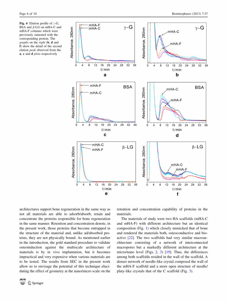
In Fig. 4 are compiled the SEC results from injection of the
three different proteins, i.e. c-G, BSA and b-LG, through
the mHA-F and mHA-C columns. As can be seen from
Fig. 4a, c, e all chromatograms were characterized by two
peaks. In SEC, the presence of these two peaks preceded by
the injection of a single protein indicates that the protein
travels through the column by two different path lengths.
There was a first narrow peak that resulted from the quick
exclusion of most of the proteins during the first 5 min, and
a second exclusion peak which appeared at later times and
was of much lower intensity and broader than the first one
(please refer to Table 1 for quantitative values). Unlike the
first peak, which clearly overlapped in all chromatograms
independent of the protein and column, the second peak
appeared at different times depending on the type of protein
and column. The elution time decreased with protein size:
c-G (150 kDa) eluted first, BSA (66 kDa) second and b-LG
(18 kDa) third. Furthermore, proteins eluted faster from the
mHA-C column than from the mHA-F one. It was also
noteworthy the fact that in the mHA-F column there were
less proteins eluting through the second peak (refer to
Table 1) and that they dwelled longer time in the column
than in the mHA-C column. Another striking result was that
all the injected BSA and c-G eluted completely but, a
30–40 % of the injected b-LG did not elute neither from the
C nor from the F column (refer to Table 1). Please note that
the peaks centred around 25 min in the b-LG chromato-
grams and 28 min for the c-G ones were not the result of
size exclusion effects. Their appearance was linked to the
protein injections history, i.e. the protein injection sequence
which was followed (data not shown), and the peaks
probably emerged as the result of protein desorption events.
To confirm that the separation events were of exclusion
nature and not from other forms of separation like ion
exchange or affinity separation, the elution of the proteins
was further investigated on mHA-C columns using mobile
phases of different ionic strength: Milli Q water, 0.1 M
AA, or 0.4 M AA combined with ACN (70:30 v/v).
Figure 5 summarizes the results pertaining to the elution of
the various proteins in either Milli Q water or 0.1 M AA
combined with ACN. Since the elution profile and the order
of elution did not vary with the ionic strength of the mobile
phase (AA versus water) one could conclude that the main
process governing separation was indeed size exclusion.
Investigation of the elution of BSA using 0.1 M AA or
0.4 M AA combined with ACN gave an almost identical
profile (data not shown) proving once again that the pre-
vailing mechanism was size exclusion. Furthermore, the
similarity in the chromatograms plotted in Fig. 6 corre-
sponding to the elution of c-G from columns previously
saturated with either c-G or BSA proves that the protein
eluted is a fraction of the injected protein and is not the
result of desorption from the column reinforcing that the
mechanism underlaying separation is not via adsorption–
desorption events.
4 Discussion
The awareness that biological events like osteoinduction
can be influenced and even controlled by the architecture of
a scaffold, has made the geometry of materials to become a
discriminatory point in the selection of materials for
applications in areas like bone regeneration. Not all the
Fig. 3 Pore size distributions obtained from MIP analysis of the
mHA-C and mHA-F samples
Biointerphases (2012) 7:37 Page 5 of 10
123
architectures support bone regeneration in the same way as
not all materials are able to adsorb/absorb, retain and
concentrate the proteins responsible for bone regeneration
in the same manner. Retention and concentration denote, in
the present work, those proteins that become entrapped in
the structure of the material and, unlike ad/absorbed pro-
teins, they are not physically bound. As mentioned earlier
in the introduction, the gold standard procedure to validate
osteoinduction against the multiscale architecture of
materials is by in vivo implantation, but it becomes
impractical and very expensive when various materials are
to be tested. The results from SEC in the present work
allow us to envisage the potential of this technique eluci-
dating the effect of geometry at the nano/micro scale on the
retention and concentration capability of proteins in the
materials.
The materials of study were two HA scaffolds (mHA-C
and mHA-F) with different architecture but an identical
composition (Fig. 1) which closely mimicked that of bone
and rendered the materials both, osteoconductive and bio-
active [22]. The two scaffolds had very similar macroar-
chitecture consisting of a network of interconnected
macropores but a markedly different architecture at the
micro/nano level (Figs. 2, 3) [19]. Thus, the differences
among both scaffolds resided in the wall of the scaffold. A
denser network of needle-like crystal composed the wall of
the mHA-F scaffold and a more open structure of needle/
platy-like crystals that of the C scaffold (Fig. 3).
Fig. 4 Elution profile of c-G,
BSA and b-LG on mHA-C and
mHA-F columns which were
previously saturated with the
corresponding protein. The
graphs on the right (b, d and
f) show the detail of the second
elution peak observed from the
a, c and d plots respectively
Page 6 of 10 Biointerphases (2012) 7:37
123

One important aspect to note is that the range of
microporosity found in the materials with pores below
0.1 lm (Fig. 3), was comparable to the size range of the
proteins in blood. Fibrinogen for instance which is an
abundant protein in blood has a rod-like shape and mea-
sures 46 nm in length [23]. Thus, if both materials were to
be implanted, blood would flow through them and their
specific microstructure would act as a sieve discriminating
among proteins thus leading to a specific protein adsorp-
tion, retention and concentration pattern. Assessing the
protein adsorption pattern is probably more straightforward
than investigating the latter two and, in fact, the under-
standing that we have today on protein adsorption mech-
anisms on hydroxyapatite (HA) accounts for that [24–27].
This contrasts with the lack of reports focused on protein
retention and concentration capability in 3D scaffolds for
bone regeneration applications. Interestingly, size-exclu-
sion chromatography is a chromatographic method which
emerged as molecular sieve and, though used for different
purposes, their results bear information on retention/con-
centration characteristics of a material.
Table 1 Compilation of the protein elution doses (%), their corresponding elution times (expressed as the time it starts eluting till it finishes) and
the protein elution span (Dt) for the two columns i.e., mHA-C and mHA-F
C, elution (%) F, elution (%)
1st peak 2nd peak 1st peak 2nd peak
c-G 67.0 ± 2 31.0 ± 2.7 79.0 ± 6.7 21.0 ± 6.7
BSA 55.5 ± 1.3 44.2 ± 1.1 70.5 ± 1.5 29.0 ± 1.5
b-LG 78.6 ± 1.5 – 71.6 ± 0.2 –
C, elution t (min) F, elution t (min)
1st peak 2nd peak 1st peak 2nd peak
c-G 1.2–4.7 3.8–13 1.2–4.7 5.6–20
BSA 1.2–4.5 7–18 1.2–4.5 8.3–20
b-LG 1.2–9.2 11.7–18.7 1.2–9.2 14.3–19.9
C, elution Dt (min) F, elution Dt (min)
1st peak 2nd peak 1st peak 2nd peak
c-G 3.5 9.2 3.5 14.4
BSA 3.3 11 3.3 11.7
b-LG 8 7 8 5.6
– Too low content
Fig. 5 Elution profile of c-G, BSA and b-LG from mHA-C columns
using two different mobile phases: a 70:30 v/v 0.1 M AA/ACN and
b 70:30 v/v Milli Q water/ACN. The concentration of protein injected
was 10 mg/ml in (a) and 3 mg/ml in (b)
Fig. 6 Elution profile of c-G from the mHA-C column previously
saturated with either BSA or c-G
Biointerphases (2012) 7:37 Page 7 of 10
123
Strictly speaking, the basis of ideal SEC assumes that
there is no interaction between the solute, in our case the
protein/s, and the stationary phase i.e. the material, and
therefore implies that all the proteins must elute within the
time required to renew the mobile phase that fully fills the
voids of the column, i.e. void volume [28]. The capacity of
a column (void volume) becomes then a critical factor in
the visualization of the various exclusion events. Columns
with too low capacity might not allow discriminating
separately the various exclusion events as proteins would
not have sufficient time to diffuse within the small pores of
the column before elution and columns with too high
capacity would result in long analysis times. In the present
work, the difficulties of preparing large amount of mac-
roporous material in a reproducible manner restricted the
size of the columns to a specific amount of material
insufficient to independently discriminate the various
exclusion events. Moreover, the well known affinity of
proteins towards HA further complicates visualization of
exclusion events. Thus, to overcome these difficulties a
particular strategy was developed. Before protein injection,
the stationary phase was 1st pre-adsorbed with the protein
of interest to saturate all binding sites of HA so as to
facilitate exclusion events and 2nd, following column sat-
uration, a rinsing cycle with ACN was applied in order to
selectively enrich the smaller pores of the columns with
ACN. The lower viscosity of ACN as compared to that of
the mobile phase (30:70 v/v mixture of ACN with Milli Q
water) was believed to facilitate protein diffusion into the
pores thus allowing their separation. To confirm that this
latter strategy was not altering the mechanism of interac-
tion of the proteins with the column and that size exclusion
was the main mechanism of interaction, experiments using
mobile phases with various ionic strengths were performed
(Fig. 5). In the case that a change in ionic strength modifies
the protein elution profile this would be indicative of an
affinity type of interaction mechanism between protein-
column but if no alteration occurs then the main interaction
mechanism would be of size-exclusion nature. The elution
of the various proteins in mixtures of ACN with either
Milli Q water or 0.1 M AA yielded similar protein elution
profiles and elution order thus confirming that the main
mechanism of interaction was indeed size exclusion.
Nevertheless, in spite of the similar general appearance
between chromatograms, there were some differences in
the elution of BSA and c-G in the two mobile phases which
should not be misunderstood as other types of elution
mechanisms but are simply the result of contribution of the
blank (fraction of protein eluted owing to the rinsing pro-
cess) in the chromatograms. Although the blank was usu-
ally subtracted in the various chromatograms (e.g. Figs. 4,
6), that was not the case in Fig. 5 which explains the
alteration of the tail for the second elution peak in the
results. Thus, if we obviate the contribution from the blank,
the similarity among chromatograms improves, further
supporting a separation mechanism mainly based on
exclusion events. Similar differences were also detected in
Fig. 4d in the mHA-C column regardless of subtraction of
the blank. In this case the differences arose from sub-
tracting the same blank in a series of independent runs
instead of performing a blank per run. A separate experi-
ment that was also done to corroborate the exclusion nature
mechanism of interaction was to assess the elution of c-G
on a column pre-adsorbed with either BSA or c-G (Fig. 6).
Since there were no differences in the eluted profile which
could point to an affinity type of interaction of c-G with the
scaffold, the results supported once again the exclusion
nature of the separation events.
To better understand the contribution of SEC in this
work, it is worth comparing the SEC chromatograms with
the MIP plot (Figs. 3, 4). One seems the mirror image of
the other as gauged by the presence of two peaks, one
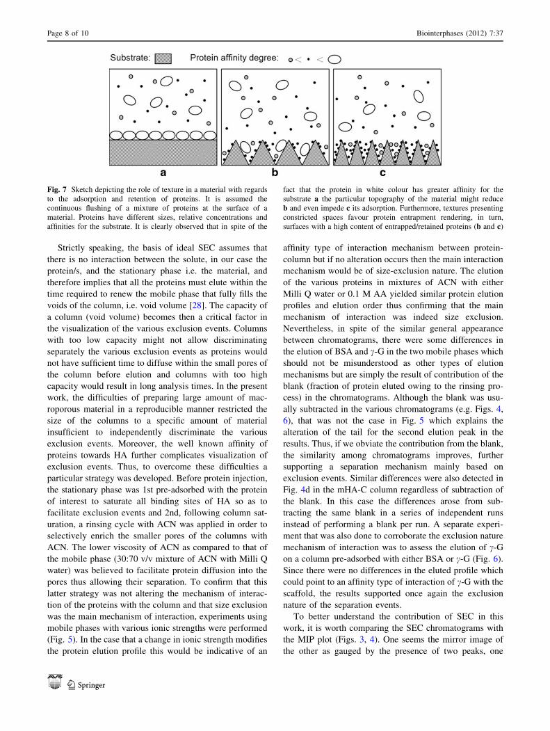
Fig. 7 Sketch depicting the role of texture in a material with regards
to the adsorption and retention of proteins. It is assumed the
continuous flushing of a mixture of proteins at the surface of a
material. Proteins have different sizes, relative concentrations and
affinities for the substrate. It is clearly observed that in spite of the
fact that the protein in white colour has greater affinity for the
substrate a the particular topography of the material might reduce
b and even impede c its adsorption. Furthermore, textures presenting
constricted spaces favour protein entrapment rendering, in turn,
surfaces with a high content of entrapped/retained proteins (b and c)
Page 8 of 10 Biointerphases (2012) 7:37
123
sharper and of higher intensity than the other. This simi-
larity is just a reflection of how tightly is linked one graph
with the other. We can picture the mHA material (SEC
column) as having an interconnected path of 80 lm width
whereby proteins travelling through the main stream would
quickly cross the column eluting first and, the proteins in
contact with the material would be more or less entertained
depending on the microarchitecture of the stationary phase
thus eluting second. Since the size of the proteins (i.e. a
few nanometers) is much inferior to the size of the inter-
connected macropores (10–200 lm), this will cause all
proteins travelling through the main stream to elute at the
same time. It is then not surprising to see that both columns
with similar macroarchitecture have the first elution peak
appearing at the same time. It is worth noting that proteins
travelling this path become irrelevant to the material as
they are simply excluded without allowing any kind of
interaction. Depending on the type of column and protein,
the amount excluded can vary from 60 % to almost 80 %
of the injected amount (Table 1).
More interesting than investigating this first exclusion
peak is to focus at the proteins travelling in contact with the
stationary phase as they can potentially interact with the
material. With regards to these proteins, and independent
of any enthalpic interaction protein-material, it is clear that
the proteins can diffuse differently into the pores affecting
their retention time and their concentration capability. It is
this type of events that become critical in order to under-
stand the enhanced biological performance observed in
certain materials and more specifically for certain geome-
tries [7–10]. Differences in the microarchitecture between
the F and C column resulted, indeed, in differences in the
concentration and retention capability of the proteins as
revealed from the second elution peak. The wall structure
of the mHA-F scaffold, consisting in a denser network of
smaller crystallites than for the C one, increased the
retention time of the proteins (higher dwelling time) and
decreased the protein concentration capability owing to the
reduced space in between crystals. This trend though, was
clearly observed for the BSA and c-G proteins but became
less obvious for the b-LG. We believe that was caused by
an unexpected strong interaction of the protein with the
material which caused b-LG to irreversibly adsorb onto the
material rather than to delay its elution (30–40 % of the
injected amount became adsorbed). Nevertheless the elu-
tion pattern for the b-LG can still be distinguished as
shown in Fig. 4f. Another aspect to highlight is that the
elution of each protein depended on their size, thus c-G
(*150 kDa), which is the largest, elutes first, BSA
(*66 kDa) second and the smallest b-LG (*18 kDa)
third. This trend was to be expected as the mechanism that
prevails is entropically driven. Looking at the quantitative
values gathered in Table 1 it is remarkable the amounts of
proteins retained per protein and column. Thus, up to a
40 % of the injected BSA was retained in the C column
while only a 25 % for the F one. In the case of c-G prac-
tically the same amount than BSA was retained in F and
only a 30 % in the C column.
The different behaviour shown in the amount retained
per column demonstrates that it is possible to implement
classic processing routes with simple characterization
techniques to modulate the retention capability of a mate-
rial. Understanding which proteins are retained and their
specific amount (dose) is particularly important from var-
ious points of view. On the one hand we cannot overlook
the fact that topography can involve the concentration/
retention of large amounts of specific proteins (this will
depend on the particular geometry of the material) which
apart from ‘hindering’ the ad/absorbed surface might end
up governing the nature of the ad/absorbed layer. If we also
take into account that biological events are not restricted to
ad/absorbed proteins but can also be triggered by concen-
tration gradients, both, the bound and unbound ‘protein
states’ have to be controlled to predict the response of the
material. In addition, is worth noting that topography can
override the intrinsic affinity of proteins for a substrate by
simply acting as a sieve (please refer to the sketch in
Fig. 7). Understanding and controlling these effects which
have clearly been observed to play an important role in
events like osteoinduction, blood clotting and cell function
in general [2, 3, 29] have become an ongoing challenging
field of investigation.
In spite of the ‘apparent’ distortion that topography
causes on the nature of the protein layer at the surface of
the material, we can take advantage of the sieving and
concentrating capability of topography to design materials
able to self-concentrate targeted proteins from the body
(e.g. BMPs) instead of current approaches which use car-
riers for the delivery of a particular protein [29–31].
Quantifying and distinguishing this retained amount from
what is adsorbed is also critical because it affects the
mechanism by which the biomaterial influences a biologi-
cal function [29].
Thus, this study which in essence has been performed
minimising any enthalpic interaction, has given a good
account on how via entropic mechanisms the different
proteins are able to discriminate the micro/nanoarchitecture
created by the entanglement of crystals of different sizes
(F vs. C). Nevertheless, it is important to bear in mind that
the present study only gives a partial view as in an in vivo
situation both, entropic and enthalpic interactions would
always occur. The immediate goal following this work will
be the preparation of columns with greater dimensions so
that the different exclusion events along with any enthalpic
event would be readily observed using a mobile phase
similar to physiological conditions. In fact, one concern in
Biointerphases (2012) 7:37 Page 9 of 10
123

the present work was the effect that organic solvents like
ACN might have caused on the protein conformation and
elution profile albeit its content in the mobile phase was
kept low enough to minimize any changes [32]. This is a
problem though, that is readily solved working with larger
columns as the requirement of using ACN to favor protein
diffusion into the pores would no longer be needed. Under
those conditions, the injection of complex protein mixtures
could then be made and fractions of the eluate could be
retrieved to further analyse its content. We believe that
SEC can become a powerful tool characterizing constructs
to be applied in the tissue engineering field.
5 Conclusions
Size-exclusion chromatography has proved to be a suc-
cessful tool for the investigation of the concentration and
entrapment behaviour of proteins in two different macro-
porous HA scaffolds (mHA) with similar macroarchitec-
ture but different microarchitecture, i.e. mHA-C versus
mHA-F. The larger size and lower density of entangled
crystals in the C material as compared to the F one allowed
for more protein concentration and a faster elution time.
Furthermore, proteins with different sizes i.e., c-G, BSA
and b-LG, were shown to discriminate differently the
microarchitecture created by both materials. This was
observed in the different retention times and the amounts
retained per column. We propose the use of SEC to screen
the geometry of TE scaffolds in order to identify the best
architecture that would enhance concentration/retention of
proteins of interest.
Acknowledgments The authors thank Eva Maria del Alamo and
Esther Miralles for technical assistance during SEC analysis. This
work was funded by the European Commission through the AN-
GIOSCAFF project NMP3-LA-2008-214402 and the Spanish Minis-
try through the project MAT2009-13547. M.P. Ginebra also
acknowledges funding from the Generalitat de Catalunya through the
prize ICREA Academia for excellence in research.
Open Access This article is distributed under the terms of the
Creative Commons Attribution License which permits any use, dis-
tribution, and reproduction in any medium, provided the original
author(s) and the source are credited.
References
1. Horbett TA (1982) In: Cooper SL, Peppas NA, Hoffman AS,
Ratner BD (eds) Biomaterials: interfacial phenomena and
applications. Advances in chemistry series. American Chemical
Society, Washington DC
2. Roach P, Farrar D, Perry CC (2006) J Am Chem Soc
128:3939–3945
3. Lord MS, Foss M, Besenbacher F (2010) Nano Today 5:66–78
4. Lord MS, Cousins BG, Doherty PJ, Whitelock JM, Simmons A,
Williams RL, Milthorpe BK (2006) Biomaterials 27:4856–4862
5. Vroman L, Adams AL, Fischer GC, Munoz PC, Standford M
(1982) In: Cooper SL, Peppas NA, Hoffman AS, Ratner BD (eds)
Biomaterials: interfacial phenomena and applications. American
Chemical Society, Washington DC
6. Ripamonti U, Herbst NN, Ramoshebi LN (2005) Cytok Growth
Factor Rev 16:357–368
7. Ripamonti U, Crooks J, Kirkbride AN (1999) S Afr J Sci
95:335–343
8. Habibovic P, Huipin Y, van der Valk CM, Meijer G, van Bli-
tterswijk CA, de Groot K (2005) Biomaterials 26:3565–3575
9. Habibovic P, Sees TM, van den Doel M, van Blitterswijk CA, de
Groot K (2006) J Biomed Mater Res 77A:747–762
10. LeGeros RZ (2008) Chem Rev 108:4742–4753
11. Yuan H, Fernandes H, Habibovic P, de Boer J, Barradas AMC, de
Ruiter A, Walsh WR, van Blitterswijk A, de Bruijn JD (2010)
Proc Natl Acad Sci USA 107:13614–13619
12. Irvine GB (1997) Anal Chim Acta 352:387–397
13. Barth HG, Boyes BE, Jackson C (1998) Anal Chem 70:251R–
278R
14. Liang H, Scott MK, Murry DJ, Sowinski KM (2001) J Chro-
matogr B Anal Technol Biomed Life Sci 754:141–151
15. Kawasaki T, Takahashi S, Ikeda K (1985) Eur J Biochem
152:361–371
16. Grodzki AC, Berenstein E (2010) Methods Mol Biol 588:33–41
17. Neville B (1996) In: Doonan S (ed) Protein purification protocols
methods in molecular biology. Humana Press Inc, Totowa
18. Mahn A, Lienqueo ME, Asenjo JA (2007) J Chromatogr B Anal
Technol Biomed Life Sci 849:236–242
19. Espanol M, Perez RA, Montufar EB, Marichal C, Sacco A,
Ginebra MP (2010) Acta Biomater 5:2752–2762
20. Montufar EB, Traykova T, Planell JA, Ginebra MP (2011) Mat
Sci Eng C 31:1498–1504
21. Ginebra MP, Fernandez E, de Maeyer EAP, Verbeeck RMH,
Boltong MG, Ginebra J, Driessens FCM, Planell JA (1997)
J Dent Res 76:905–912
22. Hench LL, Polak JM (2002) Science 295:1014–1017
23. Hall CE, Slayter HS (1959) J Biophys Biochem Cytol 5:11–16
24. Kawasaki T (1991) J Chrom A 544:147–184
25. Yin G, Liu Z, Zhan J, Ding F, Yuan N (2002) Chem Eng J
87:181–186
26. Mura-Galelli MJ, Voegel JC, Behr S, Bres EF, Schaaf P (1991)
Proc Natl Acad Sci USA 88:5557–5561
27. Barroug A, Lernoux E, Lemaitre J, Rouxhet PG (1998) J Coll Int
Sci 208:147–152
28. Berek D (2010) J Sep Sci 33:315–335
29. Uludag H, Gao T, Porter TJ, Friess W, Wozney JM (2001)
Delivery systems for BMPs: factors contributing to protein
retention at an application site. J Bone Joint Surg
83A(1):128–135
30. Bessa PC, Casal M, Reis RL (2008) J Tissue Eng Regener Med
2:81–96
31. De Groot K (1998) Tissue Eng 4:337–341
32. Gekko K, Ohmae E, Kameyama K, Takagi T (1998) Biochim
Biophys Acta 1387:195–205
Page 10 of 10 Biointerphases (2012) 7:37
123