Embed Size (px)
Citation preview
Arterivirus RNA Synthesis Dissected Nucleotides, membranes, amino acids, and a bit of zinc ...
ERIC J. SNIJDER Department of Virology, Center of Infectious Diseases, Leiden University Medical Center, LUMC P4-26, p.o. Box 9600,2300 RC Leiden, the Netherlands. E-mail: [email protected]
1. INTRODUCTION
Over the past ten years, research on by far the largest nidovirus protein, the non structural gene 1 or replicase gene, has gone through a number of characteristic stages. First, cDNA copies of various nidovirus replicase genes were cloned and sequenced, with those of the coronaviruses infectious bronchitis virus (IBV) (Boursnell et al 1987) and mouse hepatitis virus (MHV) (Lee et al 1991) and the arterivirus equine arteritis virus (EA V) (den Boon et al 1991) being the first to be completed. Subsequently, extensive sequence comparisons revealed very interesting similarities/homologies within the nidovirus group (then still classified as corona-, toro-, and arteriviruses) and between nidoviruses and other positive-stranded RNA viruses. About five years later, the taxonomic debate on the position of the various nidovirus subgroups ended at the 1996 International Congress of Virology in Jerusalem. The family Arteriviridae and the order Nidovirales (containing the Coronaviridae and Arteriviridae) were formally established to acknowledge both the unique properties of arteriviruses and coronaviruses as well as their intriguing ancestral relationship at the level of replicase genes, genome organization, and replication strategy (Snijder and Spaan 1995, de Vries et aI1997).
In the meantime, the experimental characterization of the replicase gene products had mainly been focused on the identification of protease domains
The Nidoviruses (Coronaviruses and Arteriviruses). Edited by Ehud Lavi l'f al .. Kluwer Academic/Plenum Puhlishers. 200 I. 241
242 Arterivirus RNA Synthesis Dissected
and the elucidation of the pathways used for proteolytic processing of the replicase ORFla and ORFlab polyproteins (for a review, see Ziebuhr et al 2000). A more recent point of interest is the membrane-association of the nidovirus replication complex. With the advent of reverse genetics systems for arteriviruses in 1996 (Meulenberg et a11998, van Dinten et a11997) and, more recently, for coronaviruses (Almazan et al 2000), the stage has now been set for the functional characterization of replicase gene products in the context of their main task in the viral life cycle: RNA synthesis. The size of the nidovirus nonstructural polyprotein varies between 3175 residues for the arterivirus EA V and approximately 7200 amino acids for the coronavirus MHV. This size difference of the replicase again illustrates how both conservation and variation have played a part in nidovirus evolution. In addition to a number of highly conserved domains/functions that can be considered to form the "core" of the nidovirus replicase, each virus (or cluster of viruses) appears to have developed its own set of accessory non structural functions.
2. PROTEOLYTIC PROCESSING
Proteolytic processing of nonstructural proteins fulfils a key role in the life cycle of most viruses. In the course of virus evolution, highly specific virus-encoded proteases have evolved and their importance for the regulation of virus replication is becoming more and more evident (for reviews, see Dougherty and Semler 1993, Gorbalenya and Snijder 1996). A comprehensive review on the properties and activities of nidovirus proteases has been published recently in the Journal of General Virology (Ziebuhr et al 2000). A basic characterization of the various protease domains has been carried out and a cleavage site map has been obtained for prototypic coronavirus and arterivirus replicases. Still, given the size of the nidovirus replicase and the details that have emerged for the replicases of other groups of positive-stranded RNA viruses, it is quite likely that -in processing termswe have merely revealed the tip of an iceberg. Some of the most challenging topics that remain to be investigated are clearly related to the role of proteolytic processing in the regulation of the nidovirus life cycle. That investigating these aspects is not a simple exercise of "knocking out" cleavage sites, has become clear from our work with the EA V infectious cDNA clone. The systematic mutagenesis of each of the EA V replicase cleavage sites (van Dinten et al 1999, M. A. Tijms and E. J. Snijder; unpublished data) has revealed that -basically- they are all essential for nonnal viral RNA synthesis. This conclusion includes sites that are part of the previously described "minor" pathway for processing of the C-terminal
Eric J. Snijder 243
half of the ORFla protein (Wassenaar et al1997). This indicates that -as far as the in vivo analysis of proteolytic processing by mutagenesis is concerned- a more sophisticated approach is required, for example by using partially cleavable mutant sites and/or partially active protease mutants.
3. MEMBRANE ASSOCIATION OF THE REPLICATION COMPLEX
Having raised antibodies against replicase subunits, using them in immunofluorescence assays to analyze infected cells was a logical next step that was taken by several nidovirus research groups. In Leiden, the initial results obtained for EAV were followed by a comparative analysis for MHV.
Possibly due to the number of labs working on either virus group, the results for arteriviruses seem to be more consistent than those for coronaviruses: all EA V replicase subunits (with the exception of a part of nsp1) and de novo RNA synthesis (visualized by BrUTP labeling) colocalize in the perinuclear region of the three cell types tested (Pedersen et al 1999, van der Meer et al 1998). Also a substantial part of the EA V N protein was found in this region of the cell, but its involvement in genome replication or subgenomic RNA transcription is highly unlikely, since N gene expression can be inactivated without a significant effect on either process (Molenkamp et al2000).
For MHV, the situation is much less clear: different replicase subunits have been reported to localize to different membrane compartments in different cell types (Bost et al2000, Shi et al1999, Sims et al2000, van der Meer et al1999). Co-localization of RNA synthesis and N protein has been reported, but is not as complete as for EAV.
Two technical complications that are sometimes overlooked are (i) the fact that coronavirus-infected cells fuse into syncytia that are unsuitable for a reliable cell biological analysis, and (ii) (true for both virus groups) that replicase processing takes time and antisera do not discriminate between processing intermediates and end products.
There is no doubt that -in the end- we will need the power of the electron microscope to understand the details of nidoviral membrane-associated RNA synthesis. The advantages of a 100-fold higher resolution need hardly be explained, and also allows for the identification of cellular compartments on the basis of their morphology and the presence of well-defined markers. So far, however, only one replicase study for each nidovirus subgroup included electron microscopy (Pedersen et al 1999, van der Meer et al 1999) and therefore it is good to interpret these data with caution. For the moment, two main differences between arteri- and coronaviruses appear to exist: a

244 Arterivirus RNA Synthesis Dissected
replication complex associated with double ER-derived membranes for arteriviruses (EA V) versus a complex associated with single membranes of another origin, most likely endosomal/lysosomal, for coronaviruses (MHV). The general distribution in the cell of these two replication complexes is somewhat different (Fig. 1): the arterivirus signal is generally closer to the nucleus and a bit less punctate.
Figure 1. Immunofluorescence staining for specific replicase subunits of the arterivirus EA V (left) in BHK-2l cells and the coronavirus MHV (right) in mouse L cells at a time point around the peak of viral RNA synthesis. For both viruses, the staining is representative for the majority of replicase subunits and de novo RNA synthesis (see text).
The formation of paired membranes and double membrane vesicles (DMVs) is a typical feature of arterivirus replication, although the use of double membranes as matrix for RNA synthesis is not restricted to arteriviruses. Double membrane structures in arterivirus-infected cells were seen over 30 years ago and have now been convincingly linked to RNA synthesis. A number of suggestive EM pictures indicate that they originate from ER membranes (Pedersen et al 1999), although a thorough biochemical characterization remains to be carried out. We have previously shown that we can induce DMV s that strikingly resemble those in infected cells by expressing EA V nsp2-7, which comprises 1,400 residues of the 0 RF 1 a protein, including four major hydrophobic regions. Recently, we have narrowed this down to nsp2 and nsp3 (K. W. Pedersen and E. 1. Snijder; unpublished data), which were already known to have a very strong

Eric J Snijder 245
interaction from our analysis of the proteolytic processing of the ORFla protein (Snijder et al 1994). Co-expression of nsp2 and nsp3, but not the expression of either protein by itself, results in the generation of DMV s.
Clearly we have no well-defined ideas about the way in which the EA V ORFla proteins associate with membranes. Computer predictions suggest up to 10 trans-membrane domains and furthermore a sort of "processing logic" can be applied (Fig. 2).
1b" 1'------===---=='----==-----'=..::==--===--==='------'-... ·.··· . nsp1 nsp2 nsp3 nsp4 nsp5-12 ~ ~
lumen
cytoplasm
Figure 2. Tentative model for membrane association of the EAV ORFla protein. The processing scheme of the ORFla protein is depicted and the hydrophobic domains in nsp2, nsp3 and nsp5 are indicated. The model is based on computer predictions and processing infonnation. Abbreviations: P, papain-like cysteine protease; S, chymotrypsin-like serine protease; h, hydrophobic domain.
For example, the nsp2 protease and the nsp2/3 cleavage site should obviously be on the same side of the membrane, making it logical that the nsp2 hydrophobic domain spans the membrane twice. Likewise, three (and not two or four) is the obvious number of transmembrane segments in the second (largest) hydrophobic domain of nsp3, because otherwise the nsp4 protease would end up in the lumen and not have access to the sites it is known to cleave.
Particularly interesting questions that remain to be addressed relate to (i) the mechanism of translocation, which is probably post-translational since there are no N-terminal signal sequences on the replicase polyprotein, (ii) the coordination of processing and membrane association, (iii) the role of established interactions, like the one between nsp2 and nsp3 (Snijder et al 1994) and the cofactor role of nsp2 for the nsp4 protease in cleaving the nsp4/5 site (Wassenaar et aI1997), and (iv) the mechanism of the formation of double membranes and DMV s.

246 Arterivirus RNA Synthesis Dissected
4. RNA SYNTHESIS
Following the generation of the EA V infectious cDNA clone in 1996 (van Dinten et at 1997), the RNA sequences thought to be involved in discontinuous mRNA transcription were an obvious primary target for sitedirected mutagenesis. In our opinion, the discontinuous minus strand extension model (Fig. 3), proposed by Sawicki and Sawicki (Sawicki and Sawicki 1995), is the nidovirus transcription model that is best supported by the experimental data available for corona- and arteriviruses.
According to this model, nidovirus minus strand transcription is either continuous, yielding the genomic minus strand, or discontinuous. The latter process involves a jump, which partially depends on a base pairing interaction between conserved transcription-regulating sequences (TRSs), and yields subgenomic minus strands that serve as templates for the transcription of sub genomic mRNAs. An important aspect of the nidovirus transcription strategy is that the 3' end of the genome contains an array of body TRS sequences, all directing this jump with their own specific activity, and probably also influencing each other's activity in making sub genomic minus strands.
If we try to dissect the discontinuous step in transcription, which may strongly resemble an RNA recombination event (Brian and Spaan, 1997, Nagy and Simon, 1997), we can discriminate at least four steps, each of which may involve specific RNA-RNA and RNA-protein interactions. There is probably attenuation of the transcriptase, translocation of the nascent strand, base-pairing at the leader TRS, and reinitiation of transcription. Although, in our EA V reverse genetics system, we can analyze the impact of mutations only after conversion of the sub genomic minus strand into sub genomic mRNAs, a number of predictions could be made on the basis of the transcription model. First, leader and body TRS mutations were expected to affect base-pairing and identical mutations in leader and body TRSs should complement each other. Second, since not all body TRSs are completely identical, leader TRS mutations might have different effects on the transcription of different subgenomic mRNAs. Third, body TRS mutations mayor may not affect attenuation: if they do not, the number of transcriptases reaching upstream body TRSs should remain the same; but if they do for example reduce attenuation, transcription from upstream TRSs may go up.

Eric J Snijder
body junction
- genome - body TRS
subgenomic mRNA
247
5'
3'
5'
Figure 3. Discontinuous minus strand extension model for nidovirus transcription as proposed by Sawicki and Sawicki (1995). The genomic plus strand is copied into a genomic minus strand that serves as the template for genome replication. In addition, minus strand synthesis can be interrupted at a body TRS, after which the nascent strand is translocated and the TRS complement at the 3' end of the nascent minus strand base-pairs with the genomic leader TRS. Transcription is resumed to add the anti-leader sequence and the resulting subgenomic minus strand (containing the body TRS at its leader-body junction site) is the template for subgenomic mRNA transcription.
Evidence for the importance of the TRS and the existence of a basepairing interaction between plus leader and minus body TRSs was previously published in a paper by van MarIe et al. (1999a). However, in this study only the two C residues of the conserved 5' UCAACU 3' sequence that forms the EA V TRS were targeted by mutagenesis. A more recent, comprehensive mutagenesis study of the leader TRS and RNA 7 body TRS (A. O. Pasternak, E. van den Born, W. J. M. Spaan, and E. J. Snijder; unpublished data) has revealed that, depending on the nucleotide that is changed, the results can vary quite dramatically. For example, mutants in which identical (leader and RNA 7 body) TRS mutations do not restore transcription at all have been obtained. Likewise, body TRS mutations that appear to up-regulate the activity of more upstream body TRSs have been identified. A detailed characterization of these TRS mutants is now in progress.

248 Arterivirus RNA Synthesis Dissected
In addition to RNA primary structure, RNA secondary structure is being studied as a factor that may be involved in the regulation of transcription. A conserved "leader TRS hairpin" has been predicted in the 5' end of all arterivirus genomes and seems a very good candidate to be involved in the base-pairing step of transcription (van Marie et al 1999a). At the other end of the genome, a recent analysis of the body TRS regions suggests a link between body TRS activity and its presence in a non-base-paired region of the predicted RNA structure (Pasternak et al 2000). Experiments to corroborate this prediction are now in progress.
5. PROTEIN FUNCTIONS IN RNA TRANSCRIPTION
We have previously proposed that the nidovirus replicase may consist of a basic or "core" transcriptase, containing subunits like the RNA-dependent RNA polymerase and helicase found in other systems, that is supplemented with more specialized domains/proteins involved exclusively in subgenomic RNA synthesis (van Marie et al 1999a). Thus, one can envision multiple possibilities for discontinuous transcription-specific protein functions, e.g. proteins interacting with body TRSs, leader TRS, or the leader TRS hairpin (Fig. 4).
3'
5'
-----~
Figure 4. Potential sites for protein-RNA interactions during EA V discontinuous minus strand synthesis. The nidovirus RdRp complex may operate like a core transcriptase that is supplemented with specific protein factors to direct discontinuous minus strand synthesis.
We were confronted with such a transcription-specific function even before the full-length cDNA clone was generated (van Dinten et al 1997). Mutant EA V030F had a single replicase mutation in nsp 1 0 (Ser-2429 to Pro)

Eric 1. Snijder 249
that selectively knocked out sub genomic RNA transcription, thereby rendering the clone noninfectious. The EAV030F mutation maps to nsplO, which can be considered the most conserved nidovirus replicase subunit, with an N-terminal predicted zinc finger domain (van Dinten et al2000) and a C-terminal helicase, for which unique enzymatic properties were recently revealed that are shared by corona- and arteriviruses (Seybert et al 2000). The EA V030F mutation is located in a region that seems to connect zinc fmger and helicase (Fig. 5).
We have recently shown that the EAV030F mutant is not completely defective in subgenomic RNA synthesis (van MarIe et al 1999b). It was estimated that sub genomic plus and minus strand synthesis are about 500-fold reduced, which was supported by a construct in which we inserted the CAT gene at the position of the N protein gene and quantified mRNA7 transcription on the basis of reporter gene expression.
Figure 5. Tentative model for zinc binding by the N-terrninal domain of the EAV nsplO helicase protein (van Dinten et aI2000).
More recently, an extensive mutagenesis of the nspl0 zinc finger region and the region containing the original EA V030F mutation was performed, details of which can be found in a paper by van Dinten et al (2000). Briefly, all 13 conserved Cys and His residues, which are predicted to be part of an unusual zinc-binding domain (Fig. 5), were found to be important for both genome replication and subgenomic RNA synthesis. However, two additional mutants in the region downstream of the zinc finger displayed EAV030F-like phenotypes and suggested that this region may indeed function as a "spacer" between the zinc finger and helicase domains. The flexibility of this spacer may be essential for a function of nsp lOin sub genomic RNA transcription.
Complementation of the EA V030F defect was attempted using a replicon containing the EA V030F replicase and an IRES/nsp 1 0 cassette in the
250 Arterivirus RNA Synthesis Dissected
genomic 3' end (van Dinten et a/2000). Expression of wild-type nsplO from this locus in the genome was indeed possible, but unfortunately complementation did not occur. Immunofluor-escence studies suggested that the IRES-expressed nsp 1 0 did not localize to the replication complex and may have been unable to force the mutant nsp 10, which is still active in genome replication, out of the complex.
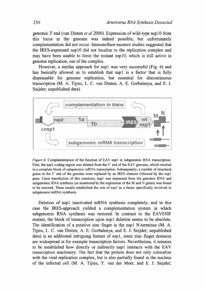
However, a similar approach for nsp 1 was very successful (Fig. 6) and has basically allowed us to establish that nsp 1 is a factor that is fully dispensable for genome replication, but essential for discontinuous transcription (M. A. Tijms, L. C. van Dinten, A. E. Gorbalenya, and E. J. Snijder; unpublished data).
I complementation in trans I
1a 1b
~nsp1
CS I subgenomic mRNA transcription I
Figure 6. Complementation of the function of EA V nsp I in subgenomic RNA transcription. First, the nspl-coding region was deleted from the 5' end of the EAV genome, which resulted in a complete block of subgenomic mRNA transcription. Subsequently, a number of structural genes in the 3' end of the genome were replaced by an IRES element followed by the nsp I gene. Upon transfection of this construct, nsp I was expressed from the genomic RNA and subgenomic RNA synthesis (as monitored by the expression of the M and N genes) was found to be restored. These results established the role of nsp I as a factor specifically involved in subgenomic mRNA synthesis.
Deletion of nsp 1 inactivated mRNA synthesis completely, and in this case the IRES-approach yielded a complementation system in which sub genomic RNA synthesis was restored. In contrast to the EA V030F mutant, the block of transcription upon nsp 1 deletion seems to be absolute. The identification of a putative zinc finger in the nspl N-terminus (M. A. Tijms, L. C. van Dinten, A. E. Gorbalenya, and E. J. Snijder; unpublished data) is an additional intriguing feature of nspl, since zinc finger domains are widespread in for example transcription factors. Nevertheless, it remains to be established how directly or indirectly nsp 1 interacts with the EA V transcription machinery. The fact that the protein does not only colocalize with the viral replication complex, but is also partially found in the nucleus of the infected cell (M. A. Tijms, Y. van der Meer, and E. J. Snijder;
Eric 1. Snijder 251
unpublished data), suggests that it may be a multifunctional replicase subunit.
ACKNOWLEDGMENTS
A large number of people have contributed to the work described in this chapter, and to arteriviruslnidovirus research in Leiden as a whole. I would like to acknowledge the contributions of Ketil Pedersen (Oslo University), lacomine Krijnse Locker (EMBL, Heidelberg), Yvonne van der Meer (LUMC), lohan den Boon (LUMC), Peter Bredenbeek (LUMC) , Willem Luytjes (LUMC), Guido van Marle (LUMC), Richard Molenkamp (LUMC), Babette Rozier (LUMC), Sasha Pasternak (LUMC), Erwin van den Born (LUMC) , Sasha Gultyaev (Department of Chemistry, Leiden University), Fred Wassenaar (LUMC), Leonie van Dinten (LUMC), Sietske Rensen (LUMC) , Hans van Tol (LUMC), Marieke Tijms (LUMC), Sasha Gorbalenya (SAIC/NCI-FCRDC, Frederick), lessika Dobbe (LUMC) , Sophie Greve (LUMC), and (in many ways) Willy Spaan. This work was supported by several grants from the Council for Chemical Sciences of the Netherlands Organization for Scientific Research (CW-NWO).
REFERENCES
Almazan, F., Gonzalez, J.M., Penzes, Z., Izeta, A., Calvo, E., Plana-Duran, J., and Enjuanes, L., 2000, Engineering the largest RNA virus genome as an infectious bacterial artificial chromosome. Proc. Natl. Acad. Sci. U.S.A. 97:5516-5521.
Bost, AG., Carnahan, R.H., Lu, X.T., and Denison, M.R., 2000, Four proteins processed from the replicase gene polyprotein of mouse hepatitis virus colocalize in the cell periphery and adjacent to sites of virion assembly. J. Virol. 74: 3379-3387.
Boursnell, M.E., Brown, T.D.K., Foulds, U., Green, P.F., Tomley, F.M., and Binns, M.M., 1987, Completion of the sequence of the genome of the coronavirus avian infectious bronchitis virus. J. Gen. Virol. 68: 57-77.
Brian, D.A and Spaan, W.J.M., 1997, Recombination and coronavirus defective interfering RNAs. Semin. Virol. 8: !OI-III.
den Boon, J.A, Snijder, E.J., Chimside, E.D., de Vries, A.A.F., Horzinek, M.e., and Spaan, W.J.M., 1991, Equine arteritis virus is not a togavirus but belongs to the coronaviruslike superfamily. J. Virol. 65: 2910-2920.
de Vries, A.A.F., Horzinek, M.e., Rottier, P.J.M., and de Groot, R.J., 1997, The genome organization of the Nidovirales: similarities and differences between arteri-, toro-, and coronaviruses. Semin. Virol. 8: 33-47.
Dougherty, W.G. and Semler, B.L., 1993, Expression of virus-encoded proteinases: functional and structural similarities with cellular enzymes. Microbiol. Rev. 57: 781-822.
Gorbalenya, A.E. and Snijder, E.J., 1996, Viral cysteine proteases. Persp. Drug Discov. Design 6: 64-86.

252 Arterivirus RNA Synthesis Dissected
Lee, H.J., Shieh, C.K., Gorbalenya, A.E., Koonin, E.V., La Monica, N., Tuler, 1., Bagdzhadzhyan, A., and Lai, M.M.C., 1991, The complete sequence (22 kilobases) of murine coronavirus gene 1 encoding the putative proteases and RNA polymerase. Virology 180: 567-582.
Meulenberg, J.1.M., Bos-de Ruijter, 1.N.A, Wensvoort, G., and Moormann, R.J.M., 1998, Infectious transcripts from cloned genome-length cDNA of porcine reproductive respiratory syndrome virus. J. Virol. 72: 380-387.
Molenkamp, R., van Tol, H., Rozier, B.C.D., van der Meer, Y., Spaan, W.J.M., and Snijder, E.J., 2000, The arterivirus replicase is the only viral protein required for genome replication and subgenomic mRNA transcription. J. Gen. Virol. 81: 2491-2496.
Nagy, P.D. and Simon, A.E., 1997, New insights into the mechanisms of RNA recombination. Virology 235: 1-9.
Pasternak, A.O., Gultyaev, A.P., Spaan, W.J.M., and Snijder, E.J., 2000, Genetic manipulation of arterivirus alternative mRNA leader-body junction sites reveals tight regulation of structural protein expression. J. Virol. 74, in press.
Pedersen, K.W., van der Meer, Y., Roos, N., and Snijder, E.J., 1999, Open reading frame laencoded subunits of the arterivirus replicase induce endoplasmic reticulum-derived double-membrane vesicles which carry the viral replication complex. J. Virol. 73: 2016-2026.
Sawicki, S.G. and Sawicki, D.L., 1995, Coronaviruses use discontinuous extension for synthesis of subgenome-length negative strands. Adv. Exp. BioI. Med. 380: 499-506.
Seybert, A., van Dinten, L.C., Snijder, E.J., and Ziebuhr, 1., 2000, The biochemical characterization of the equine arteritis virus helicase suggests a close functional relationship between arterivirus and coronavirus helicases. J. Virol. 74: 9586-9593.
Shi, S.T., Schiller, J.J., Kanjanahaluethai, A., Baker, S.C., Oh, 1.W., and Lai, M.M., 1999, Colocalization and membrane association of murine hepatitis virus gene 1 products and De novo-synthesized viral RNA in infected cells. J. Virol. 73: 5957-5969.
Sims, AC., Ostermann, 1., and Denison, M.R., 2000, Mouse hepatitis virus replicase proteins associate with two distinct populations of intracellular membranes. J. Virol. 74: 5647-5654.
Snijder, EJ., and Spaan, WJ.M., 1995, The coronaviruslike superfamily. In: The Coronaviridae (S.G.Siddell, ed), Plenum Press, New York, N.Y., pp. 239-255.
Snijder, E.J., Wassenaar, AL.M., and Spaan, W.J.M., 1994, Proteolytic processing of the replicase ORFla protein of equine arteritis virus. J. Virol. 68: 5755-5764.
van der Meer, Y., Snijder, E.J., Dobbe, 1.c., Schleich, S., Denison, M.R., Spaan, W.J.M., and Krijnse Locker, 1., 1999, Localization of mouse hepatitis virus non structural proteins and RNA synthesis indicates a role for late endosomes in viral replication. J. Virol. 73: 7641-7657.
van der Meer, Y., van Tol, H., Krijnse Locker, 1., and Snijder, E.J., 1998, ORFla-encoded replicase subunits are involved in the membrane association of the arterivirus replication complex. J. Virol. 72: 6689-6698.
van Dinten, L.C., den Boon, lA., Wassenaar, A.L.M., Spaan, W.J.M., and Snijder, E.J., 1997, An infectious arterivirus cDNA clone: identification of a replicase point mutation which abolishes discontinuous mRNA transcription. Proc. Natl. Acad. Sci. U.S.A. 94: 991-996.
van Dinten, L.c., Rensen, S., Spaan, W.J.M., Gorbalenya, A.E., and Snijder, E.J., 1999, Proteolytic processing of the open reading frame 1 b-encoded part of arterivirus replicase is mediated by nsp4 serine protease and is essential for virus replication. J. Virol. 73: 2027-2037.

Eric 1. Snijder 253
van Dinten, L.e., van Tol, H., Gorbalenya, AE., and Snijder, EJ., 2000, The predicted metalbinding region of the arterivirus helicase protein is involved in subgenomic mRNA synthesis, genome replication, and virion biogenesis. J. Virol. 74: 5213-5223.
van Marie, G., Dobbe, lC., Gultyaev, A.P., Luytjes, W., Spaan, WJ.M., and Snijder, E.l, 1999a, Arterivirus discontinuous mRNA transcription is guided by base- pairing between sense and antisense transcription-regulating sequences. Proc. Natl. Acad. Sci. U.S.A. 96: 12056-12061.
van Marie, G., van Dinten, L.C., Luytjes, W., Spaan, WJ.M., and Snijder, EJ., 1999b, Characterization of an equine arteritis virus replicase mutant defective in subgenomic mRNA synthesis. J. Virol. 73: 5274-5281.
Wassenaar, AL.M., Spaan, WJ.M., Gorbalenya, AE., and Snijder, EJ., 1997, Alternative proteolytic processing of the arterivirus replicase ORFla polyprotein: evidence that NSP2 acts as a cofactor for the NSP4 serine protease. J. Virol. 71: 9313-9322.
Ziebuhr, 1., Snijder, EJ., and Gorbalenya, A.E., 2000, Virus-encoded proteinases and proteolytic processing in the Nidovirales. J. Gen. Virol. 81: 853-879.