Embed Size (px)
Citation preview
1
Antibody Recognition of the Pandemic H1N1 Influenza 1 Hemagglutinin Receptor Binding Site 2 3 Running title: Pandemic H1N1 Recognition by Human Antibody 5J8 4 5 Minsun Honga,1,2, Peter S. Leea,b,1, Ryan M. B. Hoffmana, Xueyong Zhua, Jens C. Krausec, 6 Nick S. Laursena, Sung-il Yoona,3, Langzhou Songe, Lynda Tusseye, James E. Crowe Jr.c,d, 7 Andrew B. Warda, Ian A. Wilsona,b,4 8 9 aDepartment of Integrative Structural and Computational Biology and bThe Skaggs Institute for 10 Chemical Biology, The Scripps Research Institute, 10550 North Torrey Pines Road, La Jolla, CA 11 92037, USA. 12 Departments of cPediatrics and dMicrobiology and Immunology, Vanderbilt University Medical 13 Center, Nashville, TN 37232, USA. 14 eVaxInnate Corporation, 3 Cedar Brooke Drive, Cranbury, NJ 08512, USA. 15 16 1M.H. and P.S.L. contributed equally to the work. 17 2Present address: Division of Biological Science and Technology, Yonsei University, Wonju, 18 220-710, Republic of Korea. 19 3Present address: Department of Systems Immunology and Institute of Antibody Research, 20 College of Biomedical Science, Kangwon National University, Chuncheon 200-701, Republic of 21 Korea. 22
JVI Accepts, published online ahead of print on 11 September 2013J. Virol. doi:10.1128/JVI.01388-13Copyright © 2013, American Society for Microbiology. All Rights Reserved.
on March 13, 2018 by guest
http://jvi.asm.org/
Dow
nloaded from

2
4Address correspondence to Ian A. Wilson, The Scripps Research Institute, 10550 North Torrey 23 Pines Road, La Jolla, CA 92037, USA. (858) 784-2939. [email protected]. 24 25 Conflict of interest statement: Vanderbilt University submitted a patent covering the diagnostic 26 and therapeutic use of the antibody described in this paper; J.C.K. and J.E.C. are two of the co-27 inventors on that application. 28 29 Data deposition: The atomic coordinates and structure factors reported in this paper are deposited 30 in the Protein Data Bank, www.pdb.org (PDB ID codes 4M5Y, 4M5Z, and 4M4Y). The 31 reconstruction data reported in this paper are deposited in the Electron Microscopy Data Bank, 32 www.emdatabank.org (EMDB ID code EMDB-5731 and EMDB-5733). 33 34 Word count (abstract): 246 35 Word count (text): 4,641 excluding acknowledgment, references, and figure legends. 36 Figures: 9 37 Tables: 2 38
on March 13, 2018 by guest
http://jvi.asm.org/
Dow
nloaded from

3
ABSTRACT 39 Influenza virus is a global health concern due to its unpredictable pandemic potential. This 40 potential threat was realized in 2009 when an H1N1 virus emerged that resembled the 1918 41 virus in antigenicity, but fortunately was not nearly as deadly. 5J8 is a human antibody 42 that potently neutralizes a broad spectrum of H1N1 viruses, including the 1918 and 2009 43 pandemic viruses. Here, we present the crystal structure of 5J8 Fab in complex with a 44 bacterially expressed and refolded globular head domain from the hemagglutinin (HA) of 45 the A/California/07/2009 (H1N1) pandemic virus. 5J8 recognizes a conserved epitope in 46 and around the receptor binding site (RBS) and its HCDR3 closely mimics interactions of 47 the sialic acid receptor. EM reconstructions of 5J8 Fab in complex with an HA trimer from 48 a 1986 H1 strain and with an engineered stabilized HA trimer from the 2009 H1 pandemic 49 virus showed a similar mode of binding. As for other characterized RBS-targeted 50 antibodies, 5J8 uses avidity to extend its breadth and affinity against divergent H1 strains. 51 5J8 selectively interacts with HA insertion residue 133a, which is conserved in pandemic 52 H1 strains and has precluded binding of other RBS-targeted antibodies. Thus, the RBS of 53 divergent HAs is targeted by 5J8 and adds to the growing arsenal of common recognition 54 motifs for design of therapeutics and vaccines. Moreover, consistent with previous studies, 55 bacterially expressed H1 HA properly refolds, retaining its antigenic structure, and 56 presents a low-cost and rapid alternative for engineering and manufacturing candidate flu 57 vaccines. 58
on March 13, 2018 by guest
http://jvi.asm.org/
Dow
nloaded from

4
INTRODUCTION 59 Influenza virus is the cause of seasonal epidemic and sporadic pandemic flu outbreaks. The 60 hemagglutinin (HA) surface glycoprotein mediates viral recognition of host cells through its 61 interaction with sialic acid receptors (1, 2). The globular “head” domain of HA is 62 immunodominant, likely due to its accessibility on the surface of viruses and, consequently, 63 antibodies are rapidly generated against it. However, the HA head undergoes continual antigenic 64 drift, which results in escape from the host immune response through amino-acid changes on its 65 surface or by masking neutralizing epitopes with glycans. Antibodies generated against HA are 66 typically strain specific, which necessitates nearly annual vaccine strain reformulations. In 67 contrast, the residues that form the receptor binding site (RBS) are functionally constrained for 68 receptor binding and, thus, have restricted mutational freedom. As such, the RBS is a prime 69 target for virus neutralization by broadly neutralizing antibodies that prevent viral-host 70 interactions (3). However, the footprint of the sialoglycan receptor on the RBS is much smaller 71 than that of an antibody. Hence, most antibodies that block the RBS also contact the 72 hypervariable regions surrounding it, which leads to strain-specific binding. Nevertheless, a few 73 antibodies that target the RBS display a broader spectrum of reactivity compared to those that 74 target HA elsewhere on the head (4-10). S139/1 reaches into the RBS and has heterosubtypic 75 neutralizing activity (7). C05, which also neutralizes highly divergent viruses, similarly enters 76 the RBS and remarkably accomplishes this interaction using essentially a single antibody loop 77 (6). CH65 and CH67 are broadly neutralizing H1-specific antibodies and, unlike S139/1 and 78 C05, make use of receptor mimicry (5, 9); however, CH65 notably does not neutralize 1918 or 79 2009 H1 pandemic strains that are now the current seasonal H1 epidemic strains (5). 80
on March 13, 2018 by guest
http://jvi.asm.org/
Dow
nloaded from

5
We have previously reported the identification and characterization of a human 81 monoclonal antibody 5J8 that possesses neutralization activity and therapeutic efficacy against 82 H1 viruses spanning decades, including the 1918 and 2009 pandemic viruses (11). Notably, the 83 pandemic strains contain a basic amino-acid insertion at residue 133a (between residues 133 and 84 134) that has been proposed to sterically clash with other RBS-targeted antibodies (6, 7). Here, 85 we present the crystal structure of the bacterially expressed HA1 globular head domain from the 86 A/California/07/2009 (H1N1) (Cali07/2009-H1) virus in complex with 5J8 Fab. The complex 87 structure reveals that Lys133a, which is conserved in pandemic H1N1 strains, makes favorable 88 electrostatic interactions with an acidic patch on the antibody. Similar to other RBS-targeted 89 antibodies (6, 7), avidity through a bivalent IgG extends the antibody’s breadth of neutralization 90 and allows it to bind divergent HA strains within the H1 subtype. Most notably, 5J8 reaches into 91 the RBS and utilizes receptor mimicry, similar to that of CH65 and CH67 (5, 9). That these three 92 antibodies all use receptor mimicry and, hence, display a common theme for receptor site 93 recognition, as well as provide complementary coverage of H1 viruses spanning the past four 94 decades, reveals a promising site of vulnerability on the HA. 95 96 MATERIALS AND METHODS 97 Fab and IgG cloning, expression, and purification. 5J8 and CH65 Fab were cloned in a 98 pFastBac Dual vector (Invitrogen) with N-terminal gp67 and honeybee melittin secretion signal 99 peptides fused to the heavy and light chains, respectively, and a C-terminal His6 tag fused to the 100 heavy chain. Recombinant bacmid DNA and baculovirus were generated as previously described 101 (7). The Fabs were purified by Ni-NTA (Qiagen) and MonoS (GE Healthcare) chromatography. 102
on March 13, 2018 by guest
http://jvi.asm.org/
Dow
nloaded from
6
The purified Fabs were then dialyzed into 20 mM HEPES, pH 7.4, 150 mM NaCl, flash frozen 103 with liquid nitrogen, and stored at -80 ºC. 104
The heavy and light chains of the 5J8 and CH65 IgGs were cloned separately in a 105 phCMV3 vector (Genlantis) with an N-terminal IgKappa secretion signal peptide and a C-106 terminal His6 tag fused to the heavy chain. The heavy and light chain plasmids were transiently 107 transfected at a 2:1 ratio into HEK293F suspension cells and incubated for six days. The IgGs 108 were purified by Ni-NTA (Qiagen) and protein A (GE Healthcare) chromatography and then 109 buffer exchanged into 20 mM Tris pH 8, 150 mM NaCl. 110
HA expression and purification. HA was prepared for binding studies and 111 crystallization as previously described (7, 12). Briefly, each HA was fused with an N-terminal 112 gp67 signal peptide and a C-terminal BirA biotinylation site, thrombin cleavage site, 113 trimerization domain, and a His6 tag. The HAs were expressed, as described above for the Fabs, 114 and purified by Ni-NTA. The HAs were either matured by trypsin (NEB) for crystallization or 115 biotinylated with BirA (12) for binding studies. 116
Flagellin-HA1 preparation. The HA1 protein was derived from a recombinant fusion 117 protein, STF2.HA1, that contains N-terminal Salmonella typhimurium flagellin residues 1-505 118 linked to the N-terminus of A/California/07/2009 (H1N1) (Cali07/2009-H1) residues 55-271 (H3 119 numbering), which was expressed in batch bioreactor cultures as previously described (13). 120
Crystallization. Apo 5J8 Fab crystals were grown by sitting drop vapor diffusion at 4 ºC 121 by mixing 0.5 µL of protein (6.3 mg/mL) with 0.5 µL of reservoir solution (0.2 M calcium 122 acetate, 11% (w/v) PEG 3,350). Crystals were cryo-protected in mother liquor supplement with 123 20% (v/v) ethylene glycol, flash cooled, and stored in liquid nitrogen until data collection. 124
on March 13, 2018 by guest
http://jvi.asm.org/
Dow
nloaded from
7
The 5J8 Fab-STF2.HA1 complex was prepared by mixing individually prepared proteins 125 in a 1.1:1 molar ratio and then purified by gel filtration. Fractions corresponding to the complex 126 were pooled and concentrated to 16 mg/mL for crystallization screening. However, STF2.HA1 127 degraded over time, likely at or around the linker connecting flagellin and HA1, as only HA1 in 128 complex with 5J8 Fab crystallized. Crystals grew at 23 °C by sitting drop vapor diffusion by 129 mixing 0.6 µL of protein solution (16 mg/mL) with 0.5 µL of reservoir solution (0.1 M Tris pH 130 8.5, 23% (w/v) PEG 8,000, 0.2 M magnesium chloride). Crystals were cryo-protected in mother 131 liquor supplemented with 15% (v/v) glycerol, flash cooled, and stored in liquid nitrogen until 132 data collection. 133
The trimer-stabilized A/California/04/2009 (H1N1) (Cali04/2009-H1 HA2 E47G) HA 134 crystals were grown by sitting drop vapor diffusion. The HA was concentrated to 17 mg/mL in 135 20 mM Tris pH 8.0, 100 mM NaCl, 0.02% (v/v) NaN3 and crystallized in 0.1 M Tris pH 8.8, 136 25% (w/v) methoxypolyethylene glycol (MPEG) 2,000 at 23 ºC. Crystals were flash cooled in 137 mother liquor supplemented with 12% (v/v) ethylene glycol and stored in liquid nitrogen until 138 data collection. 139
X-ray structure determination and refinement. X-ray diffraction data for the apo 5J8 140 Fab were collected to 1.55 Å resolution at the GM-CA CAT 23ID-D beamline at the Advanced 141 Photon Source (APS). The data were processed in spacegroup P212121 using XDS (14). The 142 structure was determined by molecular replacement with Phaser (15) using the variable and 143 constant domains of the anti-HIV-1 V3 Fab 3074 (PDB ID code 3MLY, chains H and L) as 144 search models and two Fab copies were found in the asymmetric unit. The crystal exhibited 145 pseudo-translational symmetry. The model was iteratively built using Coot (16) and refined in 146 Phenix (17). Refinement parameters included rigid body refinement (set for each Ig domain), 147
on March 13, 2018 by guest
http://jvi.asm.org/
Dow
nloaded from
8
simulated annealing, and restrained refinement including TLS refinement (set for each Ig 148 domain). 149
X-ray diffraction data for the 5J8-Cali07/2009-H1 HA1 complex were collected to 2.25 150 Å at the Canadian Light Source beamline 08B1-1 (CMGF-BM). The data were processed in 151 spacegroup P3121 using XDS (14). The complex was determined by molecular replacement with 152 Phaser (15) first using one copy of the HA1 from Cali04/2009-H1 (PDB ID code 3LZG, chain 153 A) residues 55-271 (H3 numbering). Next, one copy of the variable and constant domains of the 154 high-resolution 5J8 Fab structure were used as search models after fixing the position and 155 orientation of the HA head. The model was iteratively built using Coot (16) and refined in 156 Phenix (17). Refinement parameters included rigid body refinement (set for the HA1 and each Ig 157 domain), simulated annealing, and restrained refinement including TLS refinement (set for the 158 HA1 and each Ig domain). 159
X-ray diffraction data for the trimer-stabilized HA (Cali04/2009-H1 HA2 E47G) were 160 collected to 2.20 Å resolution at beamline 12-2 at the Stanford Synchrotron Radiation 161 Lightsource (SSRL). The mutant HA structure was determined by molecular replacement using 162 the program Phaser (15) using the native Cali04/2009-H1 HA (PDB ID code 3LZG) as the 163 starting model. The refinement was performed in Refmac5 (18) and Phenix (17) and model 164 building was carried out with Coot (16). 165
Sample preparation and imaging by electron microscopy. 400-mesh copper grids 166 were coated in nitrocellulose and a thin layer of carbon. The grids received samples shortly (<20 167 min) after glow discharging. Negative staining was performed through application of 4 µL 168 sample (~0.02 mg/mL of Fab-HA complex diluted in TBS) to the grid, blotting to remove excess 169 sample, followed by two cycles of staining with 4 µL of “Nano W” stain (2% methylamine 170
on March 13, 2018 by guest
http://jvi.asm.org/
Dow
nloaded from

9
tungstate, Nanoprobes [Yaphank, NY USA]), 20 sec incubation, and blotting to remove excess 171 stain. 172 Micrographs were acquired on an FEI Tecnai Spirit TEM operating at an accelerating 173 voltage of 120 kV. A Tietz charge-coupled device camera was used to record 2,048 x 2,048 174 images at a magnification of 52,000x and a defocus range of 900–1,300 nm. The stage was tilted 175 in five-degree increments, from 0º to 55°, to increase the number of observed orientations. The 176 pixel size was previously calibrated to be 2.65 Å using a two-dimensional catalase crystal. The 177 Leginon software package (19, 20) was used to automate some steps of data acquisition. 178
Image processing, volume map determination and interpretation. Particles were 179 automatically selected using a difference-of-Gaussian algorithm (21) provided in the Appion 180 package (22) and most subsequent processing steps were facilitated using Appion. Particle 181 boxing was performed using Eman1.9 (23) and Spider (24). Xmipp (25) was used to normalize 182 the boxed images. There was no correction applied for the contrast transfer function. Initial 183 classification was performed with the CL2D program (26) provided in the Xmipp package (25). 184 Classification steps were followed by manual analysis in which heterogeneities or low-quality 185 picks were excluded. 186 Images corresponding to homogeneous complexes were inputted into a projection-187 matching algorithm implemented in Eman 1.9 (23). An unliganded HA (PDB ID code 4FQV) 188 was low-pass filtered to 30 Å and used as an initial model for the reconstruction by first refining 189 against class averages. The resulting map was then refined against raw particles (128 x 128 box 190 size) for 80–90 cycles. Volumes were visualized and interpreted using UCSF Chimera (27). 191 Fourier shell correlation (FSC) curves were calculated using the ‘eotest’ protocol in Eman 1.9 192 (23) and were to fit to a tanh-based function to estimate the resolutions at FSC=0.5. 193
on March 13, 2018 by guest
http://jvi.asm.org/
Dow
nloaded from

10
Structural analyses. Hydrogen bonds and van der Waals contacts were calculated using 194 HBPLUS and CONTACSYM, respectively (28, 29). Surface area upon Fab binding was 195 calculated using MS (30). MacPyMOL (DeLano Scientific) was used to render structure figures. 196 Kabat numbering was applied to the coordinate files using the AbNum server (31). The final 197 coordinates were validated using the JCSG quality control server (v2.8), which includes 198 Molprobity (32). Structural alignments to calculate RMSD values were performed by iterative 199 fitting on the alpha carbons using the McLachlan algorithm (33) as implemented in the program 200 ProFit (Martin, A.C.R. and Porter, C.T., http://www.bioinf.org.uk/software/profit). 201
Sequence analysis of the antibody epitopes. The full-length and non-redundant 202 influenza A HA sequences were downloaded from the Influenza Virus Resource at the NCBI 203 database (34). At the time of download (March 20, 2013), the dataset includes 3,700 human 204 sequences from the H1 subtype. The sequences were aligned using MUSCLE (35) and analyzed 205 using GCG (Accelrys) and custom shell scripts (available from the authors upon request). 206
Kd determination. Kd values were determined by bio-layer interferometry using an Octet 207 RED instrument (ForteBio, Inc.) as previously described (12). Briefly, biotinylated HAs at ~10-208 50 µg/mL in 1x kinetics buffer (1x PBS pH 7.4, 0.01% BSA, and 0.002% Tween 20) were 209 immobilized onto streptavidin-coated biosensors and incubated with varying concentrations of 210 Fab or IgG of 5J8 or CH65. All binding data were collected at 30 ºC. The kon and koff values of 211 each Fab or IgG were measured in real-time to determine the Kd values for each HA tested. The 212 sequences of the HA proteins used in the binding studies and the experimental binding curves for 213 each Fab or IgG for fitting kon and koff are reported in the Supplemental Material. 214 215 RESULTS 216
on March 13, 2018 by guest
http://jvi.asm.org/
Dow
nloaded from

11
Crystal structure of 5J8 Fab in complex with bacterially expressed 2009 H1 HA1. To 217 understand the mechanism that antibody 5J8 uses to neutralize a large number of H1 viruses, we 218 determined the crystal structure of its Fab in complex with the globular HA1 head domain of 219 Cali07/2009-H1 (HA1 residues 55-271 based on H3 numbering) at 2.25 Å resolution (Table 1). 220 In addition, the crystal structure of the 5J8 Fab alone was determined at 1.55 Å resolution and 221 served as a high-resolution starting model for the complex (Table 1). The asymmetric unit of the 222 complex contains one copy of the 5J8 Fab bound to the monomeric HA1 (Fig. 1). Interestingly, 223 the HA1 was generated from a flagellin-HA1 recombinant fusion protein in E. coli and is a 224 vaccine candidate termed STF2.HA1 (13). However, STF2.HA1 was cleaved over time in our 225 storage buffer and crystallization conditions (Fig. 2), which differ from the vaccine formulation 226 buffer where the vaccine candidates are stable, and only the 5J8-HA1 complex crystallized. In 227 spite of being produced in bacteria and undergoing refolding procedures, the HA1 does indeed 228 properly fold and aligns well with that of a similar bacterially expressed and refolded HA1 (36) 229 as well as full-length HAs produced from insect cells (4) (Fig. 3A). The lack of carbohydrates on 230 the bacterially expressed HA1 does not affect the folding and antigenic integrity; almost all of 231 the conventional HA antigenic sites Sa, Sb, Ca1, Ca2, and Cb, which have been classically 232 characterized using polyclonal murine antisera (37, 38), have good structural integrity and align 233 well with the bacterially expressed and refolded HA1 and full-length HAs from the H1 subtype 234 (4, 36) (Fig. 3A). However, residues in and around the Cb site as well as the eight C-terminal 235 residues, which are located at the opposite end of the RBS, have weak electron density and could 236 not be completely modeled. This conformational heterogeneity is likely attributable to the 237 absence of normal stabilizing secondary structural elements in the minimal construct used. A 238
on March 13, 2018 by guest
http://jvi.asm.org/
Dow
nloaded from

12
longer construct may provide additional secondary structural elements that stabilize this region 239 of the structure, as observed in other antibody complexes with HA1 molecules (6, 39) (Fig. 3B). 240
Consistent with previous epitope mapping and hemagglutination inhibition studies (11), 241 5J8 recognizes the area in and around the RBS and contacts the Sb and Ca2 antigenic sites, a 242 region of HA that has not previously been observed to be involved in crystal structures of other 243 HA complexes with antibodies (Fig. 4). In particular, 5J8 contacts conserved residues in the 244 structural elements that form the RBS: the 130 loop, 190 helix, and 220 loop. Additional contacts 245 are made outside of the RBS at the 140 loop. The antibody recognizes HA using all three 246 complementary determining region loops (CDRs) of both the heavy and light chains as well as 247 the light chain framework 3 region. A total of 1,298 Å2 is buried upon binding (657 Å2 on HA 248 and 641 Å2 on the Fab). The heavy chain and light chain contribute 56% and 44% of the buried 249 surface area, respectively, where the light chain contributes a larger proportion of the buried 250 surface area than seen for some other Fab-HA complexes that target the RBS (5-7, 9, 10, 39). 251
5J8 binds HA using receptor mimicry. The HCDR3 of 5J8 reaches into the HA RBS 252 and makes up 47% of the buried surface area contributed by the antibody, and contacts highly 253 conserved residues that participate in sialic acid receptor binding (Fig. 4). Most notably, the 254 carboxylate moiety of AspH100b overlaps closely with the sialic acid carboxylate and utilizes the 255 same network of hydrogen bonding interactions with conserved RBS residues (Fig. 5). In 256 addition, ProH100a inserts into a hydrophobic pocket formed by the highly conserved Trp153 and 257 Leu194, which are conserved in 100% and 95.8% of human H1 strains, respectively, in 3,700 258 sequences from the Influenza Virus Resource at the NCBI database (34). Other RBS-targeted 259 antibodies have also been observed to insert a hydrophobic residue into this RBS pocket 260 indicating a common recurring recognition motif at this site (5-7, 9, 10). 261
on March 13, 2018 by guest
http://jvi.asm.org/
Dow
nloaded from

13
In addition to the RBS contacts, the 140 loop of HA is buried by the antibody and 262 comprises 22% of the buried surface on HA. Although the 140 loop is established as part of the 263 Ca2 antigenic site, no other antibody complex has exhibited such extensive contacts with this 264 site. Most contacts between 5J8 and the 140 loop are van der Waals interactions. Additionally, a 265 salt bridge is formed between Lys145 and AspL51, as well as a main-chain–main-chain hydrogen 266 bond between Lys145 and GlyL29 that stabilize the interaction (Fig. 6A). Three other electrostatic 267 and seven other hydrogen bonding interactions stabilize the interface between 5J8 and 268 Cali07/2009-H1. 269
EM reconstructions of 5J8 in complex with full-length stabilized HA trimers. Due to 270 the weak association of the HA ectodomain protomers in the wild-type 2009 H1 protein that 271 causes it to have a high propensity to be purified as a monomer (40-42), we investigated 272 mutations that would stabilize the HA stem trimer interface. A mutation from HA2 Glu47 (H3 273 numbering), which interacts with a γ-turn at HA1 position 30 from a neighboring protomer, to 274 Gly47 was found to stabilize the trimer (Fig. 7). We then determined the stabilized-trimer 2009 275 H1 crystal structure at 2.20 Å resolution (Table 1) and observed that the HA2 Glu47Gly 276 mutation appears to relieve steric restraints between HA protomers. We previously reported use 277 of a similar HA2 Ala47Gly mutation to stabilize the bat H17 HA to determine its structure (43). 278 The crystal structures of the native and mutant HA are nearly identical with Cα root mean square 279 deviation (RMSD) values of 0.5 Å (HA1), 0.4 Å (HA2), and 0.6 Å (HA protomer). We are 280 currently investigating whether this mutation is also beneficial for increased stability of other HA 281 trimers that have weak association between protomers. 282
Extensive crystallization trials were performed with 5J8 Fab in complex with full-length 283 HAs and, although we were able to grow crystals of the Fab in complex with full-length HAs 284
on March 13, 2018 by guest
http://jvi.asm.org/
Dow
nloaded from

14
from 1918, 1986, and 2009 viruses, these crystals diffracted only to ~8 Å and quickly decayed 285 from radiation damage. Instead, we turned to EM and generated reconstructions of 5J8 Fab in 286 complex with full-length A/Singapore/6/1986 (H1N1) and trimer-stabilized 287 A/California/04/2009 (H1N1) HAs at resolutions of 22 Å and 23 Å, respectively (Fig. 8). These 288 EM reconstructions confirm the binding data and show that 5J8 does indeed target the apex of 289 HA at and around the RBS in the HA trimer, occupying all three potential binding sites. 290
Sequence analysis of the 5J8 epitope. To further probe the binding specificity and 291 breadth of 5J8, binding studies of Fab and IgG were performed by bio-layer interferometry 292 against a panel of 12 H1 strains that spanned from 1918 to 2009. Nine strains were bound by 5J8 293 ranging from very weak (Kd > 1 µM) to very strong (Kd < 1 nM) affinity (Table 2). Remarkably, 294 5J8 extends its binding breadth using avidity, as the bivalent binding of IgG has substantially 295 higher apparent affinity to HA in comparison to the monovalent Fab. Moreover, the IgG 296 recognizes divergent H1 strains where no detectable binding was observed for the Fab, as for 297 A/AA/Marton/1943, A/Beijing/262/1995, and A/New Caledonia/20/1999. However, these strains 298 are bound by the IgG with a Kd around or greater than 250 nM, which we have previously shown 299 is not sufficient for neutralization, as for S139/1 (7). These binding data are in good agreement 300 with the published neutralization data, which show that 5J8 binds HAs that span decades, 301 including the pandemic strains that circulated in 1918, a sporadic case in 1977, and the most 302 recent 2009 outbreak (11). 303
Among the pandemic HA strains bound by 5J8, the single residue insertion at position 304 133a is in common. This insertion is present in 71.7% of H1 isolates (Fig. 4). The 133a residue is 305 typically lysine (97.5% of the human H1 strains that possess the 133a insertion), but can also be 306 a similarly positively charged arginine. The Lys133a amine forms an electrostatic interaction 307
on March 13, 2018 by guest
http://jvi.asm.org/
Dow
nloaded from

15
with the AspL53 carboxylate and appears to be a common determinant for recognition by 5J8 (Fig. 308 6B). The importance of this residue has been previously determined from escape mutants and 309 mutagenesis studies, as deletion of the 133a residue, or mutation to Gln or Ile, eliminates binding 310 by 5J8 (11). Another important electrostatic interaction is formed between Lys222 (conserved in 311 99.7% of human H1 strains) and AspL95a, which are both buried in the interface (Fig. 6C). The 312 Lys222Gln escape mutant (11) would disrupt favorable electrostatic interactions and bury a 313 charged residue in the interface and, hence, eliminate recognition by 5J8. In addition, 314 A/duck/Alberta/345/1976 HA possesses Glu222, which would likely form a destabilizing 315 interaction with AspL95a. In the strains that are not bound by 5J8, Glu190 is present compared to 316 Asp190 for all strains that bind 5J8. Although Asp190 and Glu190 are both acidic, it is probable 317 that the longer Glu side chain would clash with the antibody (Fig. 6D). Interestingly, Asp190 318 mediates favorable interactions with α2,6-linked glycans (42) and is highly conserved in 90.7% 319 of human H1 strains. 320 321 DISCUSSION 322 Here, we describe the recognition of H1 pandemic viruses by the human monoclonal antibody 323 5J8. This antibody inserts its HCDR3 into the RBS of HA and thus blocks viral-host interactions. 324 In addition, 5J8 utilizes avidity through bivalency to extend its breadth of recognition and 325 increase its affinity against highly divergent HA strains, as the bivalent IgG is more potent 326 compared to monovalent Fab. Avidity has also been observed in other previously characterized 327 antibodies that target the RBS (6, 7) as well as CH65, which has been further characterized in 328 this study (Table 2). These data indicate that avidity through bivalency is critical for extending 329 the breadth of neutralization and may be a general mode of antibody recognition against the HA 330
on March 13, 2018 by guest
http://jvi.asm.org/
Dow
nloaded from
16
RBS. In addition, a bivalent IgG relaxes the specificity of antibody recognition, allowing it to 331 tolerate some of the hypervariable residues in divergent strains that would have otherwise been 332 moderately or weakly bound by monovalent Fab. 333
The HCDR3 of 5J8 inserts into the HA RBS and closely mimics the natural sialoglycan 334 receptor. The carboxylate of AspH100b is oriented nearly identically to that of the sialic acid 335 carboxylate and makes similar hydrogen bonding networks to conserved receptor binding 336 residues. This mode of receptor mimicry has also been observed in related broadly neutralizing 337 H1 antibodies CH65 and CH67 (5, 9) (n.b. 5J8 uses D3-3*02 and J4*02, while CH65 and CH67 338 use D1-1 and J6 germline genes). In addition, the 5J8 ProH100a inserts into a universally 339 conserved hydrophobic pocket in the HA RBS that would be occupied by the acetamide group of 340 sialic acid, which has also been similarly targeted by other antibodies (5-7, 9, 10). The 341 compounding structural information has revealed a number of common binding modes and 342 recognition hot spots. For instance, these binding details may serve as a template for structure-343 guided drug discovery by combining common recognition elements for the design of small 344 molecules. The antibody-antigen interactions can also be recapitulated through proteins 345 engineered proteins engineered to target the HA RBS, as has been successfully performed 346 against the HA stem (44, 45). In addition, the structural information can be used to design 347 immunogens that elicit RBS-targeted antibodies, along similar lines as the germline-targeting 348 engineered outer domain of HIV-1 gp120 (46). 349
Although 5J8 and other antibodies mimic certain moieties of the receptor, the region of 350 the binding site occupied by the glycerol moiety of sialic acid is not contacted by these 351 antibodies (Fig. 9B). As there is only space for a single antibody loop to enter into the binding 352 groove, the level of receptor mimicry therefore has spatial limitations. Sterics also play a role in 353
on March 13, 2018 by guest
http://jvi.asm.org/
Dow
nloaded from

17
antibody recognition, as the 133a insertion present in pandemic H1 strains, appears to be an 354 important binding determinant for these H1-specific antibodies. For example, binding by 5J8 355 largely depends on the presence of the 133a insertion, whereas CH65 appears to favor binding to 356 strains without the insertion (Table 2), although CH67 modestly neutralizes pandemic strains (5). 357 The 133a insertion may thus dictate the specificity of any subsequent design efforts against the 358 RBS of H1 isolates. Obviously, it is overly simplistic to distinguish the antibodies by a single 359 amino acid, considering they have distinct binding footprints on HA and use different angles of 360 approach (Fig. 9). However, it is compelling to note that these antibodies complement each other 361 and jointly recognize all H1 human isolates tested since the H1N1 virus reemerged in humans in 362 1977 (Table 2). As avidity by bivalent IgG increases the affinity of each antibody to HA, it could 363 be possible to use a bispecific antibody (47), i.e., with one arm as 5J8 and the other as CH65 or 364 CH67, as a potential therapeutic or diagnostic for existing and emerging H1 viruses. 365
Our study also highlights the potential for developing an alternative immunogenic and 366 effective vaccination strategy using an E. coli expression system. Eukaryotic expression systems 367 are widely used for the preparation of recombinant HAs as these proteins have glycans on their 368 N-linked glycosylation sites. However, it has been previously reported that HA1 can be 369 recombinantly produced and refolded from E. coli inclusion bodies with similar biophysical 370 properties to HA produced in insect cells (36). Moreover, E. coli-expressed HA1 has been shown 371 to elicit a protective immune response in ferrets (48), suggesting that a protective antibody 372 response can be generated despite the lack of glycan shielding on the surface of HA, at least in 373 the case of the 2009 H1 pandemic strain. In support of this, E. coli-expressed fusions of flagellin 374 and Cali07/2009-H1 HA1 or A/Solomon Islands/3/1986 HA1 have been shown to elicit 375 protective antibody titers in humans (49, 50). We show that the bacterially expressed HA1, 376
on March 13, 2018 by guest
http://jvi.asm.org/
Dow
nloaded from

18
which was originally expressed as a fusion protein with flagellin, is properly refolded and is 377 recognized by broadly neutralizing human antibody 5J8. As such, these results provide further 378 evidence that this strategy of producing recombinant HA in E. coli is suitable for vaccination, as 379 also noted from the ferret experiments (48) and human studies (49, 50). The structural 380 knowledge of how 5J8 functions, in combination with the other RBS-targeted antibodies, will 381 potentially further aid and inform vaccine and therapeutic design against the influenza A H1 382 subtype. 383 384 ACKNOWLEDGMENTS 385 We thank H. Tien of the Robotics Core at the Joint Center for Structural Genomics for 386 automated crystal screening, the staff of the APS GM/CA-CAT 23ID-D, CLS 08ID-1, and SSRL 387 BL12-2 for beamline support, X. Dai and M. Elsliger for crystallographic and computational 388 support, W. Yu, Y. Hua, and A. Arnell for materials and expertise, as well as J.-P. Julien, R. L. 389 Stanfield, and L. Kong for helpful discussions. The work was funded in part by an NIH R56 390 AI099275 (I.A.W.), the Skaggs Institute for Chemical Biology, GM080209 from the NIH 391 Molecular Evolution Training Program (P.S.L.), a Saper Aude fellowship from the Danish 392 Council for Independent Research, Natural Sciences (N.S.L.), and with Federal funds from the 393 Office of the Assistant Secretary for Preparedness and Response, BARDA, HHS, Contract No. 394 HHSO100201100011C. Isolation and production of the antibody was supported by NIH grant 395 R01 AI106002 and NIH contract HHSN272200900047C (both to J.E.C.). The electron 396 microscopy data presented here were collected at the National Resource for Automated 397 Molecular Microscopy at TSRI, which is supported by the NIH though the P41 program 398 (RR017573) at the National Center for Research Resources. We thank A. Cheng, T. Nieusma, 399
on March 13, 2018 by guest
http://jvi.asm.org/
Dow
nloaded from

19
and J. H. Lee for assistance with EM data collection and interpretation. X-ray diffraction data 400 were collected at the Canadian Light Source, which is supported by the Natural Sciences and 401 Engineering Research Council of Canada, the National Research Council Canada, the Canadian 402 Institutes of Health Research, the Province of Saskatchewan, Western Economic Diversification 403 Canada, and the University of Saskatchewan. The GM/CA CAT has been funded in whole or in 404 part with Federal funds from National Cancer Institute (Y1-CO-1020) and National Institute of 405 General Medical Sciences (Y1-GM-1104). Use of the Advanced Photon Source was supported 406 by the U.S. Department of Energy, Basic Energy Sciences, Office of Science, under contract No. 407 DE-AC02-06CH11357. The content is solely the responsibility of the authors and does not 408 necessarily represent the official views of NIGMS or the NIH. This is The Scripps Research 409 Institute manuscript number 23085. 410
on March 13, 2018 by guest
http://jvi.asm.org/
Dow
nloaded from

20
REFERENCES 411 1. Wilson IA, Skehel JJ, Wiley DC. 1981. Structure of the haemagglutinin membrane 412
glycoprotein of influenza virus at 3 Å resolution. Nature. 289:366-373. 413 2. Weis W, Brown JH, Cusack S, Paulson JC, Skehel JJ, Wiley DC. 1988. Structure of 414
the influenza virus haemagglutinin complexed with its receptor, sialic acid. Nature. 415 333:426-431. 416
3. Julien JP, Lee PS, Wilson IA. 2012. Structural insights into key sites of vulnerability on 417 HIV-1 Env and influenza HA. Immunol. Rev. 250:180-198. 418
4. Xu R, Ekiert DC, Krause JC, Hai R, Crowe JE, Jr., Wilson IA. 2010. Structural basis 419 of preexisting immunity to the 2009 H1N1 pandemic influenza virus. Science. 328:357-420 360. 421
5. Whittle JR, Zhang R, Khurana S, King LR, Manischewitz J, Golding H, Dormitzer 422 PR, Haynes BF, Walter EB, Moody MA, Kepler TB, Liao HX, Harrison SC. 2011. 423 Broadly neutralizing human antibody that recognizes the receptor-binding pocket of 424 influenza virus hemagglutinin. Proc. Natl. Acad. Sci. U. S. A. 108:14216-14221. 425
6. Ekiert DC, Kashyap AK, Steel J, Rubrum A, Bhabha G, Khayat R, Lee JH, Dillon 426 MA, O'Neil RE, Faynboym AM, Horowitz M, Horowitz L, Ward AB, Palese P, 427 Webby R, Lerner RA, Bhatt RR, Wilson IA. 2012. Cross-neutralization of influenza A 428 viruses mediated by a single antibody loop. Nature. 489:526-532. 429
7. Lee PS, Yoshida R, Ekiert DC, Sakai N, Suzuki Y, Takada A, Wilson IA. 2012. 430 Heterosubtypic antibody recognition of the influenza virus hemagglutinin receptor 431 binding site enhanced by avidity. Proc. Natl. Acad. Sci. U. S. A. 109:17040-17045. 432
8. Tsibane T, Ekiert DC, Krause JC, Martinez O, Crowe JE, Jr., Wilson IA, Basler 433 CF. 2012. Influenza human monoclonal antibody 1F1 interacts with three major 434 antigenic sites and residues mediating human receptor specificity in H1N1 viruses. PLoS 435 Pathog. 8:e1003067. 436
9. Schmidt AG, Xu H, Khan AR, O'Donnell T, Khurana S, King LR, Manischewitz J, 437 Golding H, Suphaphiphat P, Carfi A, Settembre EC, Dormitzer PR, Kepler TB, 438 Zhang R, Moody MA, Haynes BF, Liao HX, Shaw DE, Harrison SC. 2013. 439 Preconfiguration of the antigen-binding site during affinity maturation of a broadly 440 neutralizing influenza virus antibody. Proc. Natl. Acad. Sci. U. S. A. 110:264-269. 441
10. Xu R, Krause JC, McBride R, Paulson JC, Crowe JE, Jr., Wilson IA. 2013. A 442 recurring motif for antibody recognition of the receptor-binding site of influenza 443 hemagglutinin. Nat. Struct. Mol. Biol. 20:363-370. 444
11. Krause JC, Tsibane T, Tumpey TM, Huffman CJ, Basler CF, Crowe JE, Jr. 2011. A 445 broadly neutralizing human monoclonal antibody that recognizes a conserved, novel 446 epitope on the globular head of the influenza H1N1 virus hemagglutinin. J. Virol. 447 85:10905-10908. 448
12. Ekiert DC, Friesen RH, Bhabha G, Kwaks T, Jongeneelen M, Yu W, Ophorst C, 449 Cox F, Korse HJ, Brandenburg B, Vogels R, Brakenhoff JP, Kompier R, Koldijk 450 MH, Cornelissen LA, Poon LL, Peiris M, Koudstaal W, Wilson IA, Goudsmit J. 451 2011. A highly conserved neutralizing epitope on group 2 influenza A viruses. Science. 452 333:843-850. 453
13. Liu G, Tarbet B, Song L, Reiserova L, Weaver B, Chen Y, Li H, Hou F, Liu X, 454 Parent J, Umlauf S, Shaw A, Tussey L. 2011. Immunogenicity and efficacy of 455
on March 13, 2018 by guest
http://jvi.asm.org/
Dow
nloaded from

21
flagellin-fused vaccine candidates targeting 2009 pandemic H1N1 influenza in mice. 456 PLoS One. 6:e20928. 457
14. Kabsch W. 2010. XDS. Acta Crystallogr. D Biol. Crystallogr. 66:125-132. 458 15. McCoy AJ, Grosse-Kunstleve RW, Adams PD, Winn MD, Storoni LC, Read RJ. 459
2007. Phaser crystallographic software. J. Appl. Crystallogr. 40:658-674. 460 16. Emsley P, Lohkamp B, Scott WG, Cowtan K. 2010. Features and development of 461
Coot. Acta Crystallogr. D Biol. Crystallogr. 66:486-501. 462 17. Adams PD, Afonine PV, Bunkoczi G, Chen VB, Davis IW, Echols N, Headd JJ, 463
Hung LW, Kapral GJ, Grosse-Kunstleve RW, McCoy AJ, Moriarty NW, Oeffner R, 464 Read RJ, Richardson DC, Richardson JS, Terwilliger TC, Zwart PH. 2010. 465 PHENIX: a comprehensive Python-based system for macromolecular structure solution. 466 Acta Crystallogr. D Biol. Crystallogr. 66:213-221. 467
18. Murshudov GN, Vagin AA, Dodson EJ. 1997. Refinement of macromolecular 468 structures by the maximum-likelihood method. Acta Crystallogr. D Biol. Crystallogr. 469 53:240-255. 470
19. Suloway C, Pulokas J, Fellmann D, Cheng A, Guerra F, Quispe J, Stagg S, Potter 471 CS, Carragher B. 2005. Automated molecular microscopy: the new Leginon system. J. 472 Struct. Biol. 151:41-60. 473
20. Carragher B, Kisseberth N, Kriegman D, Milligan RA, Potter CS, Pulokas J, Reilein 474 A. 2000. Leginon: an automated system for acquisition of images from vitreous ice 475 specimens. J. Struct. Biol. 132:33-45. 476
21. Voss NR, Yoshioka CK, Radermacher M, Potter CS, Carragher B. 2009. DoG Picker 477 and TiltPicker: software tools to facilitate particle selection in single particle electron 478 microscopy. J. Struct. Biol. 166:205-213. 479
22. Lander GC, Stagg SM, Voss NR, Cheng A, Fellmann D, Pulokas J, Yoshioka C, 480 Irving C, Mulder A, Lau PW, Lyumkis D, Potter CS, Carragher B. 2009. Appion: an 481 integrated, database-driven pipeline to facilitate EM image processing. J. Struct. Biol. 482 166:95-102. 483
23. Ludtke SJ, Baldwin PR, Chiu W. 1999. EMAN: semiautomated software for high-484 resolution single-particle reconstructions. J. Struct. Biol. 128:82-97. 485
24. Frank J, Radermacher M, Penczek P, Zhu J, Li Y, Ladjadj M, Leith A. 1996. 486 SPIDER and WEB: processing and visualization of images in 3D electron microscopy 487 and related fields. J. Struct. Biol. 116:190-199. 488
25. Marabini R, Masegosa IM, San Martin MC, Marco S, Fernandez JJ, de la Fraga 489 LG, Vaquerizo C, Carazo JM. 1996. Xmipp: an image processing package for electron 490 microscopy. J. Struct. Biol. 116:237-240. 491
26. Sorzano CO, Bilbao-Castro JR, Shkolnisky Y, Alcorlo M, Melero R, Caffarena-492 Fernandez G, Li M, Xu G, Marabini R, Carazo JM. 2010. A clustering approach to 493 multireference alignment of single-particle projections in electron microscopy. J. Struct. 494 Biol. 171:197-206. 495
27. Pettersen EF, Goddard TD, Huang CC, Couch GS, Greenblatt DM, Meng EC, 496 Ferrin TE. 2004. UCSF Chimera--a visualization system for exploratory research and 497 analysis. J. Comput. Chem. 25:1605-1612. 498
28. McDonald IK, Thornton JM. 1994. Satisfying hydrogen bonding potential in proteins. 499 J. Mol. Biol. 238:777-793. 500
on March 13, 2018 by guest
http://jvi.asm.org/
Dow
nloaded from

22
29. Sheriff S, Hendrickson WA, Smith JL. 1987. Structure of myohemerythrin in the 501 azidomet state at 1.7/1.3 Å resolution. J. Mol. Biol. 197:273-296. 502
30. Connolly ML. 1983. Analytical molecular surface calculation. J. Appl. Crystallogr. 503 16:548-558. 504
31. Abhinandan KR, Martin AC. 2008. Analysis and improvements to Kabat and 505 structurally correct numbering of antibody variable domains. Mol. Immunol. 45:3832-506 3839. 507
32. Chen VB, Arendall WB, 3rd, Headd JJ, Keedy DA, Immormino RM, Kapral GJ, 508 Murray LW, Richardson JS, Richardson DC. 2010. MolProbity: all-atom structure 509 validation for macromolecular crystallography. Acta Crystallogr. D Biol. Crystallogr. 510 66:12-21. 511
33. McLachlan AD. 1982. Rapid comparison of protein structures. Acta Crystallogr. A. 512 38:871-873. 513
34. Bao Y, Bolotov P, Dernovoy D, Kiryutin B, Zaslavsky L, Tatusova T, Ostell J, 514 Lipman D. 2008. The influenza virus resource at the National Center for Biotechnology 515 Information. J. Virol. 82:596-601. 516
35. Edgar RC. 2004. MUSCLE: multiple sequence alignment with high accuracy and high 517 throughput. Nucleic Acids Res. 32:1792-1797. 518
36. DuBois RM, Aguilar-Yanez JM, Mendoza-Ochoa GI, Oropeza-Almazan Y, Schultz-519 Cherry S, Alvarez MM, White SW, Russell CJ. 2011. The receptor-binding domain of 520 influenza virus hemagglutinin produced in Escherichia coli folds into its native, 521 immunogenic structure. J. Virol. 85:865-872. 522
37. Gerhard W, Yewdell J, Frankel ME, Webster R. 1981. Antigenic structure of 523 influenza virus haemagglutinin defined by hybridoma antibodies. Nature. 290:713-717. 524
38. Caton AJ, Brownlee GG, Yewdell JW, Gerhard W. 1982. The antigenic structure of 525 the influenza virus A/PR/8/34 hemagglutinin (H1 subtype). Cell. 31:417-427. 526
39. Bizebard T, Gigant B, Rigolet P, Rasmussen B, Diat O, Bosecke P, Wharton SA, 527 Skehel JJ, Knossow M. 1995. Structure of influenza virus haemagglutinin complexed 528 with a neutralizing antibody. Nature. 376:92-94. 529
40. Yang H, Carney P, Stevens J. 2010. Structure and receptor binding properties of a 530 pandemic H1N1 virus hemagglutinin. PLoS Curr. 2:RRN1152. 531
41. Zhang W, Qi J, Shi Y, Li Q, Gao F, Sun Y, Lu X, Lu Q, Vavricka CJ, Liu D, Yan J, 532 Gao GF. 2010. Crystal structure of the swine-origin A (H1N1)-2009 influenza A virus 533 hemagglutinin (HA) reveals similar antigenicity to that of the 1918 pandemic virus. 534 Protein Cell. 1:459-467. 535
42. Xu R, McBride R, Nycholat CM, Paulson JC, Wilson IA. 2012. Structural 536 characterization of the hemagglutinin receptor specificity from the 2009 H1N1 influenza 537 pandemic. J. Virol. 86:982-990. 538
43. Zhu X, Yu W, McBride R, Li Y, Chen LM, Donis RO, Tong S, Paulson JC, Wilson 539 IA. 2013. Hemagglutinin homologue from H17N10 bat influenza virus exhibits divergent 540 receptor-binding and pH-dependent fusion activities. Proc. Natl. Acad. Sci. U. S. A. 541 110:1458-1463. 542
44. Fleishman SJ, Whitehead TA, Ekiert DC, Dreyfus C, Corn JE, Strauch EM, Wilson 543 IA, Baker D. 2011. Computational design of proteins targeting the conserved stem 544 region of influenza hemagglutinin. Science. 332:816-821. 545
on March 13, 2018 by guest
http://jvi.asm.org/
Dow
nloaded from

23
45. Whitehead TA, Chevalier A, Song Y, Dreyfus C, Fleishman SJ, De Mattos C, Myers 546 CA, Kamisetty H, Blair P, Wilson IA, Baker D. 2012. Optimization of affinity, 547 specificity and function of designed influenza inhibitors using deep sequencing. Nat 548 Biotechnol. 30:543-548. 549
46. Jardine J, Julien JP, Menis S, Ota T, Kalyuzhniy O, McGuire A, Sok D, Huang PS, 550 MacPherson S, Jones M, Nieusma T, Mathison J, Baker D, Ward AB, Burton DR, 551 Stamatatos L, Nemazee D, Wilson IA, Schief WR. 2013. Rational HIV immunogen 552 design to target specific germline B cell receptors. Science. 340:711-716. 553
47. Ridgway JB, Presta LG, Carter P. 1996. 'Knobs-into-holes' engineering of antibody 554 CH3 domains for heavy chain heterodimerization. Protein Eng. 9:617-621. 555
48. Aguilar-Yanez JM, Portillo-Lara R, Mendoza-Ochoa GI, Garcia-Echauri SA, 556 Lopez-Pacheco F, Bulnes-Abundis D, Salgado-Gallegos J, Lara-Mayorga IM, 557 Webb-Vargas Y, Leon-Angel FO, Rivero-Aranda RE, Oropeza-Almazan Y, Ruiz-558 Palacios GM, Zertuche-Guerra MI, DuBois RM, White SW, Schultz-Cherry S, 559 Russell CJ, Alvarez MM. 2010. An influenza A/H1N1/2009 hemagglutinin vaccine 560 produced in Escherichia coli. PLoS One. 5:e11694. 561
49. Treanor JJ, Taylor DN, Tussey L, Hay C, Nolan C, Fitzgerald T, Liu G, Kavita U, 562 Song L, Dark I, Shaw A. 2010. Safety and immunogenicity of a recombinant 563 hemagglutinin influenza-flagellin fusion vaccine (VAX125) in healthy young adults. 564 Vaccine. 28:8268-8274. 565
50. Taylor DN, Treanor JJ, Sheldon EA, Johnson C, Umlauf S, Song L, Kavita U, Liu 566 G, Tussey L, Ozer K, Hofstaetter T, Shaw A. 2012. Development of VAX128, a 567 recombinant hemagglutinin (HA) influenza-flagellin fusion vaccine with improved safety 568 and immune response. Vaccine. 30:5761-5769. 569
570 on March 13, 2018 by guest
http://jvi.asm.org/
Dow
nloaded from

24
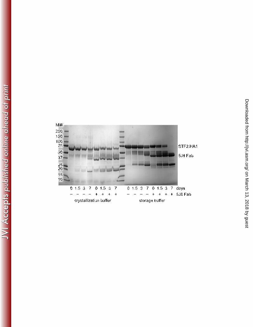
FIG 1 Human monoclonal antibody 5J8 recognizes the HA receptor binding site. (A) Crystal 571 structure of 5J8 Fab in complex with the bacterially expressed Cali07/2009-H1 HA1. The HA1 is 572 colored yellow, the Fab heavy chain in blue, the Fab light chain in light blue, and the HCDR3 in 573 red. (B) 5J8 inserts its HCDR3 into the HA receptor binding site and (C) overlaps with the 574 human α2,6 sialoglycan receptor (PDB ID code 3UBE). 575 576 FIG 2 Degradation of unliganded STF2.HA1 and 5J8 Fab-STF2.HA1 complex. Unliganded 577 STF2.HA1 (~77 kDa) and 5J8 Fab (~45 kDa) in complex with STF2.HA1 were incubated at 578 room temperature in the crystallization buffer or the storage buffer (150 mM NaCl, 20 mM Tris 579 pH 8). 10 µg of each reaction was quenched at each time point by the addition of non-reducing 580 SDS buffer and was boiled for ~2 minutes. Samples were analyzed by SDS-PAGE. Over time, a 581 band at ~25 kDa appears, which is consistent with the mass of HA1 in the 5J8 Fab-Cali07/2009-582 H1 complex crystal. 583 584 FIG 3 Structural relationships between the bacterially expressed Cali07/2009-H1 HA1 subunit to 585 other HAs. (A) Comparison of the structure of the antigenic sites between the bacterially 586 expressed HA1 (colored yellow) and the full-length baculovirus-expressed Cali04/2009-H1 HA 587 (colored green; PDB ID code 3LZG) with RMSD values for each site labeled. The Cb antigenic 588 site in our structure is disordered and is not fully built. The global fit RMSD between these HAs 589 is 0.64 Å. A previously solved bacterial HA1 (PDB ID code 3MLH) is shown in pink and has a 590 global fit RMSD of 0.6 Å to our structure. (B) Alignment of the HA1 subunits between 591 Cali07/2009-H1 and A/Hong Kong/1/1968 (H3N2) (PDB ID code 4FP8) colored yellow and 592 grey, respectively. The longer H3 HA1 construct contains additional secondary structural 593
on March 13, 2018 by guest
http://jvi.asm.org/
Dow
nloaded from

25
elements of the vestigial esterase domain that extend away from the RBS towards the stem 594 domain as well as two additional disulfide bridges, which are circled and depicted as green 595 sticks. The 5J8 Fab is colored in light and dark blue (heavy and light chains) and its HCDR3 in 596 red. 597 598 FIG 4 Interaction of 5J8 with Cali07/2009-H1 illustrating the neutralizing epitope. (A) Sequence 599 conservation of the 5J8 epitope across human H1 strains. HA residues contacted by 5J8 are 600 represented as yellow sticks. The percent conservation for the most common residue at each 601 position is shown, which is identical to the residues of Cali07/2009-H1. (B) HA is illustrated as a 602 surface in the same orientation as A with the HA contact residues on the surface colored by 603 sequence conservation according to the inserted scale. 5J8 contact residues are labeled and 604 shown as sticks with the heavy chain in dark blue and the light chain in light blue. 605 606 FIG 5 5J8 binds to the HA receptor binding site using receptor mimicry. (A) The carboxylate of 607 AspH100b overlaps with (B) the carboxylate of the α2,6 sialoglycan (PDB ID code 3UBE) and 608 uses identical hydrogen bonding interactions, which are shown as black dashed lines. 609 610 FIG 6 Electrostatic and hydrogen bonding interactions between 5J8 Fab and Cali07/2009-H1 611 HA1 with hydrogen bonds depicted by dashed lines. The HA is colored yellow, the Fab is 612 colored blue, and the HCDR3 is colored red. 613 614 FIG 7 Engineered trimer-stabilized Cali04/2009-H1 HA E54G used for the EM reconstructions. 615 (A) Overview of the HA structure with the HA2 Glu47Gly mutation circled in red. (B) Zoomed-616
on March 13, 2018 by guest
http://jvi.asm.org/
Dow
nloaded from

26
in view of the HA trimer interface. The wild-type HA is colored gray with Glu47 and the HA1 γ-617 turn residues (Leu30’ and Glu31’) from the neighboring HA protomer shown as sticks. The 618 trimer-stabilized chains are differentially colored. 619 620 FIG 8 EM reconstructions of 5J8 in complex with HA. Negative-stain EM was used to 621 determine volume maps of 5J8 Fab (red) in complex with the full-length HAs (blue) from the 622 A/Singapore/6/1986 (H1N1) and trimer-stabilized A/California/04/2009 (H1N1) strains. The 623 reconstructions show that the HA-antibody interactions described in the 5J8-Cali07/2009-H1 624 HA1 crystal structure (red and cyan) are recapitulated with the HA trimers. The HA from PDB 625 ID code 3LZG (blue) was used as a reference structure. Fourier shell correlation (FSC) curves 626 indicate that the resolution for the 5J8-A/Singapore/6/1986 map is 22 Å is and the 5J8-627 Cali04/2009-H1 map is 23 Å. 628 629 FIG 9 Comparison of 5J8 and CH65 contacts on HA. (A) Sequence alignment of the H1 strains 630 tested for binding by bio-layer interferometry. Residues contacted by 5J8 (blue), CH65 (red), or 631 both antibodies (violet) are mapped onto the alignment and (B) colored on the surface of 632 Cali07/2009-H1. The sialoglycan (PDB ID code 3UBE) is represented as sticks. The glyph “.” 633 indicates sequence identity with the Cali07/2009-H1 strain. 634
on March 13, 2018 by guest
http://jvi.asm.org/
Dow
nloaded from

HCDR3
SIA
GAL
A B
C
HA1“head”
5J8Fab
on March 13, 2018 by guest
http://jvi.asm.org/
Dow
nloaded from
MW250150100755037
25201510
days01.53 7 01.53 7 01.53 7 01.53 7– – – – – – – –+ + + + + + + +crystallization buffer storage buffer
5J8 Fab
5J8 Fab
STF2.HA1
on March 13, 2018 by guest
http://jvi.asm.org/
Dow
nloaded from

A BSa0.82 Å
Sb1.00 Å
Ca10.50 Å
Ca21.04 Å
Cb0.65* Å
H1 HA1 55-263H3 HA1 43-309
on March 13, 2018 by guest
http://jvi.asm.org/
Dow
nloaded from

Lys133a72%
Val13599%Thr136
74%
Ala13798%Gly143100%
Ala14469%
Lys14569%
Trp153100%
Ala18969%
Asp19091%
Gln19270%
Ser19369%Leu19496%
Lys222100%
Asp22590%
Gln22698%
AAsnH32
TyrH52
ArgH97
TyrH100ProH100a
AspH100b
AsnL27
ThrL30LysL31
ValL32 AspL51
AspL53
AspL66IleL93SerL94
AspL95a
B
Conservation (%)60 100
on March 13, 2018 by guest
http://jvi.asm.org/
Dow
nloaded from

A
Gln226
Ala137Thr136
AspH100b
ProH100a
HCDR3B
SIA
GAL
Gln226
Ala137Thr136
on March 13, 2018 by guest
http://jvi.asm.org/
Dow
nloaded from

Lys133a
AspL53
A
Lys145
AspL51
GlyL29
B
Asp190
ArgH97
C
Lys222
AspL95a
D
on March 13, 2018 by guest
http://jvi.asm.org/
Dow
nloaded from

Glu47
Leu30’
Glu31’
A B
on March 13, 2018 by guest
http://jvi.asm.org/
Dow
nloaded from

TABLE 1 X-ray data collection and refinement statistics
Data collection 5J8 Fab 5J8-Cali07/2009-H1 HA1 Cali04/2009-H1 HA2 E47G Beamline APS 23ID-D CLS 08ID-1 SSRL 12-2 Wavelength (Å) 1.0332 0.97949 0.97950 Space group P212121 P3121 P212121 Unit cell parameters a = 90.6, b = 100.0, c = 144.3 a = b = 67.5, c = 259.6 a = 71.4, b = 132.2, c = 201.8
(Å, º) α = β = γ = 90.0 α = β = 90.0, γ = 120.0 α = β = γ = 90.0 Resolution (Å) 50 – 1.55 (1.57 – 1.55)a 50 – 2.25 (2.32 – 2.25)a 50 – 2.20 (2.24 – 2.20)a Observations 1,340,148 219,878 321,115 Unique reflections 184,085 (6,687)a 33,082 (2,526) 95,786 (4,847)a Rmerge (%) b 4.6 (91.0)a 6.0 (75.5)a 13.5 (71.1)a Rpim (%) b 1.8 (41.2)a 2.4 (37.6)a 8.0 (42.0)a I/sigma 21.9 (1.8)a 14.9 (1.9)a 11.2 (1.5)a Completeness (%) 96.9 (72.5)a 97.7 (83.1)a 96.3 (98.9)a Multiplicity 7.3 (5.6)a 6.6 (4.4)a 3.4 (3.2)a Refinement statistics Resolution (Å) 49.2 – 1.55 48.5 – 2.25 48.5 – 2.20 Reflections (total) 183,666 32,999 95,720 Reflections (test) 9,196 1,667 4,796 Rcryst (%)c 17.2 19.1 17.9 Rfree (%)d 18.7 23.8 23.2 Protein atoms 6,607 4,928 11,752 Carbohydrate atoms – – 200 Waters 866 90 940 Other 97 0 0 Average B-value (Å2) Overall 27.9 64.1 36.1 HA – 74.1 35.3 Fab 26.6 59.2 – Wilson 20.1 54.2 41.7 RMSD from ideal geometry Bond length (Å) 0.009 0.010 0.008 Bond angles (°) 1.26 1.55 1.13 Ramachandran statistics (%)e Favored 97.8 96.0 97.2 Outliers 0 0.3 0.1 PDB ID 4M5Y 4M5Z 4M4Y
aNumbers in parenthesis refer to the highest resolution shell. bRmerge = Σhkl Σi | Ihkl,i - <Ihkl> | / Σhkl Σi Ihkl,I and Rpim = hkl[1/(N-1)]1/2i |Ihkl,i - <Ihkl>| /hkli Ihkl,i, where Ihkl,i is the scaled intensity of the ith measurement of reflection h, k, l, < Ihkl> is the average intensity for that reflection, and N is the redundancy. cRcryst = Σ | Fo - Fc | / Σ | Fo | x 100, where Fo and Fc are the observed and calculated structure factors, respectively. dRfree was calculated as for Rcryst, but on a random test set comprising 5% of the data excluded from refinement. eCalculated using MolProbity (32).
on March 13, 2018 by guest
http://jvi.asm.org/
Dow
nloaded from

TABLE 2 Binding of influenza HA strains by 5J8 and CH65 Fab and IgG
H1N1 strain 5J8 Fab 5J8 IgG CH65 Fab CH65 IgG A/South Carolina/1/1918 ++ ++++ – –
A/WSN/1933 – – – – A/Puerto Rico/8/1934 – – – – A/AA/Marton/1943 – ++ – +
A/duck/Alberta/345/1976 – – – – A/USSR/90/1977 + ++++ – ++
A/Singapore/6/1986 +++ ++++ +++ ++++ A/Texas/36/1991 ++ ++++ ++++ ++++
A/Beijing/262/1995 – + ++++ ++++ A/New Caledonia/20/1999 – ++ ++ ++++ A/Solomon Islands/3/2006 ++ +++ +++ ++++
A/California/04/2009 +++ ++++ – – Dissociation constants: – no detectable binding > 5 µM + Kd = 500–5,000 nM ++ Kd = 50–500 nM +++ Kd = 5–50 nM ++++ Kd < 5 nM
on March 13, 2018 by guest
http://jvi.asm.org/
Dow
nloaded from