Embed Size (px)
Citation preview

Silva et al.: Annona squamosa L. (Annonaceae): Chemical bioprospection and biological activity in two phenological stages
- 133 -
APPLIED ECOLOGY AND ENVIRONMENTAL RESEARCH 14(4): 133-147.
http://www.aloki.hu ● ISSN 1589 1623 (Print) ● ISSN 1785 0037 (Online)
DOI: http://dx.doi.org/10.15666/aeer/1404_133147
2016, ALÖKI Kft., Budapest, Hungary
ANNONA SQUAMOSA L. (ANNONACEAE): CHEMICAL
BIOPROSPECTION AND BIOLOGICAL ACTIVITY IN TWO
PHENOLOGICAL STAGES
SILVA, I. T. S. S.1 ‒ FERNANDES, M. J. B.
2 ‒ OLIVEIRA, R. A.
3 ‒ CARVALHO, L. D.
1 ‒
CORTEZ, P. A.1 ‒ SÃO JOSÉ, A. R.
4 ‒ CONCEIÇÃO, A. O.*
1
1Department of Biological Science, Universidade Estadual de Santa Cruz, Ilhéus, Bahia, Brazil
(phone:+1 55 73 3680-5105; fax: +1 55 73 3680-5230)
2Center of Research and Development of Animal Health, Instituto Biológico, São Paulo, São
Paulo, Brazil (phone: +1 55 11 fax: +1 55 11 5087-1791)
3Department of Mathematical Sciences and Technology, Universidade Estadual de Santa Cruz,
Ilhéus, Bahia, Brazil (phone:+1 55 73 3680-5620; fax: +1 55 73 3680-5230)
4Department of Animal and Plant Science, Universidade Estadual do Sudoeste da Bahia,
Vitória da Conquista, Bahia, Brazil (phone:+1 55 77 34237548; fax: +1 55 077 3423-7038)
*Corresponding author
e-mail: [email protected]; phone:+1 55 73 36805105; fax: +1 55 73 3680-5230
(Received 21st Apr 2016; accepted 16
th Jun 2016)
Abstract. Annona squamosa L. has been recognized by having compounds with important in vitro
biological activity against human disease or as insecticide. However, knowledge of the antiviral potential
and micro molecules production of A. squamosa L. at different phenological stages is scarce. The secondary metabolites detection and biological activity from seeds and leaves in 4 and 14 years old trees
from semi arid region were performed. Phytochemical identification was obtained by chemical
bioprospection and histochemical localization in leaves and seeds’. Seeds acids content was analyzed by
gas chromatography. Cytotoxic effect and antiviral activities against equine and suid herpesvirus were
also performed. Results showed differences in the amount of flavonoids, carotenoids, and phenolic
compounds between leaves and seeds and by stage of production. Carotenoids and phenolic compounds,
especially tannins, were higher in older trees’ leaves. Fat acids detected in seeds were oleic, linoleic
arachidic, Cis-11-eicosenoic, elaidic and palmitoidic acids. Cytotoxicity was higher in hydrophobic than
hydroalcoolic extracts. Broad spectrum antiviral activity was more marked for younger trees’ aqueous
extract. The high chemical adaptability of A. squamosa to semi arid environment was evident and the
identification of A. squamosa secondary metabolites regarding the plant senescence may guide a better utilization of plant organs in order to obtain substances of pharmacological interest.
Keywords: natural products, secondary metabolites, apoptosis, antiherpetic
Introduction
Annona squamosa L. (Annonaceae Juss.), popularly known as custard apple, fruta-
do-conde, ata or pinha, is recognized as one of the most important traditional edible
medicine tree as well as by its economical sustainability (São José et al., 2014; Maas et
al., 2014). The medicinal usage of A. squamosa includes the treatment of
hypertireoidism, cancer, and heart and infectious diseases. The tea from root is used as
purgative and the powder of unripe fruit is used to treat worms and protozoa (Di stasi
and Hiruma-Lima, 2003). The leaves’ infusion is also used to treat ulcer, wounds and
swoon, and the seeds are recognized by their pesticide effects (Gajalakshmi et al., 2011;
Luna et al., 2015). Economically, A. squamosa crop has been characterized mainly by

Silva et al.: Annona squamosa L. (Annonaceae): Chemical bioprospection and biological activity in two phenological stages
- 134 -
APPLIED ECOLOGY AND ENVIRONMENTAL RESEARCH 14(4): 133-147.
http://www.aloki.hu ● ISSN 1589 1623 (Print) ● ISSN 1785 0037 (Online)
DOI: http://dx.doi.org/10.15666/aeer/1404_133147
2016, ALÖKI Kft., Budapest, Hungary
small farmers using the family labor and presents increasing important socio-economic
values considering international market because of its status as exotic fruit showing
excellent qualities (São José et al., 2014).
Review of Literature
Several secondary metabolites have been identified from Annonaceae species,
including phenolic acids, tannins, flavonoids, benzenics compounds, lipids, proteins,
lactones, vitamins, carotenes and saponins (Leboeuf et al., 1980). From A. squamosa
leaves, two main chemical classes were described: steroids and terpenoids
(Savithramma et al., 2011). Particularly, acetogenins and benzylisoquinoline alkaloids
isolated from this family have received special attention due to their variety and
distribution in Annonaceae representatives in distinct stages of development, including
A. squamosa (Yogesh, 1994; Araya et al., 2002; De-La-Cruz et al., 2013).
In vitro studies have validated the medicinal potential of Annona secondary
metabolites being Leishmanicidal (Vila-Nova et al., 2011), antioxidant (Panda and Kar,
2007; Mariod et al., 2012), antimicrobial (Padmaja et al., 1995; Rahma et al., 2005), and
antitumoral (Tormo et al., 2001; Yang et al., 2009; Chen et al., 2012) activities most
described in the literature. Also, hyperglycemiant properties of fat acids from seeds
(Sultana, 2008), anti-HIV activity of diterpenes from A. squamosa (Wu et al., 1996),
and anti-herpetic activity of ethanolic (Padmaja et al., 1995) and methanolic (Betancur-
Galvis et al., 1999) extracts from A. muricata were reported. More specifically,
acetogenins isolated from Annona seeds demonstrated cytotoxicity (Vila-Nova et al.,
2011) and immunosuppression related to antitumoral activity (Tormo et al., 2001; Araya
et al., 2002; Yang et al., 2009; Chen et al., 2012).
The relation between biological potential of plants and the plant-environment
interface is another point of interest in the A. squamosa studies. Seasonality, circadian
cycle, ultraviolet radiation, and temperature, can be considered the main environmental
factors involved in reducing and increasing the production rate of secondary metabolites
(Taiz and Zeiger, 2008) since A. squamosa seems to show a greater control of
transpiration through stomata closure in water deficit situation (Endres, 2007).
Although biological properties are evident, there is a lack of information about
phytochemical prospection description, histochemical localization, and antiviral
potential elucidation of A. squamosa tree which could guide the search for biologically
active molecules. Consequently, in this study, two stage of cultivation of A. squamosa -
initial production stage (4 years) and peak production stage (14 years) - were chosen to
better understand the variability on secondary metabolites of A. squamosa in semi arid
environment and its influence on antiviral activity against suid (SHV-1) and equid
(EHV-1) alphaherpesviruses.
Materials and Methods
Botanical field collection
The A. squamosa L., Annonaceae, mature leaves and fruits from both 4 and 14 years
of cultivation where collected from a commercial rural property located in the
municipality of Anage, Bahia state, Brazil (14°26.159' S; 41°05.216' WO), where
cultivation is monitored from seed germination. The environment where the plants
Silva et al.: Annona squamosa L. (Annonaceae): Chemical bioprospection and biological activity in two phenological stages
- 135 -
APPLIED ECOLOGY AND ENVIRONMENTAL RESEARCH 14(4): 133-147.
http://www.aloki.hu ● ISSN 1589 1623 (Print) ● ISSN 1785 0037 (Online)
DOI: http://dx.doi.org/10.15666/aeer/1404_133147
2016, ALÖKI Kft., Budapest, Hungary
grown is defined as caatinga area of Brazilian Northeast, characterized by a xeric
shrubland (less dry) and thorny trees with an annual precipitation varying from 400 to
600 mm. Plant material was identified and deposited at herbarium of Universidade
Estadual Santa Cruz (HUESC) under register number HUESC#18501
(http://inct.splink.org.br/) and identified by Prof. Luiz Alberto da Costa Matos. A.
squamosa is not native from Brazil (Datiles and Acevedo-Rodriguez, 2016) and it is not
considered an endangered or a protected plant once it is largely cultivated around the
world.
Extract production
For extract production, leaves were submitted to dryness at 50ºC under forced
ventilation and reduced a powder with mechanical knifes. Seeds from several fruits
were cleaned and dried at room temperature. Seeds (S) and leaves (L) powder were then
submitted to aqueous (Aq), hexanic (Hex) and ethanolic (Et) extraction. The aqueous
extract of leaves was done by infusion at 10% for 1 hour, filtered and lyophilized. The
ethanolic extracts from seeds and leaves were obtained by exhaustive extraction, using
10 g of powder plant material in 100 mL of ethanol 99% (Biotec, Brazil). The solvent
was evaporated under reduced pressure and the extract recovered. The hexanic extract
was obtained by Soxhlet method following the International Union of Pure ad Applied
Chemistry (IUPAC) procedure with hexane for 6 hours. The solvent was evaporated
under reduced pressure and the extract recovered. The ethanolic extracts were stored at
4ºC in the dark while hexanic and lyophilized extracts were kept at room temperature in
the dark.
For biological test, immediately before use, hexanic and ethanolic extracts were
dissolved with 0.8 mL of dimethyl sulfoxide (DMSO, Sigma-Aldrich, Brazil) and
Minimal Essential Medium (MEM - Vitrocell/Embriolife®, Atená-SP) to obtain an 8
mg.mL-1
stock solution. Work solutions varied from 0.03 to 4 mg.mL-1
. Aqueous
extracts were dissolved directly in MEM at the same stock and work solutions.
Phytochemical prospection
To perform phytochemical prospection, the NI 1600 UV-VIS spectophotometer
(Novainstruments®, São Paulo, Brazil) was used and all solutions were high purity
quality.
Carotenoid
The carotenoids determination was performed according to the technique and
equation described by Kimura and Rodriguez (2003). For that, to 0.3 g of plant powder
material, 50 ml of acetone was added. After 30 minutes the solution was vacuum
filtered and 1.5 mL of clarifying solution [1,5 mL de Ba(OH)2 (0.3 M) e 1.5 mL de
ZnSO4(5%)] was added and after 30 minutes, all solution was vacuum filtered again.
Next, 30 mL of petroleum ether was added to the mixture directly in the filter funnel.
The ether phase was harvested and exhaustively washed. The ether extract was then
transferred to a volumetric flask and the 50 mL final volume was completed with
petroleum ether. The ether extract was transferred to a 50 mL volumetric flask by
adjusting the volume of solutions with petroleum ether. A 450 nm wave length was used
to read samples.

Silva et al.: Annona squamosa L. (Annonaceae): Chemical bioprospection and biological activity in two phenological stages
- 136 -
APPLIED ECOLOGY AND ENVIRONMENTAL RESEARCH 14(4): 133-147.
http://www.aloki.hu ● ISSN 1589 1623 (Print) ● ISSN 1785 0037 (Online)
DOI: http://dx.doi.org/10.15666/aeer/1404_133147
2016, ALÖKI Kft., Budapest, Hungary
Total phenolic compounds
To determine phenolic compounds, the Folin-Ciocauteau reagent (RFC) was used
following the method described by Wettasingue and Shahidi (1999) and Brito et al.
(2013). To a final volume of 10 mL, 0.5 ml of RFC, 0.5 mL of the plant extract and 1
mL of saturated NaHCO3 solution were added to water. After 25 minutes, the analysis
at 773 nm wavelength was performed. Gallic acid (GA, 1 mg.mL-1
stock solution) was
used to prepare an analytical curve varying from 5 to 217.5 μg.mL-1
. The analytical
curve were made in triplicate and presented the equation y=0.0034x + 0.0194 with
R2=0.990 being detection (DL) and quantification (QL) limit values of 1.973 μg.mL-1
and 6.576 μg.mL-1
, respectively. The total phenolic compounds were expressed in mg of
GA equivalents per 100 g of dried plant.
Flavonoid
The quantification of flavonoid followed Sobrinho et al. (2008) procedures. In a 25
mL volumetric flask 1.0 mL of methanolic solution was added to the sample, followed
by 0.6 mL glacial acetic acid (concentrated), 1 mL of methanolic piridin (20% v/v) and
2.5 mL methanolic AlCl3 (50 mg. L-1
), reaching a final volume with destiled water.
After 30 minutes at room temperature, samples were detected by espectophotometry at
420 nm wave lenght. Rutine (RU, 0.5 mg.mL-1
stock solution) was used to prepare an
analytical curve varing from 5 to 30 μg.mL-1
. The analytical curve were made in
triplicate and presented the equation y=0.0075x + 0.1, R20.998 being detection (DL)
and quantification (QL) limit values of 0.4799 μg.mL-1
and 1.599 μg.mL-1
, respectively.
The flavonoids content was expressed in mg of RU equivalents per 100 g of dried plant.
Tannin
The tannins content was determined following Tiitto-Julkunem (1985) procedures.
For that, 0.5 g of sample was soaked in 30 mL of 80% aqueous acetone (80:20, PA
acetone, water, v.v-1) under continuous stirring at room temperature (24 ° C ± 2 ° C) for
20 minutes. The mixture was filtered and another extraction was done. The procedure
was repeated twice. Then, extractions were combined and 3 mL of a methanol solution
of vanillin (4% MV-1) was added to the 50 mL final volume adjusted with water. Then,
in a tube covered with aluminum foil, 0.5 mL of the extract was put together with 3 mL
of vaniline methanolic solution (4% w/v) and 1.5 mL of concentrated HCl. The mixture
was stirred vigorously, and read at 500 nm wave length. Catechin (25 mg.mL-1
stock
solution) was used to prepare an analytical curve varying from 5 to 125 μg.mL-1
. The
analytical curve were made in triplicate and presented the equation y = 0.0024x +
0.2173, R2= 0.9990 being detection (DL) and quantification (QL) limit values of
0.62500 μg.mL-1 and 2.0833 μg.mL-1
, respectively. The tannins content was expressed
as catechin equivalents per 100 g of dried plant.
Histochemical localization
Leaves and seeds of 4 and 14 years of cultivation of A. squamosa L. were selected
for in situ localization of their main chemical compounds. Some fresh samples were cut
by hand using a razor blade. Additionally, part of the samples were fixed in 2.5%
glutaraldeyde, 4.0% formaldehyde and 0.2 M cacodylate buffer solution, pH 7.2
(modified from Karnovsky, 1965), embedded using the HistoResin Embedding Kit
Silva et al.: Annona squamosa L. (Annonaceae): Chemical bioprospection and biological activity in two phenological stages
- 137 -
APPLIED ECOLOGY AND ENVIRONMENTAL RESEARCH 14(4): 133-147.
http://www.aloki.hu ● ISSN 1589 1623 (Print) ● ISSN 1785 0037 (Online)
DOI: http://dx.doi.org/10.15666/aeer/1404_133147
2016, ALÖKI Kft., Budapest, Hungary
(Leica, Heidelberg, Germany) and sectioned using a RM 2145 rotary microtome (Leica,
Heidelberg, Germany) with glass knife. All the sections obtained were mounted onto
glass slides and submitted to specific tests, including their respective controls. All the
analyses were made using a DM 2500 light microscope (Leica, Heidelberg, Germany)
equipped with a DFC 295 digital camera (Leica, Heidelberg, Germany). Also, scanning
electron microscopic (Quanta 250 (FEI Company) was used to verify
micromorphological aspects of the the epidermal cells.
Gas chromatography analysis from hexanic extract
Ester methyl production
Three mL of hexane and 4 mL of NaOH (0.5 N) methanolic solution were added to a
50 mg of oil. The mixture was heated at 65-70oC for 3 to 5 minutes until total
solubilization. After cooling down, 5 mL of transestherifing solution [NH4Cl (10 g) in
methanol (300 mL) + H2SO4 concentrated (15 mL)] and again heated at 65-70oC. Five
minutes later, the mixture was cooled down and transferred to a separation funnel,
where 4 mL of NaCl saturated solution was added. The hexane phase was selected,
stored under refrigeration and analyzed by GC.
CG analysis
The methyl ester were analyzed by gas chromatography using Varian Saturno 3800
equipped with flame ionized detector (FID) and melted silica capillary column (30 m x
0.25 mm) with Carbowax (0.25 µm film thin) stationary phase, having helium as drag
gas, flow of 1.0 mL/minute. The injector and detector temperatures were 220°C e
240°C, respectively. The column temperature began at 60°C and it was increased from
5°C to 200°C, maintaining the latest for 5 minutes more. One µL of hexane phase (split
1:10) was injected. A fat acids certified standards mixture varying from 8 to 24 carbons
(Sigma-Aldrich - USA) was used to identify the sample fat acids content by
comparison. The fat acid methyl esters’ concentrations were obtained through integral
area of picks related to all sample components total area, normalized method.
Biological assay
Vero cell lines, (Vero, ATCC-CL 81) kindly provided by Instituto Butantan (São
Paulo, Brazil), were used to perform cytotoxic and antiviral assays. Cells were
cultivated in MEM supplemented with 8% of fetal bovine serum (FBS,
Vitrocell/Embriolife®, Atená) with 5% CO2 atmosphere at 37°C. For all tests, cells
were seeded in a 96 well plate at 3.0x10⁴ cell/well density. After 24 h seeding, cells
were treated with different concentrations of extracts (0.03 mg.mL-1 a 4 mg.mL-1) and
incubated at 5% CO2 atmosphere and 37°C for 48-72 h.
To verify citotoxicity of hexanic, ethanolic, and aqueous extracts, the 3-(4, 5-di-
metilazol-2-il)-2,5-difeniltetrazolium bromide (MTT, Sigma-Aldrich, Brazil)
mitochondrial reduction assay was used (Valadares et al., 2007). Also, cells were stained
with May-Grünwald-Giemsa to analyze cell morphological changes.
The cytotoxic concentration to 50% of cell culture (CC50) was calculated using
GraphPad Prism version 7.00 for Windows, GraphPad Software, San Diego California
USA (free trial version) and the maximum non cytotoxic concentration (MNCC) for this
study was considered the concentration 50% below the CC50.

Silva et al.: Annona squamosa L. (Annonaceae): Chemical bioprospection and biological activity in two phenological stages
- 138 -
APPLIED ECOLOGY AND ENVIRONMENTAL RESEARCH 14(4): 133-147.
http://www.aloki.hu ● ISSN 1589 1623 (Print) ● ISSN 1785 0037 (Online)
DOI: http://dx.doi.org/10.15666/aeer/1404_133147
2016, ALÖKI Kft., Budapest, Hungary
For cytotoxic experiments, controls consisted of cells treated with MEM only,
DMSO at 0.5% and DMSO at 1.25%.
The antiviral activity assay was performed using suid alphaherpesvirus type 1
(SuHV-1; EMBRAPA: BRMSA 3, 00588 strain) and equid alphaherpesvirus type 1
(EHV-1; 4/72 strain). Due to high cytoxicity of some extracts, only aqueous extracts
from seeds and leaves and hexanic extract from seeds were tested for antiviral activity.
The methodology was adapted from Kaziyama et al. (2012). After 24 h seeding and
75% confluence of Vero cells, supernatants were removed and cells were treated with
MNCC extracts and MNCC extracts with virus. Controls with only virus or cells were
also used. Cells were incubated at 37°C and 5% de CO2 atmosphere for three days.
Cytopathic effects (cell tumefaction and death) were observed every day and tissue
culture infectious dose to 50% of the cell culture (TCID50) was determined. The viral
inhibition index (VII) was obtained through Reed and Muench (1938) method giving
the proportional difference between viral titer from extract treated cells and not treated
cells. Following criteria established by Simoni et al. (2007), the extract was antiviral
eligible when VII was ≥ 1.5 corresponding to percentage of inhibition (PI) of 97%.
Statistical analysis
Student’s t test and one-way ANOVA, followed by Tukey’s test were done.
Statistical analyses were performed with the Prism software (version 7.00; GraphPad
Software, 2007, San Diego, California, USA). All experiments samples were made in
replicates and repeated at least three times.
Results
Phytochemical prospection
Phytochemical difference was seen between plant organ and tree age being
carotenoides and flavonoids found in significatively higher amounts in leaves from
older trees (Table 1). Also, phenolic compounds were found only in leaves of 14 years
individuals with more then a half of components constituted by condensed tanins.
Table 1. A. squamosa L. leaf and seed chemical bioprospection according to two plant tree age. Anagé, Bahia State, Brazil, 2015.
Leaves Seed
Tree age (years) 4 14 4 14
Carotenoids
(mg.100g-1
)
0.11b ** 0.29a ** 0.011B ** 0.014A **
Flavonoids
(mg.100g-1
)
240.37b * 488.25a* 21.06A * 12.13A *
Total phenols (mg.g
–1GAE)
ND 562.25 ND ND
Condensed tannins
(mg.100g-1
)
ND 343.98 ND ND
ND – not detected. *p<0.1 **p<0.001

Silva et al.: Annona squamosa L. (Annonaceae): Chemical bioprospection and biological activity in two phenological stages
- 139 -
APPLIED ECOLOGY AND ENVIRONMENTAL RESEARCH 14(4): 133-147.
http://www.aloki.hu ● ISSN 1589 1623 (Print) ● ISSN 1785 0037 (Online)
DOI: http://dx.doi.org/10.15666/aeer/1404_133147
2016, ALÖKI Kft., Budapest, Hungary
Histochemical localization
Through histochemical technique plant metabolites such as alkaloids, lipids, phenolic
compounds, terpenoids, and starch were located in leaves and seeds from both 4 and 14
years trees (Table 2). The differences observed were more at plant organ level then at
phenological stage, highlighting the presence of acid lipid and starch only in seeds and
phenolic compounds in leaves.
Table 2. Histochemical analysis of Annona squamosa seeds and leaves regarding two plant
tree ages.
Leaves Seeds
4 years 14 years 4 years 14 years
Alkaloid + + + +
Total lipid (Sudan) + + + +
Acid lipid - - + +
Structural phenolic compounds + + - -
Total phenolic compounds + + - -
Terpenoids (essential oil) + + + +
Terpenoids (resinified acid) + + + +
Starch - - + +
Presence of compound (+); Compound not detected (-)
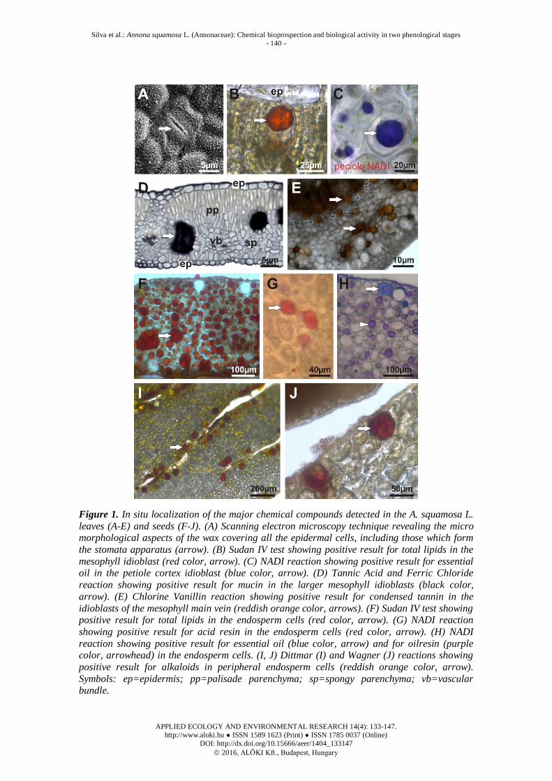
In situ localization associated to scanning electron microscopy technique revealed
the micromorphological aspects of the wax covering all the epidermal cells, including
those which form the stomata apparatus (Fig. 1A). In leaves, the results showed the
presence of total lipids in the mesophyll idioblast (Fig. 1B) and essential oil in the
petiole cortex idioblast (Fig. 1C) (Fig. 1D). Tannic Acid and Ferric Chloride reaction
revelead the presece of mucin in the larger mesophyll idioblasts as well as condensed
tannin in the idioblasts of the mesophyll main vein (Fig. 1E). In seeds, total lipids (Fig.
1F), acid resin (Fig. 1G), essential oil and oilresin (Fig. 1H) were detected in the
endosperm cells. In this same plant organ, alkaloids were detected only in some
peripheral endosperm cells (Fig. 1I-J).
Silva et al.: Annona squamosa L. (Annonaceae): Chemical bioprospection and biological activity in two phenological stages
- 140 -
APPLIED ECOLOGY AND ENVIRONMENTAL RESEARCH 14(4): 133-147.
http://www.aloki.hu ● ISSN 1589 1623 (Print) ● ISSN 1785 0037 (Online)
DOI: http://dx.doi.org/10.15666/aeer/1404_133147
2016, ALÖKI Kft., Budapest, Hungary
Figure 1. In situ localization of the major chemical compounds detected in the A. squamosa L.
leaves (A-E) and seeds (F-J). (A) Scanning electron microscopy technique revealing the micro morphological aspects of the wax covering all the epidermal cells, including those which form
the stomata apparatus (arrow). (B) Sudan IV test showing positive result for total lipids in the
mesophyll idioblast (red color, arrow). (C) NADI reaction showing positive result for essential oil in the petiole cortex idioblast (blue color, arrow). (D) Tannic Acid and Ferric Chloride
reaction showing positive result for mucin in the larger mesophyll idioblasts (black color,
arrow). (E) Chlorine Vanillin reaction showing positive result for condensed tannin in the
idioblasts of the mesophyll main vein (reddish orange color, arrows). (F) Sudan IV test showing positive result for total lipids in the endosperm cells (red color, arrow). (G) NADI reaction
showing positive result for acid resin in the endosperm cells (red color, arrow). (H) NADI
reaction showing positive result for essential oil (blue color, arrow) and for oilresin (purple color, arrowhead) in the endosperm cells. (I, J) Dittmar (I) and Wagner (J) reactions showing
positive result for alkaloids in peripheral endosperm cells (reddish orange color, arrow).
Symbols: ep=epidermis; pp=palisade parenchyma; sp=spongy parenchyma; vb=vascular bundle.

Silva et al.: Annona squamosa L. (Annonaceae): Chemical bioprospection and biological activity in two phenological stages
- 141 -
APPLIED ECOLOGY AND ENVIRONMENTAL RESEARCH 14(4): 133-147.
http://www.aloki.hu ● ISSN 1589 1623 (Print) ● ISSN 1785 0037 (Online)
DOI: http://dx.doi.org/10.15666/aeer/1404_133147
2016, ALÖKI Kft., Budapest, Hungary
Gas chromatography
To elucidate lipid compounds detected in seeds by qualitative analysis, gas
chromatography was performed from hexanic extraction (Table 3). The majority of fat
acids analyzed had similar amount considering the two tree ages studied. However,
differences were found between tree age in relation to arachidic acid and Cis-11-
eicosenoic acid contents, being this compound found only in older trees seed.
Table 3. A.squamosa fat acids from seed hexanic extraction by gas chromatography
analysis according to two plant tree age. Anage, Bahia State, Brazil, 2015.
A. squamosa tree age 4 years 14 years
Methyl esters standards RT1
RT2 Content
(%)
RT2 Content
(%)
Palmitoleic acid (C16: 1n9-c) 22,080 22,101 6.20 22,141 7.50
Elaidic acid (C18: 1n9 t) 25,606 25,627 16.04 25,695 11.55
Oleic acid (C18: 1n9C) 25,941 25,956 52.72 26,080 49.50 Linoleic acid (C18: 2n6-c) 26,651 26,676 21.78 26,760 25.80
Arachidic acid (C20: 0) 27,655 ND ND 27,698 0.98
Cis-11-eicosenoic acid (C20: 1) 28,857 ND ND 28,905 1.37
Biological assay
The biological assay results indicated the more functional plant organ and
phenological stage regarding antiviral activity potential. Significant differences between
phenological stages was not seen, however compounds extraction had a role in the
toxicity. In relation to antiviral activity, a difference in virus sensibility was seen. In
general, SuHV-1 was more sensitive than EHV-1, including an unusual elevation on
virus titer observed. Exception was seen for 14 years tree aqueous extract from seeds
which was more active against EHV-1. Regarding compounds selected, aqueous
extraction from leaves showed the best effect. Finally, with exception of the seeds
aqueous extract for EHV-1, the tree age did not play a significant role in antiviral
activity when comparing same extraction solvent (Table 4).
Table 4. Cytotoxic concentration (CC50), Viral inhibition index (VII) and percentage of
inhibition (PI) of A. squamosa extracts, obtained from two plant tree age against SuHV-1
and EHV-1.
Plant Extracts Tree age CC50 SuHV-1 EHV-1 [mg.mL
-1] IIV PI (%) IIV PI (%)
LAq 4 0.32 0.55 71.60 0.60 74.88
14 0.42 0.50 68.38 0.59 68.86
SAq 4 1.43 -0.16 -46.78 0.00 0.00 14 1.51 0.17 32.91 0.50 53.58
SHex 4 0.49 0.50 68.38 0.13 25.87
14 0.40 0.25 44.02 0.39 37.77
CC50 - 50% cytotoxic concentration for cell culture. LAq – leaves aqueous extract; SHex –
seeds hexane extract; SAq – seeds aqueous extract.

Silva et al.: Annona squamosa L. (Annonaceae): Chemical bioprospection and biological activity in two phenological stages
- 142 -
APPLIED ECOLOGY AND ENVIRONMENTAL RESEARCH 14(4): 133-147.
http://www.aloki.hu ● ISSN 1589 1623 (Print) ● ISSN 1785 0037 (Online)
DOI: http://dx.doi.org/10.15666/aeer/1404_133147
2016, ALÖKI Kft., Budapest, Hungary
Discussion
The genus Annona has been studied by its biological potential in health and
agriculture. In this study some secondary metabolites and biological activity of seed and
leaves extracts obtained from A. squamosa are reported. Differences in cultivation stage
and plant organ were seen. Through bioprospecting, mainly secondary metabolites such
as carotenoids, phenolic compounds including flavonoids and condensed tannins were
detected and quantified in leaves and seeds (Table 1). The histochemical localization
analyses allowed identifying plant organ and the cell types in which the major chemical
compounds were produced and stored (Table 2, Fig. 1). Palmitoleic, elaidic, oleic,
linoleic, arachidic, and cis-11-eicosenoic acids were also identified and quantified in
seeds hexanic extract by gas chromatography (Table 3).
Carotenoids content was significantly higher (p <0.01) in leaves and seeds of 14 years
trees when compared to 4 years ones. Carotenoid levels usually remain constant in leaves
until early senescence (Uenojo et al., 2007), however an increase during ripening can be
seen, at which there is an intensification of the synthesis of this compound (John et al.,
1970). Here, the higher content found is in accordance with the stage of production of
older trees at the moment of collection. Moreover, the high content of carotenoids may be
connected to photo protection process against oxidative damage in function of the
adaptability to climate (Taiz and Zeiger, 2008; Mertz et al., 2009). It is worth to note that
there is a tendency to recover this metabolite from the leaves instead of seeds, since the
amount of this metabolite was very close to lower detection limit by the method used.
Naturally, regarding to photo protection, seeds are protected by fruit tissues.
The presence of phenolic compounds in A. squamosa leaves has been already
reported (Mariod et al., 2012), however differences between trees of different stage of
production is one notable point of our study. Flavonoids were significantly higher
(p<0.5) in leaves of 14 years individuals when compared with plants in the beginning of
fruit production (4 years). Total phenolic compounds were observed only in leaves of
14 years production (562.25 mg GAE.g–1
), and it is worth to note that from total
phenolic compounds, 343.98 mg.100g-1
were condensed tannins. The sites of tannin
production were the large cells located in the leaf mesophyll, where its protective
feature became evident against the UV radiation damage (Wang et al., 2014) and
herbivory (Lokvam and Kursar, 2005). Interestingly, phenolic substances such as
flavonoids and tannins have allelopathic function contributing to the establishment of
seedlings photo protection and plant adaptability along their development (Waterman
and Mole, 1994; Giner-Chaves, 1996; Rawat et al., 1998; Hassanpour et al., 2011). In
this study, the microclimate adaptability, more specifically the high incidence of
sunshine in the semiarid area, lead us to think that the amount found in older trees is due
to protection against oxidative stress or as accessory pigments on photosynthesis
(Hassanpour et al., 2011).
The fat acids from A. squamosa seeds deserve attention for their quantitative and
qualitative variation, being considered the seeds as the main lipid storage organ (Corner,
1949). The fat acids in the seeds where histologically detected in almost every
endosperm cells, which are the main storage tissue of the Annonaceae species (Kessler,
1993). In this context, differences in fat acids content have been reported in relation to
seeds stage of development representing the potential of the plant to plasticity (Endres,
2007; Chacón et al., 2013). In this study, an expressive amount of polyunsaturated fat
acids (PUFAs) such as oleic and linoleic acids were detected in seeds from both tree age
of production. However, arachidic and Cis-11-eicosenoic acids were detected only in

Silva et al.: Annona squamosa L. (Annonaceae): Chemical bioprospection and biological activity in two phenological stages
- 143 -
APPLIED ECOLOGY AND ENVIRONMENTAL RESEARCH 14(4): 133-147.
http://www.aloki.hu ● ISSN 1589 1623 (Print) ● ISSN 1785 0037 (Online)
DOI: http://dx.doi.org/10.15666/aeer/1404_133147
2016, ALÖKI Kft., Budapest, Hungary
seeds of later production stage (14 years) and higher amount of elaidic and palmitoidic
acids were detected in seeds of initial production stage (4 years) trees. Although less
expressive, the fat acids from leaf where derived from the epidermal wax, a layer that is
related to the dry protection among other functions (Samuels et al., 2008), and also from
some mesophyll and petiole cells, reinforcing the great importance of this class of
compounds to plant adaptation.
As plant metabolism reflects its plasticity, the variety of secondary metabolites
originated from this adaptation gives to the vegetal a great potential for biological
proposals. In this study, cytotoxicity and antiviral tests were biological properties
explored for A. squamosa biological potential. The cytotoxic morphological findings
confirmed the potential of A. squamosa compounds to cause apoptotic effects on
tumoral cells (Pardhasaradhi et al., 2005). Cells treated with hexanic and aqueous
extracts showed apoptosis signs characterized by reduction of cell volume, nuclear
fragmentation, and the presence of apoptotic bodies (Table 4 and Fig. 2). It was also
noted that aqueous extract from leaves were more toxic than aqueous extract from seeds
(Tables 4 and 5) which is in agreement with their tannins content. Although tannins
have biological properties, they can be very harmful to mammalian cells and removing
them from extract can help other phenolic compounds to positively act on infection
agents and in the mammalian systems as reported elsewhere (Schmitt et al., 2001;
Hassanpour et al., 2011; George et al., 2012; Zhao and Hu, 2013). Furthermore, the high
cytotoxicity of hexane extract from seeds and ethanolic extracts from seeds and leaves
was verified in our study.
The antiviral activity of Annona extracts is an additional biological activity not yet
described in the literature. However these data confirmed the antiviral potential of the
Annona species, following the criteria established by Simoni et al. (2007), the antiviral
activity of A. squamosa extracts was considered weak. Both viruses tested seemed to be
sensitive to water-soluble compounds such as tannins, for which inhibition related to
virus protein interaction has been reported in the literature Kikulskie et al. (1992). On
the other hand, it is worth to note that although some flavonoids have already been
identified as anti-herpetic (Cushnie and Lamb, 2005; Lin et al., 2011; Kumar and
Pandey, 2013), in the 4 years trees seeds aqueous extracts, which are rich in
flavonoides, there was no antiviral activity on EHV-1. In addition, in our study, the
influence of some A. squamosa products on SuHV-1 cell interaction seen by enhanced
virus titer (%PI of -46.78%) should receive more attention in the future.
Analyzing the effect of hexanic extracts from seeds against both viruses it seems to
confirm the interaction between virus and PUFAs reported in the literature (Apostolov
et al., 1989; Leu et al. 2004; Orhan et al., 2011). For example, the pretreatment of cells
with extracts having oleic and linoleic acid as major compounds, lead to maximal
antiviral effect against hepatitis C virus (HCV) (Oh and Chung, 2014). In addition, the
elaidic acid has shown promising action against herpesvirus compared to gancyclovir
(Andrei et al., 2000) and it was found in A. squamosa seeds reported here. Finally, the
stronger action of seeds’ hexanic extracts from 4 years trees compared to 14 years trees
on SuHV-1 indicate a potentiating on antiviral action in the absence of arachidic and, or
eicosanoic acid.
Finally, the great diversity of secondary metabolites provides plants distinct
therapeutic properties. One point of concern is to have substances of interest at
qualitative and quantitative level at the right moment of plant’s life. Thereby, through a
chemical and biological approach, the diversity of substances in seeds and leaves of two

Silva et al.: Annona squamosa L. (Annonaceae): Chemical bioprospection and biological activity in two phenological stages
- 144 -
APPLIED ECOLOGY AND ENVIRONMENTAL RESEARCH 14(4): 133-147.
http://www.aloki.hu ● ISSN 1589 1623 (Print) ● ISSN 1785 0037 (Online)
DOI: http://dx.doi.org/10.15666/aeer/1404_133147
2016, ALÖKI Kft., Budapest, Hungary
different ages of A. squamosa showed the adaptation of these plants to the hostile
condition of a xeric shrub land and thorn forest environment and indicated the leaves of
plants in the beginning of production (4 years) as the suitable ones to obtain biological
potential directed to viral-natural products interaction. Besides those chemical
compounds analyzed here, we considered the importance of the alkaloids and
mucilaginous substances, which are detected by our histological evaluation in the seeds
and leaves, respectively. We emphasize that these kinds of metabolites are often related
with the protection for plants that grown in dry environments (Di Stasi and Hiruma-
Lima, 2003) and has some pharmacological properties (Gajalakshmi et al., 2011) and,
for these, deserve accurate attention.
Acknowledgements. This work was financially supported by the Fundação de Amparo a Pesquisa da
Bahia fund (PET-0058/2012) and Coordenação de Aperfeiçoamento de Pessoal de Nivel Superior
(CAPES) for ITSS scholarship. The authors thank Electronic Microscopic Center and Laboratory of
Anatomy from Universidade Estadual de Santa Cruz.
REFERENCES
[1] Andrei, G., Snoeck, R., Neyts, J., Sandvold, M.L., Myhren, F., De Clercq, E. (2000): Antiviral activity of ganciclovir elaidic acid ester against herpesviruses. - Antiviral
Research 45:157–167.
[2] Apostolov, K., Barker, W., Galpin, S.A., Habib, N.A., Wood, C.B., Kinchington, D. (1989): Syncytia formation in HIV-1 infected cells is associated with an increase in
cellular oleic acid. - FEBS Letters 250:241–244.
[3] Araya, H., Sahai, M., Singh, S., Singh, A.K., Yoshida, M., Hara, N., Fujimoto, Y. (2002): Squamocin-O1 and squamocin-O2, new adjacent bis-tetrahydrofuran acetogenins from
the seeds of Annona squamosa. – Phytochemistry 61:999–1004.
[4] Betancur-Galvis, L.A., Saez, J., Granados, H., Salazar, A., Ossa, J.E. (1999): Antitumor
and Antiviral Activity of Colombian Medicinal Plant Extracts – Memorias do Instituto Oswaldo Cruz 94:531–535.
[5] Brito, N, Amarante Junior, O, Polese, L, Ribeiro, M. (2003): Validação de métodos
analíticos: Estratégias e dicussão. - Pesticida R. Ecotoxicologia e Meio Ambiente13:129–146.
[6] Chen, Y, Chen, JW, Wang, Y, Xu, SS, Li, X. (2012): Six cytotoxic annonaceous
acetogenins from Annona squamosa seeds. - Food Chemistry135:960–966. [7] George, V.C., Kumar, D.R., Rajkumar, V., Suresh, P.K., Kumar, R.A. (2012):
Quantitative assessment of the relative antineoplastic potential of the n-butanolic leaf
extract of Annona Muricata Linn. in normal and immortalized human cell lines. - Asian
Pacific Journal of Cancer Prevention 13:699–704. [8] Corner, E. (1949): The annonaceous seed and its four integuments. - New Phytology
48:332–64.
[9] Cushnie, T.P., Lamb, AJ. (2005): Antimicrobial activity of flavonoids. - International Journal of Antimicrobial Agents 26: 343–356.
[10] Datiles, M.J., Acevedo-Rodriguez, P. (contributers) (2016): Invasive Species
Compendium - Annona aquamosa (sugar apple). In:
http://www.cabi.org/isc/datasheet/5820. Accessed in 11 jun, 2016. [11] Chacón, I. D.-C., Riley-Saldaña, C.A., González-Esquinca, A.R. (2013): Secondary
metabolites during early development in plants. - Phytochemistry Reviews 12: 47–64.
[12] Di Stasi, L.C., Hiruma-Lima, C.A. (2003): Medicinal plants in the Amazon and Atlantic Forest. 2 nd. Stasi LC Di, Hiruma-Lima CA, editors. UNESP.

Silva et al.: Annona squamosa L. (Annonaceae): Chemical bioprospection and biological activity in two phenological stages
- 145 -
APPLIED ECOLOGY AND ENVIRONMENTAL RESEARCH 14(4): 133-147.
http://www.aloki.hu ● ISSN 1589 1623 (Print) ● ISSN 1785 0037 (Online)
DOI: http://dx.doi.org/10.15666/aeer/1404_133147
2016, ALÖKI Kft., Budapest, Hungary
[13] Endres, L. (2007): Daily and seasonal variation of water relationship in sugar apple
(Annona squamosa L.) under different irrigation regimes at semi-arid Brazil. Scientia
Horticulturae 113: 149–154. [14] Gajalakshmi, S., Divya, R., Deepika, V.D., Mythili, S., Sathiavelu, A. (2011):
Pharmacological activities of Annona squamosa: a Review Taxonomic Classification. -
International Journal of Pharmaceutical Sciences Review and Research 10:24–29.
[15] Giner-Chaves, B.I. (1996): Condensed tannins in tropical forages. Cornell University, Ithaca, NY.
[16] Hassanpour, S., Maherisis, N., Eshratkhah, B., Baghbani Mehmandar, F. (2011): Plants
and secondary metabolites (Tannins): A Review. - International Journal of Forest, Soil and Erosion (IJFSE) 1:47–53.
[17] John, J., Subbarayan, C., Cama, H.R. (1970): Carotenoids in 3 stages of ripening of
mango. - Journal of Food Science 35:262–265.
[18] Kaziyama, V., Fernandes, M., Simoni, I. (2012): Antiviral activity of commercially available medicinal plants on suid and bovine herpesviruses. - Brazilian Journal of
Medicina Plants 14:522–528.
[19] Kimura, M., Rodriguez-Amaya, D.B. (2003): Carotenoid composition of hydroponic leafy vegetables. – Journal of Agriculture and Food Chemistry 51:2603–2607.
[20] Kumar, S., Pandey, A.K. (2013): Chemistry and biological activities of flavonoids: An
overview. - The Scientific World Journal. ScientificWorld Ltd. [21] Leboeuf, M., Cavé, A., Bhaumik, P.K., Mukherjee, B., Mukherjee, R. (1980): The
phytochemistry of the annonaceae. - Phytochemistry. 21: 2783–2813.
[22] Leu, G.Z., Lin, T.Y., Hsu, J.T.A. (2004): Anti-HCV activities of selective
polyunsaturated fatty acids. - Biochemical and Biophysical Research Communications 318:275–280.
[23] Lin, L.-T., Chen, T.-Y., Chung, C.-Y., Noyce, R.S., Grindley, T.B., McCormick, C.,
Wang GH, Lin, C.C., Richardson, C.D. (2011): Hydrolyzable tannins (chebulagic acid and punicalagin) target viral glycoprotein-glycosaminoglycan interactions to inhibit
herpes simplex virus 1 entry and cell-to-cell spread. Journal of Virologt 85:4386–4398.
[24] Luna, J.S., Santos, A.F., Lima, M.R.F., Omena, M.C., Mendonça, F.A.C., Bieber, L.W., Sant’Ana, A.E.G. (2005): A study of the larvicidal and molluscicidal activities of some
medicinal plants from northeast Brazil. - Journal of Ethnopharmacology 97:199–206.
[25] Mace, M.E., Howell, C. R. (1974): Histochemistry and identification of condensed
tannin precursors in roots of cotton seedlings. - Canadian Journal of Botany 52:2423–2426.
[26] Maas, P., Lobao, A., Rainer, H. (2014): Annonaceae in Species List flora of Brazil.
Botanical Garden of Rio de Janeiro [Internet]. [cited 2014 Jan 1]. Available from: http://floradobrasil.jbrj.gov.br/jabot/floradobrasil/FB110219.
[27] Mariod, A.A., Abdelwahab, S.I., Elkheir, S., Ahmed, Y.M., Fauzi, P.N.M., Chuen, C.S.
(2012): Antioxidant activity of different parts from Annona squamosa, and Catunaregam
nilotica methanolic extract. - Acta scientiarum Polonorum / Technologia alimentaria 11:249–258.
[28] Mertz, C., Gancel, A.L., Gunata, Z., Alter, P., Dhuique-Mayer, C., Vaillant, F., Perezc,
A.M., Rualesd, J., Brata, P. (2009): Phenolic compounds, carotenoids and antioxidant activity of three tropical fruits. - Journal of Food Composition and Analysis 22:381–387.
[29] Oh, M., Chung, M.S. (2014): Effects of Oils and Essential Oils from Seeds of
Zanthoxylum schinifoliumagainst Foodborne Viral Surrogates. - Evidence-Based Complementary and Alternative Medicine 2014:135797 (6 pages).
[30] Orhan, İ., Özçelik, B., Şener, B. (2011): Evaluation of antibacterial, antifungal, antiviral,
and antioxidant potentials of some edible oils and their fatty acid profiles. _ Turkish
Journal of Biology 35:251–258. [31] Padmaja, V., Thankamany, V., Hara, N., Fujimoto, Y., Hisham, A. (1995): Biological
activities of Annona glabra. – Journal of Ethnopharmacology 48:21–24.

Silva et al.: Annona squamosa L. (Annonaceae): Chemical bioprospection and biological activity in two phenological stages
- 146 -
APPLIED ECOLOGY AND ENVIRONMENTAL RESEARCH 14(4): 133-147.
http://www.aloki.hu ● ISSN 1589 1623 (Print) ● ISSN 1785 0037 (Online)
DOI: http://dx.doi.org/10.15666/aeer/1404_133147
2016, ALÖKI Kft., Budapest, Hungary
[32] Panda, S., Kar, A. (2007): Antidiabetic and antioxidative effects of Annona squamosa
leaves are possibly mediated through quercetin-3-O-glucoside. – Biofactor 31:201–210.
[33] Pardhasaradhi, B.V.V., Reddy, M., Ali, A.M., Kumari, A.L., Khar, A. (2005): Differential cytotoxic effects of Annona squamosa seed extracts on human tumour cell
lines: role of reactive oxygen species and glutathione. – Journal of Bioscience 30:237–
244.
[34] Pearse, A.G.E. (1980): Histochemistry: theoretical and applied. - Churchill Livingstone, London.
[35] Pizzolato, T., Lillie, R. (1973): Mayer’s tannic acid-ferric chloride stain for mucins. –
Journal of Histochemistry and Cytochemistry 21:56–64. [36] Rahman, M.M., Parvin, S., Haque, M.E., Islam, M.E., Mosaddik, M.A. (2005):
Antimicrobial and cytotoxic constituents from the seeds of Annona squamosa. -
Fitoterapia 76: 484–489.
[37] Rawat, M., Pant, B., Prasad, D., Joshi, R.K., Pande, C.P. (1998): Plant growth inhibitors (Proanthocyanidins) from Prunus armeniaca. - Biochemical Systematic and Ecology 26:
13–23.
[38] Reed, L.J., Muench, H. (1938): A simple method of estimating fifty per cent endpoints. - American Journal of Epidemiology. - Oxford University Press 27:493–497.
[39] São José, A.R., Pires, M.M., Freitas, A.L.G.E., Ribeiro, D.P., Perez, L.A.A. (2014 ):
Atualidades e perspectivas das Anonáceas no mundo. - Revista Brasileira de Fruticultura 36: 86-93.
[40] Savithramma, N., Linga Rao, M., Suhrulatha, D. (2011): Screening of Medicinal Plants
for Secondary Metabolites. - Middle-East Journal of Scientific Research 8:579–584.
[41] Schmitt, A.C., Ravazzolo, A.P., Von Poser, G.L. (2001): Investigation of some Hypericum species native to Southern of Brazil for antiviral activity. – Journal of
Ethnopharmacology 77:239–245.
[42] Simoni, I. (2007): Evaluation of the antiviral activity of Brazilian cerrado plants against animal viruses. - Virus Review and Research 12:25–31.
[43] Sobrinho, T.J.S.P., Silva, C.H.T.P., Nascimento, J.E., Monteiro, J.M., Albuquerque, U.P.,
Amorim, E.L.C. (2008): Validação de metodologia espectrofotométrica para quantificação dos flavonóides de Bauhinia cheilantha (Bongard) Steudel. - Revista
Brasileira de Ciências Farmacêuticas 44:683–689.
[44] Sultana, N. (2008): Lipoxygenase inhibition by novel fatty acid ester from Annona
squamosa seeds. – Journal of Enzyme Inhibition and Medicinal Chemistry 23:877–881. [45] Taiz, L., Zeiger, E. (2008): Fisiologia Vegetal. – Artemed, Porto Alegre.
[46] Tormo, J.R., Estornell, E., Gallardo, T., González, M.C., Cavé, A., Granell, S., Zafra-
Polo, M.C. (2001): γ-lactone-functionalized antitumoral acetogenins are the most potent inhibitors of mitochondrial complex I. - Bioorganic and Medicinal and Chemistry Letters
11:681–684.
[47] Uenojo, M., Marostica, J., Pastore, G.M. (2007): Carotenoids: Properties, applications
and biotransformation in flavor compounds. - Quim Nova 30:616–622. [48] Valadares, M. C., Castro, N. C., Cunha, L. C. (2007): Synadenium umbellatum:
citotoxicidade e danos ao DNA de células da medula óssea de camundongos. - Brazilian
Journal of Pharmaceutical Sciences 43:631-638.
[49] Vila-Nova, N.S., Morais, S.M., Falcão, M.J.C., Machado, L.K.A., Beviláqua, C.M.L.,
Costa, I.R.S., Gramosa, N.V, Brasil, PS., Andrade Junior, H.F. (2011): Leishmanicidal
activity and cytotoxicity of compounds from two Annonacea species cultivated in Northeastern Brazil. - Revista da Sociedade Brasileira de Medicina Tropical 44: 567–571.
[50] Waterman, P.G., Mole, S. (1994 ): Analysis of phenolic plant metabolites. - Blackwell
Scientific Publications, Oxford. [51] Wu, Y.C., Hung, Y.C., Chang, F.R., Cosentino, M., Wang, H.K., Lee, K.H. (1996):
Identification of ent-16 beta, 17-dihydroxykauran-19-oic acid as an anti-HIV principle

Silva et al.: Annona squamosa L. (Annonaceae): Chemical bioprospection and biological activity in two phenological stages
- 147 -
APPLIED ECOLOGY AND ENVIRONMENTAL RESEARCH 14(4): 133-147.
http://www.aloki.hu ● ISSN 1589 1623 (Print) ● ISSN 1785 0037 (Online)
DOI: http://dx.doi.org/10.15666/aeer/1404_133147
2016, ALÖKI Kft., Budapest, Hungary
and isolation of the new diterpenoids annosquamosins A and B from Annona squamosa. –
Journal of Natural Product 59:635–637.
[52] Yang, H.-J., Li, X., Zhang, N., Chen, J.-W., Wang, M.-Y. (2009): Two new cytotoxic acetogenins from Annona squamosa. – Journal of Asian Natural Products Research
11:250–256.
[53] Fujimoto, Y., Murasaki, C., Shimada, H., Nishioka, S., Kakinuma, K., Singh, S., Singh,
M., Gupta, Y.K., Sahai, M. (1994): Annonaceous Acetogenins from the Seeds of Annona squamosa.Non-adjacent Bis-tetrahydrofuranic Acetogenins. - Chemical and
Pharmaceutical Bulletin 42:1175–1184.
[54] Zhao, B., Hu, M. (2013): Gallic acid reduces cell viability, proliferation, invasion and angiogenesis in human cervical cancer cells. - Oncology Letters 6:1749–1755.