Embed Size (px)
Citation preview

Animal Reproduction & Development

Acrosomal Process

Fertilization (Page 2)
The signal transduction pathway causes large amounts Ca2+ to be released into the cytoplasm of the egg
Ca2+ causes the cortical reaction Ca2+ causes changes in the granules of the egg
(vitelline) membrane to become a hard fertilization envelope, so it resists the entry of other sperm
Sharp rise in Ca2+ caused the egg to activate & develop
** HOWEVER, in parthenogenesis, the cortical reaction is triggered by electrical stimulation or Ca2+ injection

Embryonic Development
3 Stages1. Cleavage2. Gastrulation3. Organogenesis
Cleavage – rapid mitotic division of the zygote Immediately following fertilization Early cellular divisions follow 1 of 2 patterns
Protosome or deuterosome

Protosome vs. Deuterosome
Protosome Cleavage Spiral & Determinant Determinant – future of each cell is determined by the
time it reaches the 4-cell stage At the time of determination, if a cell is separated it
will NOT develop into a complete embryo
Deuterosome Cleavage Radial & indeterminant Indeterminant – each cell retains the capacity to
develop into a complete & normal embryo

Cleavage Process
-- Results in a fluid-filled ball called a blastula
-- Blastomere – blastula cells
-- Blastocoel – fluid-filled center

Gastrulation
Def – process of rearrangement of the blastula
Begins with formation of the blastophore (opening into the blastula)
In protosomes, blastophore becomes mouthIn deuterosomes, blastophore becomes anus
Some of the cells at the surface of the embryo migrate into the blastophore = cavity called archenteron (primitive gut)

Gastrulation (Page 2)
This cellular migration produces a 3-layered embryo Called a gastrula
Gastrula – consists of 3 differentiated cell layers These 3 layers are collectively referred to as
embryonic germ layers They develop into all tissues of the adult
Ectoderm – skin & nervous systemEndoderm – viscera (lungs, liver, digestive
organs, etc)Mesoderm – Muscle, blood, & bones

Gastrulation (Invertebrate)

Gastrulation (Vertebrate)
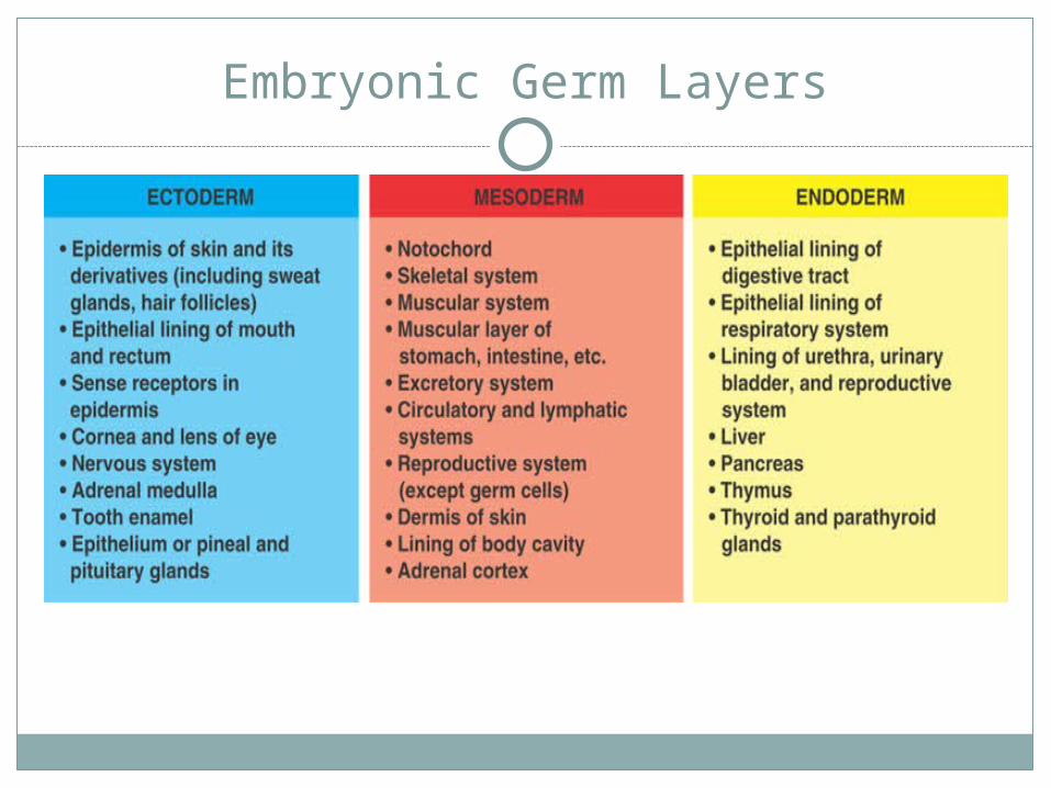
Embryonic Germ Layers

3 Questions
What are the 3 stages of embryonic development?
What are the 3 embryonic germ layers?
What are the 3 differences between oogenesis & spermatogenesis?

Le Frog Embryo
Fertilization – 1/3 of egg is yolk, and is limited to the lower half (called the vegetal pole) Top half called animal pole and has a pigmented cap Grey crescent appears on opposite side of sperm entry
point
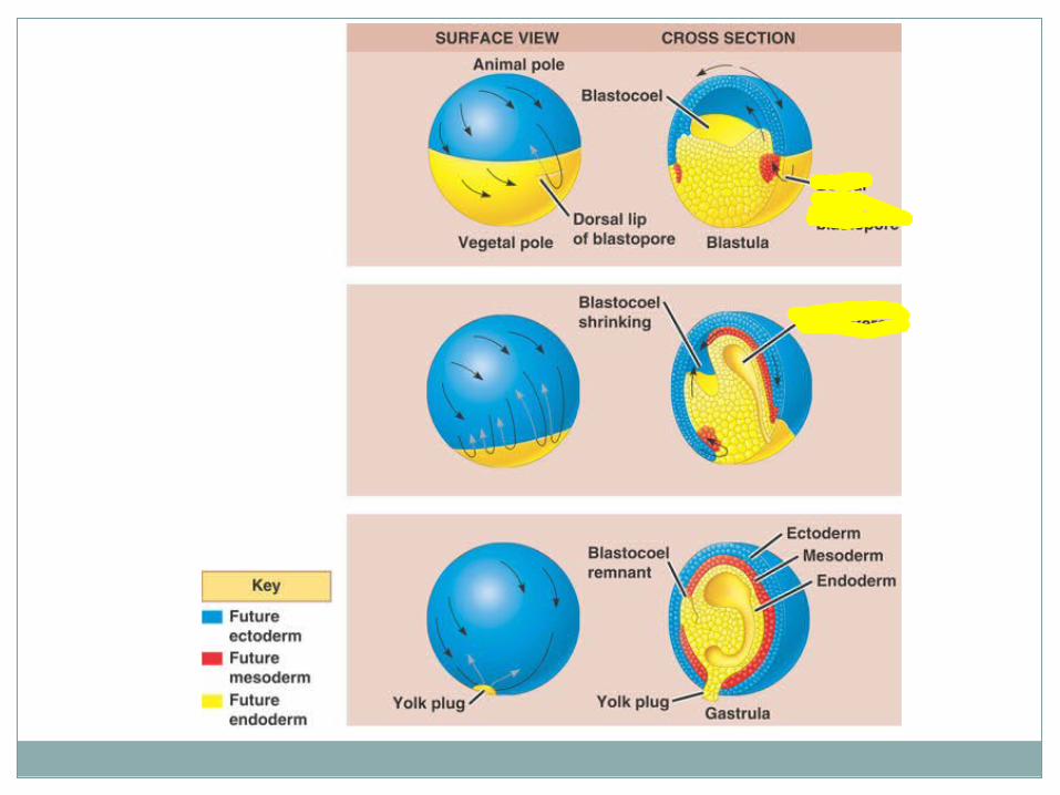
Cleavage & Gastrulation Yolk presence = uneven cleavage Blastophore forms on the border of the gray crescent
and the vegetal pole

Le Frog Embryo (Page 2)
Involution – cells at the dorsal lip, but above the blastophore stream over the dorsal lip and into the blastophore
These cells then become endoderm & mesoderm
The ectoderm streaming inward is called Epibolic movement
The blastocoel disappears and is replaced by another cavity called the archenteron
The mesoderm opposite the blastophore = dorsal mesoderm

Le Frog Embryo (Page 3)
Organogenesis – In chordates, forming first are: Notochord (skeletal rod characteristic of all chordates)
Forms the dorsal mesoderm Neural Tube (becomes CNS)
Forms the dorsal ectoderm Formed by embryonic induction
After the blueprints for the organs are established, embryo develops into a larval stage (tadpole)
Metamorphosis takes the tadpole to frog

Le Bird Embryo
Cleavage & Gastrulation – contains so much yolk that embryo develops on a flat disc (blastodisc) that sits on top of the yolk Instead of grey streak, it is called primitive streak
Cells migrate over the primitive streak and flow inward to the archenteron
As cleavage & gastrulation occurs, the yolk gets smaller
Extraembryonic Membranes – Tissue outside the embryo forms 4 extraembryonic membranes These membranes support the growing embryo inside
the shell

Le Bird Embryo (Page 2)
The 4 embryonic membranes are:1. Yolk Sac2. Amnion3. Chorion4. Allantois
1. Yolk Sac – contains the yolk (food for embryo)
2. Amnion – encloses the embryo in protective amniotic fluid

Le Bird Embryo (Page 3)
3. Chorion – lies underneath the shell-- Allows for the diffusion of respiratory gases
between the outside and the growing embryo
4. Allantois – like the placenta in mammals-- Conduit for respiratory gases between the
environment and the embryo-- Stores uric acid from the embryo
-- Uric acid is the nitrogenous waste from the embryo that accumulates until the chick
hatches


Factors that influence Embryonic Development
Cytoplasmic determinants – the importance of the cytoplasm surrounding the nucleus for embryonic development
For example, if a sea-urchin embryo is cut to influence development: If cut Longitudinally (some animal pole & vegetal
pole cells), subsequent development is normal If cut Horizontally (all animal pole or all vegetal pole
cells), subsequent development is abnormal

8-Cell Stage
Blastula
Gastrula
--If cut Longitudinally (some animal pole & vegetal pole cells), subsequent development is normal
--If cut Horizontally (all animal pole or all vegetal pole cells), subsequent development is abnormal

Example of Cytoplasmic Determinants

The Grey Cresent
Hans SpemannEmbryonic development is affected by how
their distribution is affected by the zygote’s characteristic pattern of cleavage
Demonstrated that if the grey crescent was constricted to one side of the blastomere, then there was abnormal separation & development

Spemann’s Experiment

Embryonic Induction
Def – the ability of one group of embryonic cells to influence the development of another group of embryonic cells
Example: Ordinarily dorsal ectoderm in gastrula neural plate If other ectoderm is transplanted in the same gastrula
neural plate (same as b4)
At late stage of development in gastrula,If transplant ectoderm NO neural plate

Homeotic, Homeobox, or Hox Genes
Def – master genes that control the expression of genes responsible for specific anatomical structures
Play critical role in normal embryonic development

Example of Embryonic Induction
-- Dorsal tube usually initiates a chain of inductions neural tube development
-- Speeman & Mangold
-- Grafted dorsal lip onto ventral side of embryo
-- The dorsal lip relocated induced the abdomen tissue to become neural tissue
-- Termed primary organizer due to its crucial role in development

Videos
http://youtube.com/watch?v=UgT5rUQ9EmQhttp://www.youtube.com/watch?v=AoisqOGQI
VE&feature=relatedhttp://www.youtube.com/watch?v=si20cxSHr
WU&NR=1http://youtube.com/watch?v=jvanNDQhlYIhttp://youtube.com/watch?v
=Tf9mXrl0tUI&feature=related


![Ocw [animal reproduction]](https://img.dokumen.tips/doc/110x75/58835b371a28ab42678b649f/ocw-animal-reproduction.jpg)