Embed Size (px)
Citation preview

Anatomical Identi�cation of Ischial SpinesApplicable to Intrapartum Transperineal UltrasoundBased on Magnetic Resonance Imaging of PregnantWomenEriko Yano
University of TokyoTakayuki Iriyama ( [email protected] )
University of TokyoShouhei Hanaoka
University of TokyoSeisuke Sayama
University of TokyoMari Ichinose
University of TokyoMasatake Toshimitsu
University of TokyoTakahiro Seyama
University of TokyoKenbun Sone
University of TokyoKeiichi Kumasawa
University of TokyoTakeshi Nagamatsu
University of TokyoKoichi Kobayashi
JCHO Tokyo Yamate Medical CenterTomoyuki Fujii
Sanno HospitalYutaka Osuga
University of Tokyo
Research Article

Keywords: ITU, IQR, SID, ultrasound, Gynecology
Posted Date: March 5th, 2021
DOI: https://doi.org/10.21203/rs.3.rs-269362/v1
License: This work is licensed under a Creative Commons Attribution 4.0 International License. Read Full License

1
Anatomical identification of ischial spines applicable to intrapartum transperineal 1
ultrasound based on magnetic resonance imaging of pregnant women 2
3
Eriko Yano*1, Takayuki Iriyama*1, Shouhei Hanaoka2, Seisuke Sayama1, Mari Ichinose1, 4
Masatake Toshimitsu1, Takahiro Seyama1, Kenbun Sone1, Keiichi Kumasawa1, Takeshi 5
Nagamatsu1, Koichi Kobayashi 3, Tomoyuki Fujii4, and Yutaka Osuga1 6
7
1 Department of Obstetrics and Gynecology, Faculty of Medicine, The University of 8
Tokyo, Tokyo, Japan. 9
2 Department of Radiology, Faculty of Medicine, The University of Tokyo, Tokyo, Japan. 10
3 Department of Obstetrics and Gynecology, JCHO Tokyo Yamate Medical Center, Tokyo, 11
Japan. 12
4 Department of Obstetrics and Gynecology, Sanno Hospital, Tokyo, Japan. 13
14
* These authors contributed equally to this work. 15
16
Correspondence: Takayuki Iriyama M.D., Ph.D. 17
Department of Obstetrics and Gynecology, Faculty of Medicine, University of Tokyo 7-3-1 18
Hongo, Bunkyo-ku, Tokyo, 113-8655, Japan 19
Tel; +81-03-3815-5411 20
Fax; +81-03-5800-6937 21
Email; [email protected] 22

2
Abstract 23
Intrapartum transperineal ultrasound (ITU) is considered useful in judging fetal head 24
descent; however, the inability to detect ischial spines on ITU has been a drawback to its 25
legitimacy. The current study aimed to determine the anatomical location of ischial spines, 26
which can be directly applied to ITU. Based on magnetic resonance imaging of 67 pregnant 27
women at 33+2 [31+6-34+0] weeks gestation (median [interquartile range: IQR]), we 28
calculated the angle between the pubic symphysis and the midpoint of ischial spines 29
(midline symphysis-ischial spine angle; mSIA), which is theoretically equivalent to the 30
angle of progression at fetal head station 0 on ITU, by determining spatial coordinates of 31
pelvic landmarks and utilizing spatial vector analysis. Furthermore, we measured 32
symphysis-ischial spine distance (SID), defined as the distance between the vertical plane 33
passing the lower edge of the pubic symphysis and the plane that passes the ischial spines. 34
As a result, mSIA was 109.6 ° [105.1-114.0] and SID 26.4 mm [19.8-30.7] (median, 35
[IQR]). There was no correlation between mSIA or SID and maternal characteristics, 36
including physique. Our results provide valuable evidence to enhance the reliability of ITU 37
in assessing fetal head descent by considering the location of ischial spines. 38
39
40
41
42
43
44

3
Introduction 45
Intrapartum transperineal ultrasound (ITU) has been proposed for evaluating 46
labor progression. The angle of progression (AoP), defined as the angle between the 47
midline of the pubic symphysis and a line running from the inferior edge of the symphysis 48
to the fetal skull, is regarded as more reliable in accuracy and reproducibility than vaginal 49
examination (VE) to assess fetal head descent 1-5. However, some researchers have 50
questioned the accuracy of ITU in assessing fetal head descent because ischial spines, the 51
most important landmark in evaluating fetal head station (St), cannot be obtained by 52
ultrasound 6,7. Previous studies have tried to identify a landmark equivalent to ischial spines 53
or St 0 on ITU images by evaluating the angle or the distance between the pubic symphysis 54
and the ischial spine 4,6-9. Tustchek et al. depicted a plane perpendicular to the pubis, which 55
runs ischial spines (level of ischial spines), on ITU images; they reported that the AoP was 56
116° when the most presenting part of the fetus’ head reached this plane 10,11. This is one of 57
the reference angles for evaluating St on ITU images, which is covered by the International 58
Society of Ultrasound in Obstetrics and Gynecology (ISUOG) practice guidelines 12. 59
However, this plane was defined based on a symphysis-ischial spine distance (SID) of 3 60
cm, which was obtained based on single computed tomography (CT) images from only one 61
non-pregnant woman 9. Considering the anatomical changes of the pelvis during pregnancy 62
and the differences in pelvic structure among individuals 13, this reference remains 63
arguable. 64
To date, there is only one report from France by Arthuis et al. that has evaluated 65
the anatomical position of ischial spines by analyzing pelvic images during pregnancy 7. By 66
4
analyzing CT images of pregnant women, they calculated the angle between the upper-67
lower edge of the pubic symphysis and the midpoint of ischial spines in a mid-sagittal plane 68
(midline symphysis-ischial spine angle; mSIA) and reported that it was 110°. This angle 69
was considered nearly equivalent to the AoP at St 0, and it contributed to the understanding 70
of the fetal head location on ITU images. 71
Considering racial or physique differences in pelvic anatomy 14,15, it has not been 72
clarified whether an mSIA 110° can be applied to other races; thus, more evidence needs to 73
be accumulated to determine ischial spines or St 0 on ITU images universally. Additionally, 74
if there is a method to evaluate the components of the bony pelvis more easily, we can 75
evaluate the pelvic structure and apply this further to ITU. Therefore, by calculating the 76
structural relationship between the pubis and other bony birth canal components, we can 77
establish an absolute index that divides the pelvic cavity, or the positional relationship 78
between the pubis and fetal head changes, quantitatively along the pelvic curve. 79
The current study aimed to quantify the relative position between ischial spines 80
and the pubic symphysis to assess St 0 on ITU images in an East Asian population by 81
establishing a novel and practical method to evaluate pelvic anatomy that can be applied to 82
ITU by analyzing three-dimensional coordinates of magnetic resonance images (MRI) in 83
pregnant women. 84
85
Results 86
There were 76 cases whose MRI scans were performed during pregnancy [31+6 87
weeks to 34+0 weeks] between January 2016 and December 2018; 67 cases were analyzed 88

5
as nine cases were excluded due any of the landmarks, including the superior, or inferior 89
edge of pubic symphysis or ischial spines, was unclear. Table 1 shows the maternal 90
characteristics; age, height, pre-pregnancy body mass index (BMI), and the gestational age 91
when the MRI was taken (35 years old [31-39] (median [interquartile range: IQR]), 159 cm 92
[156-162], 20.0 kg/m2 [19.1-21.2], and 33+2 weeks [31+6 - 34+0], respectively). Fifty 93
patients (74%) were primiparous, 11 cases (16.4%) had a history of vaginal delivery, and 94
six cases (8.9%) had a history of cesarean section. The indications for MRI were placental 95
previa, low set placenta, or placenta accreta (59 cases [88.0%]); fetal malformation (four 96
cases [5.9%]); and birth canal evaluation for maternal complication with Klippel-97
Trenaunay-Weber syndrome (one case [1.5%]]. All pregnant women were East Asians. 98
We measured the spatial coordinates of each of the following points on MRI 99
images: superior edge of the pubic symphysis, inferior edge of the pubic symphysis, and 100
bilateral ischial spines, utilizing software as described in the Methods. Pubic symphysis 101
was evaluated using T2-weighted images (T2WI) (Figure 1a-d). We determined the mid-102
sagittal plane using the sagittal and axial views. Next, we identified the superior edge of the 103
pubic symphysis (A) and inferior edge of the pubic symphysis (B) using mid-sagittal and 104
axial views and measured each coordinate: A (Ax, Ay, Az) and B (Bx, By, Bz). The left 105
ischial spine (C) and right ischial spine (D) were identified by evaluating the axial and 106
coronal views of T1-weighted images (T1WI) and measuring each coordinate: C (Cx, Cy, 107
Cz) and D (Dx, Dy, Dz) (Figure 1e and f). The point corresponding to the level of the 108
ischial spine in the mid-sagittal plane was calculated as the midpoint of the bilateral ischial 109
spines (E). 110

6
E (Ex, Ey, Ez) = (Cx+Dx2 , Cy+Dy
2 , Cz+Dz2 ) (1) 111
The vector connecting the inferior edge of pubis (B) and the superior edge of 112
pubis (A) is defined as the vector BA, and the vector connecting the inferior edge of the 113
pubis (B) and midpoint of the ischial spines (E) is defined as the vector BE. The angle 114
between the vector BA and BE is defined as θ, which corresponds to the midline 115
symphysis-ischial spine angle (mSIA) on MRI (Figure 2a). 116
Vector BA = (Ax-Bx, Ay-By, Az-Bz) (2) 117
Vector BE = (Ex-Bx, Ey-By, Ez-Bz) (3) 118
The angle θ between line AB and BE was measured using the Arc cos function. 119
Angle θ can be expressed by vector BA and BE, and the Arc cos function as follows 120
(Figure 2b): 121
122
θ= Arc cos ( BA∙BE|BA||BE|) (4) 123
In other words, the angle θ can be calculated by introducing the value obtained 124
by taking the inner product of the vector BA and the vector BE as the numerator and 125
multiplying the absolute values of the vectors BA and BE as the denominator into the Arc 126
cos function. The angle θ obtained from this formula is 109.6° [105.1-114.0] (median 127

7
[IQR]. There was no significant statistical correlation between mSIA and maternal 128
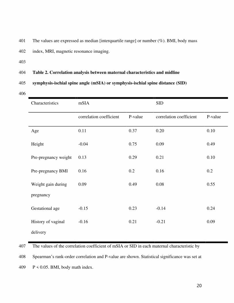
characteristics (age, gestational days, height, body weight, BMI, or parity) (Table 2). 129
The infrapubic plane, which is defined as the plane perpendicular to the pubis, 130
which passes the inferior edge of the pubis, and the level of the ischial spines, which is 131
defined as the plane parallel to the infrapubic plane, which runs ischial spines, and the 132
distance between two planes is defined as the symphysis-ischial spine distance (SID) 133
(Figure 3a). E' is the foot of the perpendicular line drawn from the midpoint of the ischial 134
spine (E) to the extension of the pubis; the distance corresponding to the SID was 135
calculated as the distance (|BE'|) between the infrapubic plane (BB') and the level of the 136
ischial spine (EE') (Figure 3b). The distance between the infrapubic plane and the level of 137
ischial spines, |BE'| or SID, can be calculated as follows: 138
SID = |BE'| = |BE| cos (180-θ) = - |BE| cos (θ) (5) 139
The median SID was 26.4 mm [19.8-30.7] (median [IQR]). There was no significant 140
statistical correlation with maternal characteristics (Table 2). 141
142
Discussion 143
The current study demonstrated the anatomical angles and distance that can help 144
identify the location of ischial spines on ITU by analyzing the anatomical relationship 145
between the pubic symphysis and ischial spines using MRI data from pregnant women in 146
the third trimester. In this study, we also established a novel method to measure the 147
components of the pelvic anatomy by analyzing the three-dimensional coordinates of MRI. 148

8
Although several studies have attempted to identify St 0 or ischial spines on ITU 149
images based on image analysis, there is only one report by Arthuis et al. prior to the 150
current study that measured the anatomical relationship between the ischial spines and the 151
pubis on the mid-sagittal plane using images during pregnancy 7. Since it is known that 152
there are differences in pelvic size and structures depending on race or physique 14,15, it has 153
been questioned whether the mSIA of 110° calculated by Arthuis et al. can be applied to 154
other races. In the current study, we demonstrated that the mSIA was 109.6° [105.1-114.0] 155
(median [IQR]), regardless of the differences in maternal characteristics including height. 156
Arthuis et al. also demonstrated that the angle between the pubis and left ischial spine was 157
106° [105-109] (median [IQR]). Based on our MRI analysis, the angles between the pubis 158
and left or right ischial spine were 105.5° [102.0-107.9] and 106.8° [101.9-109.3] (median 159
[IQR]), respectively. Intriguingly, these values are almost equal to those reported by 160
Arthuis et al. Although they did not describe the target race, the height was 164 cm [160-161
169] (median, [IQR]), which is different from ours, 159 cm [156-162] (median [IQR]), 162
obtained from our Japanese and East Asian population. This comparison shows that a 163
mSIA of 109.6° can be considered as a universal value corresponding to St 0 in applying to 164
ITU, regardless of race or physique. 165
According to ISUOG practice guidelines 12, AoP equivalent to ITU head station 0 is 166
116°, which is a result calculated based on single CT images of only one pregnant woman 167
reported by Tutchek et al 10,11. They calculated this value, 116°, by depicting the level of 168
ischial spines on ITU images by drawing a perpendicular line to the pubis, which passes 30 169
mm from the lower edge of the pubis. The rationale for this distance, 30 mm, was based on 170

9
the measurement of the symphysis-ischial spine distance (SID), which was 30 mm, 171
although this was obtained from only one non-pregnant woman. Therefore, their result 172
remains controversial to be used as a universal value and is in fact different from our result 173
of 109°. In our study, we calculated SID to be 26.4 mm [19.8-30.7] (median [IQR]) based 174
on 67 pregnant women. Intriguingly, this SID value was almost the same at 26.1 mm [23.4-175
29.5] (Median [IQR]) as Arthuis et al. reported 7, implying that SID may be consistent 176
among pregnant women. The pelvic cavity changes gradually but dramatically during 177
pregnancy to prepare for delivery, such as an increase in the mobility of the sacroiliac joint 178
and pubic symphysis 13. For example, Barbera et al. calculated mSIA as 99° using 3DCT of 179
non-pregnant women 4, which is clearly different from our current result of 109.6°, 180
calculated from 67 pregnant women. Therefore, we speculate that mSIA increases during 181
pregnancy, which implies that the value obtained from non-pregnant women should not be 182
directly applied to pregnant women. Taken together, we must reconsider the value obtained 183
from one non-pregnant woman in assessing fetal head descent, which is still used to assess 184
head station in the ISUOG guidelines. The figure calculated from multiple pregnant women 185
is more reliable in accurately assessing fetal descent. 186
Arthuis et al. analyzed 3DCT of pregnant women and reported the angle between 187
the pubis and left ischial spine as 106° [105-109] (median [IQR]) 7. However, since the 188
view equivalent to the ITU images is the mid-sagittal plane, this angle cannot be directly 189
applied to ultrasound. Although the midpoint of the ischial spines can be used as an index 190
for the ischial spine on ITU images, it cannot be measured directly by their method. Thus, 191
they calculated the angle between the pubis and the midpoint of the ischial spine (mSIA) as 192

10
110° by using the distance measurement by 3DCT and trigonometric function. In contrast, 193
the method that we newly developed enables us to calculate the mSIA directly and easily 194
by measuring the spatial coordinates of the lower and upper edges of the pubis and bilateral 195
ischial spines and applying them to vector calculation. Thus, the anatomical relationship on 196
the mid-sagittal plane can be easily obtained. Furthermore, as our method enables the 197
evaluation of anatomical landmarks other than the ischial spines, we can assess other 198
components of the birth canal, which has not been evaluated in previous studies, but is of 199
value in determining fetal descent, including the sacrum. We can easily quantify the 200
positional relation of the pubis, sacrum, and fetal head. According to the American College 201
of Obstetricians and Gynecologists (ACOG) forceps delivery classification 16, namely 202
mid/low/outlet, clinicians rely on VE to assess fetal descent, and its correspondence to ITU 203
findings is still undetermined. With our novel method for calculating the exact position of 204
the pubis, sacrum, and ischial spines within the pelvis, we might be able to theoretically 205
divide the pelvic cavity into a mid/low/outlet in relation to the position of pubic symphysis 206
as a future study, which would ultimately lead to safer application of operative vaginal 207
delivery. 208
The limitation of our study is that we did not consider the fetal head. Since the 209
fetal head progresses in the birth canal three-dimensionally, the presenting part does not 210
necessarily exist on the mid-sagittal plane. Since the presenting part is evaluated on the 211
mid-sagittal plane when evaluating AoP, there may be a discrepancy from VE findings. 212
Another limitation is that the pelvic anatomy may change during labor. To determine the 213
position of the ischial spines during labor more accurately, angle evaluation during labor 214

11
may be necessary. Another limitation is that we did not examine intra-observer or inter-215
observer differences. However, identification of ischial spines and pubis on MRI images is 216
easy, and it is unlikely that a large error will occur. 217
We established a novel method to analyze the pelvic anatomy by evaluating 218
spatial coordinates on MRI images, and calculated the mSIA as 109.6° and SID as 26.4 mm 219
in pregnant women in the third trimester, which can be applied to ITU as positional 220
landmarks of ischial spines. Furthermore, since these values are almost equivalent to the 221
value from other races and physique, our figure can be a universal parameter. With this 222
index, the anatomical relation between the fetal head and the ischial spines can be evaluated 223
on ITU images, and it should increase the reliability and accuracy of labor progression 224
assessment using ITU. 225
226
Methods 227
Under the approval of the Institutional Review Board of the University of Tokyo 228
[3053-(4)], we retrospectively analyzed MRI scans performed during pregnancy between 229
January 2016 and December 2018 in our hospital. This was a retrospective observational 230
study, and consent was obtained in the form of opt-out on the internet homepage of our 231
institution, in accordance with the request of the ethics committee of the University of 232
Tokyo. All research was performed in accordance with the relevant guidelines and 233
regulations. Either MRI, 1.5T MAGNETOM Avanto (Siemens, Germany), or 1.5T 234
EXCITE HDX (GE Healthcare, United States) were used. The mid-sagittal plane of T2WI 235
and axial and coronal planes of fast-suppressed T1WI were taken with the patient placed in 236

12
a supine position with their knees bent. In 1.5T MAGNETOM Avanto (Siemens, 237
Germany), T2WI were acquired by 2D single shot fast spin echo with the following 238
settings: time of echo (TE) of 75 ms, time of repetition (TR) of 1500 ms, and slice interval 239
of 5 mm; fast-suppressed T1WI were acquired by 3-D spoiled gradient echo with TE of 240
1.07, TR of 3.2, and slice interval of 5 mm. In 1.5T EXCITE HDX (GE Healthcare, United 241
States), T2WI were acquired by 2D single-shot fast spin echo with TR of 1268 ms, TE of 242
91.50, and slice interval of 5 mm; fast-suppressed T1WI were acquired by 3D spoiled 243
gradient echo with TR of 3.532, TE of 1.688, slice interval of 1.5 mm. 244
MRI data were analyzed with OsiriX Lite (Pixmeo SARL, Switzerland), free and 245
open source of the Digital Imaging and Communications in Medicine (DICOM) viewer. 246
OsiriX Lite can simultaneously evaluate three cross sections of DICOM images: horizontal, 247
coronal, and sagittal sections. By selecting any point on the image, the spatial coordinates 248
(x, y, z) are automatically measured when the image is constructed in 3D. The 249
identification and measurement of each anatomical were conducted by the same 250
obstetrician (E. Y.) under the guidance of a radiologist (S. H.). 251
Data of background characteristics were expressed as median (interquartile range 252
[IQR]). Correlation analysis was conducted using Spearman’s rank-order correlation with 253
JMP pro version 15 (SAS Institute Inc., Japan). 254
255
Data availability 256
All data generated or analyzed during this study are included in this published article. 257
258

13
259
260
References 261
1 Molina, F. S., Terra, R., Carrillo, M. P., Puertas, A. & Nicolaides, K. H. What is the 262
most reliable ultrasound parameter for assessment of fetal head descent? 263
Ultrasound Obstet Gynecol 36, 493-499, doi:10.1002/uog.7709 (2010). 264
2 Ramphul, M. et al. Instrumental delivery and ultrasound : a multicentre 265
randomised controlled trial of ultrasound assessment of the fetal head 266
position versus standard care as an approach to prevent morbidity at 267
instrumental delivery. BJOG 121, 1029-1038, doi:10.1111/1471-0528.12810 268
(2014). 269
3 Duckelmann, A. M. et al. Measurement of fetal head descent using the 'angle 270
of progression' on transperineal ultrasound imaging is reliable regardless of 271
fetal head station or ultrasound expertise. Ultrasound Obstet Gynecol 35, 216-272
222, doi:10.1002/uog.7521 (2010). 273
4 Barbera, A. F., Imani, F., Becker, T., Lezotte, D. C. & Hobbins, J. C. Anatomic 274
relationship between the pubic symphysis and ischial spines and its clinical 275
significance in the assessment of fetal head engagement and station during 276
labor. Ultrasound in Obstetrics and Gynecology 33, 320-325, 277
doi:10.1002/uog.6322 (2009). 278
5 Barbera, A. F., Pombar, X., Perugino, G., Lezotte, D. C. & Hobbins, J. C. A new 279
method to assess fetal head descent in labor with transperineal ultrasound. 280
Ultrasound Obstet Gynecol 33, 313-319, doi:10.1002/uog.6329 (2009). 281
6 Bamberg, C. et al. Relationship between fetal head station established using 282
an open magnetic resonance imaging scanner and the angle of progression 283
determined by transperineal ultrasound. Ultrasound Obstet Gynecol 37, 712-284
716, doi:10.1002/uog.8944 (2011). 285
7 Arthuis, C. J., Perrotin, F., Patat, F., Brunereau, L. & Simon, E. G. Computed 286

14
tomographic study of anatomical relationship between pubic symphysis and 287
ischial spines to improve interpretation of intrapartum translabial ultrasound. 288
Ultrasound Obstet Gynecol 48, 779-785, doi:10.1002/uog.15842 (2016). 289
8 Iliescu, D. et al. The Angle of Progression at Station 0 and in Magnetic 290
Resonance and Transperineal Ultrasound Assessment. Case Rep Obstet 291
Gynecol 2015, 748327, doi:10.1155/2015/748327 (2015). 292
9 Henrich, W., Dudenhausen, J., Fuchs, I., Kamena, A. & Tutschek, B. Intrapartum 293
translabial ultrasound (ITU): sonographic landmarks and correlation with 294
successful vacuum extraction. Ultrasound Obstet Gynecol 28, 753-760, 295
doi:10.1002/uog.3848 (2006). 296
10 Tutschek, B., Braun, T., Chantraine, F. & Henrich, W. A study of progress of 297
labour using intrapartum translabial ultrasound, assessing head station, 298
direction, and angle of descent. BJOG 118, 62-69, doi:10.1111/j.1471-299
0528.2010.02775.x (2011). 300
11 Tutschek, B., Torkildsen, E. A. & Eggebo, T. M. Comparison between ultrasound 301
parameters and clinical examination to assess fetal head station in labor. 302
Ultrasound Obstet Gynecol 41, 425-429, doi:10.1002/uog.12422 (2013). 303
12 Ghi, T. et al. ISUOG Practice Guidelines: intrapartum ultrasound. Ultrasound 304
Obstet Gynecol 52, 128-139, doi:10.1002/uog.19072 (2018). 305
13 Cunningham, F. G. et al. Williams Obstetrics 25 edn, Vol. 1 14-32 (McGraw Hill 306
Education, 2018). 307
14 Arima, H. et al. Differences in lumbar and pelvic parameters among African 308
American, Caucasian and Asian populations. Eur Spine J 27, 2990-2998, 309
doi:10.1007/s00586-018-5743-5 (2018). 310
15 Handa, V. L. et al. Racial differences in pelvic anatomy by magnetic resonance 311
imaging. Obstet Gynecol 111, 914-920, doi:10.1097/AOG.0b013e318169ce03 312
(2008). 313
16 ACOG Practice Bulletin No. 154: Operative Vaginal Delivery. Obstet Gynecol 314
126, e56-e65, doi:10.1097/AOG.0000000000001147 (2015). 315

15
316
Acknowledgements 317
None. 318
319
Author Information 320
Department of Obstetrics and Gynecology, Faculty of Medicine, The University of Tokyo, 321
Tokyo, Japan 322
Eriko Yano, Takayuki Iriyama, Seisuke Sayama, Mari Ichinose, Masatake Toshimitsu, 323
Takahiro Seyama, Kenbun Sone, Keiichi Kumasawa, Takeshi Nagamatsu, and Yutaka 324
Osuga 325
Department of Radiology, Faculty of Medicine, The University of Tokyo, Tokyo, Japan 326
Shouhei Hanaoka 327
Department of Obstetrics and Gynecology, JCHO Tokyo Yamate Medical Center, Tokyo, 328
Japan 329
Koichi Kobayashi 330
Department of Obstetrics and Gynecology, Sanno Hospital, Tokyo, Japan 331
Tomoyuki Fujii 332
333
Author contributions 334
E.Y. and T.I. designed the research. E.Y. and S.H. analyzed MRI images and acquired the 335
data. E.Y., T.I., and S.S. wrote the manuscript. M.I, M.T., T.S., K.S., K.K., T.N., and K.K. 336

16
have made substantial contribution to study design, interpretation of data, and made critical 337
comments on the manuscript. T.F. and Y.O. organized this research as project managers. 338
339
Competing interests 340
The Authors declare no competing interests. 341
342
Corresponding author 343
Correspondence to Takayuki Iriyama 344
345
Figure Legends 346
Figure 1. Identification of superior and inferior edge of the pubic symphysis and 347
bilateral ischial spines on magnetic resonance imaging (MRI) of pregnant women. 348
Superior edge of the pubic symphysis (A) on T2 weighted mid-sagittal plane MRI (a) and 349
T1 weighted axial plane (b) MRI. Inferior edge of the pubic symphysis (B) on T2 weighted 350
mid-sagittal plane (c) and T1 weighted axial plane MRI (d). Right ischial spine (C) and left 351
ischial spine (D) on axial and coronal plane of T1 weighted image MRI (e, f). 352
353
Figure 2. Calculation of the angle between pubic symphysis and midpoint of ischial 354
spines, termed the midline symphysis-ischial spine angle (mSIA). 355
(a) T2-weighted mid-sagittal plane of MRI. θ is the angle between superior edge of the 356
pubic (A), inferior edge of pubic symphysis (B), and midpoint of ischial spines (E). 357

17
(b) Vectors and θ can be calculated by each coordinate (x, y, z) as follows: 358
Vector BA = (Ax-Bx, Ay-By, Az-Bz) 359
Vector BE = (Ex-Bx, Ey-By, Ez-Bz) 360 |BA| = length of the vector BA =√(Ax-Bx)2+(Ay-By)2+(Az-Bz)2 361
BA∙BE = inner product of the vector BA and the vector BE 362
= (Ax-Bx)∙(Ex-Bx)+(Ay-By)∙(Ey-By)+(Az-Bz)∙(Ez-Bz) 363
θ= Arc cos ( BA∙BE|BA||BE|) 365
364
366
Figure 3. Calculation of symphysis-ischial spine distance (SID) 367
(a) Schema of the relationship between SID, pubic symphysis, infrapubic plane, and level 368
of ischial spines. (b) T2-weighted mid-sagittal plane of MRI. E is defined as the midpoint 369
of the ischial spines. E' is defined as the foot of the perpendicular line drawn from E on the 370
extension line of the pubis. EE' (level of ischial spines) is parallel to the infrapubic plane. 371
The distance between B and E' (|B E'| =SID) can be calculated as follows: 372
SID = |BE'| = |BE| cos (180-θ) = - |BE| cos (θ) 373
|BE| = length of the vector BE =√(Ex-Bx)2+(Ey-By)2+(Ez-Bz)2 374
375
376

18
377
Table 1. Maternal characteristics of 67 pregnant women analyzed in the current study 378
379

19
380
381
382
383
384
385
386
387
388
389
390
391
392
393
394
395
396
397
398
399
400
Characteristics Value
Age (years) 35.0 [31.0-39.0]
Height (cm) 159.0 [156-162]
Pre-pregnancy weight (kg) 51.0 [48.0-54.0]
Weight gain during pregnancy (kg) 7.0 [5.2-9.0]
Pre-pregnancy BMI (kg/m2) 20.0 [19.1-21.2]
Gestational age when MRI was taken 33+2 [31+6 – 34+0]
Parity
Nulliparous 50 (74.6)
History of vaginal delivery 11 (16.4)
History of caesarian section 6 (8.9)
Indication for MRI
Placenta previa or low set placenta 59 (88.0)
Fetal malformation 4 (5.9)
Other 1 (1.5)
Race (East Asian) 67 (100)
20
The values are expressed as median [interquartile range] or number (%). BMI, body mass 401
index, MRI, magnetic resonance imaging. 402
403
Table 2. Correlation analysis between maternal characteristics and midline 404
symphysis-ischial spine angle (mSIA) or symphysis-ischial spine distance (SID) 405
406
The values of the correlation coefficient of mSIA or SID in each maternal characteristic by 407
Spearman’s rank-order correlation and P-value are shown. Statistical significance was set at 408
P < 0.05. BMI, body math index. 409
Characteristics mSIA SID
correlation coefficient P-value correlation coefficient P-value
Age 0.11 0.37 0.20 0.10
Height -0.04 0.75 0.09 0.49
Pre-pregnancy weight 0.13 0.29 0.21 0.10
Pre-pregnancy BMI 0.16 0.2 0.16 0.2
Weight gain during
pregnancy
0.09 0.49 0.08 0.55
Gestational age -0.15 0.23 -0.14 0.24
History of vaginal
delivery
-0.16 0.21 -0.21 0.09

21
410
411

22
412
413
414
415

Figures
Figure 1
Identi�cation of superior and inferior edge of the pubic symphysis and bilateral ischial spines onmagnetic resonance imaging (MRI) of pregnant women. Superior edge of the pubic symphysis (A) on T2weighted mid-sagittal plane MRI (a) and T1 weighted axial plane (b) MRI. Inferior edge of the pubic

symphysis (B) on T2 weighted mid-sagittal plane (c) and T1 weighted axial plane MRI (d). Right ischialspine (C) and left ischial spine (D) on axial and coronal plane of T1 weighted image MRI (e, f).
Figure 2
"See the Supplemental Files section for the complete �gure caption".
Figure 3

"See the Supplemental Files section for the complete �gure caption".
Supplementary Files
This is a list of supplementary �les associated with this preprint. Click to download.
FigureCaptions.docx