Embed Size (px)
Citation preview

Analysis of Innate DefencesAgainst Plasmodium Falciparum
in Immunodeficient MiceThe Harvard community has made this
article openly available. Please share howthis access benefits you. Your story matters
Citation Arnold, Ludovic, Rajeev Kumar Tyagi, Pedro Mejia, Nico Van Rooijen,Jean-Louis Pérignon, and Pierre Druilhe. 2010. Analysis of innatedefences against Plasmodium falciparum in immunodeficient mice.Malaria Journal 9:197.
Published Version doi:10.1186/1475-2875-9-197
Citable link http://nrs.harvard.edu/urn-3:HUL.InstRepos:4547524
Terms of Use This article was downloaded from Harvard University’s DASHrepository, and is made available under the terms and conditionsapplicable to Other Posted Material, as set forth at http://nrs.harvard.edu/urn-3:HUL.InstRepos:dash.current.terms-of-use#LAA
Arnold et al. Malaria Journal 2010, 9:197http://www.malariajournal.com/content/9/1/197
Open AccessR E S E A R C H
© 2010 Arnold et al; licensee BioMed Central Ltd. This is an Open Access article distributed under the terms of the Creative CommonsAttribution License (http://creativecommons.org/licenses/by/2.0), which permits unrestricted use, distribution, and reproduction inany medium, provided the original work is properly cited.
ResearchAnalysis of innate defences against Plasmodium falciparum in immunodeficient miceLudovic Arnold†1, Rajeev Kumar Tyagi†1, Pedro Mejia1,2, Nico Van Rooijen3, Jean-Louis Pérignon1 and Pierre Druilhe*1
AbstractBackground: Mice with genetic deficiencies in adaptive immunity are used for the grafting of human cells or pathogens, to study human diseases, however, the innate immune responses to xenografts in these mice has received little attention. Using the NOD/SCID Plasmodium falciparum mouse model an analysis of innate defences responsible for the substantial control of P. falciparum which remains in such mice, was performed.
Methods: NOD/SCID mice undergoing an immunomodulatory protocol that includes, clodronate-loaded liposomes to deplete macrophages and an anti-polymorphonuclear leukocytes antibody, were grafted with human red blood cells and P. falciparum. The systematic and kinetic analysis of the remaining innate immune responses included the number and phenotype of peripheral blood leukocytes as well as inflammatory cytokines/chemokines released in periphery. The innate responses towards the murine parasite Plasmodium yoelii were used as a control.
Results: Results show that 1) P. falciparum induces a strong inflammation characterized by an increase in circulating leukocytes and the release of inflammatory cytokines; 2) in contrast, the rodent parasite P. yoelii, induces a far more moderate inflammation; 3) human red blood cells and the anti-inflammatory agents employed induce low-grade inflammation; and 4) macrophages seem to bear the most critical function in controlling P. falciparum survival in those mice, whereas polymorphonuclear and NK cells have only a minor role.
Conclusions: Despite the use of an immunomodulatory treatment, immunodeficient NOD/SCID mice are still able to mount substantial innate responses that seem to be correlated with parasite clearance. Those results bring new insights on the ability of innate immunity from immunodeficient mice to control xenografts of cells of human origin and human pathogens.
BackgroundDefences against foreign cells, including pathogens, relyon both innate or non-adaptive responses, and adaptiveor antigen-specific immune responses. However, modernimmunology has focused primarily or almost exclusivelyon the latter.
Therefore, various strains of mice having a genetic defi-ciency in cells responsible for adaptive immunity (i.e. Tand B lymphocytes) have been selected, which have beenused for the grafting of xenogenic cells, particularly thoseof a human origin. Indeed, the vast majority of thesestudies have focused on the grafting of human lympho-cytes, haematopoietic stem cells and to a lesser extent
tumor cells [1]. However, it was previously shown thatone can take advantage of these immunocompromizedmice to develop a mouse model for human malaria.
The initial report that bovine red blood cells (RBC)injected intra-peritoneally in SCID mice could cross theperitoneum and colonize the peripheral blood [2] was anincentive to repeat the experiment using human redblood cells (HuRBC). Although there was regrettably nounderstanding of the underlying mechanism of transportthrough the peritoneum, SCID mice harbouring up to 70- 80% HuRBC among total RBC in mice peripheral bloodwere obtained [3].
However, when Plasmodium falciparum was injectedinto SCID mice, the parasites became pycnotic withinhours in erythrocytes. After excluding a potential toxicityof mouse serum by in vitro methods, [3], it was hypothe-sized that innate immune defences could constitute themain limiting factor in parasite survival, and this was
* Correspondence: [email protected] Laboratoire de Parasitologie Bio-Médicale, Institut Pasteur, 28, rue du Dr Roux, 75015 Paris, France† Contributed equallyFull list of author information is available at the end of the article

Arnold et al. Malaria Journal 2010, 9:197http://www.malariajournal.com/content/9/1/197
Page 2 of 12
confirmed by employing agents able to control mac-rophages (MP) and other cells involved in innate defences[4,5]. Further steps were performed in an empirical man-ner. The model was improved by moving from the origi-nal SCID mouse to the NIHIII (Beige Xid Nude) mouse,then to the NOD/SCID mouse that have more defectiveinnate immunity, and by identifying two componentswhich, when combined, allowed to obtain a stable P. falci-parum parasitaemia in some of the NOD/SCID mice [6](namely: liposomes encapsulating dichloromethylene-diphosphonate, named clo-lip [5] and a monoclonal anti-body (NIMP-14) [4] directed to mouse polymorphonu-clear cells (PMN)).
Though this empirical approach demonstrated the fea-sibility of the objective, and could be applied to vaccinedevelopment [6,7] and drug screening [8], the control ofinnate defences was far from optimal in this P. falciparumblood stage rodent model. Indeed, the P. falciparum para-sitaemia remained stable for extended periods of time, upto four months only in a minor subset of mice, whereas itwas cleared within a few days in the majority of the ani-mals.
The above data stress the importance of innatedefences and are in agreement with long standing obser-vations made in humans. However, despite their crucialimportance innate defences have been far less studiedthan adaptive responses, and remain poorly known.Innate responses in P. falciparum infected patients wererecently analysed in detail and this unveiled several dra-matic modifications in the phenotypes and functions ofblood monocyte/macrophage populations [9].
To complement the above descriptive analysis inhumans by an experimental approach in a model, it wasdecided to perform in the P. falciparum NOD/SCIDmodel a systematic and stepwise analysis of innate cellresponses and inflammation mediators produced inresponse to the grafting of HuRBC, of P. falciparum, aswell as to agents employed to control innate defences.Results bring new insights about the role and potency ofinnate defences against human xenografts, such asHuRBC, and human pathogens, such as P. falciparum.
MethodsMiceTwo to six months old male and female NOD/SCID micewere used. They were purchased from Charles River, andkept in an A3 animal house, i.e. in sterile isolators. Theywere housed in sterilized cages equipped with filter topsduring the experimentation. Mice were provided withautoclaved tap water and a γ-irradiated pelleted diet adlibitum. They were manipulated under pathogen freeconditions using laminar flux hoods. All animals weretreated according to the French legislation.
Human red blood cellsHuman whole blood was provided by the French bloodbank (Etablissement français du sang, Paris, France).Blood donors had no history of malaria and all the bloodgroups were used without observing any difference onparasite survival. Whole blood was washed three times bycentrifugation at 900 ×g, 5 minutes at room temperatureand buffy coat was separated in order to eliminate whiteblood cells and platelets. Packed HuRBC were suspendedin SAGM (Adenine, Glucose and mannitol solution) andkept at 4°C for a maximum of 2 weeks. Before use HuRBCwere washed three times in RPMI-1640 medium (Gibco/BRL, Grand Island, N.Y.) supplemented with 1 mg hypox-anthine per liter (Sigma, St Louis, MO) and warmed 10minutes at 37°C.
Parasite culturesThe P. falciparum 3D7 clone was employed in this study.This parasite strain was maintained in vitro at 5% haema-tocrit in complete culture medium at 37°C in a candle jar.This medium contained RPMI-1640 medium (Gibco/BRL), 35 mM HEPES (Sigma), 24 mM NaHCO3, 0.5%albumax (Gibco/BRL) and 1 mg of hypoxanthine (Sigma)per liter. Cryopreserved parasites were thawed using theglycerol/sorbitol method [10] and used for the furtherexperiments.
A non-lethal rodent parasite strain Plasmodium yoeliiXNL1.1 was preserved in 500 μl aliquot of cryo-preserv-ing buffer at -80°C at 22% parasitaemia. The strain wasthawed at room temperature, diluted twice in RPMI-1640medium followed by the injection of 50 × 106 parasitedirectly into the mice.
Immunomodulatory agents and suppression of innate immunityNumerous attempts were made to increase the successrate of the grafting of infected RBC. Clo-lip (provided byN. Van Rooijen) was injected through intraperitoneal(i.p.) route in order to reduce the number of tissue MP, asdescribed previously [5]. The anti-PMN monoclonal anti-body NIMP-R14 [4] was purified from a hybridomakindly provided by Dr. M. Strath (National Institute forMedical Research, London, UK). Its activity was com-pared to that of two other anti-PMN monoclonal anti-bodies: RB6-8C5 (purified from the hybridoma kindlyprovided by Geneviève Milon (Institut Pasteur, Paris,France) and 1A8 (purchased from BioXcell, Lebanon).The NIMP-R14 monoclonal antibody was used in all thestudies, unless specified. Various agents (all purchased atSigma, unless specified) were used to further reduceinnate immunity such as dexamethasone (1-5 mg/Kg/day), TGF-β100 ng - 1 μg/day) (PeproTech, Rocky Hill,NJ), cyclophosphamide (75 mg/kg/day), cisplatinium (1-10 mg/Kg/day), and TMβ-1 monoclonal antibody that

Arnold et al. Malaria Journal 2010, 9:197http://www.malariajournal.com/content/9/1/197
Page 3 of 12
targets NK cells (1 mg/kg/day). The effect of splenectomyand of irradiation (100 - 300 cGy) was also tested. Otherexperiments evaluated the addition of metabolic agentssuch as pABA (400 mg/kg/day), and folinic acid (400 mg/kg/day), or of antioxidants, such as vitamin E (20 mg/Kg/day; Nepalm, Cenexi, Fontenay sous bois), N-acetylcysteine (100 mg/kg/day), trolox (4 - 100 mg/kg/day), 8-aminoguanidine (100 mg/Kg/day).
Chemical immunomodulation protocol and mouse infectionIn the current study, a previously described immuno-modulation protocol was employed [11], modified asdescribed in [12]. On day -13, each mouse received a doseof 10 mg/kg of mAb NIMP-R14 by i.p. injection. On day -12, each mouse received 0.2 ml of the suspension of clo-lip by the same route. On days -9, -6, -3 each mousereceived 0.5 ml of HuRBC i.p. mixed with a dose of 10mg/kg of mAb NIMP-R14 and 0.2 ml of clo-lip. On day 0mice were infected with 500 μl HuRBC parasitized by P.falciparum at a parasitaemia of 1% (all the developmentalforms, i.e. trophozoite, schizont and rings, were present)mixed with a dose of 10 mg/kg of NIMP-R14 antibody.Afterwards, a dose of 10 mg/kg of antibody NIMP-R14,0.2 ml of clo-lip and 0.5 ml of HuRBC was injected i.p. at3 days interval, until the end of the study. The follow-upof the infection was performed by daily Giemsa stainedthin blood films drawn from the tail vein.
Haematological parameters and grafting of P. falciparum-HuRBCThe study blood samples were collected from mice retro-orbital sinus on heparin. Various haematological parame-ters such as haematocrit, leukocyte number and pheno-type (Ly-6C APC, Ly-6G APC (Miltenyi Biotec,Germany), CD115 PE, CD43 FITC, CD62L FITC, CD11bFITC, DX5 FITC, CD122 PE (BD Biosciences, UK) inperipheral blood samples were monitored, as well as thephenotype characterization of monocytes (CD11b+,CD115+), inflammatory monocytes (CD43-, CD62L+, Ly-6C+), PMN (CD11b+, ly6G+), and natural killer cells(DX5+, CD122+). Total leukocyte number (leukocytes/μlblood) was evaluated by lysing 20 μl of total blood withBD FACS™ Lysing solution, and counting on Malassezhaematocytometer. Since successfully grafted mice have asignificant, but variable, percentage of HuRBC in theirperipheral blood, parasitaemia in mice was expressed asthe overall percentage of P. falciparum infected RBCamong total RBC, i.e. both human and mouse RBCobserved in thin blood smears. In addition, the peritonealblood parasitaemia was measured in the smears drawnfrom the peritoneum. The percentage of HuRBC in theperipheral blood of mice was determined by an immuno-fluorescence technique using a FITC labeled anti-humanglycophorin A monoclonal antibody (DAKO, Denmark).
Assay of cytokines and chemokine100-150 μl blood samples were collected from the retro-orbital sinus with the help of Pasteur pipette, and serawere stored at -80°C. Cytokines (IL-6, MCP-1, IFNγ,TNFα, IL-12p70 and IL-10) were quantified using theBD™ Cytometric Bead Array mouse inflammatory kit fol-lowing the manufacturer's recommendations.
Statistical analysisThe paired test was used for statistical analysis. P valuesof less than 0.05 were considered significant. In theresults section only differences reaching significance arementioned.
ResultsVarious patterns of peripheral blood parasitaemia are observed in NOD/SCID miceAlthough stable long-lasting parasitaemia could beobtained and employed for various applications [8,11] thepattern of parasitaemia has never been homogeneous, i.e.varied greatly from one animal to the other, and the vari-ous attempts to modify the protocol failed to improveresults significantly. For instance, among a total of 84mice studied recently under rigorous and well-controlledconditions using the standard immunomodulation proto-col described above, four different patterns of peripheralblood parasitaemia could be described (summarized inFigure 1). Following a single infection by P. falciparum,17% of mice remained parasitologically negative, 34%showed a transient parasitaemia lasting for ca. 12 dayspost-infection, 12% showed a stable parasitaemia formore than 20 days and 37% showed an almost total para-
Figure 1 Patterns of blood parasitaemia observed in NOD/SCID mice. In four independent experiments, a total of 84 NOD/SCID mice were infected with a single i.p. infection of P. falciparum 3D7 strain on day 0 and HuRBC (500 μl of pellet at 50% haematocrit), clo-lip (200 μl) and NIMP-R14 (10 mg/kg) antibody were injected i.p. every 3 days as previously described (black arrows). The preparatory phase prior to the injection of infected HuRBC is not shown. Parasitaemia was measured on tail blood smears by counting the number of infected RBC among 10000 total human and mouse RBC. Parasitemia lower than 0,001 are not considered. The figure shows the average parasitaemia ± SD, and the percentages (and number of mice) corresponding to each pattern of parasitemia.

Arnold et al. Malaria Journal 2010, 9:197http://www.malariajournal.com/content/9/1/197
Page 4 of 12
site clearance from peripheral blood, however followedby a re-emergence a few days later, without new parasiteinoculation, i.e. a second wave of parasitaemia lasting forthe life-span of the animal. The sequence of events of thelatter pattern considered the most informative, wasselected for further analysis.
Various modifications to the standard protocol wereassessed, and resulted only in differences in the propor-tion of mice presenting the four patterns of parasitaemiadescribed above. These modifications include: 1) varia-tions in the dose and schedule of administration of thedifferent components (i.e. infected and uninfectedHuRBC, clo-lip and NIMP-R14, including modificationsproposed by others [12]); 2) use of various agents toreduce innate immunity, such as cyclophosphamide, cis-platinium, irradiation, dexamethasone, TGF-β, splenec-tomy, TMβ-1 antibody targeting NK cells 3) Addition ofmetabolic precursors such as pABA, folinic acid to supplynutritive factors for the parasite 4) Addition of antioxi-dants such as vitamin E, N-acetyl cysteine (NAC), trolox,8-aminoguanidine. These empirical approaches neverbrought a significant or reproducible improvement ofparasite survival in NOD/SCID mice.
These results raise the question of which factor(s) arecritical to control in order to obtain a stable parasitaemiaand prompted the launch of a detailed analysis of theremaining innate immune defences, mainly MP andPMN.
The strong inflammation induced by P. falciparum is associated with parasite clearanceIn mice showing a recrudescence (fourth patterndescribed above, which was thought to be the most dis-criminating, see Figure 1) the following parameters wereanalysed in kinetic manner: 1) the numbers and pheno-type of blood leukocyte subsets, 2) various cytokinesserum levels, 3) HuRBC grafting in the peripheral blood,together with 4) parasitaemia.
Results, summarized in Figure 2A, show that, whereasparasitaemia in peritoneal blood remains stable overtime, conversely, peripheral blood parasitaemia decreasesmarkedly. This decrease is related to a major clearance ofHuRBC in this compartment, despite repeated bloodinjections, which is parasite-dependant since the num-bers of circulating HuRBC in control, uninfected mice,remain stable (see below).
The parasite was found to induce a very potent pro-inflammatory effect characterized by a transient increasein peripheral leukocytes numbers mostly monocytes(MO) (1690 ± 1080 MO/μl on day 7 post-infection vs 290± 125 on day 0, i.e. 5.8 fold increase; P < 0.006) and to alesser extent PMN (3000 ± 1500 PMN/μl on day 3 post-infection vs 2100 ± 750 on day 0, i.e. 1.4 fold increase; notsignificant) (Figure 2B). Moreover, during the first few
Figure 2 P. falciparum is highly pro-inflammatory. (A) Peripheral (plain circles) and peritoneal (open circles) parasitaemia (average of 31 mice ± SEM) counted on blood smears and percentage of human gly-cophorin A+ RBC grafted in mouse peripheral blood detected by FACS among total RBC (full circle, dotted line). Parasitemia lower than 0,001 are not considered. (B) Total number of leukocyte per μl of blood (plain circle) at different times post-infection. The numbers of CD11b+ F4/80+
monocytes (black square, dotted line) and CD11b+ Ly-6G+ PMN (white triangle) were calculated from the percentages obtained by FACS anal-ysis. The percentage of CD43- CD62L+ Ly-6C+ inflammatory monocytes was also assessed by FACS (open circle, dotted line). (C) Cytokines/chemokine levels were estimated using the CBA mouse inflammatory cytokines kit by FACS. (D) Representative peritoneal blood smears showing the progressive phagocyte recruitment following P. falcipar-um infection. Results represent the means ± SEM from 3 distinct exper-iments (n = 14 mice).
Arnold et al. Malaria Journal 2010, 9:197http://www.malariajournal.com/content/9/1/197
Page 5 of 12
days following the infection, a transient increase (Figure2B) in blood MO having an inflammatory phenotypeCD43- CD62L+ Ly-6C+ was observed (57.2 ± 12.7% of MOon day -1 before infection vs 80 ± 11% on day 3 post-infection; P < 0.03) (the Gr-1 antigen is not expressed onNOD/SCID MO surface). In addition, the parasiteinduces a major recruitment of leukocytes in the perito-neal cavity as compared to controls receiving uninfectedHuRBC (Figure 2D).
The peak of blood MO was associated with a peak ofsecretion of several inflammatory cytokines such as IL-12p70 (342 ± 520 pg/ml on day 7 post-infection vs 33 ±6.5 pg/ml on day -1 before infection), IFNγ (290 ± 215 pg/ml on day 7 vs 5 ± 2.2 pg/ml on day -1) and TNFα (155 ±53 pg/ml on day 7 vs 53 ± 26 pg/ml on day -1) orchemokine such as MCP-1 (from 355 ± 290 pg/ml on day-1 to 960 ± 480 pg/ml on day 7 post-infection) (P < 0.009).
Results attest of the strong inflammation induced bythe parasite; however the latter is transient, (Figure 2C)and accordingly the number of leukocyte decreases toreach values close to the steady state.
The decrease in parasite-induced inflammation wasfollowed by a new increase in HuRBC numbers in periph-eral blood, which understandably correlated with a recru-descence of P. falciparum blood parasitaemia. Thissecond wave of parasitaemia itself induced an increase ofleukocyte counts, of IL-6 and MCP-1, however not of IL-12p70, IFNγ and TNFα, and in addition, was not associ-ated with a re-emergence of inflammatory MO. Hence,the secondary parasitaemia was less pro-inflammatorythan the first. This also point to an important role forinflammatory MO in the initial clearance of uninfectedand P. falciparum infected HuRBC.
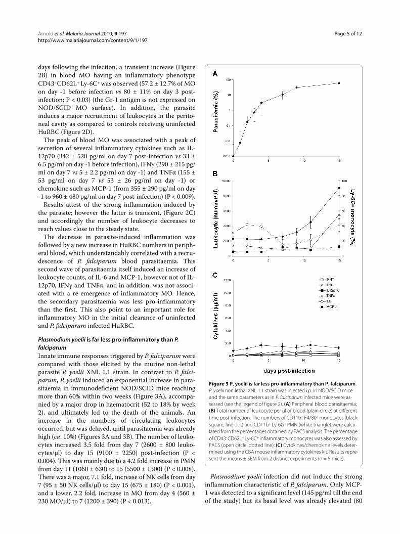
Plasmodium yoelii is far less pro-inflammatory than P. falciparumInnate immune responses triggered by P. falciparum werecompared with those elicited by the murine non-lethalparasite P. yoelii XNL 1.1 strain. In contrast to P. falci-parum, P. yoelii induced an exponential increase in para-sitaemia in immunodeficient NOD/SCID mice reachingmore than 60% within two weeks (Figure 3A), accompa-nied by a major drop in haematocrit (52 to 18% by week2), and ultimately led to the death of the animals. Anincrease in the numbers of circulating leukocytesoccurred, but was delayed, until parasitaemia was alreadyhigh (ca. 10%) (Figures 3A and 3B). The number of leuko-cytes increased 3.5 fold from day 7 (2600 ± 800 leuko-cytes/μl) to day 15 (9100 ± 2250) post-infection (P <0.004). This was mainly due to a 4.2 fold increase in PMNfrom day 11 (1060 ± 630) to 15 (5500 ± 1300) (P < 0.008).There was a major, 7.1 fold, increase of NK cells from day7 (95 ± 50 NK cells/μl) to day 15 (675 ± 180) (P < 0.001),and a lower, 2.2 fold, increase in MO from day 4 (560 ±230 MO/μl) to 7 (1200 ± 390) (P < 0.013).
Plasmodium yoelii infection did not induce the stronginflammation characteristic of P. falciparum. Only MCP-1 was detected to a significant level (145 pg/ml till the endof the study) but its basal level was already elevated (80
Figure 3 P. yoelii is far less pro-inflammatory than P. falciparum. P. yoelii non lethal XNL 1.1 strain was injected i.p. in NOD/SCID mice and the same parameters as in P. falciparum infected mice were as-sessed (see the legend of figure 2). (A) Peripheral blood parasitaemia; (B) Total number of leukocyte per μl of blood (plain circle) at different time post-infection. The numbers of CD11b+ F4/80+ monocytes (black square, line dot) and CD11b+ Ly-6G+ PMN (white triangle) were calcu-lated from the percentages obtained by FACS analysis. The percentage of CD43- CD62L+ Ly-6C+ inflammatory monocytes was also assessed by FACS (open circle, dotted line); (C) Cytokines/chemokine levels deter-mined using the CBA mouse inflammatory cytokines kit. Results repre-sent the means ± SEM from 2 distinct experiments (n = 5 mice).

Arnold et al. Malaria Journal 2010, 9:197http://www.malariajournal.com/content/9/1/197
Page 6 of 12
pg/ml) (Figure 3C). The other cytokines assayed did notshow any significant increase as compared to basal level(Figure 3C). Moreover, P. yoelii did not induce the strongincrease of CD43- CD62L+ Ly-6C+ inflammatory MOwhich was very significant with P. falciparum (Figure 2B),but only several moderate variations (i.e. a small initialincrease from 30.6 ± 6.5% on day 0 to 41.7 ± 5.2% on day 2post-infection (P < 0.0045) followed by a temporarydecrease, 15.2 ± 7.3% on day 7 post-infection (P < 0.0045)then by a new increase to reach steady state value on day15 post-infection) (Figure 3B). In the absence of adaptiveimmune responses, all mice infected with P. yoelii diedbetween day 16 to day 20 post-inoculation (not shown).
HuRBC, and anti-inflammatory agents, induce low-grade inflammationConsidering the indications that inflammation wasrelated to the clearance of the parasite, it was decided toexamine whether HuRBC, clo-lip and NIMP-R14 anti-body also induce inflammation. The effects of each com-ponent, injected i.p. upon 1) the number and phenotypeof blood leukocytes, and 2) cytokines serum levels, wereexamined. The inflammatory effect of HuRBC was notunexpected, as it represents a heterologous graft. The i.p.injection of HuRBC induced an increase in leukocytenumbers (Figure 4A) (P < 0.003 between day 0 and day 3)mainly PMN and to a lesser extent MO and NK cells (datanot shown). The inflammatory MO subset increasedtransiently to peak at 76% of total MO 10 hours postinfection (P < 0.009), and then progressively decreased tobasal level (Figure 4B). HuRBC grafting also led to a tran-sient secretion of IL-6 (732 ± 51 vs 47 ± 14.7 pg/ml; P <0.003) and TNFα (216 ± 51 pg/ml vs 75 ± 79 pg/ml; P <0.1) (Figures 4C and 4E), without substantial modifica-tions of MCP-1, IL-10, IL-12p70 or IFNγ.
Unexpectedly, it was observed that clo-lip induce a fastand strong inflammatory reaction. Indeed, as soon asthree hours post liposomes injection, the number of cir-culating leukocyte increased by 6.8 fold (Figure 4A) (MO3.14 fold, PMN 8.8 fold and NK 3.07 fold) (P < 0.01), andthereafter, decreased but slowly (remaining four timeshigher than basal level 48 hours post-injection). In addi-tion, clo-lip induced an increase of the "inflammatory"MO subset (CD43- CD62L+ Ly-6C+) that lasted for morethan three days (Figure 4B) (P < 0.001). Clo-lip injectionalso led to a transient release of IL-6 (777 ± 445 vs 48 ± 30pg/ml at 3 hours post-injection) (Figure 4C) (P < 0.03).
Finally, the anti-PMN monoclonal antibody NIMP-R14had a modest effect on the number of leukocytes and onthe percentage of inflammatory MO, but was the stron-gest inducer of IL-6 (1054 ± 292 vs 30 ± 7 pg/ml at 3hours post-injection) and of MCP-1 (2425 ± 760 pg/ml at3 hours post-injection) (Figure 4C and 4D), and alsoinduced the release of TNF(133 ± 19 vs 24 ± 3.7 pg/ml at3 hours post-injection) (Figure 4E) (P < 0.04).
Figure 4 HuRBC, clo-lip and NIMP-R14 antibody are pro-inflam-matory. HuRBC (full circle, plain line), clo-lip (open circle) and NIMP-R14 antibody (full circle, dotted line) were injected once in the perito-neum at hour 0, at doses identical to those used for the whole immu-nomodulation protocol. Leukocyte number (A), CD43- CD62L+ Ly-6C+
inflammatory monocytes (B) and inflammatory cytokines/chemokine (C-E) were assessed in mouse peripheral blood by FACS. Results are the means ± SD. from groups of 4 mice.
Arnold et al. Malaria Journal 2010, 9:197http://www.malariajournal.com/content/9/1/197
Page 7 of 12
Altogether these results indicate that the immunomod-ulators clo-lip and NIMP-R14 antibody, which have pre-viously been shown to be indispensable to the grafting ofP. falciparum, actually reduce innate responses, though atthe expense of a transient but substantial inflammation.
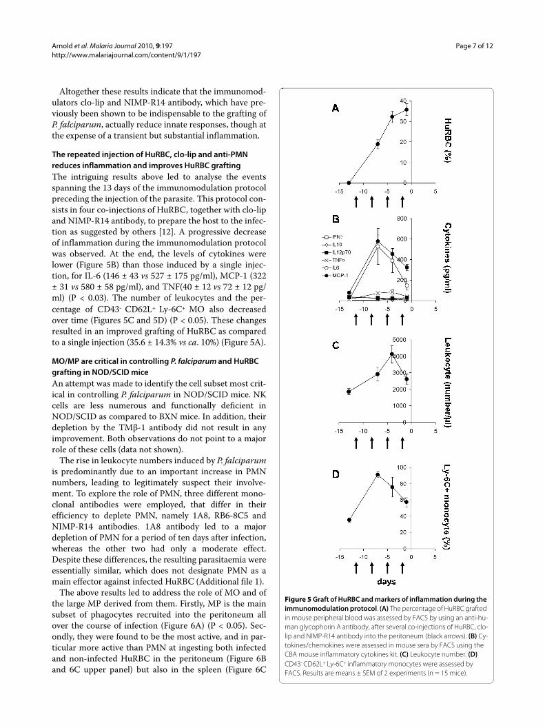
The repeated injection of HuRBC, clo-lip and anti-PMN reduces inflammation and improves HuRBC graftingThe intriguing results above led to analyse the eventsspanning the 13 days of the immunomodulation protocolpreceding the injection of the parasite. This protocol con-sists in four co-injections of HuRBC, together with clo-lipand NIMP-R14 antibody, to prepare the host to the infec-tion as suggested by others [12]. A progressive decreaseof inflammation during the immunomodulation protocolwas observed. At the end, the levels of cytokines werelower (Figure 5B) than those induced by a single injec-tion, for IL-6 (146 ± 43 vs 527 ± 175 pg/ml), MCP-1 (322± 31 vs 580 ± 58 pg/ml), and TNF(40 ± 12 vs 72 ± 12 pg/ml) (P < 0.03). The number of leukocytes and the per-centage of CD43- CD62L+ Ly-6C+ MO also decreasedover time (Figures 5C and 5D) (P < 0.05). These changesresulted in an improved grafting of HuRBC as comparedto a single injection (35.6 ± 14.3% vs ca. 10%) (Figure 5A).
MO/MP are critical in controlling P. falciparum and HuRBC grafting in NOD/SCID miceAn attempt was made to identify the cell subset most crit-ical in controlling P. falciparum in NOD/SCID mice. NKcells are less numerous and functionally deficient inNOD/SCID as compared to BXN mice. In addition, theirdepletion by the TMβ-1 antibody did not result in anyimprovement. Both observations do not point to a majorrole of these cells (data not shown).
The rise in leukocyte numbers induced by P. falciparumis predominantly due to an important increase in PMNnumbers, leading to legitimately suspect their involve-ment. To explore the role of PMN, three different mono-clonal antibodies were employed, that differ in theirefficiency to deplete PMN, namely 1A8, RB6-8C5 andNIMP-R14 antibodies. 1A8 antibody led to a majordepletion of PMN for a period of ten days after infection,whereas the other two had only a moderate effect.Despite these differences, the resulting parasitaemia wereessentially similar, which does not designate PMN as amain effector against infected HuRBC (Additional file 1).
The above results led to address the role of MO and ofthe large MP derived from them. Firstly, MP is the mainsubset of phagocytes recruited into the peritoneum allover the course of infection (Figure 6A) (P < 0.05). Sec-ondly, they were found to be the most active, and in par-ticular more active than PMN at ingesting both infectedand non-infected HuRBC in the peritoneum (Figure 6Band 6C upper panel) but also in the spleen (Figure 6C
Figure 5 Graft of HuRBC and markers of inflammation during the immunomodulation protocol. (A) The percentage of HuRBC grafted in mouse peripheral blood was assessed by FACS by using an anti-hu-man glycophorin A antibody, after several co-injections of HuRBC, clo-lip and NIMP-R14 antibody into the peritoneum (black arrows). (B) Cy-tokines/chemokines were assessed in mouse sera by FACS using the CBA mouse inflammatory cytokines kit. (C) Leukocyte number. (D) CD43- CD62L+ Ly-6C+ inflammatory monocytes were assessed by FACS. Results are means ± SEM of 2 experiments (n = 15 mice).

Arnold et al. Malaria Journal 2010, 9:197http://www.malariajournal.com/content/9/1/197
Page 8 of 12
lower left panel) and in the liver (Fig 6C lower rightpanel). Moreover, clo-lip target only circulating MO andMP, and Figure 6D clearly shows that clo-lip is necessaryfor the grafting of HuRBC: the percentage of circulatingHuRBC was 54 ± 2% after three clo-lip injections vs 0.95± 0.93% without, or 4.1 ± 2% with the anti-PMN NIMP-R14 antibody. Altogether, these results point to the MO/MP lineage as the most efficient component in control-ling the graft of P. falciparum infected HuRBC.
DiscussionResults confirm that innate defences are potent againstxenografts and pathogens in mice lacking T, B and NKcell functions, and provide an insight of their effectagainst P. falciparum in immunodeficient mice. Severalyears ago, our group has taken advantage of immuno-compromized mice, i.e. mice genetically defective in theiradaptive immunity, to generate a P. falciparum mousemodel, which could be successfully used for several typesof applications such as assessment of antibodies directedto new vaccine candidates [6,7,11] or of anti-malarialdrugs [8] with any P. falciparum strain or isolate [12]. Therepeated injection into the peritoneum of HuRBC, clo-lipand NIMP-R14 antibody allowed P. falciparum growth inmouse peripheral blood, but at the expense of a high fail-ure rate, since only a subset harboured a stable parasitae-mia (i.e. 12% among several hundred NOD/SCID micetested have a stable parasitaemia and 37% have a recru-descent parasitaemia). Therefore, it was decided to focusfurther work on the analysis of murine innate immunedefences induced by this very pro-inflammatory parasitein order to gather an understanding, i.e. to attempt toidentify the critical defence component(s) limiting suc-cessful grafting.
The present study shows that 1) P. falciparum induces amajor inflammation characterized by an increase inperipheral leukocytes and by the release of inflammatorycytokines in the serum; 2) parasitaemia in the perito-neum remains stable whereas the decrease observed inthe peripheral blood is related to HuRBC clearance in thiscompartment, triggered by the parasite; 3) P. yoelii,induces a very moderate inflammation as compared to P.falciparum, suggesting that a parasite adapted to its hostdoes not trigger the same inflammatory response (whichmay be related to host factors, for example a lower recog-nition by host receptors, and/or to parasite factors, forexample a lower production of pro-inflammatory mole-cules presented by this parasite); 4) clo-lip, NIMP-R14and HuRBC also induce an inflammation, but far lessimportant than that triggered by the parasite itself, andthe former is anyhow essential to parasite grafting; 5)none of the additional immunosuppressive, anti-inflam-matory, anti-oxidant and nutritive factors assessed inempirical manner was able to significantly, or reproduc-
Figure 6 Evaluation of the role of PMN, and of monocytes/mac-rophages, in the graft of infected and non-infected HuRBC. A peri-toneal smear was drawn at different times post-infection in order to assess the phenotype of peritoneal phagocytes. (A) MP (black bar) and PMN (white bar) were identified by microscopy in order to determine the percentage of each subset among the total number of phagocytes counted on the smear (at least 500). (B) Percentages of MP (black bar) and PMN (white bar) having ingested at least one particle of malaria pigment. Results are the mean ± SD from 4 infected NOD/SCID mice. (C) Representative Giemsa staining of different organs smears per-formed 45 days post-infection from 3 different NOD/SCID mice (upper panel left and right: peritoneum; lower left panel: spleen, lower right panel: liver). Arrows indicate haemozoin loaded MP. (D) Effect of clo-lip and NIMP-R14 antibody on HuRBC grafting in NOD/SCID peripheral blood. HuRBC are injected alone (Black square) or in combination with clo-lip (plain circle) or with NIMP-R14 antibody (open circle). Black ar-rows represent the days of injection. Results are means ± SEM of 2 ex-periments (n = 6 mice per group).
Arnold et al. Malaria Journal 2010, 9:197http://www.malariajournal.com/content/9/1/197
Page 9 of 12
ibly, improve P. falciparum survival; and 6) MP seem tobear the most critical function in controlling P. falci-parum in these mice.
The innate immune defences that remain in immuno-compromized mice have seldom been analysed. Surpris-ingly, only one study has so far focused on this issue. Itreported the occurrence of a major inflammation in rela-tion to human leukocytes grafting in SCID mouse perito-neum [13]. It consists in a massive and early (24 h)recruitment of neutrophils and in the secretion of a widespectrum of murine cytokines, such as IL-6, TNF, IFNγ,IL-1β, IL-10, IL-12p40 in the peritoneal cavity. The roleof granulocytes is supported by improved engraftment ofhuman leukocytes following administration of the anti-granulocyte antibody RB6-8C5 [14]. Since this report,innate immune responses in immunocompromized micehave remained surprisingly little explored, despite thegeneral agreement that they are very efficient in defenceagainst xenografts [15,16]. Even NOD/SCID/IL-2rγ-/-
mice, which present substantial additional defects ininnate immunity (they lack functional T, B and NK cells,have reduced functions of MP and dendritic cells, andseveral cytokines pathways are totally impaired) stillrequire the use of pharmacological agents or irradiation/splenectomy to allow for the grafting of cells of humanorigin [15,17].
The present study model includes a "double xenograft",of HuRBC and of P. falciparum. A first limitation of thismodel is the lack of understanding of the transperitonealpassage of both uninfected and infected HuRBC in mouseperipheral blood, and its lack of reproducibility (17% ofmice were negative). The i.p. route of administration ofHuRBC was chosen as it was previously found effectiveusing bovine RBC [2], as it constitutes a reservoirwhereas HuRBC injected i.v. are quickly eliminated [18].However, the mechanism of passage being unknown,though there are indications for lymphatic drainage[19,20], the reasons for occasional blockade are even lessclear. There might be a pure mechanical component asthe simple increase in HuRBC volume (2 ml at 50% hae-matocrit daily) led us and others [21] to improve HuRBCchimerism in peripheral blood. Secondly, the increase ofclearance of HuRBC (both infected and non-infected) 7to 10 days after injection of the parasite, is likely inflam-mation dependent as it was found to be associated with aconcomitant peak of inflammatory cytokines andchemokine (IL-12p70, IFNγ, TNFα and MCP-1) in mouseserum. TNFα is known to increase erythrophagocytosisand to induce anaemia during malaria both in mouse andin human [22]. Splenectomy improves HuRBC grafting,suggesting that splenic MP play a role in this clearance[21,23]. Thirdly, in those rare cases (5%) when the perito-neum cavity was invaded by millions of MP this resulted
in HuRBC phagocytosis, and clearance of the graft in thiscompartment.
Together these data suggest a prominent role of MP inthe rejection of both uninfected and infected HuRBC bytwo main mechanisms: the release of inflammatory medi-ators reflecting the activation of MO and the resultingincreased erythrophagocytosis. The role of MP in xeno-graft rejection is well documented [24]. For instance theyaccount for the majority of infiltrating leukocytes duringthe rejection of pig-to-primate xenografts [25,26] andselective MP depletion in immunocompetent rodentsresulted in significant delays in cellular infiltration andxenograft rejection [27,28]. Finally, clo-lip which depletesMO/MP was clearly essential to both HuRBC and P. falci-parum survival, stressing also in this model the impor-tance of MP.
The mechanism of HuRBC phagocytosis by murine MPis yet to be elucidated. It can not be due to complement orby Fc receptors-mediated opsonization, as NOD/SCIDmice have neither complement activity [29] nor antibod-ies [29,30]. C-type lectins integrated in MP membranehave been reported to mediate erythrophagocytosis bydirectly binding to sugar ligands on the surface of senes-cent or altered RBC [31,32]. Moreover, the signal regula-tory protein a (SIRPα) is a critical immune inhibitoryreceptor on MP that reacts with CD47 to prevent autolo-gous phagocytosis. Since CD47 is species specific,healthy HuRBC may be phagocytozed by murine MP dueto their inability to induce murine MP SIRPα tyrosinephosphorylation [33,34]. Conversely, phagocytosis of P.falciparum infected HuRBC by MP is well documented,and the scavenger receptor CD36, which recognizesPfEMP-1 on the surface of infected RBC, is widely impli-cated in this process. Indeed, P. falciparum phagocytosisdecreases by 80% using blocking antibodies or CD36-/-
murine MP [35,36]. MP also possess a large array ofpathogen-recognition receptors such as toll-like recep-tors (TLRs) that recognize P. falciparum GPI anchors,that are considered as key malaria pathogenicity factors[37,38]. TLR-2 and to a lesser extent TLR-4 directly rec-ognize infected RBC [39] whereas TLR-9 recognize onlythe haemozoin pigment [40-42], but all trigger inflamma-tion through the MyD88/NFκB pathway. Uric acid(derived from hypoxanthine accumulated by the parasite)has been recently reported to be another major inflam-matory mediator involved in P. falciparum infection [43].The effect of haemozoin pigment on MO/MP is debatedas it was reported to impair their function such as block-ing of phagocytosis [44-46], but also to be a potent proin-flammatory agent. Indeed it can trigger the release ofNO, TNF, IL-6, IL-1β, MIP-1α, MIP-1β, MIP-2, MCP-1and of many other mediators after in vivo administration[47-49]. Successive effects, i.e. pro-inflammation fol-
Arnold et al. Malaria Journal 2010, 9:197http://www.malariajournal.com/content/9/1/197
Page 10 of 12
lowed by a relative anergy, may explain this apparent con-tradiction. Whatever the complexity of the inflammatoryresponse induced by the parasite, it probably explains thepresent observation, also made by another group, that theinjection of infected HuRBC induces a decrease of thenumber of circulating HuRBC, either infected or not. Theresults of the present study suggest that, whereas unin-fected HuRBC induce a moderate inflammation, the par-asite is a far more potent pro-inflammatory componentleading to a significant phagocytosis of HuRBC (bothinfected and uninfected).
The remarkable efficiency of the innate immune systemto control plasmodium infection is in keeping with obser-vations in humans: indeed, the parasitaemia recorded in aprimary malaria attack, in non-immune travellers, is usu-ally quite modest, 0.1% on average (PLD, unpublishedobservations in 700 infected European travellers),whereas the theoretical 16×/48 h multiplication ratewould lead to heavy parasitaemia in the absence of stronginnate defences. Results in NOD/SCID show similaritiesand differences with P. falciparum activation of innatedefences in humans. Recent studies performed in P. falci-parum infected and exposed individuals have shown astrong pro-inflammatory effect with an overall increasein intermediate and inflammatory MO expressingCD16+, mIFNγ and 2- to 10-fold increase in serum medi-ators, such as IL-6, IL-10, IFNγ, TNF and MCP-1 [9].However, two opposite phenotypes could be identified inhumans, based on chemokine receptors expression,which corresponded to differences in activity in the anti-body-dependent, MO-mediated ADCI mechanism ofdefence and with 10 fold differences in the resulting para-sitaemia in humans [9]. These two opposite phenotypescan correspond either to true differences between twogroups of individuals or, alternatively, to successive statesof MO activation by the parasite at different time-points.One of the patterns observed in NOD/SCID mice, whereparasitaemia and inflammation were followed by adecrease of both, brings support to the latter view and isreminiscent of the two states described in mouse MOexposed to haemozoin. Finally, the observation that therodent parasite P. yoelii induces much less inflammationthan the human parasite in mice challenges the generalassumption that the evolution-driven adaptation of Plas-modium to their respective hosts depends mainly on theadaptive immune system, and indicates that the role ofnon-adaptive immunity should be taken in consideration.The fine molecular tuning of parasite molecules requiredfor the adaptation through the co-evolution of the para-site with their usual host has most likely taken place formolecules interacting with the adaptive immune system("antigens") and for molecules interacting with the innatedefences. The numerous differences between P. yoelii and
P. falciparum observed in our study bring support to thishypothesis.
More generally, the results of the present study stressthat a deficiency in adaptive immunity is far from beingenough to ensure the success of xenografts. It is alsoessential to control innate defences (which can not beknocked out safely). In this respect, P. falciparum-SCIDmice constitute a convenient model as the effect of innatedefences is readily visible within hours, in contrast withother types of grafts (e.g. lymphocytes). However, innateimmunity proved as efficient as it is difficult to control.Three strategies have been used for this purpose. Firstly,splenectomy improved P. falciparum survival in NOD/SCID mice [18]. Secondly, the suicide-based approachdeveloped by Van Rooijen with clo-lip [5], despite its par-adoxical inflammatory effect, was nonetheless efficient atdepleting MP. Clo-lip proved essential at successful graft-ing of P. falciparum erythrocytic stages as well as humanhepatocytes and P. falciparum liver stages in uPA-SCID[50]. Yet, a single clo-lip i.p. injection led to an increase ofleukocytes, an increase of the CD43- CD62L+ Ly-6C+ MOsubset and an increase of IL-6 and MCP-1. These resultssuggest that conclusions from studies using clo-lip inmice should be interpreted with care. A third, and likelymore satisfactory, strategy relies on the generation ofmice with improved genetic deficiency of innate immu-nity. In this respect, the NOD/SCID/IL2rγ-/- (NOG) miceopen new perspectives [1,51], but simultaneously stressesthe need for further efforts in the same direction. Indeed,NOG mice are not as good recipient of human skin andartery as SCID/bg mice [15], and, NOG reconstitutedwith CD34+ haematopoietic stem cells did not sustain thedevelopment of human B cells, and most T cells couldneither proliferate nor produce IL-2 in response to anti-genic stimulation [17]. These results indicate that furtherimprovements in genetic deficiencies are necessary. Thisin turn implies to better identify the molecular mecha-nisms critical in graft rejection.
ConclusionsTaken together data presented in this study show thatimmunocompromized mice such as NOD/SCID mice inwhich the number of MP and PMN are controlled to acertain extent by repeated injection of clo-lip and NIMP-R14 antibody respectively, are still able to mount substan-tial innate defences against xenografts, notably HuRBCand P. falciparum. Moreover, the immunomodulatorytreatment itself induced inflammatory responses. Theseresults indicate that the use of SCID mice to study humandisease need to be carefully interpreted and that furtherimprovements are required to obtain a mouse model fullyreceptive to grafts of foreign origin.

Arnold et al. Malaria Journal 2010, 9:197http://www.malariajournal.com/content/9/1/197
Page 11 of 12
Additional material
AbbreviationsHuRBC: Human red blood cells; MP: macrophage; MO: monocyte; PMN: poly-morphonuclear; clo-lip: clodronate-loaded liposomes.
Competing interestsThe authors declare that they have no competing interests.
Authors' contributionsLA planned and carried out the research, performed experiments and analysedthe results, drafted and revised the manuscript. RKT performed experimentsand helped to write the manuscript. PM has performed experiments concern-ing the use of different reagents to improve P. falciparum survival. NVR suppliedthe clo-lip to the lab. JLP was involved in revising the manuscript. PD revisedthe manuscript and was responsible for overall strategy. All authors read andapproved the final manuscript.
AcknowledgementsWe thank Karima Brahimi and Geneviève Milon for useful advices and. Christian Roussilhon for the statistical analysis.This work was supported in greatest part by a grant from the European Union BioMalPar programme.
Author Details1Laboratoire de Parasitologie Bio-Médicale, Institut Pasteur, 28, rue du Dr Roux, 75015 Paris, France, 2Current Address; James Mitchell Laboratory, Department of Genetics and Complex Diseases, Harvard School of Public Health, Boston, MA, USA and 3Department of Molecular Cell Biology, VU University Medical Center, 1007 MB Amsterdam, the Netherlands
References1. Ito M, Kobayashi K, Nakahata T: NOD/Shi-scid IL2rgamma(null) (NOG)
mice more appropriate for humanized mouse models. Current Top Microbiol Immunol 2008, 324:53-76.
2. Tsuji M, Ishihara C, Arai S, Hiratai R, Azuma I: Establishment of a SCID mouse model having circulating human red blood cells and a possible growth of Plasmodium falciparum in the mouse. Vaccine 1995, 13:1389-1392.
3. Badell E, Pasquetto V, Van Rooijen N, Druilhe P: A mouse model for human malaria erythrocytic stages. Parasitol Today 1995, 11:235-237.
4. Lopez AF, Strath M, Sanderson CJ: Differentiation antigens on mouse eosinophils and neutrophils identified by monoclonal antibodies. Br J Haematol 1984, 57:489-494.
5. van Rooijen N, van Kesteren-Hendrikx E: "In vivo" depletion of macrophages by liposome-mediated "suicide". Methods Enzymol 2003, 373:3-16.
6. Druilhe P, Spertini F, Soesoe D, Corradin G, Mejia P, Singh S, Audran R, Bouzidi A, Oeuvray C, Roussilhon C: A malaria vaccine that elicits in humans antibodies able to kill Plasmodium falciparum. PLoS Medicine 2005, 2:e344.
7. Singh S, Soe S, Mejia JP, Roussilhon C, Theisen M, Corradin G, Druilhe P: Identification of a conserved region of Plasmodium falciparum MSP3 targeted by biologically active antibodies to improve vaccine design. J Infect Dis 2004, 190:1010-1018.
8. Moreno A, Badell E, Van Rooijen N, Druilhe P: Human malaria in immunocompromised mice: new in vivo model for chemotherapy studies. Antimicrob Agents Chemother 2001, 45:1847-1853.
9. Chimma P, Roussilhon C, Sratongno P, Ruangveerayuth R, Pattanapanyasat K, Perignon JL, Roberts DJ, Druilhe P: A distinct peripheral blood monocyte phenotype is associated with parasite inhibitory activity in acute uncomplicated Plasmodium falciparum malaria. PLoS Pathogens 2009, 5:e1000631.
10. Rowe AW, Eyster E, Kellner A: Liquid nitrogen preservation of red blood cells for transfusion; a low glycerol-rapid freeze procedure. Cryobiology 1968, 5:119-128.
11. Badell E, Oeuvray C, Moreno A, Soe S, van Rooijen N, Bouzidi A, Druilhe P: Human malaria in immunocompromised mice: an in vivo model to study defense mechanisms against Plasmodium falciparum. J Exp Med 2000, 192:1653-1660.
12. Moreno A, Ferrer E, Arahuetes S, Eguiluz C, Van Rooijen N, Benito A: The course of infections and pathology in immunomodulated NOD/LtSz-SCID mice inoculated with Plasmodium falciparum laboratory lines and clinical isolates. Int J Parasitol 2006, 36:361-369.
13. Santini SM, Rizza P, Logozzi MA, Sestili P, Gherardi G, Lande R, Lapenta C, Belardelli F, Fais S: The SCID mouse reaction to human peripheral blood mononuclear leukocyte engraftment. Neutrophil recruitment induced expression of a wide spectrum of murine cytokines and mouse leukopoiesis, including thymic differentiation. Transplantation 1995, 60:1306-1314.
14. Santini SM, Spada M, Parlato S, Logozzi M, Lapenta C, Proietti E, Belardelli F, Fais S: Treatment of severe combined immunodeficiency mice with anti-murine granulocyte monoclonal antibody improves human leukocyte xenotransplantation. Transplantation 1998, 65:416-420.
15. Kirkiles-Smith NC, Harding MJ, Shepherd BR, Fader SA, Yi T, Wang Y, McNiff JM, Snyder EL, Lorber MI, Tellides G, Pober JS: Development of a humanized mouse model to study the role of macrophages in allograft injury. Transplantation 2009, 87:189-197.
16. Takeuchi D, Jones VC, Kobayashi M, Suzuki F: Cooperative role of macrophages and neutrophils in host Antiprotozoan resistance in mice acutely infected with Cryptosporidium parvum. Infect Immun 2008, 76:3657-3663.
17. Watanabe Y, Takahashi T, Okajima A, Shiokawa M, Ishii N, Katano I, Ito R, Ito M, Minegishi M, Minegishi N, Tsuchiya S, Sugamura K: The analysis of the functions of human B and T cells in humanized NOD/shi-scid/gammac(null) (NOG) mice (hu-HSC NOG mice). Int Immunol 2009, 21:843-858.
18. Moore JM, Kumar N, Shultz LD, Rajan TV: Maintenance of the human malarial parasite, Plasmodium falciparum, in scid mice and transmission of gametocytes to mosquitoes. J Exp Med 1995, 181:2265-2270.
19. Courtice FC, Harding J, Steinbeck AW: The removal of free red blood cells from the peritoneal cavity of animals. The Australian Journal of Experimental Biology and Medical Science 1953, 31:215-225.
20. Flessner MF, Parker RJ, Sieber SM: Peritoneal lymphatic uptake of fibrinogen and erythrocytes in the rat. Am J Physiol 1983, 244:H89-96.
21. Angulo-Barturen I, Jimenez-Diaz MB, Mulet T, Rullas J, Herreros E, Ferrer S, Jimenez E, Mendoza A, Regadera J, Rosenthal PJ, Bathurst I, Pompliano DL, Gómez de las Heras F, Gargallo-Viola D: A murine model of falciparum-malaria by in vivo selection of competent strains in non-myelodepleted mice engrafted with human erythrocytes. PLoS ONE 2008, 3:e2252.
22. Clark IA, Chaudhri G: Tumour necrosis factor may contribute to the anaemia of malaria by causing dyserythropoiesis and erythrophagocytosis. Br J Haematol 1988, 70:99-103.
23. Ishihara C, Tsuji M, Hagiwara K, Hioki K, Arikawa J, Azuma I: Transfusion with xenogeneic erythrocytes into SCID mice and their clearance from the circulation. J Vet Med Sci 1994, 56:1149-1154.
24. Cadili A, Kneteman N: The role of macrophages in xenograft rejection. Transplantation proceedings 2008, 40:3289-3293.
25. Itescu S, Kwiatkowski P, Artrip JH, Wang SF, Ankersmit J, Minanov OP, Michler RE: Role of natural killer cells, macrophages, and accessory molecule interactions in the rejection of pig-to-primate xenografts beyond the hyperacute period. Human Immunol 1998, 59:275-286.
26. Lin Y, Vandeputte M, Waer M: Natural killer cell- and macrophage-mediated rejection of concordant xenografts in the absence of T and B cell responses. J Immunol 1997, 158:5658-5667.
27. Fox A, Koulmanda M, Mandel TE, van Rooijen N, Harrison LC: Evidence that macrophages are required for T-cell infiltration and rejection of fetal pig pancreas xenografts in nonobese diabetic mice. Transplantation 1998, 66:1407-1416.
Additional file 1 Comparison of the effects of three anti-PMN mono-clonal antibodies. (A) Peripheral blood parasitaemia in 3 different NOD/SCID mice treated either with NIMP-R14 (plain circle, dotted line), RB6-8C5 (open circle) or 1A8 (black square) monoclonal antibody at 10 mg/kg. Black arrows represent injection of HuRBC + clo-clip and one of the three anti-PMN antibodies. (B) Peritoneal blood parasitaemia obtained in the NOD/SCID mice treated with different anti-PMN. (C) Percentages of CD11b+ Ly-6G+ PMN in mouse peripheral blood following repeated administration of the anti-PMN monoclonal antibodies.
Received: 19 March 2010 Accepted: 9 July 2010 Published: 9 July 2010This article is available from: http://www.malariajournal.com/content/9/1/197© 2010 Arnold et al; licensee BioMed Central Ltd. This is an Open Access article distributed under the terms of the Creative Commons Attribution License (http://creativecommons.org/licenses/by/2.0), which permits unrestricted use, distribution, and reproduction in any medium, provided the original work is properly cited.Malaria Journal 2010, 9:197

Arnold et al. Malaria Journal 2010, 9:197http://www.malariajournal.com/content/9/1/197
Page 12 of 12
28. Wu GS, Korsgren O, Zhang JG, Song ZS, Van Rooijen N, Tibell A: Role of macrophages and natural killer cells in the rejection of pig islet xenografts in mice. Transplantation Proc 2000, 32:1069.
29. Shultz LD, Schweitzer PA, Christianson SW, Gott B, Schweitzer IB, Tennent B, McKenna S, Mobraaten L, Rajan TV, Greiner DL: Multiple defects in innate and adaptive immunologic function in NOD/LtSz-scid mice. J Immunol 1995, 154:180-191.
30. Bosma GC, Custer RP, Bosma MJ: A severe combined immunodeficiency mutation in the mouse. Nature 1983, 301:527-530.
31. Muller E, Schroder C, Schauer R, Sharon N: Binding and phagocytosis of sialidase-treated rat erythrocytes by a mechanism independent of opsonins. Hoppe-Seyler's Zeitschrift fur physiologische Chemie 1983, 364:1419-1429.
32. Sheiban E, Gershon H: Recognition and sequestration of young and old erythrocytes from young and elderly human donors: in vitro studies. J Lab Clin Med 1993, 121:493-501.
33. Oldenborg PA, Zheleznyak A, Fang YF, Lagenaur CF, Gresham HD, Lindberg FP: Role of CD47 as a marker of self on red blood cells. Science 2000, 288:2051-2054.
34. Wang H, VerHalen J, Madariaga ML, Xiang S, Wang S, Lan P, Oldenborg PA, Sykes M, Yang YG: Attenuation of phagocytosis of xenogeneic cells by manipulating CD47. Blood 2007, 109:836-842.
35. Patel SN, Lu Z, Ayi K, Serghides L, Gowda DC, Kain KC: Disruption of CD36 impairs cytokine response to Plasmodium falciparum glycosylphosphatidylinositol and confers susceptibility to severe and fatal malaria in vivo. J Immunol 2007, 178:3954-3961.
36. Patel SN, Serghides L, Smith TG, Febbraio M, Silverstein RL, Kurtz TW, Pravenec M, Kain KC: CD36 mediates the phagocytosis of Plasmodium falciparum-infected erythrocytes by rodent macrophages. J Infect Dis 2004, 189(2):204-213.
37. Naik RS, Branch OH, Woods AS, Vijaykumar M, Perkins DJ, Nahlen BL, Lal AA, Cotter RJ, Costello CE, Ockenhouse CF, Davidson EA, Gowda DC: Glycosylphosphatidylinositol anchors of Plasmodium falciparum: molecular characterization and naturally elicited antibody response that may provide immunity to malaria pathogenesis. J Exp Med 2000, 192:1563-1576.
38. Schofield L, Hackett F: Signal transduction in host cells by a glycosylphosphatidylinositol toxin of malaria parasites. J Exp Med 1993, 177:145-153.
39. Krishnegowda G, Hajjar AM, Zhu J, Douglass EJ, Uematsu S, Akira S, Woods AS, Gowda DC: Induction of proinflammatory responses in macrophages by the glycosylphosphatidylinositols of Plasmodium falciparum: cell signaling receptors, glycosylphosphatidylinositol (GPI) structural requirement, and regulation of GPI activity. J Biol Chem 2005, 280:8606-8616.
40. Coban C, Ishii KJ, Kawai T, Hemmi H, Sato S, Uematsu S, Yamamoto M, Takeuchi O, Itagaki S, Kumar N, Horii T, Akira S: Toll-like receptor 9 mediates innate immune activation by the malaria pigment hemozoin. J Exp Med 2005, 201:19-25.
41. Parroche P, Lauw FN, Goutagny N, Latz E, Monks BG, Visintin A, Halmen KA, Lamphier M, Olivier M, Bartholomeu DC, Gazzinelli RT, Golenbock DT: Malaria hemozoin is immunologically inert but radically enhances innate responses by presenting malaria DNA to Toll-like receptor 9. Proc Natl Acad Sci USA 2007, 104:1919-1924.
42. Tachado SD, Gerold P, McConville MJ, Baldwin T, Quilici D, Schwarz RT, Schofield L: Glycosylphosphatidylinositol toxin of Plasmodium induces nitric oxide synthase expression in macrophages and vascular endothelial cells by a protein tyrosine kinase-dependent and protein kinase C-dependent signaling pathway. J Immunol 1996, 156:1897-1907.
43. Orengo JM, Leliwa-Sytek A, Evans JE, Evans B, van de Hoef D, Nyako M, Day K, Rodriguez A: Uric acid is a mediator of the Plasmodium falciparum-induced inflammatory response. PLoS One 2009, 4:e5194.
44. Morakote N, Justus DE: Immunosuppression in malaria: effect of hemozoin produced by Plasmodium berghei and Plasmodium falciparum. International Arch Allergy Immunol 1988, 86:28-34.
45. Schwarzer E, Turrini F, Ulliers D, Giribaldi G, Ginsburg H, Arese P: Impairment of macrophage functions after ingestion of Plasmodium falciparum-infected erythrocytes or isolated malarial pigment. J Exp Med 1992, 176:1033-1041.
46. Schwarzer E, Skorokhod OA, Barrera V, Arese P: Hemozoin and the human monocyte--a brief review of their interactions. Parassitologia 2008, 50:143-145.
47. Huy NT, Trang DT, Kariu T, Sasai M, Saida K, Harada S, Kamei K: Leukocyte activation by malarial pigment. Parasitol Int 2006, 55:75-81.
48. Jaramillo M, Plante I, Ouellet N, Vandal K, Tessier PA, Olivier M: Hemozoin-inducible proinflammatory events in vivo: potential role in malaria infection. J Immunol 2004, 172:3101-3110.
49. Skorokhod OA, Schwarzer E, Ceretto M, Arese P: Malarial pigment haemozoin, IFN-gamma, TNF-alpha, IL-1beta and LPS do not stimulate expression of inducible nitric oxide synthase and production of nitric oxide in immuno-purified human monocytes. Malar J 2007, 6:73.
50. Morosan S, Hez-Deroubaix S, Lunel F, Renia L, Giannini C, Van Rooijen N, Battaglia S, Blanc C, Eling W, Sauerwein R, Hannoun L, Belghiti J, Brechot C, Kremsdorf D, Druilhe P: Liver-stage development of Plasmodium falciparum, in a humanized mouse model. J Infect Dis 2006, 193:996-1004.
51. Jimenez-Diaz MB, Mulet T, Viera S, Gomez V, Garuti H, Ibanez J, Alvarez-Doval A, Shultz LD, Martinez A, Gargallo-Viola D, Angulo-Barturen I: Improved murine model of malaria using P. falciparum competent strains and non-myelodepleted NOD-scid IL2R{gamma}null mice engrafted with human erythrocytes. Antimicrol Agents Chemother 2009, 53(10):4533-4536.
doi: 10.1186/1475-2875-9-197Cite this article as: Arnold et al., Analysis of innate defences against Plasmo-dium falciparum in immunodeficient mice Malaria Journal 2010, 9:197