Embed Size (px)
Citation preview
An in vitro digestion test that reflects rat intestinal conditions to probe the
importance of formulation digestion vs first pass metabolism in danazol
bioavailability from lipid based formulations
Mette U. Anby1,3, Tri-Hung Nguyen1, Yan Yan Yeap1,4, Orlagh M. Feeney1, Hywel D. Williams1,5, Hassan Benameur2,
Colin W. Pouton1 and Christopher JH Porter1*
1Drug Delivery, Disposition and Dynamics, Monash Institute of Pharmaceutical Sciences, Monash University (Parkville
campus), 381 Royal Parade, Parkville, Victoria 3052, Australia.
2Capsugel R&D, Strasbourg, France
3Current Address Technologie Servier, Orleans, France
4Current Address Northeastern University, Department of Chemical Engineering, Boston, MA, USA
5Current Address Capsugel Product Development Center, Cambridge, MA, USA
* Drug Delivery, Disposition and Dynamics
Monash Institute of Pharmaceutical Sciences
Monash University (Parkville Campus)
381 Royal Parade
Parkville, Victoria, Australia 3052
Ph: +61 3 9903 9649
Fax: +61 3 9903 9583
E-mail: [email protected]

1
ABSTRACT GRAPHIC
ABSTRACT
The impact of gastrointestinal (GI) processing and first pass metabolism on danazol oral bioavailability (BA) was
evaluated after administration of self-emulsifying drug delivery systems (SEDDS) in the rat. Danazol absolute BA was
determined following oral and intraduodenal (ID) administration of LFCS class IIIA medium chain (MC) formulations at
high (SEDDSH-III) and low (SEDDSL-III) drug loading and a lipid free LFCS class IV formulation (SEDDS-IV). Experiments
were conducted in the presence and absence of ABT (1-aminobenzotriazole) to evaluate the effect of first pass
metabolism. A series of modified in vitro lipolysis tests were developed to better understand the in vivo processing of
SEDDS in the rat. Danazol BA was low (< 13%) following oral and ID administration of either SEDDS. Increasing drug
loading, ID rather than oral administration, and administration of SEDDS IV rather than SEDDS III led to higher oral BA.
After pre-treatment with ABT, however, danazol oral BA significantly increased (e.g. 60% compared to 2% after
administration of SEDDSL-III), no effect was observed on increasing drug loading, and differences between SEDDS III
and IV were minimal. In vitro digestion models based on the lower enzyme activity and lower dilution conditions
expected in the rat, resulted in significantly reduced danazol precipitation from SEDDS III or SEDDS IV on initiation of
digestion. At the doses administered here (4-8 mg/kg), the primary limitation to danazol oral BA in the rat was first
pass metabolism, and the fraction absorbed was > 45% after oral administration of SEDDS III or SEDDS IV. In contrast,
previous studies in dogs suggest that danazol BA is less dependent on first pass metabolism and more sensitive to
changes in formulation processing. In vitro digestion models based on likely rat GI conditions suggest less drug
precipitation on formulation digestion when compared to equivalent dog models, consistent with the increases in in
vivo exposure (fraction absorbed) seen here in ABT pre-treated rats.

2
KEY WORDS
Absorption; Lipid-based drug delivery systems; in vitro digestion; supersaturation; danazol, first pass metabolism;
Bioavailability
ABBREVIATIONS
4-BPB bromophenyl boronic acid
ABT 1-aminobenzotriazole
AUC area under the curve
BA bioavailability
BS bile salt
Cmax peak plasma concentration
CrEL cremophor EL
CYP cytochrome P450
F absolute bioavailability
GI gastrointestinal
HPLC high performance liquid chromatography
ID Intraduodenal
IV intravenous
LBDDS lipid-based drug delivery system
LCMS liquid chromatography mass spectrometry
MC medium-chain
NaTDC sodium taurodeoxycholate
PL phospholipid
SBA serum bile acid
SEDDS self-emulsifying drug delivery system
t1/2 half life
TBU tributyrin units
Tmax time of occurrence of peak plasma concentration
Vdβ volume of distribution
3
INTRODUCTION
Self-emulsifying drug delivery systems (SEDDS) provide a means to enhance the absorption of poorly water-soluble
drugs (PWSD),1-4 and their utility has been exemplified with a range of drugs including cyclosporine, halofantrine and
danazol.5-8 Drug administration as a SEDDS circumvents traditional dissolution since the drug is presented to the
gastrointestinal (GI) tract in a molecularly dispersed state (i.e. in solution in the formulation). Incorporation of SEDDS
into lipid digestion pathways also results in the generation of a series of mixed colloidal species in the GI tract,
comprising exogenous (i.e. formulation derived) and endogenous (bile salts, phospholipids) species that collectively
promote drug solubilisation.
In response to dilution, and interaction of the formulation with pancreatic and biliary fluids, the solubilisation capacity
of SEDDS typically changes during GI processing and depending on the nature of the formulation, dispersion and
digestion can result in a decrease in solubilisation capacity and the generation of transient supersaturation.9-11
Supersaturation ultimately provides a driver for precipitation and where this is significant, drug absorption from
SEDDS is typically compromised. Realisation that drug precipitation on digestion may provide an indication of
formulation performance has led to increasingly common utilisation of in vitro models of lipid digestion to assist in
the design and evaluation of candidate formulations.12-15
Where supersaturation is maintained for extended periods of time, drug absorption continues and may be enhanced
by virtue of an increase in thermodynamic activity. The degree of supersaturation generated by either dispersion or
digestion of SEDDS is expected to increase with increases in drug dose and the likelihood of precipitation similarly
expected to increase as the quantity of drug in metastable solution reaches the critical point for nucleation.16-18
In a previous study from our laboratories, the impact of increasing drug dose on danazol bioavailability from SEDDS
formulations in beagle dogs was explored in an attempt to delineate the potentially opposing effects of increasing
dose leading to supersaturation (and an increase in thermodynamic activity and absorption) versus an increase in
precipitation (and a reduction in absorption).19 Interestingly, the data obtained varied as a function of the animal
cohort.19 In older animals increasing dose resulted in increases in bioavailability up to a critical point (consistent with
increases in supersaturation and thermodynamic activity), above which further increases resulted in a reduction in
4
bioavailability (consistent with increased precipitation). In contrast, in a younger animal cohort linear increases in
danazol exposure were evident with increases in dose and bioavailability was unchanged.
In the case of danazol, however, evaluation of the effects of dose on bioavailability is further complicated by the
possibility of changes to first pass metabolism. Thus, increases in bioavailability with increases in dose might also
reflect saturation of first pass effects. The prospect of first pass metabolism of danazol has been suggested
previously20, however, subsequent studies have also described oral bioavailabilities as high as 100% in beagle dogs21
in seeming contradiction to the possibility of significant pre-systemic metabolic limitations to bioavailability.
In light of this, the objectives of the current studies were two fold. Firstly, we sought to further explore the role of drug
dose on the bioavailability of danazol from SEDDS and in particular to evaluate the potential limitations of first pass
metabolism. Studies were conducted in rats to allow more facile comparison of the impact of first pass metabolism
versus gastric and intestinal processing on danazol bioavailability. The results of these studies, however, raised
regarding the ability of previous in vitro lipid digestion models to accurately describe events in the rat. As such a second
objective of the current studies was to identify modifications to the in vitro lipolysis model that allow better alignment
with events in the rat GI tract. The data suggest that in the rat, lipid digestion may be less efficient than it is in the dog
(or potentially in humans), that danazol absorption from SEDDS formulations is relatively high (> 50%) and that the
principle limitation to danazol bioavailability is first pass metabolism.
MATERIALS AND METHODS
Materials
Danazol (pregna-2,4-dien-20-yno[2,3-d]isoxazol-17-ol) was supplied by Coral drugs PVT (New Delhi, India),
progesterone and 1-aminobenzotriazole (ABT) was from Sigma-Aldrich (St Louis, MO, USA). Captex 300 and Capmul
MCM EP were kindly donated by Abitec Corporation (Janesville, WI, USA). Soybean oil, Cremophor EL (polyoxyl 35
castor oil), sodium taurodeoxycholate > 95% (NaTDC), porcine pancreatin (8 x USP specification activity), glyceryl
tributyrate, hydroxypropyl methylcellulose (HPMC E4M) and 4-bromophenylboronic acid (4-BPB) were from Sigma-
Aldrich (St Louis, MO, USA). Lipoid E PC S, (Lecithin from egg, approximately 99% pure phosphatidylcholine (PC)) was
5
from Lipoid GmbH (Ludwigshafen, Germany). Sodium heparin (1000 IU/mL) was obtained from DBL (Mulgrave, VIC,
Australia) and normal saline (0.9%) obtained from Baxter Healthcare (Old Toongabbie, QLD, Australia). Anaesthetics;
Parnell ketamine (100 mg/ml) from Parnell Labs, NSW, Australia, Ilium xylazine-100 (100 mg/ml) from Troy Labs, NSW,
Australia) and A.C.P 10 (13.5 mg acepromazine maleate equivalent to 10 mg acepromazine) from Ceva Delvet, NSW,
Australia were used as received. Sodium hydroxide 1 M, which was diluted to obtain 0.6 M NaOH titration solution,
was from Merck (Darmstadt, Germany) and water was purified with a Milli-Q (Millipore, Bedford, MA, USA) system.
All other chemicals and solvents were of analytical purity or high performance liquid chromatography (HPLC) grade.
Formulation preparation
SEDDS formulations for oral/intraduodenal administration
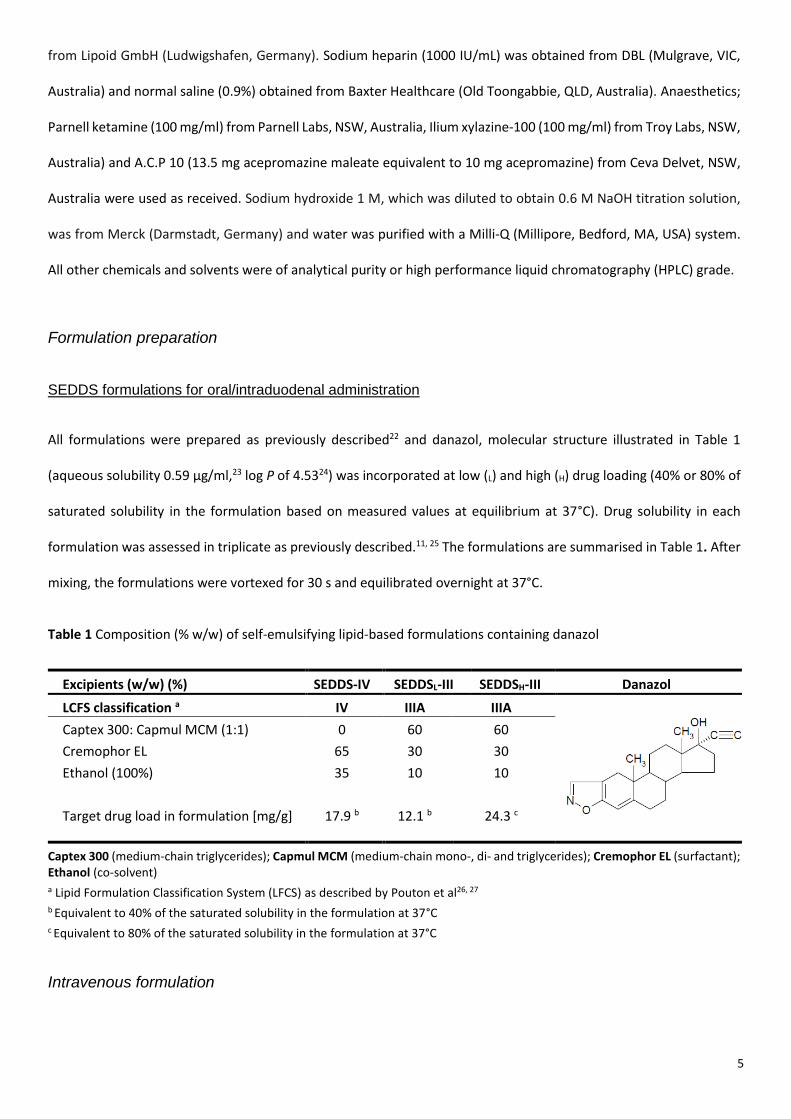
All formulations were prepared as previously described22 and danazol, molecular structure illustrated in Table 1
(aqueous solubility 0.59 µg/ml,23 log P of 4.5324) was incorporated at low (L) and high (H) drug loading (40% or 80% of
saturated solubility in the formulation based on measured values at equilibrium at 37°C). Drug solubility in each
formulation was assessed in triplicate as previously described.11, 25 The formulations are summarised in Table 1. After
mixing, the formulations were vortexed for 30 s and equilibrated overnight at 37°C.
Table 1 Composition (% w/w) of self-emulsifying lipid-based formulations containing danazol
Excipients (w/w) (%) SEDDS-IV SEDDSL-III SEDDSH-III Danazol
LCFS classification a IV IIIA IIIA
Captex 300: Capmul MCM (1:1) 0 60 60
Cremophor EL 65 30 30
Ethanol (100%) 35 10 10
Target drug load in formulation [mg/g] 17.9 b 12.1 b 24.3 c
Captex 300 (medium-chain triglycerides); Capmul MCM (medium-chain mono-, di- and triglycerides); Cremophor EL (surfactant); Ethanol (co-solvent) a Lipid Formulation Classification System (LFCS) as described by Pouton et al26, 27 b Equivalent to 40% of the saturated solubility in the formulation at 37°C c Equivalent to 80% of the saturated solubility in the formulation at 37°C
Intravenous formulation
6
An intravenous formulation of danazol (1.2 mg/mL) was prepared using 15% (w/v) hydroxypropyl-β-cyclodextrin (HP-
β-CD). The binding constant for danazol to -cyclodextrin is relatively low (9.72 x 10² M-1)28 and no significant impact
on PK parameters is therefore expected in spite of the relatively high cyclodextrin concentration employed.29
Danazol and HP-β-CD were dissolved in 0.9% saline using a magnetic stirrer (Teflon coated stirrer bar, 10 x 6mm) at
ambient temperature and filtered through a 0.22 μm filter (Millix®-GV) before use.
BIOAVAILABILITY STUDIES IN RATS
SEDDS formulations were evaluated in vivo to examine the impact of excipients on drug absorption. In some test
groups, a non-specific cytochrome P450 inhibitor (1-aminobenzotriazole, ABT), that is commonly used in animal
models to inhibit hepatic and intestinal CYP enzymes, was administered orally.30, 31 Danazol bioavailability was
explored following oral and intraduodenal (ID) administration to assess the impact of gastric processing on
formulation performance.
Surgical and experimental procedures
All surgical and experimental procedures were approved by the Monash Institute of Pharmaceutical Sciences animal
ethics committee and were conducted in accordance with EC Directive 86/609/EEC for animal experiments and the
Australian and New Zealand Council for the Care of Animals in Research and Teaching guidelines.
Experiments were conducted as a series of one-way parallel studies in male Sprague Dawley rats (250-320 g). Surgical
and recovery procedures were as described previously.32, 33 Briefly, polyethylene tubing cannulas (0.96 x 0.58) were
surgically implanted into the right carotid artery to facilitate serial blood collection. In some treatment groups,
cannulas were also inserted into the right jugular vein (for IV administration) or duodenum (for intraduodenal (ID)
administration). Animals were allowed to recover overnight prior to formulation administration. At the conclusion of
the experiments, rats were euthanised via a lethal dose of sodium pentobarbitone (100 mg/mL).
Formulation administration and sample collection
Rats were fasted for 12 h prior to dosing and remained fasted until the conclusion of the study. Drinking water was
provided ad libitum. In groups pre-treated with ABT (100 mg/kg), a single bolus dose (1.2 mL) of ABT in saline (25

7
mg/mL) was administered via oral gavage post surgery, approximately 14 h prior to IV, oral or ID dosing.30, 31 A similar
dosing protocol has been shown previously to provide almost complete inhibition (93%) of CYP-mediated antipyrine
clearance.30
The intravenous formulation (2.3 mg/kg) was administered over 0.5 min by infusion pump (1 mL/min) via the
indwelling jugular vein cannula. The cannula was subsequently flushed with 0.5 mL of saline. Blood samples (300 µL)
were taken pre-dose and at 1, 5, 15, 30, 60, 120, 180, 240, 360 and 480 min after IV administration.
For the oral and ID treatment groups, SEDDS formulations were pre-dispersed in water (100 mg formulation + 400 mg
MilliQ water) immediately prior to administration. For ID dosing, the pre-dispersed formulations were administered
into the duodenum via the ID cannula over 30 min at a constant infusion rate (1 mL/hr) followed by infusion of 0.5 mL
of MilliQ water to flush the cannula. Blood samples (300 µL) were taken at pre-dose, 15, 25, 35, 45, 60, 90 min and 2,
3, and 5 h after ID dosing. For oral administration, rats were lightly anaesthetised via inhalation of isoflurane (2.5%
v/v), and the pre-dispersed formulations were dosed via oral gavage followed by 0.5 mL MilliQ water to rinse the
gavage tube. Blood samples were taken at pre-dose, 15, 30, 45, 60, 90 min and 2, 3, 4 and 5 h after oral dosing. Blood
samples were collected into 1.5 mL eppendorf tubes containing 10 µL of sodium heparin (10 IU/mL) and cannulas
were flushed with heparinised saline (2 IU/mL) between samples to ensure patency of the cannula. Samples were
centrifuged for 5 min at 6700 x g (Eppendorf minispin plus, Eppendorf AG, Hamburg, Germany) to separate plasma.
Collected plasma samples were stored in eppendorfs at -80°C until analysis.
Quantification of danazol in plasma samples by LC-MS
Plasma samples and calibration standards for danazol were prepared and quantified by LC-MS as previously
described.34
Pharmacokinetic Data analysis
The peak plasma concentrations (Cmax) and the time for their occurrence (Tmax) were noted directly from the individual
plasma concentration vs. time profiles. The areas under the plasma concentration vs. time profiles to the last
measured timepoint (AUC0-tz) were calculated using the linear trapezoidal method. In the absent of ABT treatment,
the AUC was in general very low and the terminal phase poorly defined. The mean terminal elimination rate constant
8
from the IV study was therefore used to extrapolate the AUC to infinity (AUC0-). In the presence of ABT, the AUCs
were higher and extrapolated AUC was based on elimination rate constants obtained from individual plasma profiles.
Clearance (Cl), volume of distribution (Vdβ) and bioavailability (F) were calculated using standard calculation methods.
Statistically significant differences were determined by ANOVA followed by a Tukey test for multiple comparisons at
a significance level of α = 0.05. All statistical analysis was performed using SigmaPlot Statistics for Windows version
11.0.
IN VITRO EXPERIMENTS
In vitro dispersion of SEDDS formulations
Evaluation of the impact of gastric dispersion was performed in model gastric fluid using two dilution levels and pHs,
and also in ex vivo rat gastric fluids. First, 1 g of lipid-formulation (SEDDSL-III and SEDDS-IV) was dispersed in 36 mL
simulated gastric fluid (0.1 N HCL, pH of 1.2). Experiments were performed in a glass vessel at 37°C with a
thermostatically controlled water jacket and stirred magnetically (disc-shaped Teflon coated stirrer bar, 10 x 14 mm)
with samples (200 μL) collected after 30 min.
Subsequently, a low dilution/ intermediate pH model was utilized to better reflect rat gastric conditions. Here, 100
mg of lipid-based formulation was dispersed in 900 µL of buffer (pH 5.5)35 (i.e. 1:10 dilution consistent with volumes
administered in vivo). The dispersion was stirred using a magnetic stirrer (Teflon coated stirrer bar, 10 x 6 mm) and
samples (200 μL) collected after 30 min.
For experiments using ex-vivo gastric fluids, male Sprague Dawley (SD) rats (300-400 g) were fasted for 12 h prior to
surgery with free access to drinking water. Animals were anesthetised with isoflurane (5% v/v) and a ligature tied
around the oesophageal and duodenal apertures of the stomach. The stomach was excised and rinsed with 900 μL
MilliQ (the volume of fluid dosed with the formulations in the oral bioavailability experiments). The rinsing fluid (‘ex
vivo stomach fluid’) was collected and utilised in low volume dispersion experiments as described above.
In vitro digestion of SEDDS formulations
9
The impact of digestion on the solubilisation properties of SEDDS formulations containing danazol was examined using
a range of protocols that employed different sources and quantities of digestive enzyme in an attempt to most
effectively mirror conditions in the rat in vivo. The different conditions are summarised in Table 2 and described
below.
Dog digestion model – High dilution / High enzyme activity
In vitro digestion model (high dilution) using 100% porcine pancreatin extract (1000 TBU/mL)
This is the in vitro model used in previous in vitro studies,11 and that was designed to estimate the impact of digestion
on SEDDS formulations in larger species such as dogs or humans.12 In these studies, 1 g of formulation was dispersed
in 36 mL of digestion media in a thermostatically controlled (37°C) vessel and digestion initiated by addition of porcine
pancreatin extract (4 mL).
Rat digestion model - High dilution / Low enzyme activity
In vitro rat digestion model (high dilution) using ex-vivo rat pancreatic/biliary fluid
To better mimic conditions in the rat intestine, digestions were carried out using collected (ex vivo) rat
pancreatic/biliary secretions (collection method described below). Due to the relatively low rate of secretion of these
fluids in the rat (1-1.5 mL/h), initial studies were conducted in 10 mL in vitro digests. The ratio of formulation to
digestion medium was kept constant relative to previous studies36 and experiments conducted using 250 mg
formulation in 9 mL digestion medium with 1 mL of ex vivo rat pancreatic/biliary secretions to stimulate digestion.
Since rat pancreatic/bile secretions contain bile, experiments were conducted in digestion buffer without added bile
salt and phospholipid but were otherwise conducted as described previously.36 1 mL samples of digestion medium
were collected following 30 min dispersion and at 10, 20, 30 and 60 min post initiation of digestion.
In vitro rat digestion model (low dilution) using 1.7% porcine pancreatin extract
In vitro digests were also performed using the ‘rat’ digestion protocol described above (high dilution/low enzyme) but
using a quantity (17 μL) of porcine enzyme that provided a similar enzyme activity to that of 1 mL ex-vivo rat pancreatic
fluid. The activity of the ex-vivo rat pancreatic/biliary fluid and the quantity of porcine pancreatic fluid required to
mimic this activity was evaluated using a tributyrin assay as described below.

10
Rat digestion model – Low dilution / Low enzyme activity
In vitro rat digestion model (low dilution) using ex-vivo rat pancreatic/biliary fluid
In conducting the surgical procedures required to collect bile in the rats, it became apparent that the volume of fluid
in the rat intestine was low (certainly much lower than the dilution factor of 40 used in the initial in vitro rat digestion
experiments), and that under fasted state conditions the major source of fluid flow into the intestine was from the
bile. As such, a second series of experiments was conducted using a lower volume of digestion media (and therefore
lower dilution). Low dilution digestions were performed using a 1:1 (w/w) mixture of dispersed formulation and rat
bile/pancreatic secretion. The dispersed formulation contained 20% w/v formulation in MilliQ water reflecting the
ratio of formulation to dispersion fluid employed in the in vivo studies (i.e. 100 mg formulation to 400 µL MilliQ).
Additionally, 250 µL MilliQ water was added to simulate the low levels of basal intestinal fluids that might be present
in the GI tract during in vivo studies. Studies were performed in a 10 mL glass vial and media was stirred magnetically
(Teflon coated stirrer bar, 10 x 6 mm). Samples of the digest (300 μL) were collected after 30 min. It was not possible
to employ a pH stat for maintenance of pH during the digestions due to the low volume employed, however, following
digestion the pH of the digestion medium was 7.9 and 5.8 for SEDDS-IV and SEDDS-III, respectively.
In vitro rat digestion model (low dilution) using 1.7% porcine pancreatin extract
In vitro digestion experiments were also conducted as described above, but with ex vivo rat bile/pancreatic fluid
replaced with 17 μL porcine pancreatic extract.
Determination of pancreatic lipase activity in rat pancreatic/biliary fluid
The lipolytic activity of enzymes in ex vivo rat bile/pancreatic fluid was determined in tributyrin units (TBUs) as
previously described,37 where 1 TBU is defined as quantity of enzyme releasing 1 mol of titratable butyric acid per
minute.38 Briefly, 6 g of tributyrin was dispersed in 9 g of digestion buffer (50 mM TRIS maleate, 150 mM NaCl, 5 mM
CaCl2·2H2O, pH 7.5). Experiments were performed at 37°C. Digestion was initiated by addition of 1 mL of collected ex
vivo pancreatic fluids following 2 min dispersion and pH was maintained at 7.5 using a pH-stat titration unit
(Radiometer, Copenhagen, Denmark). Digestion was followed for 30 min and lipase activity in TBU (mol titratable
11
butyric acid per minute) calculated from the initial rate of digestion (i.e. via the slope of the titration profile (i.e. fatty
acids (mmol) liberated per min) multiplied by the molar strength of titrant (0.5 M)).
To determine the relative lipase activity of ex-vivo rat pancreatic fluid and the porcine pancreatin extract used
previously, the lipase activity of 1 mL of porcine pancreatin enzyme extract was also examined under the same
conditions (although in this case the digestion buffer contained 5 mM NaTDC and 1.25 mM phosphatidycholine (PC)
since the enzyme preparation did not contain bile). Porcine pancreatic lipase extract was prepared by dispersing 1000
mg of porcine pancreatin (8 x USP specification activity) in 5 mL digestion buffer. The mixture was stirred for 15 min
and centrifuged for 10 min (2,880 x g at 5°C, Eppendorf 5804 R centrifuge, Eppendorf AG, Hamburg, Germany). The
supernatant was separated and used for digestion studies on the day of preparation.
Further experiments were conducted using lower quantities of porcine pancreatic extract to provide a ‘standard
curve’ of enzyme activity as a function of the volume/quantity of porcine pancreatic enzyme added. This identified
the quantity of porcine pancreatic extract needed to match the activity of 1 mL of ex-vivo rat pancreatic/biliary fluid.
In this case, experiments were conducted using volumes of porcine pancreatic extract of less than 1 ml and additional
buffer was added to maintain a total volume of 1 mL added to the digestion vessel.
Pancreatic and biliary fluid collection from the rat
Ex vivo pancreatic/biliary fluid from the rat (containing bile and pancreatic enzymes) was collected via bile duct
cannulation. In the rat the bile duct also serves as the main duct for the transfer of pancreatic secretions to the GIT
and to obtain an accurate secretion ratio between biliary and pancreatic fluids secreted in vivo, fluids were collected
together. Anaesthesia and surgical procedures in rats were performed as previously described.39-41 A ligature was then
tied around the bile duct at the point of entry into the duodenum and an incision was made in the duct above the
ligature and a polyethylene cannula (0.61 x 0.28 mm, o.d. x i.d.) inserted. Bile and pancreatic fluids were collected
continuously for a 2 h period (achieving approximately 1.5 mL/h) and used immediately after collection. Rats were re-
hydrated via saline infusion (1.5 mL/h) into a cannula inserted into the right jugular vein during the collection period.

12
Table 2 Conditions for in vitro digestion experiments employing ex vivo rat bile/pancreatic fluids and porcine pancreatic extract
Digestion model
(dilution/enzyme activity) Dog (High / High) Rat (High / Low) Rat (Low / Low)
Lipase source Porcine pancreatin
extract a
Porcine pancreatin
extract a
Ex vivo rat pancreatic /
biliary fluid
Porcine pancreatin
extract a
Ex vivo rat pancreatic /
biliary fluid
Formulation [mg] 1000 250 250 200 200
Digestion medium b micelles micelles buffer micelles buffer
Pancreatic enzymes 4 mL 0.017 mL c 1 mL 0.017 mL c 1 mL
Total volume 40 mL 10 mL 10 mL 2.05 mL 2.05 mL
Formulation dilution 40 (High) 40 (High) 40 (High) 10 (Low) 10 (Low)
Enzyme activity High Low Low Low Low
a Source of porcine pancreatic extract as previously published37 b Ex vivo rat pancreatic/biliary fluid contains bile. Experiments were therefore carried out in digestion buffer without added bile salt. In vitro digestions using porcine pancreatic extract were performed in digestion medium containing bile salt micelles (5 mM NaTDC and 1.25 mM phosphatidycholine (PC)) c The total volume of pancreatic enzyme added was kept constant (1 ml). The 1 ml volume was made up of 17 µL enzyme extract and 983 µL digestion medium. The quantity of enzyme extract is listed here to emphasise comparison with other groups. The additional volume of digestion medium is included in the total volume of media employed

13
Sample work up for in vitro dispersion/digestion experiments
The lipase inhibitor 4-bromophenylboronic acid42 (4-BPB; 9 µL of a 0.5 M solution in methanol per mL of digestion
medium) was added to all in vitro digestion samples to prevent further digestion of the lipid components during phase
separation. Dispersion samples were centrifuged for 10 min and digestion samples for 30 min at 21,100 x g (Heraeus
Fresco 21 microcentrifuge, Thermo Scientific, Germany) to form a pellet phase and a solubilised aqueous phase (AP).
Samples obtained from each phase were diluted (aqueous phase: 50 µL to 950 µL in ACN; precipitate (pellet) and oil
phase: initially in 50 µL chloroform/methanol (2:1 v/v) and this further diluted with 950 µL MeOH) and then analysed
by HPLC as below.
Quantification of Danazol in in vitro Experiments
Danazol concentrations in samples taken from in vitro dispersion and digestion experiments were measured by HPLC
as described previously.11 The results are presented as the % drug distribution in the aqueous phase (AP), ie the
proportion of the drug initially dissolved in the formulation that was recovered in the aqueous phase post
centrifugation of the dispersion or digestion media.
RESULTS
In vivo evaluation
Intravenous pharmacokinetics of danazol
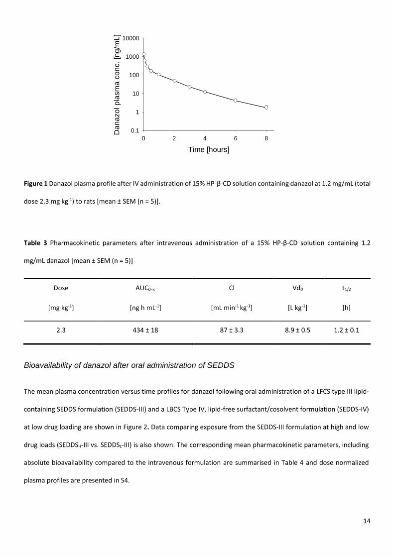
The mean plasma concentration versus time profile for danazol following intravenous administration of a 15% HP-β-
CD solution containing danazol at 1.2 mg/mL to rats is shown in Figure 1 with corresponding mean pharmacokinetic
parameters summarised in Table 3. Plasma concentrations declined bi-exponentially. The total clearance and volume
of distribution were high (87 mL min-1 kg-1 and 8.9 L kg-1 respectively) and the terminal half-life relatively short (1.2 h).
14
Figure 1 Danazol plasma profile after IV administration of 15% HP-β-CD solution containing danazol at 1.2 mg/mL (total
dose 2.3 mg kg-1) to rats [mean ± SEM (n = 5)].
Table 3 Pharmacokinetic parameters after intravenous administration of a 15% HP-β-CD solution containing 1.2
mg/mL danazol [mean ± SEM (n = 5)]
Dose AUC0-∞ Cl Vdβ t1/2
[mg kg-1] [ng h mL-1] [mL min-1 kg-1] [L kg-1] [h]
2.3 434 ± 18 87 ± 3.3 8.9 ± 0.5 1.2 ± 0.1
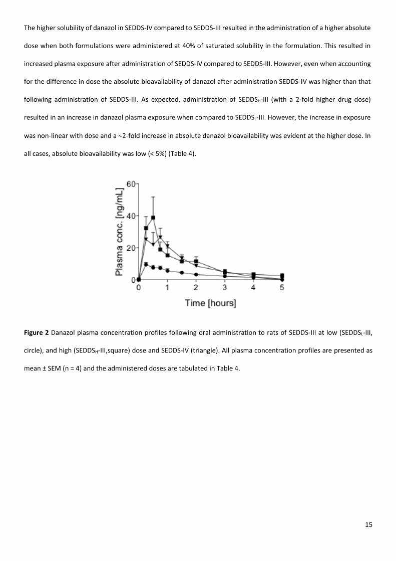
Bioavailability of danazol after oral administration of SEDDS
The mean plasma concentration versus time profiles for danazol following oral administration of a LFCS type III lipid-
containing SEDDS formulation (SEDDS-III) and a LBCS Type IV, lipid-free surfactant/cosolvent formulation (SEDDS-IV)
at low drug loading are shown in Figure 2. Data comparing exposure from the SEDDS-III formulation at high and low
drug loads (SEDDSH-III vs. SEDDSL-III) is also shown. The corresponding mean pharmacokinetic parameters, including
absolute bioavailability compared to the intravenous formulation are summarised in Table 4 and dose normalized
plasma profiles are presented in S4.
Time [hours]
0 2 4 6 8
Danazol pla
sm
a c
onc.
[ng
/mL]
0.1
1
10
100
1000
10000
15
The higher solubility of danazol in SEDDS-IV compared to SEDDS-III resulted in the administration of a higher absolute
dose when both formulations were administered at 40% of saturated solubility in the formulation. This resulted in
increased plasma exposure after administration of SEDDS-IV compared to SEDDS-III. However, even when accounting
for the difference in dose the absolute bioavailability of danazol after administration SEDDS-IV was higher than that
following administration of SEDDS-III. As expected, administration of SEDDSH-III (with a 2-fold higher drug dose)
resulted in an increase in danazol plasma exposure when compared to SEDDSL-III. However, the increase in exposure
was non-linear with dose and a 2-fold increase in absolute danazol bioavailability was evident at the higher dose. In
all cases, absolute bioavailability was low (< 5%) (Table 4).
Figure 2 Danazol plasma concentration profiles following oral administration to rats of SEDDS-III at low (SEDDSL-III,
circle), and high (SEDDSH-III,square) dose and SEDDS-IV (triangle). All plasma concentration profiles are presented as
mean ± SEM (n = 4) and the administered doses are tabulated in Table 4.
16
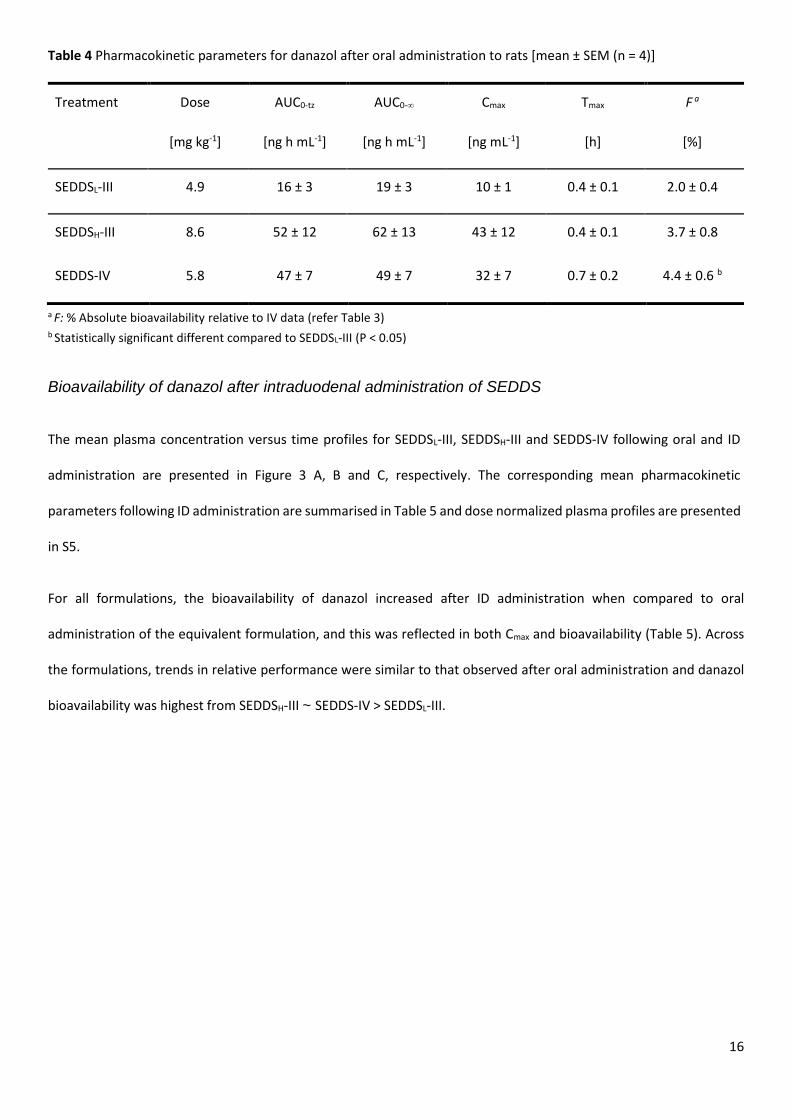
Table 4 Pharmacokinetic parameters for danazol after oral administration to rats [mean ± SEM (n = 4)]
Treatment Dose AUC0-tz AUC0-∞ Cmax Tmax F a
[mg kg-1] [ng h mL-1] [ng h mL-1] [ng mL-1] [h] [%]
SEDDSL-III 4.9 16 ± 3 19 ± 3 10 ± 1 0.4 ± 0.1 2.0 ± 0.4
SEDDSH-III 8.6 52 ± 12 62 ± 13 43 ± 12 0.4 ± 0.1 3.7 ± 0.8
SEDDS-IV 5.8 47 ± 7 49 ± 7 32 ± 7 0.7 ± 0.2 4.4 ± 0.6 b
a F: % Absolute bioavailability relative to IV data (refer Table 3) b Statistically significant different compared to SEDDSL-III (P < 0.05)
Bioavailability of danazol after intraduodenal administration of SEDDS
The mean plasma concentration versus time profiles for SEDDSL-III, SEDDSH-III and SEDDS-IV following oral and ID
administration are presented in Figure 3 A, B and C, respectively. The corresponding mean pharmacokinetic
parameters following ID administration are summarised in Table 5 and dose normalized plasma profiles are presented
in S5.
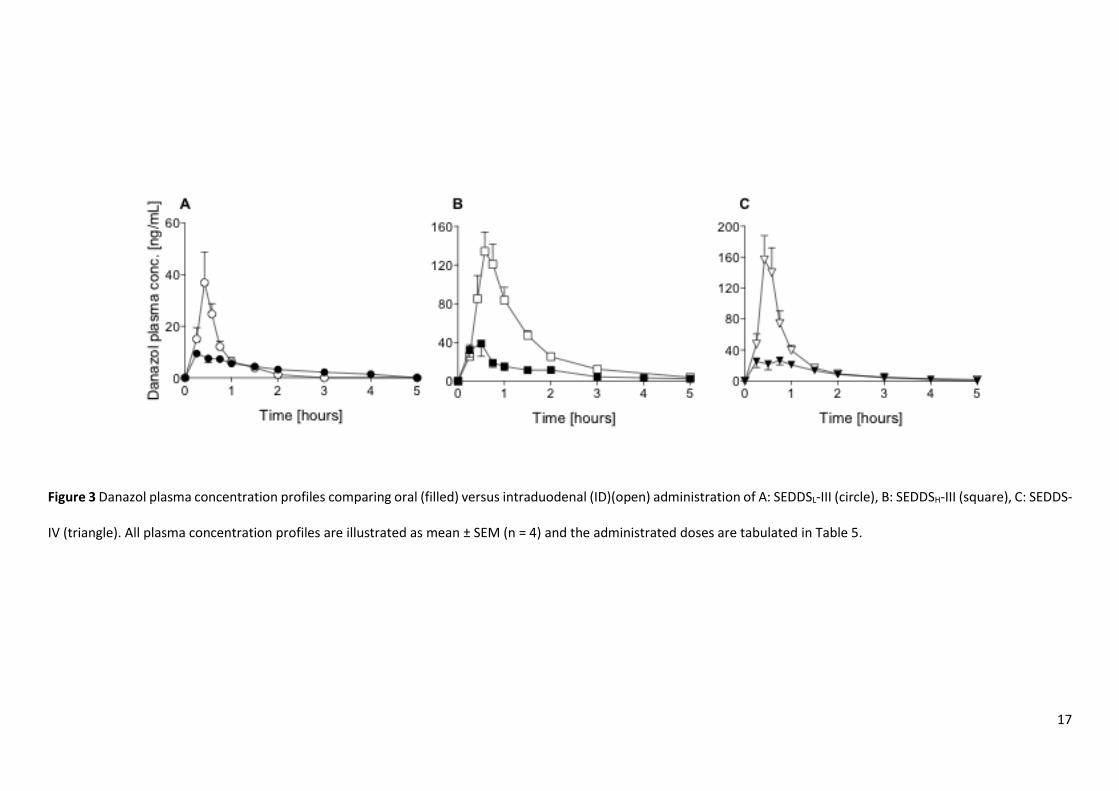
For all formulations, the bioavailability of danazol increased after ID administration when compared to oral
administration of the equivalent formulation, and this was reflected in both Cmax and bioavailability (Table 5). Across
the formulations, trends in relative performance were similar to that observed after oral administration and danazol
bioavailability was highest from SEDDSH-III ~ SEDDS-IV > SEDDSL-III.
17
Figure 3 Danazol plasma concentration profiles comparing oral (filled) versus intraduodenal (ID)(open) administration of A: SEDDSL-III (circle), B: SEDDSH-III (square), C: SEDDS-
IV (triangle). All plasma concentration profiles are illustrated as mean ± SEM (n = 4) and the administrated doses are tabulated in Table 5.
18
Table 5 Pharmacokinetic parameters for danazol after ID administration to rats [mean ± SEM (n = 4)]
Treatment Dose AUC0-tz AUC0-∞ Cmax Tmax F a
[mg kg-1] [ng h mL-1] [ng h mL-1] [ng mL-1] [h] [%]
SEDDSL-III 4.0 25 ± 7 27 ± 8 39 ± 12 0.5 ± 0.0 3.6 ± 1.0
SEDDSH-III 6.3 151 ± 18 156 ± 19 145 ± 21 0.6 ± 0.0 12.8 ± 1.5 bc
SEDDS-IV 5.3 113 ± 23 116 ± 23 161 ± 29 0.5 ± 0.0 11.3 ± 2.3 bc
a F: % Absolute bioavailability relative to IV data (refer Table 3) b Statistically significant different compared to SEDDSL-III (P <0.05) c Statistically significant different compared to oral administration of equivalent formulation (P < 0.050) (see Figure 2 and Table 4)
Impact of first pass metabolism on danazol bioavailability from SEDDS
The impact of first pass metabolism was explored by administration of a non-specific cytochrome P450 inhibitor, 1-
aminobenzotriazole (ABT), prior to danazol administration. The influence of ABT on danazol elimination was evaluated
following IV administration of a 15% HP-β-CD solution containing danazol at 1.2 mg/mL in ABT pre-treated rats (a
comparison of the mean plasma concentration versus time profiles for danazol in the presence and absence of ABT
(S1) and the corresponding tabulated mean pharmacokinetic parameters (S2) can be found in supporting
information).
ABT pre-treatment resulted in a significant reduction in danazol clearance (50 mL min-1 kg-1 compared to 87 mL min-1
kg-1 in the absence of ABT) with a corresponding increase in elimination half-life (1.8 h versus 1.2 h in the absence of
ABT). No significant difference in volume of distribution was observed (7.6 L kg-1 compared to 8.9 L kg-1 in the absence
of ABT).
The mean plasma concentration versus time profiles for danazol after oral and ID administration to ABT pre-treated
rats are shown in Figure 4 with mean pharmacokinetic parameters summarised in Table 6 (dose normalized plasma
profiles are presented in S6).
19
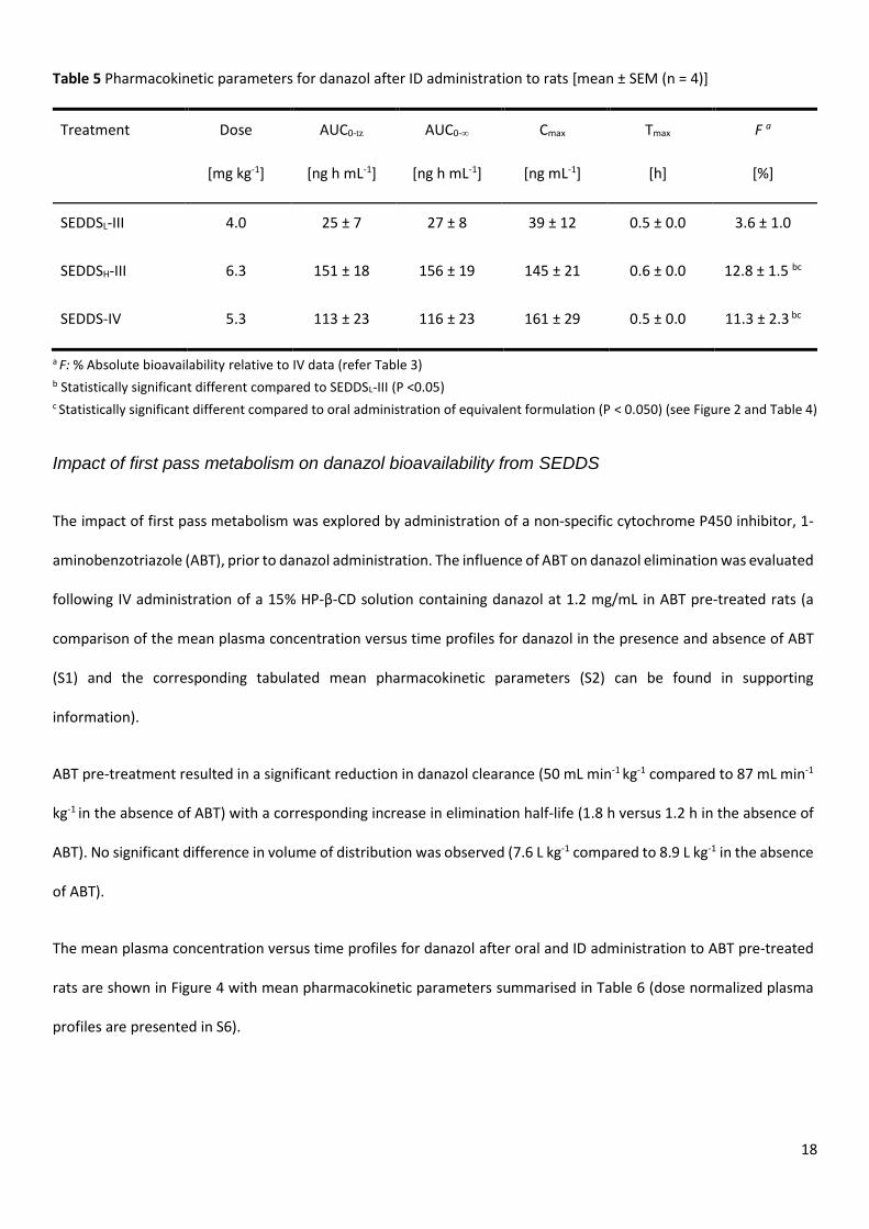
Panel A in Figure 4 shows the plasma profiles following oral administration of SEDDS-III at low and high drug load and
SEDDS-IV in ABT pre-treated animals. Inhibition of CYP-metabolism resulted in significant increases in AUC compared
to non ABT-treated animals (Figure 2). When compared with danazol exposure after IV administration in the presence
and absence of ABT, oral bioavailability was much higher (up to 30 fold) suggesting the presence of a significant first
pass effect for danazol in rats.
Oral administration of SEDDS-IV in the presence of ABT resulted in 45.4% danazol bioavailability compared to only
4.4% in the ABT untreated group. Increasing the lipid content in the formulation (and decreasing the surfactant
content) by administering SEDDSL-III, resulted in a small increase in bioavailability (to 59.7%) compared to SEDDS-IV,
but again a very large increase relative to administration of the same formulation in the absence of ABT (~30-fold).
When SEDDSH-III was administered orally to ABT pre-treated rats (Figure 4A), increasing the drug loading in SEDDS-III
led to a linear increase in Cmax and AUC and the oral bioavailability of SEDDS-III was unaffected.
Panel B in Figure 4 compares plasma profiles following ID and oral administration of SEDDSL-III to ABT-treated rats.
The oral bioavailability of danazol following ID administration in the presence of ABT was essentially complete
(111.0%), and significantly higher than bioavailability after oral administration of the same formulation, also in the
presence of ABT.
A B
Time [hours]
0 1 2 3 4 5
0
200
400
600
Time [hours]
0 1 2 3 4 5
Pla
sm
a c
onc.
[ng/m
L]
0
200
400
600
20
Figure 4 Danazol plasma concentration profile from ABT pre-treated rats following A: oral administration of SEDDSL-III
(filled circle), SEDDSH-III (filled square), SEDDS-IV (filled triangle); B: Intraduodenal (open) versus oral (filled)
administration of SEDDSL-III. All plasma concentration profiles are illustrated as mean ± SEM (n = 4).
Table 6 Pharmacokinetic parameters for danazol after oral and intraduodenal administration to ABT pre-treated rats
[mean ± SEM (n = 4)].
Treatment Dose AUC0-tz AUC0-∞ Cmax t1/2 Tmax F a
[mg kg-1] [ng h mL-1] [ng h mL-1] [ng mL-1] [h] [h] [%]
ABT-SEDDS-IV (O) b 6.4 737 ± 21 975 ± 58 284 ± 36 2.3 ± 0.3 0.7 ± 0.2 45.4 ± 3
ABT-SEDDSL-III (O) 3.9 636 ± 71 781 ± 103 258 ± 26 1.9 ± 0.3 1.3 ± 0.3 59.7 ± 8
ABT- SEDDSH-III (O) 9.5 1344 ± 127 1643 ± 152 528 ± 72 1.8 ± 0.1 0.9 ± 0.1 51.6 ± 5
ABT-SEDDSL-III (I.D) c 3.4 996 ± 102 1265 ± 141 541 ± 40 1.9 ± 0.2 0.8 ± 0.1 111.0 ± 12 d
a F: % Absolute bioavailability calculated relative to IV data in ABT pre-treated rats (data in supplementary information Fig S2) b Oral administration (O), c Intraduodenal administration (I.D.) d Statistically significant different compared to oral administration of SEDDSL-III (P < 0.050)
IN VITRO EVALUATION
Impact of gastric dispersion and ex vivo gastric fluid on drug precipitation from SEDDS
The impact of formulation processing under gastric conditions on the in vitro performance of the investigated
formulations (SEDDS-IV / SEDDSL-III) was evaluated in a series of dispersion studies (Figure 5).

21
Dispersion of SEDDS-IV under high dilution conditions at pH 1.2 led to rapid drug precipitation and only 40% of the
initial drug load was retained in a solubilized state after 30 min. In comparison, no drug precipitation was observed on
dispersion of SEDDSL-III under similar conditions (Figure 5A). Experiments were also conducted under lower dilution
conditions (1 in 10) and at pH 5.5 (Figure 5B). Decreasing the volume of dispersion medium did not affect drug
solubilisation patterns when compared to high volume conditions for either SEDDSL-III or SEDDS-IV. Increasing the
drug load in SEDDS-III, however, resulted in ~20% drug precipitation for SEDDSH-III (Figure 5B).
Solubilisation/precipitation patterns following formulation dispersion in ex vivo gastric fluids from rats are shown in
Figure 5C. The dispersion of SEDDSL-III and SEDDS-IV in ex vivo gastric fluids resulted in similar drug solubilisation
patterns to that observed using the simpler in vitro conditions. The mean pH of the collected ex vivo gastric fluid was
4.8, which is in agreement with previously published studies35 and similar to that used in the low dilution simulated
rat gastric fluid buffer (pH 5.5)
Figure 5 The extent of danazol precipitation after 30 min dispersion of SEDDS-IV and SEDDSL-III (drug loaded at 40%
saturated solubility) under gastric condition [mean ± SD (n = 3)] in A: 40 mL 0.1 N HCL pH 1.2; B: 0.9 mL buffer, pH 5.5;
C: 0.9 mL ex vivo rat gastric fluid pH 4.8. In addition, panel B also shows SEDDSH-III (80% saturated solubility) and
therefore the effect of drug loading on drug precipitation after 30 min of dispersion. Differences in drug loading in
panel B are indicated by 40% and 80% labels within the bars. Bars represent danazol in aqueous phase (light blue) and
precipitate (dark grey) [mean ± SD (n = 3)].
Impact of intestinal digestion on in vitro performance of danazol SEDDS
22
Development of a rat in vitro digestion model
Rat digestion model – high dilution / low enzyme activity
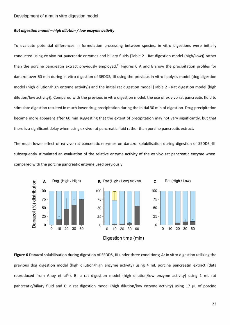
To evaluate potential differences in formulation processing between species, in vitro digestions were initially
conducted using ex vivo rat pancreatic enzymes and biliary fluids (Table 2 - Rat digestion model (high/Low)) rather
than the porcine pancreatin extract previously employed.11 Figures 6 A and B show the precipitation profiles for
danazol over 60 min during in vitro digestion of SEDDSL-III using the previous in vitro lipolysis model (dog digestion
model (high dilution/high enzyme activity)) and the initial rat digestion model (Table 2 - Rat digestion model (high
dilution/low activity)). Compared with the previous in vitro digestion model, the use of ex vivo rat pancreatic fluid to
stimulate digestion resulted in much lower drug precipitation during the initial 30 min of digestion. Drug precipitation
became more apparent after 60 min suggesting that the extent of precipitation may not vary significantly, but that
there is a significant delay when using ex vivo rat pancreatic fluid rather than porcine pancreatic extract.
The much lower effect of ex vivo rat pancreatic enzymes on danazol solubilisation during digestion of SEDDSL-III
subsequently stimulated an evaluation of the relative enzyme activity of the ex vivo rat pancreatic enzyme when
compared with the porcine pancreatic enzyme used previously.
Figure 6 Danazol solubilisation during digestion of SEDDSL-III under three conditions; A: In vitro digestion utilizing the
previous dog digestion model (high dilution/high enzyme activity) using 4 mL porcine pancreatin extract (data
reproduced from Anby et al11), B: a rat digestion model (high dilution/low enzyme activity) using 1 mL rat
pancreatic/biliary fluid and C: a rat digestion model (high dilution/low enzyme activity) using 17 µL of porcine
23
pancreatin extract to match the activity of ex vivo rat pancreatic/biliary fluid. Bars represent danazol in aqueous
colloidal (light blue), oil phase below colloidal phase (light yellow) and precipitate (dark grey) [mean ± SD (n = 3)].
Evaluation of ex vivo lipase activity
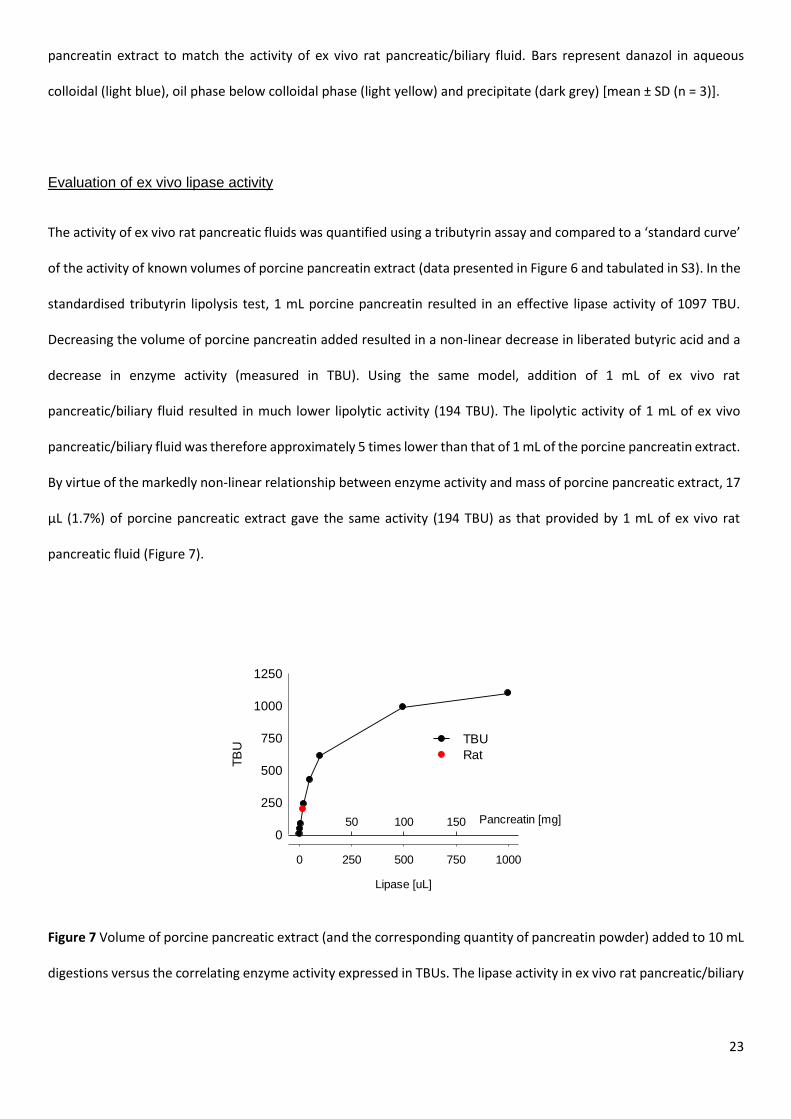
The activity of ex vivo rat pancreatic fluids was quantified using a tributyrin assay and compared to a ‘standard curve’
of the activity of known volumes of porcine pancreatin extract (data presented in Figure 6 and tabulated in S3). In the
standardised tributyrin lipolysis test, 1 mL porcine pancreatin resulted in an effective lipase activity of 1097 TBU.
Decreasing the volume of porcine pancreatin added resulted in a non-linear decrease in liberated butyric acid and a
decrease in enzyme activity (measured in TBU). Using the same model, addition of 1 mL of ex vivo rat
pancreatic/biliary fluid resulted in much lower lipolytic activity (194 TBU). The lipolytic activity of 1 mL of ex vivo
pancreatic/biliary fluid was therefore approximately 5 times lower than that of 1 mL of the porcine pancreatin extract.
By virtue of the markedly non-linear relationship between enzyme activity and mass of porcine pancreatic extract, 17
µL (1.7%) of porcine pancreatic extract gave the same activity (194 TBU) as that provided by 1 mL of ex vivo rat
pancreatic fluid (Figure 7).
Figure 7 Volume of porcine pancreatic extract (and the corresponding quantity of pancreatin powder) added to 10 mL
digestions versus the correlating enzyme activity expressed in TBUs. The lipase activity in ex vivo rat pancreatic/biliary
Lipase [uL]
0 250 500 750 1000
TB
U
0
250
500
750
1000
1250
Pancreatin [mg]50 100 150
TBU
Rat
24
fluid is illustrated by the red circle (194 ± 34 TBU). Similar activity is expected from 17 μL of porcine pancreatic extract
(~ 1.7% of the volume utilized previously).
Drug distribution patterns following addition of 17 µL of porcine pancreatic extract rather than 1 mL of ex vivo rat bile
are shown in Figure 6C. Similar data were seen when compared to the ex vivo rat pancreatic/biliary fluid up to 30 min
digestion, although the increase in digestion at 60 min was not evident.
Rat digestion model – low dilution / low enzyme activity
In an attempt to better reproduce conditions in the GI tract of the rat with volumes likely to be less than 10 mL, a
lower volume (low dilution) rat digestion model (Table 2 - Rat digestion model (low dilution/low activity)) was also
evaluated using the same quantities/sources of enzyme used in the higher volume rat model (i.e. 1 mL of ex vivo rat
pancreatic fluid or 1 mL of 1.7% porcine pancreatic extract). In this case, however, the total volume of digestion media
(buffer plus enzyme) was reduced to 2.05 mL.
Under these conditions, formulation processing and danazol solubilisation profiles were markedly different when
compared to patterns obtained using the much higher volume digests (Figure 8). The most notable change was the
generation of a dense lipid-rich phase for the SEDDS-III formulation, which migrated to the bottom of the tube on
centrifugation. In contrast, digestion and phase separation under higher enzyme loads and higher dilution led to a
pellet phase containing precipitated drug and a highly dispersed micellar aqueous phase. In the low enzyme activity/
low volume model over 99% of the drug from the SEDDSL-III formulation was recovered in the dense oil phase located
below the colloidal aqueous phase (Figure 8A). Similar data were obtained using either ex vivo pancreatic fluid or 1.7%
of the standard porcine pancreatic extract (Figure 8A).
The impact of drug load was also evaluated using the low volume digestion model (Figure 8B). For the SEDDSH-III
formulation, addition of ex vivo lipase led to a similar high-density oil phase that contained the majority of the drug.
Limited drug precipitation was observed after 30 min as shown in Figure 8B, however following 60 min digestion some
drug precipitation was observed, albeit at a relatively low level (< 20%), presumably reflecting the higher drug load.
25
The use of porcine pancreatic extract resulted in a similar profile (Figure 8B), but in this case precipitation was did not
occur at later time points.
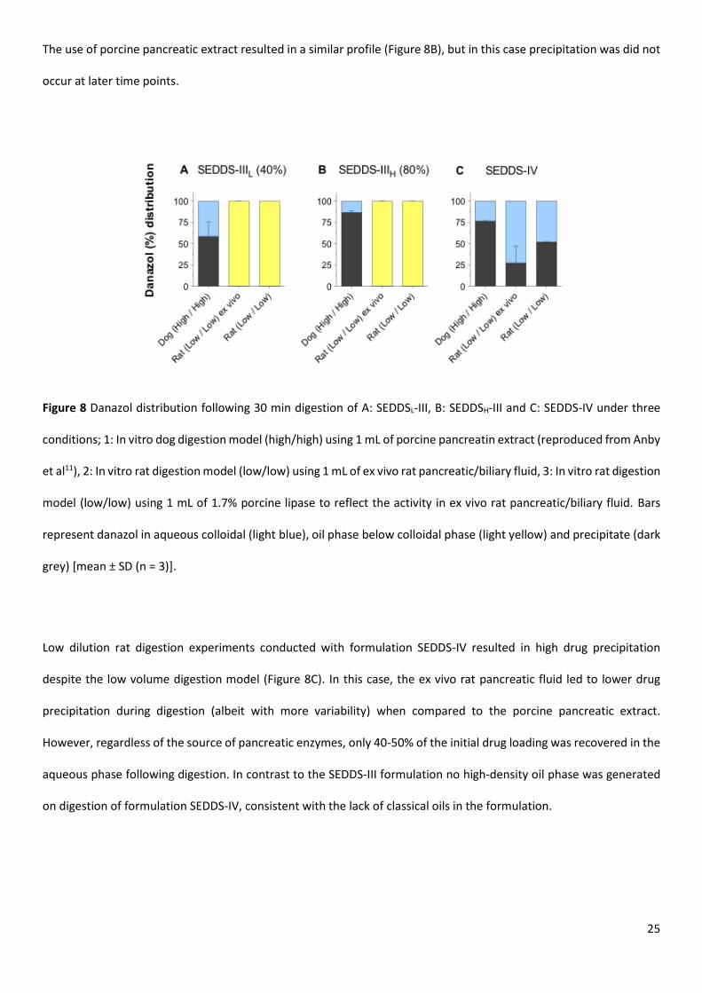
Figure 8 Danazol distribution following 30 min digestion of A: SEDDSL-III, B: SEDDSH-III and C: SEDDS-IV under three
conditions; 1: In vitro dog digestion model (high/high) using 1 mL of porcine pancreatin extract (reproduced from Anby
et al11), 2: In vitro rat digestion model (low/low) using 1 mL of ex vivo rat pancreatic/biliary fluid, 3: In vitro rat digestion
model (low/low) using 1 mL of 1.7% porcine lipase to reflect the activity in ex vivo rat pancreatic/biliary fluid. Bars
represent danazol in aqueous colloidal (light blue), oil phase below colloidal phase (light yellow) and precipitate (dark
grey) [mean ± SD (n = 3)].
Low dilution rat digestion experiments conducted with formulation SEDDS-IV resulted in high drug precipitation
despite the low volume digestion model (Figure 8C). In this case, the ex vivo rat pancreatic fluid led to lower drug
precipitation during digestion (albeit with more variability) when compared to the porcine pancreatic extract.
However, regardless of the source of pancreatic enzymes, only 40-50% of the initial drug loading was recovered in the
aqueous phase following digestion. In contrast to the SEDDS-III formulation no high-density oil phase was generated
on digestion of formulation SEDDS-IV, consistent with the lack of classical oils in the formulation.
26
DISCUSSION
Previous studies of danazol absorption from SEDDS revealed non-linear increases in exposure (bioavailability) with
increasing drug dose in dogs.19 This was suggested to reflect either saturation of first pass metabolism or an increase
in thermodynamic activity at higher doses. Interestingly, the dose-dependent increase in bioavailability was evident in
only one cohort of (older) animals, and not in another (younger) group. The uncertainty generated by these previous
studies stimulated a more detailed examination of danazol absorption from SEDDS formulations in the current study.
Here, the rat model was employed to allow more facile examination of the role of first pass metabolism and to examine
whether the trends seen previously in dogs were replicated in another species (and therefore to rule out species-
specific anomalies). The use of rats also prompted a re-evaluation of the utility of previous in vitro digestion protocols
(that were originally established to reflect events in the dog) to mirror events in the rat where GI volumes and enzyme
activities were expected to be lower.
Danazol bioavailability in rats after administration of SEDDS-III and SEDDS-IV
The plasma profiles in Figure 2 and the pharmacokinetic parameters presented in Table 4 reveal moderate differences
in in vivo exposure of danazol in rats following oral administration of two markedly different lipid based formulations
(SEDDS-IV and SEDDS-III). In both cases, bioavailability was low, in agreement with previous studies in rats using
similar lipid-based formulations where the absolute bioavailability of danazol was < 20%.43, 44 When danazol was
incorporated into SEDDS-III at a 2-fold higher dose (SEDDSH-III), bioavailability increased when compared to the lower
dose formulation (SEDDSL-III). The dose effect data for SEDDS-III was consistent with previous studies in beagle dogs,
at least in older animals.19
Danazol bioavailability after administration of SEDDS-IV, however, was significantly higher (~2-fold) than that
observed after administration of SEDDSL-III, in contrast to previous studies in beagle dogs where little difference in
oral bioavailability was seen across the two formulations.11 The very low absolute bioavailability of danazol in the rat
prompted a more detailed evaluation of the potential causes of the limited exposure. In the first instance,
intraduodenal (ID) administration was explored as a means of circumventing potential gastric processing of lipidic
formulations, on the basis that drug precipitation in the stomach may reduce drug absorption. The plasma profiles in
27
Figure 3 and the pharmacokinetic parameters presented in Table 5 are consistent with this suggestion and ID
administration of both the high cosolvent/surfactant formulation (SEDDS-IV), and the more lipid-rich formulations
(SEDDSL-III and SEDDSH-III) led to increased danazol exposure (1.8-3.5 fold) when compared to oral administration
(although the increase was not statistically significant for SEDDSL-III). Interestingly, the non-linearity in danazol
absorption with increasing dose was not only retained after intraduodenal absorption but was enhanced. Thus, the
bioavailability of danazol after intraduodenal infusion of SEDDSH-III was 12.8%v versus 3.6% for SEDDSL-III (a 3.6-fold
increase) whereas after oral administration danazol bioavailability from the same formulations was 3.6% versus 2%
(a 1.8-fold increase).
The increase in exposure evident after ID administration may reflect avoidance of precipitation events in the stomach
after oral administration. However, in light of the larger increase in intraduodenal bioavailability for the higher dose
formulation (SEDDSH-III) (where precipitation might be expected to be more prevalent, not less), it is also possible
that direct introduction of high concentrations of drug directly into the intestine, may more effectively saturate first
pass enterocyte-based metabolism than is the case after oral administration (where gastric emptying is expected to
delay and dilute entry into the small intestine).
First pass metabolism is the major limitation to danazol oral bioavailability from SEDDS formulations
in the rat
Previous studies have shown that after oral administration of (14C) labelled danazol to rats, biliary excretion of danazol
metabolites is significant (~70%) and that extensive enterohepatic re-cycling occurs.20 In vitro studies have further
shown that danazol is primarily a substrate for CYP3A4 (86%) and to a lesser extent CYP2D6 (11%) and CYP2J2 (3%).45
The role of CYP-mediated danazol metabolism in the rat was explored here using the non-specific cytochrome P450
inhibitor, 1-aminobenzotriazole (ABT). ABT has been used extensively in animal studies to probe the role of CYP3A
and CYP2D in drug clearance and first pass metabolism.30 In rats, pre-treatment with 100 mg/kg ABT 2 hours prior to
the administration of a test compound (antipyrine) substantially inhibits CYP450 enzymes, and a single dose of ABT is
sufficient to inhibit metabolism for over 24 h.30, 31
28
Comparison of oral and IV plasma AUCs obtained after ABT pre-treatment provides an indication of ‘apparent’ danazol
bioavailability in the absence of CYP450-mediated metabolism, and therefore gives a clearer indication of the likely
fraction of dose absorbed, unencumbered by first pass CYP-metabolism. Under these conditions, oral administration
of SEDDSL-III to ABT pre-treated rats resulted in a sharp increase in apparent bioavailability to 59.7% compared to true
bioavailability of 2% in non-ABT pretreated animals (Figure 4 and Figure 2, respectively). First pass metabolism is
therefore a very significant limitation to danazol oral bioavailability in rats under these conditions and at these doses.
Furthermore, in contrast to the data in non-ABT pretreated rats, increasing the drug load in the SEDDSH-III formulation
resulted in similar danazol exposure (51.6%) to that obtained after administration of SEDDSL-III The lack of difference
in danazol bioavailability after administration of the two drug doses in the presence of ABT suggests that the non-
linear increase in danazol bioavailability observed in the absence of ABT stems from saturation of first pass
metabolism rather than differences in thermodynamic activity and fraction of dose absorbed.
After pre-administration of ABT, danazol bioavailability was slightly higher after administration of SEDDSL-III when
compared to the cosolvent/surfactant-based formulation (SEDDS-IV), however, these differences were not significant
(Figure 4). This is in contrast to the data obtained in the absence of ABT where danazol bioavailability from SEDDS-IV
was higher than that from SEDDS-III. The enhanced bioavailability of danazol after administration of the SEDDS-IV
formulation in the absence of ABT is consistent with higher danazol solubility in SEDDS-IV (17.9 mg/g vs. 12.1 mg/g in
SEDDSL-III) (Table 1) and therefore administration of a slightly higher dose at 40% of the saturation solubility. Realizing
the role of first pass metabolism in dictating danazol bioavailability in the rat, it is likely that the higher dose may have
led to greater saturation of first pass metabolism in the absence of ABT. Previous studies have also suggested that
Cremophor EL (present at high concentrations in SEDDS IV) is able to inhibit CYP3A in human and rat liver
microsomes.46, 47 The results obtained here in the absence of ABT may therefore indicate differences in the ability of
the two formulations to inhibit first pass metabolism. More specifically, the higher quantity of Cremophor in SEDDS-
IV [65% (w/w)] compared to SEDDS-III [(30% (w/w)] may help to reduce first pass metabolism and promote
bioavailability. In contrast, in the presence of ABT, metabolic limitations are circumvented and solubilisation events
dominate. Under these circumstances, differences between danazol bioavailability from SEDDS-III and SEDDS IV were
not significant (consistent with the previous data in dogs), although exposure was slightly higher from SEDDS-III.

29
Bearing in mind the increase in exposure in the absence of ABT after ID administration (compared to oral
administration) SEDDSL-III was also administered intraduodenally to ABT pre-treated rats to see whether the same
trends were apparent. In this case, bioavailability increased to 111.0% after ID administration when compared to
59.7% after oral administration. Absorption of danazol was therefore essentially complete after intraduodenal
administration of SEDDSL-III. The increase in bioavailability observed after ID rather than oral administration of
SEDDSL-III to ABT pretreated animals (1.9-fold) was also consistent with the increases seen in non-ABT pretreated
animals (1.8-fold) suggesting that the drivers of enhanced absorption after ID rather than oral administration, in the
presence of ABT, were similar to the drivers of enhanced bioavailability in the absence of ABT. Alignment between
increases in bioavailability in the presence and absence of ABT suggests that in this case the differences between ID
and oral administration may have been mediated by changes to solubilisation rather than first pass metabolism.
In contrast, much greater increases in bioavailability were evident after intraduodenal versus oral administration of
SEDDSH-III to non ABT pre-treated animals (~3.5 fold) when compared to intraduodenal versus oral administration of
SEDDSL-III (1.8-fold). Since bioavailability of danazol after oral administration of SEDDSH-III to ABT pre-treated animals
was as high as ~52% (and therefore the fraction absorbed must have been at least 55%), the 3.5-fold increase in
bioavailability seen after intraduodenal administration in the absence of ABT could not have stemmed (completely)
from increases in absorption. The current data therefore indicate that at the higher drug load (i.e. after administration
of SEDDSH-III) intraduodenal administration was able to more effectively saturate first pass metabolism than was the
case at lower drug doses. This trend was also replicated for SEDDS-IV where increases in ID versus oral bioavailability
were slightly higher (2.6-fold) than were evident for SEDDS-IIIL at the lower danazol dose. Intraduodenal delivery
therefore seems more able to take advantage of direct delivery to the absorptive site and to subsequently inhibit first
pass metabolism when combined with formulations containing higher doses of danazol. Formulation strategies that
deliver high concentration of danazol rapidly to the upper small intestine therefore seem most likely to benefit from
increases in bioavailability, at least in the rat.
In summary, the in vivo data suggest that the primary limitation to danazol bioavailability in the rat is first pass
metabolism, that increasing drug dose leads to increases in bioavailability via saturation of first pass metabolism, that
intraduodenal administration results in increases in bioavailability probably as a result of both increases in absorption

30
and reductions in first pass metabolism, and that based on the data obtained in the presence of ABT, danazol
absorption from the SEDDS formulations examined here is generally good (in contrast to bioavailability). Indeed after
ID administration danazol absorption from SEDDSL-III was almost complete. This is surprising based on previous in
vitro dispersion/digestion data11 that show significant drug precipitation after initiation of digestion for both SEDDS-
IV and SEDDS-III. Interspecies differences in GI tract conditions may, however, influence formulation processing, and
the efficiency of digestion (and subsequent drug precipitation) may be different in the rat when compared to larger
species, such as the dog. The solubilisation behaviour of the SEDDS formulations was therefore also evaluated under
in vitro conditions more reflective of the GI tract in the rat, when compared to GI conditions in the dog.
The effect of gastric dispersion on SEDDS performance in the rat
To provide a comparative assessment of possible behaviour in rats and dogs, the impact of gastric dispersion on
formulation performance was initially evaluated using experimental protocols designed to mimic conditions in the dog
(high formulation dilution in simulated gastric fluid, pH 1.2). Dispersion of SEDDS-IV in pH 1.2, high dilution gastric
media resulted in significant drug precipitation and increased drug precipitation compared to dispersion data
conducted under simulated intestinal conditions (Figure 5A). To better reflect the conditions expected in the GI tract
of the rat, dispersion volume and pH were subsequently altered to 900 μL and pH 5.5, respectively, however, significant
precipitation of danazol from SEDDS-IV was still evident. In contrast, drug solubilisation during dispersion of the
formulation containing greater quantities of lipid (and lower quantities of surfactant and cosolvent), SEDDSL-III, was
not affected by pH or dispersion volume (Figure 5 A, B) and drug precipitation from SEDDSL-III was limited under both
conditions.
The interaction of lingual lipase with medium-chain triglycerides results in the liberation of fatty acid,48 and lingual
lipase activity is reportedly49 high in rodents. Subsequent experiments were therefore conducted to explore the
potential additional impact of ex vivo gastric fluids (containing any available lingual lipase) on danazol precipitation
from SEDDS formulation. These data suggest limited effects of lingual lipase on danazol solubilisation in SEDDS in the
rat stomach (Figure 5C). However, pre-processing of lipids in the stomach may affect subsequent events in the
31
duodenum (i.e., secretion of digestive enzymes and the rate and extent of lipid digestion)50-52 and as such, gastric
digestion by lingual lipase may indirectly affect drug absorption in the small intestine.
Development of a modified in vitro digestion model for SEDDS evaluation in the rat
To explore the potential impact of intestinal digestion on SEDDS performance, an in vitro lipolysis model previously
used to examine digestion events in the dog was modified here to better reflect the conditions in the GI tract of the
rat. Experiments were initially undertaken using the same formulation dilution factor as that previously used in the
‘dog’ in vitro digestion model, but where the source of digestive enzyme was replaced with ex vivo rat pancreatic fluid.
This model is described in the methods as the rat high dilution / low enzyme activity model. Ex vivo pancreatic fluids
were collected from rats by cannulation of the common bile duct resulting in collection of mixed bile and pancreatic
fluids. Analysis of the in vitro activity of recovered rat pancreatic enzyme (using a standardized tributyrin activity test)
revealed activities (~200 TBU/mL pancreatic/bile fluid) much below the values commonly reported in vivo in humans
and dogs, and therefore far below the levels commonly used in in vitro digestion experiments modelled on those
conditions.
Recently, Tønsberg et al53 examined lipase activity in luminal intestinal samples from rats and reported lower activity
(153 U/mL), consistent with the levels utilised in the rat digestion models employed here and consistent with dilution
of pancreatic fluids with bile prior to entry into the intestine. It seems likely therefore that the pancreatic enzyme
levels recorded here, whilst low, reflect lower lipase activity in the rat GI tract when compared to the dog or human.
Formulation digestion was evaluated in a series of in vitro experiments utilising different formulation dilutions, enzyme
activities and enzyme sources. As expected, the conduct of studies using the high dilution/low enzyme activity model
and employing 1 mL of ex vivo rat pancreatic/biliary fluid resulted in much lower lipid digestion and correspondingly
lower levels of drug precipitation when compared to the previously employed dog in vitro conditions (Figure 6). Based
on in vitro analysis of the activity of the ex vivo pancreatic/biliary fluids, subsequent studies were conducted using a
quantity of porcine pancreatic enzyme that was equally active in the TBU test to 1 mL of ex vivo enzyme fluid (Figure
7). Similarly reduced levels of digestion and precipitation were evident, suggesting that substitution of low levels of
porcine pancreatic enzyme may be sufficient to broadly mimic the lipolytic activity of ex vivo rat pancreatic fluids. For
32
different digestible substrates, however, different pancreatic enzymes may be required, and a more detailed series of
studies would be required to fully characterise the similarity of rat pancreatic/biliary fluids to porcine pancreatic
extract.
A digestion model was also evaluated using much lower dilution conditions (the rat low dilution/low activity model).
This was designed to better mimic the lower fluid volumes expected in the rat GI tract where the volume of fluid
administrated with the formulation was ~1 mL and the flow of fluid from the bile duct is ~1.5 mL/h. Under these
conditions, the formulations behaved quite differently, and SEDDS-III formed a dense lipid phase that phase separated
below the aqueous phase. This lipid-rich phase contained 80-99% of the incorporated drug. In contrast, no oil phase
was generated on digestion of SEDDS-IV, the lipid-free formulation, suggesting that the high density oil phase
generated by digestion of SEDDS-III consisted of fatty acids and mono/diglycerides generated via digestion of the lipids
present in SEDDS-III.
Conduct of these experiments at low volume precluded the use of the pH stat titrator and as such, the pH in the digest
was not constrained. Fatty acid liberation therefore resulted in a limited drop in pH during digestion of SEDDS-III (pH
5.8 following 60 min digestion). Nonetheless, pancreatic enzyme activity was expected to be retained at this pH 54. In
contrast, the terminal pH of digestion of SEDDS-IV was higher (pH 7.9) suggesting limited fatty acid liberation,
consistent with the lack of lipid substrate in this formulation.
The presence of an oil phase that floats on centrifugation is common during lipid digestion and typically represents
poorly digested tri- and diglycerides, and less readily solubilised monoglycerides and protonated fatty acids. In
contrast, in the current low dilution/low activity rat digests, the oil phase that was generated on digestion, was a
viscous isotropic phase that was more dense than the solubilised aqueous phase and sedimented when left unstirred,
consistent with previous observations.55, 56 The current data suggest that in the rat, under conditions of lower dilution
in vitro (and potentially in vivo), less readily dispersed lipid phases are formed that are less dense than water. This
may also be exacerbated by the lower pH, resulting in greater quantities of less polar unionised fatty acid. Where
isotropic, partially digested lipid phases are formed under conditions of low enzyme activity and low dilution, the
likelihood of drug precipitation appears to be diminished. Whether drug absorption is possible from these phases
directly or whether further dispersion into e.g. bile salt micelles is required is unknown at this point. Continued

33
dilution is likely to occur and the possibility of transition through different phases, which may not be captured with
the low dilution conditions employed here, is likely. This is supported by the difference between oral and ID
administration in the presence and absence of ABT suggesting that differences in phase generation and how the drug
is presented at the absorption site is important, and that drug precipitation from SEDDS-III may occur in spite of the
low dilution/low enzyme in vitro model.
The current studies therefore suggest that comparison of in vivo drug absorption patterns in the rat with in vitro
digestion data obtained using lipid digestion models that simulate dog/human conditions may lead to overestimation
of drug precipitation and underestimation of absorption. Grove et al57, 58 previously also suggested that the quantity
of GI fluid present in the rat may be low, and that administration of self-emulsifying drug delivery systems under these
conditions may lead to the formation of a more viscous, bicontinuous phase when compared to an emulsion system.
The current in vitro data using the low volume low enzyme activity rat model are consistent with this contention.
Impact of animal model on danazol bioavailability from SEDDS
In the current studies the primary limitation to danazol oral bioavailability in the rat was first pass metabolism, and
this was reduced (and bioavailability enhanced) by administration of higher doses or by direct infusion of the dose
into the duodenum. These data are broadly consistent with previous danazol dose-escalation studies in older beagle
dogs where administration of higher doses also resulted in increased drug exposure.19 However, in beagle dogs the
absolute oral bioavailability of danazol was higher (10-26%)19 than that seen here in rats, and much higher oral
bioavailability of danazol in beagle dogs has previously been reported (64%, 82% and 107%).21, 59 Collectively, the data
suggest that where solubilisation is ensured, danazol bioavailability in the dog may be more than an order of
magnitude higher than that observed here in the rat (< 5% after oral administration), and therefore that first pass
limitations to bioavailability are likely to be lower. Whilst the rat data presented here indicate a very large first pass
effect; under conditions where first pass metabolism was inhibited, absolute bioavailability was high suggesting that
the fraction absorbed was also high. This was not expected based on previous in vitro digestion data showing
considerable drug precipitation under simulated dog GI environments,11, 22 which was seemingly reflected in previous
bioavailability data in the dog22, 60 showing significant formulation effects on bioavailability. However, much improved
34
absorption in the rat is consistent with the lower extent of precipitation obtained in the in vitro tests conducted in
the current studies under the lower dilution conditions and lower digestive enzyme levels expected in the rat. In
contrast, under conditions of lower metabolic effects, higher intestinal dilution and higher digestive enzyme load in
the dog, bioavailability appears to be higher and more dependent on continued solubilisation. One significant lack of
congruence between the current rat studies and previous beagle studies is the differential behaviour of the SEDDS-III
and SEDDS-IV formulations. Here, in rats, SEDDS-IV outperformed SEDDS-III when first pass metabolism was the
primary limitation to bioavailability leading to the suggestion that the higher absolute dose in the SEDDS-IV
formulation, or the presence of higher quantities of Cremophor EL, may have enhanced bioavailability via a reduction
in first pass metabolism. In contrast, in beagle dogs, danazol exposure was essentially the same after administration
of SEDDS-III and SEDDS-IV.11 This may be explained by lower first pass metabolism in the dog and therefore less impact
of formulation effects that are mediated via differences in first pass rather than differences in solubilisation.
Interestingly, in the presence of ABT (i.e. in the absence of first pass effects) danazol absorption from SEDDS-III and
SEDDS-IV was similar in the rat and more consistent with the previous dog data.
This, however, also suggests that in spite of the somewhat higher prevalence of precipitation from SEDDS-IV when
compared to SEDDS-III, in both dog and rat digestion conditions (although much lower for ex vivo rat conditions), this
may not significantly impact absorption and in vivo exposure. For these formulations, in vitro digestion therefore
seems to overestimate the extent, or impact, of precipitation in vivo in some cases. Similar conclusions have recently
been drawn for correlations between the absorption of the model weakly basic drug substance AZD0865,61 and the
basic BSC class II drug mebendazole,62 and simple in vitro models of drug precipitation where the in vitro tests also
appeared to over-predict precipitation in vivo.
The current data also raise the question as to whether formulation or dose effects on first pass metabolism may have
obscured data interpretation in previous dog studies, In particular, in studies by Cuine et al22 danazol bioavailability
was previously shown to correlate well with differences in solubilisation during in vitro digestion experiments and in
particular to decrease with the inclusion of increasing proportions of surfactant (cremophor EL) and cosolvent in
SEDDS formulations. In these studies, however, the formulations with the highest quantities of Cremophor EL (and
therefore those that might be expected to more significantly inhibit first pass metabolism) resulted in the lowest in
35
vivo danazol exposure. Similarly, drugs were dosed at a fixed proportion of drug solubility in the formulation, and
since danazol is more soluble in surfactant and cosolvent than in lipids, the absolute dose in the surfactant and
cosolvent rich formulations was also higher. This in turn might be expected to lead to increased saturation of first
pass metabolism. In contrast, the reverse was true and the formulations containing the highest absolute danazol
doses led to the lowest exposure. These previous data are therefore consistent with the suggestion that whilst first
pass metabolic limitation may dominate danazol bioavailability in the rat, this may not be the case in the dog where
correlations with in vitro solubilisation profiles appear to provide good rank order indicators of in vivo exposure.
CONCLUSION
SEDDS formulations have been widely employed to enhance the oral bioavailability of poorly water-soluble drugs. In
the majority of cases, the ability of SEDDS to improve bioavailability has been ascribed to increases in apparent GI
solubility or the ability to circumvent traditional dissolution process. SEDDS are therefore commonly used to enhance
the oral bioavailability of poorly water-soluble drugs. Many poorly water-soluble drugs are also highly metabolised,
and in some cases, first pass metabolism may be an additional limitation to oral bioavailability. Danazol is employed
here as a poorly water-soluble and highly metabolised drug, to better understand the potential role of formulation,
drug dose and first pass metabolism on drug bioavailability from SEDDS formulations. In the current studies the oral
bioavailability of danazol in the rat, after administration of either a LFCS class III or class IV lipid based formulation,
was extremely low (<5%). In contrast, data obtained in the presence of the metabolic inhibitor ABT revealed that the
fraction absorbed was high from all formulations (45-60%). Since ABT is not expected to affect the fraction absorbed,
it is likely that danazol absorption was relatively high after administration of all SEDDS formulations and that the major
limitation to oral bioavailability was first pass metabolism. Interestingly, this was not consistent with data obtained
during in vitro lipolysis studies, at least with models that reflected intestinal conditions in the dog or human, since
these suggested that significant drug precipitation was expected on formulation processing in the GI tract. Efforts
were therefore made to modify the lipolysis test to better reflect the intestinal conditions in the rat. When test
conditions were modified to better reflect the much lower digestion challenge in the rat intestine and lower levels of
dilution (i.e. the rat (low/low) digestion model), the degree of drug precipitation on formulation digestion was
36
reduced, providing data that was more consistent with the in vivo data (where danazol absorption was seemingly
robust from both of the studied SEDDS formulations). The rat (low/low) in vitro digestion model may therefore
provide a better indication of the utility of SEDDS formulations to promote drug absorption in the rat. Comparison of
formulation performance across in vitro models reflecting species differences in dilution and enzyme activity may also
provide an indication of differences in drug exposure in different species, however, significantly more data is required
to support this suggestion.
Even when using the rat (low/low) digestion model, however, the class IV formulation outperforms the in vitro profile
and resulted in good exposure in spite of significant in vitro drug precipitation, Recent studies suggest that in vitro GI
models may have a tendency to overestimate drug precipitation in vivo, potentially due to non-sink conditions. Thus,
formulations providing for transiently highly supersaturated conditions (i.e. the type IV formulations) may lead to
good absorption in spite of significant in vitro drug precipitation. However a more detailed investigation of time
dependent drug absorption, including the impact of gastric emptying, is required.
In the absence of ABT, danazol bioavailability was higher after administration of the class IV formulation when
compared to class III formulations, whereas in the presence of ABT this difference was ameliorated. This suggests that
the Cremophor rich type IV formulation may reduce first pass metabolism. However, the absolute drug dose was
slightly higher in this formulation (which in turn may have led to saturation of first pass metabolism), the effect was
only moderate and first pass metabolism was still highly significant. The ability of the SEDDS formulations studied
here, to reduce first pass metabolism, was therefore limited, at least for danazol.
In contrast to the data obtained here in the rat (where digestive enzyme activity was low, metabolic activity high and
effects on first pass metabolism critical), previous studies suggests that danazol bioavailability in the dog is less
dependent on first pass metabolism, more dependent on continued solubilisation and therefore more sensitive to
differential formulation processing by digestion.
ACKNOWLEDGEMENT
Funding support from the Australian Research Council (ARC) and Capsugel is gratefully acknowledged.

37
ILLUSTRATIONS EMBEDDED IN THE MANUSCRIPT
SUPPORTING INFORMATION
Supporting information S1 illustrates the danazol plasma profile after IV administration of 15% HP-β-CD solution
containing danazol at 1.2 mg/mL to control (open) [mean ± SD (n = 5)] and pre-treated (filled) ABT rats [mean ± SD (n
= 4)]. S2 shows the tabulated pharmacokinetic parameter after intravenous administration of a 15% HP-β-CD solution
containing 1.2 mg/mL danazol to control [mean ± SEM (n = 5)] and ABT pre-treated rats [mean ± SEM (n = 4)]. S3 shows
the tabulated activity of ex vivo rat bile and lipase fluids in comparison to the activity of porcine pancreatin extract.
This material is available free of charge via the internet at http://pubs.asc.org.
REFERENCES
1. O'Driscoll, C. M.; Griffin, B. T. Biopharmaceutical challenges associated with drugs with low aqueous solubility - The potential impact of lipid-based formulations. Advanced Drug Delivery Reviews 2008, 60, (6), 617-624.
2. Humberstone, A. J.; Charman, W. N. Lipid-based vehicles for the oral delivery of poorly water soluble drugs. Advanced Drug Delivery Reviews 1997, 25, (1), 103-128.
3. Porter, C. J. H.; Pouton, C. W.; Cuine, J. F.; Charman, W. N. Enhancing intestinal drug solubilisation using lipid-based delivery systems. Advanced Drug Delivery Reviews 2008, 60, (6), 673-691.
4. Charman, S. A.; Charman, W. N.; Rogge, M. C.; Wilson, T. D.; Dutko, F. J.; Pouton, C. W. Self-Emulsifying Drug Delivery Systems: Formulation and Biopharmaceutic Evaluation of an Investigational Lipophilic Compound. Pharmaceutical Research 1992, 9, (1), 87-93.
5. Khoo, S.-M.; Humberstone, A. J.; Porter, C. J. H.; Edwards, G. A.; Charman, W. N. Formulation design and bioavailability assessment of lipidic self-emulsifying formulations of halofantrine. International Journal of Pharmaceutics 1998, 167, (1-2), 155-164.
6. Trull, A. K.; Tan, K. K.; Tan, L.; Alexander, G. J.; Jamieson, N. V. Absorption of cyclosporin from conventional and new microemulsion oral formulations in liver transplant recipients with external biliary diversion. British Journal of Clinical Pharmacology 1995, 39, (6), 627-31.
7. Vonderscher, J.; Meinzer, A. Rationale for the development of Sandimmune Neoral. Transplantation Proceedings 1994, 26, (5), 2925-7.
8. Charman, W. N.; Rogge, M. C.; Boddy, A. W.; Barr, W. H.; Berger, B. M. Absorption of danazol after administration to different sites of the gastrointestinal tract and the relationship to single- and double-peak phenomena in the plasma profiles. The Journal of Clinical Pharmacology 1993, 33, (12), 1207-1213.
9. Anby, M. U.; Williams, H. D.; McIntosh, M.; Benameur, H.; Edwards, G. A.; Pouton, C. W.; Porter, C. J. H. Lipid digestion as a trigger for supersaturation: evaluation of the impact of supersaturation stabilisation on the in vitro and in vivo performance of self-emulsifying drug delivery systems. Molecular Pharmaceutics 2012, 9, (7), 2063-2079.

38
10. Gao, P.; Morozowich, W. Development of supersaturatable self-emulsifying drug delivery system formulations for improving the oral absorption of poorly soluble drugs. Expert Opinion on Drug Delivery 2006, 3, (1), 97-110.
11. Williams, H. D.; Anby, M. U.; Sassene, P.; Kleberg, K.; Bakala N'Goma, J. C.; Calderone, M.; Jannin, V.; Igonin, A.; Partheil, A.; Marchaud, D.; Jule, E.; Vertommen, J.; Maio, M.; Blundell, R.; Benameur, H.; Carriere, F.; Mullertz, A.; Pouton, C. W.; Porter, C. J. H. Toward the establishment of standardized in vitro tests for lipid-based formulations, Part 2: The effect of bile salt concentration and drug saturation level (dose) on the performance of Type I, II, IIIA, IIIB and IV formulations during in vitro digestion. Molecular Pharmaceutics 2012, 9, (11), 3286-3300.
12. Porter, C. J. H.; Charman, W. N. In vitro assessment of oral lipid based formulations. Advanced Drug Delivery Reviews 2001, 50, (Supplement 1), S127-S147.
13. Williams, H. D.; Sassene, P.; Kleberg, K.; Bakala N'Goma, J. C.; Calderone, M.; Jannin, V.; Igonin, A.; Partheil, A.; Marchaud, D.; Jule, E.; Vertommen, J.; Maio, M.; Blundell, R.; Benameur, H.; Carriere, F.; Mullertz, A.; Porter, C. J. H.; Pouton, C. W. Toward the establishment of standardized in vitro tests for lipid-based formulations, Part 1: Method parameterization and comparison of in vitro digestion profiles across a range of representative formulations. J. Pharm. Sci. 2012, 101, (9), 3360-3380.
14. Larsen, A. T.; Sassene, P.; Müllertz, A. In vitro lipolysis models as a tool for the characterization of oral lipid and surfactant based drug delivery systems. Int. J. Pharm. 2011, 417, (1-2), 245-255.
15. Zangenberg, N. H.; Müllertz, A.; Gjelstrup Kristensen, H.; Hovgaard, L. A dynamic in vitro lipolysis model: II: Evaluation of the model. Eur. J. Pharm. Sci. 2001, 14, (3), 237-244.
16. Pedersen, O., Precipitation in limit tests. In Pharmaceutical Chemical Analysis: methods for identification and limit tests, CRC Press / Taylor & Francis: 2006; pp 93-96.
17. Kashchiev, D. Note: On the critical supersaturation for nucleation. The Journal of Chemical Physics 2011, 134, (19), 196102-2.
18. Lindfors, L.; Forssén, S.; Westergren, J.; Olsson, U. Nucleation and crystal growth in supersaturated solutions of a model drug. J. Colloid Interface Sci. 2008, 325, (2), 404-413.
19. Anby, M. U.; Williams, H. D.; Feeney, O.; Edwards, G. A.; Benameur, H.; Pouton, C. W.; Porter, C. J. H. Non-linear increases in danazol exposure with dose in older vs. younger beagle dogs: the potential role of differences in bile salt concentration, thermodynamic activity and formulation digestion Pharmaceutical Research 2014, 31, (6), 1536-1552.
20. Davison, C.; Banks, W.; Fritz, A. The absorption, Distribution and Metabolic Fate of Danazol in Rats, Monkeys and human Volunteers. Arch Int Pharmacodyn 1976, 221, 294-310.
21. Liversidge, G. G.; Cundy, K. C. Particle size reduction for improvement of oral bioavailability of hydrophobic drugs: I. Absolute oral bioavailability of nanocrystalline danazol in beagle dogs. Int. J. Pharm. 1995, 125, (1), 91-97.
22. Cuiné, J. F.; Charman, W. N.; Pouton, C. W.; Edwards, G. A.; Porter, C. J. H. Increasing the Proportional Content of Surfactant (Cremophor EL) Relative to Lipid in Self-emulsifying Lipid-based Formulations of Danazol Reduces Oral Bioavailability in Beagle Dogs. Pharm. Res. 2007, 24, (4), 748-757.
23. Erlich, L.; Yu, D.; Pallister, D. A.; Levinson, R. S.; Gole, D. G.; Wilkinson, P. A.; Erlich, R. E.; Reeve, L. E.; Viegas, T. X. Relative bioavailability of danazol in dogs from liquid-filled hard gelatin capsules. Int. J. Pharm. 1999, 179, (1), 49-53.
24. Bakatselou, V.; Oppenheim, R. C.; Dressman, J. B. Solubilization and Wetting Effects of Bile Salts on the Dissolution of Steroids. Pharm. Res. 1991, 8, (12), 1461-1469.

39
25. Kossena, G. A.; Charman, W. N.; Boyd, B. J.; Dunstan, D. E.; Porter, C. J. H. Probing drug solubilization patterns in the gastrointestinal tract after administration of lipid-based delivery systems: A phase diagram approach. J. Pharm. Sci. 2004, 93, (2), 332-348.
26. Pouton, C. W. Lipid formulations for oral administration of drugs: non-emulsifying, self-emulsifying and 'self-microemulsifying' drug delivery systems. Eur. J. Pharm. Sci. 2000, 11, (Supplement 2), S93-S98.
27. Pouton, C. W. Formulation of poorly water-soluble drugs for oral administration: Physicochemical and physiological issues and the lipid formulation classification system. Eur. J. Pharm. Sci. 2006, 29, (3-4), 278-287.
28. Jadhav, G. S.; Vavia, P. R. Physicochemical, in silico and in vivo evaluation of a danazol-beta-cyclodextrin complex. Int J Pharm 2008, 352, (1-2), 5-16.
29. Stella, V. J.; He, Q. Cyclodextrins. Toxicologic Pathology 2008, 36, 30-42.
30. Balani, S. K.; Zhu, T.; Yang, T. J.; Liu, Z.; He, B.; Lee, F. W. Effective Dosing Regimen of 1-Aminobenzotriazole for Inhibition of Antipyrine Clearance in Rats, Dogs, and Monkeys. Drug Metabolism and Disposition 2002, 30, (10), 1059-1062.
31. Strelevitz, T. J.; Foti, R. S.; Fisher, M. B. In Vivo use of the P450 inactivator 1-aminobenzotriazole in the rat: Varied dosing route to elucidate gut and liver contributions to first-pass and systemic clearance. Journal of Pharmaceutical Sciences 2006, 95, (6), 1334-1341.
32. Nguyen, T.-H.; Hanley, T.; Porter, C. J. H.; Larson, I.; Boyd, B. J. Phytantriol and glyceryl monooleate cubic liquid crystalline phases as sustained-release oral drug delivery systems for poorly water soluble drugs I. Phase behaviour in physiologically-relevant media. J. Pharm. Pharmacol. 2010, 62, (7), 844-855.
33. Lyons, K. C.; Charman, W. N.; Miller, R.; Porter, C. J. H. Factors limiting the oral bioavailability of N-acetylglucosaminyl-N-acetylmuramyl dipeptide (GMDP) and enhancement of absorption in rats by delivery in a water-in-oil microemulsion. Int. J. Pharm. 2000, 199, (1), 17-28.
34. Van Speybroeck, M.; Williams, H. D.; Nguyen, T.-H.; Anby, M. U.; Porter, C. J. H.; Augustijns, P. Incomplete Desorption of Liquid Excipients Reduces the in Vitro and in Vivo Performance of Self-Emulsifying Drug Delivery Systems Solidified by Adsorption onto an Inorganic Mesoporous Carrier. Molecular Pharmaceutics 2012, 9, (9), 2750-2760.
35. Hamosh, M.; Scow, R. O. Lingual Lipase and Its Role in the Digestion of Dietary Lipid. The Journal of Clinical Investigation 1973, 52, (1), 88-95.
36. Sek, L.; Porter, C. J. H.; Charman, W. N. Characterisation and quantification of medium chain and long chain triglycerides and their in vitro digestion products, by HPTLC coupled with in situ densitometric analysis. J. Pharm. Biomed. Anal. 2001, 25, (3-4), 651-661.
37. MacGregor, K. J.; Embleton, J. K.; Lacy, J. E.; Perry, E. A.; Solomon, L. J.; Seager, H.; Pouton, C. W. Influence of lipolysis on drug absorption from the gastro-intestinal tract. Advanced Drug Delivery Reviews 1997, 25, (1), 33-46.
38. Alvarez, F. J.; Stella, V. J. The Role of Calcium Ions and Bile Salts on the Pancreatic Lipase-Catalyzed Hydrolysis of Triglyceride Emulsions Stabilized with Lecithin. Pharm. Res. 1989, 6, (6), 449-457.
39. Trevaskis, N.; Porter, C. J. H.; Charman, W. N. Bile Increases Intestinal Lymphatic Drug Transport in the Fasted Rat. Pharm. Res. 2005, 22, (11), 1863-1870.
40. Caliph, S. M.; Charman, W. N.; Porter, C. J. H. Effect of short-, medium-, and long-chain fatty acid-based vehicles on the absolute oral bioavailability and intestinal lymphatic transport of halofantrine and assessment of mass balance in lymph-cannulated and non-cannulated rats. J. Pharm. Sci. 2000, 89, (8), 1073-1084.
40
41. Johnson, B. M.; Chen, W.; Borchardt, R. T.; Charman, W. N.; Porter, C. J. H. A Kinetic Evaluation of the Absorption, Efflux, and Metabolism of Verapamil in the Autoperfused Rat Jejunum. J. Pharmacol. Exp. Ther. 2003, 305, (1), 151-158.
42. Garner, C. W. Boronic acid inhibitors of porcine pancreatic lipase. J. Biol. Chem. 1980, 255, (11), 5064-5068.
43. Badawy, S. I. F.; Ghorab, M. M.; Adeyeye, C. M. I. Characterization and bioavailability of danazol-hydroxypropyl [beta]-cyclodextrin coprecipitates. Int. J. Pharm. 1996, 128, (1-2), 45-54.
44. Larsen, A. T.; Holm, R.; Pedersen, M.; Müllertz, A. Lipid-based Formulations for Danazol Containing a Digestible Surfactant, Labrafil M2125CS: In Vivo Bioavailability and Dynamic In Vitro Lipolysis. Pharm. Res. 2008, 25, (12), 2769-2777.
45. Lee, C. A.; Neul, D.; Clouser-Roche, A.; Dalvie, D.; Wester, M. R.; Jiang, Y.; Jones, J. P.; Freiwald, S.; Zientek, M.; Totah, R. A. Identification of Novel Substrates for Human Cytochrome P450 2J2. Drug Metabolism and Disposition 2010, 38, (2), 347-356.
46. Christiansen, A.; Backensfeld, T.; Denner, K.; Weitschies, W. Effects of non-ionic surfactants on cytochrome P450-mediated metabolism in vitro. Eur. J. Pharm. Biopharm. 2011, 78, (1), 166-172.
47. Bravo González, R. C.; Huwyler, J.; Boess, F.; Walter, I.; Bittner, B. In vitro investigation on the impact of the surface-active excipients Cremophor EL, Tween 80 and Solutol HS 15 on the metabolism of midazolam. Biopharm. Drug Dispos. 2004, 25, (1), 37-49.
48. Liao, T. H.; Hamosh, P.; Hamosh, M. Fat Digestion by Lingual Lipase: Mechanism of Lipolysis in the Stomach and Upper Small Intestine. Pediatric Research May 1984, 18, (5), 402-409.
49. DeNigris, S. J.; Hamosh, M.; Kasbekar, D. K.; Lee, T. C.; Hamosh, P. Lingual and gastric lipases: species differences in the origin of prepancreatic digestive lipases and in the localization of gastric lipase. Biochimica et Biophysica Acta (BBA) - Lipids and Lipid Metabolism 1988, 959, (1), 38-45.
50. Plucinski, T.; Hamosh, M.; Hamosh, P. Fat digestion in rat: role of lingual lipase. American Journal of Physiology - Endocrinology And Metabolism 1979, 237, (6), E541-E547.
51. Ramirez, I. Oral stimulation alters digestion of intragastric oil meals in rats. American Journal of Physiology - Regulatory, Integrative and Comparative Physiology 1985, 248, (4), R459-R463.
52. Roy, C.; Roulet, M.; Lefebvre, D.; Chartrand, L.; Lepage, G.; Fournier, L. The role of gastric lipolysis on fat absorption and bile acid metabolism in the rat. Lipids 1979, 14, (9), 811-815.
53. Tønsberg, H.; Holm, R.; Mu, H.; Boll, J. B.; Jacobsen, J.; Müllertz, A. Effect of bile on the oral absorption of halofantrine in polyethylene glycol 400 and polysorbate 80 formulations dosed to bile duct cannulated rats. J. Pharm. Pharmacol. 2011, 63, (6), 817-824.
54. Carriére, F.; Laugier, R.; Barrowman, J. A.; Douchet, I.; Priymenko, N.; Verger, R. Gastric and Pancreatic Lipase Levels during a Test Meal in Dogs. Scand. J. Gastroenterol. 1993, 28, (5), 443-454.
55. Holt, P.; Fairchild, B.; Weiss, J. A liquid crystalline phase in human intestinal contents during fat digestion. Lipids 1986, 21, (7), 444-446.
56. Patton, J. S.; Carey, M. C. Watching fat digestion. Science 1979, 204, (4389), 145-148.
57. Grove, M.; Müllertz, A.; Nielsen, J. L.; Pedersen, G. P. Bioavailability of seocalcitol: II: Development and characterisation of self-microemulsifying drug delivery systems (SMEDDS) for oral administration containing medium and long chain triglycerides. Eur. J. Pharm. Sci. 2006, 28, (3), 233-242.
41
58. Grove, M.; Müllertz, A.; Pedersen, G. P.; Nielsen, J. L. Bioavailability of seocalcitol: III. Administration of lipid-based formulations to minipigs in the fasted and fed state. Eur. J. Pharm. Sci. 2007, 31, (1), 8-15.
59. Jain, A. C.; Aungst, B. J.; Adeyeye, M. C. Development and in vivo evaluation of buccal tablets prepared using danazol–sulfobutylether 7 β-cyclodextrin (SBE 7) complexes. J. Pharm. Sci. 2002, 91, (7), 1659-1668.
60. Cuiné, J. F.; McEvoy, C. L.; Charman, W. N.; Pouton, C. W.; Edwards, G. A.; Benameur, H.; Porter, C. J. H. Evaluation of the impact of surfactant digestion on the bioavailability of danazol after oral administration of lipidic self-emulsifying formulations to dogs. J. Pharm. Sci. 2008, 97, (2), 995-1012.
61. Carlert, S.; Pålsson, A.; Hanisch, G.; von Corswant, C.; Nilsson, C.; Lindfors, L.; Lennernäs, H.; Abrahamsson, B. Predicting Intestinal Precipitation - A Case Example for a Basic BCS Class II Drug. Pharm. Res. 2010, 27, (10), 2119-2130.
62. Carlert, S.; Åkesson, P.; Jerndal, G.; Lindfors, L.; Lennernäs, H.; Abrahamsson, B. In Vivo Dog Intestinal Precipitation of Mebendazole: A Basic BCS Class II Drug. Molecular Pharmaceutics 2012, 9, (10), 2901-2911.


![Untitled-1 [] P for website/pdf/Bp_2.pdf · Marketin ab SUCCESSFUL' 1 TYE-ARS Business Proposal ... Replenish gut flora . digestion imhun;ty • intestinal infections & regulate movement](https://img.dokumen.tips/doc/110x75/5a6ff7727f8b9ac0538b8669/untitled-1-wwwinfluxmultitradecomwwwinfluxmultitradecomimgb-p-for-websitepdfbp2pdfpdf.jpg)


![`»Z¯ÂbÌÅ ÉÁ• €] ¹|À³ Ä¿YÂm Êfœ§Zv» €iY ʇ•€] [€‡ Z] à ...armaghanj.yums.ac.ir/article-1-2458-fa.pdf · digestion and the intestinal absorption of](https://img.dokumen.tips/doc/110x75/5f2cafbcd997277fd64ca57c/zboe-a-a-ym-fzv-aiy-aaa-aa.jpg)