Embed Size (px)
Citation preview
Lipid Digestion in the Stomach
The Stomach(Interactions Animation)
You must be connected to the internet to run this animation
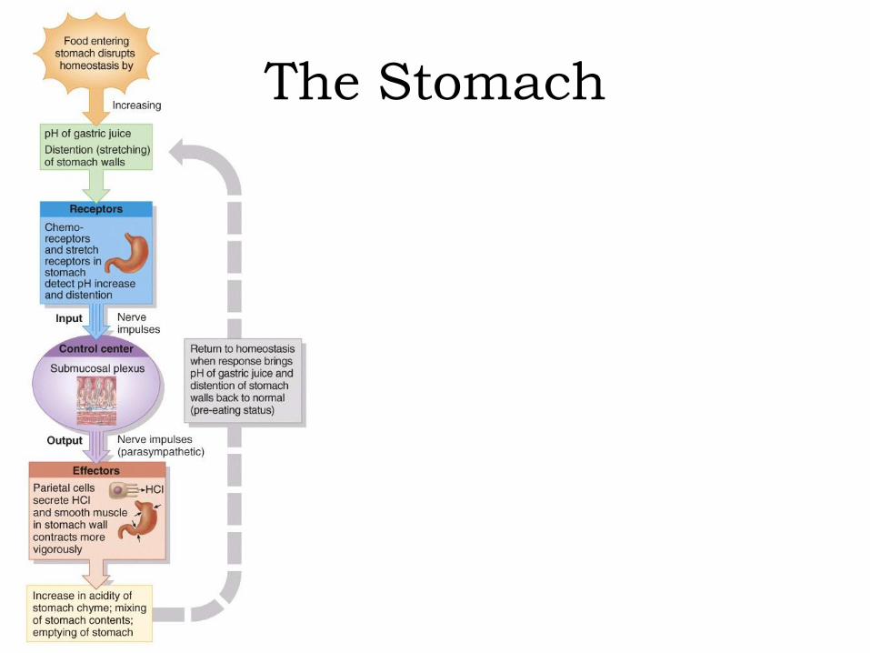
The Stomach
The StomachAlthough digestion is a major function of the stomach, its epithelial cells are impermeable to most materials, and very little absorption takes place.Within 2 to 4 hours after eating a meal, the stomach has emptied its contents into the duodenum. Foods rich in carbohydrate spend the least time. High-protein foods remain somewhat longer. Emptying is slowest after a fat-laden meal
containing large amounts of triglycerides.
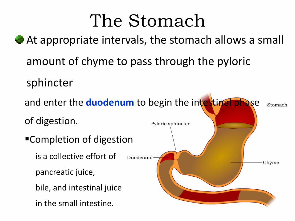
The StomachAt appropriate intervals, the stomach allows a small
amount of chyme to pass through the pyloric
sphincter
and enter the duodenum to begin the intestinal phase
of digestion.
Completion of digestion
is a collective effort of
pancreatic juice,
bile, and intestinal juice
in the small intestine.
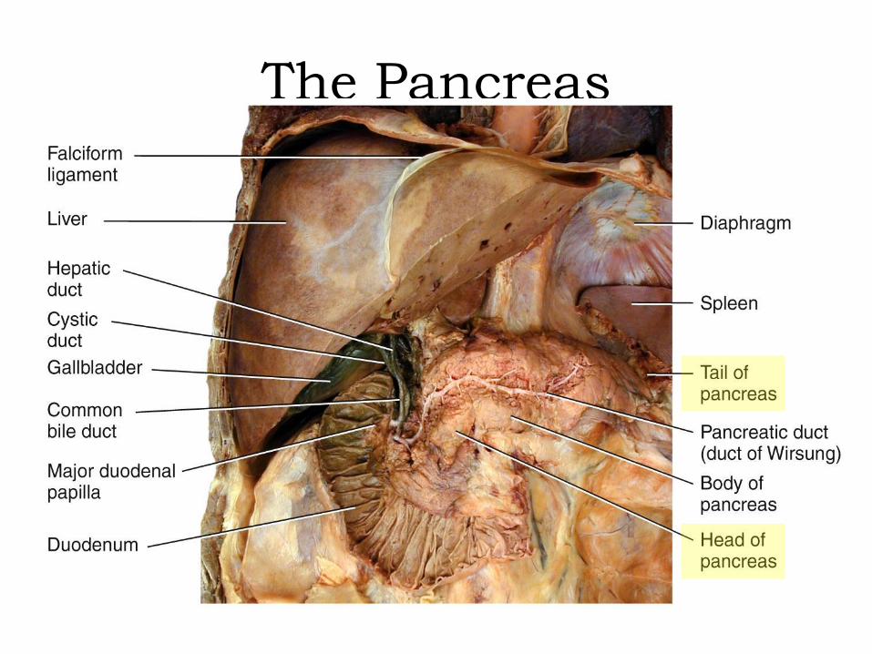
The PancreasDigestion and absorption in the small intestine depend heavily on secretions from the pancreas and gallbladder (liver). The pancreas is an oblong gland located posterior to
the stomach in the retroperitoneal space.• It is connected to the duodenum by the hepatopancreatic
ampulla and accessory ducts.• It secretes enzymes, which digest food in the small
intestine, and sodium bicarbonate, which buffers the acidic pH of chyme.
The Pancreas

The PancreasAbout 99% of pancreatic acini (glandular clusters)
participate in exocrine secretion – only 1% of the
clusters, called pancreatic islets,
form the endocrine
portion of the gland
(secreting the hormones
glucagon, insulin, and
somatostatin and
pancreatic polypeptide).
The PancreasAbout 1-1.5 liters of alkaline pancreatic juice is secreted into the duodenum each day. It creates the proper pH for the following digestive enzymes in the small intestine:
A starch digesting enzyme called pancreatic amylase Several enzymes that cleave polypeptides into
dipeptides and single amino acids: trypsin, chymotrypsin, carboxypeptidase, and elastase
Pancreatic lipase, the major triglyceride (fat) digesting enzyme in adults

Carbohydrate Digestion – The Pancreas
The Pancreas(Interactions Animation)
You must be connected to the internet to run this animation
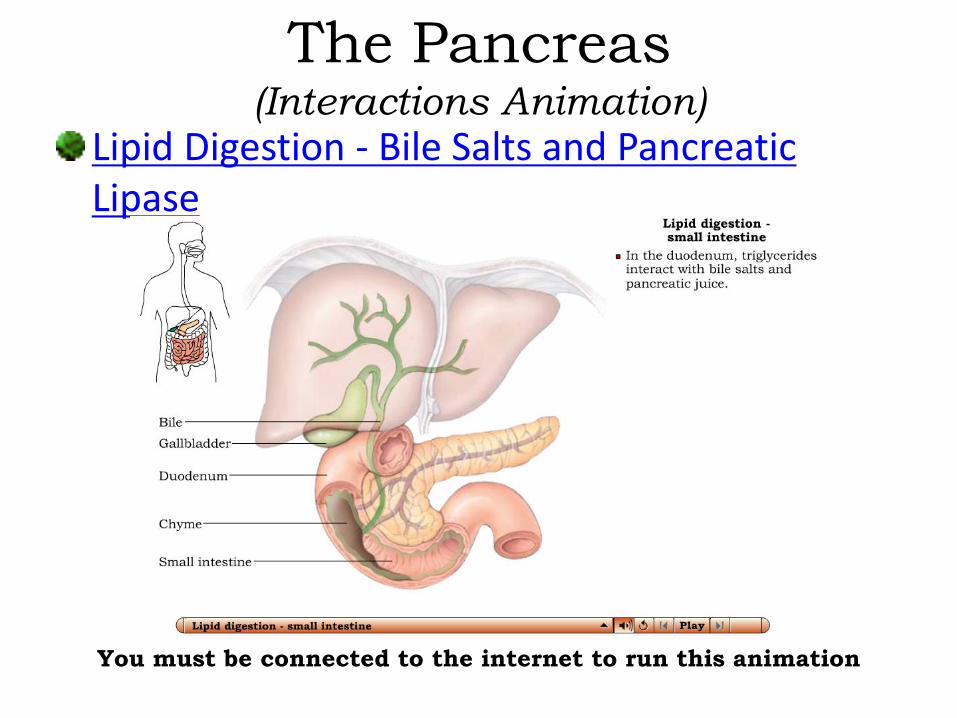
Lipid Digestion - Bile Salts and Pancreatic Lipase
The Pancreas(Interactions Animation)
You must be connected to the internet to run this animation
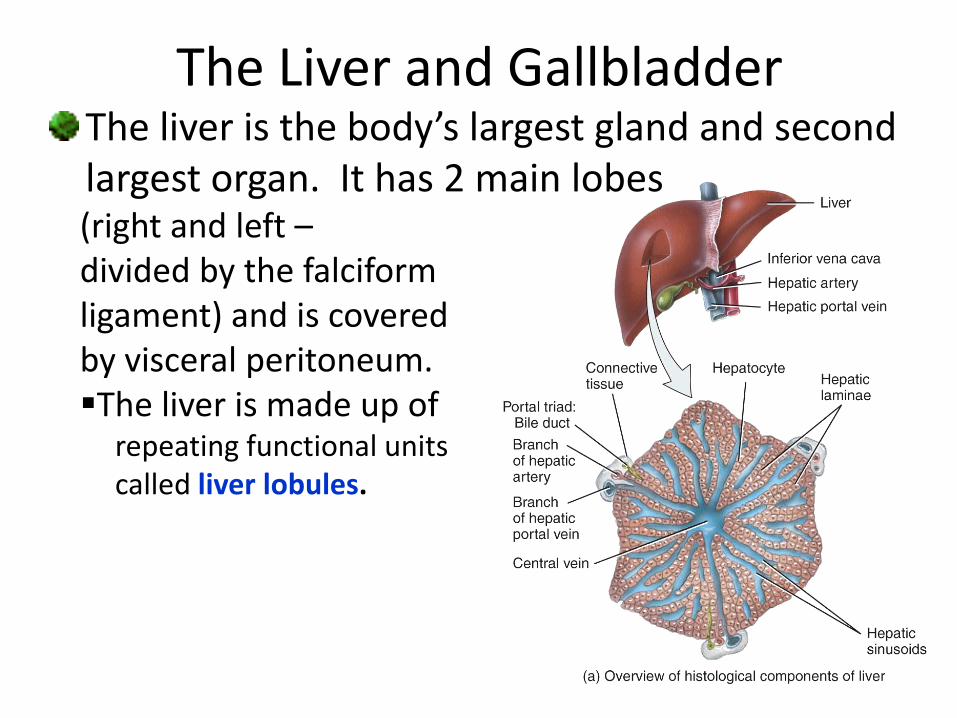
The liver is the body’s largest gland and second largest organ. It has 2 main lobes (right and left –divided by the falciform ligament) and is covered by visceral peritoneum. The liver is made up of
repeating functional units called liver lobules.
The Liver and Gallbladder

The Liver and GallbladderHepatocytes are the major functional cells of the liver. As the body’s “chemical factories”, their metabolic versatility is truly remarkable. Hepatocytes participate in a number of digestive and non-digestive functions.
Important digestive functions include:
• the synthesis, transformation, and
storage of proteins, carbohydrates,
and fats
• detoxification, modification, and excretion
of a variety of exogenous and endogenous substances

The Liver and GallbladderNon-digestive liver functions include:
Phagocytosis of old or worn-out cells
Making heparin (anticoagulant) and other plasma
proteins (prothrombin, fibrinogen, and albumin)
Modifying vitamin D to its active form
Human Albumin

Venous blood (from the hepatic portal vein) and
arterial blood (from the hepatic artery) feed the
lobule from the triad on its outer margin.
The blood mixture percolates through endothelial-
lined
spaces called
sinusoids
(a specialized
capillary)
towards the
central vein.
The Liver and Gallbladder

The Liver and Gallbladder
Path of blood in hepatic sinusoid
Microstructure of the liver lobule

The Liver and GallbladderFixed macrophages within the sinusoids called
Kupffer cells destroy red cells, white cells, and
bacteria in blood draining
from the GI tract.
An important function of lobule
hepatocytes is to secrete bile, an
excretory product that helps emulsify fats for the watery
environment of small intestine digestive juices.
Hepatocytes secrete about 1 liter of bile per day.
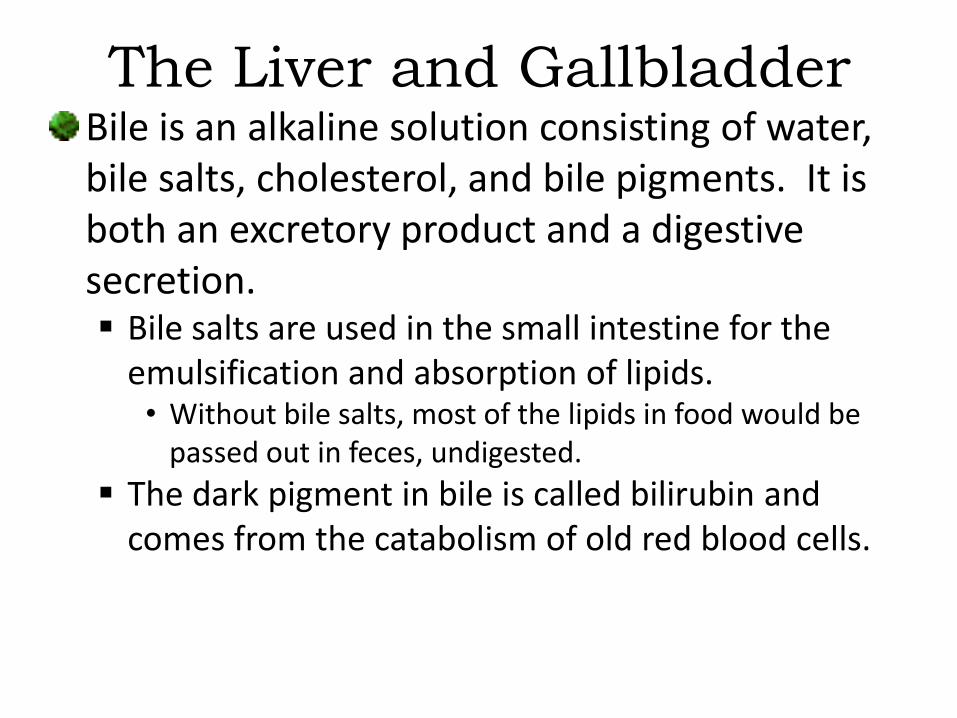
The Liver and GallbladderBile is an alkaline solution consisting of water, bile salts, cholesterol, and bile pigments. It is both an excretory product and a digestive secretion. Bile salts are used in the small intestine for the
emulsification and absorption of lipids.• Without bile salts, most of the lipids in food would be
passed out in feces, undigested.
The dark pigment in bile is called bilirubin and comes from the catabolism of old red blood cells.

The Liver and GallbladderBile secreted into the canaliculi (located between the hepatocytes) exits the liver in the common hepatic duct. This duct joins the
cystic duct from the gallbladder to form the common bile duct (CBD).

The CBD works its way towards the duodenum
and joins with the pancreatic duct to form
the hepatopancreatic
ampulla just proximal
to the second part of the
duodenum.
The duodenal papilla
(“nipple”) pierces the
intestinal mucosa to
deliver its contents.
The Liver and Gallbladder

The Liver and GallbladderBetween meals, the sphincter of the hepatopancreatic ampulla is closed – bile “backs-up” into the gall bladder where it is stored and concentrated up to ten-fold through the absorption of water and ions.

The Liver and GallbladderUnder the influence of the hormone cholecystokinin (CCK), the gallbladder contracts and ejects stored bile.Although not necessary for life, normal gall bladder function is highly desirable. After surgical
removal of the gall bladder (called a cholecystectomy), a person would experience severe indigestion if they ate a large meal high in fat content.
The Liver and Gallbladder(Interactions Animation)
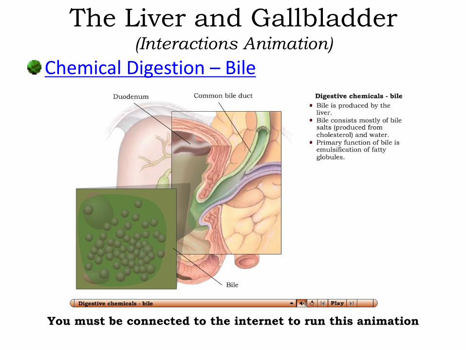
Chemical Digestion – Bile
You must be connected to the internet to run this animation
The Small IntestineThe small intestine is divided into 3 regions: The duodenum (10 in) The jejunum (8 ft) The ileum (12 ft)
• If measured in a cadaver, the intestines are longer than if measured in a live person due to the loss of smooth muscle contraction.
In the small intestine, digestion continues, even while the process of absorption begins.

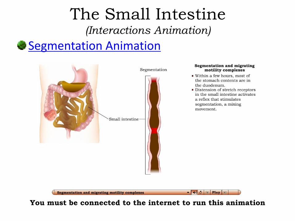
The Small IntestineMechanical digestion in the small intestine is a localized mixing contraction called segmentations. Segmentations is a type of peristalsis used to mix
chyme and bring it in contact with the mucosa for absorption.
It begins in the lower portion of the stomach and pushes food forward along a small stretch of small intestine.• It is governed by the myenteric plexus.
The Small Intestine(Interactions Animation)
Segmentation Animation
You must be connected to the internet to run this animation
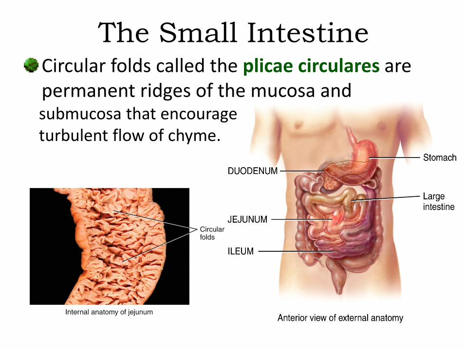
The Small IntestineCircular folds called the plicae circulares are permanent ridges of the mucosa and submucosa that encourage turbulent flow of chyme.

The Small IntestineVilli are multicellular structures that can barely
be seen by the naked eye. They form finger-like
projections that are covered with a simple
columnar epithelium.
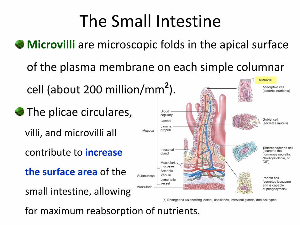
Microvilli are microscopic folds in the apical surface
of the plasma membrane on each simple columnar
cell (about 200 million/mm2).
The plicae circulares,
villi, and microvilli all
contribute to increase
the surface area of the
small intestine, allowing
for maximum reabsorption of nutrients.
The Small Intestine

• The small intestinal mucosa contains many deep crevices lined with glandular epithelium (intestinal glands) that secrete intestinal juice. Its function is to complete the digestive process begun by
pancreatic juice.
– Trypsin exists in pancreatic
juice in the inactive form
trypsinogen - it and other
enzymes are activated by
intestinal juice.
The Small Intestine

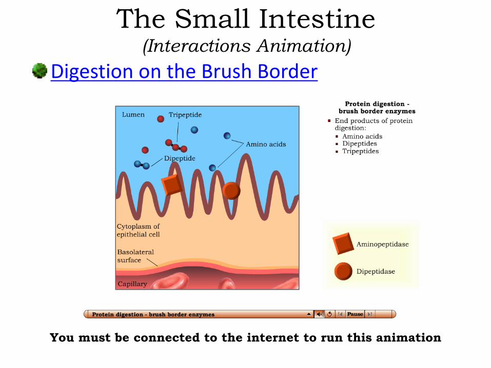
The Small IntestineMost of the enzymatic digestion in the small
intestine occurs inside the epithelial cells or
on their surfaces (rather than in
the lumen of the tube) as
intestinal juice comes in
contact with the brush
border of the villi.
The Small Intestine(Interactions Animation)
Digestion on the Brush Border
You must be connected to the internet to run this animation
The Small Intestine(Interactions Animation)
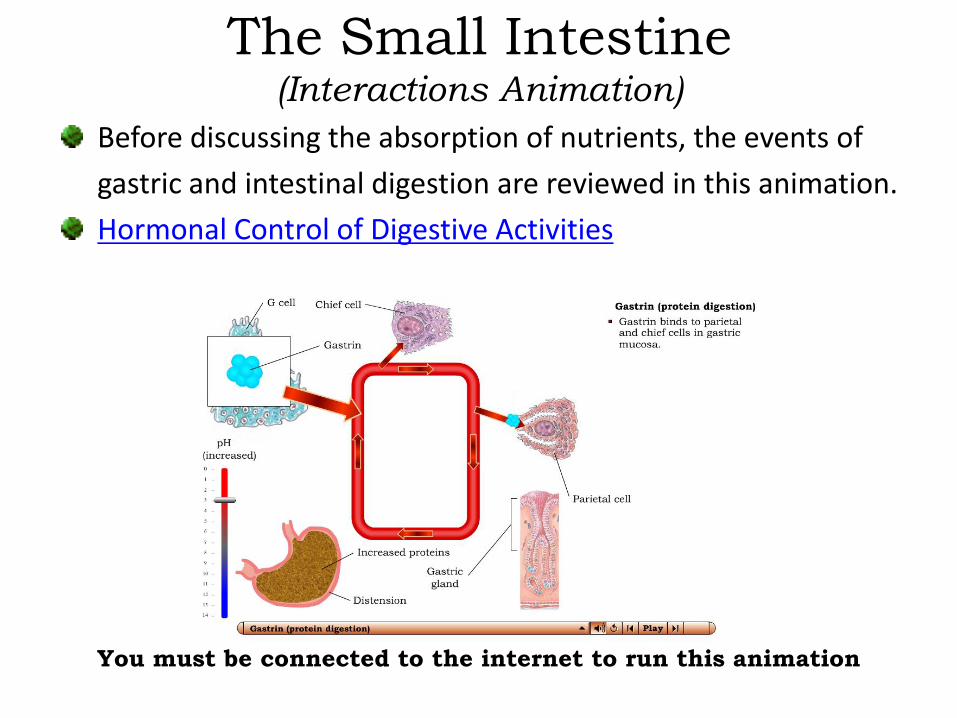
Before discussing the absorption of nutrients, the events of
gastric and intestinal digestion are reviewed in this animation.
Hormonal Control of Digestive Activities
You must be connected to the internet to run this animation
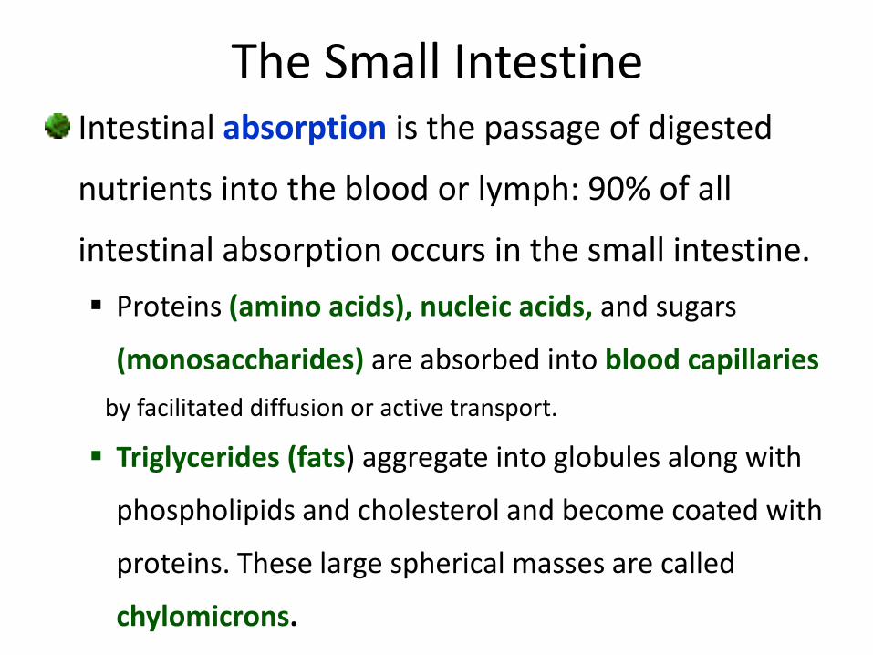
The Small IntestineIntestinal absorption is the passage of digested
nutrients into the blood or lymph: 90% of all
intestinal absorption occurs in the small intestine.
Proteins (amino acids), nucleic acids, and sugars
(monosaccharides) are absorbed into blood capillaries
by facilitated diffusion or active transport.
Triglycerides (fats) aggregate into globules along with
phospholipids and cholesterol and become coated with
proteins. These large spherical masses are called
chylomicrons.

The Small IntestineChylomicrons, too large to enter blood
capillaries, enter specialized lymphatic vessels
called lacteals and
eventually drain
into the superior
vena cava and
mix with blood.
All dietary
lipids are absorbed
by simple diffusion.
The Small Intestine(Interactions Animation)
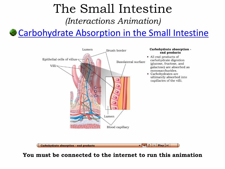
Carbohydrate Absorption in the Small Intestine
You must be connected to the internet to run this animation

Protein Absorption in the Small Intestine
The Small Intestine(Interactions Animation)
You must be connected to the internet to run this animation

Nucleic Acid Absorption in the Small Intestine
You must be connected to the internet to run this animation
The Small Intestine(Interactions Animation)
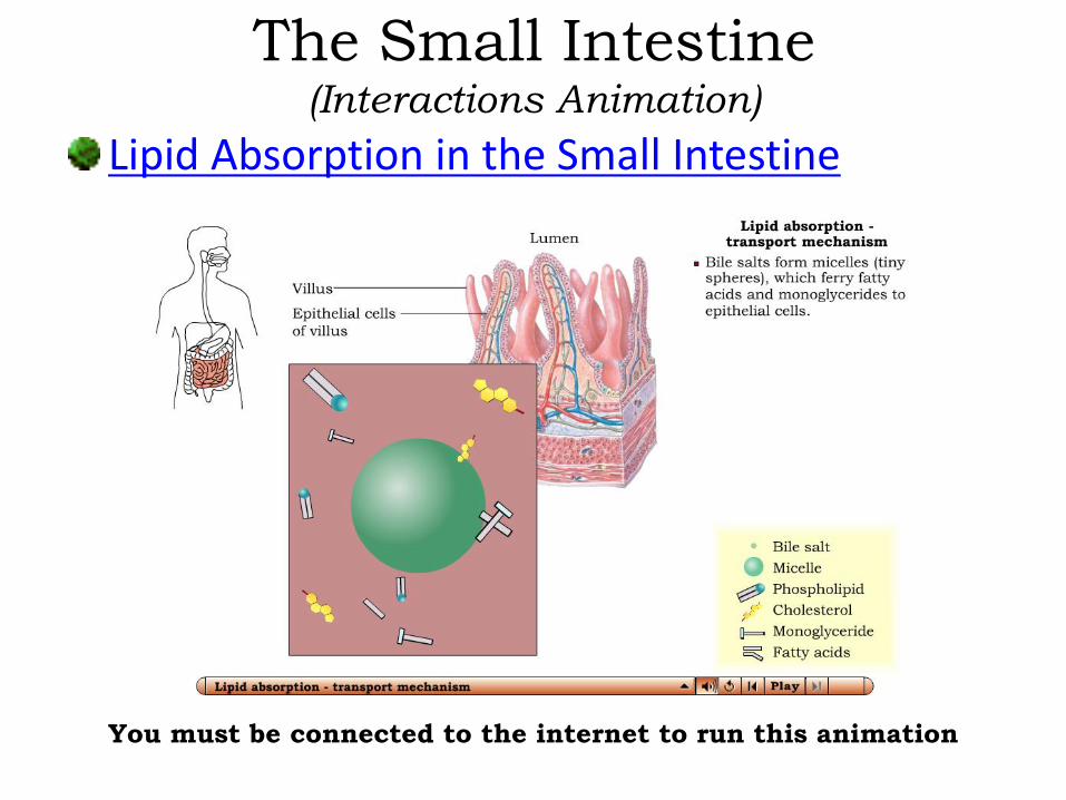
Lipid Absorption in the Small Intestine
The Small Intestine(Interactions Animation)
You must be connected to the internet to run this animation

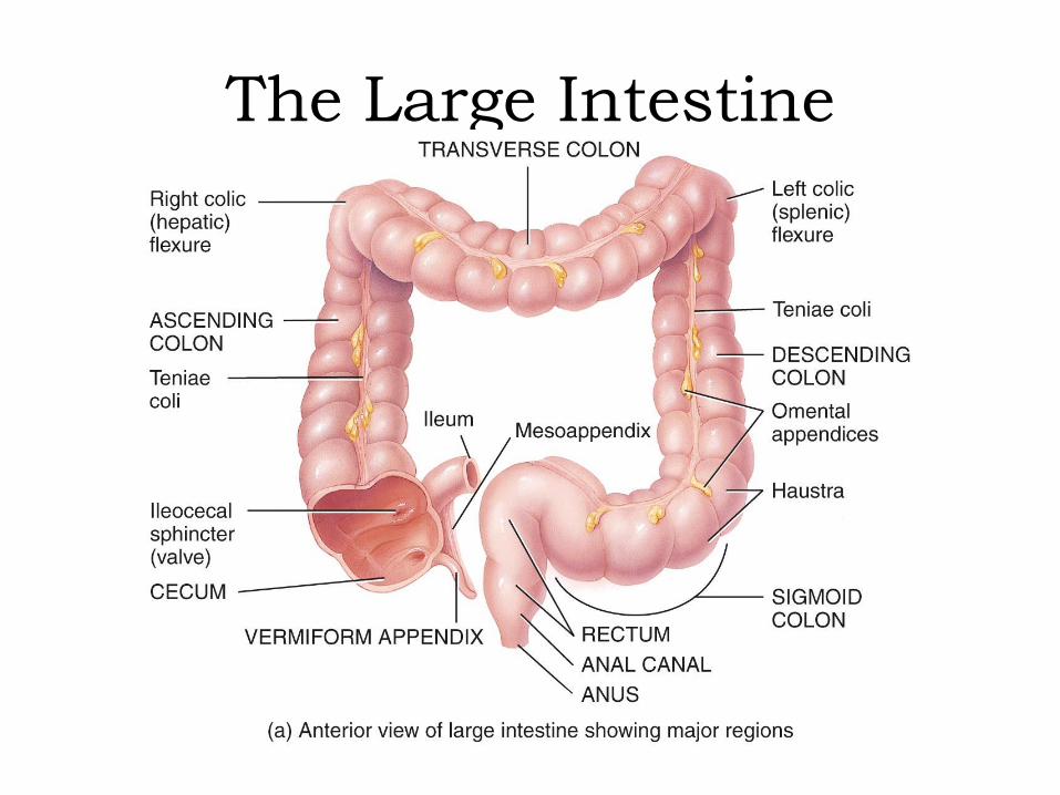
The large intestine is about 5 feet in length. Starting at the ileocecal valve, the large intestine has 4 parts: The cecum The colon
• ascending• transverse• descending• sigmoid
The rectum The anal canal
The Large Intestine
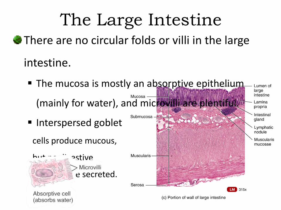
The Large IntestineThere are no circular folds or villi in the large
intestine.
The mucosa is mostly an absorptive epithelium
(mainly for water), and microvilli are plentiful.
Interspersed goblet
cells produce mucous,
but no digestive
enzymes are secreted.

The Large IntestineThe large intestine is attached to the posterior
abdominal wall by its mesocolon peritoneal
membrane.
Teniae coli are 3 separate longitudinal ribbons of
smooth muscle that run the length of the colon.
Because the teniae coli is shorter than the intestine,
the colon becomes sacculated into small pouches
called haustra (giving it a segmented appearance).
• As one haustrum distends, it stimulates muscles to contract,
pushing the contents to the next haustrum.
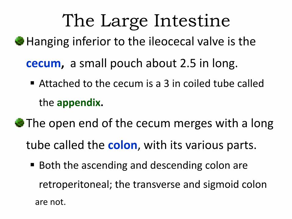
The Large IntestineHanging inferior to the ileocecal valve is the
cecum, a small pouch about 2.5 in long.
Attached to the cecum is a 3 in coiled tube called
the appendix.
The open end of the cecum merges with a long
tube called the colon, with its various parts.
Both the ascending and descending colon are
retroperitoneal; the transverse and sigmoid colon
are not.
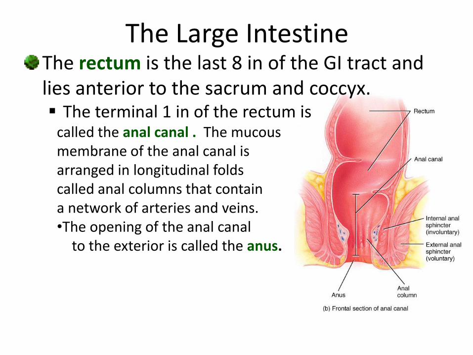
The Large IntestineThe rectum is the last 8 in of the GI tract and lies anterior to the sacrum and coccyx. The terminal 1 in of the rectum is
called the anal canal . The mucous membrane of the anal canal is arranged in longitudinal folds called anal columns that contain a network of arteries and veins.•The opening of the anal canal
to the exterior is called the anus.
The Large Intestine

The Large IntestineIncluding the 2 liters we
drink, about 9 liters of fluid
enter the small intestine
each day.
The small intestine absorbs
about 8 liters; the
remainder passes into the
large intestine, where most
of the rest of it is also
absorbed.
The Large IntestineFeces are the waste leftover after digesting and absorbing all the nutrients we can from eaten material. Though it is lower in energy than the food it came from, feces may still contain a large amount of energy, often 50% of that of the original food. The characteristic brown coloration comes from a
combination of bile and bilirubin. The distinctive odor is due to bacterial action - both
aerobic and anaerobic bacteria participate.
The Large Intestine
Though the human body consists of about 100
trillion cells, we carry about ten times as many
microorganisms in the intestines. Bacteria make
up most of the flora in the colon and about 60%
of the dry mass of feces.
As these bacteria digest/ferment left-over food,
they secrete beneficial chemicals such as
vitamin K, biotin (a B vitamin), and some amino
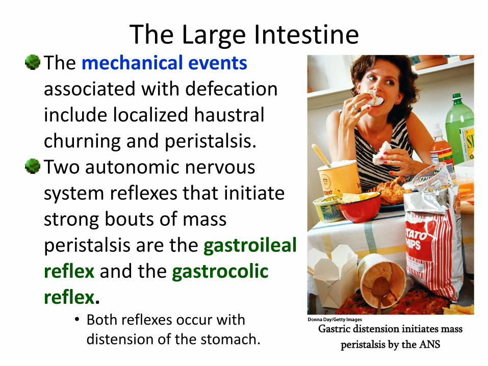
The Large IntestineThe mechanical events associated with defecation include localized haustral churning and peristalsis.Two autonomic nervous system reflexes that initiate strong bouts of mass peristalsis are the gastroileal reflex and the gastrocolic reflex.
• Both reflexes occur with distension of the stomach.
Gastric distension initiates mass
peristalsis by the ANS
The Large IntestineThe gastroileal reflex causes relaxation of the
ileocecal valve, intensifies peristalsis in the
ileum, and forces any chyme into the cecum.
The gastrocolic reflex intensifies strong
peristaltic waves that begin at about the middle
of the transverse colon and quickly drive the
contents of the colon into the rectum.
This mass peristalsis takes place three or four times
a day during or immediately after a meal, and may
The Large IntestineThe defecation reflex is activated by stretch
receptors stimulated by filling of the rectum.
The events leading to defecation include:
• Food in the stomach stimulates mass peristalsis.
• Food moves through the intestine into the rectum.
• Rectal pressoreceptors respond to distention and
longitudinal muscles shorten the rectum.
• ANS releases the internal anal sphincter and gives a
conscious awareness of distention.
• Release of external sphincter is under conscious control.
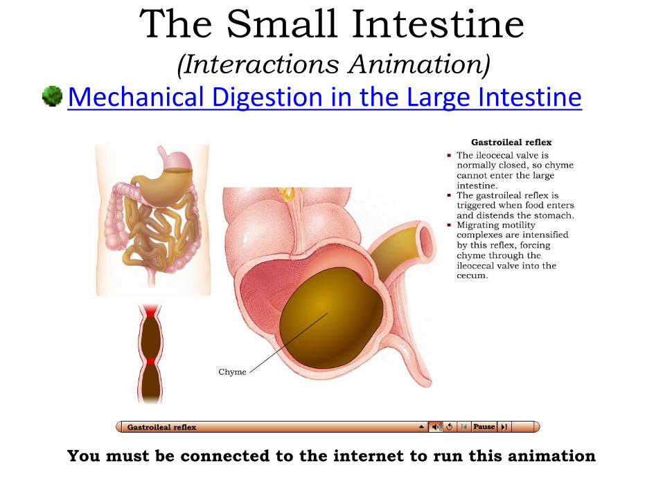
Mechanical Digestion in the Large Intestine
The Small Intestine(Interactions Animation)
You must be connected to the internet to run this animation
End of Chapter 24Copyright 2012 John Wiley & Sons, Inc. All rights reserved. Reproduction or translation of this work beyond that permitted in section 117 of the 1976 United States Copyright Act without express permission of the copyright owner is unlawful. Request for further information should be addressed to the Permission Department, John Wiley & Sons, Inc. The purchaser may make back-up copies for his/her own use only and not for distribution or resale. The Publisher assumes no responsibility for errors, omissions, or damages caused by the use of these programs or from the use of the information herein.
![[PPT]PowerPoint Presentation - Dr. Jerry Cronin - Homedrjerrycronin.weebly.com/uploads/5/9/7/4/5974564/3a.pptx · Web viewChapter 3 The Cellular Level of Organization Lecture slides](https://img.dokumen.tips/doc/110x75/5aec44127f8b9ac361903e23/pptpowerpoint-presentation-dr-jerry-cronin-viewchapter-3-the-cellular-level.jpg)