Embed Size (px)
Citation preview

J Oral Maxillofac Surg61:918-927, 2003
Alterations of Morphology andMicrodensity in the Condyle After
Mandibular Osteodistraction in the RatZi Jun Liu, DDS, PhD,* Gregory J. King, DMD, DMSc,†
and Susan W. Herring, PhD‡
Purpose: In this study, we examined the effects of mandibular distraction osteogenesis on themorphology and the microdensity of the rat condyle.
Materials and Methods: One hundred twenty-nine rats were allocated to 4 experimental groups (n �32 or 33). Each received unilateral mandibular ramus osteotomy and distraction device placement. Aftera 3-day latency, these were distracted once a day for 5 days. The slow distraction group was distracteda total of 1 mm (0.2 mm/d); the moderate group, 2 mm (0.4 mm/d); the rapid group, 3 mm (0.6 mm/d);and the sham group, no distraction (0.0 mm/d). Eight to 9 rats in each group were sacrificed at each of4 time points after device placement (6, 10, 24, and 38 days). Baseline data were obtained 3 days afterosteotomy and device placement without distraction from an additional 10 rats. Radiographs of thehemimandibles were scanned and measured to evaluate changes in condylar size (height, width, andarea), angulation, and bone microdensity converted to equivalent bone thickness using a stepwedge. Thewet weights of masseter muscle were measured at the time of harvest.
Results: 1) Muscle weight gains over time were significantly lower in the treated than the untreatedsides throughout the consolidation period (P � .001). 2) Condylar size and angulation on the untreatedside increased postoperatively, whereas there was a significant reduction of these parameters (P � .01to .001) on the treated side at 24 and/or 38 days. 3) Condylar microdensity significantly increased on theuntreated side at 24 and 38 days (P � .05 to .01) but not on the treated side. 4) Faster or larger distractioncaused more severe size reduction and more upright condylar angulation, prevented an increase in bonemicrodensity on the treated side, especially during the consolidation periods (P � .05 to .01), andretarded increase in muscle weight, whereas a slower distraction rate showed few negative, and evensome positive effects. 5) Correlations in size, angulation, and microdensity between right and leftcondyles became less significant over time. 6) There were positive correlations between muscle weightand condylar size, angulation, and microdensity.
Conclusion: An increased rate of mandibular distraction has significant negative effects on condylarmorphology and microdensity.© 2003 American Association of Oral and Maxillofacial SurgeonsJ Oral Maxillofac Surg 61:918-927, 2003
As a dynamic method for generating new bone, dis-traction osteogenesis has been widely accepted as the
treatment of choice for the surgical correction ofhypoplasias of the craniofacial skeleton. However, theprocedure can be difficult to control and its sideeffects are not well understood.1 In osteodistraction, atensile force is applied across an osteotomy tolengthen a bone. The forces produced by the distrac-tion maneuvers are superimposed on the normal me-chanics of the bone, and thus put unusual loads onthe entire craniofacial skeleton, which could alter theprocess of normal growth and/or adaptive remodel-ing in the region.
Some have raised concerns about undue loading ofjoints during lengthening of long bones by distrac-tion.2,3 Early surgery for distraction may also retardthe growth potential of the mandible and its associ-ated soft-tissue structures.4 When the mandible is
Received from the Department of Orthodontics, University of
Washington, Seattle, WA.
*Assistant Professor.
†Professor and Chair.
‡Professor.
This work was supported by National Institute of Dental and
Craniofacial Research Grant DE13061.
Address correspondence and reprint requests to Dr Liu: Depart-
ment of Orthodontics, Box 357446, University of Washington,
Seattle, WA; e-mail: [email protected]
© 2003 American Association of Oral and Maxillofacial Surgeons
0278-2391/03/6108-0092$30.00/0
doi:10.1016/S0278-2391(03)00294-5
918

lengthened by distraction, the applied force maythrust the condyle into the articular fossa, producingan excessive compressive load on the articular sur-faces of temporomandibular joint (TMJ). This unde-sirable force could cause morphologic changes, struc-tural displacements, abnormal growth, and/orremodeling of the condylar cartilage or functionaldisorders of TMJ components. Few studies have ad-dressed the effects of mandibular distraction on theTMJ. Mandibular linear distraction in dogs indicatedflattening of the condylar head and thinning of con-dylar cartilage, but these changes were transient.5
However, a similar experiment with transverse dis-traction produced permanent flattening and erosionin TMJ components and condylar displacement.6 An-other study using rabbits also revealed a positive cor-relation between mechanical loading by distractionand degeneration in TMJ cartilage, with morphologicalterations in the condyle.7
Very limited clinical follow-up studies indicatedthat unilateral distraction caused the condyles to as-sume a larger size and a more vertical orientation,normalizing their appearance overall. The contralat-eral condyle was unaffected. Bilateral distractioncaused an increase in condylar size and improvedgeometry and augmented vertical height.8 Lateral con-dylar displacement has been found in direct relation-ship to the amount of mandibular symphyseal distrac-tion, but symptoms were not introduced or, if presentbefore therapy, distraction did not exacerbate them.9
The distraction might even improve TMJ symptoms,10
but temporary TMJ complications are also reported.11
These studies have led to the conclusions that TMJbony changes from mandibular distraction are mini-mal and reversible and that the process of distractionis beneficial to the structure and position of the TMJcomplex.12 On the contrary, studies on the long-termeffects of mandibular distraction have revealed thatthe distracted side always grew at a slower rate thanthe intact side after the distraction was completed onvery young children under 48 months of age4 and thatthe condylar cartilage of the operated side was thin-ner and the bone structure was more dense withwoven bone predominating. These changes wereonly partly reversible over time in growing sheep.13
Despite most treatment designs attempting to pro-tect the TMJ from the distraction force and to allownormal motion of the joint,14 unusual loading of theTMJ may unavoidably produce changes on both theoperated and contralateral sides.13 Also, damage tomasticatory muscles due to the surgery and/or thedistraction may cause changes in that highly adaptivetissue.15 The evidence regarding the effects of man-dibular distraction osteogenesis on the TMJ, althoughsuggestive, remains conflicting and limited.
In our previous study using rats, we found thateven though linear lengthening of the mandible wasattained with increasing amounts of distraction, theincreases were 40% of what would be predicted16
(also see Fig 2, line A-F). To address this discrepancy,an extensive investigation was performed on our es-tablished rat model to elucidate the effects of man-dibular distraction osteogenesis on condylar morphol-ogy and microdensity over time after distraction. Wehypothesized that the loads produced on the TMJ bythe distraction, the detachment of masticatory mus-cles at the time of the installation of the distractiondevice, and stretching of masticatory muscles bylengthening the mandible may all affect the normalprocesses of condylar growth and/or adaptive remod-eling,17 represented by the changes in the morphol-ogy and microdensity. We further hypothesized thatthe severity of the effect may be associated with thevarying distraction forces (rates) or amounts.
Materials and Methods
ANIMALS AND SURGICAL PROCEDURES
One hundred thirty-nine 3-month-old male rats(Sprague-Dawley) were obtained from Animal Tech-nologies Ltd (Kent, WA). Both powdered and regularpellet food were provided 2 days before the surgery.
The surgery was carried out under aseptic condi-tions. The rats were anesthetized with Ketamine (70mg/kg; Animal Health Co, Fort Dodge, IA) and Xyla-zine (13 mg/1 kg; Phoenix Pharmaceutical Inc, StJoseph, MO) intraperitoneally. An antibiotic (Cefazo-lin 10 mg/kg; G.C. Hanford Mfg Co, Syracuse, NY)was delivered intraperitonealy. The inferior part ofthe right masseter was exposed, and a 5-mm incisionwas made along the inferior border of the mandible. Asmall curette was used to strip the muscle fibers fromthe bone until the mandibular angle, sigmoid notch,and mandibular body posterior to the third molarwere exposed. The attachment of the medial ptery-goid muscle to the angle was also stripped to place aprefabricated acrylic wafer (5 � 3 � 1 mm) againstthe lingual surface of the angle for additional supportfor the posterior screws of the device. The distractiondevice consisted of a Leone jackscrew (10 � 6 � 3;0.2 mm per quarter turn; Leone Co, Florence, Italy), 2Luhr L-shaped microplates (0.8 mm, 5 holes), and 4microcortical self-tapping screws (0.8 � 3 mm;Stryker-Leibinger Corp, Kalamazoo, MI). The anteriormicroplate was secured to the body of the mandible,and the posterior one was secured to the angle andthe acrylic wafer. After the device was placed, anosteotomy was performed from the sigmoid notchdown to the inferior border between the 2 plates byusing a diamond bur (round end taper, 0.5-mm tip
LIU, KING, AND HERRING 919

diameter, 10 mm long; Axis Dental Co, Irving, TX)with copious irrigation. The muscle and skin incisionswere closed in layers after a complete osteotomy wasconfirmed by activating the jackscrew (Fig 1). Thedetails of device design and surgery procedures havebeen described elsewhere.18
The postoperative care included injection of anal-gesic (Buprenorphin 0.1 mg/kg; Reckitt & ColemanProducts, Hull, England), 7 days of powdered food,followed by 3 days of both powdered and solid food,trimming of mandibular incisors to prevent over-growth, and daily weighing.
DISTRACTION STRATEGIES AND GROUPALLOCATIONS
The surgery day was day 0, and the latency periodlasted 3 days. Then, activation for mandibular distrac-tion was performed once a day for 5 days (day 3 today 7: “distraction period”). The consolidation periodbegan at day 8 and terminated at day 38.
Of 139 rats, 129 were randomly allocated into 4groups differing in distraction rate: 0.0 mm (sham),0.2 mm (slow), 0.4 mm (moderate), and 0.6 mm
(rapid), respectively. Eight to 9 rats in each groupwere further randomized to death at the following 4time points: day 6 (mid-distraction), day 10 (earlyconsolidation), day 24 (middle consolidation), andday 38 (late consolidation). The remaining 10 ratswere killed at the end of the latency period (day 3)and served as baseline. At the time of death, thebilateral masseter muscles were dissected andweighed immediately (model 100A; Denver Instru-ment Co, Denver, CO). Right and left hemimandibleswere removed separately because the symphysis inrats is not fused. All specimens were fixed in 10%formalin for 24 hours and preserved in 70% alcoholfor radiographic morphometry and microdensiom-etry.
CONDYLAR MORPHOLOGY ANDMICRODENSIOMETRY
After removal of the distraction device, the mandi-bles were placed flat on high-resolution diagnosticfilm (X-Omat TL; Kodak, Tokyo, Japan) with the lin-gual side facing the film, and radiographic imageswere obtained using an enclosed X-ray unit (Picker
FIGURE 1. (A) X-ray images of a pair of hemimandibles from a baseline animal indicating the reference line (occlusal plane, white line) andlocations of the device (white dots) and osteotomy (dotted line). (B) Components of the distraction device. (C) X-ray images of pairs of hemimandiblescollected from 4 animals killed at 4 different time points (all with moderate distraction, 0.4 mm/d). T, treated (right) side; U, untreated (left) side.Note that reference lines of images are lined up and that the untreated side was flipped horizontally.
920 CONDYLAR ALTERATIONS AFTER MANDIBULAR DISTRACTION
X-ray Corporation, Cleveland, OH) with the followingsettings: distance 65 cm, voltage 35 KVp, current 35mA, and exposure time 25 seconds. The radiographicfilms were scanned (ScanJet 4c/T; Hewlett Packard,Palo Alto, CA) and analyzed by means of NIH Imagesoftware (version 1.62; National Institutes of Health,Bethesda, MD). As a calibration tool, a customizedbone stepwedge made of RMI 450 bone tissue equiv-alent material (Gammex RMI Co, Middleton, WI) wasexposed on each film together with the mandiblesduring radiography. The values of mean microdensitywere converted into the equivalent bone thicknessesusing the algorithm generated from the stepwedge.
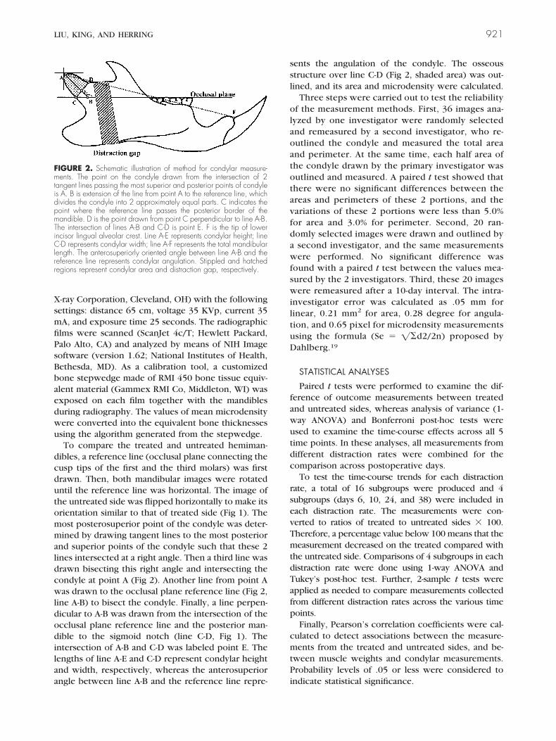
To compare the treated and untreated hemiman-dibles, a reference line (occlusal plane connecting thecusp tips of the first and the third molars) was firstdrawn. Then, both mandibular images were rotateduntil the reference line was horizontal. The image ofthe untreated side was flipped horizontally to make itsorientation similar to that of treated side (Fig 1). Themost posterosuperior point of the condyle was deter-mined by drawing tangent lines to the most posteriorand superior points of the condyle such that these 2lines intersected at a right angle. Then a third line wasdrawn bisecting this right angle and intersecting thecondyle at point A (Fig 2). Another line from point Awas drawn to the occlusal plane reference line (Fig 2,line A-B) to bisect the condyle. Finally, a line perpen-dicular to A-B was drawn from the intersection of theocclusal plane reference line and the posterior man-dible to the sigmoid notch (line C-D, Fig 1). Theintersection of A-B and C-D was labeled point E. Thelengths of line A-E and C-D represent condylar heightand width, respectively, whereas the anterosuperiorangle between line A-B and the reference line repre-
sents the angulation of the condyle. The osseousstructure over line C-D (Fig 2, shaded area) was out-lined, and its area and microdensity were calculated.
Three steps were carried out to test the reliabilityof the measurement methods. First, 36 images ana-lyzed by one investigator were randomly selectedand remeasured by a second investigator, who re-outlined the condyle and measured the total areaand perimeter. At the same time, each half area ofthe condyle drawn by the primary investigator wasoutlined and measured. A paired t test showed thatthere were no significant differences between theareas and perimeters of these 2 portions, and thevariations of these 2 portions were less than 5.0%for area and 3.0% for perimeter. Second, 20 ran-domly selected images were drawn and outlined bya second investigator, and the same measurementswere performed. No significant difference wasfound with a paired t test between the values mea-sured by the 2 investigators. Third, these 20 imageswere remeasured after a 10-day interval. The intra-investigator error was calculated as .05 mm forlinear, 0.21 mm2 for area, 0.28 degree for angula-tion, and 0.65 pixel for microdensity measurementsusing the formula (Se � ��d2/2n) proposed byDahlberg.19
STATISTICAL ANALYSES
Paired t tests were performed to examine the dif-ference of outcome measurements between treatedand untreated sides, whereas analysis of variance (1-way ANOVA) and Bonferroni post-hoc tests wereused to examine the time-course effects across all 5time points. In these analyses, all measurements fromdifferent distraction rates were combined for thecomparison across postoperative days.
To test the time-course trends for each distractionrate, a total of 16 subgroups were produced and 4subgroups (days 6, 10, 24, and 38) were included ineach distraction rate. The measurements were con-verted to ratios of treated to untreated sides � 100.Therefore, a percentage value below 100 means that themeasurement decreased on the treated compared withthe untreated side. Comparisons of 4 subgroups in eachdistraction rate were done using 1-way ANOVA andTukey’s post-hoc test. Further, 2-sample t tests wereapplied as needed to compare measurements collectedfrom different distraction rates across the various timepoints.
Finally, Pearson’s correlation coefficients were cal-culated to detect associations between the measure-ments from the treated and untreated sides, and be-tween muscle weights and condylar measurements.Probability levels of .05 or less were considered toindicate statistical significance.
FIGURE 2. Schematic illustration of method for condylar measure-ments. The point on the condyle drawn from the intersection of 2tangent lines passing the most superior and posterior points of condyleis A. B is extension of the line from point A to the reference line, whichdivides the condyle into 2 approximately equal parts. C indicates thepoint where the reference line passes the posterior border of themandible. D is the point drawn from point C perpendicular to line A-B.The intersection of lines A-B and C-D is point E. F is the tip of lowerincisor lingual alveolar crest. Line A-E represents condylar height; lineC-D represents condylar width; line A-F represents the total mandibularlength. The anterosuperiorly oriented angle between line A-B and thereference line represents condylar angulation. Stippled and hatchedregions represent condylar area and distraction gap, respectively.
LIU, KING, AND HERRING 921

Results
GENERALIZATIONS
The weight of the device (1.65 g) and minor dam-age to the tissues around the osteotomy were welltolerated. An abscess mostly around the medial platewas found in some of the 24- and/or 38-day rats, butno infection was found in the incision and distractiongap. The distraction gaps in some cases were notevenly wide perhaps because of a round end taperbur being used to cut bone and/or because of theregional bone resorption. The tissue elasticity or mus-cle tension on the segments might also have causedthis unevenness. However, neither segment relapsenor screw migration/tipping was observed through-out the experiment period. Body weight monitoringconfirmed that rats were still growing slowly through-out the experimental period (up to �20%), althoughweight dramatically dropped immediately after sur-
gery, catching up after day 7 (Fig 3A). No impairmentof mastication or other dysfunction was noted clini-cally. Undergrowth, rather than overgrowth,7 of theincisor of the distracted side was found in most ofanimals. However, we did not find significant cross-bite even in the rats with rapid-rate distraction asdescribed by other workers.20
MUSCLE WEIGHT
The masseter on the treated side was smaller thanthat on the untreated side, most obviously in its dor-sal-ventral dimension. Consistent with the long-termtrend of body weight, masseter weights on both sidesincreased continually and significantly from days 10to 38 (P � .05 to .001) (Table 1). However, the gainswere less on the treated side than on the control (P �.001) (Fig 3B). The opposite relationship of the 2sides at days 3 and 6 reflects postoperative swelling inthe treated-side muscle (P � .05 to .01) (Fig 3B).
FIGURE 3. (A) Body weightcurve for whole sample. Dashedline indicates the body weight atthe day of surgery. (B) Compari-son of wet weight of massetermuscles. Asterisks indicate thesignificance levels by ANOVA(overall difference across 5 timepoints) and paired t tests (differ-ence between treated and un-treated sides at each time point).*P � .05; **P � .01; ***P �.001.
Table 1. P VALUES FOR COMPARISONS BETWEEN POSTOPERATIVE DAYS FOR TREATED AND UNTREATED SIDES
Day
Masseterweight
Condylar size AngulationAngle
MicrodensityThicknessHeight Width Area
T U T U T U T U T U T U
38 versus 24 .041* .000‡ .793 .998 .357 .994 .938 .911 .997 .128 .998 .93738 versus 10 .000‡ .000‡ .231 .977 .000‡ .007† .276 .073 .044* .117 .148 .000‡38 versus 6 .999 .000‡ .042* .858 .000‡ .167 .031* .072 .111 .101 .917 .000‡38 versus 3 .999 .000‡ .911 .822 .003† .983 .627 .624 .373 .189 .956 .004†24 versus 10 .998 .037* .878 .978 .054 .002† .747 .417 .103 .994 .072 .000‡24 versus 6 .044* .999 .460 .862 .005† .061 .211 .420 .226 .836 .781 .000‡24 versus 3 .044* .084 .998 .825 .133 .921 .909 .927 .514 .989 .892 .000‡10 versus 6 .001† .999 .956 .995 .952 .787 .895 .999 .669 .972 .587 .63610 versus 3 .000‡ .999 .979 .964 .99 .396 .999 .996 .999 .999 .906 .9826 versus 3 .999 .961 .830 .995 .998 .861 .977 .996 .999 .999 .999 .994
Abbreviations: T, treated side; U, untreated side.*P � .05.†P � .01.‡P � .001.
922 CONDYLAR ALTERATIONS AFTER MANDIBULAR DISTRACTION

CONDYLAR SIZE
The condylar height of the treated side decreasedprogressively from days 6 to 38 (P � .05) (Table 1).However, that of the untreated side showed a veryslight increase over time. This opposite trend led tosignificant differences between the 2 sides at day 24and 38 (P � .01 and .001, respectively) (Fig 4A)
The condylar widths of both sides showed similartrends from days 3 to 10. However, they went indramatically opposite directions after day 10. Signifi-cant decreases in the treated and increases in theuntreated sides were found at days 24 and 38 (P � .01to .001) (Fig 4B). On the treated side, the width at day38 was significantly less than at 3 early time points(P � .001 to .001) (Table 1).
The condylar areas exhibited almost the same pat-terns as the widths, that is, minor changes at early 3time points (days 3, 6, and 10) and obvious, opposite
changes at late 2 time points (days 24 and 38) withdrops in the treated side (P � .01 to .001) (Table 1)and moderate increases in the untreated side, whichagain led to significant difference between the 2 sides(P � .001) (Fig 4C).
CONDYLAR ANGULATION AND MICRODENSITY
The condylar angulation of the untreated side wasroughly constant throughout the experimental pe-riod. However, the condyle of the treated side be-came more upright at the 2 late time points, resultingin a significant decrease of the angulation comparedto the untreated side (P � .01 to .001) (Fig 5A) andbetween days 38 and 10 (P � .05) (Table 1).
The converted values of microdensity (equivalentbone thickness) showed 2 features. First, the values ofthe treated side were significantly higher than thoseof untreated side at all 3 early time points (P � .01 to
FIGURE 4. Comparisons ofcondylar size. (A) Condylarheight. (B) Condylar width. (C)Condylar area. *P � .05;**P � .01; ***P � .001.
FIGURE 5. Comparisons ofcondylar angulation (A) andequivalent thickness (B). *P �.05; **P � .01; ***P � .001.
LIU, KING, AND HERRING 923

.001) (Fig 5B) with minor change over time. Second,the increase of microdensity at the 2 late time pointswas trivial on the treated side but massive on theuntreated side (P � .01 to .001) (Table 1), resulting invalues significantly lower in the treated than un-treated sides (P � .001) (Fig 5B).
EFFECTS OF DISTRACTION RATES AT EACH TIMEPOINT
The animals allocated to each time point (days 6,10, 24, and 38) were further divided by the 4distraction rates. The ratios of the treated side tothe untreated side are plotted in Figure 6. At base-line (day 3), the ratios of all were very close to100%, indicating symmetry of the 2 condyles, ex-cept for the microdensity and masseter weight (Fig6, E and F), which showed relative increase ofabout 10% on the treated side.
There is a clear trend for animals with moderate orrapid distraction rates to have relatively decreased
size of the treated condyle, statistically significant forcondylar width at all 4 time points and for condylararea at days 6 and 38. The greatest relative reductionswere found in the animals with the rapid rate, most ofwhich were only 75% to 85% of the untreated side,significantly lower than the values from sham andslow rate animals (Fig 6, B and C).
Sham and slow distraction had little effect on thecondylar angulation at any time point. However, ani-mals with rapid or moderate rates again showed sig-nificant relative decreases in their condylar angula-tion, indicating the condyle became more uprightrelative to the occlusal plane (Fig 6D).
Unlike the other measurements, an relative in-creased microdensity around 10% was found in thesham and slow rate animals at all time points, exceptfor slow rate at day 10. On the contrary, significantrelative microdensity reductions of around 10% wereobserved in animals with rapid or moderate rates ofdistraction at days 10, 24, and 38 (Fig 6E).
FIGURE 6. Comparison of effects of distraction rates at each time point. (A) Condylar height. (B) Condylar width. (C) Condylar area. (D) Condylarangulation. (E) Condylar microdensity. (F) Wet weight of masseter muscle. BL, baseline. Asterisks indicate significance levels by ANOVA (overalldifference across 4 distraction rates at each time point) and Tukey (pairwise multiple comparisons at each time point). *P � .05; **P � .01; ***P �.001. Note that the ratios of the values of treated to untreated sides were examined for these comparisons. Arrowed lines indicate the level at whichthe 2 sides are equal (100%).
924 CONDYLAR ALTERATIONS AFTER MANDIBULAR DISTRACTION

The ratios of masseter weight showed that, in gen-eral, the damage to the muscle caused a relative de-crease in its weight. The degree of this reduction wasgreatly affected by distraction rates. The influence ofthe sham was minor, but rapid or moderate rates ofdistraction resulted in a 15% to 25% relative weightreduction (Fig 6F).
CORRELATIONS OF MASSETER WEIGHT WITHCONDYLAR MEASUREMENTS AND BETWEENTHE 2 SIDES
As the masseter weight and condylar measurementsshared similar trends, the correlations between these
2 were calculated (Fig 7). There are weak but signif-icant positive correlations between masseter weightand condylar area, angulation and microdensity, indi-cating that smaller muscles are associated with asmaller size, more upright angulation and less micro-density in the condyle.
Table 2 shows clearly that the bilateral symmetriesof condylar size, angulation, microdensity, and mas-seter weight became weaker at the longer timepoints. The correlation coefficients were all signifi-cant at days 3 and 6, but significance no longer ex-isted for width after day 10, for angulation after day24, or for masseter weight after day 24.
FIGURE 7. Scatterplot of condy-lar measurements and masseterweight. Asterisks indicate signifi-cance levels of correlation coeffi-cients. *P � .05; **P � .01.Note that the ratios (refer to Fig6), instead of raw values, wereused for this analysis.
Table 2. CORRELATION COEFFICIENTS OF TREATED SIDE WITH UNTREATED SIDE
Postoperative Day
3 6 10 24 38
Condylar sizeHeight .798† .452† .597‡ .528† .367*Width .716* .750‡ .286 .206 .026Area .765‡ .540† .515† .528† .398*
Condylar angulation .932‡ .581‡ .626‡ .151 .132Condylar microdensity .952‡ .933‡ .912‡ .751‡ .775‡Masseter weight .923‡ .640‡ .812‡ .559† .137
*P � .05.†P � .01.‡P � .001.
LIU, KING, AND HERRING 925
Discussion
AGE CHANGES IN THE CONDYLE
Rats used for the present study were consideredadults, but they still grew slowly throughout the ex-perimental period as shown in the weights of thebody and the masseter muscle, as well as the size ofthe untreated condyle (Figs 3, 4). Studies on normalage changes in rats indicate that the condyle achievesa mature morphology by 50 days. However, there isan increase in the size of the bony condylar trabeculaeand a diminution of the marrow space; condylargrowth stops entirely by the age of 10 months.21 Thetransformation of the condylar cartilage from adaptiveto nonadaptive is considered complete by 220 days inrats, after which the condyle is no longer consideredcapable of adapting17 in response to changing func-tional demands and mechanical loads.22 Our 90- to130-day-old rats were clearly within the interval from50 to 220 days when adaptation can occur in themorphologically complete condyle.
Because the 2 hemimandibles do not fuse in anosseous symphysis in rats, the forces from the distrac-tion side are probably not transmitted effectively tothe other side. Thus, despite unavoidable involve-ment of the condyle of the untreated side duringfunction,13 our findings on the condyle of the un-treated side may reflect the trend of normal condylargrowth in rats reported previously.21,23 The weaken-ing correlation between the 2 condyles over timeafter distraction (Table 2) indicates that the 2 sidesunderwent different growth/remodeling trends.
LOADS OF DISTRACTION ON THE CONDYLE
TMJ components are loaded by the forces appliedto the dentition during mastication.24 Growth of thecondyle in a superior and posterior direction contrib-utes greatly to mandibular height and length, andmechanical loads play a major role in regulating thisprocess.25,26 Lessening the load by a soft diet or in-creasing the load by wearing a chin-cap causes signif-icant increase or reduction, respectively, in rat con-dylar growth.23,25 In vitro growth of the condylarcartilage in the condyle can be suppressed or evenstopped by compressive force.27
Mandibular distraction can significantly flatten theanteromedial part of the condyle on the distractedside, involving all cell layers including fibrous, pre-chondroblastic, and chondroblastic zones.7 Thesefindings are similar to those in rats wearing a chin-capdisplacing the mandible backward.15,23
On the other hand, it has been reported that pos-terior displacement of the mandible might enhancemetabolic activity in the posterior parts of the con-dyle, resulting in increased cartilage formation.28 Thismay imply that the amount of load (here equivalent to
rate of distraction) is the critical factor. The presentstudy showed that higher distraction rates or largerdistraction amounts caused significant size reductionand more upright angulation in the condyle, and thesenegative effects persisted and worsened throughoutboth distraction (day 6) and consolidation (day 10, 24,and 38) periods (Fig 7, A-D). Thus, not only did thetreated condyle not grow, but it actually becamesmaller. Because growth in mandibular length occursdepends on the condyle,26 these changes help explainwhy only 40% of predicted mandibular lengtheningwas achieved.16 However, changes in the condylemay not be the sole factor causing the discrepancybetween expected and achieved mandibular length.Other factors, such as instability within the distrac-tion gap and offset of the 2 segments due to thedistractor’s location at the inferior border, may beinvolved as well.
If overload from the excessive distraction rate hada negative impact on growth and remodeling of thecondyle, then these effects may be more significant inrapidly growing animals. A further investigation onyoung animals, combined with histologic observationon the condyles, is under way to shed light on howgrowth and remodeling patterns change in responseto these loads. Another unanswered question iswhether the negative effects on the condyle werecaused by higher distraction rates directly increasingthe TMJ load or by the larger distraction amountsresulting in a less stable, poorly ossified gap andthereby altering the local TMJ biomechanical environ-ment.
It is interesting that despite higher distraction ratespreventing increases in condylar microdensity on thetreated side, the sham and slow distraction ratestended to increase condylar microdensity beyond thatof the untreated side (5% to 15%, Fig 6E). This findingindicates that gradual distraction at an appropriaterate not only promotes successful osseous regenera-tion16 but also has a positive effect on the condyle andpossibly other bony components of the craniofacialcomplex. This observation is also supported by thefindings of higher osteocalcin expression (140% to160%) in gradual distraction rats than controls.20
MUSCLE DETACHMENT AND OVERSTRETCHING
The present procedure caused 2 major physicalchanges in the muscles: detachment and stretching.Detachment of parts of the masseter and medial ptery-goid muscles was necessary to facilitate installing thedistraction device and inserting the acrylic wafer.Passive muscle stretch was produced during length-ening of the mandible, and muscles may have beenoverstretched during consolidation. Detachment ofthe rat masseter muscle alone leads to a significantchanges in the morphology of the condyle, which
926 CONDYLAR ALTERATIONS AFTER MANDIBULAR DISTRACTION

becomes higher, narrower, and more upright com-pared to the control side.15 Muscle overstretchingresults in fiber-type transformation in the tibialis an-terior muscle during long bone distraction osteogen-esis.29
Masticatory stresses exceed distraction stresses.30 Ithas been well demonstrated that functional strains onthe TMJ are caused almost entirely by the massetermuscle in pigs.31 Our results clearly show a deleteri-ous effect of distraction on the treated-side masseter(Fig 3B). The failure of the muscle to grow may bedue to disuse atrophy and/or fibrosis as reported forlong bones32 and other mandibular distraction33 ani-mal models. In any case, the fact that the treated sidemasseter was smaller means that in addition to theeffect of direct forces on the condyle from the dis-traction, the effect of indirect forces from changes inmuscles must also be considered.34 The correlationbetween masseter weight and condylar parameters inthe present study supports the notion that relativeloss of masseter function is associated with the degreeof negative change (reduction in size, decrease indensity and more upright angulation) in the condyleduring mandibular distraction osteogenesis.
Acknowledgments
The authors would like to thank Dr Lingli Wang, visiting scholarfrom the Stomatological College, Jilin University of China, for help-ing with surgeries and animal manipulations; Xianqin Bai for vali-dating the measurement methods; and Stryker-Leibinger and LenonCo for donating the distraction devices.
References1. Yen SL, Wei S, Li S, et al: Bending of the distraction site during
mandibular distraction osteogenesis in the rabbit: A model forstudying segment control and side effects. J Oral MaxillofacSurg 59:779, 2001
2. Olney BW, Jayaraman G: Joint reaction forces during femorallengthening. Clin Orthop 130:164, 1994
3. Stanitski DF: The effect of limb lengthening on articular carti-lage: An experimental study. Clin Orthop 130:68, 1994
4. Hollier LH, Kim JH, Grayson B, et al: Mandibular growth afterdistraction in patients under 48 months of age. Plast ReconstrSurg 103:1361, 1999
5. McCormick SU, McCarthy JG, Grayson BH, et al: Effect ofmandibular distraction on the temporomandibular joint: Part 1,canine study. J Craniofac Surg 6:358, 1995
6. Stelnicki EJ, Stucki-McCormick SU, Rowe N, et al: Remodelingof the temporomandibular joint following mandibular distrac-tion osteogenesis in the transverse dimension. Plast ReconstrSurg 107:647, 2000
7. Kruse-Losler B, Meyer U, Floren C, et al: Influence of distractionrates on the temporomandibular joint position and cartilagemorphology in a rabbit model of mandibular lengthening.J Oral Maxillofac Surg 59:1452, 2001
8. McCormick SU, Grayson BH, McCarthy JG, et al: Effect ofmandibular distraction on the temporomandibular joint: Part 2,clinical study. J Craniofac Surg 6:364, 1995
9. Braun S, Bottrel JA, Legan HL: Condylar displacement related tomandibular symphyseal distraction. Am J Orthod DentofacialOrthop 121:162, 2002
10. Kewitt GF, Van Sickels JE: Long-term effect of mandibularmidline distraction osteogenesis on the status of the temporo-
mandibular joint, teeth, periodontal structures, and neurosen-sory function. J Oral Maxillofac Surg 57:1419, 1999
11. Li KK, Powell NB, Riley RW, et al: Distraction osteogenesis inadult obstructive sleep apnea surgery: A preliminary study.J Oral Maxillofac Surg 60:6, 2002
12. McCarthy JG, Stelnicki EJ, Mehrara BJ, et al: Distraction osteo-genesis of the craniofacial skeleton. Plast Reconstr Surg 107:1812, 2001
13. Karaharju-Suvanto T, Peltonen J, Laitinen O, et al: The effect ofgradual distraction of the mandible on the sheep temporoman-dibular joint. Int J Oral Maxillofac Surg 25:152, 1996
14. Denny AD, Talisman R, Hanson PR, et al: Mandibular distrac-tion osteogenesis in very young patients to correct airwayobstruction. Plast Reconstr Surg 108:302, 2001
15. Ghafari J, Heely J: Condylar adaptation to muscle alteration inthe rat. Angle Orthodontist 52:26, 1982
16. King GJ, Liu ZJ, Wang LL, et al: Effect of distraction rate andconsolidation period on bone density in mandibular osteodis-traction rats. Arch Oral Biol 48:299, 2003
17. Durkin JF, Heeley JD, Irving JT: The cartilage of the mandibularcondyle. Oral Sci Rev 2:29, 1973
18. Connolly JP, Liu ZJ, Wang LL, et al: A custom mandibulardistraction device for the rat. J Craniofac Surg 13:445, 2002
19. Dahlberg G: Statistical Method for Medical and Biological Stu-dents. London, England, Geoge Allen and Unwin, 1940, p 33
20. Warren SM, Mehrara BJ, Steinbrech DS, et al: Rat mandibulardistraction osteogenesis: Part III, gradual distraction versusacute lengthening. Plast Reconstr Surg 107:441, 2001
21. Furstman LL: Normal age changes in the rat mandibular joint. JDent Res 45:291, 1966
22. Bouvier M: Effects of age on the ability of the rat temporoman-dibular joint to respond to changing functional demands. JDent Res 6:1206, 1988
23. Petrovic AG: Mechanisms and regulation of mandibular condy-lar growth. Acta Morphol Neerl Scand 10:25, 1972
24. Kiliaridis S, Thilander B, Kjellberg H, et al: Effect of low mas-ticatory function on condylar growth: A morphometric studyin the rat. Am J Orthod Dentofacial Orthop 116:121, 1999
25. Tuominen M, Kantomaa T, Pirttiniemi P: Effect of altered load-ing on condylar growth in the rat. Acta Odontol Scand 52:129,1994
26. Buschang PH, Gandidni LG Jr: Mandibular skeletal growth andremodeling between 10-15 years of age. Eur J Orthod 24:69,2002
27. Copray JC, Jansen HW, Duterloo HS: An in-vitro system forstudying the effect of variable compressive forces on the man-dibular condylar cartilage of the rat. Arch Oral Biol 30:305,1985
28. Ingervall B, Freden H, Heyden G: Histochemical study of man-dibular joint adaptation in experimental posterior mandibulardisplacement in the rat. Arch Oral Biol 17:661, 1972
29. De Deyne PG, Hayatsu K, Meyer R, et al: Muscle regenerationand fiber-type transformation during distraction osteogenesis.J Orthop Res 17:560, 1999
30. Kessler P, Wiltfang J, Neukam FW: A new distraction device tocompare continuous and discontinuous bone distraction inmini-pigs: A preliminary report. J Craniomaxillofac Surg 28:5,2000
31. Herring SW, Liu ZJ: Loading of the temporomandibular joint:Anatomical and in vivo evidence from the bones. Cells TissuesOrgans 169:193, 2001
32. Fink B, Neuen-Jacob E, Madej M, et al: Morphometric analysisof canine skeletal muscles following experimental callus dis-traction according to the Ilizarov method. J Orthop Res 18:620,2000
33. Fisher E, Staffenberg DA, McCarthy JG, et al: Histopathologicand biochemical changes in the muscles affected by distractionosteogenesis of the mandible. Plast Reconstr Surg 99:366, 1997
34. Williams P, Simpson H, Kyberd P, et al: Effect of rate ofdistraction on loss of range of joint movement, muscle stiff-ness, and intramuscular connective tissue content during sur-gical limb-lengthening: A study in the rabbit. Anat Rec 255:78,1999
LIU, KING, AND HERRING 927