Embed Size (px)
Citation preview

UNIVERSITY OF AGRICULTURAL SCIENCE
AND VETERINARY MEDICINE
CLUJ-NAPOCA
Ph.D.SCHOOL
FACULTY OF VETERINARY MEDICINE
ADVANTAGES OF USING SEVERAL METHODS OF
PRESERVING ANATOMICAL SPECIMENS
(SUMMARY OF Ph.D. THESIS)
SCIENIFIC COORDINATOR :
Prof. DAMIAN AUREL, PhD
Ph.D. STUDENT:
DUMITRU IOANA-MIHAELA
CLUJ-NAPOCA
2015

II
TABLE OF CONTENTS
INTRODUCTION .......................................................................................................................................III
AIM, MOTIVATION AND OBJECTIVES............................................................................................... IV
STRUCTURE OF THE THESIS ................................................................................................................. V
METHODOLOGY AND RESEARCH RESULSTS ................................................................................... V
SELECTIVE REFERENCES .................................................................................................................... XX

III
INTRODUCTION
The preservation of anatomical pieces is a topical subject for various reasons, starting with the
observance of certain religious precepts, in the case of humans, to the production of didactical material
for anatomy studies. Along with the evolution of science, the prevention of tissue, organ or cadaver decay
has become a necessity for numerous domains of research in biology and medicine but also for the
teaching institutions related to them.
Currently multiple techniques of anatomical pieces preservation are applied in anatomy
laboratories, all aiming to meet certain quality standards such as: the processing and maintenance of the
pieces must be as simple are possible, the characteristics of live tissue must be preserved as closely as
possible and the items obtained must not jeopardize the health of people handling them.
Considering the need of a laboratory for this type of anatomical pieces, there are 4 tissue, organ and
cadaver processing and preservation techniques. These can be classified according to the preservation
degree in techniques for the preservation of osseous pieces, boiling, maceration and techniques for the
preservation of soft tissues, mummification and plastination.
From a historical perspective, mummification is the oldest tissue preservation technique, dating
back to the age of ancient Egypt.
Although anatomical pieces processing techniques are relatively simple and accessible, there are
very few studies offering information with scientific value on the process of obtaining them.
Key words: anatomical pieces, preservation techniques, boiling, macerations, formalin
preservation, mummification, plastination

IV
AIM, MOTIVATION AND OBJECTIVES
The main aim of this research is to bring new scientific data on techniques for obtaining anatomical
pieces.
Given the lack of a standard protocol for obtaining anatomical pieces, and that research addressing
this area are relatively brief, this study aims to make a comparative assessment of the existing techniques
and improving them.
A secondary aim was to evaluate certain adjuvant substances for the preservation process, such as:
technical glycerin, acrylic paint, fixative spray, commercial whitener and sodium bicarbonate.
The elements of originality of this study consist of using and improving techniques briefly
described in literature such as boiling and maceration; offering scientific data meant to help establish
standard procedures for obtaining osseous pieces by boiling and maceration. Another originality element
is the use of various combinations of preservative substances and evaluating their results on the final
product, for the mummification and the formalin preservation techniques. This research also stands out by
its descriptive approach of the newest methods in the field of anatomical exhibits, respectively by
applying and implementing the plastination technique for the processing of veterinary anatomical pieces.
We believe this study can be used as a practical guide for anatomy laboratories and also but also has a
high practical scientific value in the field of processing and preserving anatomical pieces. Our research
brings new knowledge in a field that, while being familiar, has been less studied from a scientific point of
view, opening the way for new studies based on the results presented by this thesis. We hope that the
elements comprised in our research will provide valuable information to those who venture in this field
interlacing science and art.
Main objectives:
The present research brings new data on the applicability of the techniques of obtaining
anatomical pieces by evaluating the results obtained by applying them on different species.
The investigations included in this paper are meant to highlight in a comparative manner the
efficacy of the existing techniques from literature and of the modified ones, and, to eventually establish a
standard of the technique application according to the desired type of piece.
The major objectives of this research are:
1. Comparative evaluation of the osseous pieces processing techniques, in view of improving
the equality of the pieces by adding various degreasing and bleaching substances, dyes and
fixative sprays;
2. Application of the mummification technique on various animal species and introducing dying
agents and ventilation in the process of obtaining the mummies.
3. Evaluation of the formalin preservation technique, on various corporal regions and viscera;
4. Implementing the plastination technique and evaluating its results.

V
STRUCTURE OF THE THESIS
The doctoral thesis titled „The advantages of using various anatomical pieces preservation
methods” extends over 148 pages, being composed of 2 parts, structured in 5 chapters each. The latter
have been complied in compliance with the norms of the Doctoral School of USAMVCluj-Napoca.
The Ist part, composed of 5 chapters extending over a number of 28 de pages, presents the state
of the art in the domain of obtaining anatomical pieces, as well as the chronological evolution of tissue
preservation methods. The literature study constitutes a solid base for the experimental investigations
comprised in the original research.
The IInd
part, structured in 5 chapters, over a number of 94 pages, is comprised of my own
research in the field of obtaining and preserving anatomical pieces. The own research chapters are
structured according to the requirements of the Doctoral School of the USAMV-Cluj-Napoca,
respectively: objectives, materials and methods, results, discussions and partial conclusions.
The last chapter, named „General conclusions and recommendations”, illustrates the
information obtained following the research carried out in this study. The thesis then presents the
reference list with a number of 173 literature titles selected according to their relevance for the conducted
studies.
Finally, the summary of the thesis can be found, in Romanian and in English.
METHODOLOGY AND RESEARCH RESULSTS
The methodology of the own research conducted in the domain of obtaining and preserving
anatomical pieces is in agreement with the usual practices of anatomy laboratories. The main parameters,
on which the evaluation of the results was based, were comprised of qualitative criteria, respectively:
aspect, preservation degree, durability.
In the following, we will present the main aspects of the original own research.
Chapter II.1 presents the application of the boiling technique for obtaining bone. This chapter
has as main objective the evaluation of the efficiency of this technique for obtaining osseous pieces from
birds and pigs. The original element of this chapter is the use of pressure containers to shorten the
duration of the boiling. The results obtained after applying the boiling technique have offered information
with practical applicability for obtaining osseous pieces in the case of the studied species.
The boiling technique is easy to apply, due to its low cost, but it demands patience and attention
from the person doing the maneuvers. After the boiling of the skeleton, the muscle and cartilage residues
were removed, to clean the bones, and then the latter were immersed in hydrogen peroxide for bleaching
and in acetone for degreasing, allowing 24 hours for each process, which, if not correctly carried out,
deteriorate the bones, leading to an unsatisfactory final result. Applying this technique on bird cadavers
has revealed the fact that it is not efficient on skulls, due to their small size (Fig. 1; Fig. 2).
The fine and fragile skull bones have been deteriorated visibly by both methods. Although more
efficient in obtaining the osseous pieces, the use of a pressure recipient has deteriorated the delicate
structures of the bird’s skull (Fig. 1).
The bird skull obtained by the classic technique has well maintained its integrity, although it
was unsatisfactory from a qualitative point of view (Fig. 2).

VI
Fig. 1 Bird skull obtained by boiling in a pressure cooker
Fig. 2 Bird skull obtained by boiling in a thermo resistant recipient
In the case of the thoracic zonoskeleton (scapula, coracoids and clavicle), the scapulo-coracoid
joint was preserved intact, but the clavicle detached itself and the segment articulating with the scapula
was destroyed. Except the thoracic wall, the skeleton of the thoracic cavity was adequately preserved. The
best results of applying this technique were obtained for the thoracic and pelvic limbs (Fig. 3).
Fig. 3 Bird skeletons obtained by boiling

VII
In the case of the thoracic autopodium, we noted that it took 2 hours and 30 minutes until the
remains of musculature and tendons started to detach off the bones. After the completion of the process,
the bones were removed, as much as possible muscle and tendon remains were cleaned off with the help
of dissection instruments, and the bones were immerged in water with a commercial detergent for
degreasing, a process which has lasted 12 hours.
In the case of the autopodium obtained by means of the classic technique, we noticed that the
bones presented a greasier aspect, with visible grease spots and a higher degree of muscle and tendon
retention, necessitating ulterior processing (Fig. 4).
Fig. 4. Pig autopodium obtained through the classic boiling technique
In the case of the autopodium obtained through the pressure boiling technique, we noticed that
the obtained pieces have a superior quality when compared to those obtained through the classic
technique (Fig. 5). Using the pressure recipient resulted in a piece with a better degreasing degree, with a
smaller number of grease spots and with a better detachment of adjacent tissues. Furthermore, we have
noticed that the adherence of the adhesive we have used has been much improved in the case of the piece
boiled under pressure. The pressure boiling was also more efficient from the point of view of the duration
of the process, the latter being reduces by 30 de minutes.
Fig. 5 Osseous pieces mounted on a base

VIII
The literature indicates that the boiling technique consists of 5 essential steps: boiling,
degreasing, drying, bleaching and mounting on a base (Allouch, et al., 2014), other authors considered
only the first 4 steps as being essential (Allouch and Sheikh, 2008), steps which deteriorate the pieces if
they are not correctly carried out.
The studies concluded in this field (Hussain, et al., 2007) indicate that the necessary maneuvers
for obtaining quality pieces may vary, according to the type of the bone and the species of the animal (
).
Table 1
Time necessary for the skeleton preservation steps
Bone type
Large animal Small animal
Boiling
(h)
Degreasing
(days)
Bleaching
(h) Boiling (h)
Degreasing
(days)
Bleaching
(h)
Long bone 6 7 48 4 7 48
Vertebras 6 7 48 4 7 48
Skull 8 7 48 4 7 48
Sesamoid
bones,
phalanges
4 5 24 2 3 24
Regarding the maneuvers, through this research we have concluded that a prior dissection of the
cadaver leads to shortening the working time, by reducing the quantity of tissue adjacent to the bones.
Taking into account the boiling steps mentioned in literature (Allouch, et al., 2014),
respectively: boiling, degreasing, drying, bleaching and mounting, our research indicate the introduction
of the dissection in this process as an initial step.
As an originality element, this research proposes the use of pressure recipients (cookers) to
shorten the processing time. Pressure cookers also offer the advantage of higher quality pieces.
Using a preliminary dissection of the anatomical pieces facilitates the processing of a larger
number of specimens and shortens the boiling time.
In 2007, Hussain et al. tested this technique by applying and comparing it on horses, bovines,
dogs and poultry, obtaining good results at an average boiling time of 6 hours for large animals and 4
hours for small animals. Using the pressure cooker has shortened the actual boiling time by 30 minutes.
The advantages of the boiling technique are that it is easy to apply and its cost is low, but it
demands patience and attention when performing the maneuvers. Its application on bird cadavers has
revealed that it is not a good method for skulls, because of their fragility.
Chapter II.2 had as main objective the investigation of the maceration technique as described
in literature (warm water maceration and insect maceration). The purpose and motivation of the research
comprised in this chapter was the application of the maceration techniques on different species and
making a comparative evaluation of the results.
The maceration is defined as a controlled form of putrefaction, a decomposing stage in which
the proteins are anaerobically consumed by bacteria (Sommer and Anderson, 1974).
Following the application of the warm water maceration and cold water maceration we have
registered notable differences regarding the processing time and the quality of the pieces.
By applying the cold water maceration technique we have obtained in 6 days a satisfying
product regarding the quality, the anatomical integrity and the color of the pieces. The only inconvenient
was the prolonged maceration time which led to the destruction of the alveolo-dental ligaments, resulting
in the detachment of the teeth.
The warm water maceration only took 3 days of working time. Thanks to this, the alveolo-
dental ligaments were preserved intact, resulting in a superior quality of the final product.
Comparatively, both techniques have produced similar items, the only inconvenient being the
reattachment of the teeth necessary in the case of the piece macerated in cold water (Fig. 6 and Fig. 7).

IX
Fig. 6 Cold water macerated skull
Fig. 7 Warm water macerated skull
By applying the sun heated water maceration technique on the roe deer skull (Fig. 8) we have
obtained a piece of a satisfactory quality. However, it must be mentioned, that compared to the other
techniques, the long waiting time necessary in this one led to the production of a soft and brittle piece.
Because of this, the nasal bones, the maxilla bines and a part of the body of the mandible broke after a
simple handling of the piece. Furthermore, the sun heated water maceration technique is especially not
recommended for young specimens, where as the ossification process is not complete and thus the
obtained pieces will be even more fragile and will not be resistant to handling.
Fig. 8 Roe deer skull
The result of the maceration process with help of insects was an anatomically whole item. The
inconvenience of this technique is that of color modifications of the piece (Fig. 9). This was not corrected
by the bath with the commercial detergent, as the latter acted as a degreasing agent only, while the piece
maintained its altered colors. Disregarding the esthetical changes due to the color modification, the
obtained piece can be considered suitable to be used in osteology studies, as the anatomical characteristics
have been adequately maintained

X
Fig. 9 Pig autopodium
The Dermetidae maceration was applied on the chicken body for 5 days, with remarkable
results on the thoracic and pelvic zonoskeletons, with an excellent preservation of the anatomical
characteristics of the small and brittle bones comprised in these regions (Fig. 10). The substances used for
degreasing and bleaching have acted adequately as well, producing a well degreased and well bleached
item.
Unlike its use on the chicken cadaver, applying this process on rabbits has revealed differences
of quality of the obtained pieces, namely that the small bones of the sternal, costal and thoracic
zonoskeleton regions detached themselves (Fig. 11). Both acetone and gasoline have adequately
degreased the pieces. The hydrogen peroxide has yielded remarkable results in bleaching the pieces,
compared to the sodium bicarbonate, which has not efficient. The applied dyes used for bone coloring
were also efficient. The fixative spray presented the advantage of maintaining the colors even after
frequent handling.
Fig. 10 Chicken skeleton

XI
Fig. 11 Rabbit skeletons mounted on bases
In the case of the tortoise (Fig. 12), following the use of the Dermestidae insects maceration
technique, we have obtained very good results, taking into consideration the fact that the cadaver was
only summarily dissected, by removing the plastron and removing the viscera.
Fig. 12 Tortoise skeleton
Regarding the kangaroo cadaver (
Fig. 13), despite the incomplete stage of ossification and the fact that bleaching and degreasing
were not possible, it is worthy of notice that we have obtained an anatomically complete skeleton, with a
satisfying quality of the bones. Assembling this skeleton would not have been possible without
deteriorating the bones, but we can mention that it can be used for separate study of each bone.
Fig. 13 Kangaroo skeleton
The use of the Dermestidae on skulls yielded pieces of a higher quality than those produced by
the warm and cold water maceration techniques. Both in the case of mammals and in the case of birds,
this technique offers advantages from the point of view of the necessary steps and of the integrity of the

XII
final products. Unlike the boiling technique, the Dermestidae insect maceration techniques have
preserved the fine structures of bird skulls intact. In mammal cadavers, the insect maceration presented an
unexpected inconvenient represented by the tainting of the bones when surpassing the optimal maceration
processing time. Still, this inconvenient was surpassed by applying a bleaching process (Fig. 16).
Using various degreasing processes on skulls has offered us valuable information on obtaining
qualitatively superior pieces. The degreasing process was able to compensate for an incomplete
maceration. Using various substances has revealed the fact that acetone, ammonia and gasoline offered
the best degree of degreasing, followed by the mixture of formalin and technical ethylic alcohol and the
commercial detergent, these two offering adequate but not extraordinary results. When choosing the
degreasing agent, we recommend taking into account the following factors: species, age, maceration
degree and brittleness. If the process is incorrectly applied, the pieces will deteriorate, becoming
unusable. Furthermore, the choice of the degreasing agent requires a careful evaluation of the piece,
because structural differences between species indicate a different usability of agents. Thus, even though
it hasn’t produced adequate results on mammal skulls, the formalin and technical ethylic acid mixture
yielded spectacular results in birds. We noticed that the bleaching applied on the skulls will yield pieces
of superior quality if certain factors are taken into account such as: the species, the animal’s age,
respecting the optimal duration of the maceration and of the degreasing, as well as respecting the optimal
duration for bleaching.
Taking into account the fact that bleaching is a chemical process relatively aggressive on the
piece, not respecting the optimal time of exposure to the bleaching agents will deteriorate the pieces,
modifying their aesthetic aspect, if the overexposure is short, and even their physical integrity, if the
overexposure is prolonged.
The fixative spray along with the acrylic dyes have offered improved aesthetics for the finite
pieces.
Bird skulls (Fig. 14) are obtained in a short time by maceration with the Dermestidae, insects,
by comparison to mammal skulls. Acetone degreasing and hydrogen peroxide bleaching were efficient on
chicken skulls. During the hydrogen peroxide bleaching we noticed the tendency of chemical
deterioration of the skull that becomes evident is the duration of the process is prolonged. Furthermore,
the piece thus obtained has attracted insects from the environment which have contributed to its
deterioration.
In the case of the crow skull, the degreasing process has performed with a formalin solution
mixed to technical ethylic alcohol. Due to the formalin’s insecticide action, this also had a repelling effect
on the insects. Compared to the hydrogen peroxide, using a commercial detergent yielded much better
results in the case of this piece.
Fig. 14 Crow and chicken skulls
The degreasing of the rabbit skulls (Fig. 15) obtained by Dermastidae insect maceration, we
used acetone and gasoline. Bleaching with hydrogen peroxide yielded an excellent result, compared to the
use of sodium bicarbonate, in which case the result was only adequate. Even if the sodium bicarbonate
whitening was not efficient, we can mention that in both cases, the skulls were correctly preserved.
The pig skull (Fig. 16) was degreased using and ammonia solution, which yielding good results.
Aesthetical deficiencies resulting from the prolongation of the maceration process, namely
brown colored spots on the bones of the splanchnocranium, were partly remedied by prolonging the
bleaching process from 4 to 7 days.
In the case of this piece it is worth noting that even if the maceration process was prolonged, the
skull remained greasy, necessitating a prolonged degreasing time. Even so, the final result was a piece of
adequate quality, useful for osteology study.

XIII
Fig. 15 Rabbit skull - final results
Fig. 16 Pig skull
Using gasoline as a degreasing agent has yielded exceptional results in the case of the jackal
skull. After applying this process for 6 days, we obtained a completely degreased piece, which, after
bleaching with commercial bleach for 4 days, was ready to be put on display or to be used for teaching.
Fig. 17 Jackal skull
We have also obtained good results in the case of the wild cat skull (Fig. 18) which we have
immersed in acetone for 4 days and then bleached with hydrogen peroxide for another 4 days.

XIV
Fig. 18 Wild cat skull
The kangaroo skull (Fig. 19) resulted in a qualitatively acceptable item, even though its
incomplete ossification made it too delicate and did not permit degreasing or bleaching treatments. Even
so, the obtained piece allows the study of the bones of the cranium.
Fig. 19 Kangaroo skull
We have also obtained an adequate result in the case of the lion skull (Fig. 20). Although the
maceration process lasted 30 days (a lot longer by comparison with other pieces), the degreasing with
ammonia, 9 days and the bleaching with a commercial agent, 6 days, the result has good. We have
obtained a well degreased and bleached piece and the fixative spray has improved its aesthetical aspect.
Fig. 20 Lion skull
The use of the hot and cold water, local insects and Dermestidae insects maceration techniques
produced notably different results in terms of specimen quality.
Applying the warm and the cold water maceration techniques indicated that the maceration time
is shorter in the case of the first one and it yielded a piece of a better quality, than the cold water
technique. Shortening the maceration time from 6 days (cold water technique) to 3 days (warm water
technique) presents advantages like preserving alveolo-dental ligaments.

XV
The sun heated water maceration technique applied on the roe deer skull yielded acceptable
results. However, when compared to the other techniques, this one tends to produce brittle pieces because
of the long processing time needed. This makes is especially not suitable for cadavers of young animals,
who’s ossification process is incomplete as the resulting pieces will be very sensible to direct handling.
The local insects maceration technique used on the pig autopodium, with a long processing time
of 12 weeks, has the inconvenient of yielding spotted bones, even though they are anatomically intact.
This aesthetic disadvantage limits its usability, even more so as it also generates an unpleasant odor that
can attract other animals that can deteriorate the piece.
Using the maceration technique with the Dermestidae insects on both skeletons and skulls, we
have obtained good results, and a variable processing time, varying according to cadaver or skull size.
The degreasing process can compensate for certain deficiencies of the maceration process,
especially in the case of the Dermestidae insects maceration technique, which takes place in a dry
environment.
Comparative use of different degreasing substances has revealed that this step requires special
attention, being influenced by factors such as: maceration time, cadaver species, and nature of the
degreasing agents.
When choosing acetone as a degreasing agent one must take into account the fact that it has a
high flammability potential and thus it requires careful manipulation. According to
http://www.collectioncare.org/MSDS/Acetonemsds.pdf, it is a substance that may endanger the health of
those exposed to it and it is also a costly product.
Although it presents certain disadvantages, the biggest advantage of this substance is it excellent
degreasing properties, the time required for the degreasing varying widely according to its size from a few
hours to a few days.
Aside the fact that it is an excellent degreasing agent, although a volatile substance, it can be
recycled by freezing at -180, (grease freezes and it can be removed, and the acetone can be reused)
(http://www.skullsite.com/misc/macerationmanual.htm). As mentioned by Greene et al., 1993, when choosing a degreasing for the bones, gasoline
produces remarkable results, but with the same inconveniences, namely its high price and flammability.
Compared to acetone, it can be stored in closed plastic recipients, to prevent evaporation
(http://www.skullsite.com/misc/macerationmanual.htm)
In 1997, Nawrocki S., in his studies of using ammonia as degreasing agent for osseous pieces,
mentions that this is a very good degreasing agent, especially for large cadavers. A great disadvantage of
this substance is that it cannot be reused. It may be used undiluted, but also diluted in water in a
concentration of 50%. It is a very volatile substance, that necessitates cautious manipulation and which
forms highly irrigative (http://archlab.uindy.edu).
According to http://www.skullsite.com/misc/macerationmanual.htm, commercial detergent,
being cheap and not presenting that many risks, can be used with remarkable results, with the condition
that the used water not to be boiled, but heated at 800C, and also being used only on skulls or bodies of
large animals, because it might destroy those of small size.
By carrying out experiments in which the degreasing was performed with a commercial
detergent, in water at room temperature, we can mention that the detergent must not necessarily be
dissolved in water at 80o C, because it is efficient at lower temperatures of 25-30
oC.
Furthermore, to choose the degreasing agents, one must take into account the present state of the
piece obtained by maceration. Thus we recommend a prior assessment of the state of the piece, before
choosing the degreasing substances.
In fact, by the comparative use of different bleaching substances, it can be mentioned also that
this stage requires special attention, being also influenced by factors such as maceration time, species, and
the nature of the bleaching agents.
As mentioned by Gram, 2006, Husain et al., 2007, Allouch G.M., 2014, the degreased osseous
pieces are whitened using hydrogen peroxide. Its concentration may vary from 37% to 4%, paying special
attention to high concentrations (37%) usable for a very short bleaching time (4-6 hours), and the lower
ones (4 %) used during several days ( 2-3 days), because this substance, as bleaching agent, presents the
risk of destroying the pieces, if case of a prolonged exposure at inadequate concentration levels.

XVI
In the experiments using hydrogen peroxide for bleaching we have obtained satisfying results,
on large size pieces, while small pieces presents in time signs of corrosion. The use of sodium bicarbonate
has not yielded relevant results.
In literature, we found no data mentioning the use of a commercial whitener to achieve the
bleaching process, therefore the use of this substance can be considered an element of originality of this
chapter.
Chapter II.3 addresses the use mummification to obtain the anatomical pieces. This process
differs from the previously presented methods, as it preserves the soft tissues, namely the muscles and the
viscera. The mummification is in essence a dehydration process that, although visibly reducing and
hardening the musculature or the viscera involved in the process, it maintains with precision the
topography and certain anatomical particularities. This chapter had as main objective the evaluation of the
mummification technique on different animal species, as well as its improvement by adding dyeing
agents. After applying this technique on a pony and rabbits, we have noted certain characteristics which
ensure an adequate mummification. These were later applied on the following specimens prepared
through this technique. Thus, when mummifying the second rabbit cadaver, removing the skin 3 days
after the injection with the 10% formalin solution was an advantage, as keeping the skin permitted a
better solution penetration and fixation of the tissues. Although, in the case of the pony mummy, where
keeping the skin was a factor that has led to its degradation, in the case of the rabbit, keeping the skin on
for 3 days favored the penetration of the formalin into the tissues. Moreover, unlike the pony, where the
skin was not subsequently removed from the body, in the rabbit, this step of injection and subsequent
removal of the skin was an element that contributed to obtaining a piece satisfactory in terms of quality.
Also, the steps of mechanical degreasing, of fixative solutions application, such as technical
ethyl alcohol and formalin, as well as freezing the piece, have facilitated the replacement of the tissular
fluids with the preservative substances and thus facilitated the preservation of the piece. Using acrylic
dyes have improved the aesthetics of the piece, giving it a more natural texture, also having a higher
degree of penetration of the pigment.
In the case of the cat mummies, applying the mummification technique has presented
advantages linked to the quality of the piece. Applying the mentioned steps in the work protocol has
ensured the production of an anatomical piece adequate in terms of maintenance of anatomical
characteristics. Using the ventilator has offered the advantage of shortening the processing time from 90
to 30 days.
The mummification technique applied on the rabbit gastro-intestinal tract has yielded
spectacular results. From the point of view of the anatomical particularities, injecting of acrylic dyes,
followed by the formalin treatment helped maintain the aspects of the vascular supply, by highlighting it.
In this case too, the ventilator has facilitated the drying process, reducing the processing time.
In case of applying the mummification techniques on reptile corpses, using formalin and
technical ethylic alcohol mixture, as well as the application of acrylic dyes, yielded very good results
In terms of the protocol used, except the pony mummy, all cases yielded remarkable results.
By adding the technical glycerin, in the case of the cat cadaver, we have noticed a more intense
luster, when compared to the rabbit cadaver that was not treated with this substance.
With the exception of the pony, all pieces have maintained the anatomical characteristics of
didactical interest, and they presented no deteriorating signs.
Regarding the processing time, we can state that this procedure is meticulous, demands patience
when performing the maneuvers and the pieces are obtained after a rather long time interval
In all the cases of using a ventilator to help dry the pieces, the device has shortened the
mummified pieces processing time.
Comparatively to the classical mummification methods, which lead to pieces losing their
original colors (Dumitru Ioana et al., 2012), applying this technique has helped preserve a large
proportion of them.
Chapter II.4 aimed to evaluate the efficiency of the technology of preservation with formalin.
This chapter intended to rate the comparative applicability of this technique for obtaining anatomical
pieces from birds and mammals.
Following the completion of our experiments, we can assert that by using this technique the
pieces lose their original colors, which will degrade in different shades of gray.
XVII
Regarding the duration of exposure to formalin, the obtained data indicate that the formalin start
to work on the organs as early as in the first 15 minutes, by decolorizing them. Exposure to a
concentration of 20% formalin, leads to a marked discoloration of the pieces associated with hardening
due to water loss. By performing dissection, it was macroscopically observed that formalin and started to
penetrate into the parts, causing discoloration.
Regarding skulls, during the experiment we have registered a changing of the color in different
shades of gray, while maintaining with high fidelity the anatomical features.
If the case of the calf skull we have noted an adequate maintenance of all anatomical
characteristics, with the exception of the original color of the fresh piece.
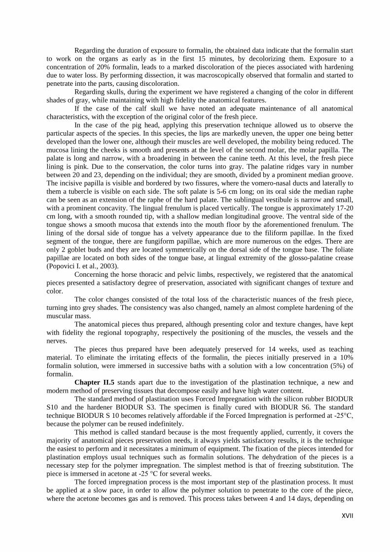
In the case of the pig head, applying this preservation technique allowed us to observe the
particular aspects of the species. In this species, the lips are markedly uneven, the upper one being better
developed than the lower one, although their muscles are well developed, the mobility being reduced. The
mucosa lining the cheeks is smooth and presents at the level of the second molar, the molar papilla. The
palate is long and narrow, with a broadening in between the canine teeth. At this level, the fresh piece
lining is pink. Due to the conservation, the color turns into gray. The palatine ridges vary in number
between 20 and 23, depending on the individual; they are smooth, divided by a prominent median groove.
The incisive papilla is visible and bordered by two fissures, where the vomero-nasal ducts and laterally to
them a tubercle is visible on each side. The soft palate is 5-6 cm long; on its oral side the median raphe
can be seen as an extension of the raphe of the hard palate. The sublingual vestibule is narrow and small,
with a prominent concavity. The lingual frenulum is placed vertically. The tongue is approximately 17-20
cm long, with a smooth rounded tip, with a shallow median longitudinal groove. The ventral side of the
tongue shows a smooth mucosa that extends into the mouth floor by the aforementioned frenulum. The
lining of the dorsal side of tongue has a velvety appearance due to the filiform papillae. In the fixed
segment of the tongue, there are fungiform papillae, which are more numerous on the edges. There are
only 2 goblet buds and they are located symmetrically on the dorsal side of the tongue base. The foliate
papillae are located on both sides of the tongue base, at lingual extremity of the glosso-palatine crease
(Popovici I. et al., 2003).
Concerning the horse thoracic and pelvic limbs, respectively, we registered that the anatomical
pieces presented a satisfactory degree of preservation, associated with significant changes of texture and
color.
The color changes consisted of the total loss of the characteristic nuances of the fresh piece,
turning into grey shades. The consistency was also changed, namely an almost complete hardening of the
muscular mass.
The anatomical pieces thus prepared, although presenting color and texture changes, have kept
with fidelity the regional topography, respectively the positioning of the muscles, the vessels and the
nerves.
The pieces thus prepared have been adequately preserved for 14 weeks, used as teaching
material. To eliminate the irritating effects of the formalin, the pieces initially preserved in a 10%
formalin solution, were immersed in successive baths with a solution with a low concentration (5%) of
formalin.
Chapter II.5 stands apart due to the investigation of the plastination technique, a new and
modern method of preserving tissues that decompose easily and have high water content.
The standard method of plastination uses Forced Impregnation with the silicon rubber BIODUR
S10 and the hardener BIODUR S3. The specimen is finally cured with BIODUR S6. The standard
technique BIODUR S 10 becomes relatively affordable if the Forced Impregnation is performed at -25°C,
because the polymer can be reused indefinitely.
This method is called standard because is the most frequently applied, currently, it covers the
majority of anatomical pieces preservation needs, it always yields satisfactory results, it is the technique
the easiest to perform and it necessitates a minimum of equipment. The fixation of the pieces intended for
plastination employs usual techniques such as formalin solutions. The dehydration of the pieces is a
necessary step for the polymer impregnation. The simplest method is that of freezing substitution. The
piece is immersed in acetone at -25 °C for several weeks.
The forced impregnation process is the most important step of the plastination process. It must
be applied at a slow pace, in order to allow the polymer solution to penetrate to the core of the piece,
where the acetone becomes gas and is removed. This process takes between 4 and 14 days, depending on

XVIII
the size of the piece, on the density of the tissue and on the viscosity of the polymer solution. The
polymer solution used for (BIODUR S 10) can be reused by simply adding a new quantity of polymer.
The curing of the impregnated specimen occurs in the presence of the BIODUR S6 reactive in a
gaseous form, which is put in contact with the piece to conclude the polymerization.
The polymers used for plastination must have the following properties: low viscosity to
facilitate tissue penetration, unlimited reusability, capacity to preserve the biological material, mechanical
and optical adequate properties (e. g.: resistance to yellowing, adequate refractive index) and not
degrading the pieces during processing.
To prevent shrinking, the pieces must be kept at a temperature -25°C and -30 °C during
dehydration.
If the pieces are left too long in acetone, they lose their flexibility, become hardened and tend to
brake, leading to the following disadvantages:
They will be very sensitive to mechanical forces such as pressure during the polymer
impregnation.
The flexibility of the specimens impregnated with silicon rubber (BIODUR S) decreases.
This is harmful for the pieces with thin walls (intestine), with a fragile structure, with filaments (nerves)
and with membranes (kidneys with intact renal pelvis).
For this reasons, all pieces impregnated with silicone rubber that must remain flexible after the
plastination process, must not the submerged in acetone longer than necessary.
Mechanical properties of dehydrated specimens play no role for the mechanical properties of the
final specimens, if a non flexible, hardened product is desired. Such pieces can be kept indefinitely in
acetone. However, they are fragile and susceptible to mechanical pressure during processing.
The disadvantages of an insufficient dehydration are white spots or shrinking of the specimen;
they can be more severe than those induced by an excessive dehydration.
Before the Forced Impregnation it is advised to always measure the water content of the
Volatile Intermedium (acetone).
The water content of the Volatile Intermedium must be under 1%. Water content higher than 2%
will lead to inferior quality products.
If one is inexperienced in carrying out the dehydration process, one should measure again the
density of the dehydrating liquid after three days to see if the water content has remained constant.
The pieces must be only pre-cooled, because direct freezing leads to the formation of ice
crystals, deteriorating the pieces due to several reasons:
The pieces will blacken due to the ice crystals and the surface cell layers will dry in certain
places, leading to white spots on the final products, the so called „freeze burning marks”

XIX
GENERAL CONCLUSIONS AND RECOMMENDATIONS
Out of the pool of general conclusions formulated through the analysis of the obtained results
we have highlighted the following:
1. The boiling preservation technique has a higher degree of applicability due to the fact that
it does not involve special equipment and it is low cost, but the risk of bone specimens degradation is
present in a various degree according to the species.
2. The maceration technique is simple and when applied it yields good results in obtaining
osseous pieces from reptiles, mammals and birds.
3. The acetone and the gasoline have produced a satisfying degreasing of the specimens;
4. The hydrogen peroxide was more efficient than the sodium bicarbonate in the process of
bone bleaching.
5. The technique applied on the pony cadaver was not adequate and the applied protocol
needs a revision.
6. The addition of the technical glycerin in the mummification process reduces its duration
and helps maintaining the consistency of the original live tissue.
7. The cold air ventilator helps accelerate the mummification process.
8. Technical ethylic alcohol has a toxic effect on the insects that are potentially harmful to
the specimens.
9. The mummification technique offers multiple advantages to the didactical process such
as: specimen durability, accuracy of the anatomical particularities and the possibility of directly handling
the items.
10. The formalin acts on the tissues as early as in the first 15 minutes.
11. The standard plastination technique S10 is complex and when applied it yields
remarkable results, producing anatomical specimens of a quality superior to those obtained though the
classical techniques.
12. We consider that the foundation of a plastination laboratory would represent a strong
asset in obtaining anatomical specimens of superior quality for both student use and research in the
veterinary medicine domain.
The following recommendations have been formulated from our results:
1. In order to obtain osseous pieces through the boiling technique, it is advisable to take into
account their origin, as an inadequate boiling time leads to the destruction of the specimens.
2. We recommend the use of degreasing and drying, because, if they are not adequately
applied, the assembling of the pieces becomes impossible, no matter the choice of adhesive.
3. When applying the mummification process, it is advisable to take into account the size of
the cadaver and the degree of its decomposing state.
4. The mummification technique using simple formalin is not adequate for the preservation
of anatomical specimens, at least not for those of large size such as intact large animal cadavers.
5. We recommend the use of technical glycerin during mummification, because it speeds up
the process and helps maintaining the original consistency of live tissue.
6. Seeing the outage of the specimens obtained by formalin preservation, we recommend
their incineration when signs of decay appear or when they lose their utility.

XX
SELECTIVE REFERENCES
1. ALLOUCH, G. M., 2014, Scientific Technique for Skeletons Preservation and
Preparation of Anatomical Models to Promote Veterinary Anatomy, J. Vet. Anat. Vol. 7 No 2, 133 - 139
http://www.vetanat.com/v13-pdf/9.pdf ;
2. ALLOUCH, G. M., AL -SHEIKH, 2008, Textbox of Comparative Anatomy, The ones,
ligaments and joints, practical part, pp 20-22, Veterinary Medicine College, Al Baath Univesity; Teaching
Effectiveness, J. Anim. Sci. 71:2270-2274;
3. DUMITRU IOANA ET AL., 2012, Comparative Study Of Two Of The Main
Consevation Technique Of Anatomical Pieces, Bulletin Of Univerity Of Agricultural Sciences And
Veterinary Medicine Cluj-Napoca; 2012; 69(1-2); 107-115; print ISSN 1843-5270, electronic ISSN 1843-
5378;
4. GRAM, C. O., 2006, Vertebrate Skeleton: Preparation and Storage, pp7-11, National
Park Service, http://www.nps.gov/Museum/publications/conserveogram/11-07.pdf.;
5. HUSSAIN, M., N. HUSSAIN, ZAINAB H., S. QAISER, 2007, Skeletal Preservation
Techniques to Enhance Veterinary Anatomy Teaching;
6. POPOVICI, I., DAMIAN A., POPOVICI N., CHIRILEAN IOANA., 2003, Tratat de
anatomie comparată, Splanhnologie, Ed. AcademicPres, Cluj-Napoca;
7. SOMMER, H.G., S. ANDERSON, 1974, Cleaning skeletons with dermestid beetles:
two refinements in the method. Curator,17:290–8;
8. *** http://archlab.uindy.edu);
9. *** http://www.collectioncare.org/MSDS/Acetonemsds.pdf;
10. *** http://www.skullsite.com/misc/macerationmanual.htm;
11. *** http://www.skullsite.com/misc/macerationmanual.htm;
12. *** http://www.skullsite.com/misc/macerationmanual.htm).