Embed Size (px)
Citation preview

Adult cardiopulmonary
resuscitationUD FM DAIC
English program practice
2018

www.erc.edu
www.reanimatio.com

Terminology
CPR – Cardiopulmonary Resuscitation
BLS – Basic Life Support CPR without the help of medical devices
EBLS – Extended Basic Life Support CPR with easy to use tools or devices (special
expertise not needed) (NPT / OPT, BVM + O2, automated defibrillator)
ALS – Advanced Life Support CPR with devices for the qualified personnel
ROSC – Return Of Spontaneous Circulation

Chain of survival


Response? (vocal and tactile stimulus)
Call for help!
Open the airway
Head tilt
Chin lift
Foreign body removal
Check vital signs
Breathing? (see / hear / feel!)
(Normal: RR≥2/10 sec, noticeable
chest/abdominal movement with
audible and sensible airflow without
pathological noises)
(Circulation / pulse?)
Monitor? (Pulse oxymetry /capnography
/ECG /IBPM)
Vital signs / open airways

CPR 30:2Until defibrillation /
monitoring is
available
Provide a patent airwayLook for vital signs
Call for paramedicsor Medical Emergency Team
Cardiac arrest
Calling the ambulance:
Your name
What has happened
Where did it happen
How to get there
Stay calm and follow
the advices

Chest compression Right („high quality”) technique:
Firm surface
Proper posture
Extended straight arms
Position of the hands: lower half of the sternum
Compression / ventilation rate
30:2 (adult)
15:2 (pediatric)
In the case of a primary cardiac cause COCPR (Compression Only CPR) is allowed in the first 3 minutes (?)
In the case of a secured patent airway (ETT) continuous compression without disruption
Depth:
Approx. 5, max. 6 cms (adult)
1/3rd of the chest (pediatric)
Frequency: 100-120 / min (metronome?)
Duty cycle 1:1; allow complete recoil
(Compression feedback devices?)
Avoid:
fatigue (→change)
> 5 (10) sec disruption (→planning)

Rescue breaths (BLS and EBLS)
Technique:
Mouth-to-mouth/ Mouth-to-nose
BVM (bag-valve mask) ventilation
NB: patent airway!
Head tilt + chin lift
jaw thrust (Esmarch-Heiberg)
NPT / OPT
Foreign body removal(→ FBAO protocol)
Normal blow in
Vt: approx. a normal breath intake (6-8 ml/ttkg)
Approx. 1 sec duration
30:2 (compressions:breaths)
Avoid:
Hyperventilation
→ hypocapnia → cerebral vasoconstriction → cerebral ischaemia
High pressures
→ abdominal insufflation → risk of aspiration

CPR 30:2Until AED or manual
defibrillator is applied
Rhythm
analysis
Shockable(VF/ pulseless VT)
Non-shockable(PEA/Asystole)
Patent airway
Vital signs
Call for
resuscitation
team

Ventricular Fibrillation
(VF)
Pulseless Ventricular
Tachycardia
(pulseless VT)
Rhythm
analysis
Shockable
(VF/ pulseless VT)
1 Shock150-360 J biphasic
or 360 J monophasic
Immediately resume
CPR 30:2
for 2 minutes

Shockable rhythm(VF)
Irregular wave
No recognizeable QRS complex
Ever changing frequency and amplitude
coarse / fine
NB: artefacts
movement
electric interference

Shockable rhythm(pulseless VT)
Monomorphic pulseless VT
widened QRS complex
high frequency
No QRS morphologic changes
Polimorphic pulselessVT
Torsades de pointes

(Precordial thump)
For immediate treatment of a witnessed VF/VT
Monitored patient
If defibrillation is not a prompt option
BUT: according to the 2015 ERC guidelines, it is NOT RECOMMENDED!

Defibrillators AED
Manual
Diagnostic / monitor function
ECG (monitor +/- 12 lead)
(+/- rhythm analysis)
NIBP
Pulse oxymetry
Capnography
Therapeutic options
Defibrillation
SCV (synchronized
cardioversion)
Transcutaneous pacing

Defibrillation energy
Factory recommendation / device learning
Monophasic: 1x360 J
Biphasic: 1x150-360 J (escalating)
If not sure, use the highest possible energy (don't waste time)!!!
(3 shock strategy?: witnessed VF/pulselessVT)

„Good” defibrillation
Efficient
Time !!!
Biphasic wave
Provide contact:
– Adhesive electrodes
– Defibrillator paddles
Contact gel or gel pad
Compression of the paddles
Shock during exhalation
(ventilated patients!)
Anterolat. vs. anteropost. vs. bilat.
position
Safe
Adhesive electrodes
Adequate communication
Correct manipulation (NB:
moving, contact, charging)
Avoidance of wet circumstances
O2 flow and NTTS removal from
defibrillation area
PM / ICD avoidance (>15 cm)

If VF/VT remains
Second shock and after:
150 - 360 J biphasic
360 J monophasic
Minimize the delay between the CPR
and a shock (< 10 s)
Adinistration of medication mustn’t
delay the shock
First 1 mg of Adrenaline + 300 mgs of
Amiodarone after the third shock
Repeat 1 mg Adrenaline if needed in
every 3-5 minutes
Repeat 150 mgs of Amiodarone if
needed once more
Third shock
Fourth shock
CPR for 2 minutes
Adrenaline 1mg iv.
+ Amiodarone 300 mgs iv.
Recheck:
if VF/VT remains

After the shock Immediately continue CPR for 2 minutes
Stop only if the patient shows clear vital signs (breaths,
coughs, opens his/her eyes, moves limbs, regains
consciousness)
After 2 minutes of CPR reassess ECG rhythm:
VT / pulseless VT: keep on with shockable algorithm
Any other electrical activities (compatible with circulation):
ROSC: continue with postresuscitation care
No signs of circulazion (PEA): swap to non-
shockable algorithm
Asystole: swap to non-shockable algorithm

Asystole (Asy)
Pulseless Electrical
Activity (PEA)
Rhythm
analysis
Non-shockable
(PEA/Asystole)
Immediately
resume
CPR 30:2
For 2 minutes

Non shockable rhythmAsystole
No ventricular activity (QRS)
Atrial activity is possible (P-wave asystole – PM indicated)
Rarely straight line (look out: electrical contact!)
Fine VF => treat as asystole

Asystole
During CPR:
Check ECG leads
Adrenaline 1 mg iv in every 3 – 5 minutes (Atropine 3 mg iv.)

Non-shockable rhythm(PEA)
Clinical signs of cardiac arrest with any
ECG pattern (VF excluded)

Pulseless Electrical Activity
During CPR:
Exclude / treat reversible causes
Adrenaline 1 mg iv in every 3-5 minutes
(Atropine 3 mg iv., in case of PEA with frequency below 60 / min)

During CPR:
• Maintain high quality chest compressions → change as frequently as needed
• Shortest possible disruption of CPR → planning
• Mechanical chest compression devices if available or needed. (CPR in or on the
way to the catheterization laboratory, fibrinolysis with CPR, hypothermic patient, etc)
• Ventilation with 100 % FiO2
• Route of administration (iv., io.)
• Adrenaline in every 3-5 minutes
• Advanced airway management
• Continuous compressions after securing airways
• Capnography (tube position? effectiveness? ROSC?)
• Reversible causes (4H/4T) and treatment
• Check devices, positions, functions regularly

Airway and ventilation Secured Airway:
ETT (gold standard)
LMA / iGel
LT
ETC, etc.
After a secured airway
Continuous chest compressions!
BVM/mechanical ventilation
FiO2: 100 % !!!
Avoid hyperventilation
Vt: 6-8 ml/bwkgs
RR: approx. 10/min

Mechanical chest
compression devices
LUCAS AutoPulse Weil SCC

Route of administration
Peripheral?
Veins on the upper limbs
external jugular vein
Central?
Femoral vein
Internal jugular vein
Subclavian vein
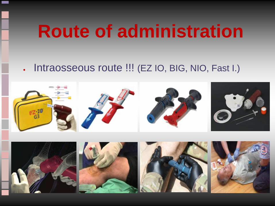
Route of administration
Intraosseous route !!! (EZ IO, BIG, NIO, Fast I.)

ALS medicines

4H Hypoxia (asphyxia)
Hypovolemia (absolute, eg. bleeding / relative, eg. anaphylaxis)
Hypo/hyperkalemia & metabolic changes (acidosis, hypoglycaemia)
Hypo/hyperthermia
4T Thrombosis (ACS, PE)
Tension PTX
Tamponade (cardiac)
Toxin / Tablets (side effects)
Dg: Heteroanamnesis
Medical documentations
Physical examination
Blood gas analysis
Emergency Ultrasound (EUS)
THINK!!!
Potentially reversible causes

After ROSC (postresuscitation care)
ABCDE approach
Advanced monitoring - ECG, BGA, laboratory tests
Imaging (X-Ray, US, CT – HRCT; skull, thorax, abdomen)
Consider coronarography +/- PCI
Causal (4H-4T) treatment ASAP
Supportive therapy: provide tissue oxygenation
Augmentation of the cardiac function (DO2)
O2, NIV, invasive ventilation (normoxia, normocapnia)
Katecholamines, PDE inhibitor drugs, Ca-sensitizers
IABP, ECMO (failing all else)
Decreasing metabolic demand (VO2)
Mechanical ventilation
Analgo-sedation, seizure prophylaxis
(Neuromuscular blockade???)
Targeted Temperature Management (TTM) → 32-36 ⁰C, avoid fever

Any
questions ?

Thank you for your attention