Embed Size (px)
Citation preview

Acetylcholinesterase Staining Version: January 2, 2016
ACETYLCHOLINESTERASE STAIN
Figure 1. Mouse smooth muscle stained using the acetylcholinesterase method of Paxinos & Watson (1998). Darker staining indicates higher levels of acetylcholinesterase.
Overview.
Acetylcholinesterase is the main enzyme responsible for regulating cholinergic neurotransmission in the CNS by catalyzing the hydrolysis of the cholinergic neurotransmitter acetylcholine (Silver, 1974). Acetylcholinesterase activity can be viewed histologically in fresh or fixed muscle (Figure 1) and brain (Figure 2). The methods contained in this protocol are based on those used in the widely used rodent brain atlases produced by Watson, Paxinos and Franklin (e.g., Franklin & Paxinos, 2008; Watson & Paxinos, 1998; see also Shute & Lewis, 1963).
The availability of atlases based on this stain provides an invaluable resource with which to compare the results of your
experiments. The depth of available background information also makes the stain suitable for inclusion in teaching labs.
Figure 2. Mouse brain stained for ACHE 40 µm thick section.
Version 1-2-16
Applications
Limitations
Experiments where the goal is to study the distribution and location of acetylcholinesterase and by inference cholinergic neurotransmission. Experiments requiring an estimate of effects on cholinergic neurotransmission in the brain. Studies of the neuromuscular junction.
Results with fixed tissue is poorer than with fresh frozen sections.
Slides are stable in light. Storage time is not known but we have stored slides for at least seven years without noticeable fading. Slides are easily viewed and photographed with a light microscope at 4x to 40x.
Slide Characteristics
Acetylcholinesterase Staining
2
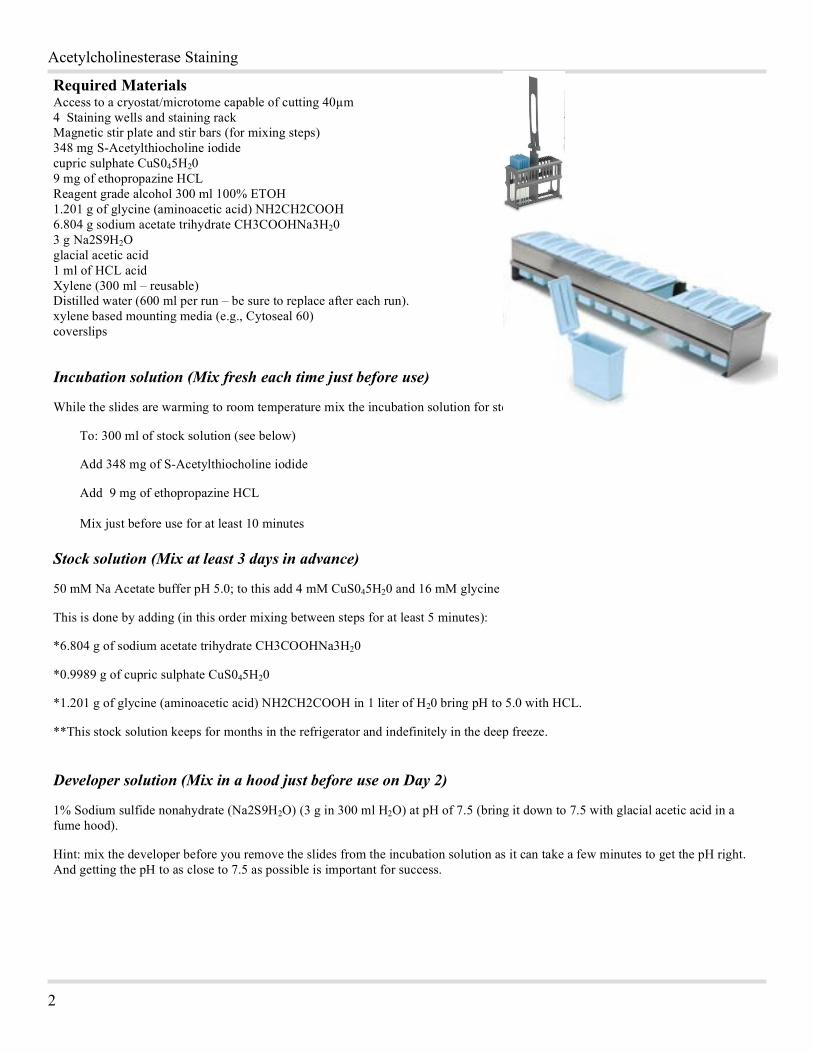
Required Materials Access to a cryostat/microtome capable of cutting 40µm 4 Staining wells and staining rack Magnetic stir plate and stir bars (for mixing steps) 348 mg S-Acetylthiocholine iodide cupric sulphate CuS045H20 9 mg of ethopropazine HCL Reagent grade alcohol 300 ml 100% ETOH 1.201 g of glycine (aminoacetic acid) NH2CH2COOH 6.804 g sodium acetate trihydrate CH3COOHNa3H20 3 g Na2S9H2O glacial acetic acid 1 ml of HCL acid Xylene (300 ml – reusable) Distilled water (600 ml per run – be sure to replace after each run). xylene based mounting media (e.g., Cytoseal 60) coverslips
Incubation solution (Mix fresh each time just before use)
While the slides are warming to room temperature mix the incubation solution for step 5.
To: 300 ml of stock solution (see below)
Add 348 mg of S-Acetylthiocholine iodide
Add 9 mg of ethopropazine HCL Mix just before use for at least 10 minutes Stock solution (Mix at least 3 days in advance)
50 mM Na Acetate buffer pH 5.0; to this add 4 mM CuS045H20 and 16 mM glycine
This is done by adding (in this order mixing between steps for at least 5 minutes):
*6.804 g of sodium acetate trihydrate CH3COOHNa3H20
*0.9989 g of cupric sulphate CuS045H20
*1.201 g of glycine (aminoacetic acid) NH2CH2COOH in 1 liter of H20 bring pH to 5.0 with HCL.
**This stock solution keeps for months in the refrigerator and indefinitely in the deep freeze.
Developer solution (Mix in a hood just before use on Day 2)
1% Sodium sulfide nonahydrate (Na2S9H2O) (3 g in 300 ml H2O) at pH of 7.5 (bring it down to 7.5 with glacial acetic acid in a fume hood).
Hint: mix the developer before you remove the slides from the incubation solution as it can take a few minutes to get the pH right. And getting the pH to as close to 7.5 as possible is important for success.
Version: January 2, 2016
3
1
Staining and Sectioning Procedure for Fresh Frozen Sections (no perfusion method) Important: Preset the cryostat to – 15 C the day before sectioning.
Steps 1.) Immediately after testing rapidly remove and freeze brains on the “rapid freeze” bar/cryobar in your cryostat. Do not leave the tissue on the bar for more than 5 minutes. Move to lower storage tray in the cryostat or start sectioning. 2.) Sectioning should be completed at 40 to 50 µm and mounted directly onto slides (do NOT use subbed slides). 3.) Keep the completed slides in a slide box placed in the bottom of the cryostat until you are ready to process the day’s slides (no more than a few hours). 4.) Remove the slides and place flat (tissue side up) on paper towels for 10 minutes at room temperature.
While the slides are warming to room temperature mix the incubation solution for step 5. To: 300 ml of stock solution (see below) Add 348 mg of S-Acetylthiocholine iodide Add 9 mg of ethopropazine HCL Mix these just before use for at least 10 minutes
5.) Places slides in a slide staining rack and leave overnight in the incubation solution: 6.) Rinse in distilled H2O for five minutes. 7.) Develop for 10 minutes in 1% Na2S9H2O (3 g in 300 ml H2O) at pH of 7.5 (bring it down to 7.5 with glacial acetic acid in a fume hood). 8.) Rinse in distilled H2O for five minutes. 9.) Fix in 3% buffered formalin for 24 hours. 10.) Remove slides from straining rack and place slides tissue side up on paper towels until dry (about an hour – do not over dry to avoid cracking the tissue). DO NOT DO THIS IN THE HOOD as the tissue will dry too fast and is likely to crack. 11.) Return slides to staining rack and clear in 100% ETOH for 3-5 minutes 12.) Put in Xylene for at least 30 minutes but no longer than 60 minutes. 13.) Coverslip
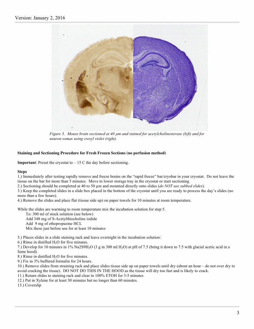
Figure 3. Mouse brain sectioned at 40 µm and stained for acetylcholinesterase (left) and for neuron somas using cresyl violet (right).

Acetylcholinesterase Staining
4
2
Donec interdum
Pellentesque:
Consectetuer:
Technical Notes:
1.) We have found that this stain is very pH sensitive so it is important to adjust the pH correctly.
2.) We have tried substituting 15 mg of haloperidol for ethopropazine and did not notice a difference in outcomes.
3.) This is a long stain and sometimes the edge of a few sections will tend to fold over (about 5% of the sections). You can take an artist’s fine paintbrush and fold them back when removing from the xylene but you must work fast as the tissue will be damaged if left out of the xylene for more than a few seconds. We usually don’t attempt the repair the folds that occur in as it usually ends up that the sections are damaged more then if just left alone.
References
Franklin, B. J. & Paxinos, G. (2008). The Mouse Brain in Sterotaxic Coordinates, Compact Third Edition, Academic Press, San Diego.
Paxinos, G. & Watson, C. (1998). The Rat Brain in Stereotaxic Coordinates, Fourth Edition, Academic Press, San Diego.
Shute C.C. & Lewis, P. R. (1963). Cholinesterase-containing systems of the brain of the rat. Nature, 199:1160–1164.
Silver, A. (1974). The biology of cholinesterases. Amsterdam: Elsevier.
Protocol obtained from neurosciencecourses.com.
This protocol may be reprinted and used for teaching or research purposes without permission and without charge as long as the reproduction notice is left on the document. Reproduction of the protocol, any portion, or image for any commercial purpose requires written permission from neurosciencecourses.com. No portion of this protocol or image used within may be posted on other web sites without the written permission of neurosciencecourses.com.