Embed Size (px)
Citation preview
Glutamate Neurotransmission Excitatory Amino Acid Neurotransmitters
Neurochemistry MS 532September 11, 2014
Dr. Dan SavageBMSB 145A
Reference: Brady et al., Basic Neurochemistry 8th ed., pp 342-366
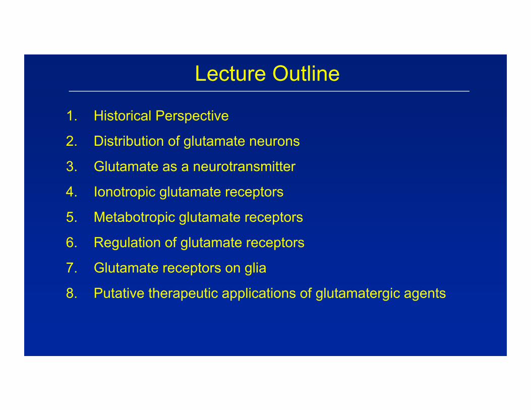
Lecture Outline
1. Historical Perspective
2. Distribution of glutamate neurons
3. Glutamate as a neurotransmitter
4. Ionotropic glutamate receptors
5. Metabotropic glutamate receptors
6. Regulation of glutamate receptors
7. Glutamate receptors on glia
8. Putative therapeutic applications of glutamatergic agents
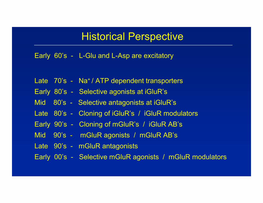
Historical Perspective
Early 60’s - L-Glu and L-Asp are excitatory
Late 70’s - Na+ / ATP dependent transportersEarly 80’s - Selective agonists at iGluR’sMid 80’s - Selective antagonists at iGluR’sLate 80’s - Cloning of iGluR’s / iGluR modulatorsEarly 90’s - Cloning of mGluR’s / iGluR AB’sMid 90’s - mGluR agonists / mGluR AB’sLate 90’s - mGluR antagonistsEarly 00’s - Selective mGluR agonists / mGluR modulators
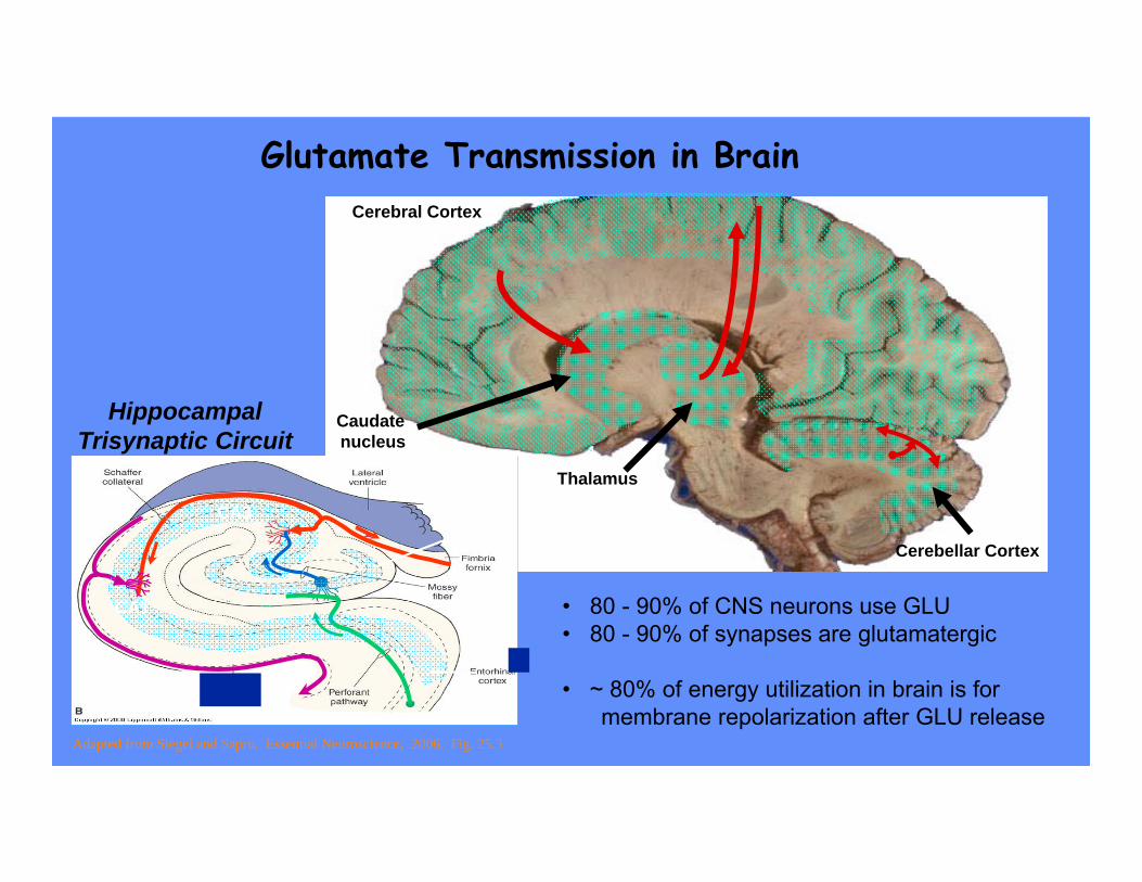
Caudatenucleus
Thalamus
Cerebellar Cortex
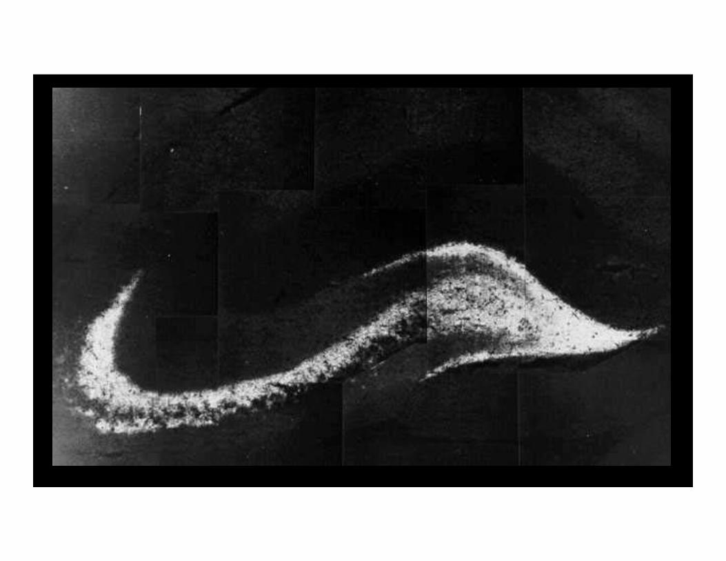
HippocampalTrisynaptic Circuit
Adapted from Siegel and Sapru, Essential Neuroscience, 2006, Fig. 25.3
Glutamate Transmission in Brain
Dentate gyrus
CA3
CA1
Cerebral Cortex
• 80 - 90% of CNS neurons use GLU• 80 - 90% of synapses are glutamatergic
• ~ 80% of energy utilization in brain is formembrane repolarization after GLU release
Glutamate as a Neurotransmitter
Synthesis
- Ubiquitous; amino acid in highest concentration in CNS
- Small proportion is “transmitter specific”
- No identifiable “synthetic machinery”
- Sources:Glucose via TCA cycle to KG → GlutamateGlutamine via Phosphate-activated glutaminase
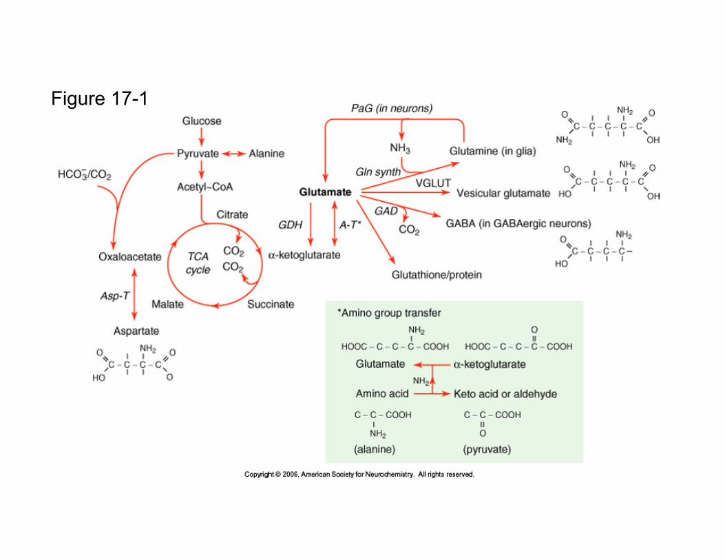
Figure 17-1
Glutamate as a Neurotransmitter Synthesis
Storage
- Small clear vesicles
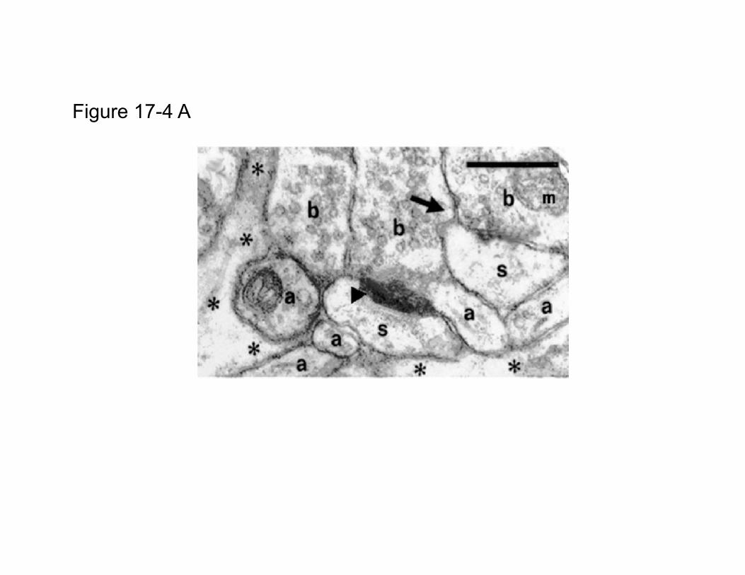
Figure 17-4 A
Glutamate as a Neurotransmitter Synthesis Storage
- Small clear vesicles (~ 17 nm diameter)- SV Transporters (VGLUT 1 & 2)- Proton pump- SV GLU Conc.: ~ 60 to 250 mM
Co-localized substances:- Peptides (CCK, DYN, others)- Zinc (via ZNT3 transporter)
Glutamate as a Neurotransmitter
Synthesis
Storage
Release
Ca++ / voltage dependent release
N, P, Q type VSCCs
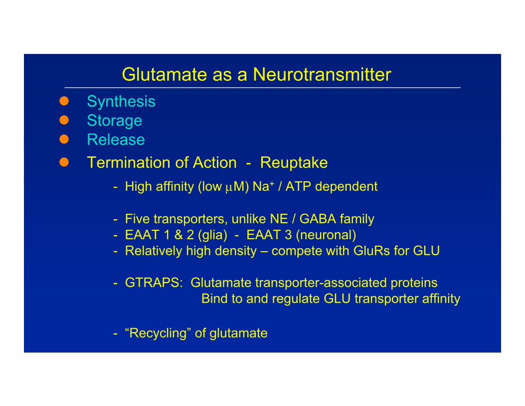
Glutamate as a Neurotransmitter Synthesis Storage Release Termination of Action - Reuptake
- High affinity (low M) Na+ / ATP dependent
- Five transporters, unlike NE / GABA family- EAAT 1 & 2 (glia) - EAAT 3 (neuronal)- Relatively high density – compete with GluRs for GLU
- GTRAPS: Glutamate transporter-associated proteinsBind to and regulate GLU transporter affinity
- “Recycling” of glutamate
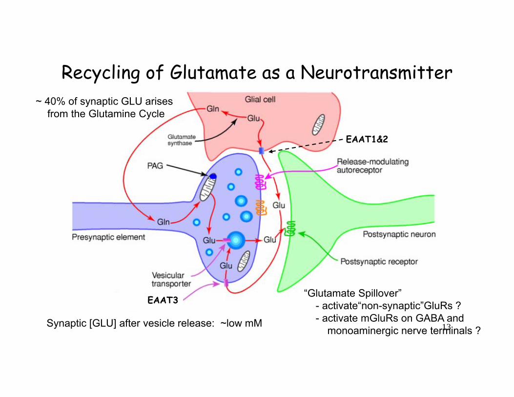
Recycling of Glutamate as a Neurotransmitter
EAAT1&2
EAAT3
~ 40% of synaptic GLU arises from the Glutamine Cycle
“Glutamate Spillover”- activate“non-synaptic”GluRs ?- activate mGluRs on GABA and
monoaminergic nerve terminals ?Synaptic [GLU] after vesicle release: ~low mM 13

Glutamate as a Neurotransmitter Synthesis Storage Release Termination of transmitter action Transmitter-specific receptors
- Ionotropic- Metabotropic
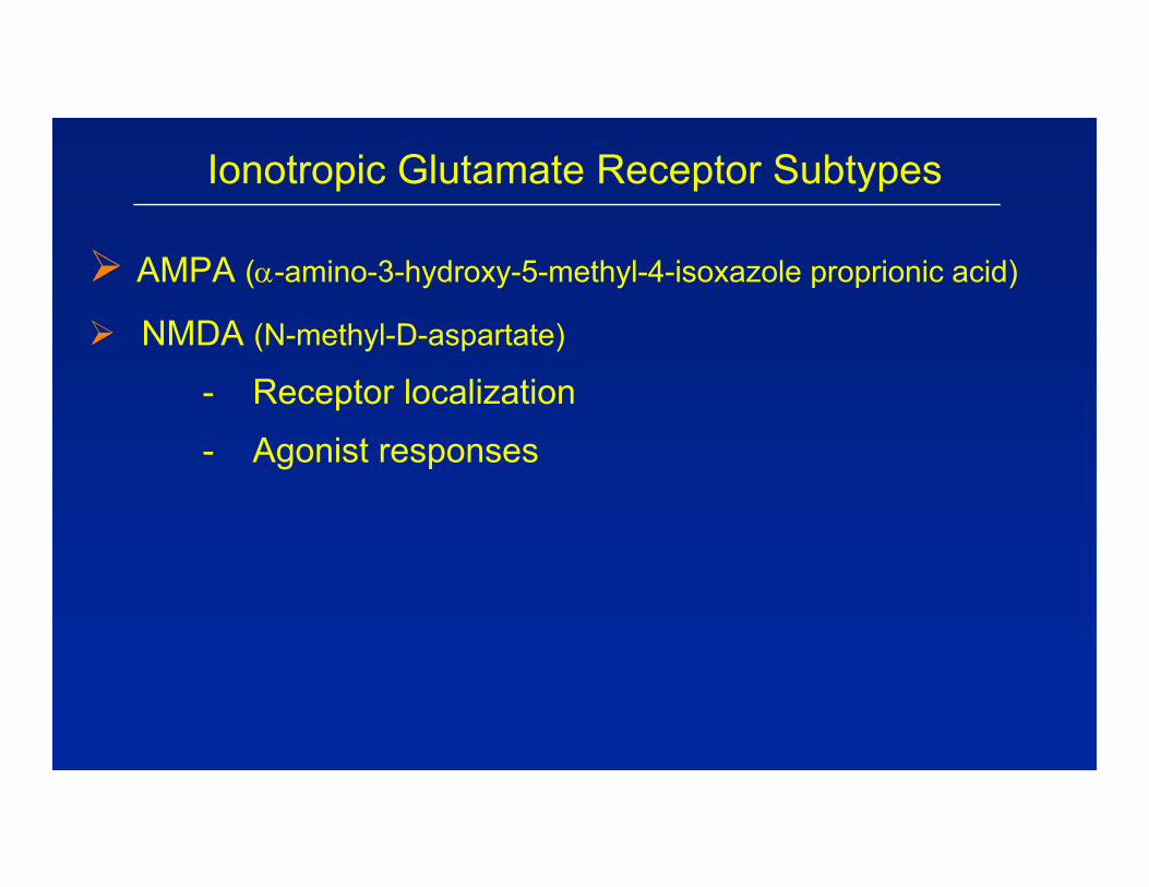
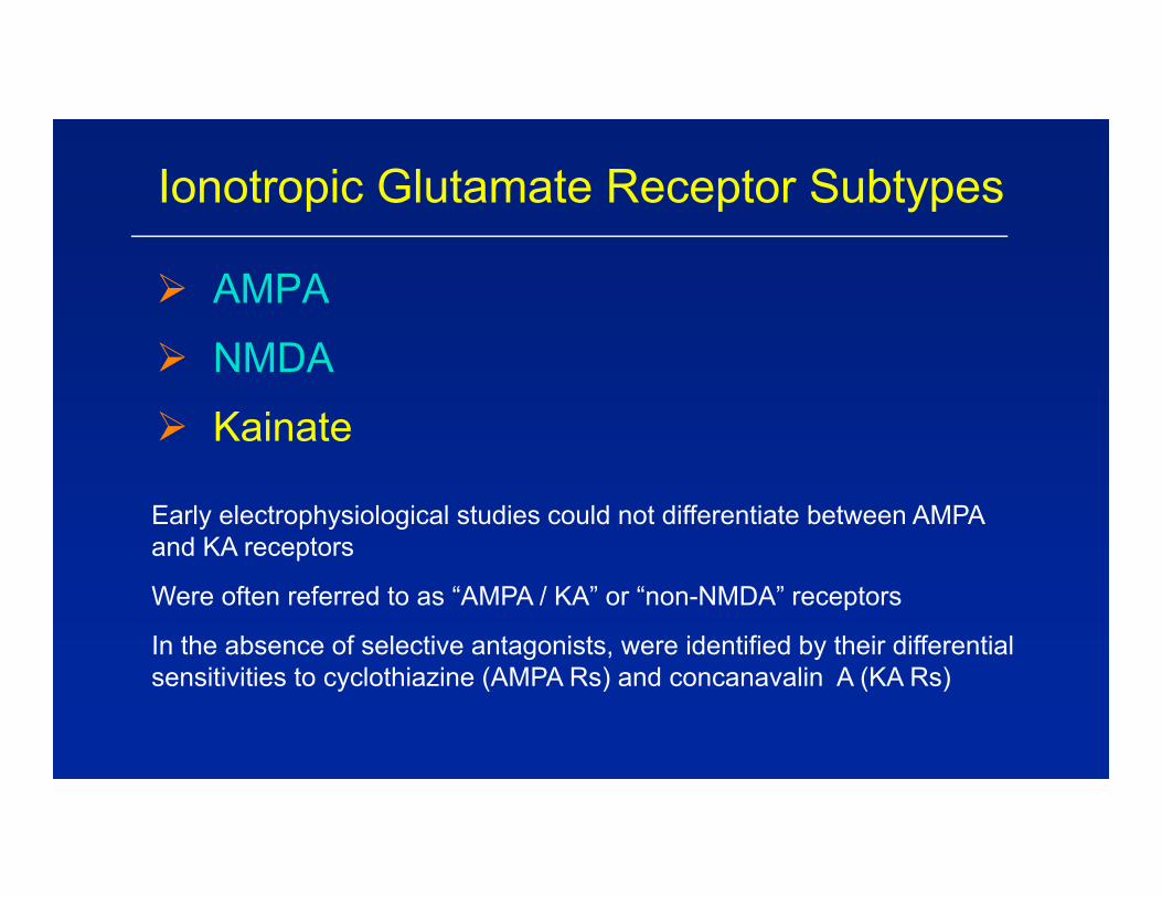
Ionotropic Glutamate Receptor Subtypes
AMPA (-amino-3-hydroxy-5-methyl-4-isoxazole proprionic acid)
NMDA (N-methyl-D-aspartate)
- Receptor localization
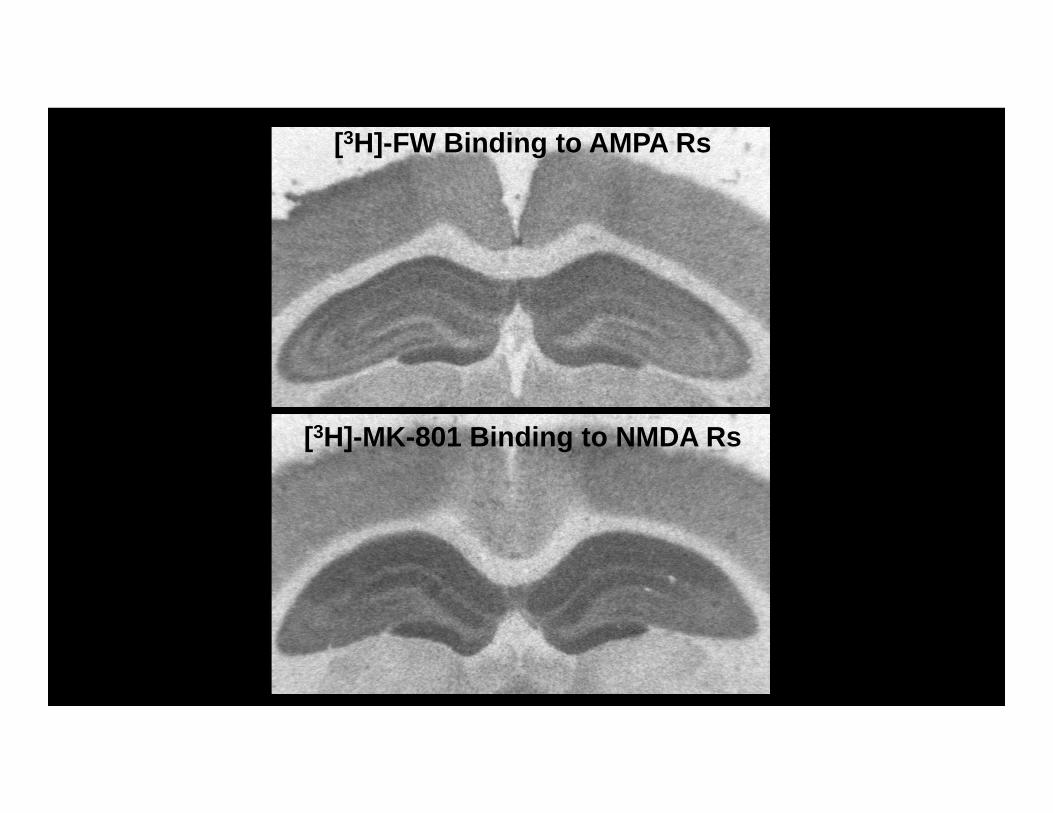
[3H]-FW Binding to AMPA Rs
[3H]-MK-801 Binding to NMDA Rs
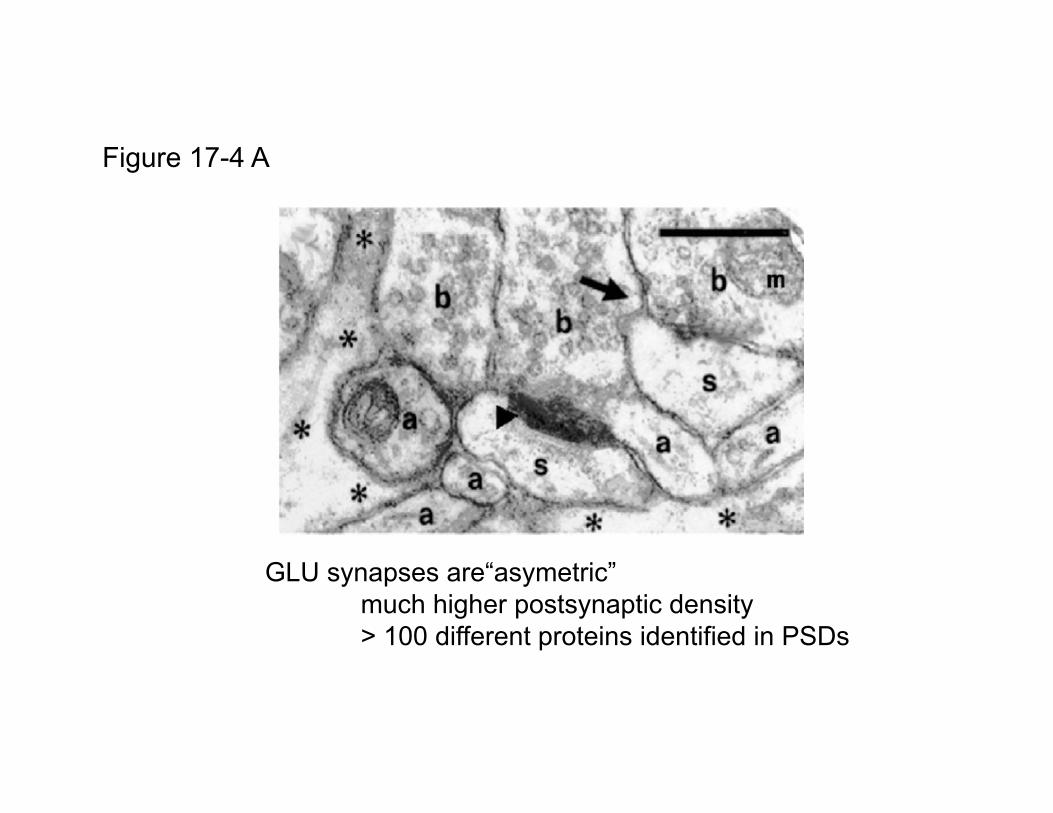
Figure 17-4 A
GLU synapses are“asymetric”much higher postsynaptic density> 100 different proteins identified in PSDs

Figure 17-4 B(Group I)
Ionotropic Glutamate Receptor Subtypes
AMPA (-amino-3-hydroxy-5-methyl-4-isoxazole proprionic acid)
NMDA (N-methyl-D-aspartate)
- Receptor localization
- Agonist responses
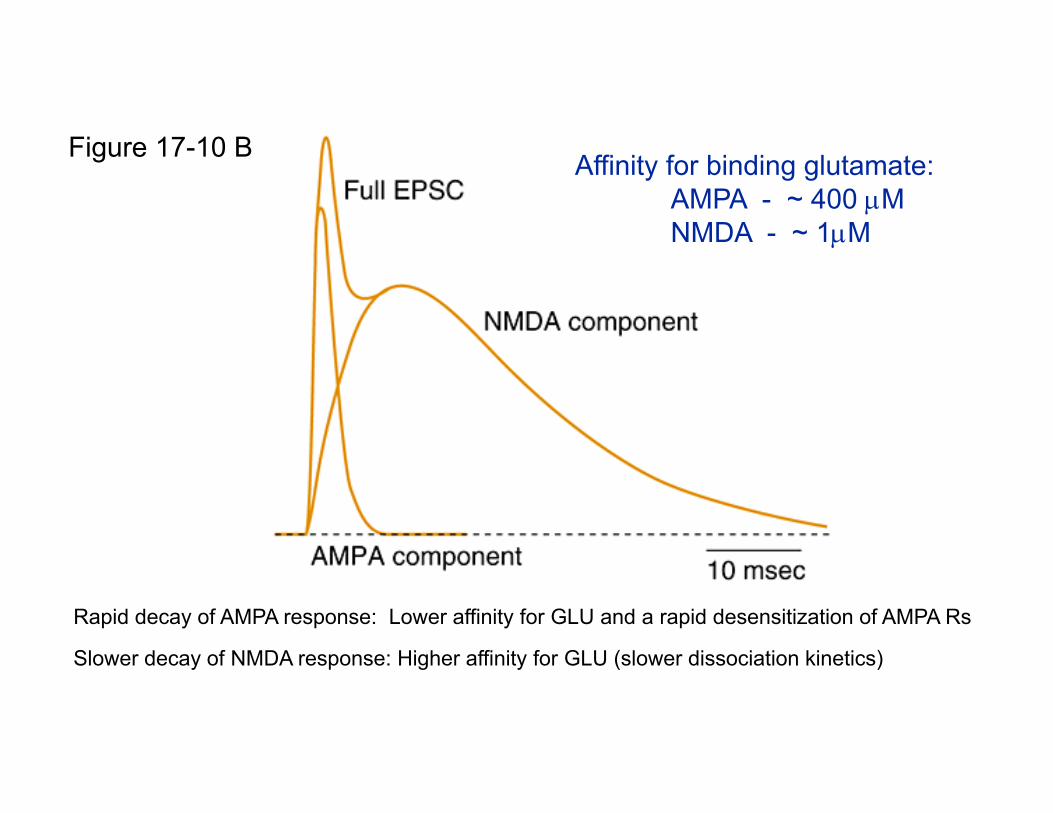
Affinity for binding glutamate:AMPA - ~ 400 MNMDA - ~ 1M
Figure 17-10 B
Rapid decay of AMPA response: Lower affinity for GLU and a rapid desensitization of AMPA Rs
Slower decay of NMDA response: Higher affinity for GLU (slower dissociation kinetics)
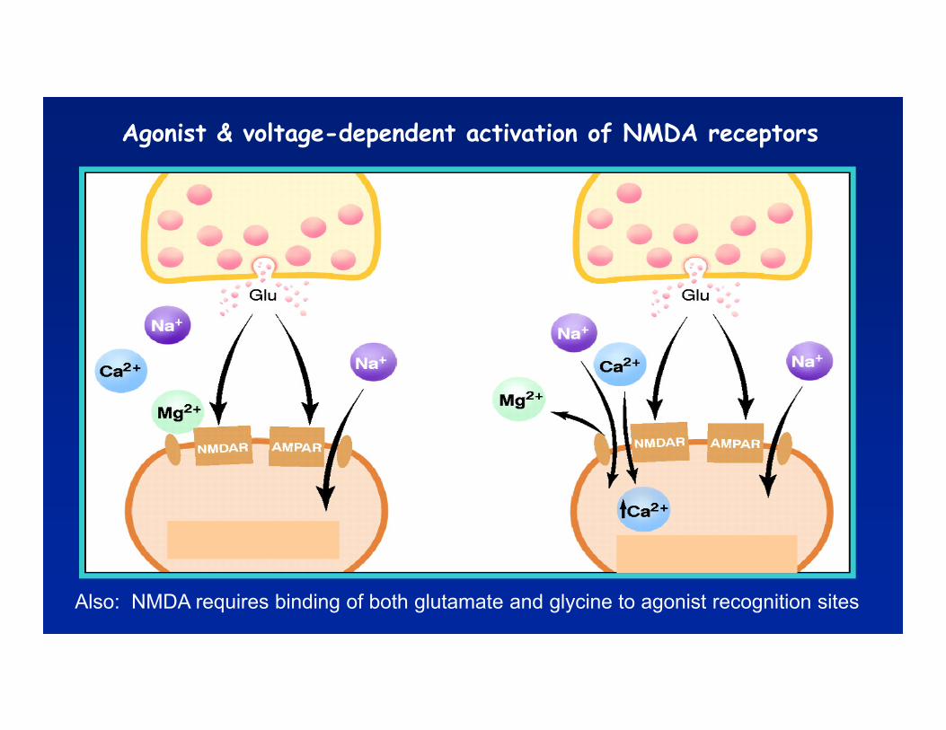
Agonist & voltage-dependent activation of NMDA receptors
Also: NMDA requires binding of both glutamate and glycine to agonist recognition sites
Ionotropic Glutamate Receptor Subtypes
AMPA
NMDA
Kainate
Early electrophysiological studies could not differentiate between AMPA and KA receptors
Were often referred to as “AMPA / KA” or “non-NMDA” receptors
In the absence of selective antagonists, were identified by their differential sensitivities to cyclothiazine (AMPA Rs) and concanavalin A (KA Rs)
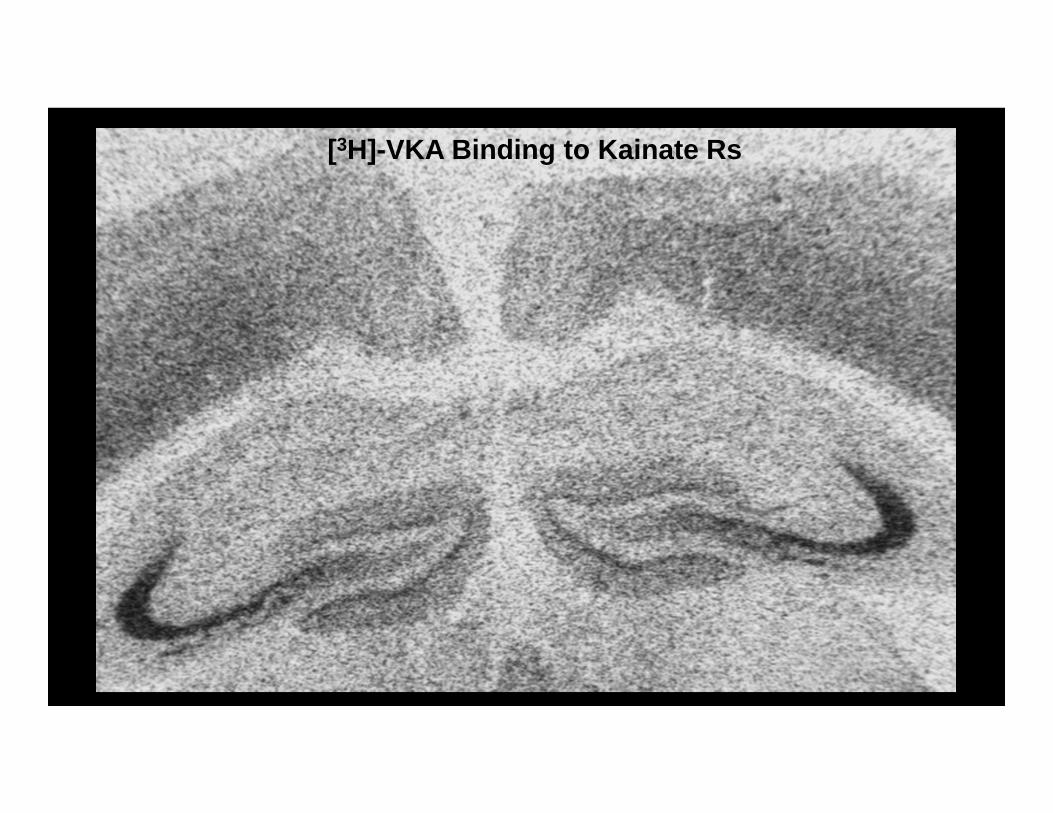
[3H]-VKA Binding to Kainate Rs
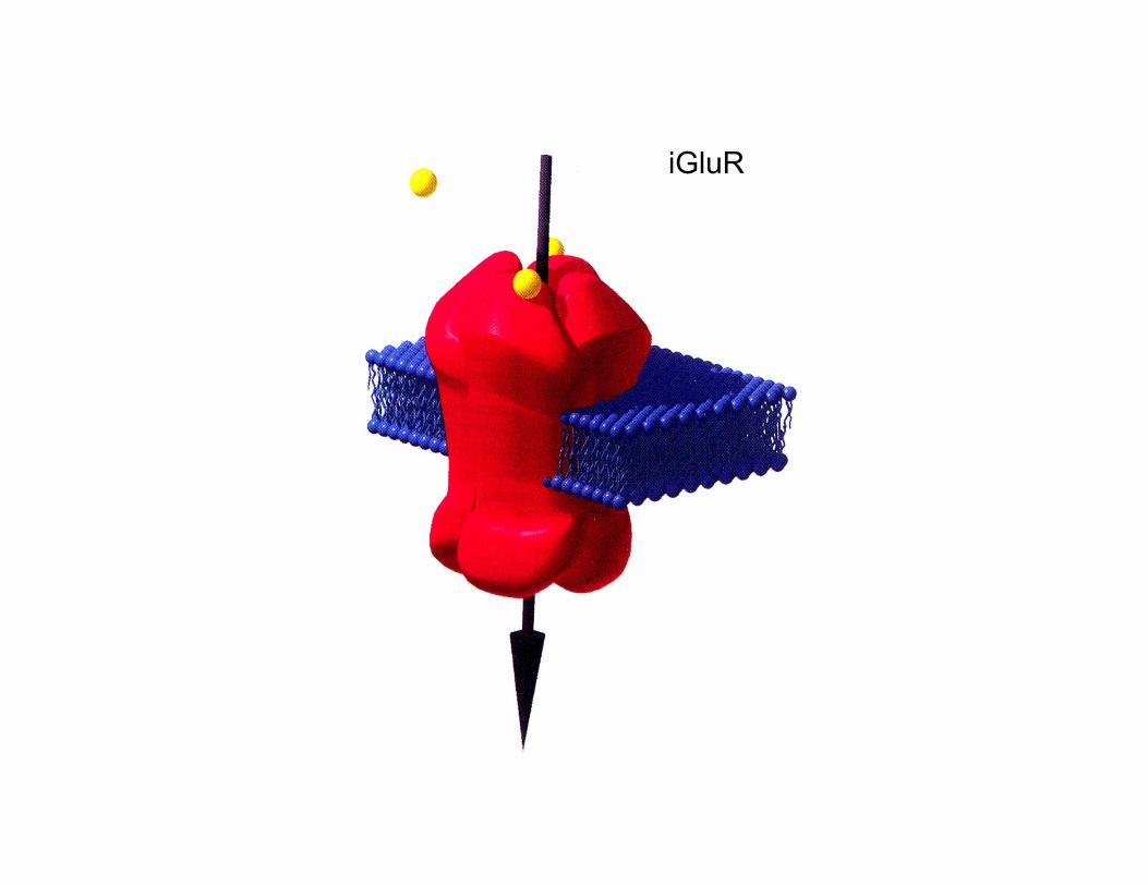
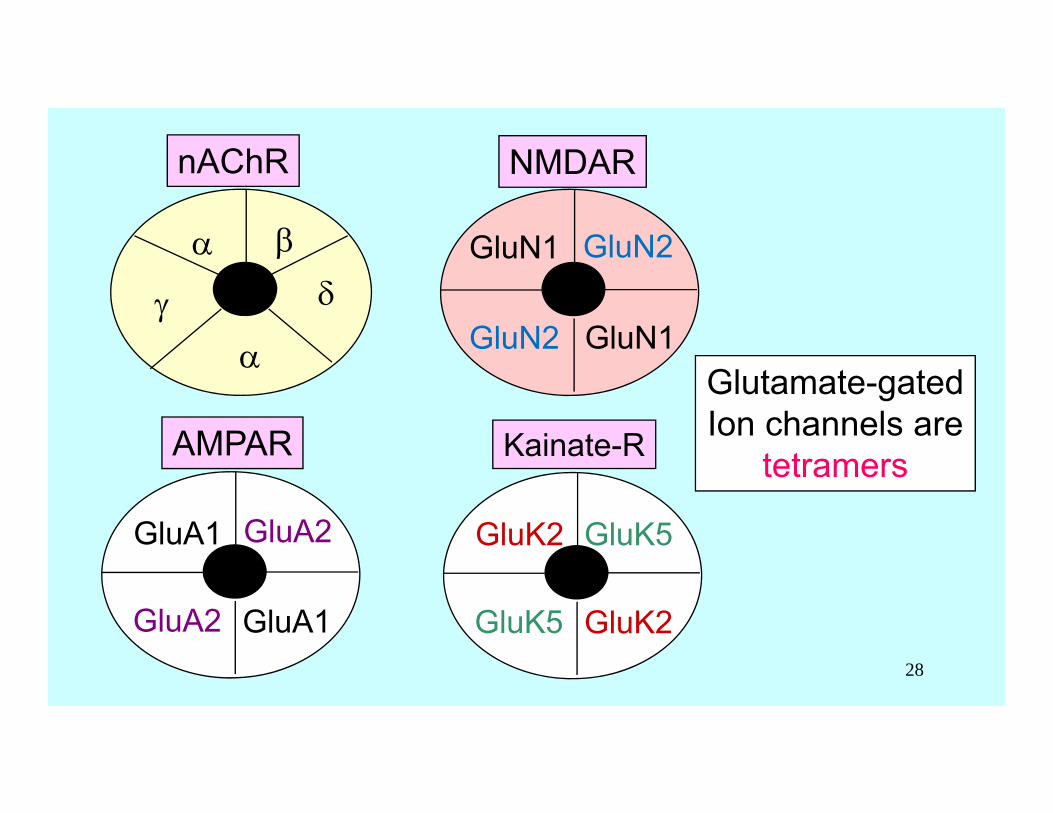
iGluR
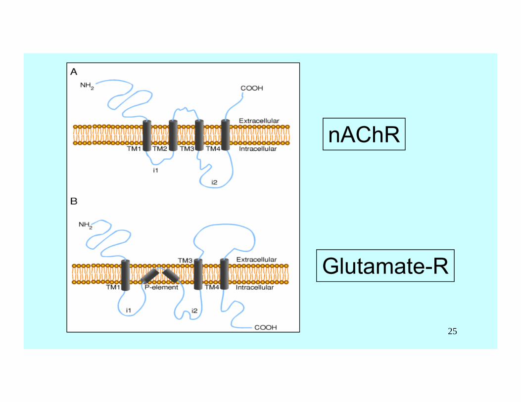
nAChR
Glutamate-R
25
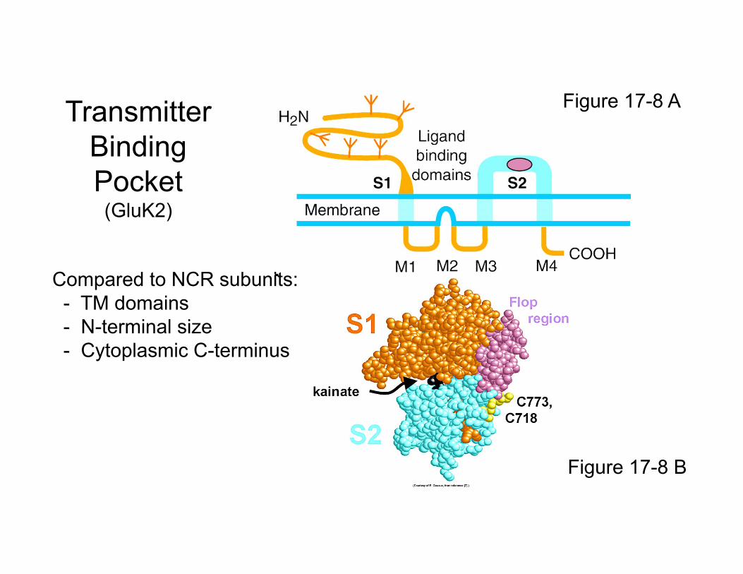
Figure 17-8 A
Figure 17-8 B
TransmitterBindingPocket(GluK2)
Compared to NCR subunits:- TM domains- N-terminal size- Cytoplasmic C-terminus
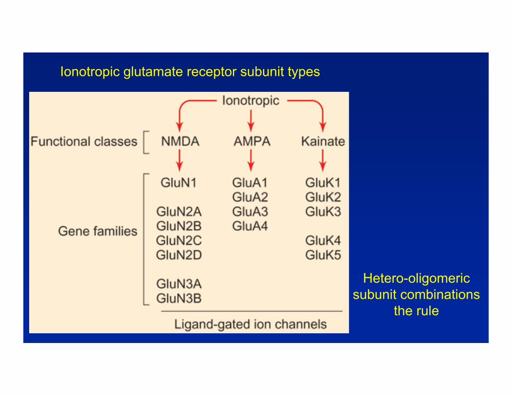
Hetero-oligomeric subunit combinations
the rule
Ionotropic glutamate receptor subunit types
nAChR
AMPAR
GluA1
GluA1
GluA2
GluA2
GluN1
NMDAR
Glutamate-gatedIon channels are
tetramersKainate-R
GluK2
GluK2
GluN1
GluN2
GluN2
GluK5
GluK528
NR1
NR1
NR2
NR2
GluN1
GluN1
GluN2
GluN2
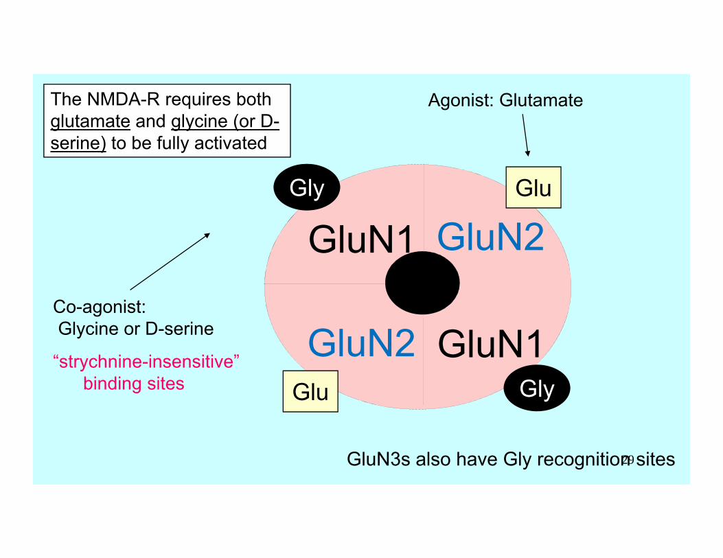
The NMDA-R requires both glutamate and glycine (or D-serine) to be fully activated
Agonist: Glutamate
Glu
Glu
Co-agonist:Glycine or D-serine
“strychnine-insensitive”binding sites Gly
Gly
GluN3s also have Gly recognition sites29
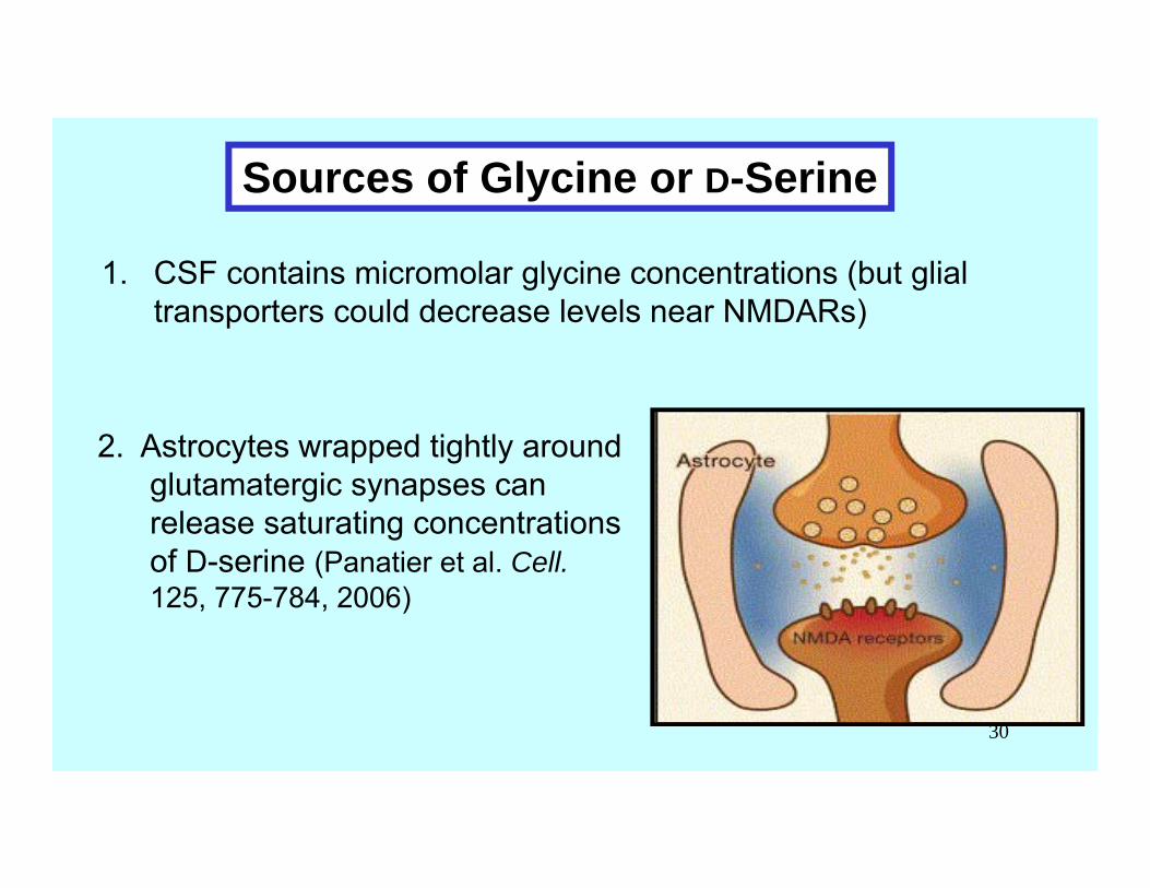
Sources of Glycine or D-Serine
1. CSF contains micromolar glycine concentrations (but glial transporters could decrease levels near NMDARs)
2. Astrocytes wrapped tightly around glutamatergic synapses can release saturating concentrations of D-serine (Panatier et al. Cell.125, 775-784, 2006)
30
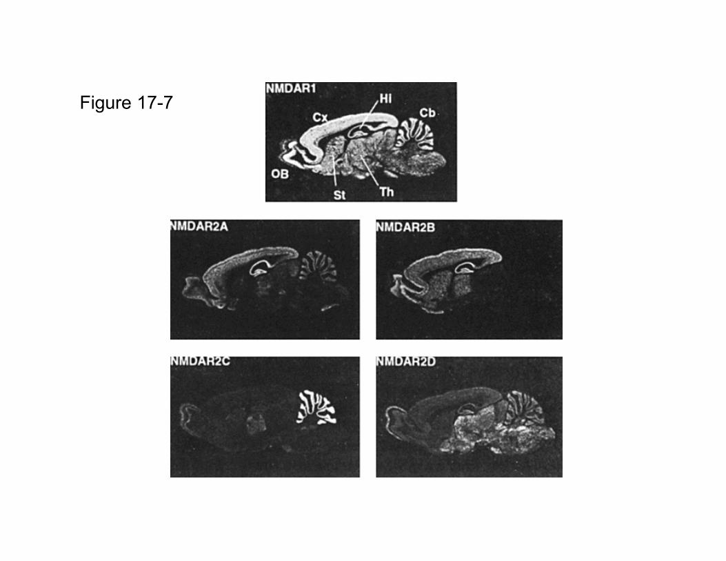
Figure 17-7
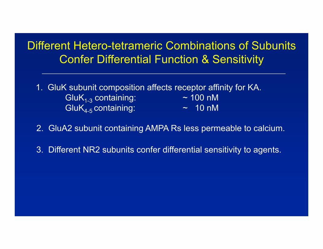
Different Hetero-tetrameric Combinations of Subunits Confer Differential Function & Sensitivity
2. GluA2 subunit containing AMPA Rs less permeable to calcium.
3. Different NR2 subunits confer differential sensitivity to agents.
1. GluK subunit composition affects receptor affinity for KA.GluK1-3 containing: ~ 100 nMGluK4-5 containing: ~ 10 nM
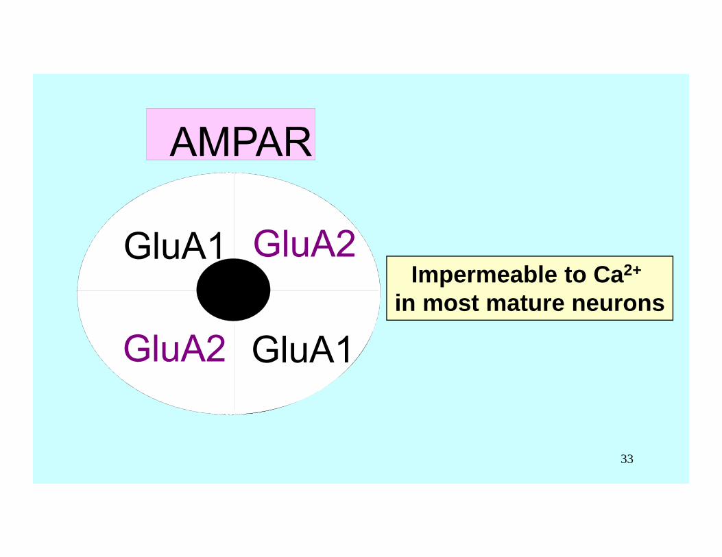
Impermeable to Ca2+
in most mature neurons
AMPAR
GluR1
GluR1
GluR2
GluR2
AMPAR
GluA1
GluA1
GluA2
GluA2
33
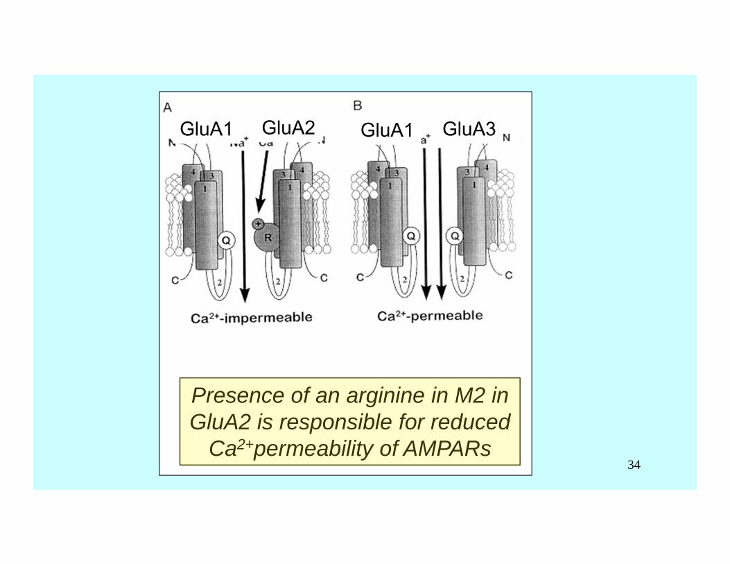
Presence of an arginine in M2 in GluA2 is responsible for reduced
Ca2+permeability of AMPARs
GluA2GluA1 GluA3GluA1
34
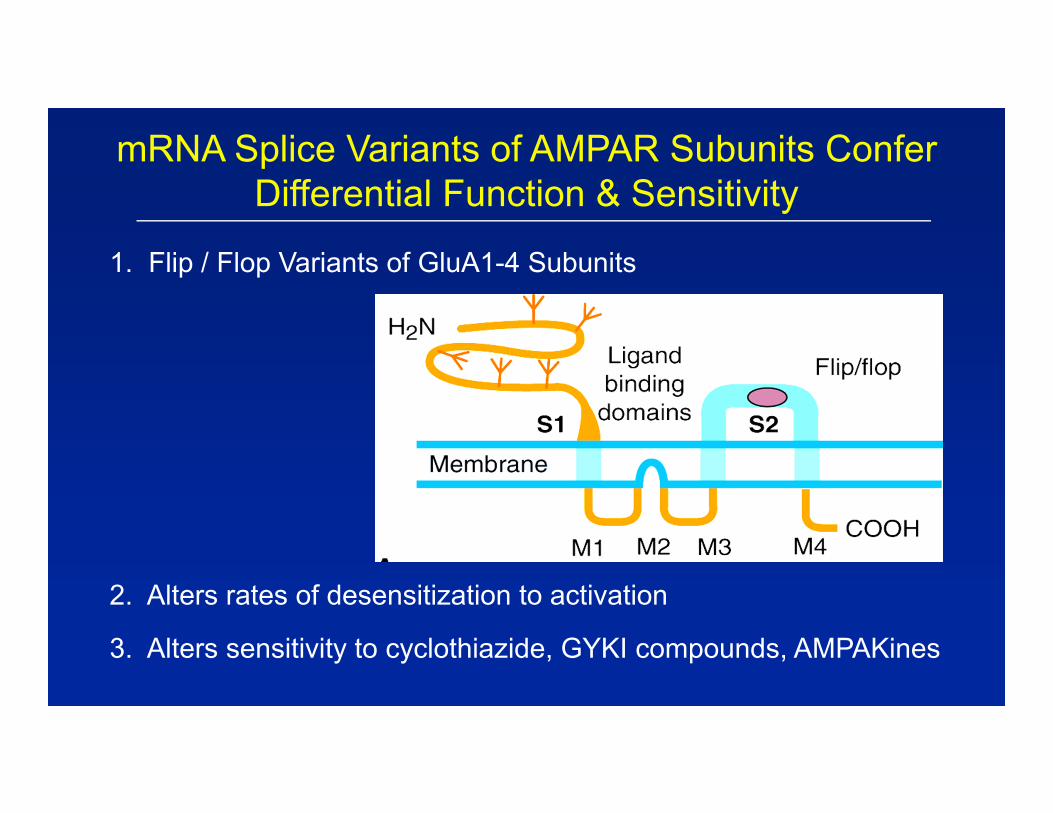
mRNA Splice Variants of AMPAR Subunits Confer Differential Function & Sensitivity
1. Flip / Flop Variants of GluA1-4 Subunits
2. Alters rates of desensitization to activation
3. Alters sensitivity to cyclothiazide, GYKI compounds, AMPAKines
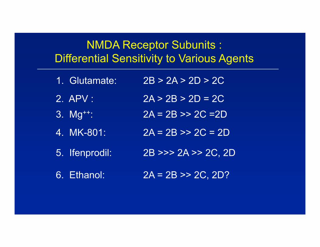
NMDA Receptor Subunits :Differential Sensitivity to Various Agents
1. Glutamate: 2B > 2A > 2D > 2C
4. MK-801: 2A = 2B >> 2C = 2D
3. Mg++: 2A = 2B >> 2C =2D
5. Ifenprodil: 2B >>> 2A >> 2C, 2D
2. APV : 2A > 2B > 2D = 2C
6. Ethanol: 2A = 2B >> 2C, 2D?
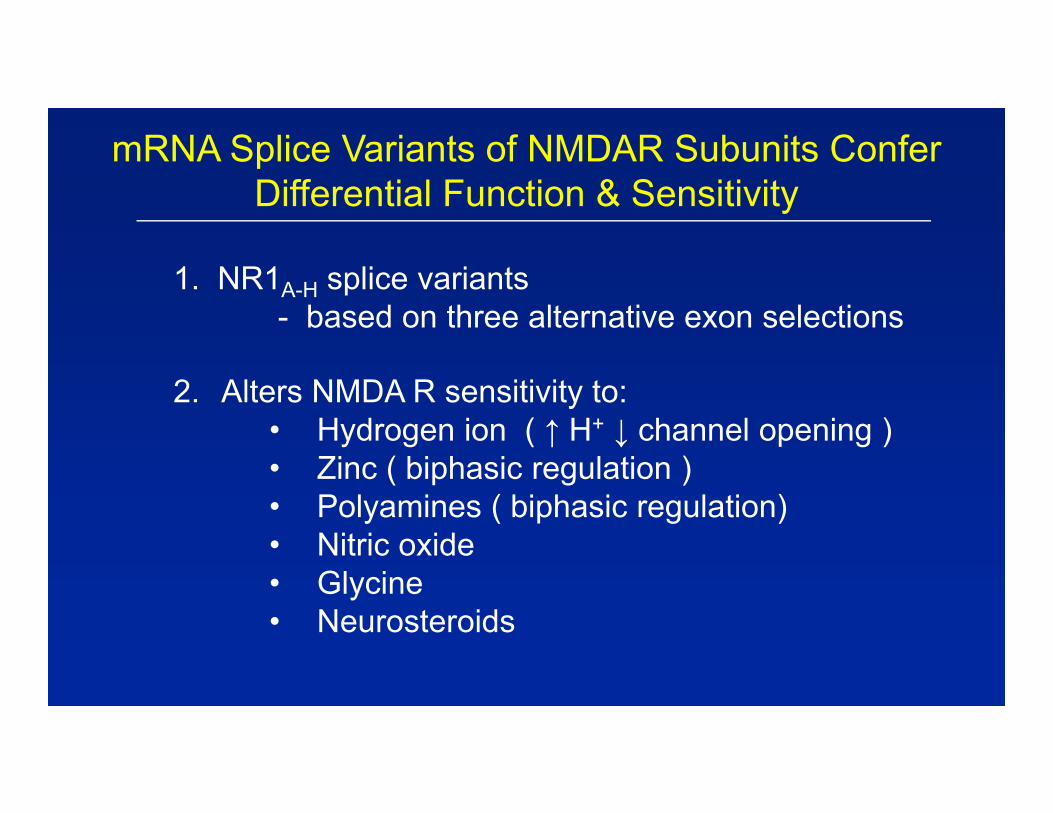
mRNA Splice Variants of NMDAR Subunits Confer Differential Function & Sensitivity
1. NR1A-H splice variants- based on three alternative exon selections
2. Alters NMDA R sensitivity to:• Hydrogen ion ( ↑ H+ ↓ channel opening ) • Zinc ( biphasic regulation )• Polyamines ( biphasic regulation) • Nitric oxide• Glycine• Neurosteroids
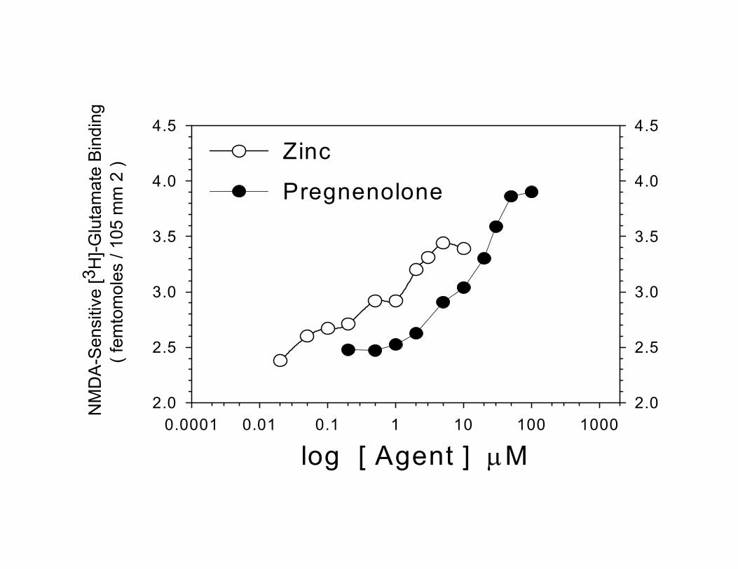
log [ Agent ] M0.0001 0.01 0.1 1 10 100 1000
NM
DA
-Sen
sitiv
e [3
H]-G
luta
mat
e B
indi
ng( f
emto
mol
es /
105
mm
2 )
2.0
2.5
3.0
3.5
4.0
4.5
2.0
2.5
3.0
3.5
4.0
4.5
Zinc
Pregnenolone

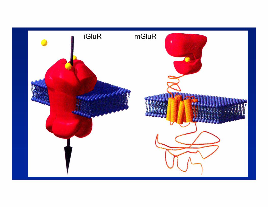
iGluR mGluR
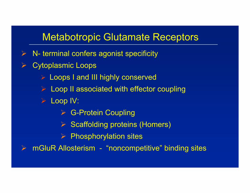
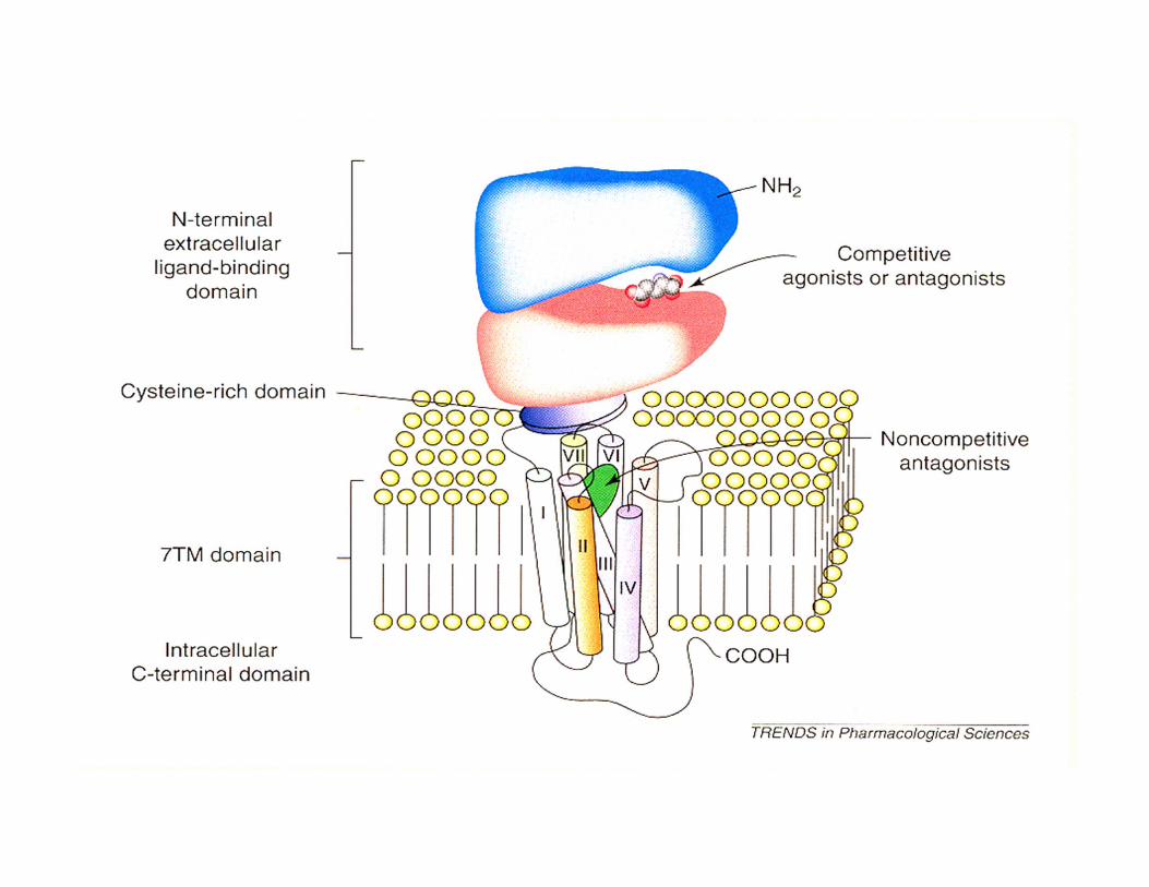
Metabotropic Glutamate Receptors N- terminal confers agonist specificity Cytoplasmic Loops
Loops I and III highly conserved Loop II associated with effector coupling Loop IV:
G-Protein Coupling Scaffolding proteins (Homers) Phosphorylation sites
mGluR Allosterism - “noncompetitive” binding sites
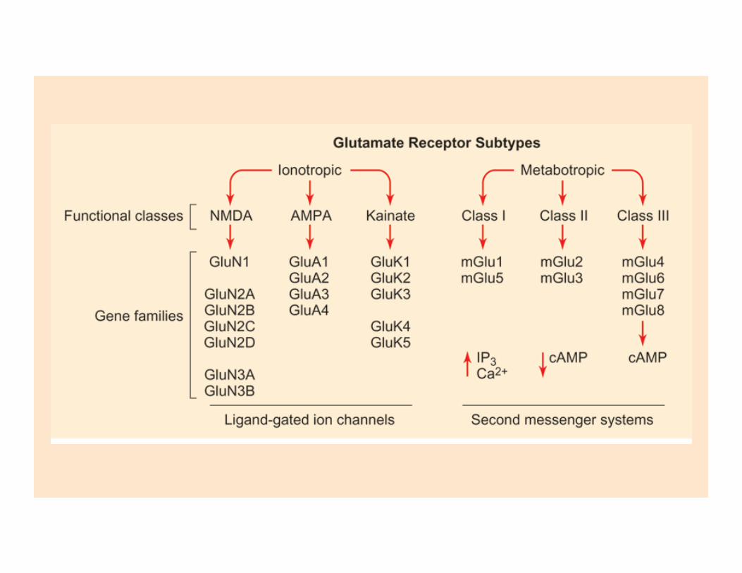
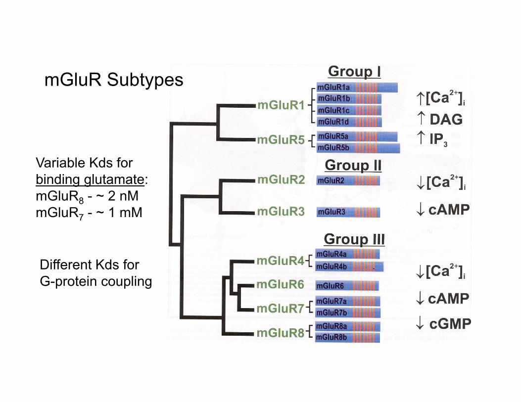
mGluR Subtypes
Variable Kds forbinding glutamate:mGluR8 - ~ 2 nMmGluR7 - ~ 1 mM
Different Kds for G-protein coupling
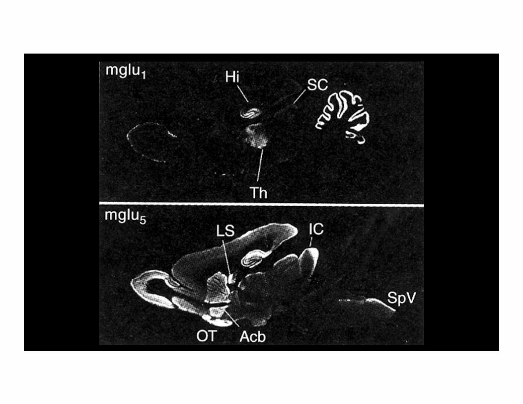
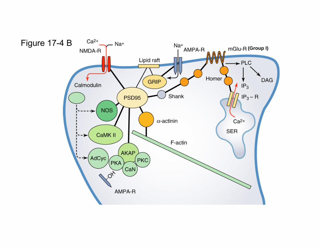
Figure 17-4 B(Group I)
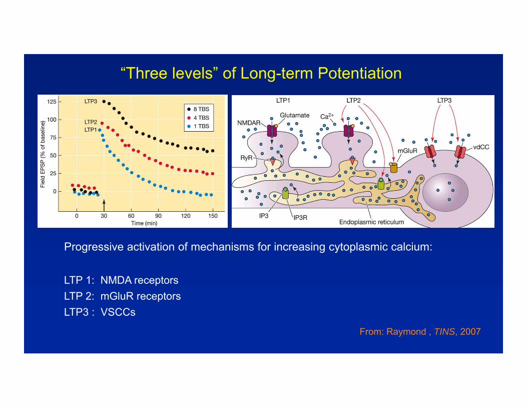
“Three levels” of Long-term Potentiation
Progressive activation of mechanisms for increasing cytoplasmic calcium:
LTP 1: NMDA receptorsLTP 2: mGluR receptorsLTP3 : VSCCs
From: Raymond , TINS, 2007
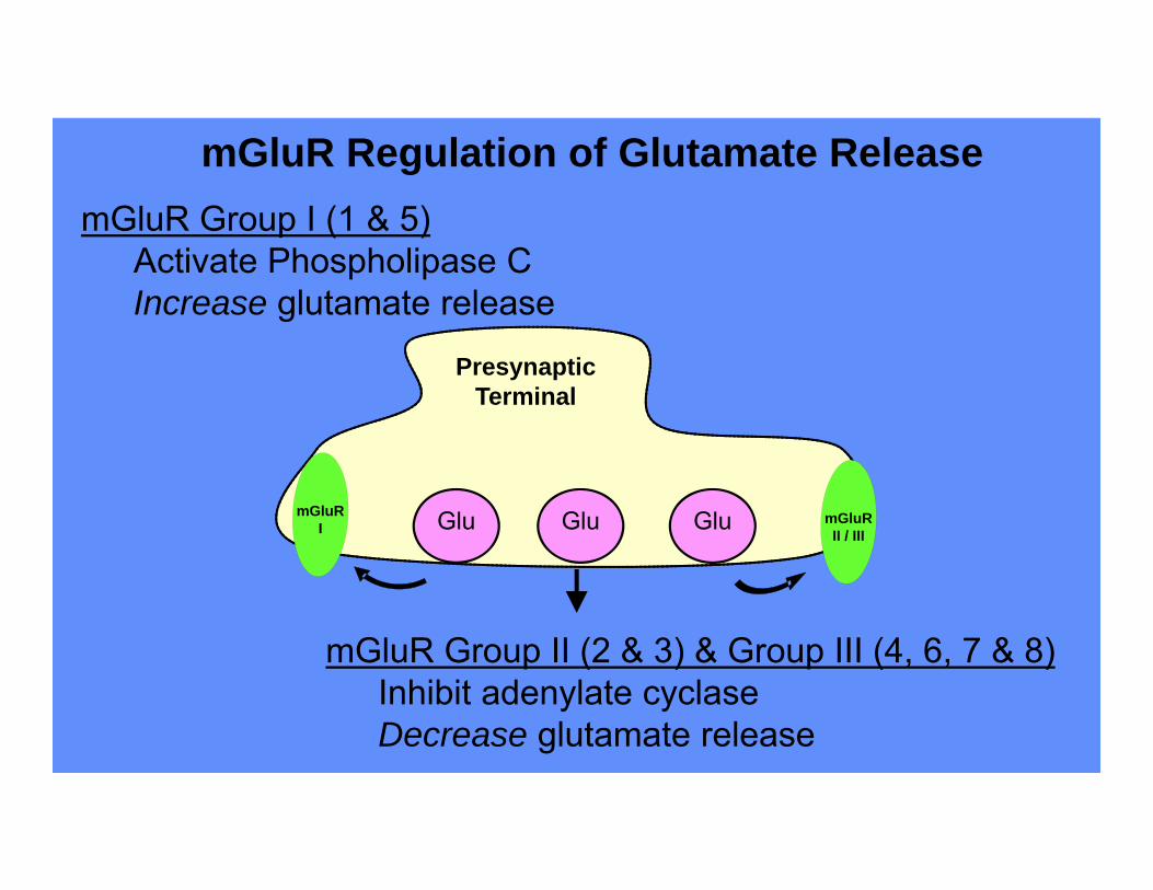
PresynapticTerminal
Glu Glu Glu
mGluR Group II (2 & 3) & Group III (4, 6, 7 & 8)Inhibit adenylate cyclaseDecrease glutamate release
mGluR Group I (1 & 5)Activate Phospholipase CIncrease glutamate release
mGluR Regulation of Glutamate Release
mGluRI mGluR
II / III
Gq
GluSynapsin I
GAP-43:CaM
CaM
CaMKII
PP-2B
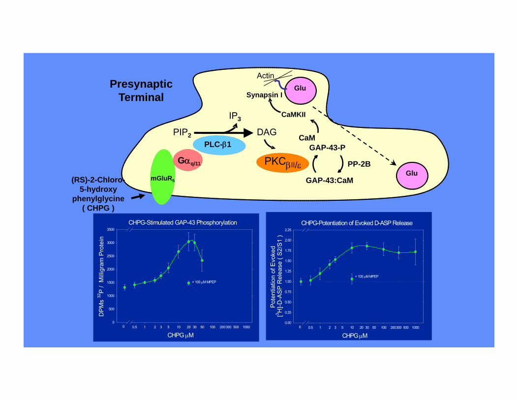
PresynapticTerminal
Actin
mGluR5
Gq/11
PLC-1PIP2
IP3
DAG
PKCII/
GAP-43-P
Glu(RS)-2-Chloro-
5-hydroxyphenylglycine
( CHPG )
CHPG-Stimulated GAP-43 Phosphorylation
CHPG M0.5 2 3 5 20 30 50 200300 5001 10 100 1000
DP
Ms
32P
/ M
illig
ram
Pro
tein
500
1500
2500
3500
0
1000
2000
3000
+ 100 M MPEP
0
CHPG-Potentiation of Evoked D-ASP Release
CHPG M0.5 2 3 5 20 30 50 200300 5001 10 100 1000
Pot
entia
tion
of E
voke
d [3 H
]-D-A
SP
Rel
ease
( S
2/S
1 )
0.25
0.75
1.25
1.75
2.25
0.00
0.50
1.00
1.50
2.00
+ 100 M MPEP
0
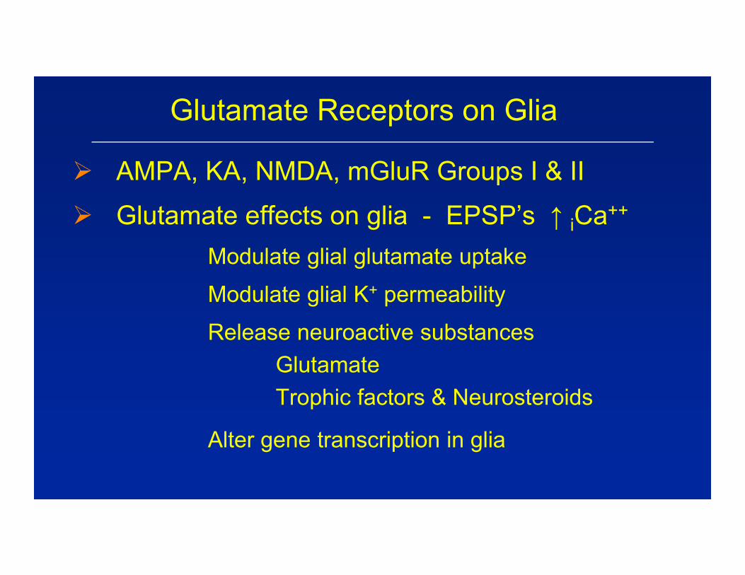
Glutamate Receptors on Glia
AMPA, KA, NMDA, mGluR Groups I & II
Glutamate effects on glia - EPSP’s ↑ iCa++
Modulate glial glutamate uptake
Modulate glial K+ permeability
Release neuroactive substancesGlutamateTrophic factors & Neurosteroids
Alter gene transcription in glia
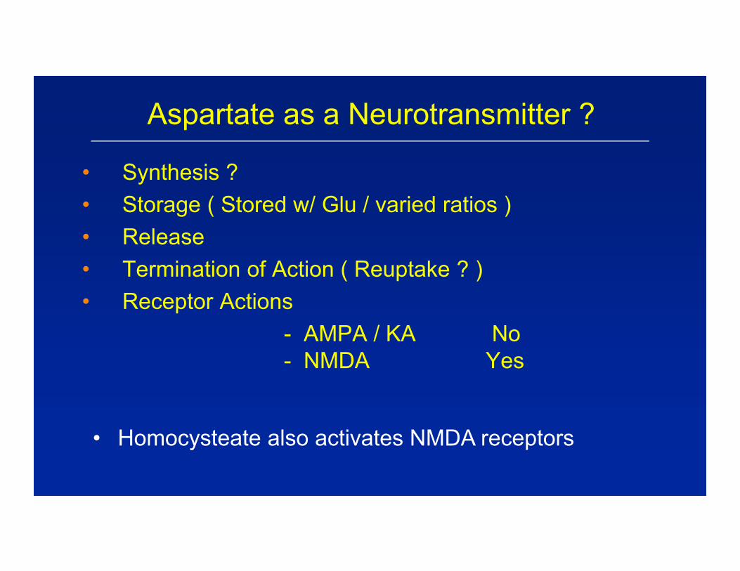
Aspartate as a Neurotransmitter ?
• Synthesis ?• Storage ( Stored w/ Glu / varied ratios )• Release• Termination of Action ( Reuptake ? )• Receptor Actions
- AMPA / KA No- NMDA Yes
• Homocysteate also activates NMDA receptors
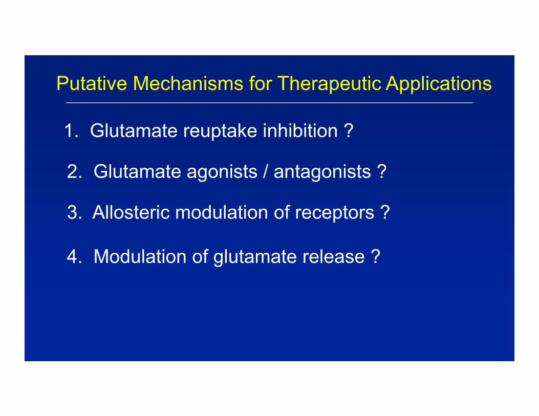
Putative Mechanisms for Therapeutic Applications
1. Glutamate reuptake inhibition ?
2. Glutamate agonists / antagonists ?
3. Allosteric modulation of receptors ?
4. Modulation of glutamate release ?
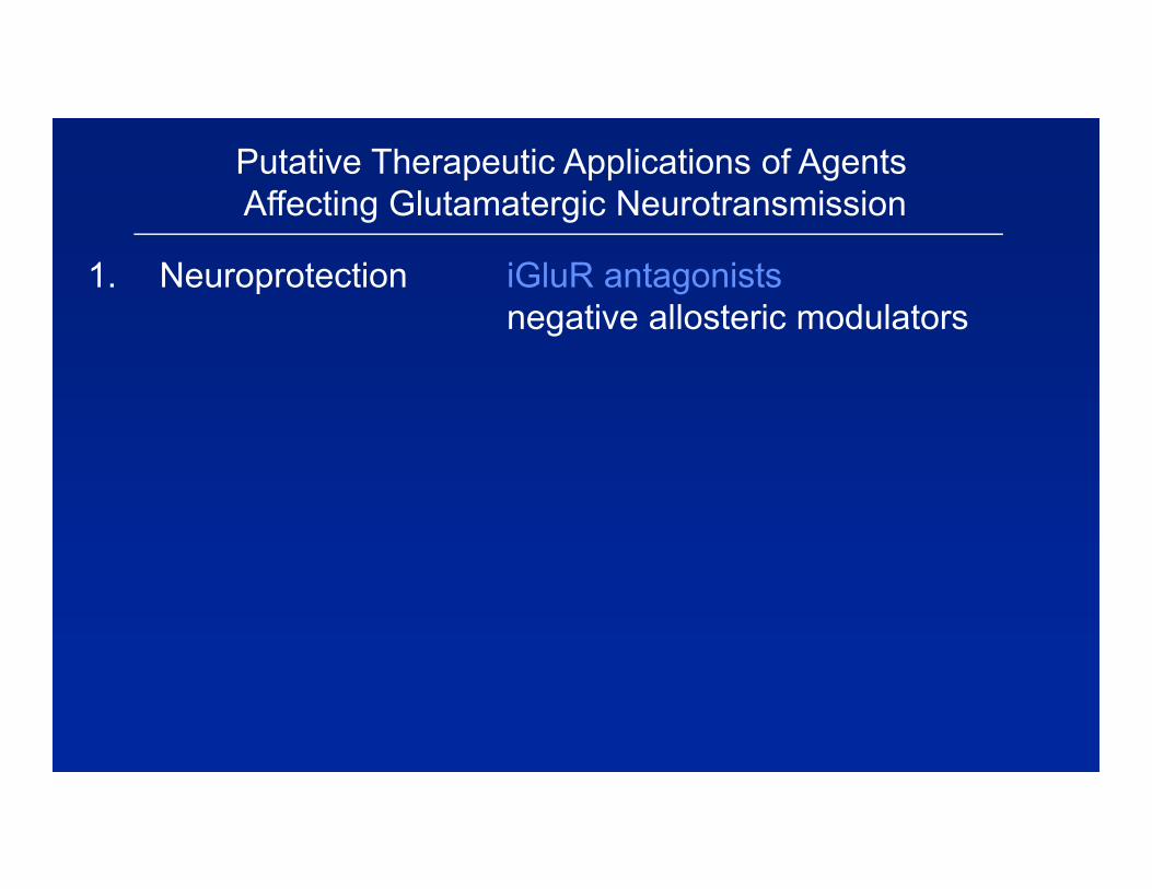
Putative Therapeutic Applications of AgentsAffecting Glutamatergic Neurotransmission
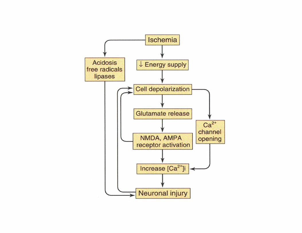
1. Neuroprotection iGluR antagonistsnegative allosteric modulators
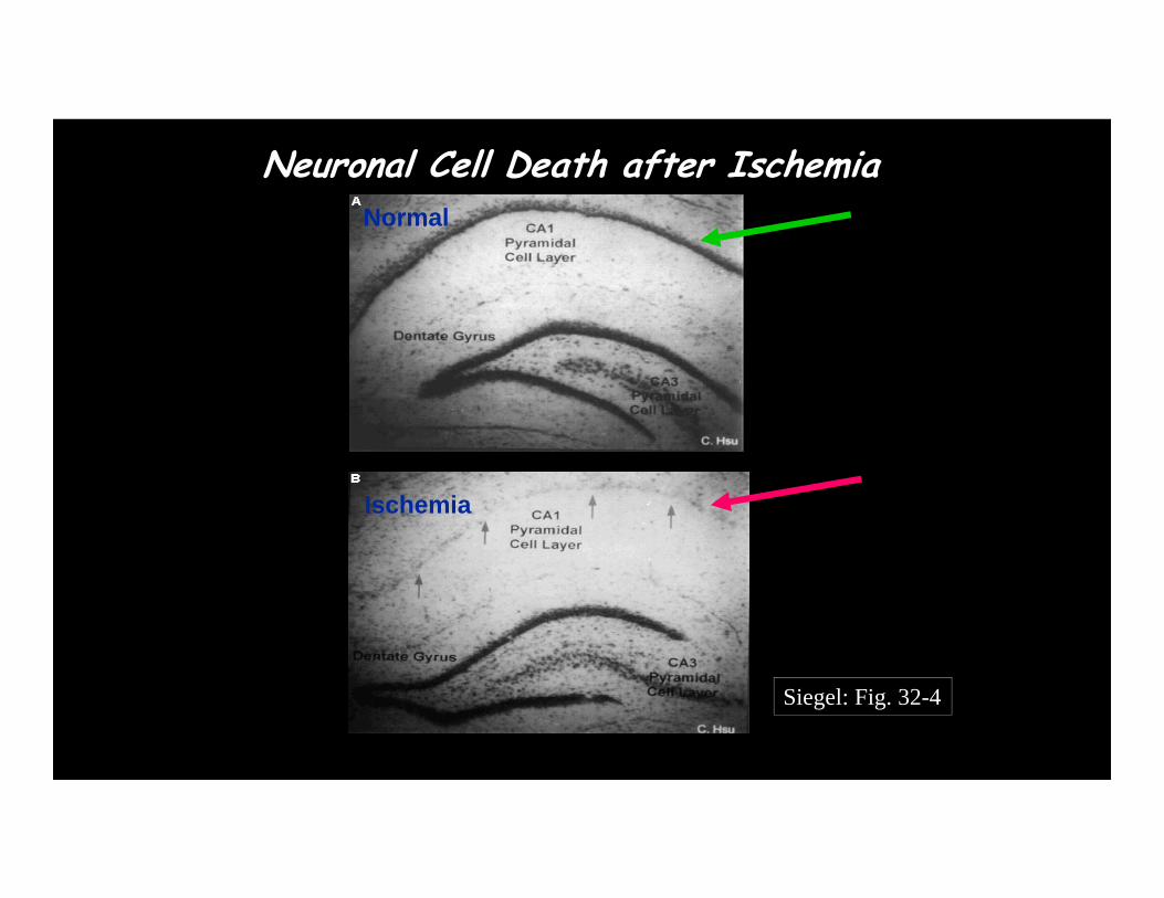
Neuronal Cell Death after IschemiaNormal
Ischemia
Siegel: Fig. 32-4
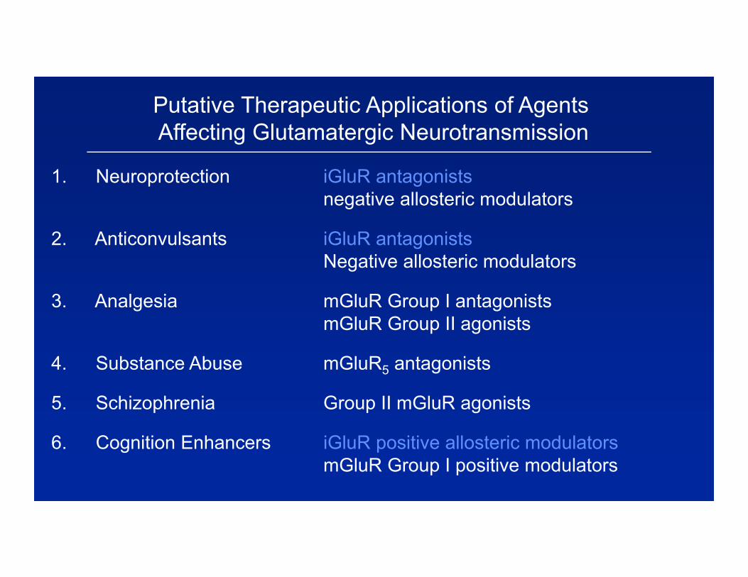
Putative Therapeutic Applications of AgentsAffecting Glutamatergic Neurotransmission
1. Neuroprotection iGluR antagonistsnegative allosteric modulators
2. Anticonvulsants iGluR antagonistsNegative allosteric modulators
3. Analgesia mGluR Group I antagonistsmGluR Group II agonists
4. Substance Abuse mGluR5 antagonists
5. Schizophrenia Group II mGluR agonists
6. Cognition Enhancers iGluR positive allosteric modulatorsmGluR Group I positive modulators
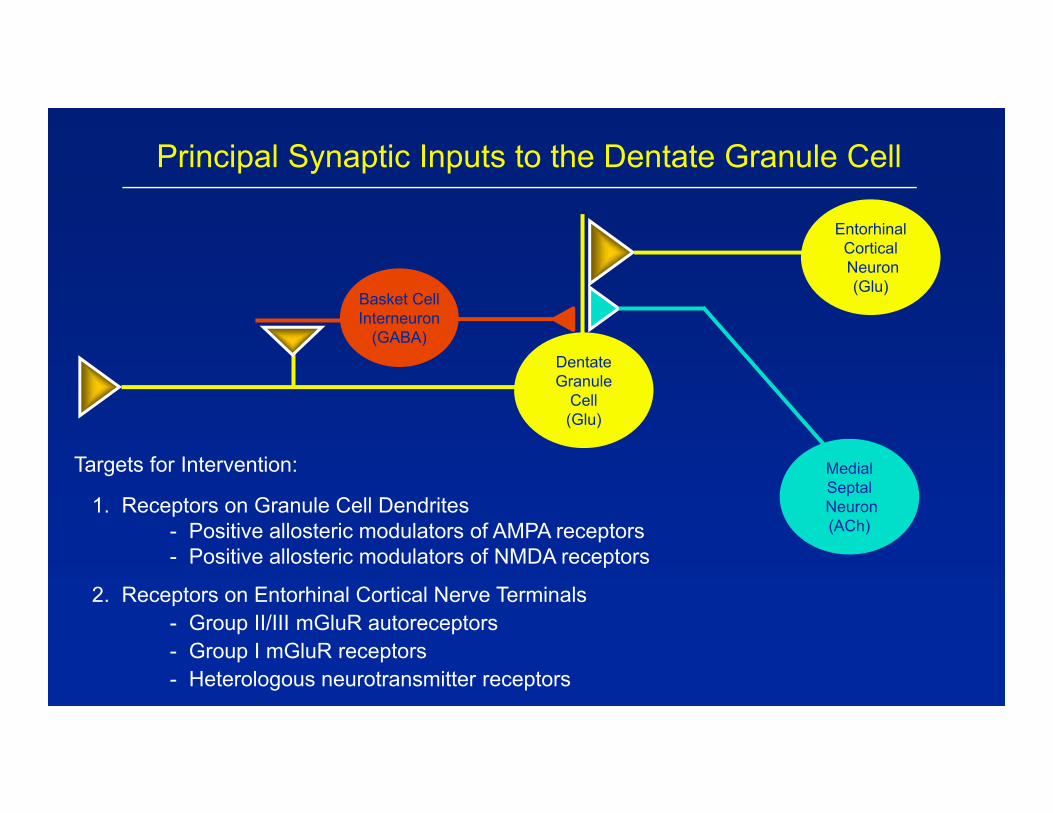
Principal Synaptic Inputs to the Dentate Granule Cell
Targets for Intervention:
1. Receptors on Granule Cell Dendrites- Positive allosteric modulators of AMPA receptors- Positive allosteric modulators of NMDA receptors
2. Receptors on Entorhinal Cortical Nerve Terminals- Group II/III mGluR autoreceptors- Group I mGluR receptors- Heterologous neurotransmitter receptors
DentateGranule
Cell(Glu)
EntorhinalCorticalNeuron(Glu)
Basket CellInterneuron
(GABA)
MedialSeptalNeuron(ACh)
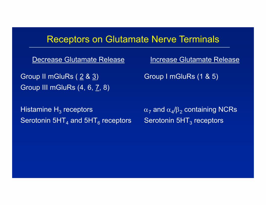
Receptors on Glutamate Nerve Terminals
Increase Glutamate Release
Group I mGluRs (1 & 5)
7 and 4/2 containing NCRsSerotonin 5HT3 receptors
Decrease Glutamate Release
Group II mGluRs ( 2 & 3)Group III mGluRs (4, 6, 7, 8)
Histamine H3 receptorsSerotonin 5HT4 and 5HT6 receptors
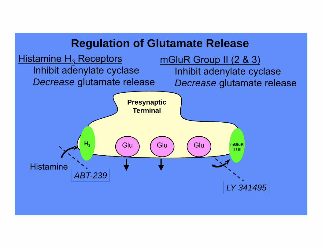
PresynapticTerminal
Glu Glu Glu
Histamine H3 ReceptorsInhibit adenylate cyclaseDecrease glutamate release
Regulation of Glutamate Release
H3 mGluRII / III
HistamineABT-239
mGluR Group II (2 & 3)Inhibit adenylate cyclaseDecrease glutamate release
LY 341495
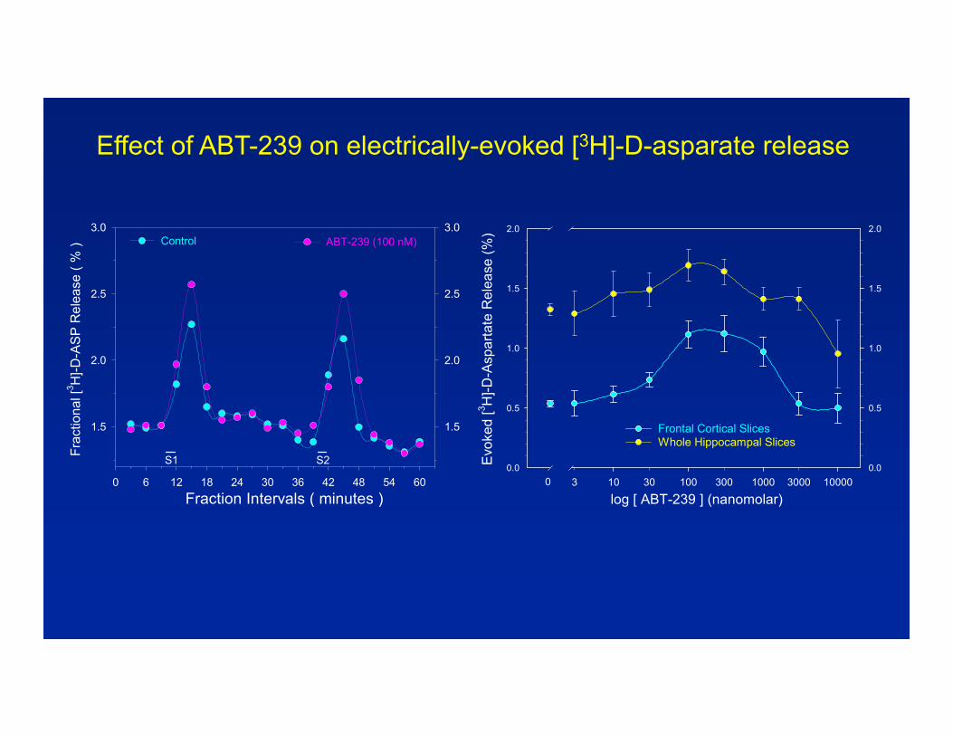
log [ ABT-239 ] (nanomolar)3 30 300 300010 100 1000 10000
Evo
ked
[3 H]-D
-Asp
arta
te R
elea
se (%
)0.0
0.5
1.0
1.5
2.0
0.0
0.5
1.0
1.5
2.0
Frontal Cortical SlicesWhole Hippocampal Slices
0
Fraction Intervals ( minutes )0 6 12 18 24 30 36 42 48 54 60
Frac
tiona
l [3 H
]-D-A
SP
Rel
ease
( %
)
1.5
2.0
2.5
3.0
1.5
2.0
2.5
3.0Control ABT-239 (100 nM)
S1 S2
Effect of ABT-239 on electrically-evoked [3H]-D-asparate release