Embed Size (px)
Citation preview
Expanded and highly active proliferation centers identify a histological subtype of chronic lymphocytic leukemia (“accelerated“ chronic lymphocytic leukemia) with aggressive clinical behavior
by Eva Giné, Antonio Martinez, Neus Villamor, Armando Lopez-Guillermo,Mireia Camos, Daniel Martinez, Jordi Esteve, Xavier Calvo, Ana Muntañola,Pau Abrisqueta, Maria Rozman, Ciril Rozman, Francesc Bosch, Elias Campo,and Emili Montserrat
Haematologica 2010 [Epub ahead of print]
Citation: Giné E, Martinez A, Villamor N, Lopez-Guillermo A, Camos M, Martinez D,Esteve J, Calvo X, Muntañola A, Abrisqueta P, Rozman M, Rozman C, Bosch F, Campo E,and Montserrat E. Expanded and highly active proliferation centers identify a histologicalsubtype of chronic lymphocytic leukemia (“accelerated” chronic lymphocytic leukemia) withaggressive clinical behavior.Haematologica. 2010; 96:xxx doi:10.3324/haematol.2010.022277
Publisher's Disclaimer.E-publishing ahead of print is increasingly important for the rapid dissemination of science.Haematologica is, therefore, E-publishing PDF files of an early version of manuscripts thathave completed a regular peer review and have been accepted for publication. E-publishingof this PDF file has been approved by the authors. After having E-published Ahead of Print,manuscripts will then undergo technical and English editing, typesetting, proof correction andbe presented for the authors' final approval; the final version of the manuscript will thenappear in print on a regular issue of the journal. All legal disclaimers that apply to the journal also pertain to this production process.
Haematologica (pISSN: 0390-6078, eISSN: 1592-8721, NLM ID: 0417435, www.haemato-logica.org) publishes peer-reviewed papers across all areas of experimental and clinicalhematology. The journal is owned by the Ferrata Storti Foundation, a non-profit organiza-tion, and serves the scientific community with strict adherence to the principles of openaccess publishing (www.doaj.org). In addition, the journal makes every paper publishedimmediately available in PubMed Central (PMC), the US National Institutes of Health (NIH)free digital archive of biomedical and life sciences journal literature.
Official Organ of the European Hematology AssociationPublished by the Ferrata Storti Foundation, Pavia, Italy
www.haematologica.org
Early Release Paper
Support Haematologica and Open Access Publishing by becoming a member of the European Hematology Association (EHA)and enjoying the benefits of this membership, which include free participation in the online CME program
Copyright 2010 Ferrata Storti Foundation.Published Ahead of Print on April 26, 2010, as doi:10.3324/haematol.2010.022277.

DOI: 10.3324/haematol.2010.022277
Expanded and highly active proliferation centers identify a histological
subtype of chronic lymphocytic leukemia (“accelerated” chronic
lymphocytic leukemia) with aggressive clinical behavior
Running title: Accelerated CLL
Eva Giné,1 Antoni Martinez,2 Neus Villamor,2 Armando López-Guillermo,1
Mireia Camos,2 Daniel Martinez,2 Jordi Esteve,1 Xavier Calvo,1 Ana Muntañola,1
Pau Abrisqueta,1 Maria Rozman,2 Ciril Rozman,1 Francesc Bosch,1
Elias Campo,2 and Emili Montserrat1
1Hematology Department, Institute of Hematology and Oncology, Hospital
Clinic, Barcelona, Spain, and 2Hematopathology Unit, Hospital Clínic,
Barcelona, Spain
Correspondence
Francesc Bosch, MD, Department of Hematology, Hospital Clínic Villarroel
nº 170, 08036-Barcelona, Spain. Phone: international +34.93.2275475.
Fax: international 34.93.2275484. E-mail: [email protected]
Funding
This work was supported by grants from the Spanish Ministry of Health, FIS-
PI07/0409, PI08/0095, Pi08/0304, RT06/0020/002051, 2009-SGR-1008,
CICYT SAF 2008-03630 and by the RTICC (Red Temática de Investigación
Cooperativa en Cáncer, RD06/0020/0051).
DOI: 10.3324/haematol.2010.022277
Current address of F. Bosch and P. Abrisqueta: Department of Hematology, University
Hospital Vall d’Hebron, P. Vall d’Hebron 119-129, 08035 Barcelona, Spain. Current
address of M. Camos: Diagnòstic de Laboratori, Hospital Sant Joan de Déu, Passeig
Sant Joan de Déu, 2, 08950, Esplugues de Llobregat, Spain. Current address of A.
Muntañola: Department of Hematology, Hospital Mútua de Terrassa, Dr. Robert 5,
08221 Terrassa, Spain.
Key words: accelerated chronic lymphocytic leukemia, lymph node biopsy,
ZAP-70,
DOI: 10.3324/haematol.2010.022277
Background
The concept of “accelerated” chronic lymphocytic leukemia (CLL) is frequently
used by both pathologists and clinicians. However, neither histological criteria
to define this form of CLL nor its clinical correlates and prognostic impact have
been formally defined in large series of patients.
Design and Methods
Tissue biopsies from 100 patients with CLL were analyzed for the size of
proliferation centers (PCs) and their proliferation rate as assessed by mitosis
count and Ki-67 immunostaining. Histological patterns were correlated with
main clinico-biological features and outcome.
Results
The suspicion of disease transformation was the main reason to carry out
tissue biopsy, which was performed at a median time of 14 months (range, 0 to
204) from CLL diagnosis. The biopsy showed histological transformation to
diffuse large B-cell lymphoma (DLBCL-t) in 22 cases. In the remaining 78
patients, the presence of expanded PCs (broader than a 20x field) and high
proliferation rate (either >2.4 mitosis/PC or Ki-67>40%/PC) predicted poor
outcome and were selected to define a highly proliferative group. Thus, 23
patients with either expanded PCs or high proliferation rate were considered as
having “accelerated” CLL. These patients displayed particular features,
including higher serum LDH levels and more frequently elevated ZAP-70 than
“non-accelerated” cases. Median survival from biopsy of patients with “non-
accelerated” CLL, “accelerated” CLL and DLBCL-t was 76, 34, and 4.3 months,
respectively (p<0.001).
Conclusions
The presence of expanded PCs and/or highly active PCs identifies a group of
patients with “accelerated” CLL characterized by an aggressive clinical
behavior.

DOI: 10.3324/haematol.2010.022277
1
Introduction
Chronic lymphocytic leukemia (CLL) is the most frequent form of leukemia in
Western countries and is considered to be mainly due to the accumulation of
neoplastic CD5+ B lymphocytes with a typical CD19+, CD23+
immunophenotype.1,2 According to the WHO classification3 CLL and small
lymphocytic lymphoma (SLL) are considered to be the same disease, primarily
involving peripheral blood in the case of CLL and lymph nodes in SLL. A
number of parameters, particularly disease stage, lymphocyte doubling time,
IGVH mutational status, ZAP-70 expression and cytogenetics allow to establish
the prognosis of the disease at diagnosis.2,4-6 Over the course of the disease,
refractoriness to treatment has poor prognosis. Likewise, disease
transformation implies a very short survival.
Although considered as a cumulative rather than a proliferative CD5+ B cell
neoplasm, CLL cells have a proliferation rate higher than previously
recognized, particularly in the lymphoid tissues.1,7 Enlarged nodes from patients
with CLL show effacement of the lymphoid architecture with a pseudofollicular
pattern of pale areas in a dark background of small cells. These pale areas
correspond to proliferation centers (PCs)7-10 and are predominantly composed
of clusters of prolymphocytes and paraimmunoblasts. PCs contain numerous T
cells, most of them CD4+, and in some cases a fine network of dendritic cells.11
Notably, as compared to the non-PC component CLL, cells clustered into the
PCs have an increased expression of proliferation associated markers Ki-67
and CD71, co-expression of survivin and bcl-2 and also a higher expression of
CD20, CD23, and MUM1/IRF-4.11-18
Histology of the lymph nodes in CLL is heterogeneous and the relationship
between different histologic patterns and clinical outcome has been poorly
studied.8-10,12,18-20 The main reason for this fact is that tissue biopsy is not a
standard procedure in the diagnostic workup of patients with CLL. The
commonest reason for performing a biopsy is to rule out histological
transformation. In general, two major histological patterns in lymph nodes of
patients with CLL are recognized: typical CLL involvement and transformation
of CLL to DLBCL. However, the existence of no clear-cut histopathological
patterns not pertaining into these two broad categories has been known for
DOI: 10.3324/haematol.2010.022277
2
many years,10 being poorly characterized and belonging to a kind of “grey-
zone” in the histopathology of CLL. Although the term “accelerated” CLL is
frequently used to describe most of these cases, no histological criteria for
identifying this form have been yet proposed and its biological background and
clinical significance is largely unknown. In the present study we report criteria to
identify “accelerated” CLL, its clinical correlates and prognostic impact.
Design and Methods
Patients and samples
From January 1990 to December 2008, 146 patients fulfilling the diagnostic
criteria of CLL according to NCI-WG guidelines21,22 underwent a tissue biopsy
(except bone marrow). These cases represented 24% of the 616 patients
diagnosed with CLL during this period in our institution. One-hundred patients
with biopsies available for pathological review were the basis of this study.
Forty-six patients were not included in the study due to different reasons
including a small size of the biopsy precluding a histological review or
immunohistological analysis (31 cases); incidental diagnosis of CLL in patients
with solid tumors, 3 cases; composite lymphomas, 3 cases; transformation to
prolymphocytic leukemia, 2 cases; T-cell lymphomas, 2 cases; and Hodgkin’s
lymphoma, 5 cases. The tissue analyzed was lymph node in 89 cases, spleen
in 1, Waldeyer’s ring in 1 and different extranodal tissues in 9.
From each patient the following biological and clinical data were evaluated and
recorded both at diagnosis and at the time of the tissue biopsy: age, gender,
performance status (according to the Eastern Cooperative Oncology Group
[ECOG] scale), presence of B-symptoms, presence of splenomegaly or
hepatomegaly, extranodal involvement, hemoglobin, WBC count, lymphocyte
count, lymphocyte doubling time, platelet count, serum albumin, LDH, and !2-
microglobulin levels, Rai’s and Binet’s staging systems, degree of lymphocytic
bone marrow infiltration, ZAP-70 and CD38 expression, IGVH mutational status
and fluorescence in situ hybridization (FISH) analysis for recurrent genetic
abnormalities in CLL. Moreover, treatment-related variables (type of treatment
DOI: 10.3324/haematol.2010.022277
3
and response to therapy) and the M.D. Anderson score for Richter’s
syndrome23 were also recorded and evaluated.
FISH studies were performed in peripheral blood lymphocytes by the LSI
p53/LSI ATM and LSI D13S319/LSI 13q34/CEP 12 Multicolor Probe Sets
provided by Vysis (Downers Grove, Il, USA) using defined cut-off levels.5 ZAP-
70 expression was assessed in peripheral blood samples by flow cytometry
(n=42 cases) or by immunostaining in lymph nodes (23 cases) as previously
described, with a 95% of concordance between both methods.4,24 CD38
expression was analyzed by flow cytometry in peripheral blood and considered
as increased when equal or superior to 30%. The mutational status of the IGVH
genes was analyzed in 27 cases following protocols described elsewhere.4
Clonality was assessed by studying immunoglobulin heavy chain gene (IgH)
rearrangement from peripheral blood and paraphine-embedded tissue samples
following BIOMED-2 protocols in 12 patients.25
Histological analysis
Tissue samples were analyzed for the presence and size of PCs, and for the
proliferation status as assessed by the mitotic index and Ki-67 immunostaining.
Histological review was performed by three investigators (A.M., D.M., E.C.)
blinded to clinical records. The samples were examined in a BX51 Olympus
microscope (Olympus Tokio, Japan) with an 20X objective Plan CN20x/0.40
and an ocular WHN 10x/22. Immunostaining was performed in formalin fixed
paraffin embedded tissue sections with p27 (1B4, Novocastra, Newcastle Upon
Tyne, UK), Ki-67 (Mib1, Dako, Glostrup, Denmark), and p53 (DO7, Dako) in an
automated immunostainer (BondMax, Vision BioSystems, Mount Waverley,
Australia).
PCs were defined as the pale areas containing large cells, prolymphocytes and
paraimmunoblasts, surrounded by a dark background of small lymphocytes.
PCs were also identified and delineated by a p27 negative staining.26 PCs were
arbitrarily considered as expanded when two or more PCs exceeded a 20x
power field in any of its major dimensions which is approximately equivalent to
an area of 0.95 mm2 in the microscope used to evaluate the samples.

DOI: 10.3324/haematol.2010.022277
4
Proliferation inside PCs was determined by both mitotic count and Ki-67
immunostaining and quantified in a total of 8 to 10 PCs per case. In addition,
global Ki-67 and p53 were quantified in 8 to 10 40x high power field (hpf)
randomly selected in the whole tissue section that included both PCs and small
cell component areas. The threshold to consider a p53 staining as positive was
30%.
The diagnosis of transformation to diffuse large cell lymphoma was based on
the WHO 2008 criteria.3 In addition, Ki-67 and p53 immunostainings were
performed in these cases and separately analyzed.
Statistical analysis
The correlation between different clinical and biological variables and the
histological pattern was performed by means of the Fisher’s exact test or
nonparametric tests when necessary.
Parameters obtained in the histological analysis, namely the size of PCs and
the proliferation index, were correlated with patients survival. To select the
optimal cut-off of the quantitative histological variables (mitotic index and Ki-67
expression) for predicting survival, a maximally selected rank statistics test was
performed using the Maxstat package (R statistical package, v. 2.8.1, Vienna,
Austria)27 and the cut-off was ultimately delimitated by the Kaplan and Meier
method.28 To minimize a possible selection bias, survival was calculated from
the time of biopsy and analyzed according to the method described by Kaplan
and Meier and the curves compared by the log-rank test.29 All prognostic
variables in the univariate analysis with enough number of cases available were
included in the multivariate analysis using the stepwise proportional hazard Cox
regression model.30 All statistical tests were two-sided and the significance
level was 0.05.
DOI: 10.3324/haematol.2010.022277
5
Results
Patients and tissue biopsies
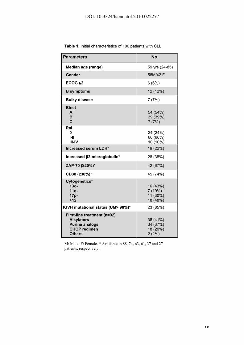
The main initial clinico-biological characteristics of the patients are detailed in
Table 1. Median age at diagnosis was 59 years-old. Forty-six per cent of
patients were in Binet stage B or C and 10% in Rai stage III-IV. The proportion
of patients with increased ZAP-70 (!20%) and CD38 (!30%) expression was
67% and 74%, respectively. Forty-four patients were treated immediately after
diagnosis and 50 additional cases required treatment during evolution with an
overall median time to progression of 5 months (range, 0 to 112 months).
The reason for performing a tissue biopsy in 73 patients was the clinical
suspicion of transformation to aggressive lymphoma (i.e. general symptoms,
rapid lymph node enlargement, bulky extranodal involvement or increasing
serum LDH levels). Other reasons for carrying out a biopsy were the diagnostic
workup of a lymphoproliferative disorder without overt peripheral blood
involvement (12 patients); re-assessment of the disease before treatment (9
cases); and biopsies routinely obtained in surgical procedures (6 cases).
Median time from diagnosis to biopsy was 14 months (range, 0 to 204 months)
and median survival from the time of tissue biopsy was 49 months (95% CI: 24
to 74). Clinical features and outcome of patients with CLL without biopsy
(median OS, 154 months) were more favorable than those with biopsy (median
OS, 90 months) (detailed data not shown).
Impact of expanded and proliferating PCs in the patients’ outcome
From the 100 patients included in this series, the diagnosis of CLL was
established in 78 cases, whereas the remaining 22 patients were diagnosed
with transformation into DLBCL (DLBCL-t).
In the 78 cases included in this study not showing disease transformation to
DLBCL, PCs were observed in 69 and were absent in 9. In the latter, isolated
prolymphocytes and paraimmunoblasts could be recognized at high
magnification and corresponded to the diffuse CLL pattern described by K.
Lennert.10,31 These cases (n=9) did not exhibit other particular histological or
clinical features.

DOI: 10.3324/haematol.2010.022277
6
Overall, 22 cases (28%) showed expanded and confluent PCs broader than a
20x field. The median number of mitosis per PC was 0.9 (range, 0 to 12
mitosis) and median Ki-67 proliferation index per PC was 10% (range, 0 to
75%). Median Ki-67 in the whole tissue section was 2% (range, 0 to 30%). In
addition, p53 positive immunostaining was observed in 5% of the samples.
The following histological features were analyzed for survival: presence of two
or more expanded and confluent PCs, mitotic index and Ki-67 expression inside
PCs and global Ki-67 expression. Patients with expanded and confluent PCs
broader than a 20x field (n=22) had a shorter survival than those with small or
absent PCs (Hazard ratio [HR] 2.72, 95% CI HR 1.47 – 5.04; p=0.001) (Figure
1A). Moreover, patients with a mitotic index >2.4 (n=14) had a shorter survival
than patients with <2.4 mitosis (HR 2.31, 95% CI HR 1.1 – 4.5; p=0.016)
(Figure 1B). In addition, patients with Ki-67 expression inside PCs "40% lived
longer than patients with a Ki-67 >40% (n=6) (HR 3.36, 95%CI 1.27-8.87;
p=0.014) (Figure 1C). Global Ki-67 expression analysis did not yield any
significant cut-off for survival. Among patients with expanded PCs, 65% had
either a mitotic rate >2.4 or Ki67 >40% within the PCs.
Patients with any of the histological parameters with adverse prognosis, namely
expanded PCs, mitosis count > 2.4 or Ki67 >40% per PC, were considered as
having an “accelerated” CLL (n=23). The remaining patients with no adverse
histological features were considered as having a “non-accelerated” CLL
(n=55). The main histological features of patients with “non-accelerated” CLL,
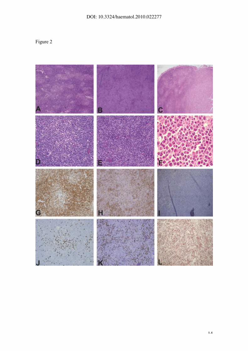
“accelerated” CLL, and DLBCL-t are summarized in Table 2. Representative
histological images are shown in Figure 2. Among other histological variables
studied, the global Ki-67 immunostaining was higher in DLBCL-t than in
“accelerated” CLL and “non-accelerated” CLL (mean+/- SD of Ki-67: 68%+/-14
vs. 12%+/-8.5 vs. 2%+/-2.4, respectively; p= 0.001). In addition, p53 was
positive by immunostaining in 67%, 18% and 2% of patients with DLBCL-t,
“accelerated” CLL and “non-accelerated” CLL, respectively (p<0.0001).

DOI: 10.3324/haematol.2010.022277
7
Analysis of clonality
To ascertain whether tissue samples of “accelerated” CLL were clonally related
to the CLL neoplastic lymphocytes from peripheral blood, IGVH rearrangement
was analyzed in 8 patients. The same kind of analysis was also performed in 4
patients with DLBCL-t. All patients with “accelerated” CLL showed the same
clonal rearrangement in tissue and peripheral blood. In contrast, one out of four
DLBCL-t was clonally unrelated.
Clinico-biological features and outcome of “accelerated” CLL
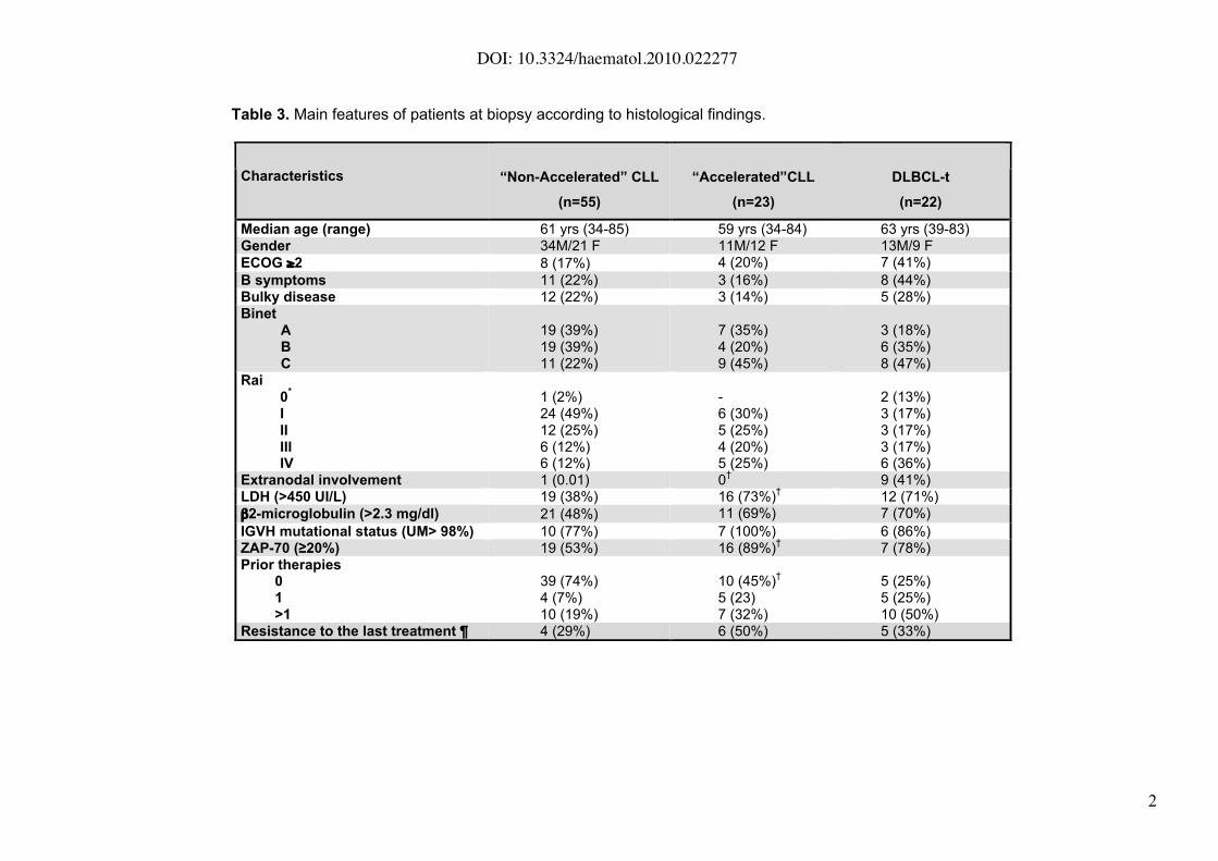
The main characteristics of the patients at the time of tissue biopsy are listed in
Table 3. Patients with “accelerated” CLL presented similar characteristics
compared to the “non-accelerated” CLL cases regarding the presence of B
symptoms, performance status, bulky disease or clinical stage. However,
patients with “accelerated” CLL” had higher serum LDH levels and more
elevated ZAP-70 expression (p<0.01), than patients with “non-accelerated”
CLL.
On the other hand, patients with DLBCL-t displayed more frequently poor
performance status and B symptoms than “accelerated” and “non-accelerated”
CLL. It is of note that in 9 out of 22 cases (41%) with DLBCL-t, disease
transformation was diagnosed in an extranodal site in contrast to patients with
CLL (either “accelerated” or “non-accelerated”), in whom only one extranodal
case (breast mass) was observed (p<0.001). Serum LDH levels were found
progressively increased from “non-accelerated” CLL, “accelerated“ CLL and
DLBCL-t (mean values+/- SD in IU/L: 455±194 vs. 543±198 vs. 820±538,
respectively, p=0.008). Among the three histological groups, no differences in
the distribution of poor risk cytogenetic alterations (17p and 11q deletions) were
observed.
Time from diagnosis to tissue biopsy was longer in DLBCL-t than in CLL (mean:
69 vs. 45 months, respectively, p=0.001). Whilst 9 out of the 23 “accelerated”
CLL cases were observed at diagnosis, only 2 out of the 22 DLBCL-t cases
were observed at that moment. Forty-five percent of patients with “accelerated”
CLL were diagnosed before starting treatment.

DOI: 10.3324/haematol.2010.022277
8
In two patients with “accelerated” CLL new tissue biopsies were obtained
during follow-up. In one case the pattern of “accelerated” CLL remained stable
over time, whereas in the second one a transformation to DLBCL was
demonstrated after 5 years.
The presence of expanded or highly proliferative PCs did not influence the
treatment decision. Treatment for “accelerated” CLL cases varied over time.
Whereas 52% of patients received doxorubicin-containing regimens, 30% of
patients were treated with purine analogs alone or in combination. In contrast,
73% of patients diagnosed with DLBCL-t were treated with doxorubicin-
containing regimens. Intensification with stem cell transplantation was carried
out in 16 patients, including 6 patients with “non-accelerated” CLL, 6 patients
with “accelerated” CLL, and 4 patients with DLBCL-t.
After a median follow-up of 63 months (range, 5.5 to 162 months) from tissue
biopsy 64 of the 100 patients eventually died. The cause of death was related
to the disease in the 18 patients with DLBCL-t who died, in 17/19 “accelerated”
cases and in 26/29 of “non-accelerated” CLL. Median survival from the time of
tissue biopsy for DLBCL-t, “accelerated” CLL and “non-accelerated” CLL was
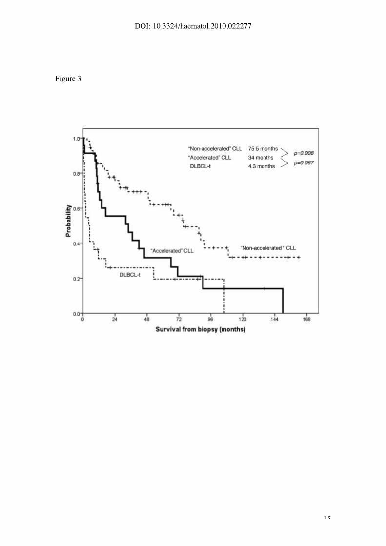
4.3, 34 and 76 months, respectively (p<0.001) (Figure 3). In addition, when
cases without suspicion of transformation at the moment of biopsy (n=27) were
excluded from the analysis, median survival was 4.3 months in DLBCL-t
(n=22), 34 months in “accelerated” CLL (n=21) and 49 months in “non-
accelerated” CLL (n=30) (p=0.032).
Other variables that correlated with poor survival at the time of biopsy were:
age (>60 yrs.), ECOG performance status (!2), Binet’s and Rai’s advanced
clinical stage, B symptoms, bulky disease, hemoglobin level (<110 g/L), platelet
count (<100x109/L), elevated LDH and beta-2-microglobulin serum levels, high
ZAP-70 expression and 17p deletion. The M.D. Anderson score for Richter’s
syndrome was also predictive for survival in the whole series and in the
DLBCL-t and “accelerated” histological subsets.
In a multivariate survival analysis, the histological subtype (“non accelerated”
vs. “accelerated” vs. DLBCL-t) (p=<0.001; RR 2.7 and 5 .5 for “accelerated”
and DLBCL-t vs. “non-accelerated”, respectively) together with age (p=<0.001;
DOI: 10.3324/haematol.2010.022277
9
RR 1.05) and the M.D. Anderson score for Richter’s syndrome (0-1 vs. !2;
p<0.001; RR 3.9) were the most important prognostic variables in the global
series (analysis performed in 94 patients in whom all the data were available).
When the same analysis was performed after excluding patients with DLBCL-t,
the histological subtype (“non accelerated” vs. “accelerated”) (p=0.007; RR 2.5)
along with age (p=0.002; RR 1.05) and M.D. Anderson score for Richter’s
syndrome (p=0.001; RR 3.6) maintained its prognostic importance.
Discussion
Increasing data suggest that PCs have an important role in the biology of CLL
as they constitute its proliferative compartment.1,7 However, the complexity of
PCs microenvironment has not been completely elucidated.32 In addition, the
clinical significance of the number and size of PCs, where proliferation of CLL
takes place, remains to be established. Historically, Lennert10,31 recognized a
histological “tumor-forming” type of CLL characterized by an excessive
prolymphocyte proliferation, resulting in large lighter regions when examined
under the microscope. These “tumor-forming” type CLL cases were considered
to convey poor prognosis but no clinical studies were performed at that time.
Since then, other studies have attempted to correlate histological
characteristics of lymph nodes with prognosis but most of these reports are
flawed by a number of reasons, including lack of immunophenotypic
studies,8,9,20 limited number of subjects and short follow-up.12,18,19
In our series, PCs were found in 88% of the biopsies and their size and
proliferation activity were important predictors for survival from the time of
biopsy. Thus, patients with either PCs broader than 20x hpf or an increased
proliferation rate within PCs as assessed by a mitotic count >2.4 per PC and/or
Ki-67 index >40% per PC, have a particularly poor outcome.
From a clinical point of view, the features of “non-accelerated” and
“accelerated” CLL patients were comparable except for the observed higher
frequency of elevated LDH levels and ZAP-70 expression in “accelerated”
cases. While the latter were also clinically undistinguishable from DLBCL-t
cases, “accelerated” CLL was observed in around half of the cases early on the

DOI: 10.3324/haematol.2010.022277
10
clinical course of the disease, in untreated patients and mainly with nodal
involvement. On the contrary, DLBCL-t tended to occur later in the course of
the disease, usually in pretreated patients and frequently displaying extranodal
involvement. Moreover, survival curves for “accelerated” CLL and DLBCL-t
were different, although this difference did not reach statistical significance (34
months vs. 4 months, p=0.07).
A predominance of poor prognostic markers was found in our series compared
to standard CLL.33,34 This is not unexpected since the main reason to perform a
biopsy was the suspicion of histological transformation. An increasing number
of p53 positive cases were found comparing “non-accelerated” CLL to
“accelerated” CLL and to DLBCL-t. Although the high frequency of p53
alterations has been extensively recognized in DLBCL transformation,35,36 the
relative small number of cases in the last two subsets precludes to reach any
definitive conclusion.
“Accelerated” CLL probably reflects a biological state of tumor CLL cells
characterized by a high proliferation resulting into an aggressive form of the
disease and, hence, with a poor prognosis.
Data obtained in the present work emphasize the importance of the lymph node
biopsy in CLL. As a general recommendation, all patients with suspicion of
clinical transformation and/or aggressive clinical behavior (refractoriness to
treatment, high cell turnover) should undergo tissue biopsy. Moreover, the
identification of accelerated CLL is easy in the clinical practice since it is based
in standard laboratory techniques and it should improve the clinical and
therapeutic management of patients with CLL. Ideally, these observations
should be confirmed by other groups. On the other hand, whether “accelerated”
CLL cases should be treated immediately with an intensive treatment, should
be addressed in prospective clinical trials.
In conclusion, in this study we present easy-to-apply histological criteria to
identify “accelerated” CLL and show that this form of disease is associated with
poor prognostic features and short survival. Further studies are needed to
clarify the molecular events behind this form of the disease, its boundaries with
DOI: 10.3324/haematol.2010.022277
11
disease transformation, and the more appropriate treatment approach for these
patients.
Autorship and Disclosures
AM, DM and EC performed histological review; EG and XC collected clinical
data, NV, MR, MC, JE, AL, AM, PA provided study materials and patients; FB,
AL, CR and EG performed statistical analysis; EG, AM, FB, AL and EC
designed the study; EG, AL, FB, EC and EM wrote the paper. The authors do
no report potential conflicts of interest.
EG and AM contributed equally to this manuscript. FB, EC and EM were the
senior authors of the manuscript
DOI: 10.3324/haematol.2010.022277
12
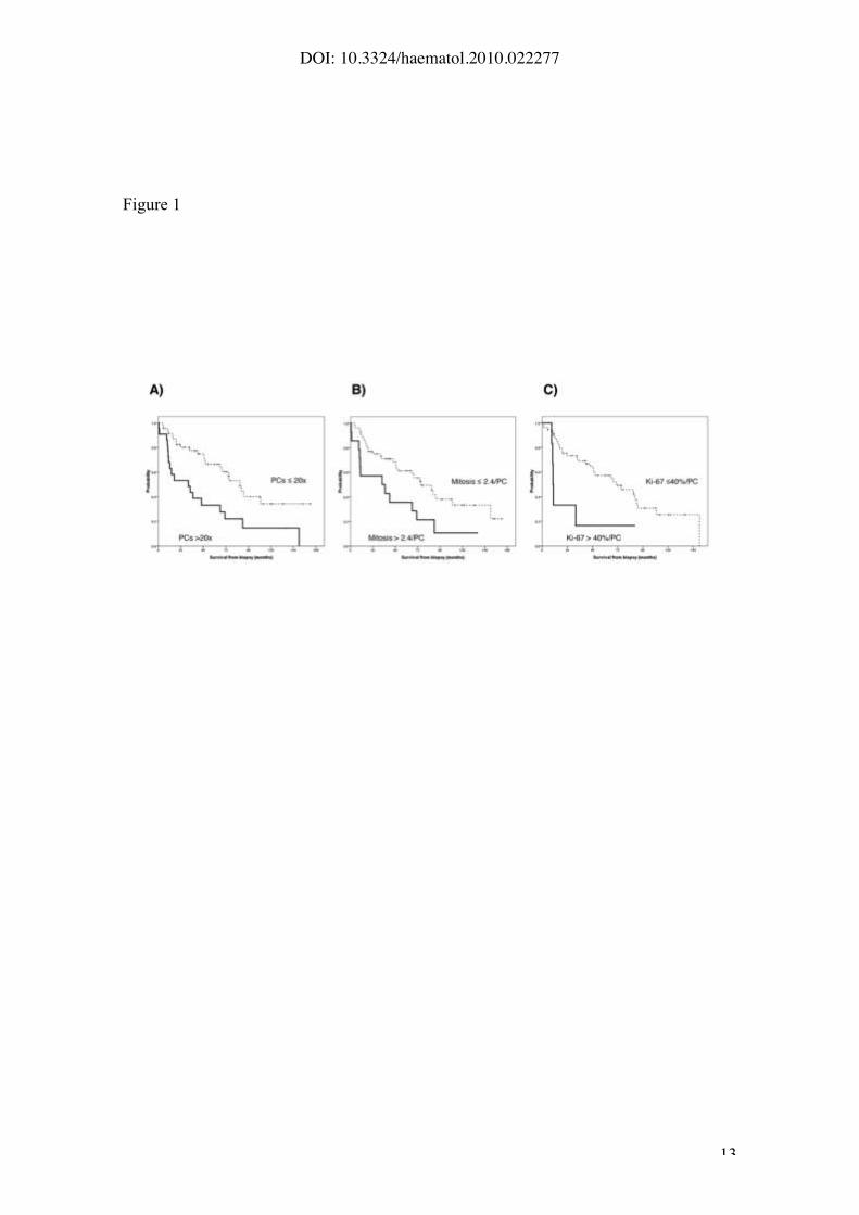
Figure 1. Survival from the time of tissue biopsy of patients with non-
transformed CLL according to the characteristics of proliferation centers (PCs):
(A) presence of expanded PCs >20x (17 vs. 75 months, PCs >20x vs. PCs
"20x, respectively; p=0.001); (B) > 2.4 mitosis per PC (34 vs. 75 months, >2.4
mitosis/PC vs. " 2.4 mitosis/PC, respectively; p=0.016); (C) >40% Ki-67
positivity per PC (11 vs. 68 months, >40%/PC vs. "40%/PC, respectively;
p=0.014).
Figure 2. (A-D-G-J): A representative case of lymph node involvement by CLL.
(A) At low magnification the tumor shows the typical biphasic growth pattern
with dark areas representing the small lymphocytic component and the clear
areas corresponding to small proliferation centers (PCs) (hematoxilyn-eosin
stain, Olympus DP70, X10). (D) The PCs are small (lesser than 20x) and
contain some large prolymphocytic cells. Scattered mitosis may also be seen
(hematoxilyn-eosin stain, Olympus DP70, X20). PCs are highlighted by
negative staining with p27 (G), and low Ki-67 labelling index (J). (B-E-H-K): A
representative CLL case with expanded proliferation centers (B, E) wider than
20X field. PCs are highlighted by negative stainig with p27 (H). The mitotic
count as well as the Ki-67 index (K) is high. (C-F-I-L): Representative sections
of DLBCL transformation of a CLL case. (C) Partial involvement of the lymph
node by a diffuse proliferation (bottom) of large immunoblastic cells (F). p27 is
negative in the tumor cells (I) that shows a very high proliferative rate (L).
Figure 3. Survival from biopsy according to the histological patterns: “non-
accelerated” CLL, “accelerated” CLL and DLBCL transformation (median
survival 76 months, 34 months and 4.3 months, respectively (p<0.001)).
DOI: 10.3324/haematol.2010.022277
13
Figure 1
DOI: 10.3324/haematol.2010.022277
14
Figure 2
DOI: 10.3324/haematol.2010.022277
15
Figure 3
DOI: 10.3324/haematol.2010.022277
16
REFERENCES
1. Chiorazzi N, Rai KR, Ferrarini M. Chronic lymphocytic leukemia. N Engl J Med
2005;352:804-15.
2. Rozman C, Montserrat E. Chronic lymphocytic leukemia. N Engl J Med
1995;333:1052-7.
3. Swerdlow SH, Campo E, Harris NL, Jaffe ES, Pileri SA, Stein H et al. WHO
Classification of Tumours of Haematopoietic and Lymphoid Tissues. 4th ed. Lyon:International Agency for Research on Cancer; 2008.
4. Crespo M, Bosch F, Villamor N, Bellosillo B, Colomer D, Rozman M et al. ZAP-70
expression as a surrogate for immunoglobulin-variable-region mutations inchronic lymphocytic leukemia. N Engl J Med 2003;348:1764-75.
5. Dohner H, Stilgenbauer S, Benner A, Leupolt E, Krober A, Bullinger L et al.
Genomic aberrations and survival in chronic lymphocytic leukemia. N Engl J Med
2000;343:1910-6.
6. Hamblin TJ, Davis Z, Gardiner A, Oscier DG, Stevenson FK. Unmutated Ig V(H)
genes are associated with a more aggressive form of chronic lymphocytic
leukemia. Blood 1999;94:1848-54.
7. Caligaris-Cappio F, Ghia P. Novel insights in chronic lymphocytic leukemia: are
we getting closer to understanding the pathogenesis of the disease? J Clin Oncol
2008;26:4497-03.
8. Ben-Ezra J, Burke JS, Swartz WG, Brownell MD, Brynes RK, Hill LR et al. Small
lymphocytic lymphoma: a clinicopathologic analysis of 268 cases. Blood
1989;73:579-87.
9. Dick FR, Maca RD. The lymph node in chronic lymphocytic leukemia. Cancer1978;41:283-92.
10. Lennert K, Mohri N, Stein Hea. Malignant lymphomas other than Hodgkin's
disease. Berlin·Heidelberg·New York: Springler-Verlag; 1978.
11. Schmid C, Isaacson PG. Proliferation centres in B-cell malignant lymphoma,
lymphocytic (B-CLL): an immunophenotypic study. Histopathology 1994;24:445-
451.
12. Bonato M, Pittaluga S, Tierens A, Criel A, Verhoef G, Wlodarska I et al. Lymph
node histology in typical and atypical chronic lymphocytic leukemia. Am J Surg
Pathol 1998;22:49-56.
13. Damle RN, Ghiotto F, Valetto A, Albesiano E, Fais F, Yan XJ et al. B-cell chroniclymphocytic leukemia cells express a surface membrane phenotype of activated,
antigen-experienced B lymphocytes. Blood 2002;99:4087-93.
14. Ginaldi L, De MM, Matutes E, Farahat N, Morilla R, Catovsky D. Levels ofexpression of CD19 and CD20 in chronic B cell leukaemias. J Clin Pathol
1998;51:364-9.

DOI: 10.3324/haematol.2010.022277
17
15. Granziero L, Ghia P, Circosta P, Gottardi D, Strola G, Geuna M et al. Survivin is
expressed on CD40 stimulation and interfaces proliferation and apoptosis in B-
cell chronic lymphocytic leukemia. Blood 2001;97:2777-83.
16. Lampert IA, Wotherspoon A, Van NS, Hasserjian RP. High expression of CD23 in
the proliferation centers of chronic lymphocytic leukemia in lymph nodes and
spleen. Hum Pathol 1999;30:648-54.
17. Soma LA, Craig FE, Swerdlow SH. The proliferation center microenvironment and
prognostic markers in chronic lymphocytic leukemia/small lymphocytic lymphoma.
Hum Pathol 2006;37:152-9.
18. Swerdlow SH, Murray LJ, Habeshaw JA, Stansfeld AG. Lymphocytic
lymphoma/B-chronic lymphocytic leukaemia--an immunohistopathological study
of peripheral B lymphocyte neoplasia. Br J Cancer 1984;50:587-99.
19. Asplund SL, McKenna RW, Howard MS, Kroft SH. Immunophenotype does notcorrelate with lymph node histology in chronic lymphocytic leukemia/small
lymphocytic lymphoma. Am J Surg Pathol 2002;26:624-9.
20. Morrison WH, Hoppe RT, Weiss LM, Picozzi VJ, Jr., Horning SJ. Smalllymphocytic lymphoma. J Clin Oncol 1989;7:598-606.
21. Cheson BD, Bennett JM, Grever M, Kay N, Keating MJ, O'Brien S et al. National
Cancer Institute-sponsored Working Group guidelines for chronic lymphocyticleukemia: revised guidelines for diagnosis and treatment. Blood 1996;87:4990-7.
22. Hallek M, Cheson BD, Catovsky D, Caligaris-Cappio F, Dighiero G, Dohner H et
al. Guidelines for the diagnosis and treatment of chronic lymphocytic leukemia: a
report from the International Workshop on Chronic Lymphocytic Leukemiaupdating the National Cancer Institute-Working Group 1996 guidelines. Blood
2008;111:5446-56.
23. Tsimberidou AM, O'Brien S, Khouri I, Giles FJ, Kantarjian HM, Champlin R et al.Clinical outcomes and prognostic factors in patients with Richter's syndrome
treated with chemotherapy or chemoimmunotherapy with or without stem-cell
transplantation. J Clin Oncol 2006;24:2343-51.
24. Carreras J, Villamor N, Colomo L, Moreno C, Cajal S, Crespo M et al.Immunohistochemical analysis of ZAP-70 expression in B-cell lymphoid
neoplasms. J Pathol 2005;205:507-13.
25. Halldorsdottir AM, Zehnbauer BA, Burack WR. Application of BIOMED-2 clonalityassays to formalin-fixed paraffin embedded follicular lymphoma specimens:
superior performance of the IGK assays compared to IGH for suboptimal
specimens. Leuk Lymphoma 2007;48:1338-43.
26. Sanchez-Beato M, Saez AI, Martinez-Montero JC, Sol MM, Sanchez-Verde L,
Villuendas R et al. Cyclin-dependent kinase inhibitor p27KIP1 in lymphoid tissue:
p27KIP1 expression is inversely proportional to the proliferative index. Am J
Pathol 1997;151:151-60.
27. Hothorn T, Lausen B. On the exact distribution of maximally selected rank
statistics. Computational Statistics & Data Analysis 2003;43:121-37.
DOI: 10.3324/haematol.2010.022277
18
28. Kaplan EL, Meier P. Non-parametric estimation from incomplete observations. J
Am Stat assoc 1958;53:457-81.
29. Peto R, Pike MC. Conservatism of the approximation _ (0-E)2/E in the log-ranktest for survival data on tumor incidence data. Biometrics 1973;29:759-84.
30. Cox DR. Regression models and life tables. J R Stat Assoc 1972;34:187-220.
31. Lennert K, Soehring M. Histopathology of Non-Hodgkin's Lymphomas (Based onthe Updated Kiel Classification). 2nd ed. New York, Berlin, Heidelberg: Springler-
Verlag; 1990.
32. Herreros B, Rodriguez-Pinilla SM, Pajares R, Martinez-Gonzalez MA, Ramos R,Munoz I et al. Proliferation centers in chronic lymphocytic leukemia: the niche
where NF-kappaB activation takes place. Leukemia 2010.
33. Schroers R, Griesinger F, Trumper L, Haase D, Kulle B, Klein-Hitpass L et al.
Combined analysis of ZAP-70 and CD38 expression as a predictor of diseaseprogression in B-cell chronic lymphocytic leukemia. Leukemia 2005;19:750-8.
34. Stilgenbauer S, Krober A, Busch R, Eichhorst B, Kienle D, Winkler D et al. 17p
Deletion Predicts for Inferior Overall Survival after Fludarabine - Based First LineTherapy in Chronic Lymphocytic Leukemia: First Analysis of Genetics in the CLL4
Trial of the GCLLSG. Blood 2005;106a:715.
35. Bea S, Lopez-Guillermo A, Ribas M, Puig X, Pinyol M, Carrio A et al. Geneticimbalances in progressed B-cell chronic lymphocytic leukemia and transformed
large-cell lymphoma (Richter's syndrome). Am J Pathol 2002;161:957-68.
36. Cobo F, Martinez A, Pinyol M, Hernandez L, Gomez M, Bea S et al. Multiple cell
cycle regulator alterations in Richter's transformation of chronic lymphocyticleukemia. Leukemia 2002;16:1028-34.
DOI: 10.3324/haematol.2010.022277
19
Table 1. Initial characteristics of 100 patients with CLL.
M: Male; F: Female. * Available in 88, 74, 63, 61, 37 and 27
patients, respectively.
Parameters No.
Median age (range) 59 yrs (24-85)
Gender 58M/42 F
ECOG "2 6 (6%)
B symptoms 12 (12%)
Bulky disease 7 (7%)
BinetABC
54 (54%)39 (39%)7 (7%)
Rai0I-IIIII-IV
24 (24%)66 (66%)10 (10%)
Increased serum LDH* 19 (22%)
Increased !2-microglobulin* 28 (38%)
ZAP-70 (!20%)* 42 (67%)
CD38 (!30%)* 45 (74%)
Cytogenetics*13q-11q-17p-+12
16 (43%)7 (19%)11 (30%)18 (48%)
IGVH mutational status (UM> 98%)* 23 (85%)
First-line treatment (n=92)AlkylatorsPurine analogsCHOP regimenOthers
38 (41%)34 (37%)18 (20%)2 (2%)

DOI: 10.3324/haematol.2010.022277
20
Table 2. Histological characteristics of “non-accelerated”, “accelerated” CLL and DLBCL-tcases.
Features“Non-Accelerated”
CLL (n=55)
“Accelerated”
CLL (n=23)
DLBCL-t
(n=22)
Proliferation Centers (PCs)
Absent 9 (16%) 0
Small 46 (84%) 1 (4%) NA
Expanded 0 22 (96%)
Mitoses/PCs*
Median (range) 0.4 (0-2.4) 2.5 (1-12)
! 2.4 46 (100%) 6 (27%)
>2.4 0 16 (73%)
NA
Ki67/PCs (%)†
Median (range) 5 (0-30) 35 (3-75)
! 40% 39 (100%) 16 (73%)
>40% 0 6 (27%)
NA
Ki67 global (%)
Median (range) 1 (0-10) 13 (5-30) 70 (50-90)
DLBCL-t: Diffuse large B cell lymphoma transformation; NA: Not apply;*evaluable in 69 cases;
† evaluable in 61 cases
DOI: 10.3324/haematol.2010.022277
21
Table 3. Main features of patients at biopsy according to histological findings.
Characteristics “Non-Accelerated” CLL
(n=55)
“Accelerated”CLL
(n=23)
DLBCL-t
(n=22)
Median age (range) 61 yrs (34-85) 59 yrs (34-84) 63 yrs (39-83)Gender 34M/21 F 11M/12 F 13M/9 F
ECOG !2 8 (17%) 4 (20%) 7 (41%)
B symptoms 11 (22%) 3 (16%) 8 (44%)Bulky disease 12 (22%) 3 (14%) 5 (28%)Binet
ABC
19 (39%)19 (39%)11 (22%)
7 (35%)4 (20%)9 (45%)
3 (18%)6 (35%)8 (47%)
Rai0
*
IIIIIIIV
1 (2%)24 (49%)12 (25%)6 (12%)6 (12%)
-6 (30%)5 (25%)4 (20%)5 (25%)
2 (13%)3 (17%)3 (17%)3 (17%)6 (36%)
Extranodal involvement 1 (0.01) 0†
9 (41%)LDH (>450 UI/L) 19 (38%) 16 (73%)
†12 (71%)
"2-microglobulin (>2.3 mg/dl) 21 (48%) 11 (69%) 7 (70%)
IGVH mutational status (UM> 98%) 10 (77%) 7 (100%) 6 (86%)ZAP-70 (!20%) 19 (53%) 16 (89%)
†7 (78%)
Prior therapies01>1
39 (74%)4 (7%)10 (19%)
10 (45%)†
5 (23)7 (32%)
5 (25%)5 (25%)10 (50%)
Resistance to the last treatment ¶ 4 (29%) 6 (50%) 5 (33%)
DOI: 10.3324/haematol.2010.022277
22
DLBCL-t: Diffuse large B cell lymphoma transformation. * corresponding to abdominal mass. ¶ % over
previously treated patients. Significant p values for Accelerated vs. Non-Accelerated cases (†): increased
serum LDH (p=0.01), elevated ZAP-70 (p=0.014) and "1 lines of treatment (p=0.032). Significant p value for
Accelerated vs. DLBCL-t (†) cases: extranodal involvement (p=0.0006)