Embed Size (px)
Citation preview

TheprostaglandinreceptorEP4
triggers remodellingof thecardiovascularsystematbirthMyTrang Nguyen*, Todd Camenisch*, John N. Snouwaert*,Elizabeth Hicks*, Thomas M. Coffman†,Page A. W. Anderson‡, Nadia N. Malouf§& Beverly H. Koller*
* Department of Medicine, University of North Carolina at Chapel Hill,Chapel Hill, North Carolina 27599-7248, USA† Department of Medicine, Duke University and Durham Veterans AffairsMedical Centers, Durham, North Carolina 27710, USA‡ Division of Pediatric Cardiology, Duke University Medical Center, Durham,North Carolina 27710, USA§ Department of Pathology, University of North Carolina at Chapel Hill,Chapel Hill, North Carolina 27599-7525, USA. . . . . . . . . . . . . . . . . . . . . . . . . . . . . . . . . . . . . . . . . . . . . . . . . . . . . . . . . . . . . . . . . . . . . . . . . . . . . . . . . . . . . . . . . . . . . . . . . . . . . . . . . . . . . . . . . . . . . . . . .
Survival of newborn placental mammals depends on closure ofthe ductus arteriosus (DA), an arterial connection in the fetuswhich directs blood away from the pulmonary circulation andtowards the placenta where oxygenation occurs1. Here we showthat morphological changes resulting in closure of the DA in miceare virtually identical to those observed in larger mammals,including humans2, and that maintenance of the DA in theopen, or patent, state in fetal mice is dependent on prostaglandinsynthesis. This requirement is absent in mice lacking the pros-taglandin E2 EP4 receptor (EP4(−/−) mice). In EP4(−/−) mice of the129 strain, remodelling of the DA fails to occur after birth,resulting in a left-to-right shunt of blood and subsequently indeath. This suggests that the neonatal drop in prostaglandin E2
(refs 3–7) that triggers ductal closure is sensed through the EP4
receptor. In contrast, 5% of EP4(−/−) mice of mixed geneticbackground survive, and selective breeding of these mice leads
to a 21% survival rate, suggesting that alleles at other loci canprovide an alternative mechanism for ductal closure.
In about 6 in 10,000 full-term infants, and even more frequentlyin the premature neonate, the DA fails to close8. This allowstransmission of high aortic pressures to the pulmonary circuit,which in turn may lead to pulmonary venous hypertension,pulmonary oedema, and congestive heart failure9. Permanent clo-sure of the DA depends on a series of structural changes that areindependent of the reversible muscular contraction of the DA.Endothelial cells detach from the internal elastic lamina, and thesubendothelial region begins to accumulate extracellular matrixmaterial and fluid, initiating the formation of intimal cushions. Theinternal elastic lamina becomes fragmented, allowing smoothmuscle cells to migrate into the subendothelial region, wideningthe intimal cushions and obstructing the vessel lumen. Eventuallythe DA degenerates into the fibrous cord referred to as theligamenum arteriosus10,11.
Examination of the DA in normal mice indicates that, as in otherspecies, the DA is a large muscular artery characterized by numerouslayers of immature smooth muscle cells separated by layers of elastin(Fig. 1a). The DA constricts within 30 min of birth, obstructingblood flow, and events leading to permanent closure of the mouseDA are initiated within three hours of birth. As noted in otherspecies, DA closure proceeds from the pulmonary to the aortic end10
of the vessel (Fig. 1a).Prostaglandin E2 (PGE2) is effective in inhibiting the natural tone
letters to nature
78 NATURE | VOL 390 | 6 NOVEMBER 1997
Table 1 Survival of EP4-deficient mice
Genotype of offspring
Strain* Parentalgenotype
+/+ +/− −/−
.............................................................................................................................................................................
129 þ=2 3 þ =2 68 113 0MB þ=2 3 þ =2 90 150 4SMB1 2 =2 3 þ =2 0 79 2SMB4 2 =2 3 þ =2 0 66 14.............................................................................................................................................................................* 129 mice are a mixture of 129/SvEv and 129/Olac substrains. MB, mixed background mice,are derived from 129/Olac, C57BL/6 and DBA/2 mice. SMB, selected mixed background,are derived from EP4(−/−) MB mice (see Methods).
Bright field Antisensedark field
Sensedark field
18.5d 19.5d-P 19.5d-A
a
b
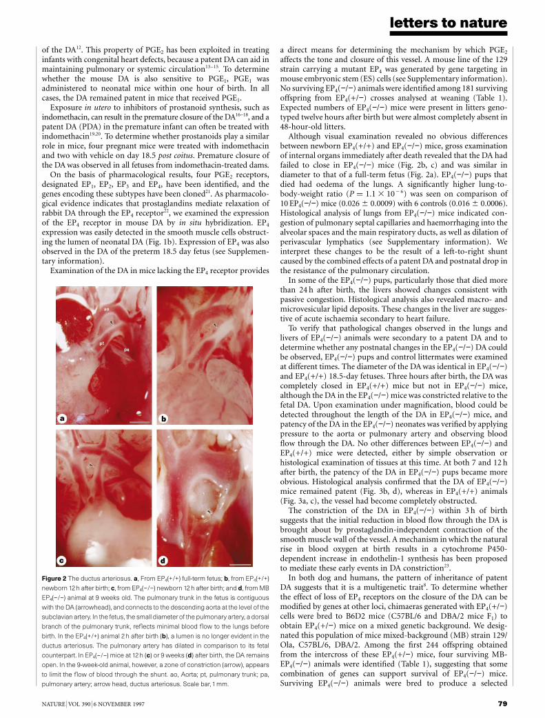
Figure 1 Mouse DA morphology and expression of the EP4 receptor. a,
Transverse sections of the ductus arteriosus from fetus at term (18.5 d) and
neonate, stained with toluidine blue. Sections are taken from the neonatal DA at
the region adjacent to the pulmonary trunk (19.5d-P) and towards its junction with
the aorta (19.5-A). The wall of the fetal ductus consists of multiple layers of
immature smooth-muscle cells alternating with layers of elastic lamina. This
structure alters quickly after birth, as the elastin layer becomes fragmented and
the lumen of the vessel is decreased. Projection of the endothelial cells into the
lumen, marked oedema, and infiltration of cells into the subendothelial region are
seen. Obstruction of the lumen is less marked in sections of the ductus adjacent
to the aorta. Scale bar,150 mm. b, Transverse sections through the DA obtained
from a neonate within 12h of birth were hybridized with 35S-labelled EP4 antisense
probe. Expression of the EP4 gene is easily detected in the migrating smooth-
muscle cells. Bright-field microscopy of the section stained with haematoxylin
and eosin is shown on the left. A serial section of the ductus was hybridized with35S-labelled EP4 sense probe to establish levels of nonspecific hybridization.
Scale bar,150 mm.
of the DA12. This property of PGE2 has been exploited in treatinginfants with congenital heart defects, because a patent DA can aid inmaintaining pulmonary or systemic circulation13–15. To determinewhether the mouse DA is also sensitive to PGE1, PGE1 wasadministered to neonatal mice within one hour of birth. In allcases, the DA remained patent in mice that received PGE1.
Exposure in utero to inhibitors of prostanoid synthesis, such asindomethacin, can result in the premature closure of the DA16–18, and apatent DA (PDA) in the premature infant can often be treated withindomethacin19,20. To determine whether prostanoids play a similarrole in mice, four pregnant mice were treated with indomethacinand two with vehicle on day 18.5 post coitus. Premature closure ofthe DA was observed in all fetuses from indomethacin-treated dams.
On the basis of pharmacological results, four PGE2 receptors,designated EP1, EP2, EP3 and EP4, have been identified, and thegenes encoding these subtypes have been cloned21. As pharmacolo-gical evidence indicates that prostaglandins mediate relaxation ofrabbit DA through the EP4 receptor22, we examined the expressionof the EP4 receptor in mouse DA by in situ hybridization. EP4
expression was easily detected in the smooth muscle cells obstruct-ing the lumen of neonatal DA (Fig. 1b). Expression of EP4 was alsoobserved in the DA of the preterm 18.5 day fetus (see Supplemen-tary information).
Examination of the DA in mice lacking the EP4 receptor provides
a direct means for determining the mechanism by which PGE2
affects the tone and closure of this vessel. A mouse line of the 129strain carrying a mutant EP4 was generated by gene targeting inmouse embryonic stem (ES) cells (see Supplementary information).No surviving EP4(−/−) animals were identified among 181 survivingoffspring from EP4(+/−) crosses analysed at weaning (Table 1).Expected numbers of EP4(−/−) mice were present in litters geno-typed twelve hours after birth but were almost completely absent in48-hour-old litters.
Although visual examination revealed no obvious differencesbetween newborn EP4(+/+) and EP4(−/−) mice, gross examinationof internal organs immediately after death revealed that the DA hadfailed to close in EP4(−/−) mice (Fig. 2b, c) and was similar indiameter to that of a full-term fetus (Fig. 2a). EP4(−/−) pups thatdied had oedema of the lungs. A significantly higher lung-to-body-weight ratio (P ¼ 1:1 3 10 2 6) was seen on comparison of10 EP4(−/−) mice (0:026 6 0:0009) with 6 controls (0:016 6 0:0006).Histological analysis of lungs from EP4(−/−) mice indicated con-gestion of pulmonary septal capillaries and haemorrhaging into thealveolar spaces and the main respiratory ducts, as well as dilation ofperivascular lymphatics (see Supplementary information). Weinterpret these changes to be the result of a left-to-right shuntcaused by the combined effects of a patent DA and postnatal drop inthe resistance of the pulmonary circulation.
In some of the EP4(−/−) pups, particularly those that died morethan 24 h after birth, the livers showed changes consistent withpassive congestion. Histological analysis also revealed macro- andmicrovesicular lipid deposits. These changes in the liver are sugges-tive of acute ischaemia secondary to heart failure.
To verify that pathological changes observed in the lungs andlivers of EP4(−/−) animals were secondary to a patent DA and todetermine whether any postnatal changes in the EP4(−/−) DA couldbe observed, EP4(−/−) pups and control littermates were examinedat different times. The diameter of the DA was identical in EP4(−/−)and EP4(+/+) 18.5-day fetuses. Three hours after birth, the DA wascompletely closed in EP4(+/+) mice but not in EP4(−/−) mice,although the DA in the EP4(−/−) mice was constricted relative to thefetal DA. Upon examination under magnification, blood could bedetected throughout the length of the DA in EP4(−/−) mice, andpatency of the DA in the EP4(−/−) neonates was verified by applyingpressure to the aorta or pulmonary artery and observing bloodflow through the DA. No other differences between EP4(−/−) andEP4(+/+) mice were detected, either by simple observation orhistological examination of tissues at this time. At both 7 and 12 hafter birth, the patency of the DA in EP4(−/−) pups became moreobvious. Histological analysis confirmed that the DA of EP4(−/−)mice remained patent (Fig. 3b, d), whereas in EP4(+/+) animals(Fig. 3a, c), the vessel had become completely obstructed.
The constriction of the DA in EP4(−/−) within 3 h of birthsuggests that the initial reduction in blood flow through the DA isbrought about by prostaglandin-independent contraction of thesmooth muscle wall of the vessel. A mechanism in which the naturalrise in blood oxygen at birth results in a cytochrome P450-dependent increase in endothelin-1 synthesis has been proposedto mediate these early events in DA constriction23.
In both dog and humans, the pattern of inheritance of patentDA suggests that it is a multigenetic trait8. To determine whetherthe effect of loss of EP4 receptors on the closure of the DA can bemodified by genes at other loci, chimaeras generated with EP4(+/−)cells were bred to B6D2 mice (C57BL/6 and DBA/2 mice F1) toobtain EP4(+/−) mice on a mixed genetic background. We desig-nated this population of mice mixed-background (MB) strain 129/Ola, C57BL/6, DBA/2. Among the first 244 offspring obtainedfrom the intercross of these EP4(+/−) mice, four surviving MB-EP4(−/−) animals were identified (Table 1), suggesting that somecombination of genes can support survival of EP4(−/−) mice.Surviving EP4(−/−) animals were bred to produce a selected
letters to nature
NATURE | VOL 390 | 6 NOVEMBER 1997 79
Figure 2 The ductus arteriosus. a, From EP4(+/+) full-term fetus; b, from EP4(+/+)
newborn 12h after birth; c, from EP4(−/−) newborn 12h after birth; and d, from MB
EP4(−/−) animal at 9 weeks old. The pulmonary trunk in the fetus is contiguous
with the DA (arrowhead), and connects to the descending aorta at the level of the
subclavianartery. In the fetus, the small diameter of the pulmonary artery, a dorsal
branch of the pulmonary trunk, reflects minimal blood flow to the lungs before
birth. In the EP4(+/+) animal 2 h after birth (b), a lumen is no longer evident in the
ductus arteriosus. The pulmonary artery has dilated in comparison to its fetal
counterpart. In EP4(−/−) mice at 12h (c) or 9 weeks (d) after birth, the DA remains
open. In the 9-week-old animal, however, a zone of constriction (arrow), appears
to limit the flow of blood through the shunt. ao, Aorta; pt, pulmonary trunk; pa,
pulmonary artery; arrow head, ductus arteriosus. Scale bar,1mm.
mixed-background (SMB) population. Five per cent of EP4(−/−)offspring survive when EP4(+/−) mice of the MB generation arecrossed, and this number is increased to 21% in the SMB4 offspring.
The DAs of EP4(−/−) mice that survived were examined, and in allcases closure of the DAwas observed. In some MB EP4(−/−) animals,however, closure was restricted to the region of the DA immediatelyadjacent to the pulmonary trunk. This produced a funnel-shapeddiverticulum that was open at the aortic end (Fig. 2d), similar to theDA diverticulum present in dog strains with susceptibility to patentDA10. The lack of difference in overall health between survivingEP4(−/−) mice and control littermates supports the contentionthat the lung oedema and liver disease seen in the EP4(−/−)mice that died perinatally was secondary to a patent DA.
We examined the effect of inhibition of prostaglandin synthesison EP4(−/−) fetuses by administering indomethacin to EP4(+/−)females impregnated 18.5 days earlier by MB EP4(−/−) males. Fourhours after treatment, the DA of each pup was examined and itsDNA prepared to determine the genotype. In all cases, indometha-cin induced closure of the DA in the wild type but not in EP4(−/−)fetuses (see Supplementary information).
Previous studies indicate that the smooth muscle of the DA has anintrinsic tone that is inhibited by PGE2 (refs 24, 25). To reconcilethese data with those from the genetic experiments presented here,we propose a model in which the EP4 receptor in the DA acts as asensor that responds to the perinatal drop in circulating levels ofPGE2 (refs 3–7) by triggering closure of the DA (Fig. 4). In mostcells, the EP4 receptor is coupled to adenylate cyclase through astimulatory G protein, so that stimulation of the receptor by PGE2
increases intracellular cyclic AMP levels21,26. We suggest that, follow-ing EP4 receptor expression in utero, other pathways contributing tocAMP generation in the cells of the DA are downmodulated so thatthe EP4 receptor becomes the principal regulator of intracellularlevels of cAMP. In EP4-deficient mice, however, patency of the DAfails to become dependent on circulating levels of PGE2, explainingwhy the DA in these animals remains patent after birth and isunresponsive to administration of indomethacin in utero.
Genes required for normal closure of the mouse DA, such as theEP4 receptor, represent candidates for the inherited components ofhuman PDA. An understanding of these genes and the mechanismby which they influence closure of the DA is important, not only forthe study and treatment of patent DA, but also for understandingvessel intimal thickening in general and identifying genes that caninfluence the structure of blood-vessel walls. The DA also provides amodel system for unravelling the complex mechanisms by which
PGE2 and its receptors modulate cell structure and function inresponse to the changing physiological state of the organism. M. . . . . . . . . . . . . . . . . . . . . . . . . . . . . . . . . . . . . . . . . . . . . . . . . . . . . . . . . . . . . . . . . . . . . . . . . . . . . . . . . . . . . . . . . . . . . . . . . . . . . . . . . . . . . . . . . . . . . . . . .
Methods
Tissue preparation for light microscopy. To minimize contraction of the DAin response to exposure to light and oxygen, mouse carcasses were cooled onice before exposure of the DA. Ducti shown in Fig. 1a were fixed in 2%paraformaldehyde/2.5% glutaraldehyde, embedded in a mixture of Spurr’s andPoly-bed 812 resin, and transverse sections, 1 mm in thickness, were cut andstained with 1% toluidine blue in 1% sodium borate. All other tissue wasfixed in 10% phosphate-buffered formalin, pH 7.2, embedded in paraffin, and5-mm-thick sections prepared.RNA in situ hybridization. The EP4-specific cDNA probe was synthesized byreverse transcription and PCR amplification using oligonucleotide primersEP4-1F (59-GTTTGGCTGATATAACTGGTTAAT-39) and EP4-2R (59-ACCTGGTGCTTCATCGACTGGACC-39) and total thymus RNA as a template.These primers correspond to the published EP4 sequence26. Originally desig-nated as the EP2 receptor, it is now recognized that this sequence represents themurine homologue of the human EP4 receptor27. The PCR fragment was clonedusing a TA cloning kit (In Vitrogen) and 35S-labelled probes prepared using acommercially available kit (Ambion). The DA was embedded in OCT com-pound, transverse sections 8 to 10 mm in thickness were prepared, and in situhybridization carried out as described28.Generation of EP4-deficient mice. The EP4 cDNA probe was used to isolategenomic clones from a 129/Sv mouse genomic library. Sequence analysisconfirmed that these clones corresponded to the mouse EP4 gene. In thetargeting vector, exons 1 and 2 are replaced by the neomycin-resistance genefrom the plasmid pJNS2. These exons include the initiation codon and regionsof the gene encoding six of the seven transmembrane-spanning domains andputative extracellular loops. 129/Ola derived E14Tg2a ES cells29 were grown,transformed and screened using standard method30. A probe prepared fromDNA immediately downstream of the targeted locus was used to identifytargeted ES cells and later to genotype the mice generated from these ES cells.Chimaeras derived from targeted ES cells were mated with 129/SvEv or B6D2(C57BL=6 3 DBA=2 F1) mice. Mice obtained from crosses with 129/SvEv arehere referred to as 129 mice. Analysis of RNA prepared from the thymus andspleen of EP4(−/−) and EP4(+/+) newborn pups with EP4-specific cDNA probesverified the loss of EP4 expression in EP4(−/−) mice. The first EP4(−/−) miceobtained from crossing EP4(+/−) offspring of chimaeras and B6D2 animals aredesignated ‘mixed background’ (MB). EP4(−/−) obtained from the crossing ofMB EP4(−/−) mice with either EP4(+/−) sibs or offspring are designatedselected mixed background generation one (SMB1). EP4(−/−) mice derivedfrom crosses of SMB1EP4(−/−) mice and EP4(+/−) sibs and offspring aredesignated SMB2.
letters to nature
80 NATURE | VOL 390 | 6 NOVEMBER 1997
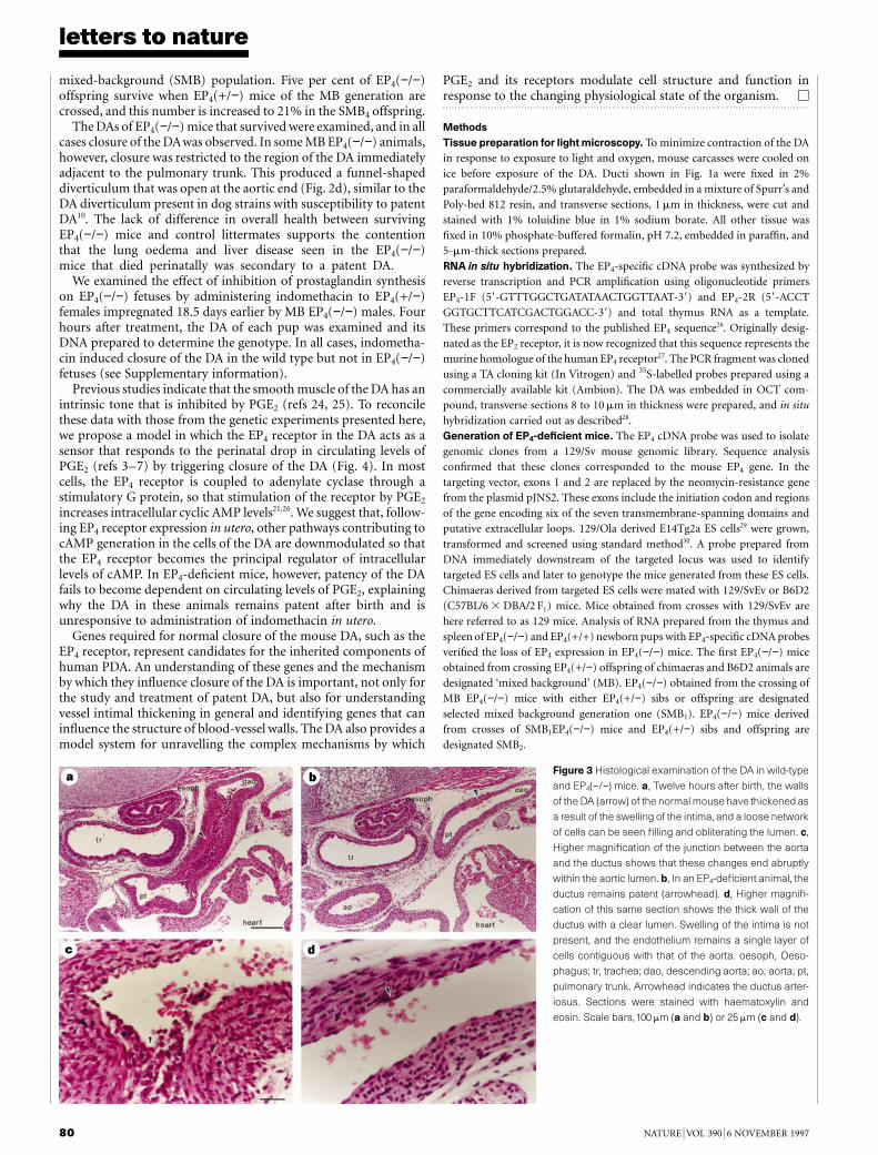
Figure 3 Histological examination of the DA in wild-type
and EP4(−/−) mice. a, Twelve hours after birth, the walls
of the DA (arrow) of the normal mouse have thickened as
a result of the swelling of the intima, and a loose network
of cells can be seen filling and obliterating the lumen. c,
Higher magnification of the junction between the aorta
and the ductus shows that these changes end abruptly
within the aortic lumen. b, In an EP4-deficient animal, the
ductus remains patent (arrowhead). d, Higher magnifi-
cation of this same section shows the thick wall of the
ductus with a clear lumen. Swelling of the intima is not
present, and the endothelium remains a single layer of
cells contiguous with that of the aorta. oesoph, Oeso-
phagus; tr, trachea; dao, descending aorta; ao, aorta; pt,
pulmonary trunk. Arrowhead indicates the ductus arter-
iosus. Sections were stained with haematoxylin and
eosin. Scale bars,100 mm (a and b) or 25 mm (c and d).
DA response to PGE2 and indomethacin. At E18.5, pregnant mice received asingle intravenous dose of indomethacin (10 mg kg−1 in bicarbonate buffer, pH7.4). At the time indicated, the mice were killed, fetuses removed andimmediately placed on ice, and the DA examined. PGE2 was prepared inethanol and diluted to 1 mg ml−1 in sterile PBS. Neonates received a singlesubcutaneous injection of 15 ml; controls received vehicle. Pups were returnedto their mother until the time indicated.
Received 17 July; accepted 22 September 1997.
1. Heymann, M. A. & Rudolph, A. M. Control of the ductus areteriosus. Physiol. Rev. 55, 62–78 (1975).2. Jager, B. V. & Wollenman, O. J. An anatomical study of the closure of the ductus arteriosus. Am. J.
Pathol. 18, 595–613 (1942).3. Challis, J. R. G., Dilley, S. R., Robinson, J. S. & Thorburn, G. D. Prostaglandins in the circulation of the
fetal lamb. Prostaglandins 11, 1041–1046 (1976).4. Clyman, R. I., Mauray, F., Roman, C., Rudolph, A. M. & Heymann, M. A. Circulating prostaglandin E2
concentrations and patent ductus arteriosus in fetal and neonatal lambs. J. Pediatr. 97, 455–461(1980).
5. Clyman, R. I., Wong, L., Heymann, M. A. & Rudolph, A. M. Responsiveness of the lamb ductusarteriosus to prostaglandins and their metabolites. Prostaglandins 15, 325–331 (1980).
6. Mitchell, M. D., Brunt, J., Clover, L. & Walker, D. W. Prostaglandins in the umbilical and uterinecirculations during late pregnancy in the ewe. J. Reprod. Fertil. 58, 283–287 (1980).
7. Piper, P. J., Vane, J. R. & Wyllie, J. H. Inactivation of prostaglandins by the lungs. Nature 225, 600–603(1970).
8. Patterson, D. F. Genetic factors in persistence of the ductus arteriosus. Paediatr. Cardiol. 2, 45-57(1979).
9. Gersony, W. M. Patent ductus arteriosus in the neonate. Pediatr. Clin. North. Am. 33, 545–560 (1986).10. Gittenberger-de Groot, A. C., Strengers, J. L. M., Mentink, M., Poelmann, R. E. & Patterson, D. F.
Histologic studies on normal and persistent ductus arteriosus in the dog. J. Am. Coll. Cardiol. 6, 394–404 (1985).
11. Jarkovska, D., Janatova, T., Hruda, J., Ostadal, B. & Samanek, M. Effect of prostaglandin E2 on the ductusarteriosus in the newborn rat: An ultrastructural study. Physiological Reviews 41, 323–329 (1992).
12. Coceani, F. & Olley, P. M. The response of the ductus arteriosus to prostaglandins. Can. J. Physiol.Pharmacol. 51, 220–225 (1973).
13. Christiansen, N. C. & Fabricus, J. Medical manipulation of the ductus arteriosus. Lancet 2, 24–26(1975).
14. Elliot, R. B., Starling, M. B. & Neutze, J. M. Medical manipulation of the ductus arteriosus. Lancet 1,140–142 (1975).
15. Olley, P. M., Coceani, F. & Bocach, E. E-type prostaglandins: A new emergency therapy for certaintypes of congenital heart malformations. Circulation 53, 728–731 (1976).
16. Sharpe, G. L., Larsson, K. S. & Thalme, B. Studies on closure of the ductus arteriosus. Prostaglandins 9,585–596 (1975).
17. Moise, K. J. et al. Indomethacin in the treatment of premature labor. Effects on the fetal ductusarteriosus. N. Engl. J. Med. 319, 327–331 (1988).
18. Momma, K. & Takao, A. In vivo constriction of the ductus arteriosus by nonsteroidal anti-inflammatory drugs in near-term and preterm fetal rats. Pediatr. Res. 22, 567–572 (1987).
19. Heymann, M. A., Rudolph, A. M. & Silverman, N. H. Closure of the ductus arteriosus in prematureinfants by inhibition of prostaglandin synthesis. N. Engl. J. Med. 295, 530–533 (1976).
20. Friedman, W. F., Hirschklau, M. J., Printz, M. P., Pitlick, P. T. & Kirkpatrick, S. E. Pharmacologicclosure of patent ductus arteriosus in the premature infant. N. Engl. J. Med. 295, 526–529 (1976).
21. Coleman, R. A., Smith, W. L. & Narumiya, S. International union of pharmacology classification ofprostanoid receptors: properties, distribution, and structure of the receptors and their subtypes.Phamacol. Rev. 46, 205–229 (1994).
22. Smith, G. C. S., Coleman, R. A. & McGrath, J. C. Characterization of dilator prostanoid receptors inthe fetal rabbit ductus arteriosus. J. Pharmacol. Exp. Ther. 271, 390–396 (1994).
23. Coceani, F. Control of the ductus arteriosus—a new function for cytochrome P450, endothelin andnitric oxide. Biochem. Pharmacol. 48, 1315–1318 (1994).
24. Clyman, R. I. Ductus arteriosus: Current theories of prenatal and postnatal regulation. Sem. Perinatol.11, 64–71 (1987).
25. Coceani, F. & Olley, P. M. Role of prostaglandins, prostacyclins, and thromboxanes in the control ofprenatal patency and postnatal closure of the ductus arteriosus. Sem. Perinatol. 4, 109–113 (1980).
26. Honda, A. et al. Cloning and expression of a cDNA for mouse prostaglandin E receptor EP2 subtype.J. Biol. Chem. 268, 7759–7762 (1993).
27. Regan, J. W. et al. Cloning of a novel human prostaglandin receptor with characteristics of thepharmacologically defined EP2 subtype. Mol. Pharmacol. 46, 213–220 (1994).
28. Engelhardt, J. F., Yankaskas, J. R. & Wilson, J. M. In vivo retroviral gene transfer into human bronchialepithelia of xenografts. J. Clin. Invest. 90, 2598–2607 (1992).
29. Hooper, M., Hardy, K., Handyside, A., Hunter, S. & Monk, M. HPRT-deficient (Lesch–Nyhan) mouseembryos derived from germline colonization by cultured cells. Nature 326, 292–295 (1987).
30. Mohn, A. & Koller, B. H. in DNA Cloning 4 (eds Glover, D. M. & Hames, B. D.) 143–184 (OxfordUniversity Press, New York, 1995).
Supplementary information is available on Nature’s World-Wide Web site (http://www.nature.com) oras paper from Mary Sheehan at the London editorial office of Nature.
Acknowledgements. We thank B. Anderson and L. Johnson for assistance in cloning the EP4 gene;B. Garges for animal husbandry; A. Latour for help with tissue culture; K. Burns, T. Bartologga andR. Bagnell for assistance with histology and photomicroscopy; and A. Oakely for help with manuscriptpreparation. This work was supported by grants from Pfizer, the US NHLBI and the NIDDK.
Correspondence and requests for materials should be addressed to B.H.K. (e-mail: [email protected]).
letters to nature
NATURE | VOL 390 | 6 NOVEMBER 1997 81
Figure 4 Model for closure of the DA in normal and EP4-deficient mice. As the
fetus approaches term, intracellular cAMP levels in the cells of the DA are
maintained largely by the binding of PGE2 to the EP4 receptors and stimulation of
adenyl cyclase through GS. To maintain normal levels of cAMP, other pathways
that maintain cAMP levels are downregulated. At birth, a dramatic decrease in
circulating levels of PGE2 occurs. This results in a drop in intracellular cAMP,
which in turn triggers the series of events leading to remodelling of the DA. In the
absence of the EP4 receptor, the cells in the DA maintain intracellular cAMP levels
independently of PGE2. As a result, these cells fail to respond to the loss of this
pathway at birth. Indomethacin mimics the events that occur at birth by inhibiting
synthesis of PGE2. Black circles, PGE2; question marks indicate that the sources
of cAMP have not been defined. Indo, indomethacin.
Complementationof dominantsuppression implicatesCD98in integrinactivationCsilla A. Fenczik, Tariq Sethi*, Joe W. Ramos,Paul E. Hughes & Mark H. Ginsberg
Department of Vascular Biology, The Scripps Research Institute,10550 North Torrey Pines, La Jolla, California 92037, USA* Department of Respiratory Medicine, University of Edinburgh, Medical School,Teviot Place, Edinburgh EH8 9AG, UK. . . . . . . . . . . . . . . . . . . . . . . . . . . . . . . . . . . . . . . . . . . . . . . . . . . . . . . . . . . . . . . . . . . . . . . . . . . . . . . . . . . . . . . . . . . . . . . . . . . . . . . . . . . . . . . . . . . . . . . . .
The integrin family of adhesion receptors are involved in cellgrowth, migration and tumour metastasis1. Integrins are hetero-dimeric proteins composed of an a and a b subunit, each with alarge extracellular, a single transmembrane, and a short cytoplas-mic domain. The dynamic regulation of integrin affinity forligands in response to cellular signals is central to integrinfunction2. This process is energy dependent and is mediatedthrough integrin cytoplasmic domains3. However, the cellularmachinery regulating integrin affinity remains poorly under-stood. Here we describe a genetic strategy to disentangle integrinsignalling pathways. Dominant suppression occurs when over-expression of isolated integrin b1 cytoplasmic domains blocksintegrin activation. Proteins involved in integrin signalling wereidentified by their capacity to complement dominant suppressionin an expression cloning scheme. CD98, an early T-cell activationantigen that associates with functional integrins4, was found toregulate integrin activation. Furthermore, antibody-mediatedcrosslinking of CD98 stimulated b1 integrin-dependent cell adhe-sion. These data indicate that CD98 is involved in regulatingintegrin affinity, and validate an unbiased genetic approach toanalysing integrin signalling pathways.
Strategies based on genetic selection have proven to be powerfulin the dissection of a number of different signal-transductionpathways. We therefore designed a new genetic cloning strategy,based on changes in integrin affinity, to identify potential mod-ulators of integrin activation. This strategy used a Chinese hamsterovary (CHO) cell line (abpy) stably expressing a chimaeric integrin(aIIba6Ab3b1) that contains the extracellular and transmembranedomains of aIIbb3 fused to cytoplasmic domains of a6Ab1 (ref. 5).This chimaeric integrin has the ligand-binding properties of aIIbb3
and is activated through the a6Ab1 cytoplasmic domains. In abpycells this chimaeric integrin is constitutively active, as measured by


![Integrating the Healthcare Enterprise€¦ · Document Source Document ConsumerOn Entry [ITI Document Registry Document Repository Provide&Register Document Set – b [ITI-41] →](https://img.dokumen.tips/doc/110x75/5f08a1eb7e708231d422f7c5/integrating-the-healthcare-enterprise-document-source-document-consumeron-entry.jpg)


