Embed Size (px)
Citation preview
NATURE | VOL 406 | 10 AUGUST 2000 | www.nature.com 637
letters to nature
.................................................................YidC mediates membrane proteininsertion in bacteriaJames C. Samuelson*, Minyong Chen*, Fenglei Jiang*, Ines MoÈ ller²,Martin Wiedmann², Andreas Kuhn³, Gregory J. Phillips§& Ross E. Dalbey*
* Department of Chemistry, The Ohio State University, 100 West 18th Avenue,
Columbus, Ohio 43210, USA² Cellular Biochemistry and Biophysic Program, Memorial Sloan-KetteringCancer Centre, 1275 York Avenue, New York, New York 10021, USA³ University of Hohenheim, Institute for Microbiology and Molecular Biology,
Garbenstrasse 30, D-70599 Stuttgart, Germany
§ Department of Microbiology, Iowa State University, 207 Science I,Osborn Drive, Ames, Iowa 50011, USA
..............................................................................................................................................
The basic machinery for the translocation of proteins into oracross membranes is remarkably conserved from Escherichia colito humans. In eukaryotes, proteins are inserted into the endo-plasmic reticulum using the signal recognition particle (SRP) andthe SRP receptor, as well as the integral membrane Sec61 trimericcomplex (composed of alpha, beta and gamma subunits)1. Inbacteria, most proteins are inserted by a related pathway thatincludes the SRP homologue Ffh2±5, the SRP receptor FtsY6,7, andthe SecYEG trimeric complex8, where Y and E are related to theSec61 alpha and gamma subunits, respectively. Proteins in bac-teria that exhibit no dependence on the Sec translocase werepreviously thought to insert into the membrane directly withoutthe aid of a protein machinery9,10. Here we show that membraneinsertion of two Sec-independent proteins requires YidC. YidC isessential for E. coli viability and homologues are present inmitochondria and chloroplasts. Depletion of YidC also interfereswith insertion of Sec-dependent membrane proteins, but it hasonly a minor effect on the export of secretory proteins. Theseresults provide evidence for an additional component of thetranslocation machinery that is specialized for the integrationof membrane proteins.
Recent studies in mitochondria, bacteria and chloroplasts suggesta new membrane protein insertion pathway that is evolutionarilyrelated11±15. In mitochondria, a subset of proteins is sorted into theinner membrane from the matrix by a conservative pathway thatseems analogous to that found in bacteria16. However, one impor-tant difference in mitochondria is that the Sec proteins are absent17.Instead, Oxa1p is required for insertion of some inner membraneproteins from the mitochondrial matrix11,12. Oxa1p homologues arepresent in the thylakoid membrane of chloroplasts (Albino3 inArabidopsis thaliana13) and the inner membrane of bacteria (YidC inEscherichia coli14).
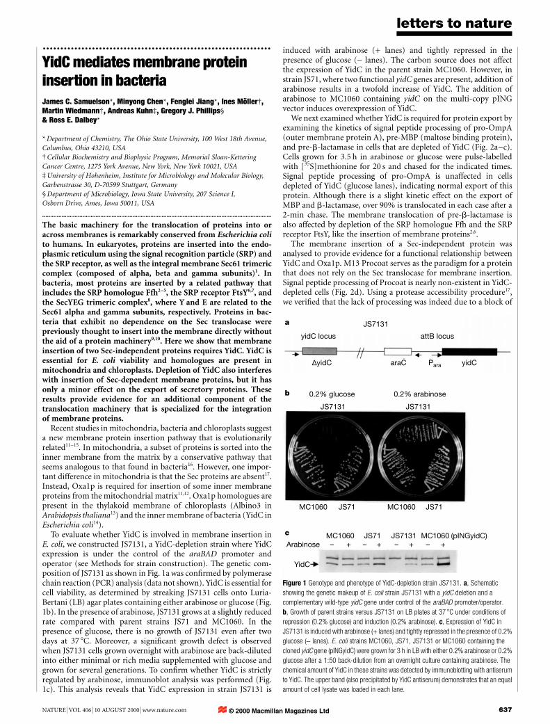
To evaluate whether YidC is involved in membrane insertion inE. coli, we constructed JS7131, a YidC-depletion strain where YidCexpression is under the control of the araBAD promoter andoperator (see Methods for strain construction). The genetic com-position of JS7131 as shown in Fig. 1a was con®rmed by polymerasechain reaction (PCR) analysis (data not shown). YidC is essential forcell viability, as determined by streaking JS7131 cells onto Luria-Bertani (LB) agar plates containing either arabinose or glucose (Fig.1b). In the presence of arabinose, JS7131 grows at a slightly reducedrate compared with parent strains JS71 and MC1060. In thepresence of glucose, there is no growth of JS7131 even after twodays at 37 8C. Moreover, a signi®cant growth defect is observedwhen JS7131 cells grown overnight with arabinose are back-dilutedinto either minimal or rich media supplemented with glucose andgrown for several generations. To con®rm whether YidC is strictlyregulated by arabinose, immunoblot analysis was performed (Fig.1c). This analysis reveals that YidC expression in strain JS7131 is
induced with arabinose (+ lanes) and tightly repressed in thepresence of glucose (- lanes). The carbon source does not affectthe expression of YidC in the parent strain MC1060. However, instrain JS71, where two functional yidC genes are present, addition ofarabinose results in a twofold increase of YidC. The addition ofarabinose to MC1060 containing yidC on the multi-copy pINGvector induces overexpression of YidC.
We next examined whether YidC is required for protein export byexamining the kinetics of signal peptide processing of pro-OmpA(outer membrane protein A), pre-MBP (maltose binding protein),and pre-b-lactamase in cells that are depleted of YidC (Fig. 2a±c).Cells grown for 3.5 h in arabinose or glucose were pulse-labelledwith [35S]methionine for 20 s and chased for the indicated times.Signal peptide processing of pro-OmpA is unaffected in cellsdepleted of YidC (glucose lanes), indicating normal export of thisprotein. Although there is a slight kinetic effect on the export ofMBP and b-lactamase, over 90% is translocated in each case after a2-min chase. The membrane translocation of pre-b-lactamase isalso affected by depletion of the SRP homologue Ffh and the SRPreceptor FtsY, like the insertion of membrane proteins2,6.
The membrane insertion of a Sec-independent protein wasanalysed to provide evidence for a functional relationship betweenYidC and Oxa1p. M13 Procoat serves as the paradigm for a proteinthat does not rely on the Sec translocase for membrane insertion.Signal peptide processing of Procoat is nearly non-existent in YidC-depleted cells (Fig. 2d). Using a protease accessibility procedure17,we veri®ed that the lack of processing was indeed due to a block of
yidC locus
∆yidC yidC
YidC
araC Para
attB locus
JS7131
JS7131
MC1060
MC1060 MC1060 (pINGyidC)JS71 JS7131
MC1060JS71 JS71
0.2% glucose 0.2% arabinose
Arabinose – – – – ++++
JS7131
a
b
c
Figure 1 Genotype and phenotype of YidC-depletion strain JS7131. a, Schematic
showing the genetic makeup of E. coli strain JS7131 with a yidC deletion and a
complementary wild-type yidC gene under control of the araBAD promoter/operator.
b, Growth of parent strains versus JS7131 on LB plates at 37 8C under conditions of
repression (0.2% glucose) and induction (0.2% arabinose). c, Expression of YidC in
JS7131 is induced with arabinose (+ lanes) and tightly repressed in the presence of 0.2%
glucose (- lanes). E. coli strains MC1060, JS71, JS7131 or MC1060 containing the
cloned yidC gene (pINGyidC) were grown for 3 h in LB with either 0.2% arabinose or 0.2%
glucose after a 1:50 back-dilution from an overnight culture containing arabinose. The
chemical amount of YidC in these strains was detected by immunoblotting with antiserum
to YidC. The upper band (also precipitated by YidC antiserum) demonstrates that an equal
amount of cell lysate was loaded in each lane.
© 2000 Macmillan Magazines Ltd

membrane insertion. When YidC is present (Fig. 3a, arabinose)Procoat inserts across the membrane and is digested with proteinase Kto a small carboxy-terminal fragment that is not recognized by M13antiserum. In YidC-de®cient cells (Fig. 3a, glucose), Procoat does notinsert across the membrane even after a 2-min chase. To our knowl-edge, this shows for the ®rst time that the membrane insertion of M13Procoat depends on a proteinaceous factor because it requires YidCfor insertion. In the same cells exhibiting a block of Procoatmembrane insertion, OmpA export is essentially unaffected.
The amino-terminal region of Leader peptidase (Lep) also insertsinto the inner membrane in a Sec-independent manner as demon-strated by studies using Pf3±Lep. Pf3±Lep contains the ®rst 18amino acids of Pf3 coat fused to the third amino acid of Lep. Theintroduction of an arginine at position 79 blocks Lep P2 domaintranslocation and allows speci®c assay of N-terminal translocation.As shown in Fig. 3b (arabinose), translocation of the N-terminalregion of Pf3±Lep is detected by a slight shift in molecular weight inprotease-treated cells. In cells grown with glucose, translocation ofthe N terminus of Pf3±Lep is only 60% ef®cient, indicating adependence on YidC for ef®cient insertion (see doublet in the +proteinase K lane).
How general is this inhibitory effect on membrane proteininsertion? We investigated the effect of YidC depletion on translo-cation of the Lep P2 domain, a Sec-dependent process. In YidCdepleted cells (Fig. 3c, glucose), a signi®cant amount of the Lep P2domain is not inserted across the membrane after a 2-min chase.As expected, when YidC is not depleted (Fig. 3c, arabinose), the
C-terminal domain of Lep is ef®ciently translocated and digestedwith proteinase K.
Next we analysed ProW, a multi-spanning membrane protein.ProW has a long N-terminal domain that is translocated into theperiplasm by a Sec-dependent pathway19 not requiring SecA forinsertion18. For our studies, we analysed ProW1-182±Lep, whichcontains the ®rst three transmembrane segments of ProW fusedto the P2 domain of Lep. We note that the membrane translocationof this fusion protein is less than 100% ef®cient in wild-type strainMC1061 (ref. 18). In JS7131 in the presence of arabinose to express
letters to nature
638 NATURE | VOL 406 | 10 AUGUST 2000 | www.nature.com
d
Chase time (s) 10 60 120 10 60 120
0.2% arabinose 0.2% glucose
ProcoatCoat
proOmpAOmpA
c
Chase time (s) 10 60 120 10 60 1200.2% arabinose 0.2% glucose
pre-β-lamβ-lam
b
Chase time (s) 10 60 120 10 60 1200.2% arabinose 0.2% glucose
pre-MPBMPB
a
Chase time (s) 10 60 120 10 60 1200.2% arabinose 0.2% glucose
proOmpAOmpA
Figure 2 Effect of YidC depletion on the translocation of pre-proteins. Signal peptide
processing was used as a measure of membrane translocation in JS7131 cells expressing
YidC (arabinose) or depleted of YidC (glucose). a, Outer membrane protein A (OmpA) was
expressed constitutively from the chromosome. b, Maltose binding protein (MBP) was
induced by 1 mM IPTG (isopropyl-b-D-thiogalactopyranoside) from pMAL-p2. c, b-
lactamase (b-lam) was expressed constitutively from pMS119. d, Procoat was induced by
1 mM IPTG from pMS119PC. The proteins were immunoprecipitated and analysed by
SDS±PAGE and ¯uorography.
Triton X-100 – – + – – +Proteinase K
Proteinase K
– + + – + +
a0.2% arabinose 0.2% glucose
0.2% arabinose 0.2% glucose
0.2% arabinose 0.2% glucose
arabinose glucose
ProcoatCoat
OmpA
b
c
d
– + – +
Pf3-Leppdf
proOmpAOmpA
Triton X-100 – – + – – +Proteinase K – + + – + +
Lep
band XproOmpA
OmpA
Proteinase K – + – +
ProW
band X
OmpAproOmpA
Sec-independent
periplasm
cytoplasm
SP
N C
Sec-independent
periplasm
cytoplasm
P2
N
+
C
Sec-dependent
periplasm
cytoplasm
P2
NC
Sec-dependent
periplasm
cytoplasm
P2
N
C
Figure 3 YidC is required for ef®cient membrane insertion of Sec-independent and
Sec-dependent proteins as determined by a protease accessibility assay. a, Procoat.
b, N-terminal translocation of Pf3±Lep. c, C-terminal translocation of the P2 domain of
Lep. d, Translocation of the N-terminal domain of ProW. All proteins were expressed from
the pMS119 vector by 1 mM IPTG addition for 10 min. After subjecting the cells to a
protease accessibility assay, the proteins were immunoprecipitated and analysed by
SDS±PAGE and ¯uorography. SP, signal peptide cleavage site; pdf, proteinase K digested
fragment; band X, unidenti®ed cytoplasmic protein precipitated by AraB antiserum which
serves as a lysis control3.
© 2000 Macmillan Magazines Ltd

YidC, 80% of the N-terminal tail of ProW1-182±Lep is translocated(Fig. 3d). This is in contrast to YidC-depleted cells where only 30%of ProW1-182±Lep is translocated after a 2-min chase. An additionalobservation is that pro-OmpA now accumulates in YidC depletedcells overexpressing Lep and ProW1±182±Lep (Fig. 3c, d). Wespeculate that this phenomenon is due to increased traf®c of aSec-dependent membrane protein that becomes stalled in thetranslocase in the absence of YidC.
To obtain further information about how YidC may function, weused an in vitro translocation system to perform crosslinking studieswhere truncated messenger RNA is translated to produce truncatedproteins bound to ribosomes20 and a photoactivatable amino-acid
analogue, (Tmd)Phe, is introduced into the nascent protein bymeans of an amber suppressor tRNA21. These nascent chain/ribosome complexes act as intermediates in the translocationprocess, allowing crosslinking of the nascent chains to translocationmachinery proteins. Photocrosslinking experiments using T7Lep asa model substrate demonstrate the interaction of YidC with thetransmembrane segment of an inner membrane protein duringinsertion. T7Lep is Leader peptidase containing a T7 epitope tag atthe N terminus. In the ®rst experiment, (Tmd)Phe was incorporatedat position 11 in the middle of the ®rst transmembrane segment(H1) of T7Lep. After translation of the truncated (Mr 12K) nascentprotein (indicated by an asterisk) in the presence of inner mem-
letters to nature
NATURE | VOL 406 | 10 AUGUST 2000 | www.nature.com 639
IP: OmpA YidC Prebleed Rib
DSS: + – + – + – + –
148
60
Mr(K)d
YidC+YidC++ YidC–
YidC+YidC++ YidC+YidC IPTotals PB IP
Mr(K)
b
a
c
148
60
IMVs
Amberposition
IMVs
+
*+ –– + + ––
Phe5 Ile11 Trp20
+ + –– + –
Ultraviolet
Ultraviolet
Ultraviolet
+ + –– + –
Figure 4 Crosslinking of T7Lep to YidC. The arrow indicates the photocrosslinked
product, T7Lep±YidC. The molecular weight of the complex is within the range of 60±
90K, which agrees with the expected molecular weight, 72K. a, T7Lep (I11Amber) as
numbered in the wild-type Lep sequence was used in a photocrosslinking reaction with
IMVs prepared from YidC overproducing cells (YidC++) or from wild-type cells (YidC+). The
total photocrosslinking reaction products are shown on the left side. Photocrosslinking
products were immunoprecipitated with YidC antiserum or prebleed serum (right side). IP,
immunoprecipitation; PB, prebleed. b, Immunoprecipitation of the T7Lep±YidC complex
from photocrosslinking reactions with IMVs containing overproduced (YidC++), wild-type
(YidC+), or depleted (YidC-) levels of YidC. T7Lep (Ile11Amber) was used in these
reactions. c, T7Lep mutants Phe5Amber, Ile11Amber and Trp20Amber, which allow
incorporation of the photoactivatable amino-acid analogue (Tmd)Phe in different locations
of the ®rst transmembrane segment, were used in photocrosslinking reactions. After
correcting for different suppression ef®ciencies, the degree of photocrosslinking is similar
in the three locations. d, Chemical crosslinking of T7Lep with YidC. In this study, T7Lep
without an amber mutation was used. The crosslinking reactions were immunopreci-
pitated with anti-YidC, anti-OmpA, anti-ribulokinase or prebleed serum.
© 2000 Macmillan Magazines Ltd

brane vesicles (IMVs), the reactions were incubated at 4 8C with orwithout exposure to ultraviolet radiation. Shown in Fig. 4a (leftside) are the total radiolabelled proteins with an arrow indicatingthe location of a crosslinked complex that is consistent with thecombined molecular weights of T7Lep and YidC (72 K). Immuno-precipitation with YidC antiserum (right side) con®rms the identityof the complex as T7Lep±YidC. The complex is detected at a lowlevel with wild-type IMVs (YidC+), whereas complex formation isgreatly increased when IMVs containing an overexpressed level ofYidC are added to the translocation reaction (YidC++).
Next we tested the amount of YidC immunoprecipitable complexwhen IMVs are prepared from JS7131, MC1060 and the YidCoverproducing cells MC1060 (pINGyidC). As displayed in Fig. 4b,the amount of T7Lep±YidC complex is increased with IMVsprepared from the YidC overproducer, whereas no T7Lep±YidCcomplex is observed with YidC- IMVs prepared from JS7131. Thefact that increased photocrosslinking to the N-terminal region ofLep is detected in YidC++ IMVs supports the idea of YidC not beingpresent as an integral, stoichiometric member of the Sec complex(Fig. 4a, b). Crosslinking of YidC to other positions within H1 ofT7Lep was analysed by creating amber mutations at positions 5 and20 designated as Phe5Amber and Trp20Amber, respectively (Fig.4c). The amount of photocrosslinking to YidC was similar aftercorrecting for different suppression ef®ciencies (data not shown). Inaddition, chemical crosslinking con®rms the T7Lep±YidC interac-tion (Fig. 4d). These studies indicate that YidC has a direct role inmembrane protein insertion.
YidC appears to cooperate with the Sec machinery for integrationof proteins into the inner membrane. This is consistent with a recentstudy showing that YidC is associated with the puri®ed SecYEGtranslocase22. A transient association with the Sec translocase wouldallow YidC to function in concert with other translocase subunits toinsert membrane proteins into the bilayer. Possibly YidC catalysesthe lateral exit of transmembrane segments from the translocaseallowing integration into the lipid bilayer, a function analogous tothat suggested for TRAM in the endoplasmic reticulum (ER)system23,24. The crosslinking studies with Lep (Fig. 4) and FtsQ22
demonstrate that YidC interacts with hydrophobic regions ofmembrane proteins. As YidC may play a role in the membraneinsertion of one or more of the Sec components, we cannot discountthis as a factor in the effects on Sec-dependent proteins. Theabsolute requirement of YidC for Procoat membrane insertionsuggests that YidC may function differently for proteins or domainsof proteins not requiring the Sec components. A recent study of thechloroplast homologue, Albino3, provides additional evidence for aconserved Sec-independent pathway. Antibody to Albino3 wasfound to inhibit thylakoid membrane insertion of light-harvestingchlorophyll-binding protein, but have no effect on translocation ofthe Sec-dependent protein iOE33 (ref. 15). Most importantly, theabsence of Sec proteins in mitochondria is in accordance with our®nding of YidC being an essential and evolutionarily conservedcomponent for the membrane insertion of Sec-independentproteins. M
MethodsConstruction of the YidC-depletion strain
The yidC gene was ampli®ed from chromosomal DNA of strain MC1060 which resulted ina 1,749-base-pair (bp) PCR product with an NcoI site at the initiation codon and an EcoRIsite 100 bp downstream of the stop codon. The PCR product was cloned into the pCRIIvector (Invitrogen) by TA cloning. After removal of the NcoI site at nucleotide 1,136 of theyidC ORF by Quikchange site-directed mutagenesis (Stratagene), the yidC fragment wassubcloned into the NcoI/EcoRI sites of pRD8, thus creating pINGyidC. In this construct,yidC is preceded by the lepB upstream region to provide a Shine±Delgarno sequence, andthe araBAD operator/promoter region including the araC gene to allow tightly controlledexpression. The ara±yidC 4-kb fragment of pINGyidC was cloned into pCD13PKS25(-Specr) in an orientation opposite to that of all other transcription within the vector. Theresulting clone pCD13PKSara-yidC was permanently integrated into the attB site of strainMC1060 (D�codB ÿ lac�3; galK16; galE15, l-, relA1, rpsL150, spoT1, hsdR2, ara+), thuscreating strain JS71 (MC1060, attB::R6Kori, ParaBAD-yidC+, (Specr)).
To generate the YidC-depletion strain JS7131, 822 bp of the yidC locus of strain JS71were deleted using the knockout vector pMAKDyidC, derived from pMAK705 (Camr)which is temperature-sensitive for replication26. The DyidC mutation comprises an in-frame deletion of nucleotides 745±1566, and was generated by restriction at the naturallyoccurring BstEII and BstBI sites, Klenow ®ll-in and ligation of the blunt ends. Theknockout vector also carries 370 bp of upstream DNA and the downstream thdF gene.pMAKDyidC was transformed into strain JS71 and chromosomal integrants were isolatedby plating on LB agar at 44 8C in the presence of 20 mg ml-1 chloramphenicol. Individualintegrants were grown for several generations at 30 8C in LB broth with arabinose andchloramphenicol to reactivate the plasmid origin. The cultures were then diluted andplated at 30 8C to obtain individual colonies. Upon allelic exchange at the yidC locus, theknockout vector carried yidC+and the strain of interest was expected to be arabinose-dependent for growth at the non-permissive temperature. This outcome was observed forten of eighty colonies streaked at 44 8C on 0.2% arabinose-LB versus 0.2% glucose-LBplates. The ®nal step of the strain construction was to eliminate pMAKyidC+ by a curingprocess combining cycloserine enrichment with growth at 44 8C. The plasmid-free strainJS7131 (JS71, DyidC) proved to be arabinose-dependent for growth, con®rming that yidCis essential for cell viability.
Crosslinking
For site-speci®c photocrosslinking, the procedure of ref. 27 was used to probe theenvironment of T7Lep during translocation. Amber nonsense mutations were introducedto the T7Lep gene corresponding to positions Phe 5, Ile 11 and Trp 20 within the ®rsttransmembrane segment (H1) to allow incorporation of the photoactivatable amino-acidanalogue (Tmd)Phe (L-49-(3-(tri¯uoromethyl)-3H-diazirin-3-yl)phenylalanine)28.(Tmd)Phe-pdCpA was ligated to E. coli suppressor tRNAAsn (ref. 29) using T4 RNA ligase(Boehringer Mannheim). Transcripts were produced by the MEGAshortscript T7 kit(Ambion) from PCR fragments terminating at codon 98 of the T7Lep open reading frame.The truncated mRNA was added to an E. coli S100 in vitro translation system30 to produce35S-labelled nascent proteins which remain ribosome-bound through transfer RNA. IMVswere added to the translation reactions 5 min after initiation and the reactions wereallowed to proceed for 45 min at 37 8C. The IMVs were prepared from YidC overproducingcells (MC1060(pINGyidC)), wild-type cells (MC1060) and YidC de®cient cells (JS7131).MC1060(pINGyidC) was grown in 0.2% arabinose-LB to overexpress YidC, and JS7131was grown in 0.2% glucose-LB to deplete YidC. Photocrosslinking was carried out using aportable ultraviolet lamp (20 W) at 364 nm for 30 min at 4 8C. Chemical crosslinking withDSS (disuccinimidyl suberate) was carried out using the same in vitro system except thatthe truncated mRNA did not contain an amber codon. The products resulting from thecrosslinking reactions were either analysed directly (totals) or immunoprecipitated beforeSDS±PAGE and autoradiography.
Received 10 February; accepted 19 May 2000.
1. Rapoport, T. A., Jungnickel, B. & Kutay, U. Protein transport across the eukaryotic endoplasmic
reticulum and bacterial inner membranes. Annu. Rev. Biochem. 65, 271±303 (1996).
2. Phillips, G. J. & Silhavy, T. J. The E. coli ffh gene is necessary for viability and ef®cient protein export.
Nature 359, 744±746 (1992).
3. de Gier, J.-W. L. et al. Assembly of a cytoplasmic membrane protein in Escherichia coli is dependent on
the signal recognition particle. FEBS Lett. 399, 307±309 (1996).
4. Bernstein, H. D., Zopf, D., Freyman, D. M. & Walter, P. Functional substitution of the signal recognition
particle 54kD subunit by its Escherichia coli homolog. Proc. Natl Acad. Sci. USA 90, 5229±5233 (1993).
5. Ulbrandt, N. D., Newitt, J. A. & Bernstein, H. D. The E. coli signal recognition particle is required for
the insertion of a subset of inner membrane proteins. Cell 88, 187±196 (1997).
6. Luirink, J. et al. An alternative protein targeting pathway in Escherichia coli: studies on the role of FtsY.
EMBO J. 13, 2289±2296 (1994).
7. Seluanov, A. & Bibi, E. FtsY, the prokaryotic signal recognition particle receptor homologue, is
essential for biogenesis of membrane proteins. J. Biol. Chem. 272, 2053±2055 (1997).
8. Wickner, W. & Leonard, M. R. Escherichia coli preprotein translocase. J. Biol. Chem. 22, 29514±29516
(1996).
9. Geller, B. L. & Wickner, W. (1985). M13 procoat inserts into liposomes in the absence of other
membrane proteins. J. Biol. Chem. 260, 13281±13285.
10. de Gier, J.-W. L. et al. Differential use of the signal recognition particle translocase targeting pathway
for inner membrane protein assembly in Escherichia coli. Proc. Natl Acad. Sci. USA 95, 14646±14651
(1998).
11. Hell, K., Herrmann, J., Pratje, E., Neupert, W. & Stuart, R. A. Oxa1p mediates the export of the N- and
C-termini of pCoxII from the mitochondrial matrix to the intermembrane space. FEBS Lett. 418,
367±370 (1997).
12. Hell, K., Herrmann, J. M., Pratje, E., Neupert, W. & Stuart, R. A. Oxa1p, an essential component of the
N-tail protein export machinery in mitochondria. Proc. Natl Acad. Sci. USA 95, 2250±2255 (1998).
13. Sundberg, E. et al. ALBINO3, an Arabidopsis nuclear gene essential for chloroplast differentiation,
encodes a chloroplast protein that shows homology to proteins present in bacterial membranes and
yeast mitochondria. Plant Cell 9, 717±730 (1997).
14. Bonnefoy, N., Chalvet, F., Hamel, P., Slonimski, P. P. & Dujardin, G. Oxa1, a Saccharomyces cerevisiae
nuclear gene whose sequence is conserved from prokaryotes to eukaryotes controls cytochrome
oxidase biogenesis. J. Mol. Biol. 239, 201±212 (1994).
15. Moore, M., Harrison, M. S., Peterson, E. C. & Henry, R. Chloroplast Oxa1p homolog Albino3 is
required for post-translational integration of the light harvesting chlorophyll-binding protein into
thylakoid membranes. J. Biol. Chem. 275, 1529±1532 (2000).
16. Stuart, R. A. & Neupert, W. Topogenesis of inner membrane proteins in mitochondria. Trends Biol.
Sci. 21, 261±267 (1996).
17. Glick, B. S. & von Heijne, G. Saccharomyces cerevisiae mitochondria lack a bacterial-type sec
machinery. Protein Sci. 5, 2651±2652 (1996).
18. Whitley, P. et al. Sec-independent translocation of a 100-residue periplasmic N-terminal tail in the E.
coli inner membrane protein proW. EMBO J. 13, 4653±4661 (1994).
letters to nature
640 NATURE | VOL 406 | 10 AUGUST 2000 | www.nature.com© 2000 Macmillan Magazines Ltd

19. Cristobal, S., Scotti, P., Luirink, J., von Heijne, G. & de Gier, J.-W. L. The signal recognition particle-
targeting pathway does not necessarily deliver proteins to the Sec-translocase in Escherichia coli. J. Biol.
Chem. 274, 20068±20070 (1999).
20. Gilmore, R., Collins, P., Johnson, J., Kellaris, K. & Rapiejko, P. Transcription of full-length and
truncated mRNA transcripts to study protein translocation across the endoplasmic reticulum.
Methods Cell Biol. 34, 223±237 (1991).
21. Martoglio, B., Hofmann, M. W., Brunner, J. & Dobberstein, B. The protein-conducting channel in the
membrane of the endoplasmic reticulum is open laterally toward the lipid bilayer. Cell 18, 207±214
(1995).
22. Scotti, P. A. et al. YidC, the Escherichia coli homologue of mitochondrial Oxa1p, is a component of the
Sec translocase. EMBO J. 19, 542±549 (2000).
23. GoÈrlich, D., Hartmann, E., Prehn, S. & Rapoport, T. A. A protein of the endoplasmic reticulum
involved early in polypeptide translocation. Nature 357, 47±52 (1992).
24. Do, H., Falcone, D., Lin, J., Andrews, D. W. & Johnson, A. E. The cotranslational integration of
membrane proteins into the phospholipid bilayer is a multistep process. Cell 85, 369±378 (1996).
25. Platt, R., Drescher, C. D., Park, S. K. & Phillips, G. J. Genetic system for reversible integration of DNA
constructs and lacZ gene fusions into the Escherichia coli chromosome. Plasmid 43, 12±23 (2000).
26. Hamilton, C. M., Aldea, M., Washburn, B. K., Babitzke, P. & Kushner, S. R. New method for generating
deletions and gene replacements in Escherichia coli. J. Bacteriol. 171, 4617±4622 (1989).
27. Graf, R., Brunner, J., Dobberstein, B. & Martoglio, B. in Cell Biology: a Laboratory Handbook 2nd edn,
Vol. 4, (ed. Celis, J. E.) 495±501 (Academic, San Diego, 1998).
28. Brunner, J. Use of photocrosslinkers in cell biology. Trends Cell Biol. 6, 154±157 (1996).
29. Cload, S. T., Liu, D. R., Froland, W. A. & Schultz, P. Development of improved tRNAs for in vitro
biosynthesis of proteins containing unnatural amino acids. Chem. Biol. 3, 1033±1038 (1996).
30. Gold, L. M. & Schweiger, M. in Methods in Enzymology Vol. 20, (eds Moldave, K. & Grossman, L.)
537±542 (Academic, London and New York, 1971).
Acknowledgements
We thank P. G. Schultz for the E. coli suppressor tRNAAsn gene, S. R. Kushner for knockoutvector pMAK705, J.-W. de Gier for the ProW construct, J. Brunner for the photocros-slinking reagent (Tmd)Phe-pdCpA, and Matthias Muller for advice with the ambersuppression studies. This work was supported by an NSF grant (to R.E.D) and by a DFGgrant (to A.K.).
Correspondence and requests for materials should be addressed to R.E.D.(e-mail: [email protected]).
letters to nature
NATURE | VOL 406 | 10 AUGUST 2000 | www.nature.com 641
.................................................................Telomere dysfunction promotesnon-reciprocal translocationsand epithelial cancers in miceSteven E. Artandi*, Sandy Chang*², Shwu-Luan Lee*, Scott Alson*,Geoffrey J. Gottlieb³§, Lynda Chin*k & Ronald A. DePinho*¶
* Department of Adult Oncology, Dana-Farber Cancer Institute, Boston,
Massachusetts 02115, USA² Department of Pathology, Brigham and Women's Hospital, Boston,
Massachusetts 02115, USA³ CPI Ameripath Laboratory, Beachwood, Ohio 44122, USA
§ Ackerman Academy of Dermatopathology, New York, New York 10016, USA
kDepartment of Dermatology, Harvard Medical School, Boston,
Massachusetts 02115, USA¶ Departments of Genetics and Medicine, Harvard Medical School, Boston,
Massachusetts 02115, USA
..............................................................................................................................................
Aged humans sustain a high rate of epithelial cancers such ascarcinomas of the breast and colon, whereas mice carryingcommon tumour suppressor gene mutations typically developsoft tissue sarcomas and lymphomas. Among the many factorsthat may contribute to this species variance are differences intelomere length and regulation. Telomeres comprise the nucleo-protein complexes that cap the ends of eukaryotic chromosomesand are maintained by the reverse transcriptase, telomerase1. Inhuman cells, insuf®cient levels of telomerase lead to telomereattrition with cell division in culture2 and possibly with ageingand tumorigenesis in vivo3±5. In contrast, critical reduction intelomere length is not observed in the mouse owing to promis-cuous telomerase expression and long telomeres6±10. Here we
provide evidence that telomere attrition in ageing telomerase-de®cient p53 mutant mice promotes the development of epithelialcancers by a process of fusion-bridge breakage that leads to theformation of complex non-reciprocal translocationsÐa classicalcytogenetic feature of human carcinomas. Our data suggest amodel in which telomere dysfunction brought about by continualepithelial renewal during life generates the massive ploidychanges associated with the development of epithelial cancers.
Chromosomal rearrangement mechanisms are intimately linkedto cancer development and are thought to generate the numerousgains and losses of segments of chromosomes needed for epithelialcarcinogenesis. These wholesale and complex rearrangements typi-cally occur early, by the carcinoma-in-situ stage, but appear to belargely unchanged with progression to invasive and metastaticdisease11±13. Additional genomic alterations and mutations nodoubt accrue during tumour progression, but these data suggestthat the instability of cancer genomes is episodic, primarily occur-ring early during carcinogenesis and resolving to relative stability inadvanced malignancy. These genomic changes are evident by thestage when telomerase is ®rst activated14,15, which suggests that anearly and transient period of telomere dysfunction could contributeto complex genomic alterations encountered in mature epithelialcancers.
Mice lacking the RNA component of telomerase (mTERC)exhibit progressive telomere shortening and ultimately chromoso-mal instability (end-to-end fusions) as a function of age and ofsuccessive generational matings16,17. Paradoxically, although telo-merase facilitates oncogenic transformation of cultured humancells18, telomere shortening in ageing mTERC-/- mice is associatedwith increased rates of cancer, suggesting that the genetic instability
Age (weeks)
% T
umou
r fr
ee
% T
umou
r fr
ee
Age (weeks)
b
a
p53+/–
P = 0.0023
G0,G1,G2
G5,G6
G7,G8
t/n
8/185
44/245
10/73
p53–/–
P = 0.0034
G1,G2
t/n
19/37
33/59
13/21
G5,G6
G7,G8
0 10 20 30 400
50
100
0 25 50 75 1000
50
100
Figure 1 Kaplan±Meier analysis of tumour incidence in p53 mutant mice divided on the
basis of generation of telomerase de®ciency. a, p53-/- mice. The number of tumours
identi®ed (t) and the total number of mice (n) in each cohort is indicated. Hatched
line, G1±G2 mTERC-/-; triangles, G5±G6 mTERC-/-; circles, G7±G8 mTERC-/-.
b, p53+/- mice. Hatched line, mTERC+/+, mTERC+/- or G1±G2 mTERC-/-; triangles,
G5±G6 mTERC-/-; circles, G7±G8 mTERC-/-.
© 2000 Macmillan Magazines Ltd


