Embed Size (px)
Citation preview

1.
Benign Bone
Tumors
Acetabular
Revision
4.
CHAPTERS
2.
Calcaneus
Fracture
1.
Benign Bone
Tumors
3.
White Wound
Drainage
5.
Tibia Plateau
Fracture
6.
High Tibial
Osteotomy
PR 0408-02 en EU/US

4.
Acetabular
Revision
Hip Prosthesis Revision Surgery
Background:
15 - 20% of all prosthetic hip surgeries are revisions [1], with rising numbers of primary hip arthroplasties [2] this
fi gure will increase signifi cantly within the next years [3]. Septic or aseptic loosening of hip prosthesis can lead to
massive destruction of bone stock [4], bone loss can be more severe at the acetabulum, where it is often diagnosed
late [4]. Moreover, treatment options are limited at the acetabulum, since fi lling of the bone defect with bone cement
(PMMA) plus a cemented cup has demonstrated a high early revision rate up to 40 % [5].
Acetabular Bone Loss:
Classifi cations:
AAOS-classifi cation [6]
R Type I (segmental)
Loss of part of the acetabular rim or medial wall
R Type II (cavitary)
Volumetric loss in the bony substance
of the acetabular cavity
R Type III (combined defi ciency)
Combination of segmental bone loss
and cavitary defi ciency
R Type IV (pelvic discontinuity)
Complete separation between the superior
and inferior acetabulum
R Type V (arthrodesis)
Arthrodesis with cancellous bone.
CERAMENT™|BONE VOID FILLER
SURGICAL TECHNIQUES
(Taken from: Orthopäde. 2010; 39: 931-94 Mega cups and partial pelvic replacement. von Eisenhart-Rothe R, Gollwitzer H, Toepfer A, Pilge H, Holzapfel BM, Rechl H, Gradinger R.)
PR 0408-02 en EU/US
4.
Acetabular
Revision
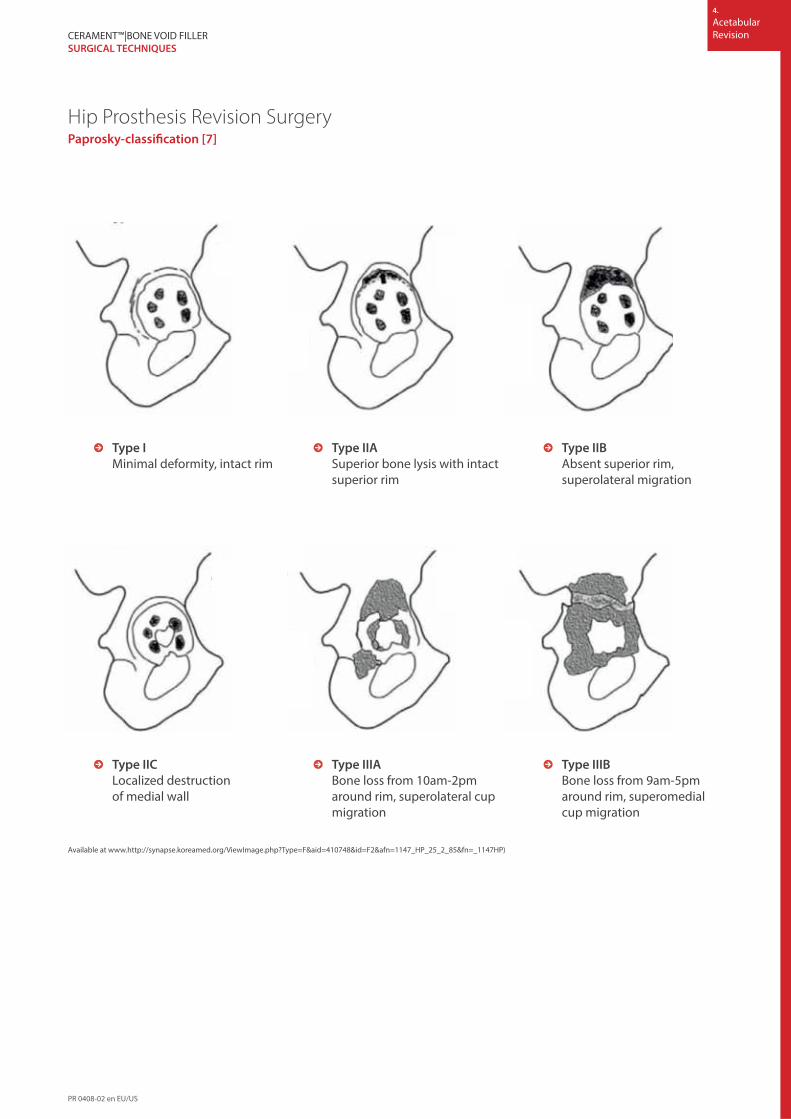
Hip Prosthesis Revision SurgeryPaprosky-classifi cation [7]
Available at www.http://synapse.koreamed.org/ViewImage.php?Type=F&aid=410748&id=F2&afn=1147_HP_25_2_85&fn=_1147HP)
CERAMENT™|BONE VOID FILLER
SURGICAL TECHNIQUES
R Type I
Minimal deformity, intact rim
R Type IIC
Localized destruction
of medial wall
R Type IIA
Superior bone lysis with intact
superior rim
R Type IIIA
Bone loss from 10am-2pm
around rim, superolateral cup
migration
R Type IIB
Absent superior rim,
superolateral migration
R Type IIIB
Bone loss from 9am-5pm
around rim, superomedial
cup migration
PR 0408-02 en EU/US
4.
Acetabular
Revision
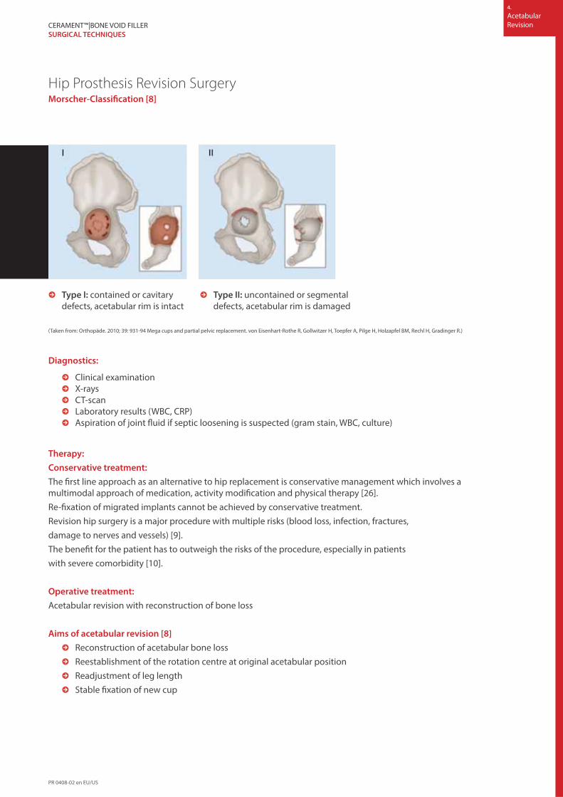
Hip Prosthesis Revision SurgeryMorscher-Classifi cation [8]
R Type I: contained or cavitary
defects, acetabular rim is intact
R Type II: uncontained or segmental
defects, acetabular rim is damaged
(Taken from: Orthopäde. 2010; 39: 931-94 Mega cups and partial pelvic replacement. von Eisenhart-Rothe R, Gollwitzer H, Toepfer A, Pilge H, Holzapfel BM, Rechl H, Gradinger R.)
CERAMENT™|BONE VOID FILLER
SURGICAL TECHNIQUES
Diagnostics:
R Clinical examination
R X-rays
R CT-scan
R Laboratory results (WBC, CRP)
R Aspiration of joint fl uid if septic loosening is suspected (gram stain, WBC, culture)
Therapy:
Conservative treatment:
The fi rst line approach as an alternative to hip replacement is conservative management which involves a
multimodal approach of medication, activity modifi cation and physical therapy [26].
Re-fi xation of migrated implants cannot be achieved by conservative treatment.
Revision hip surgery is a major procedure with multiple risks (blood loss, infection, fractures,
damage to nerves and vessels) [9].
The benefi t for the patient has to outweigh the risks of the procedure, especially in patients
with severe comorbidity [10].
Operative treatment:
Acetabular revision with reconstruction of bone loss
Aims of acetabular revision [8]
R Reconstruction of acetabular bone loss
R Reestablishment of the rotation centre at original acetabular position
R Readjustment of leg length
R Stable fi xation of new cup
PR 0408-02 en EU/US
4.
Acetabular
Revision
Hip Prosthesis Revision SurgeryMorscher-Classifi cation [8]
CERAMENT™|BONE VOID FILLER
SURGICAL TECHNIQUES
Steps of the surgical treatment
Removal of loosened cup: The primary incision and approach should be used. Membranes and fi brotic soft tissues
are excised and the cup is removed.
Sometimes the cup is better integrated than radiographically suspected. In those cases a curved osteotome or a
pneumatic impact wrench [11] can be used. If the cap was fi xed additional with screws, screws and other devices
will have to be removed as well.
Next the acetabulum is debrided with diff erent curettes, bone nibblers or high speed burrs. The sclerotic bone
should be removed, but the ventral and dorsal acetabular rim has to remain intact.
Intraoperative evaluation of the acetabulum:
R After debridement the remaining bone stock of the acetabulum has to be evaluated.
R A cup trial is used to decide if press-fi t technique gives enough stability.
R Following the suggestions from Morscher et. al. [8] and based on their classifi cation, treatment options
for two diff erent acetabular situations are described below.
Situation I:
R Morscher Type I acetabular bone loss:
- Contained or cavitary defects, acetabular rim is intact
- Intra-operative evaluation with cup trial positive: press- fi t can be achieved
Procedure: Implantation of press-fi t cup and reconstruction of
bone loss with morsellised allograft
R Debrided bone cysts and bone voids can be fi lled with morsellised allograft or bone graft substitute, a
press-fi t cup is implanted and intrinsic stability achieved [12].
R
PR 0408-02 en EU/US
4.
Acetabular
Revision
Hip Prosthesis Revision SurgeryMorscher-Classifi cation [8]
CERAMENT™|BONE VOID FILLER
SURGICAL TECHNIQUES
Situation II:
R Morscher Type II acetabular bone loss:
- Uncontained or segmental defects, acetabular rim is destroyed
- Intra-operative evaluation with test-cup negative: press-fi t cannot be achieved
Procedure: Implantation of a reinforcement ring (Müller or Ganz) or an
anti- protrusio cage (Burch-Schneider) plus cemented cup and reconstruction
of bone loss with morsellised allograft
R If segmental defects of the acetabular rim, large bone voids or a non-spherical form of the acetabulum
do not allow the implantation of a press-fi t cup, reinforcement acetabular rings, for example established
by Müller [13, 14] and Ganz [15] or anti-protrusio cages by Burch-Schneider [16, 17] are indicated.
R The bone loss of the acetabulum is reconstructed with morsellised allograft and impacted with a cup trial.
The reinforcement rings or anti-protrusio cages are fi xed with cancellous screws.
R The devices give mechanical support to the cup [16]. If the ring or cage is placed correctly, the rotation
centre of the hip is reconstructed at its original anatomical position.
R A polyethylene cup is fi xed with bone cement (PMMA) onto the reinforcement ring or the anti-protrusio
cage. PMMA-leakage behind the device increases the stability of the cage-cup combination.
Reconstruction in acetabular bone loss
There is still a lack of evidence to determine the best method for reconstructing acetabular bone loss [18-25].
Diff erent treatment options exist, some are listed below:
R autologous bone graft [18,19]
R impacted morsellised cancellous bone allografts (impact grafting) [4, 20]
R bulk allograft bone [21]
R freeze-dried, irradiated and chemically-treated allograft vitalised with autologous marrow
bone substitutes [22]
R demineralized bone matrix [23]
R bone substitutes [24, 25]
When following Morscher’s ideas and principles to reconstruct acetabular bone loss [8], it is possible to use
CERAMENT™|BONE VOID FILLER in conjunction with or instead of morsellised allograft.
R
PR 0408-02 en EU/US
4.
Acetabular
RevisionCERAMENT™|BONE VOID FILLER
SURGICAL TECHNIQUES
Literature
1. Sporer SM, Paprosky WG, O’Rourke MR. Managing bone loss in acetabular revision. Instr Course Lect. 2006; 55: 287–297.
2. Aqua- Institut für angewandte Qualitätsförderung und Forschung im Gesundheitswesen GmbH. Bundesauswertung zum
Erfassungsjahr 2013 – 17/2 Hüft-Endoprothesen Erstimplantation, https://www.sqg.de/downloads/Bundesauswertungen
/2013/bu_Gesamt_17N2-HUEFT-TEP_2013.pdf
3. Jämsen E1, Furnes O, Engesaeter LB, Konttinen YT, Odgaard A, Stefánsdóttir A, Lidgren L. Prevention of deep infection in joint
replacement surgery. Acta Orthop. 2010; 81: 660-666
4. Slooff TJ, Schreurs BW, Gardeniers JW, Buma P. Rekonstruktion des Acetabulums mit impaktierten Knochentransplantaten und
Zement. In Duparc J.: Chirurgische Techniken in Orthopädie und Traumatologie, 2005, Elsevier, München, 301-307
5. Engelbrecht DJ, Weber FA, Sweet MB, Jakim I. Long-term results of revision total hip arthroplasty.
J Bone Joint Surg Br. 1990; 72: 41-45
6. D’Antonio JA, Capello WN, Borden LS, Bargar WL, Bierbaum BF, Boettcher WG, Steinberg ME, Stulberg SD, Wedge JH.
Classifi cation and management of acetabular abnormalities in total hip arthroplasty. Clin Orthop Relat Res. 1989; 243: 126-137
7. Paprosky WG, Perona PG, Lawrence JM. Acetabular defect classifi cation and surgical reconstruction in revision arthroplasty.
A 6-year follow-up evaluation. J Arthroplasty. 1994; 9: 33-44
8. Morscher EW, Elke R, Berli B. Klassifi kation und Behandlung von Acetabulumdefekten. In Duparc J.: Chirurgische Techniken
in Orthopädie und Traumatologie, 2005, Elsevier, München, 293-299
9. Bischel O, Seeger JB, Krüger M, Bitsch RG. Multiple Acetabular Revisions in THA - Poor Outcome Despite Maximum Eff ort.
Open Orthop J. 2012; 6: 488-494.
10. Elke R et al. Revisionsendoprothetik. In: Tschauner [Hrsg]. Orthopädie und orthopädische Chirurgie. 2004. Thieme, Stuttgart,
New York, S.383 – 384
11. Anspach WE 3rd, Lachiewicz PF. A new technique for removal of the total hip arthroplasty acetabular component.
Clin Orthop Relat Res. 1991; 268: 152-156.
12. Morscher E, Berli B, Jockers W, Schenk R. Rationale of a fl exible press fi t cup in total hip replacement. 5-year followup
in 280 procedures. Clin Orthop Relat Res. 1997; 341: 42-50.
13. Rosson J, Schatzker J. The use of reinforcement rings to reconstruct defi cient acetabula. J Bone Joint Surg Br. 1992; 74: 716-720.
14. Zehntner MK, Ganz R. Midterm results (5.5-10 years) of acetabular allograft reconstruction with the acetabular reinforcement
ring during total hip revision. J Arthroplasty. 1994; 9:469-479
15. Uchiyama K, Takahira N, Fukushima K, Yamamoto T, Moriya M, Itoman M. Radiological evaluation of allograft reconstruction in
acetabulum with Ganz reinforcement ring in revision total hip replacement. J Orthop Sci. 2010; 15: 764-771
16. Gill TJ, Sledge JB, Müller ME. The Bürch-Schneider anti-protrusio cage in revision total hip arthroplasty: indications,
principles and long-term results. J Bone Joint Surg Br. 1998; 80: 946-953.
17. Perka C, Ludwig R. Reconstruction of segmental defects during revision procedures of the acetabulum with the
Burch-Schneider anti-protrusio cage. J Arthroplasty. 2001; 16: 568-574
18. Figueras Coll G, Salazar Fernandez de Erenchu J, Roca Burniol J. Results of acetabular wiremesh and autograft in protrusio
acetabuli. Hip Int. 2008; 18: 23-28
19. Welten ML, Schreurs BW, Buma P, Verdonschot N, Slooff TJ. Acetabular reconstruction with impacted morsellised cancellous
bone autograft and cemented primary total hip arthroplasty: a 10- to 17-year follow-up study. J Arthroplasty. 2000; 15: 819-824
20. Schreurs BW, Slooff TJ, Buma P, Gardeniers JW, Huiskes R. Acetabular reconstruction with impacted morsellised cancellous bone
graft and cement. A 10- to 15-year follow-up of 60 revision arthroplasties. J Bone Joint Surg Br. 1998; 80: 391-395
21. Kerboull M, Hamadouche M, Kerboull L. The Kerboull acetabular reinforcement device in major acetabular reconstructions.
Clin Orthop Relat Res. 2000; 378: 155-68.
22. Ochs BG, Schmid U, Rieth J, Ateschrang A, Weise K, Ochs U. Acetabular bone reconstruction in revision arthroplasty:
a comparison of freeze-dried, irradiated and chemically-treated allograft vitalised with autologous marrow versus frozen
non-irradiated allograft. J Bone Joint Surg Br. 2008; 90: 1164-1171
23. Patil N, Hwang K, Goodman SB. Cancellous impaction bone grafting of acetabular defects in complex primary and revision
total hip arthroplasty. Orthopedics. 2012; 35: e306-312
24. Schwartz C, Vautrin M. Phosphocalcium ceramics are effi cient in the management of severe acetabular loss in revision hip
arthroplasties. A 22 cases long-term follow-up study. Eur J Orthop Surg Traumatol. 2014 May
25. Whitehouse MR, Dacombe PJ, Webb JC, Blom AW. Impaction grafting of the acetabulum with ceramic bone graft substitute:
high survivorship in 43 patients with a mean follow-up period of 4 years. Acta Orthop. 2013; 84: 371-376
26. http://en.wikipedia.org/wiki/Hip_replacement
PR 0408-02 en EU/US
4.
Acetabular
Revision
Fig. Images reproduced by kind permission of Dr Lawrence DiDomenico, Adjunct Professor,
Ohio College of Podiatric Medicine ,Youngstown, Ohio , USA.
Acetabular revisionImplantation of an anti-protrusio cage (Bruch-Schneider) plus cemented cup and reconstruction of
acetabular bone loss with an uncontained or segmental defect (acetabular rim is not intact)
Surgical positioning and preoperative procedures:
R Mark the site of surgery while informed consent of patient is obtained
R The use of a radiolucent table is recommended
R Prepare mobile C-arm
R Antibiotic prophylaxis 30 min before incision [1]
R Usually the primary incision is used for the revision.
R Place the patient in a lateral or supine position according to the planed approach [2]
R Skin preparation and draping as usual
R Team time-out
Surgery:
R The primary incision and approach should be used
for the revision [2]
R Fibrotic or necrotic soft tissue and the (neo-) capsule
are excised [2]
R The loosened cup is removed. If the cup is more stable
than suspected, curved osteotomes may be used [3]
R The acetabulum is debrided with diff erent curettes,
bone nibblers or high speed burrs
R Remove all screws, implants, PE-wear, bone cement
(PMMA) and debris [2]
R Take samples for bacterial cultures and histological
examination [4]
R After debridement the remaining bone stock of the
acetabulum has to be evaluated. A cup trial is used to
decide, if press-fi t can be achieved [2]
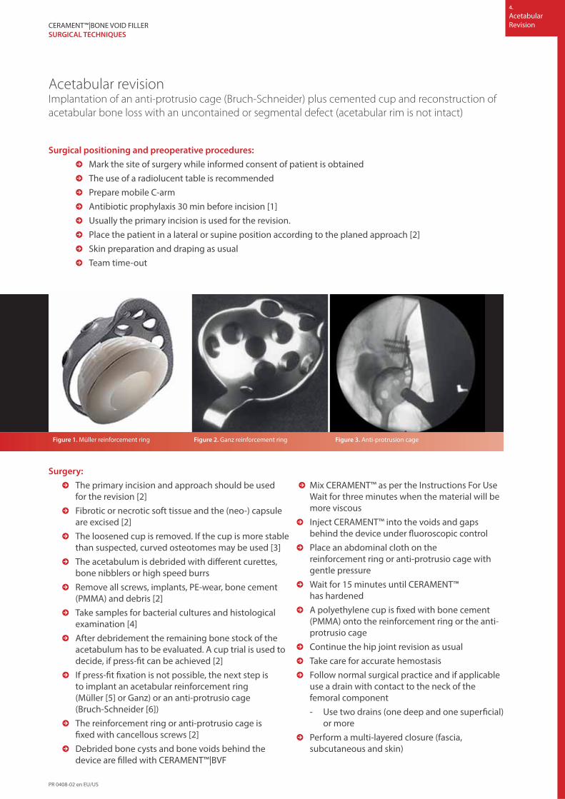
R If press-fi t fi xation is not possible, the next step is
to implant an acetabular reinforcement ring
(Müller [5] or Ganz) or an anti-protrusio cage
(Bruch-Schneider [6])
R The reinforcement ring or anti-protrusio cage is
fi xed with cancellous screws [2]
R Debrided bone cysts and bone voids behind the
device are fi lled with CERAMENT™|BVF
R Mix CERAMENT™ as per the Instructions For Use
Wait for three minutes when the material will be
more viscous
R Inject CERAMENT™ into the voids and gaps
behind the device under fl uoroscopic control
R Place an abdominal cloth on the
reinforcement ring or anti-protrusio cage with
gentle pressure
R Wait for 15 minutes until CERAMENT™
has hardened
R A polyethylene cup is fi xed with bone cement
(PMMA) onto the reinforcement ring or the anti-
protrusio cage
R Continue the hip joint revision as usual
R Take care for accurate hemostasis
R Follow normal surgical practice and if applicable
use a drain with contact to the neck of the
femoral component
- Use two drains (one deep and one superfi cial)
or more
R Perform a multi-layered closure (fascia,
subcutaneous and skin)
Figure 1. Müller reinforcement ring Figure 2. Ganz reinforcement ring Figure 3. Anti-protrusion cage
CERAMENT™|BONE VOID FILLER
SURGICAL TECHNIQUES
PR 0408-02 en EU/US
4.
Acetabular
Revision
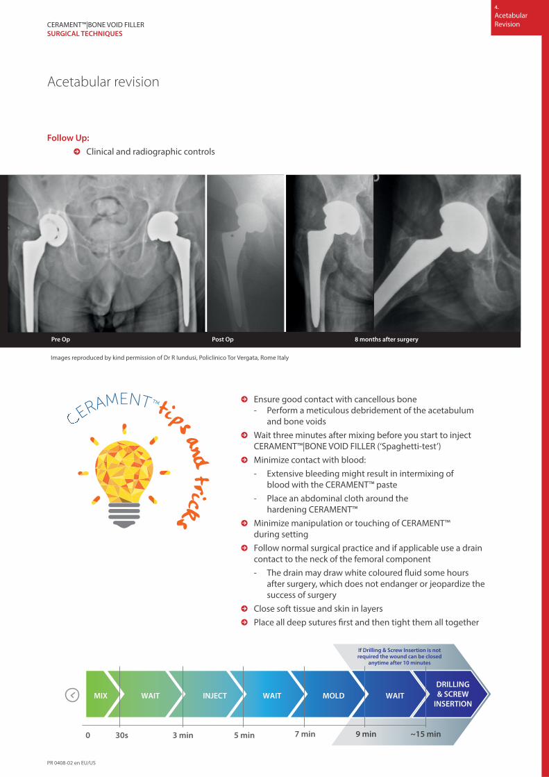
Follow Up:
R Clinical and radiographic controls
R Ensure good contact with cancellous bone
- Perform a meticulous debridement of the acetabulum
and bone voids
R Wait three minutes after mixing before you start to inject
CERAMENT™|BONE VOID FILLER (‘Spaghetti-test’)
R Minimize contact with blood:
- Extensive bleeding might result in intermixing of
blood with the CERAMENT™ paste
- Place an abdominal cloth on the reinforcement ring or anti-
protrusio cage with gentle pressure
R Follow normal surgical practice and if applicable use a drain with
contact to the neck of the femoral component
- The drain may draw white coloured fl uid some hours
after surgery, which does not endanger or jeopardise
the success of surgery
R Close soft tissue and skin two layers: Place all deep sutures fi rst
and then tighten them all together
Acetabular revision
3 min 5 min30s0 7 min 9 min ~15 min
If Drilling & Screw Insertion is not required the wound can be closed
anytime after 10 minutes
MIX WAIT WAIT
DRILLING
& SCREW
INSERTIONINJECT WAIT MOLD
a) b)
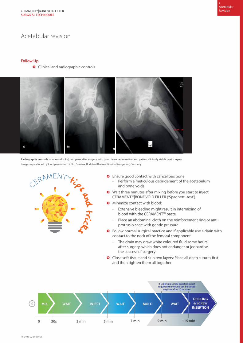
Radiographic controls :a) one and b & c) two years after surgery, with good bone regeneration and patient clinically stable post surgery.
Images reproduced by kind permission of Dr J Svacina, Bodden-Kliniken Ribnitz-Damgarten, Germany
CERAMENT™|BONE VOID FILLER
SURGICAL TECHNIQUES
PR 0408-02 en EU/US
4.
Acetabular
RevisionCERAMENT™|BONE VOID FILLER
SURGICAL TECHNIQUES
Literature
1. Bratzler DW, Houck PM. Clin Infect Dis. 2004; 38: 1706-1715
2. Morscher EW, Elke R, Berli B.. In Duparc J.: Chirurgische Techniken in Orthopädie und Traumatologie, 2005,
Elsevier, München, 293-299
3. Anspach WE 3rd, Lachiewicz PF. Clin Orthop Relat Res. 1991; 268: 152-156.
4. Atkins BL, Athanasou N, Deeks Jj et al. J Clin Microbiol 1998; 36: 2932–2939
5. Rosson J, Schatzker J. J Bone Joint Surg Br. 1992; 74: 716-720.
6. Gill TJ, Sledge JB, Müller ME. J Bone Joint Surg Br. 1998; 80: 946-953.
PR 0408-02 en EU/US

4.
Acetabular
Revision
Fig. Images reproduced by kind permission of Dr Lawrence DiDomenico, Adjunct Professor,
Ohio College of Podiatric Medicine ,Youngstown, Ohio , USA.
Implantation of a press-fi t cup and reconstruction of bone loss in
a contained acetabular defect (acetabular rim is intact)
Surgical positioning and preoperative procedures:
R Mark the site of surgery while informed consent of patient is obtained
R The use of a radiolucent table is recommended
R Prepare mobile C-arm
R Antibiotic prophylaxis 30 min before incision [1]
R The primary incision is normally used for the revision
R Place the patient in a lateral or supine position according to the planned approach [2]
R Skin preparation and draping as usual
R Team time-out
Surgery:
R The primary incision and approach should be used
for the revision [2]
R Fibrotic or necrotic soft tissue and the (neo-) capsule
are excised [2]
R The loosened cup is removed. (Fig. 1) If the cup is
more stable than suspected, curved osteotomes
can be used [3]
R The acetabulum is debrided with diff erent curettes,
bone nibblers or high speed burrs
R Remove all screws, implants, PE-wear, bone cement
(PMMA) and debris [2] (Fig.2)
R Take samples for bacterial cultures and
histological examination [4]
R Sclerotic bone is removed, the ventral and dorsal
acetabular rim should remain intact. (Fig. 3)
R After debridement the remaining bone stock of
the acetabulum is evaluated.
R A cup trial is used to decide, if press-fi t fi xation
can be achieved [2]
R If press-fi t fi xation is possible fi ll debrided
bone cysts and bone voids with
CERAMENT™|BONE VOID FILLER (Fig. 4)
R Mix CERAMENT™ as per the Instructions For Use
R Wait for three minutes when the material will be
more viscous
R Inject CERAMENT™ in the voids of the
acetabulum
R Place an abdominal cloth around the
hardening CERAMENT™
R In this indication you don‘t have to wait for the
CERAMENT™ to set
R Implant a press-fi t cup
R Continue the hip joint revision as usual
R Take care for accurate hemostasis
R Use two drains (one deep and one superfi cial)
or more
R Perform a multi-layered closure (fascia,
subcutaneous and skin)
Figure 1. Figure 2. Figure 3. Figure 4.
CERAMENT™|BONE VOID FILLER
SURGICAL TECHNIQUES
Images reproduced by kind permission of Dr R Iundusi, Policlinico Tor Vergata, Rome Italy
PR 0408-02 en EU/US
4.
Acetabular
Revision
Follow Up:
R Clinical and radiographic controls
R Ensure good contact with cancellous bone
- Perform a meticulous debridement of the acetabulum
and bone voids
R Wait three minutes after mixing before you start to inject
CERAMENT™|BONE VOID FILLER (‘Spaghetti-test’)
R Minimize contact with blood:
- Extensive bleeding might result in intermixing of
blood with the CERAMENT™ paste
- Place an abdominal cloth around the
hardening CERAMENT™
R Minimize manipulation or touching of CERAMENT™
during setting
R Follow normal surgical practice and if applicable use a drain
contact to the neck of the femoral component
- The drain may draw white coloured fl uid some hours
after surgery, which does not endanger or jeopardize the
success of surgery
R Close soft tissue and skin in layers
R Place all deep sutures fi rst and then tight them all together
Acetabular revision
3 min 5 min30s0 7 min 9 min ~15 min
If Drilling & Screw Insertion is not required the wound can be closed
anytime after 10 minutes
MIX WAIT WAIT
DRILLING
& SCREW
INSERTIONINJECT WAIT MOLD
Pre Op Post Op 8 months after surgery
Images reproduced by kind permission of Dr R Iundusi, Policlinico Tor Vergata, Rome Italy
CERAMENT™|BONE VOID FILLER
SURGICAL TECHNIQUES
PR 0408-02 en EU/US

4.
Acetabular
RevisionCERAMENT™|BONE VOID FILLER
SURGICAL TECHNIQUES
Literature
1. Bratzler DW, Houck PM. Clin Infect Dis. 2004; 38: 1706-1715.
2. Morscher EW, Elke R, Berli B.. In Duparc J.: Chirurgische Techniken in Orthopädie und Traumatologie, 2005,
Elsevier, München, 293-299.
3. Anspach WE 3rd, Lachiewicz PF. Clin Orthop Relat Res. 1991; 268: 152-156.
4. Atkins BL, Athanasou N, Deeks Jj et al. J Clin Microbiol 1998; 36: 2932–2939.
PR 0408-02 en EU/US