Embed Size (px)
Citation preview

37
3
3.1 HISToRICAL INTRoDUCTIoN
The history of allergen nomenclature dates to the time when extracts of allergen sources were fractionated using a vari-ety of classical biochemical separation techniques and the active (most allergenic) fraction was usually named accord-ing to the whim of the investigator. In the 1940s and 1950s, attempts were made to purify pollen and house dust allergens, using phenol extraction, salt precipitation, and electropho-retic techniques. In the 1960s, ion exchange and gel filtration media were introduced and ragweed “antigen E” was the first allergen to be purified [1]. This allergen was named so by King and Norman because it was one of the five precipitation lines (labeled A–E) that reacted with rabbit polyclonal anti-bodies to ragweed in Ouchterlony immunodiffusion tests. Following purification, precipitation line E, or “antigen E” was shown to be a potent allergen. Later, Marsh, working in Cambridge, England, isolated an important allergen from rye grass pollen (Lolium perenne) and used the name “Rye 1” to indicate that this was the first allergen purified from this spe-cies [2,3]. In the 1970s, many allergens were purified from ragweed, rye grass, insect venoms, and other sources. The field was led by the laboratory of the late Dr. David Marsh, who had moved to the Johns Hopkins University, Baltimore,
Maryland. At Johns Hopkins, the ragweed allergens Ra3, Ra4, Ra5, and Ra6, and the rye grass allergens Rye 2 and Rye 3 were isolated and used for immunological and genetic studies of hay fever [4–6]. At the same time, Ohman et al. identified Cat-I, the major cat allergen [7]. Elsayed purified allergen M from codfish [8,9].
The state of the art in the early 1970s was reviewed in a chapter by Marsh in a seminal book, The Antigens, which described the molecular properties of allergens, the fac-tors that influenced allergenicity, the immune response to allergens, and immunogenetic studies of immunoglobulin E (IgE) responses to purified pollen allergens [10]. This chapter provided the first clear definition of a “major” aller-gen, which Marsh defined as a highly purified allergen that induced immediate skin test responses in >90% of aller-gic individuals, in contrast to a “minor” allergen, to which <20% of patients reacted with skin test responses. Today, a major allergen is generally regarded as one to which >50% of patients with an allergy to its source react [11].
With the introduction of crossed immunoelectrophore-sis (CIE) and crossed radioimmunoelectrophoresis (CRIE) for allergen identification by Løwenstein and colleagues in Scandinavia, there was a tremendous proliferation of the
Allergen Nomenclature
Heimo BreitenederMedical University of Vienna
Martin D. ChapmanIndoor Biotechnologies, Inc.
CoNTeNTS
3.1 Historical Introduction....................................................................................................................................................... 373.1.1 Three Men in a Boat .............................................................................................................................................. 38
3.2 The Revised Allergen Nomenclature ................................................................................................................................. 383.2.1 Allergens ................................................................................................................................................................ 38
3.2.1.1 Inclusion Criteria .................................................................................................................................... 383.2.1.2 Resolving Ambiguities in the Nomenclature .......................................................................................... 39
3.2.2 Isoallergens, Isoforms, and Variants ..................................................................................................................... 413.3 Nomenclature for Allergen Genes and Recombinant or Synthetic Peptides ..................................................................... 443.4 Future Perspectives of Allergen Nomenclature ................................................................................................................. 44
3.4.1 Protein Family Membership .................................................................................................................................. 443.5 The IUIS Subcommittee on Allergen Nomenclature ........................................................................................................ 45
3.5.1 Allergen Databases ................................................................................................................................................ 453.6 Concluding Remarks ......................................................................................................................................................... 45Salient Points .............................................................................................................................................................................. 47Acknowledgments ....................................................................................................................................................................... 48References ................................................................................................................................................................................... 48
Copyrighted Material - Taylor and Francis

38 Allergens and Allergen Immunotherapy
number of antigenic proteins and CIE/CRIE peaks identi-fied as allergens. Typically, 10–50 peaks could be detected in a given allergen source based on reactivity with rabbit polyclonal antibodies or IgE antibodies [6,11–13]. These peaks were given a multitude of names such as Dp5, Dp42, Ag12, and so on. Inevitably, the same allergens were referred to by different names in different laboratories; for example, mite antigen P1 was also known as Dp42 or Ag12. It was clear that a unified nomenclature was urgently needed.
3.1.1 Three Men in a BoaT
The origins of the systematic allergen nomenclature can be traced to a meeting among Drs. David Marsh (at that time, Johns Hopkins University, Baltimore, MD, USA), Henning Løwenstein (at that time, University of Copenhagen, Denmark), and Thomas Platts-Mills (at that time, Clinical Research Centre, Harrow, UK) on a boat ride on Lake Constance (Bodensee), Konstanz, Germany, during the 13th Symposium of the Collegium Internationale Allergologicum in July 1980 [14]. The idea was to develop a systematic aller-gen nomenclature based on the Linnaean binomial nomen-clature for naming all living things, with added numerals to indicate different allergens from the same source. It was decided to adopt a system whereby the allergen was named based on the first three letters of the genus and the first letter of the species (both in italics) followed by a Roman numeral to indicate the allergen in the chronological order of purifica-tion. Thus ragweed antigen E became Ambrosia artemisiifo-lia allergen I or Amb a I and Rye 1 became Lolium perenne allergen I or Lol p I.
An allergen nomenclature subcommittee was formed under the auspices of the World Health Organization (WHO) and International Union of Immunological Societies (IUIS) and the criteria for including allergens in the systematic nomenclature were established. These included strict criteria for biochemical purity, as well as criteria for determining the allergenic activ-ity of the purified protein. A committee chaired by Marsh, and including Henning Løwenstein, Thomas Platts-Mills, Te Piao King (Rockefeller University, New York), and Larry Goodfriend (McGill University, Montreal, Canada) prepared a list of allergens that fulfilled the inclusion criteria and estab-lished a process for investigators to submit names of newly identified allergens. The original list, published in the Bulletin of the WHO in 1986, included 27 highly purified allergens from grass, weed, tree pollens, and house dust mites [15].
The systematic allergen nomenclature was quickly adopted by allergy researchers and proved to be a great suc-cess. It was logical, easily understood, and readily assimi-lated by allergologists and other clinicians who were not directly involved with the details of allergen immunochem-istry. The nomenclature and allergen designations, such as Der p I, Fel d I, Lol p I, and Amb a I, were used at scien-tific meetings and in the literature, and expanded rapidly to include newly isolated allergens.
3.2 THe ReVISeD ALLeRGeN NoMeNCLATURe
3.2.1 allergens
The widespread use of molecular cloning techniques to iden-tify allergens in the late 1980s and 1990s led to an expo-nential increase in the number of allergens described. Many allergen nucleotide sequences were obtained by cDNA clon-ing or PCR amplifications and it soon became apparent that the use of Roman numerals was unwieldy, for example, Lol p I through Lol p XI [16,17]. The use of italics to denote a purified protein was inconsistent with the nomenclature used in bacterial genetics and the human leukocyte antigen (HLA) system, where italicized names denote a gene product and a regular typeface indicates expressed proteins. In 1994, the allergen nomenclature was revised so that the allergen desig-nation was shown in regular type. Arabic numerals replaced the Roman ones. Thus Amb a I, Lol p I, and Der p I of the original 1986 nomenclature are now referred to as Amb a 1, Lol p 1, and Der p 1 in the current nomenclature, which has been published in several scientific journals [18–20].
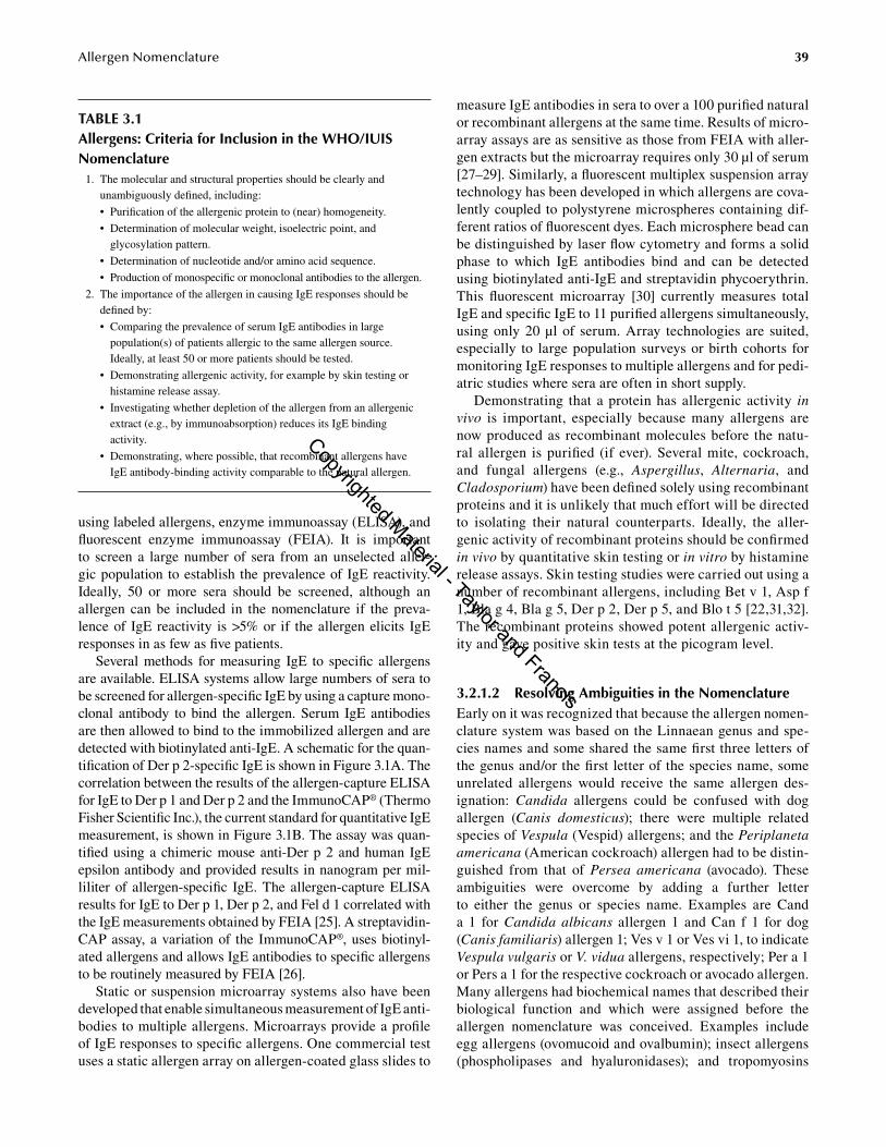
3.2.1.1 Inclusion CriteriaA key part of the systematic WHO/IUIS nomenclature is that the allergen should satisfy biochemical criteria, which define the molecular structure of the protein, and immuno-logic criteria, which define its importance as an allergen. Originally, the biochemical criteria were based on protein purity, established for example by sodium dodecyl sulfate-polyacrylamide gel electrophoresis, isoelectric focusing, or high-pressure liquid chromatography, and physicochemi-cal properties including molecular weight, isoelectric point, and N-terminal amino acid sequence [20]. Today, the full nucleotide or amino acid sequence is generally required. An outline of the inclusion criteria is shown in Table 3.1. A more detailed list of requirements for the inclusion of an allergen in the WHO/IUIS nomenclature can be found in the allergen submission form (http://www.allergen.org/submission.php). An important aspect of these criteria is that the submis-sion information should provide an unambiguous descrip-tion whereby other investigators can identify the very same allergen and make comparative studies. Originally, this was achieved by purifying the protein, developing monospecific or monoclonal antibodies to it, and providing either the allergen or antibodies to other researchers for verification. Nucleotide and amino acid sequencing unambiguously iden-tifies the allergen and enables sequence variation between cDNA clones of the same allergen definition [21–24]. Allergen preparations, sequences, and antibodies submitted for inclusion in the systematic nomenclature are expected to be made available to other investigators for research studies.
A second set of inclusion criteria involves demonstrating that the purified allergen has allergenic activity, both in vitro and in vivo. Researchers use a variety of techniques for measuring IgE antibodies in vitro, including radioallergosor-bent-based techniques, immunoblotting, radioimmunoassays
Copyrighted Material - Taylor and Francis
39Allergen Nomenclature
using labeled allergens, enzyme immunoassay (ELISA), and fluorescent enzyme immunoassay (FEIA). It is important to screen a large number of sera from an unselected aller-gic population to establish the prevalence of IgE reactivity. Ideally, 50 or more sera should be screened, although an allergen can be included in the nomenclature if the preva-lence of IgE reactivity is >5% or if the allergen elicits IgE responses in as few as five patients.
Several methods for measuring IgE to specific allergens are available. ELISA systems allow large numbers of sera to be screened for allergen-specific IgE by using a capture mono-clonal antibody to bind the allergen. Serum IgE anti bodies are then allowed to bind to the immobilized allergen and are detected with biotinylated anti-IgE. A schematic for the quan-tification of Der p 2-specific IgE is shown in Figure 3.1A. The correlation between the results of the allergen-capture ELISA for IgE to Der p 1 and Der p 2 and the ImmunoCAP® (Thermo Fisher Scientific Inc.), the current standard for quantitative IgE measurement, is shown in Figure 3.1B. The assay was quan-tified using a chimeric mouse anti-Der p 2 and human IgE epsilon antibody and provided results in nanogram per mil-liliter of allergen-specific IgE. The allergen-capture ELISA results for IgE to Der p 1, Der p 2, and Fel d 1 correlated with the IgE measurements obtained by FEIA [25]. A streptavidin-CAP assay, a variation of the ImmunoCAP®, uses biotinyl-ated allergens and allows IgE antibodies to specific allergens to be routinely measured by FEIA [26].
Static or suspension microarray systems also have been developed that enable simultaneous measurement of IgE anti-bodies to multiple allergens. Microarrays provide a profile of IgE responses to specific allergens. One commercial test uses a static allergen array on allergen-coated glass slides to
measure IgE antibodies in sera to over a 100 purified natural or recombinant allergens at the same time. Results of micro-array assays are as sensitive as those from FEIA with aller-gen extracts but the microarray requires only 30 µl of serum [27–29]. Similarly, a fluorescent multiplex suspension array technology has been developed in which allergens are cova-lently coupled to polystyrene microspheres containing dif-ferent ratios of fluorescent dyes. Each microsphere bead can be distinguished by laser flow cytometry and forms a solid phase to which IgE antibodies bind and can be detected using biotinylated anti-IgE and streptavidin phycoerythrin. This fluorescent microarray [30] currently measures total IgE and specific IgE to 11 purified allergens simultaneously, using only 20 µl of serum. Array technologies are suited, especially to large population surveys or birth cohorts for monitoring IgE responses to multiple allergens and for pedi-atric studies where sera are often in short supply.
Demonstrating that a protein has allergenic activity in vivo is important, especially because many allergens are now produced as recombinant molecules before the natu-ral allergen is purified (if ever). Several mite, cockroach, and fungal allergens (e.g., Aspergillus, Alternaria, and Cladosporium) have been defined solely using recombinant proteins and it is unlikely that much effort will be directed to isolating their natural counterparts. Ideally, the aller-genic activity of recombinant proteins should be confirmed in vivo by quantitative skin testing or in vitro by histamine release assays. Skin testing studies were carried out using a number of recombinant allergens, including Bet v 1, Asp f 1, Bla g 4, Bla g 5, Der p 2, Der p 5, and Blo t 5 [22,31,32]. The recombinant proteins showed potent allergenic activ-ity and gave positive skin tests at the picogram level.
3.2.1.2 Resolving Ambiguities in the NomenclatureEarly on it was recognized that because the allergen nomen-clature system was based on the Linnaean genus and spe-cies names and some shared the same first three letters of the genus and/or the first letter of the species name, some unrelated allergens would receive the same allergen des-ignation: Candida allergens could be confused with dog allergen (Canis domesticus); there were multiple related species of Vespula (Vespid) allergens; and the Periplaneta americana (American cockroach) allergen had to be distin-guished from that of Persea americana (avocado). These ambiguities were overcome by adding a further letter to either the genus or species name. Examples are Cand a 1 for Candida albicans allergen 1 and Can f 1 for dog (Canis familiaris) allergen 1; Ves v 1 or Ves vi 1, to indicate Vespula vulgaris or V. vidua allergens, respectively; Per a 1 or Pers a 1 for the respective cockroach or avocado allergen. Many allergens had biochemical names that described their biological function and which were assigned before the allergen nomenclature was conceived. Examples include egg allergens (ovomucoid and ovalbumin); insect allergens (phospholipases and hyaluronidases); and tropomyosins
TABLe 3.1Allergens: Criteria for Inclusion in the WHo/IUIS Nomenclature 1. The molecular and structural properties should be clearly and
unambiguously defined, including:
• Purification of the allergenic protein to (near) homogeneity.
• Determination of molecular weight, isoelectric point, and glycosylation pattern.
• Determination of nucleotide and/or amino acid sequence.
• Production of monospecific or monoclonal antibodies to the allergen.
2. The importance of the allergen in causing IgE responses should be defined by:
• Comparing the prevalence of serum IgE antibodies in large population(s) of patients allergic to the same allergen source. Ideally, at least 50 or more patients should be tested.
• Demonstrating allergenic activity, for example by skin testing or histamine release assay.
• Investigating whether depletion of the allergen from an allergenic extract (e.g., by immunoabsorption) reduces its IgE binding activity.
• Demonstrating, where possible, that recombinant allergens have IgE antibody-binding activity comparable to the natural allergen.
Copyrighted Material - Taylor and Francis

40 Allergens and Allergen Immunotherapy
from shrimps, mites, and cockroaches. Sequence homology searches have assigned allergens to particular protein fami-lies and have provided important clues to their biological function. To some extent, allergens segregate among pro-tein families according to whether they are indoor aller-gens, outdoor allergens, plant or animal food allergens, or injected allergens:
• Indoor allergens (e.g., animal dander; fecal particles from mites and cockroaches; mold spores):
– Proteolytic enzymes (serine and cysteine prote-ases), lipocalins (ligand-binding proteins), tropo-myosins, albumins, calcium-binding proteins, and protease inhibitors [22,33].
• Outdoor allergens (e.g., pollens from grasses, trees, and weeds; mold spores): – Plant pathogenesis-related (PR-10) proteins,
pectate lyases, β-expansins, calcium-binding proteins (polcalcins), defensin-like proteins, and trypsin inhibitors [21,23,34,35].
Streptavidin peroxidase
Biotinylatedanti-human IgE
Chimeric antibody
Der p 2
Anti-DpX
A
10,000
1,000
100
10
1
0.1
GMClass 0
0.47Class 1
0.57Class 2
0.69Class 3
1.86CAP system class
Chi
mer
ic E
LISA
toD
er p
1 +
Der
p 2
IgE
(IU
/ml)
Class 49.91
Class 523.66
Class 6105.51
B
FIGURe 3.1 (See color insert.) Chimeric ELISA for measuring allergen-specific IgE. (A) Schematic graphic of the ELISA. Microtiter plates are coated with a monoclonal anti-Der p 2 antibody followed by the allergen and incubated with a chimeric mouse Fab/human Fcε anti-Der p 2 antibody or patient’s serum. The chimeric and the IgE antibodies that bind to the immobilized allergen are detected using bio-tinylated anti-IgE and streptavidin peroxidase. The chimeric antibody is used to generate a control curve and IgE values for patients’ sera are interpolated from this curve. (B) Correlation between the chimeric ELISA for IgE to Der p 1 and Der p 2 and FEIA (ImmunoCAP®) for measuring IgE to house dust mite. There was an excellent quantitative correlation between the ELISA and FEIA results for 212 sera from patients with asthma, wheezing, and/or rhinitis (r = .86, p < .001). (Reproduced from Trombone, AP et al. Clin Exp Allergy 2002; 32: 1323–1328. With permission.) Abbreviations: CAP, ImmunoCAP®; GM, Geometric mean.
Copyrighted Material - Taylor and Francis

41Allergen Nomenclature
• Plant or animal food allergens (e.g., fruits, vegeta-bles, nuts, milk, eggs, shellfish, and fish):
– Lipid transfer proteins, profilins, seed storage proteins, lactoglobulins, caseins, tropomyosins, and parvalbumins [36–38].
• Injected allergens (e.g., insect venoms and some therapeutic proteins):
– Phospholipases, hyaluronidases, pathogenesis-related proteins, and asparaginase [39,40].
Allergens belonging to these protein families have bio-logic functions that are important to the organism that is the allergen source. Proteolytic enzymes are involved in diges-tion, tropomyosins and parvalbumins in muscle contraction, and profilins in actin polymerization in plants. The mouse lipocalin allergen, Mus m 1, is produced in the liver of male mice, secreted in large amounts in the urine, and serves to mark the territories of male mice [41]. The cockroach aller-gen Bla g 4, a lipocalin homolog, is produced in the accessory glands of the male reproductive system and is speculated to bind pheromones (Figure 3.2) [42,43]. Crystallographic stud-ies show that Bet v 1, a plant pathogenesis-related (PR-10) protein, contains a hydrophobic pocket that could bind brassinosteroids and hence may function as a plant steroid carrier [44].
In the allergy literature, it is preferable to use the system-atic allergen nomenclature. However, in other contexts, such as comparisons of biochemical activities or protein structure, it may be appropriate or more useful to use the biochemical names. A selected list of the allergen nomenclature and bio-chemical names of inhalant, food, and venom allergens is shown in Table 3.2. There are now over 90 three-dimensional
allergen structures in the Protein Database (http://fermi.utmb.edu/SDAP) and allergens are found in approximately 250 domains of the currently defined 14,831 protein families in the Pfam protein family database (http://pfam.sanger.ac.uk). Thus allergens are represented by a mere 1.7% of the Pfam domains, which are equivalent to approximately 190 protein families representing a fair degree of diversity at both struc-tural and biological levels. Such diversity is likely to preclude the existence of a few common structural features, for example, amino acid sequence motifs or protein structures, which predis-pose proteins to act as allergens [21,24].
3.2.2 isoallergens, isoforMs, and VarianTs
Originally, isoallergens were broadly defined as multiple molecular forms of the same allergen, sharing extensive antigenic (IgE) cross-reactivity. The revised nomenclature defines an isoallergen as an allergen from a single species, sharing a similar molecular size, identical biological func-tion, and ≥67% amino acid sequence identity [8]. A two-digit number, following the dot after the number given to the aller-gen, designates the isoallergen. Some allergens, which were previously included into the nomenclature as separate enti-ties, share extensive sequence identities and some antigenic cross-reactivity, but were named independently as they do not meet all the criteria to be classified as isoallergens. Examples include Lol p 2 and Lol p 3 (65% identity) and Amb a 1 and Amb a 2 (65% identity).
The designation “Group” is still used to describe structur-ally related allergens from different species within the same genus, or from closely related genera. In these cases, the lev-els of amino acid sequence identity can range from as little
UU
MS
CGEP
ED
UG
FIGURe 3.2 (See color insert.) Tissue localization of German cockroach allergen Bla g 4 mRNA in the large apical utricles (U) and the base of the conglobate gland (CG) of the male reproductive system by in situ hybridization (left panel). Right panel shows higher magnifica-tion. Bla g 4 mRNA is found only in the male accessory reproductive glands and is transferred to the female during copulation. (Reproduced from Fan, Y et al. Insect Mol Biol 2005; 14: 45–53. With permission.)
Copyrighted Material - Taylor and Francis

42 Allergens and Allergen Immunotherapy
as 40% to approximately 90%. Similarities in tertiary struc-ture and biologic function are also taken into account when describing allergen groups. Examples include the Group 2 mite allergens (Der p 2, Der f 2 and Lep d 2, Gly d 2, and Tyr p 2) showing 40% to 88% identity, and the Group 5 rag-weed allergens (Amb a 5, Amb t 5, and Amb p 5) show-ing approximately 45% identity. The Dermatophagoides Group 2 allergen structures have been determined by x-ray crystallography and nuclear magnetic resonance spectros-copy. The structures of Group 2 allergens from other mite
species were modeled on the Dermatophagoides Der p 2 structure (Figure 3.3). This enabled the structural basis for antigenic relationships between members of the group to be defined [20–22].
The term “variant” or “isoform” is used to indicate aller-gen sequences that differ from each other by only a limited number of amino acid substitutions (i.e., polymorphic vari-ants of the same allergen). Typically, variants may be identi-fied by sequencing several cDNA clones of a given allergen. Variants have been reported for Der p 1, Der p 2, Amb a 1,
TABLe 3.2Molecular Properties of Common Allergens
Source Allergen MW (kDa) Homology/Function
InhalantsIndoorHouse dust mite (Dermatophagoides pteronyssinus) Der p 1
Der p 2
Der p 3
2514
30
Cysteine proteasea
MD-2-related lipid-recognition domain family membera
Serine protease
Cat (Felis domesticus) Fel d 1 36 Secretoglobina
Dog (Canis familiaris) Can f 1 25 Lipocalin
Mouse (Mus musculus) Mus m 1 21 Lipocalin (territory marking protein)a
Rat (Ratus norvegicus) Rat n 1 21 Pheromone-binding lipocalina
Cockroach (Blatella germanica) Bla g 2 36 Inactive aspartic proteasea
OutdoorPollen—grassesRye (Lolium perenne) Lol p 1 28 β-expansin
Timothy (Phleum pratense) Phl p 5 32 Putative ribonucleasea
Bermuda (Cynodon dactylon) Cyn d 1 32 β-expansin
Pollen—weedsMugwort (Artemisia vulgaris) Art v 1 28 Defensin-like proteina
Ragweed (Ambrosia artemisiifolia) Amb a 1 38 Pectate lyase
Pollen—treesBirch (Betula verrucosa) Bet v 1 17 Pathogenesis-related proteina
FoodsCow’s milk (Bos domesticus) Bos d 5
(β-lactoglobulin)18 Lipocalina
Hen’s egg (Gallus domesticus) Gal d 1 (ovomucoid)
28 Trypsin inhibitor
Codfish (Gadus callarias) Gad c 1 12 Calcium-binding muscle protein (parvalbumin)
Peanut (Arachis hypogaea) Ara h 1 63 Vicilin seed storage proteina
VenomsBee (Apis mellifera) Api m 1 19.5 Phospholipase A2
a
Wasp (Polystes annularis) Pol a 2 38 Hyaluronidase
European hornet (Vespa crabro) Vesp c 1 34 Phospholipase A1B
Fire ant (Solenopsis invicta) Sol i 1 18 Phospholipase A1B
FungiAspergillus fumigatus Asp f 1 18 Cytotoxin (mitogillin)a
Alternaria alternata Alt a 10 53 Aldehyde dehydrogenase
LatexRubber tree (Hevea brasiliensis) Hev b 1 14 Rubber elongation factor
Hev b 2 34 β-1,3-glucanase
a Allergens of known three-dimensional structure.
Copyrighted Material - Taylor and Francis

43Allergen Nomenclature
Cry j 1, and, for the most prolific Bet v 1, 42 sequences have been deposited in the GenBank database. Isoallergens and variants are denoted by the addition of four numeral suffixes to the allergen name. The last two numerals, positions 3 and 4 after the dot, distinguish between allergen variants and polymorphic isoforms.
One of the tasks of the IUIS allergen nomenclature sub-committee is a regular reevaluation process of the data depos-ited in the nomenclature database. Many allergens recorded in the IUIS allergen database were originally submitted with partial or missing sequence data. Later on, full sequences became available, which in some cases led to inconsistent allergen numbers. Therefore, the database has to be screened periodically and allergen designations need to be corrected from time to time. The ragweed (Ambrosia artemisiifolia) pollen allergens Amb a 1 and Amb a 2 both belong to the pectate lyase family. Amb a 2 isoallergens showed 61% to 70% sequence identity to Amb a 1 isoallergens and were hence renamed as Amb a 1.05. Entries for the larvae of the nonbiting midge Chironomus thummi thummi contained nine allergens (Chi t 1–Chi t 9) with 16 variants, all of which are hemoglobin subunits with sequence identities between
28% and 99%. As Chi t 5 through Chi t 8 showed sequence identities >50% to Chi t 3 they received new designations as Chi t 3 isoallergens. This renaming was based on more detailed phylogenetic analyses of the proteins that showed a clear grouping of Chi t 5 through Chi t 8 with Chi t 3. The recommended 67% sequence identity for two allergens to be designated as isoallergens is only a guide.
The Group 1 allergens from tree pollen have an unusually high number of isoallergens and variants. The Bet v 1 sequences that show 73% to 98% sequence homology are named Bet v 1.01 through Bet v 1.30. At the time of this writing (11/2012), the Bet v 1 allergen nomenclature was under intense discussion that will likely result in a substan-tial reduction of the number of isoforms. The Group 1 aller-gen from hornbeam (Carpinus betulus), Car b 1, has three isoallergens, which show 74% to 88% identity (Car b 1.01, 1.02, and 1.03) and the nomenclature committee’s most recent records show 16 sequences of Car b 1. Four isoaller-gens of the hazel pollen allergen, Cor a 1, with 10 sequences have also been recorded. The reasons for the Group 1 tree pollen allergens having so many variants are unclear. Latex provides another example of distinctions in nomenclature.
Der p 2 Eur m 2
Lep d 2 Tyr p 2
FIGURe 3.3 (See color insert.) Space-filling models of Group 2 allergens from house dust mite. Amino acid substitutions are shown in gray scale. The space-filling model of Der p 2 was generated from nuclear magnetic resonance spectroscopy studies and has subsequently been confirmed by x-ray crystallography [22]. Eur m 2 shows 85% sequence identity with Der p 2 and seven of the substituted amino acids are shown in gray on the surface structure. There is extensive cross-reactivity between Der p 2 and Eur m 2 [61]. In contrast, Lep d 2 and Tyr p 2 show only 40% amino acid identity with the other Group 2 allergens [62]. They show many substitutions on the antigenic surface of the molecules and have limited antigenic cross-reactivity for monoclonal antibody and human IgE. (Reproduced from Smith, AM et al. J Allergy Clin Immunol 2001; 107: 977–984; Gafvelin, G et al. J Allergy Clin Immunol 2001; 107: 511–518. With permission.)
Copyrighted Material - Taylor and Francis

44 Allergens and Allergen Immunotherapy
The 20 kDa hevein precursor or prohevein, Hev b 6.01, gives rise to the most important 5 kDa latex allergen hevein, designated as Hev b 6.02. It is one of the two fragments derived from Hev b 6.01, the other being Hev b 6.03, the 14 kDa C-terminal fragment. This is one of the few examples where the two digits behind the dot were used to identify fragments derived from one protein rather than isoforms. Studies have also uncovered a prodigious number of iso-forms among mite Group 1 and Group 2 allergens. High-fidelity PCR sequencing of environmental isolates of dust mites revealed 23 isoforms of Der p 1 and 13 isoforms of Der p 2 [24]. Because isoforms differ by only a few amino acid residues, analysis of immunoreactivity to isoforms can be useful in defining antibody-binding sites and T-cell epit-opes on allergens [45].
3.3 NoMeNCLATURe FoR ALLeRGeN GeNeS AND ReCoMBINANT oR SYNTHeTIC PePTIDeS
In the revised nomenclature, italicized letters are reserved to designate gene coding for allergens. Two genomic aller-gen sequences have been determined from animal dander allergens: cat allergen, Fel d 1 and mouse urinary allergen, Mus m 1. Fel d 1 has two separate genes encoding chain 1 and chain 2 of the molecule, which are designated as Fel d 1A and Fel d 1B, respectively [24]. Genomic sequences of Bet v 1, Cor a 1, and an apple PR-10 family member (“Mal d 1”) have also been determined [46,47]. Mal d 1 is an example of an incomplete or nonsensitizing allergen, that is, an allergen that can interact with preformed Bet v 1-specific IgE anti-bodies, but is unable to induce the production of IgE. Thus, symptoms of the pollen food syndrome in birch pollen aller-gic patients who eat apples are due to IgE cross-reactivity between Bet v 1 (the primary sensitizer) and Mal d 1 (with which the IgE anti-Bet v 1 interacts).
When recombinant allergens were first introduced, researchers often used the term “native allergen” to distin-guish the natural protein from the recombinant allergen. However, because “native” has implications for protein struc-ture (i.e., native conformation), it was decided that the term “natural allergen” should be used to indicate any allergen purified from a natural source material. Natural allergens may be denoted by the prefix (n) to distinguish them from recombinant allergens, which are indicated by the prefix (r) before the allergen name (e.g., nBet v 1 and rBet v 1). No dis-tinction is made between recombinant allergens produced in bacterial, yeast, or mammalian expression systems. Synthetic peptides are indicated by the prefix (s), with the particular peptide residues indicated in parentheses after the allergen name. Thus a synthetic peptide encompassing residues 100 to 120 of Bet v 1.0101 would be indicated: sBet v 1.0101 (100–120). At such a detailed level, the nomenclature, while technically sound, begins to become cumbersome and rather long-winded for most purposes. There are also additional refinements to the nomenclature which cover substitutions of different amino acid residues within synthetic peptides. This
aspect of the nomenclature (which is based on that used for synthetic peptides of immunoglobulin sequences) is detailed in the revised nomenclature document to which aficionados are referred for full details [20].
3.4 FUTURe PeRSPeCTIVeS oF ALLeRGeN NoMeNCLATURe
3.4.1 ProTein faMily MeMBershiP
When the cDNA of the major birch pollen allergen Bet v 1 was published in 1989 as the first nucleotide sequence of a plant allergen [48], it became clear that this birch pollen protein belonged to a family of proteins. As more and more members of this family were discovered, the emerging superfamily of proteins was eventually named after Bet v 1 [49]. Today, the Bet v 1-like superfamily of proteins contains 14 families and 23,609 member proteins from 4,418 species distributed across all superkingdoms of life (http://pfam.sanger.ac.uk/clan/CL0209). In general, the steadily increasing number of aller-gen sequences allows the study of the evolutionary biology of allergens [50] and the determination of their distribution into protein families [23,51]. The AllFam database (http://www.meduniwien.ac.at/allergens/allfam) provides information about the protein family membership of allergens. Interestingly, all known allergens belong to only 1.7% of the 14,831 protein fam-ilies described in version 27.0 (3/2013) of the Pfam database (http://pfam.sanger.ac.uk) [52]. Although the current official allergen nomenclature is directly linked to the source organ-ism, it provides no information on the protein family member-ship of allergens. This is not surprising as the classification of allergens by protein family membership has only become available in more recent years and its implications on aller-gen nomenclature were first discussed in 2008 [53]. Initially, the same allergen numbers were given to the same type of proteins, a procedure that could not be completely adhered to as the number of allergens rapidly increased. Hence, the Bet v 1 homologs or profilins could not be assigned identical numbers in all sources (Table 3.3). However, the grouping of allergens by protein families provides valuable information on possible cross-reactions for researchers as well as clinicians. Der p 1 from Dermatophagoides pteronyssinus and Der f 1 from D. farinae belong to the protein family of cysteine pro-teases. Both proteins are potent cross-reactive mite allergens. Chruszcz and colleagues were able to determine a common epitope on Der p 1 and Der f 1 by resolving the crystal struc-tures of both natural allergens in complex with a monoclonal antibody [54]. While indicating possible cross-reactivities, it seems advisable to link the allergen nomenclature database to a more specialized database, such as the AllFam, that can provide the information on protein family memberships of allergens. Very recently, the high- resolution structure of the Alternaria alternata allergen, Alt a 1, became available [55]. Alt a 1 is a unique β-barrel composed of 11 β-strands and forms a butterfly-like dimer linked by a disulfide bond. This structure has no equivalent in the Protein Data Bank and, hence, Alt a 1 is the founding member of a new protein family.
Copyrighted Material - Taylor and Francis

45Allergen Nomenclature
3.5 THe IUIS SUBCoMMITTee oN ALLeRGeN NoMeNCLATURe
Allergens to be considered for inclusion in the nomenclature are reviewed by the IUIS allergen nomenclature subcom-mittee, which is currently chaired by Dr. Heimo Breiteneder (Medical University of Vienna, Austria) and has 20 members from all over the world (Table 3.4). The committee meets annually at an international allergy/immunology meet-ing and discusses new proposals it has received during the year, together with any proposed changes or additions to the nomenclature and the database. There is also a committee- at-large, which is open to any scientist with an interest in allergens, to whom decisions made by the subcommit-tee are circulated. The procedure for submitting candidate names for allergens to the subcommittee is straightforward. Having purified the allergen, determined its sequence, and shown that it reacts with IgE from individuals who are aller-gic to its source investigators should download the allergen submission form from the nomenclature committee website (http://www.allergen.org/submission.php) and send the com-pleted form to the subcommittee prior to publishing articles describing the allergen. The subcommittee will provision-ally accept the author’s suggested allergen name, or assign an allergen a name, provided that the inclusion criteria are satisfied. The name will later be confirmed at a full meeting of the subcommittee. Occasionally, the subcommittee has to resolve differences between investigators who may be using different names for the same allergen, or disputes concern-ing the chronological order of allergen identification. These issues can normally be resolved by objective evaluation of each case.
3.5.1 allergen daTaBases
The official website for the WHO/IUIS Subcommittee on Allergen Nomenclature is http//:www.allergen.org. This site lists all allergens and isoforms that are recognized by the subcommittee and is updated on a regular basis. Over the past several years, a number of other allergen databases have been generated by academic institutions, research orga-nizations, and industry-sponsored groups (Table 3.5). These
sites differ in their focus and emphasis but are useful sources of information about allergens. The Structural Database of Allergenic Proteins (SDAP) was developed at the Sealy Center for Structural Biology, University of Texas Medical Branch, and provides detailed structural data on allergens in the WHO/IUIS nomenclature, including sequence informa-tion and Protein Data Bank (http://www.rcsb.org) files and programs to analyze IgE epitopes. Amino acid and nucleo-tide sequence information are also compiled in the SWISS-PROT (http://www.ebi.ac.uk/uniprot) and NCBI (http://www.ncbi.nlm.nih.gov/guide/all) databases.
The Food Allergy Research and Resource Program (FARRP) and InformAll databases focus on food allergens and provide sequence similarity searches (FARRP) and clini-cal data (skin tests and provocation tests) on food allergens (InformAll). The InformAll food allergen database is in the process of being moved (11/2012) from its current location at the Institute of Food Research, Norwich, UK, most likely to the University of Manchester. The Allergome database provides regular updates on allergens from publications in the scientific literature. The AllFam database, which was developed based on allergen information in the Allergome database and the data on protein families from the Pfam database, contains all aller-gens with known sequences that can be assigned to at least one Pfam family. The database is maintained by Drs Breiteneder and Radauer at the University of Vienna and can be accessed at http://www.meduniwien.ac.at/allergens/allfam.
3.6 CoNCLUDING ReMARKS
The three men in a boat did a remarkably good job. The use of the systematic allergen nomenclature has been extremely successful, has significantly enhanced research in the area, and continues to be revised and updated. The use of the generic terms “major” and “minor” allergen continues to evoke discussion. Relatively few allergens fulfill the crite-ria originally used by Marsh to define a major allergen, (i.e., an allergen that causes IgE responses in ≥90% of allergic patients, such as Bet v 1, Fel d 1, Der p 2, and Lol p 1) [10]. However, a large number of allergens cause sensitization in >50% of patients and Løwenstein used this figure of 50% to define major allergens in the early 1980s [6]. Scientists like
TABLe 3.3Protein Family Membership and Allergen Designations
Allergen Source PR-10 Family Profilin Family
Birch (Betula verrucosa) Bet v 1 Bet v 2
Apple (Malus domestica) Mal d 1 Mal d 4
Celery (Apium graveolens) Api g 1 Api g 4
Kiwi (Actinidia deliciosa) Act d 8 Act d 9
Peanut (Arachis hypogaea) Ara h 8 Ara h 5
Soybean (Glycine max) Gly m 4 Gly m 3
Copyrighted Material - Taylor and Francis

46 Allergens and Allergen Immunotherapy
TABLe 3.5Allergen Databases
Database Host Curation URL
WHO-International Union of Immunological Societies
University of Nebraska,Lincoln, NE, USA
Expert panel reviews submissions of new allergens; the only body officially be able to give allergen designations
http://www.allergen.org
Structural Database of Allergen Proteins (SDAP)
University of Texas, USA Curated by host scientists with oversight by an expert review panel
http://fermi.utmb.edu/SDAP/sdap_ver.html
Allergen Online University of Nebraska, Food Allergy Research and Resource Program, USA
Reviewed by an expert panel with annual database updates
http://allergenonline.com
InformAll Institute of Food Research, UK Curated by host scientists with oversight by an expert review panel
http://www.foodallergens.info
Allergome Allergy Data Laboratories s.c., Italy
Not defined http://www.allergome.org
The Allergen Database
The Food and Environment Research Agency
Not defined http://www.csl.gov.uk/allergen
AllFam Medical University of Vienna, Austria
Curated by host scientists http://www.meduniwien.ac.at/allergens/allfam
TABLe 3.4The World Health organization and International Union of Immunological Societies Subcommittee on Allergen Nomenclature, 2006–2012a
Name Institution Country
Heimo Breiteneder PhD, Chairman Medical University of Vienna Vienna, Austria
Richard Goodman PhD, Secretary University of Nebraska Lincoln, NE, USA
Wayne R. Thomas PhD, Past Chair TVW Telethon Institute for Child Health Perth, Australia
Naveen Arora PhD Institute of Genomics and Integrative Biology
Delhi, India
L. Karla Arruda MD, PhD School of Medicine of Ribeirão Preto, University of São Paulo
Ribeirão Preto, Brazil
Martin D. Chapman PhD INDOOR Biotechnologies, Inc. Charlottesville, VA, USA
Fatima Ferreira PhD University of Salzburg Salzburg, Austria
Viswanath P. Kurup PhD Medical College of Wisconsin Milwaukee, WI, USA
Jørgen N. Larsen PhD ALK-Abelló Hørsholm, Denmark
Jonas Lidholm PhD Phadia, AB Uppsala, Sweden
Kåre Meno PhD ALK-Abelló Hørsholm, Denmark
Andreas Nandy PhD Allergopharma Joachim Ganzer KG Reinbek, Germany
Anna Pomés PhD INDOOR Biotechnologies, Inc. Charlottesville, VA, USA
Thomas A.E. Platts-Mills MD, PhD University of Virginia Charlottesville, VA, USA
Christian Radauer PhD Medical University of Vienna Vienna, Austria
Monika Raulf-Heimsoth Ruhr University Bochum Bochum, Germany
Marianne van Hage MD, PhD Karolinska Institute Stockholm, Sweden
Ronald van Ree PhD Academic Medical Centre Amsterdam, The Netherlands
Stefan Vieths PhD Paul Ehrlich Institute Langen, Germany
a Past Chairs of the Committee: David G. Marsh PhD (1980–1989); Te Piao King PhD (1990–1994); Henning Løwenstein PhD (1994–1997); Wayne R. Thomas PhD (1997–2006). Henning Løwenstein PhD, DMSc (Hørsholm, Denmark) is an Emeritus mem-ber of the committee.
Copyrighted Material - Taylor and Francis

47Allergen Nomenclature
to describe their allergen as “major” because it is effective in promoting their research and carries some weight in secur-ing research funding. The question continues to be “What defines a major allergen?” Demonstrating a high prevalence of IgE-mediated sensitization and that the protein has aller-genic activity is a minimal requirement, given the increasing sensitivity of assays to detect IgE antibodies. The contribu-tion of an allergen to the total potency of an allergen extract should be considered (e.g., by absorption studies), as well as the amount of IgE antibody directed against the allergen, compared to other allergens purified or cloned from the same source. Other criteria include whether the allergen induces strong T-cell responses and, for indoor allergens, whether it is a suitable marker of exposure in house dust and air samples.
It is clear from many studies that some allergens play a pre-eminent role in causing immune responses in atopic indi-viduals, are better marker proteins for immunologic, clini-cal, and epidemiologic studies, and are usually considered to be high-profile targets for allergy diagnostics and therapeu-tics. Table 3.6, which was developed together with Dr. Rob Aalberse (University of Amsterdam), lists the eight criteria for defining the properties of these “allergens that make a difference.” Examples of allergens and their sources that we consider to fulfill most of these criteria are as follows:
Mite Group 1 and Group 2 (Dermatophagoides sp) allergens
Animal Fel d 1, Mus m 1, Rat n 1
Tree pollen Bet v 1 (and structurally homologous allergens); Ole e 1
Grass pollen Phl p 1, Phl p 5
Weed pollen Amb a 1
Peanut Ara h 1, Ara h 2
Shellfish Pen a 1 and other tropomyosins from shellfish
Insect Api m 1 (and homologous insect venom allergens)
Some of these recombinant allergens have already been shown to be effective as vaccines in clinical trials (Phl p 1 and Phl p 5) and most of the other allergens listed are being used as targets for vaccine development [4,22,56,57]. Recombinant Bet v 1 and recombinant Phl p 5a were produced under Good Manufacturing Practice and recently established as reference
standards in the European Pharmacopeia for the determination of the respective proteins both in natural allergen extracts as well as in recombinant allergen products [58]. The standards adopted to determine the presence and concentrations of allergens are also widely used in environmental exposure assessments and for measuring the allergen content of vaccines. To this end, more well-defined purified allergen standards are being developed. In 2012, eight purified natural allergens were formulated into a single multiallergen standard containing the major allergens of mite, cockroach, cat, dog, mouse, and rat [59,60].
For most purposes, allergists need only be familiar with the nomenclature for allergens, rather than isoallergens, iso-forms, peptides, etc. However, as measurements of allergens in diagnostics and vaccines and in environmental exposure assessments are becoming a routine part of the care of aller-gic patients, allergists will need to understand more about the structure and functions of allergens and how to distinguish them. Having a systematic nomenclature is an important part of this process. The systematic nomenclature is a proven success and is versatile enough to evolve with advances in molecular biology and proteomics that will continue to occur.
SALIeNT PoINTS
1. A systematic nomenclature for all allergens that cause disease in humans has been formulated by a subcommittee of the WHO and the International Union of Immunological Societies.
2. Allergens are described using the first three or four letters of the genus, followed by one or two letters for the species and an Arabic numeral to indicate the chronological order of allergen purification (e.g., Dermatophagoides pteronyssinus allergen 1 = Der p 1) or their protein family membership.
3. To be included in the systematic nomenclature, allergens have to satisfy the criteria of biochemi-cal purity and criteria to establish their allergenic importance. It is important that the amino acid sequence of an allergen is defined without ambigu-ity and its allergenic activity is demonstrated in a large, unselected population of allergic patients who are exposed to the allergen.
TABLe 3.6eight Criteria for Defining Allergens That Make a Difference1. A sensitization rate of >80% (>2 ng allergen-specific IgE/ml) in a large panel of allergic patients.
2. A significant proportion of total IgE (>10%) can be allergen specific.
3. Removal of an allergen from the source material significantly reduces the potency of the extract.
4. Absorption of a serum with a purified allergen significantly reduces the specific IgE to the allergen extract.
5. An allergen accounts for a significant proportion of the extractable protein in the source material.
6. An allergen can be used as a marker for environmental exposure assessment.
7. Both antibody and cellular responses to an allergen can be measured in a high proportion of allergic patients.
8. Allergens have been shown to be effective as part of allergy vaccines.
Copyrighted Material - Taylor and Francis

48 Allergens and Allergen Immunotherapy
4. Modifications of the nomenclature are used to iden-tify isoallergens, isoforms, allergen genes, recombi-nant allergens, and synthetic peptides. For example, Bet v 1.01 is an isoallergen of Bet v 1 and Bet v 1.0101 is an isoform or a variant of the Bet v 1.01 isoallergen. Allergen genes are denoted by italics; for example, Fel d 1A and Fel d 1B are the genes encoding chain 1 and chain 2 of Fel d 1, respectively.
ACKNoWLeDGMeNTS
This chapter has reviewed the systematic IUIS allergen nomen-clature as revised in 1994. Other views expressed in the chapter are personal opinions and do not necessarily reflect the views of the WHO/IUIS Subcommittee on Allergen Nomenclature. The authors are grateful to Drs. Chad Gore and Coby Schal for permission to reproduce Figure 3.2. Author HB wishes to
acknowledge the Austrian Science Fund grant SFB F4608.
ReFeReNCeS
1. King TP, Norman PS. Isolation of allergens from ragweed pollen. Biochemistry 1962; 1: 709–720.
2. Johnson P, Marsh DG. “Isoallergens” from rye grass pollen. Nature 1965; 206: 935–937.
3. Johnson P, Marsh DG. Allergens from common rye grass pol-len (Lolium perenne). II. The allergenic determinants and car-bohydrate moiety. Immunochemistry 1966; 3: 101–110.
4. Marsh DG, Bias WB, Santilli J Jr, Schacter B, Goodfriend L. Ragweed allergen Ra5: a new tool in understanding the genetics and immunochemistry of immune response in man. Immunochemistry 1975; 12: 539–543.
5. Platts-Mills TA, Chapman MD, Marsh DG. Human immu-noglobulin E and immunoglobulin G antibody responses to the “minor” ragweed allergen Ra3: correlation with skin tests and comparison with other allergens. J Allergy Clin Immunol 1981; 67: 129–134.
6. Lowenstein H, King TP, Goodfriend L, Hussain R, Roebber M, Marsh DG. Antigens of Ambrosia elatior (short ragweed) pollen. II. Immunochemical identification of known antigens by quantitative immunoelectrophoresis. J Immunol 1981; 127: 637–642.
7. Ohman JL Jr, Lowell FC, Bloch KJ. Allergens of mammalian origin. III. Properties of a major feline allergen. J Immunol 1974; 113: 1668–1677.
8. Elsayed SM, Aas K. Characterization of a major allergen (cod.) chemical composition and immunological properties. Int Arch Allergy Appl Immunol 1970; 38: 536–548.
9. Elsayed S, Bennich H. The primary structure of allergen M from cod. Scand J Immunol 1975; 4: 203–208.
10. Marsh DG. Allergens and the genetics of allergy. In Sela M, ed. The Antigens. New York: Academic Press, 1975: 271–350 [III].
11. Lowenstein H. Quantitative immunoelectrophoretic meth-ods as a tool for the analysis and isolation of allergens. Prog Allergy 1978; 25: 1–62.
12. Lind P, Korsgaard J, Lowenstein H. Detection and quantita-tion of Dermatophagoides antigens in house dust by immuno-chemical techniques. Allergy 1979; 34: 319–326.
13. Lowenstein H. Timothy pollen allergens. Allergy 1980; 35: 188–191.
14. DeWeck A, Ring J. Collegium Internationale Allergologicum. History and Aims of a Special International Community Devoted to Allergy Research, 1954–1996. Munich: MMV Medizin Verlag, 1996: 1–120.
15. Marsh DG, Goodfriend L, King TP, Lowenstein H, Platts-Mills TA. Allergen nomenclature. Bull World Health Organ 1986; 64: 767–774.
16. Scheiner O, Kraft D. Basic and practical aspects of recombi-nant allergens. Allergy 1995; 50: 384–391.
17. Thomas WR. Mite allergens groups I-VII. A catalogue of enzymes. Clin Exp Allergy 1993; 23: 350–353.
18. King TP, Hoffman D, Lowenstein H, Marsh DG, Platts-Mills TA, Thomas W. Allergen nomenclature. WHO/IUIS Allergen Nomenclature Subcommittee. Int Arch Allergy Immunol 1994; 105: 224–233.
19. King TP, Hoffman D, Lowenstein H, Marsh DG, Platts-Mills TA, Thomas W. Allergen nomenclature. Allergy 1995; 50: 765–774.
20. King TP, Hoffman D, Lowenstein H, Marsh DG, Platt-Mills TA, Thomas WR. Allergen nomenclature. Bull World Health Org 1994; 72: 797–800.
21. Aalberse RC. Structural biology of allergens. J Allergy Clin Immunol 2000; 106: 228–238.
22. Chapman MD, Smith AM, Vailes LD, Arruda LK, Dhanaraj V, Pomés A. Recombinant allergens for diagnosis and therapy of allergic disease. J Allergy Clin Immunol 2000; 106: 409–418.
23. Radauer C, Breiteneder H. Pollen allergens are restricted to few protein families and show distinct patterns of spe-cies distribution. J Allergy Clin Immunol 2006; 117: 141–147.
24. Chapman MD, Pomes A, Breiteneder H, Ferreira F. Nomenclature and structural biology of allergens. J Allergy Clin Immunol 2007; 119: 414–420.
25. Trombone AP, Tobias KR, Ferriani VP, et al. Use of a chi-meric ELISA to investigate immunoglobulin E antibody responses to Der p 1 and Der p 2 in mite-allergic patients with asthma, wheezing and/or rhinitis. Clin Exp Allergy 2002; 32: 1323–1328.
26. Erwin EA, Custis NJ, Satinover SM, et al. Quantitative measurement of IgE antibodies to purified allergens using streptavidin linked to a high-capacity solid phase. J Allergy Clin Immunol 2005; 115: 1029–1035.
27. Harwanegg C, Laffer S, Hiller R, et al. Microarrayed recom-binant allergens for diagnosis of allergy. Clin Exp Allergy 2003; 33: 7–13.
28. Wohrl S, Vigl K, Zehetmayer S, et al. The performance of a component-based allergen-microarray in clinical practice. Allergy 2006; 61: 633–639.
29. Hiller R, Laffer S, Harwanegg C, et al. Microarrayed aller-gen molecules: diagnostic gatekeepers for allergy treatment. FASEB J 2002; 16: 414–416.
30. King EM, Vailes LD, Tsay A, Satinover SM, Chapman MD. Simultaneous detection of total and allergen-specific IgE using purified allergens in a fluorescent multiplex array. J Allergy Clin Immunol 2007; 120: 1126–1131.
31. Schmid-Grendelmeier P, Crameri R. Recombinant allergens for skin testing. Int Arch Allergy Immunol 2001; 125: 96–111.
32. Pauli G, Purohit A, Oster JP, et al. Comparison of geneti-cally engineered hypoallergenic rBet v 1 derivatives with rBet v 1 wild-type by skin prick and intradermal testing: results obtained in a French population. Clin Exp Allergy 2000; 30: 1076–1084.
33. Arruda LK, Vailes LD, Ferriani VP, Santos AB, Pomés A, Chapman MD. Cockroach allergens and asthma. J Allergy Clin Immunol 2001; 107: 419–428.
Copyrighted Material - Taylor and Francis

49Allergen Nomenclature
34. Radauer C, Willerroider M, Fuchs H, et al. Cross-reactive and species-specific immunoglobulin E epitopes of plant profil-ins: an experimental and structure-based analysis. Clin Exp Allergy 2006; 36: 920–929.
35. Gadermaier G, Dedic A, Obermeyer G, Frank S, Himly M, Ferreira F. Biology of weed pollen allergens. Curr Allergy Asthma Rep 2004; 4: 391–400.
36. Breiteneder H, Radauer C. A classification of plant food aller-gens. J Allergy Clin Immunol 2004; 113: 821–830.
37. Reese G, Schicktanz S, Lauer I, et al. Structural, immunologi-cal and functional properties of natural recombinant Pen a 1, the major allergen of brown shrimp, Penaeus aztecus. Clin Exp Allergy 2006; 36: 517–524.
38. Vieths S, Scheurer S, Ballmer-Weber B. Current understand-ing of cross-reactivity of food allergens and pollen. Ann N Y Acad Sci 2002; 964: 47–68.
39. Henriksen A, King TP, Mirza O, et al. Major venom aller-gen of yellow jackets, Ves v 5: structural characterization of a pathogenesis-related protein superfamily. Proteins 2001; 45: 438–448.
40. King TP, Spangfort MD. Structure and biology of sting-ing insect venom allergens. Int Arch Allergy Immunol 2000; 123: 99–106.
41. Hurst JL, Payne CE, Nevison CM, et al. Individual recogni-tion in mice mediated by major urinary proteins. Nature 2001; 414: 631–634.
42. Fan Y, Gore JC, Redding KO, Vailes LD, Chapman MD, Schal C. Tissue localization and regulation by juvenile hormone of human allergen Bla g 4 from the German cockroach, Blattella germanica (L.). Insect Mol Biol 2005; 14: 45–53.
43. Gore JC, Schal C. Cockroach allergen biology and miti-gation in the indoor environment. Annu Rev Entomol 2007; 52: 439–463.
44. Markovic-Housley Z, Degano M, Lamba D, et al. Crystal structure of a hypoallergenic isoform of the major birch pol-len allergen Bet v 1 and its likely biological function as a plant steroid carrier. J Mol Biol 2003; 325: 123–133.
45. Piboonpocanun S, Malainual N, Jirapongsananuruk O, Vichyanond P, Thomas WR. Genetic polymorphisms of major house dust mite allergens. Clin Exp Allergy 2006; 36: 510–516.
46. Schenk MF, Gilissen LJ, Esselink GD, Smulders MJ. Seven different genes encode a diverse mixture of isoforms of Bet v 1, the major birch pollen allergen. BMC Genomics 2006; 7: 168.
47. Gao ZS, van de Weg WE, Schaart JG, et al. Genomic clon-ing and linkage mapping of the Mal d 1 (PR-10) gene fam-ily in apple (Malus domestica). Theor Appl Genet 2005; 111: 171–183.
48. Breiteneder H, Pettenburger K, Bito A, Valenta R, Kraft D, Rumpold H, Scheiner O, Breitenbach M. The gene coding for the major birch pollen allergen Betv1, is highly homologous to a pea disease resistance response gene. EMBO J 1989; 8: 1935–1938.
49. Radauer C, Lackner P, Breiteneder H. The Bet v 1 fold: an ancient, versatile scaffold for binding of large, hydrophobic ligands. BMC Evol Biol 2008; 8: 286.
50. Radauer C, Breiteneder H. Evolutionary biology of plant food allergens. J Allergy Clin Immunol 2007; 120: 518–525.
51. Jenkins JA, Griffiths-Jones S, Shewry PR, Breiteneder H, Mills EN. Structural relatedness of plant food allergens with specific reference to cross-reactive allergens: an in silico anal-ysis. J Allergy Clin Immunol 2005; 115: 163–170.
52. Radauer C, Bublin M, Wagner S, Mari A, Breiteneder H. Allergens are distributed into few protein families and pos-sess a restricted number of biochemical functions. J Allergy Clin Immunol 2008; 121: 847–852.
53. Breiteneder H. Protein families: implications for allergen nomenclature, standardisation and specific immunotherapy. Arb Paul Ehrlich Inst Bundesinstitut Impfstoffe Biomed Arzneim Langen Hess 2009; 96: 249–254.
54. Chruszcz M, Pomés A, Glesner J, Vailes LD, Osinski T, Porebski PJ, Majorek KA, Heymann PW, Platts-Mills TA, Minor W, Chapman MD. Molecular determinants for anti-body binding on group 1 house dust mite allergens. J Biol Chem 2012; 287: 7388–7398.
55. Chruszcz M, Chapman MD, Osinski T, Solberg R, Demas M, Porebski PJ, Majorek KA, Pomés A, Minor W. Alternaria alternata allergen Alt a 1: a unique β-barrel protein dimer found exclusively in fungi. J Allergy Clin Immunol 2012; 130: 241–247.
56. Jutel M, Jaeger L, Suck R, Meyer H, Fiebig H, Cromwell O. Allergen-specific immunotherapy with recom-binant grass pollen allergens. J Allergy Clin Immunol 2005; 116: 608–613.
57. Larche M, Akdis CA, Valenta R. Immunological mechanisms of allergen-specific immunotherapy. Nat Rev Immunol 2006; 6: 761–771.
58. Vieths S, Barber D, Chapman M, Costanzo A, Daas A, Fiebig H, Hanschmann KM, Hrabina M, Kaul S, Ledesma A, Moingeon P, Reese G, Schörner C, Van Ree R, Weber B, Bucheit KH. Establishment of recombinant major allergens Bet v 1 and Phl p 5a as Ph. Eur. reference standards and vali-dation of ELISA methods for their measurements. Pharmeur Bio Sci Notes 2012; 2012: 118–134.
59. Filep S, Tsay A, Vailes L, Gadermaier G, Ferreira F, Matsui E, King EM, Chapman MD. A multi-allergen standard for the calibration of immunoassays: CREATE principles applied to eight purified allergens. Allergy 2012; 67: 235–241.
60. Filep S, Tsay A, Vailes LD, Gadermaier G, Ferreira F, Matsui E, King EM, Chapman MD. Specific allergen concentration of WHO and FDA reference preparations measured using a multiple allergen standard. J Allergy Clin Immunol 2012; 129: 1408–1410.
61. Smith AM, Benjamin DC, Hozic N, et al. The molecular basis of antigenic cross-reactivity between the group 2 mite aller-gens. J Allergy Clin Immunol 2001; 107: 977–984.
62. Gafvelin G, Johansson E, Lundin A, et al. Cross-reactivity studies of a new group 2 allergen from the dust mite Glycyphagus domesticus, Gly d 2, and group 2 allergens from Dermatophagoides pteronyssinus, Lepidoglyphus destructor, and Tyrophagus putrescentiae with recombinant allergens. J Allergy Clin Immunol 2001; 107: 511–518.
Copyrighted Material - Taylor and Francis


![Institute for In Vitro Sciences, Inc., Gaithersburg, MD ... · 3/18/2018 · (hexachlorophene), photoirritant (anthracene), allergen [cinnamic aldehyde (CA)], and non-allergen and](https://img.dokumen.tips/doc/110x75/6070554067c9f60d435f45bf/institute-for-in-vitro-sciences-inc-gaithersburg-md-3182018-hexachlorophene.jpg)