Embed Size (px)
DESCRIPTION
Lymphoid-Histology notes
Citation preview

HISTOLOGY Lymphatic System
1
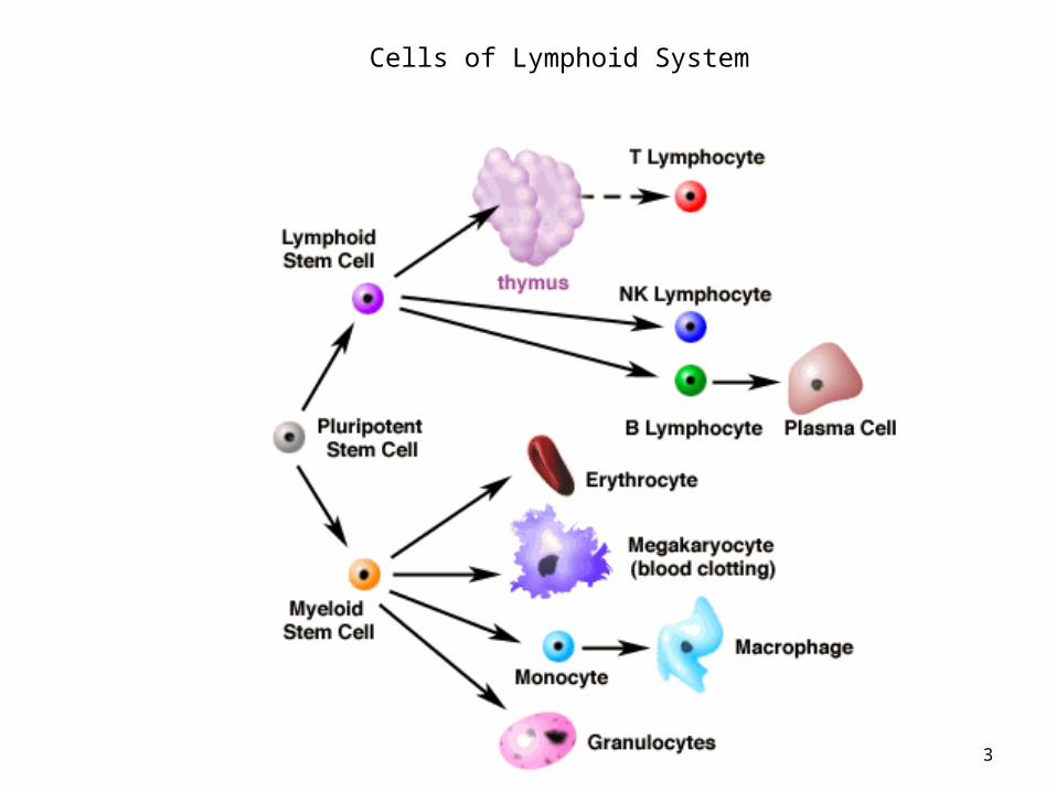
Lymphatic System
• Protect body against pathogens or antigens• Basis for this self-defense – distinguish self
from nonself• Functions carried out by – cells, effector
molecules, tissues and organs.
2
3
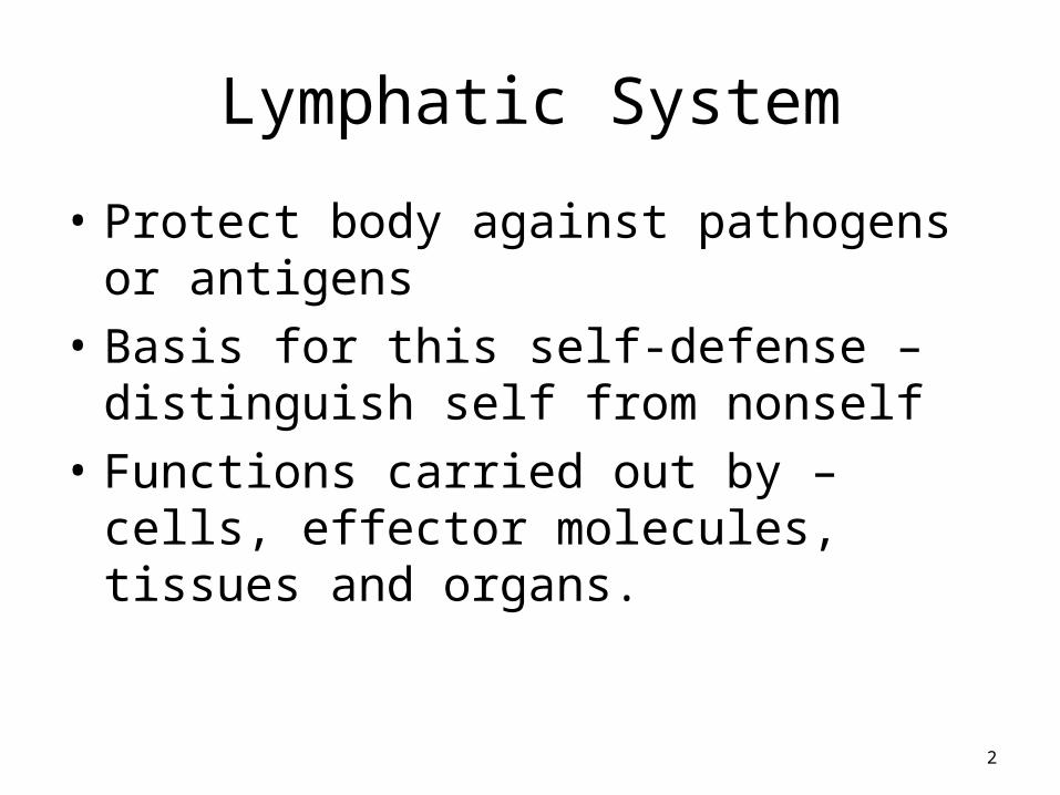
Cells of Lymphoid System

LYMPHOCYTES - T cells, B cells, NK cellsDistinguished by immunocytochemical methods. Differ
based on life history, surface receptors and behavior during immune response.
T cells - • 60-70% of circulating lymphocytes • Bone marrow to the thymus gland. • Mature and become immunocompetent – thymus• Migrate to peripheral lymphoid tissue and organs• Destroy the antigen - cytotoxic action or by
activating B cells. 4
5
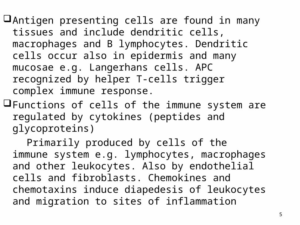
Antigen presenting cells are found in many tissues and include dendritic cells, macrophages and B lymphocytes. Dendritic cells occur also in epidermis and many mucosae e.g. Langerhans cells. APC recognized by helper T-cells trigger complex immune response.
Functions of cells of the immune system are regulated by cytokines (peptides and glycoproteins)
Primarily produced by cells of the immune system e.g. lymphocytes, macrophages and other leukocytes. Also by endothelial cells and fibroblasts. Chemokines and chemotaxins induce diapedesis of leukocytes and migration to sites of inflammation

B cells- •Produced within bone marrow. •B cells carried by blood to lymph nodes, spleen and connective tissue. •Immunocompetent B cells activated by specific antigen•Activated B cells – differentiate into plasma cells •Plasma cells – produce monoclonal antibodies specific to that antigen.•Natural Killer cells attack virally infected cells and cancer cells.
6
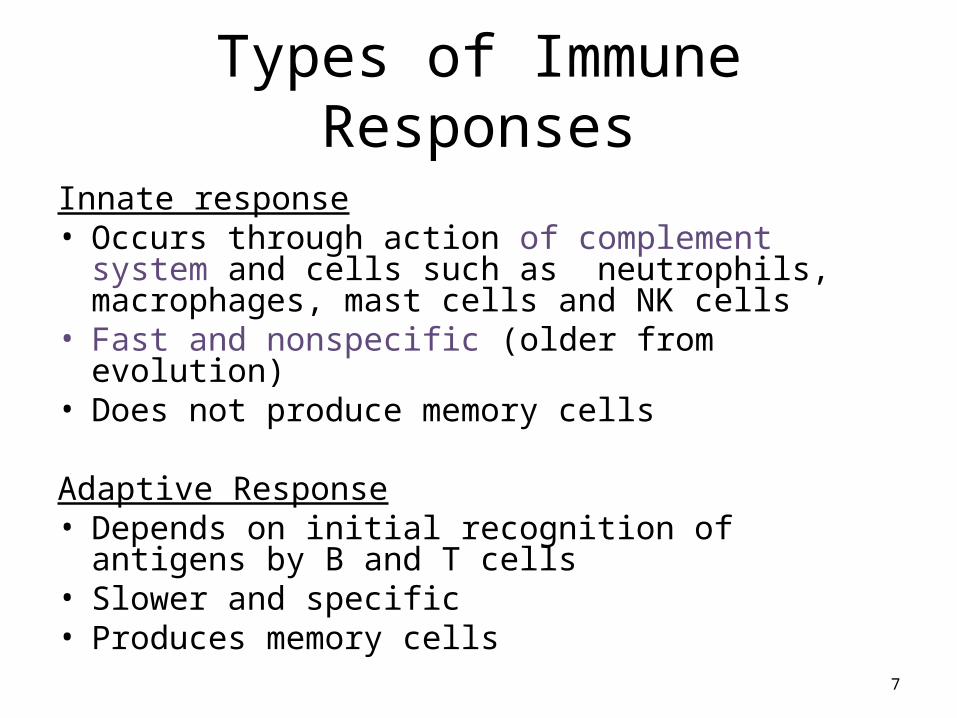
Types of Immune Responses
Innate response• Occurs through action of complement system and cells
such as neutrophils, macrophages, mast cells and NK cells
• Fast and nonspecific (older from evolution)• Does not produce memory cells
Adaptive Response• Depends on initial recognition of antigens by B and T
cells• Slower and specific• Produces memory cells
7

DIVISIONS OF adaptive or acquired IMMUNITY:ANTIBODY MEDIATED IMMUNITY (Humoral Immune Response)-• Bacterial infections• Helper T cells, B cells, Plasma cells• Effectors - immunoglobulin molecules
CELL MEDIATED IMMUNITY-• Viral and fungal infections & tumor cells - involved in rejection of
transplanted organs and tissue grafts• Cytotoxic T-cells (act on B-cells, other T-cells and macrophages &
neutrophils)• Effectors –T-cells or memory T- cells
8
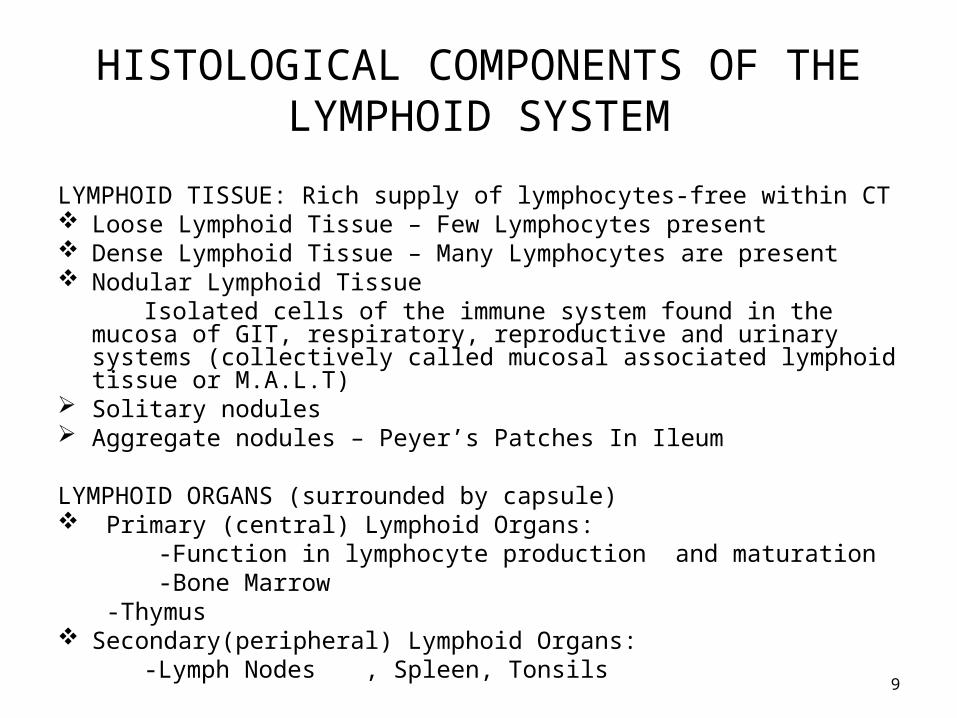
HISTOLOGICAL COMPONENTS OF THE LYMPHOID SYSTEM
LYMPHOID TISSUE: Rich supply of lymphocytes-free within CT Loose Lymphoid Tissue – Few Lymphocytes present Dense Lymphoid Tissue – Many Lymphocytes are present Nodular Lymphoid Tissue Isolated cells of the immune system found in the mucosa of GIT, respiratory,
reproductive and urinary systems (collectively called mucosal associated lymphoid tissue or M.A.L.T)
Solitary nodules Aggregate nodules – Peyer’s Patches In Ileum LYMPHOID ORGANS (surrounded by capsule) Primary (central) Lymphoid Organs: -Function in lymphocyte production and maturation -Bone Marrow
-Thymus Secondary(peripheral) Lymphoid Organs: -Lymph Nodes , Spleen, Tonsils
9
10
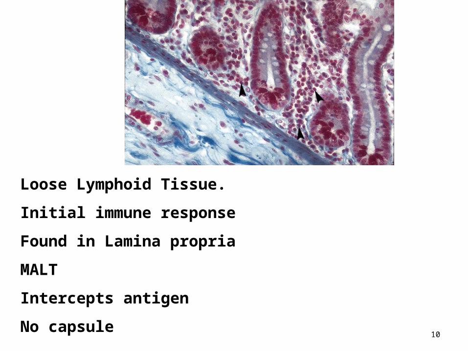
Loose Lymphoid Tissue.
Initial immune response
Found in Lamina propria
MALT
Intercepts antigen
No capsule

11
Dense Lymphoid Tissue
Contained in meshwork
Walls of GIT, Genitourinary tract, Respiratory tract
No capsule
12
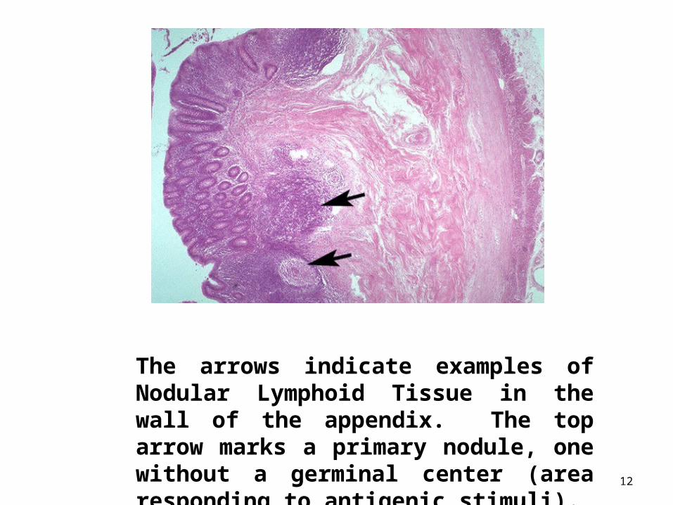
The arrows indicate examples of Nodular Lymphoid Tissue in the wall of the appendix. The top arrow marks a primary nodule, one without a germinal center (area responding to antigenic stimuli).
13
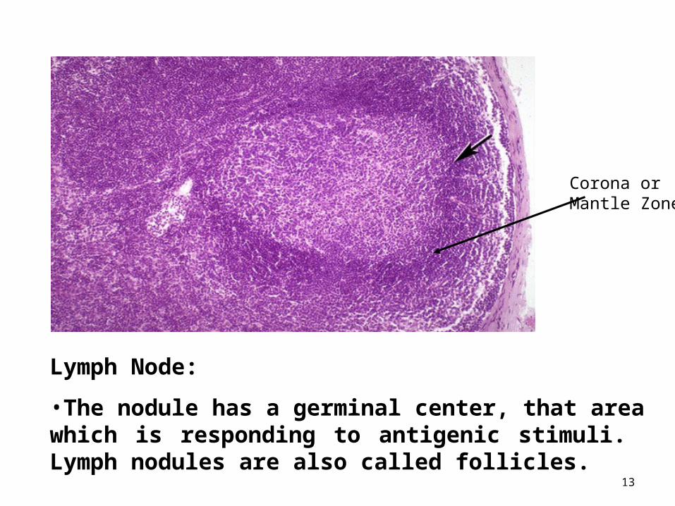
Lymph Node:
•The nodule has a germinal center, that area which is responding to antigenic stimuli. Lymph nodules are also called follicles.
Corona orMantle Zone
THE THYMUS
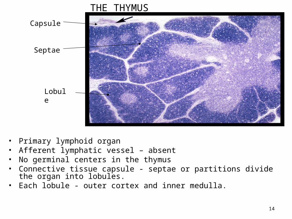
• Primary lymphoid organ• Afferent lymphatic vessel – absent• No germinal centers in the thymus• Connective tissue capsule - septae or partitions divide the organ into lobules. • Each lobule - outer cortex and inner medulla.
14
Capsule
Lobule
Septae
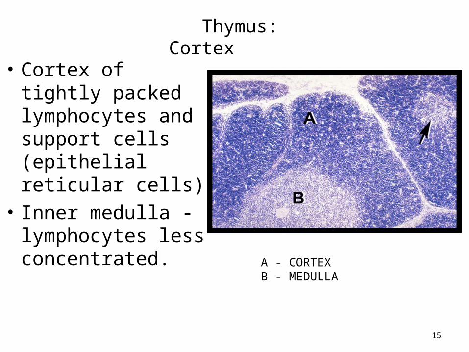
• Cortex of tightly packed lymphocytes and support cells (epithelial reticular cells)
• Inner medulla - lymphocytes less concentrated.
15
Thymus: Cortex
A - CORTEXB - MEDULLA
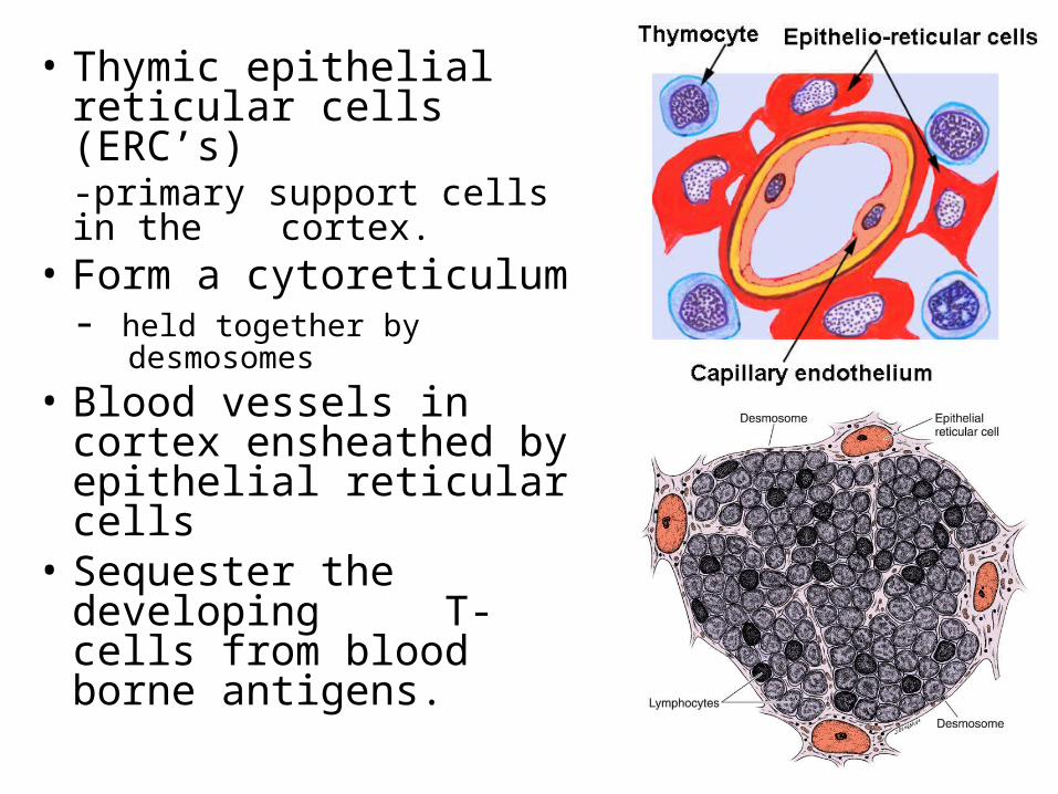
• Thymic epithelial reticular cells (ERC’s)
-primary support cells in the cortex.
• Form a cytoreticulum- held together by desmosomes
• Blood vessels in cortex ensheathed by epithelial reticular cells
• Sequester the developing T- cells from blood borne antigens.
16

17
The arrows mark thymic epithelial reticular cells
18
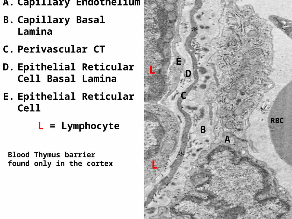
BLOOD THYMUS BARRIER
A. Capillary Endothelium
B. Capillary Basal Lamina
C. Perivascular CT
D. Epithelial Reticular Cell Basal Lamina
E. Epithelial Reticular Cell
L = LymphocyteA
B
C
DE
L
L
RBC
Blood Thymus barrier found only in the cortex

19
Thymus: Medulla

Medulla- • Blood vessels in medulla
- loose epithelial reticular cell covering• Lymphocytes differentiate and enlarge• Mature T-cells migrate into medullary blood
vessels• ERC’s contract into spherical, degenerative
masses - forming Hassall’s Corpuscle(s)
20

21
Cortex = A
Medulla = B
Arrow marks a Hassall’s Corpuscle, a key histological feature of the thymus.
Hassall’s Corpuscles are found only in the thymic medulla and consist of rings of degenerating epithelial reticular cells

22
Thymus: undergoes involution after puberty
Hassall’s Corpuscles (arrow) seen in remnants of the medulla.
Adipose cells accumulate
DiGeorge Syndrome- • Caused by a defect in chromosome 22 =
Thymic absence or underdevelopment• Inherited immunodeficiency disease • Affects cortical epithelial cells • Thymus rudimentary developed• Affects production of T-cells

LYMPH NODES
• Kidney shaped organs • Distribution – course of
lymphatic vessels• Filters lymphatic fluid• Mounts immune response by
recirculation of lymphocytes (route of metastasis)
• Capsule surrounds lymph node
• Hilum - arteries and nerves enter veins and lymphatic vessels leave the node
• Afferent lymphatic vessel – drains lymph through convex margin.
24

25
Connective tissue capsule Distinct cortex and medulla.
26
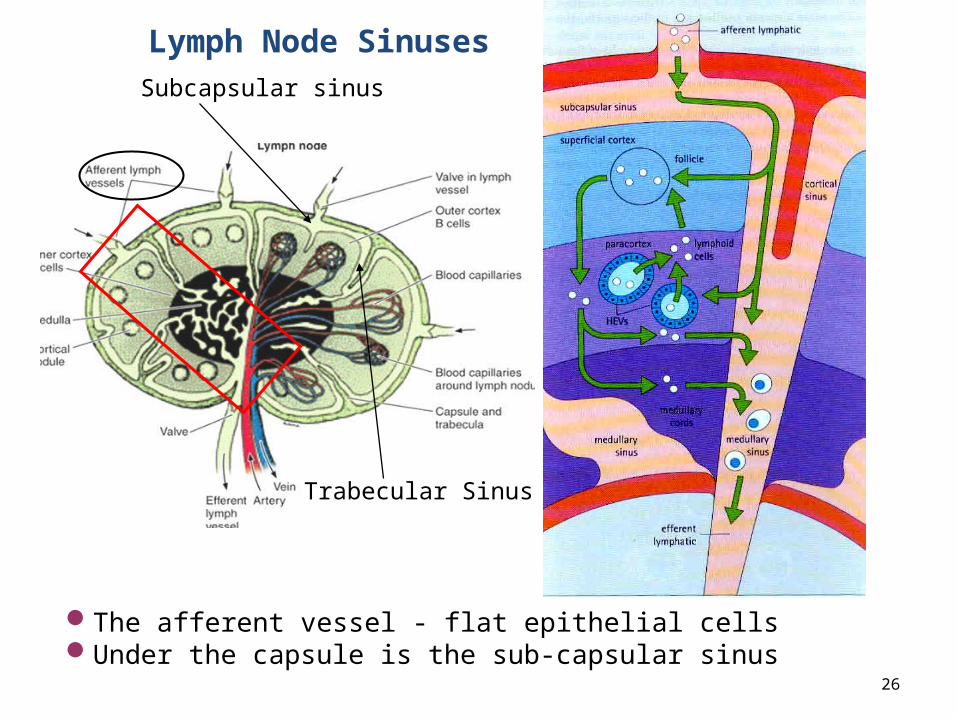
Subcapsular sinus
Trabecular Sinus
The afferent vessel - flat epithelial cells Under the capsule is the sub-capsular sinus
Lymph Node Sinuses

Subcapsular sinuses –
composed of loose reticular cells, and fibers.
Radial sinuses or trabecular sinuses – run between nodules along the trabeculae.
27
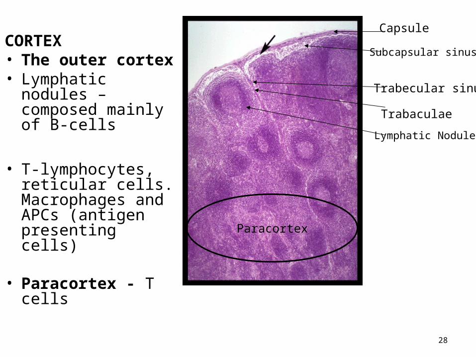
CORTEX• The outer cortex • Lymphatic nodules –
composed mainly of B-cells
• T-lymphocytes, reticular cells. Macrophages and APCs (antigen presenting cells)
• Paracortex - T cells
28
Capsule
Paracortex
Subcapsular sinus
Trabecular sinus
Trabaculae
Lymphatic Nodule
29
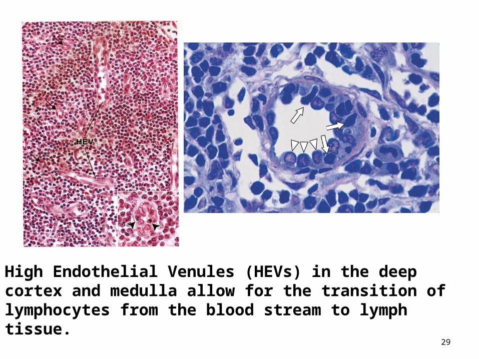
High Endothelial Venules (HEVs) in the deep cortex and medulla allow for the transition of lymphocytes from the blood stream to lymph tissue.

Medulla has two components:
• Medullary cords – A• Medullary sinus – B
30

Medullary cord-• Branched cord-like extensions of dense
lymphoid tissue• Contain primarily B lymphocytes, plasma cells
and macrophagesMedullary Sinus-• Dilated spaces separating medullary cords• High concentration of lymph, macrophages• Granulocytes may be present when lymph node
is draining an infected region
31
Medullary sinus – macrophage processes, reticular fibers and reticular cells form meshwork
• Retard free flow of lymph and enhances filtration
• Antigenic materials trapped and phagocytosed • Metastatic cancer- system overwhelmed by
excessive number of cancer cells• New metastatic site
32

Lymph node enlarges when responding to antigens-
• Reflecting- germinal center and proliferation of lymphocytes
• Enlarged lymph nodes = “swollen glands”• Painful - distension of capsule by cellular
proliferation and edema.
33
HISTOLOGY Lymphatic System II
If your exams were easy, then your degree would be worthless.
34
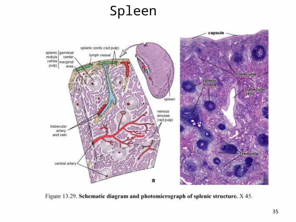
Spleen
35

THE SPLEEN
• The spleen - largest lymphoid organ
• Filters blood - site of immune responses to blood borne antigens.
• Dense connective tissue capsule - myofibroblast
• Trabaculae• Red Pulp- cords of Billroth &
sinuses engorged by blood• White pulp- Malphigian
bodies and splenic lypmhoid nodules = hyperplasia of lymphoid tissue
36
37
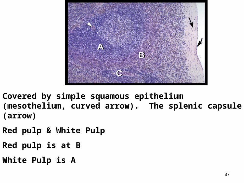
Covered by simple squamous epithelium (mesothelium, curved arrow). The splenic capsule (arrow)
Red pulp & White Pulp
Red pulp is at B
White Pulp is A

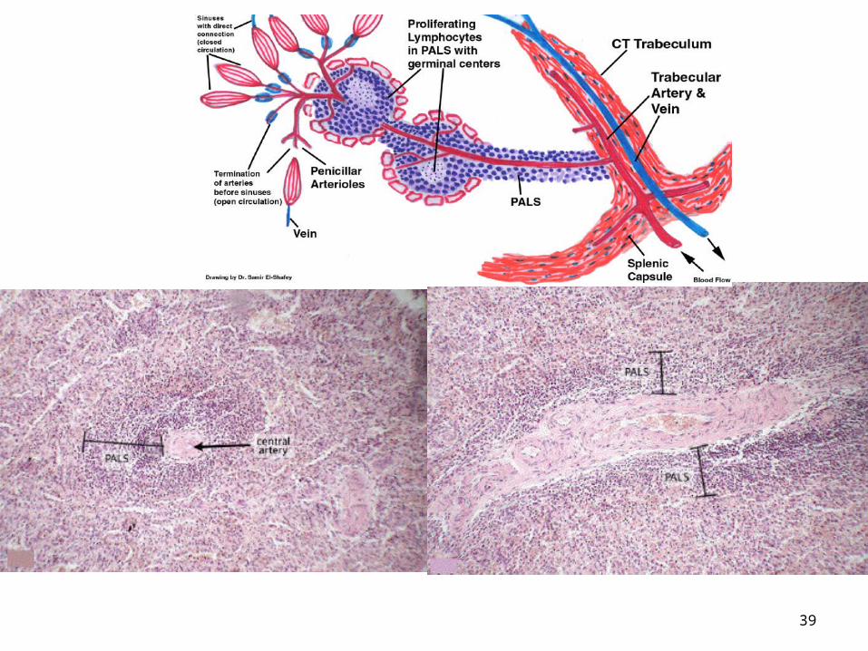
• The white pulp – thick accumulation of lymphocytes. • Lymphatic nodules - germinal centers that decrease with age.• Nodule - exhibits a peripheral zone, the periarterial lymphatic
sheath (PALS)
38
White pulp
39

40
Lymphatic nodule - central artery (CA) which has an eccentric position. B cells develop in germinal centers.Marginal zones of the nodule – trap antigens from the circulation and present the antigen to the lymphocytes of the spleen
Marginal Zone
Mantle Zone
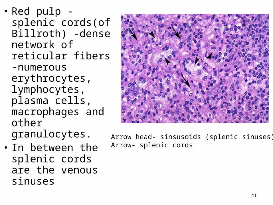
• Red pulp - splenic cords(of Billroth) -dense network of reticular fibers -numerous erythrocytes, lymphocytes, plasma cells, macrophages and other granulocytes.
• In between the splenic cords are the venous sinuses
41
Arrow head- sinsusoids (splenic sinuses)Arrow- splenic cords

• Venous sinuses - tall endothelial cells and supported by an incomplete basal lamina of reticular fibers.
• Macrophages function in the removal of damaged erythrocytes from circulation.
42
43
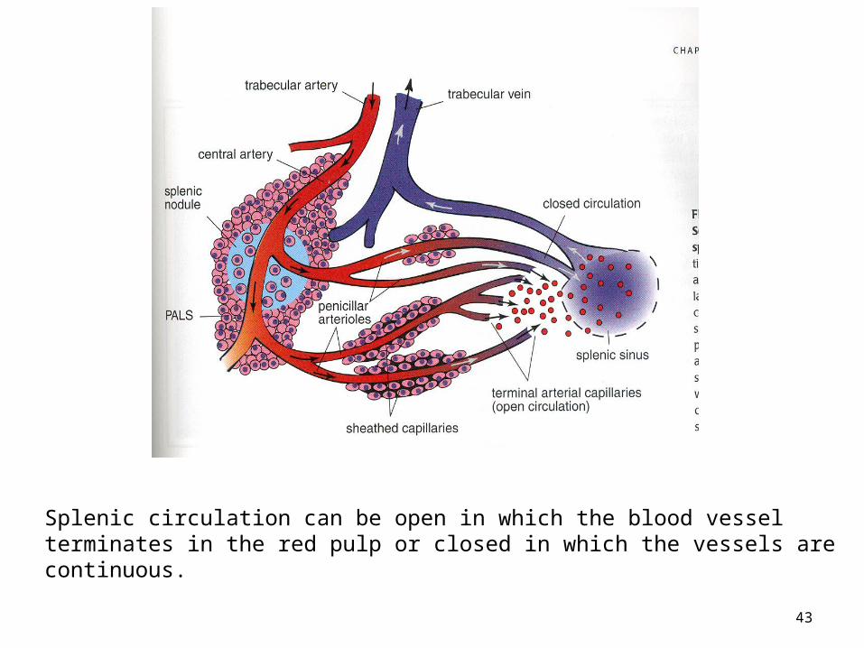
Splenic circulation can be open in which the blood vessel terminates in the red pulp or closed in which the vessels are continuous.
Splenectomy - traumatic rupture, autoimmune diseses or malignant tumor
• Adults who have antibodies less prone to bacteremia
• Children – more vulnerable
44
Tonsils
• Tonsils are part of Gut Associated Lymphoid Tissue (GALT).
• Located at the back of the oral cavity, in the tongue and pharynx
• Process antigens coming into the body
45
46
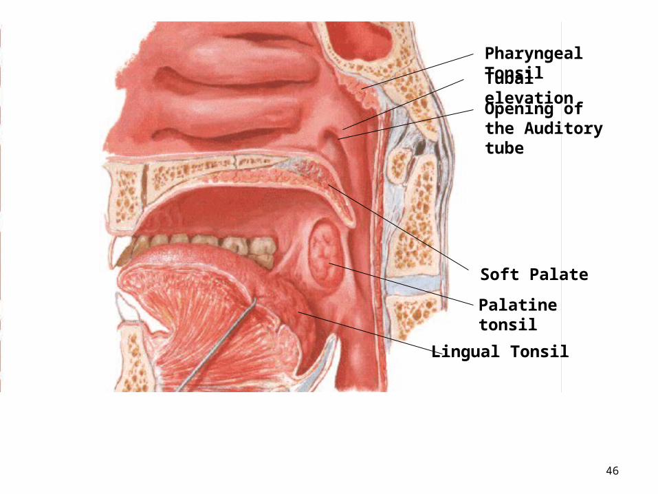
Tubal elevation
Opening of the Auditory tube
Pharyngeal Tonsil
Soft Palate
Palatine tonsil
Lingual Tonsil

47
crypt
Palatine tonsil

• Capsule prevents spread of infection to deeper structures
• Numerous lymphoid nodules are located in the palatine tonsil.
• Germinal centers• Tonsillar crypts, whose lumen
contains desquamated epithelial cells, live and dead lymphocytes and bacteria.
• The crypts are lined with epithelium.
• Stratified squamous epithelium is at the surface of palatine and lingual tonsils
48Palatine Tonsil