Embed Size (px)
Citation preview
THE IMMUNE SYSTEM & LYMPHOID ORGANS
THE IMMUNE SYSTEM & LYMPHOID ORGANS:
• The body has a system of cells—the immune system—that has the ability to distinguish "self" (the organism's own molecules) from "nonself" (foreign substances).
• This system has the ability to neutralize or inactivate foreign molecules (such as soluble molecules as well as molecules present in viruses, bacteria, and parasites) and to destroy microorganisms or other cells (such as virus-infected cells, cells of transplanted organs, and cancer cells).
• On occasion, the immune system of an individual reacts against its own normal body tissues or molecules, causing autoimmune diseases.
• The cells of the immune system are distributed throughout the body in the blood, lymph, and epithelial and connective tissues; are arranged in small spherical nodules called lymphoid nodules found in connective tissues and inside several organs; and are organized as differently sized organs called lymphoid organs—the lymph nodes, the spleen, the thymus, and the bone marrow.
• Lymphoid nodules and isolated cells of the immune system found in the mucosa of the digestive system (tonsils, Peyer's patches, and appendix), the respiratory system, the reproductive system, and the urinary system are collectively known as mucosa-associated lymphoid tissue (MALT) and may be considered a lymphoid organ.
• The wide distribution of immune system cells and the constant traffic of lymphocytes through the blood, lymph, connective tissues, and lymphoid organs provide the body with an elaborate and efficient system of surveillance and defense.
THE IMMUNE SYSTEM
Antigens
• A molecule that is recognized by cells of the immune system is called an antigen and may elicit a response from these cells.
• Antigens may consist of soluble molecules (such as proteins, polysaccharides, and nucleoproteins) or molecules belonging to whole cells (bacteria, protozoa, tumor cells, or virus-infected cells).
• The cells of the immune system do not recognize and react to the whole antigen molecule but instead react to small molecular domains of the antigen known as antigenic determinants or epitopes. (=An epitope refers to the specific target against which an individual antibody binds.)
• The response of the organism to antigens may be called cellular (in which lymphocytes are primarily in charge of eliminating the antigen) or humoral (in which molecules secreted by plasma cells, called antibodies, are primarily responsible for the response).
Antibodies
• An antibody is a glycoprotein that interacts specifically with an antigenic determinant.
• Antibodies belong to the immunoglobulin (Ig) protein family.
• Free molecules of antibodies are secreted by plasma cells that arise by proliferation and terminal differentiation of clones of B lymphocytes whose receptors recognize and bind specific epitopes.
• These secreted antibodies either circulate in the plasma and may leave the blood vessels reaching the tissues or are present in the secretion of some epithelia (eg, of the mammary gland and salivary glands).
• Other antibodies are not free molecules, but are integral membrane proteins of the surface of lymphocytes.
• In any case, each antibody combines with the epitope that it specifically recognizes.
Classes of Antibodies• The main classes of immunoglobulins in humans are immunoglobulin G (IgG), IgA, IgM, IgE,
and IgD .
• IgG is the most abundant class representing 75% of serum immunoglobulins. It is produced in large amounts during immune responses. IgG is the only immunoglobulin that crosses the placental barrier and is transported to the circulatory system of the fetus, protecting the newborn against infections for a certain period of time.
• IgA is the main immunoglobulin found in secretions, such as nasal, bronchial, intestinal, and prostatic, as well as in tears, colostrum, saliva, and vaginal fluid.
• IgM constitutes about 10% of blood immunoglobulins. Together with IgD, it is the major immunoglobulin found on the surface of B lymphocytes.
• IgD concentration in blood plasma constitutes only 0.2% of the immunoglobulins. IgD is found on the plasma membranes of B lymphocytes.
Classes of Antibodies
• IgE has a great affinity for receptors present on the surfaces of mast cells and basophils, it attaches to these cells after being secreted by plasma cells and only small amounts are found in the blood.
• When IgE molecules present on the surface of mast cells or basophils encounter the antigen that elicited the production of this specific IgE, the antigen–antibody complex triggers the liberation of several biologically active substances, such as histamine, heparin, leukotrienes, and eosinophil-chemotactic factor of anaphylaxis.
• This characterizes an allergic reaction, which is thus mediated by the binding of cell-bound IgE with the antigens (allergens) that stimulated its production.
Cells of the Immune System
• The primary cells that participate in the immune response are lymphocytes, plasma cells, mast cells, neutrophils, eosinophils, and cells of the mononuclear phagocyte system.
• Antigen-presenting cells, a group of very diverse cell types, assist other cells in the immune response. This group includes, among other cells, lymphocytes, macrophages, and dendritic cells.
Lymphocytes
• Lymphocytes are classified as B,T, or natural killer (NK) cells.
• The B and T cells are the only cells that have the ability to selectively recognize a specific epitope among a vast number of different epitopes (of the order of 1018).
• B and T cells differ based on their life history, surface receptors, and behavior during an immune response.
Lymphocytes
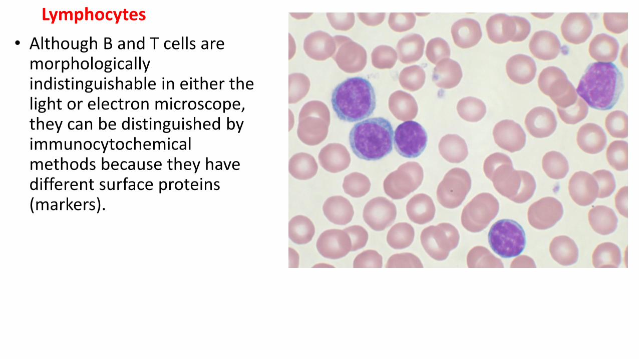
• Although B and T cells are morphologically indistinguishable in either the light or electron microscope, they can be distinguished by immunocytochemicalmethods because they have different surface proteins (markers).

Lymphocytes
The precursors of all lymphocyte types originate in the bone marrow; some lymphocytes mature and become functional in the bone marrow, and after leaving the bone marrow enter the blood circulation to colonize connective tissues, epithelia, lymphoid nodules, and lymphoid organs. These are the B lymphocytes.
Note:In birds, B cells mature in the bursa of Fabricius.
• T lymphocyte precursors, on the other hand, leave the bone marrow, and through the blood circulation reach the thymus where they undergo intense proliferation and differentiation or die by apoptosis.
• After their final maturation, T cells leave the thymus and are distributed throughout the body in connective tissues and lymphoid organs .
• Because of their function in lymphocyte production and maturation, the bone marrow, Bursa of fabricius and the thymus are called the primary or central lymphoid organs.
Lymphocytes• The other lymphoid structures are the secondary or peripheral lymphoid organs (spleen,
lymph nodes, solitary lymphoid nodules, tonsils, appendix, and Peyer's patches of the ileum).
• B and T cells are not anchored in the lymphoid organs; instead, they continuously move from one location to another, a process known as lymphocyte recirculation.
• B and T cells are not uniformly distributed in the lymphoid system but occupy preferential sites in these organs.
• A very important feature of B and T lymphocytes involves the receptors they have on their surface.
• These receptors are fundamental for recognition of antigen epitopes and, thus, for triggering an immune response.
B Lymphocytes• In B lymphocytes, the surface receptors able to recognize antigens are molecules of IgM;
each B cell is covered by about 150,000 molecules of IgM.
• The encounter of a B lymphocyte with the epitope it recognizes leads to several cycles of cell proliferation, followed by a redifferentiation of most of these lymphocytes into plasma cells.
• This population of plasma cells secretes antibodies against the same epitope as that of the B cell that originated them.
• In most cases, the activation of B cells requires the assistance of a subclass of T lymphocytes known as T-helper lymphocytes.
• Not all activated B cells, however, become plasma cells; some remain B memory lymphocytes, which react rapidly to a second exposure to the same epitope.
B Lymphocytes
• The Bursa of Fabricius is an organ that is unique to birds and is the only site for B cell differentiation and maturation. Located in the rump of birds, this organ is full of stem cells and very active in young birds but atrophies after six months.
• The central organ for B cell development in birds is the Bursa of Fabricius.
• It is now clear that the bursa is the primary site of B cell lymphopoeisis
T Lymphocytes• T cells constitute 65–75% of blood lymphocytes.
• To recognize epitopes, all T cells have on their surfaces a molecule called a T cell receptor (TCR).
• In contrast to B cells, which recognize soluble antigens or antigens present on cell surfaces, T lymphocytes recognize only epitopes (mostly small peptides) that form complexes with special proteins of the cell surface of other cells (proteins of the major histocompatibility complex).
• The two main subpopulations of T cells are helper and cytotoxic lymphocytes (CTLs).
• Helper cells play very important roles in the immune response, being responsible for cytokine production, interaction with B cells to promote their differentiation into plasma cells, activation of macrophages to phagocytose, activation of cytotoxic lymphocytes, and induction of an inflammatory reaction.
T Lymphocytes• Many of these actions are mediated by cytokines.
• Helper cells have a marker called CD4 on their surfaces and are, hence, called CD4+ T cells.
• Cytotoxic, or CD8+ T cells, can act against foreign cells or virus-infected cells by means of two main mechanisms.
• In one, they attach to the cells to be killed and release proteins called perforins that create holes in the cell membrane of the target cell, with consequent cell lysis.
• In the other, they attach to a cell and kill it by triggering mechanisms that induce programmed cell death, or apoptosis.
•
Natural Killer Cells
• The natural killer lymphocytes lack the marker molecules characteristic of B and T cells.
• They comprise about 10–15% of the lymphocytes of circulating blood.
• Their name derives from the fact that they attack virus-infected cells, transplanted cells, and cancer cells without previous stimulation; for this reason they are involved in what is called an innate immune response.
Major HistocompatibilityComplex• The major histocompatibility complex (MHC) is a set of cell surface proteins essential for
the acquired immune system to recognize foreign molecules in vertebrates, which in turn determines histocompatibility.
• The main function of MHC molecules is to bind to peptide fragments derived from pathogens and display them on the cell surface for recognition by the appropriate T-cells.
• MHC molecules mediate interactions of leukocytes, also called white blood cells (WBCs), which are immune cells, with other leukocytes or with body cells.
• MHC molecules are integral membrane proteins present on the cell surface.
• They are synthesized by polyribosomes and are inserted in the membrane of the rough endoplasmic reticulum, as a regular membrane protein.
Antigen-Presenting Cells
• An antigen-presenting cell (APC) is a cell that displays antigen complexed with major histocompatibility complexes (MHCs) on their surfaces; this process is known as antigen presentation. T cells may recognize these complexes using their T cell receptors (TCRs). These cells process antigens and present them to T-cells.
Types of Immune Responses• The two basic types of immune responses are the innate response and the adaptive
response.
• The innate response, which occurs through the action of neutrophils, macrophages, mast cells, and natural killer cells, is fast, nonspecific, and older from an evolutionary point of view.
• It does not produce memory cells.
• The adaptive response, which depends on the initial recognition of antigens by B and T cells, is much more complex, is slower and specific, produces memory cells, and is a more recent evolutionary development.
• The adaptive mechanisms that lead to the elimination of antigens are classified as humoralor cellular responses.
• Humoral immunity is accomplished by antibodies produced by plasma cells derived from clones of activated B lymphocytes.
Types of Immune Responses
• Cellular immunity is mediated by T lymphocytes that secrete cytokines that act on B lymphocytes, on other T cells, and on inflammatory cells such as macrophages and neutrophils, and attack foreign cells or cells that exhibit foreign epitopes on their surfaces, such as cells infected by viruses or parasites, and tumor cells.
• With few exceptions, both types of immune response are activated when foreign epitopes are recognized by lymphocytes.
• Antigens (such as a microorganism or molecules) that reach the skin or a mucosa (or the connective tissue in the case of an injected vaccine) have two main fates.
• In the first, the antigen is phagocytosed either by a macrophage or by a dendritic cell and is transported by these cells through the lymphatic vessels to the lymph node that drains that region of the body (satellite lymph node).
Types of Immune Responses
• In the second, the molecules, the whole microorganism, or its debris are transported by the lymph to the satellite lymph node where macrophages or other APCs phagocytose them.
• Antigens that reach the lymph node are recognized by B lymphocytes.
• APCs that arrived from the skin or mucosa as well as APCs that processed antigens within the lymph node display the antigens to helper T lymphocytes as complexes with class II MHC molecules.
• B lymphocytes that recognize antigens are activated by helper cells to enter several cycles of cell division.
• Many of the daughter cells of B lymphocytes differentiate into plasma cells that secrete antibodies against the antigen recognized by the first B lymphocyte.
• The plasma cells secrete most of the antibodies into the lymph and the antibodies eventually reach the blood circulation and act on antigens in different ways.
• B cells that are not transformed into plasma cells remain B memory cells.
Types of Immune Responses• Intracellular antigens, such as those synthesized in the cytosol of virus-infected cells, tumor
cells, or transplanted cells, are presented to cytotoxic T lymphocytes bound to class I MHC molecules.
• Concurrently, APCs that phagocytose fragments of viruses, tumor cells, or transplanted cells display the antigens to helper T lymphocytes, bound to class II MHC molecules.
• Cytotoxic T cells, activated by helper cells, enter several cycles of proliferation and some of these cells become effector cytotoxic T cells that will destroy the cells that hold the antigens.
• Some cells, instead of becoming effector cells, remain memory cytotoxic T cells. A humoral response resulting from recognition of antigens by B lymphocytes generally occurs simultaneously.
Lymphoid Tissue• Lymphoid tissue is a type of connective tissue characterized by a rich supply of
lymphocytes.
• It exists free within the regular connective tissue or is surrounded by capsules, forming the lymphoid organs.
• Because lymphocytes have very little cytoplasm, lymphoid tissue stains dark blue in hematoxylin and eosin-stained sections.
• Lymphoid tissues are basically made up of free cells; as a result, they typically have a rich network of reticular fibrils (made principally of type III collagen) that supports the cells.
• In most lymphoid organs, the fibrils are produced by a fibroblastic cell called a reticular cell, whose many processes rest on the reticular fibrils. The thymus is an exception in so far as its cells are supported by a reticulum of epithelial cells of endodermic origin.
•
Lymphoid Tissue• The network of reticular fibrils of the lymphoid tissue may be relatively closed (dense
lymphoid tissue) and is, thus, able to hold many free cells (mostly lymphocytes, macrophages, and plasma cells).
• Another type is loose lymphoid tissue, whose network has fewer but larger spaces, providing means for easy movement of the free cells.
• In the nodular lymphoid tissue, groups of lymphocytes are arranged as spheres, called lymphoid nodules or lymphoid follicles, that primarily contain B lymphocytes.
• When lymphoid nodules become activated as a result of the arrival of antigen-carrying APCs and recognition of the antigens by B lymphocytes, these lymphocytes proliferate in the central portion of the nodule, which then stains lighter and is called a germinativecenter.
Lymphoid Tissue• After completion of the immune response, the germinative center may disappear.
• The germinative centers contain a special cell, the follicular dendritic cell (distinct from the epithelial dendritic APCs), that has many processes that bind antigen on their surfaces, to be presented to B lymphocytes.
• Lymphoid nodules vary widely in size, typically measuring a few hundred micrometers to 1 mm in diameter.
• They are found free in connective tissues anywhere in the body or within lymphoid organs (lymph nodes, spleen, tonsils, but not in the thymus).
• They are, however, never covered by a capsule.
• Free lymphoid nodules are commonly present in the lamina propria of several mucosal linings, where, together with free lymphocytes, they constitute the mucosa-associated lymphoid tissue (MALT).
Avian lymphatic tissue
• Many birds do not have lymph nodes.
• Solitary accumulations of lymph nodules characterize their digestive tract, serousmembranes, and skin.

Mucosa-Associated Lymphoid Tissue & Tonsils• The digestive, respiratory, and genitourinary tracts are common sites of microbial invasion
because their lumens are open to the external environment.
• To protect the organism, the mucosa and submucosa of these tracts contain a large amount of diffuse collections of lymphocytes, IgA-secreting plasma cells, APCs, and lymphoid nodules.
• Most of the lymphocytes are B cells; among T cells, CD4+ helper cells predominate.
• In some places, these aggregates form conspicuous structures such as the tonsils and the Peyer's patches in the ileum. Similar aggregates are found in the appendix.
Mucosa-Associated Lymphoid Tissue & Tonsils• In the Peyer's patches, some of the regular surface epithelial cells may be replaced by
special M cells.
• The M cells do not have microvilli as do the regular cells that line the intestine.
• By pinocytosis they actively capture and transport antigens from the intestinal lumen to the connective tissues where APCs and B lymphocytes are usually present.
• The plasma cells derived from these lymphocytes secrete mostly IgA, which is transported through the epithelium toward the intestinal cavity.
• Primary lymphatic organs are where lymphocytes are formed and mature. Theyprovide an environment for stem cells to divide and mature into B- and T- cells:
• There are three primary lymphatic organs: the bone marrow, the thymus gland andbursa Fabricius.
Bone Marrow
• The two types of bone marrow are "red marrow", which consists mainly of hematopoietic tissue, and "yellow marrow", which is mainly made up of fat cells.
• Red blood cells, platelets, and most white blood cells arise in red marrow.
• Both types of bone marrow contain numerous blood vessels and capillaries.
• At birth, all bone marrow is red.
• With age, more and more of it is converted to the yellow type; only around half of adult bone marrow is red.
• Red marrow is found mainly in the flat bones, such as the pelvis,sternum, cranium, ribs, vertebrae and scapulae, and in the cancellous("spongy") material at the epiphyseal ends of long bones such as thefemur and humerus.
Bone Marrow
• Yellow marrow is found in the medullary cavity, the hollow interior of the middle portion ofshort bones.
• In cases of severe blood loss, the body can convert yellow marrow back to red marrow toincrease blood cell production.
• Progenitor cell (stem cell) lines in the bone marrow produce new blood cells and stromalcells. Bone marrow is also an important part of the lymphatic system.
• Bone marrow consists of stem cells, which are large, "primitive," undifferentiated cells supported by fibrous tissue called stroma.
• There are 2 main types of stem cells and, therefore, the bone marrow consists of 2 types of cellular tissue. One type of stem cell is involved in producing blood cells and the other is involved in producing stromal cells, which are responsible for the supporting stroma.
Bone Marrow• The bone marrow stroma contains mesenchymal stem cells. These cells are multipotent
stem cells that can differentiate into a variety of cell types, including osteoblasts, osteoclasts, chondrocytes, myocytes, fibroblasts, macrophages, adipocytes, andendothelial cells.
• The stroma is not directly involved in the primary function of hematopoiesis, but it provides the microenvironment and colony-stimulating factors needed to facilitate hematopoiesis by the parenchymal cells.
Thymus• The thymus is a lymphoepithelial organ located in the mediastinum; it attains its peak
development during youth.
• Whereas the other lymphoid organs originate exclusively from mesenchyme (mesoderm), the thymus has a dual embryonic origin.
• Its lymphocytes arise in the bone marrow from cells of mesenchymal origin that invade an epithelial primordium that has developed from the endoderm of the third and fourth pharyngeal pouches.
• The thymus has a connective tissue capsule that penetrates the parenchyma and divides it into incomplete lobules, so that there is continuity between the cortex and medulla of adjoining lobules.
• Each lobule has a peripheral dark zone known as the cortex and a central light zone called the medulla.

Thymus• The cortex is composed of an extensive population of T cell precursors (also called
thymocytes), dispersed epithelial reticular cells, and macrophages.
• Because the cortex is richer in small lymphocytes than the medulla, it stains more darkly.
• The epithelial reticular cells are stellate cells with light-staining oval nuclei.
• They are usually joined to similar adjacent cells by desmosomes.
• Bundles of intermediate keratin filaments (tonofibrils) in their cytoplasm are evidence of the epithelial origin of these cells.
• A subpopulation of epithelial reticular cells present in the cortex consists of thymic nurse cells (TNCs), which contain many (20–100) maturing lymphocytes in their cytoplasm.
• The medulla contains epithelial reticular cells, many differentiated T lymphocytes, and structures called thymic corpuscles or Hassall corpuscles, which are characteristic of this region, although their function is unknown.
• These corpuscles contain flattened epithelial reticular cells that are arranged concentrically and are filled with keratin filaments. They sometimes calcify.
Thymus
Vascularization of the Thymus:• Arterioles and capillaries in the thymus are surrounded by
processes of epithelial reticular cells. • Thymus capillaries have a nonfenestrated endothelium and a very
thick basal lamina, making these blood vessels particularly impermeable to proteins.
• This prevents most circulating antigens from reaching the thymus cortex, thus creating the so-called thymic–blood barrier.
• The thymus has no afferent lymphatic vessels and does not constitute a filter for the lymph, as do lymph nodes.
• The few lymphatic vessels encountered in the thymus are all efferent; they are located in the walls of blood vessels and in the connective tissue of the septa and the capsule.
Thymus
Role of the Thymus in T Cell Differentiation
• The thymus is the site of the terminal differentiation and selection of T lymphocytes.
• The thymus reaches its maximum development in relation to body weight immediately after birth; it undergoes involution after attaining its greatest size in puberty, but continues to produce lymphocytes until old age.
• T cell precursors, committed to produce T lymphocytes, do not exhibit the T cell receptor on their surfaces and are CD4– and CD8–.
• They arise in the fetal liver in early fetal life and later migrate from the bone marrow to the thymus during both fetal and adult life.
• After penetrating the thymus, the T cell precursors populate the cortex where they divide by mitosis.
• In the cortex they are presented to self-antigens bound to class I and class II MHC molecules present on the surface of the epithelial cells, macrophages, and dendritic cells.
Thymus• The maturation and selection of T lymphocytes within the thymus are very complex
processes that include positive and negative selection of T cells.
• Part of these processes is supposed to occur within nurse cells.
• In brief, thymocytes whose T cell receptors are unable to bind or, in contrast, that bind too avidly to self-antigens (about 95% of the total) are induced to die by apoptosis and are removed by macrophages.
• The remaining T cells survive and migrate to the medulla.
• Migration depends on the action of chemokines and on the interaction of thymocytes with the thymic extracellular matrix.
• Mature CD4+ or CD8+ T cells with T cell receptors on their surfaces leave the thymus, enter the blood circulation passing through the walls of medullary veins, and are distributed throughout the body.
Secretion by the Thymus
• The thymus produces several proteins that act as growth factors to stimulateproliferation and differentiation of T lymphocytes.
• They seem to be paracrine secretions, acting in the thymus.
• At least four hormones have been identified: thymosin-, thymopoietin, thymulin, andthymus humoral factor.
Bursa of Fabricius• The bursa Fabricius occurs in birds.
• It is a blind sac opening into the caudodorsal surface of cloaca.
• The bursa consists of a number of lymphoid lobules and crypt-like folds surrounding a lumen, which is enclosed in a thin layer of stratified squamous epithelium. The lumen opens into the proctodeum.
• Like the thymus the lobules have a cortex and medulla and the lympocytes are supported by epithelial cells.
• B lymphocytes developing from the lymphoid precursors gather in the cortex, hence the cortex stains stronger than the medulla.
• Lymph nodules are located between epithelial folds, and germinal centers are present.
• The bursa’s primary function is maturating and causing the differentiation of B lymphocytes, it also produces the hormone bursin which activates B lymphocytes.
• SECONDARY LYMPHOID ORGANS are arranged as a series of filters monitoring the contents of the extracellular fluids, i.e. lymph, tissue fluid and blood. The lymphoid tissue filtering each of these fluids is arranged in different ways.
• Secondary lymphoid tissues are also where lymphocytes are activated.
• Secondary lymphoid organs are lymph nodes, tonsils, hemal nodes, hemolymph nodesand spleen.
SECONDARY LYMPHOID ORGANS:Tonsils• Tonsils belong to the MALT, but because they are incompletely encapsulated, they are
considered organs and will be studied apart from the MALT.
• The tonsils constitute a lymphoid tissue that lies beneath, and in contact with, the epithelium of the initial portion of the digestive tract.
• Depending on their location, tonsils in the mouth and pharynx are called palatine, pharyngeal, or lingual.
Palatine Tonsils• The two palatine tonsils are located in the lateral walls of the oral part of the pharynx.
• They are lined with a squamous stratified epithelium that often becomes so densely infiltrated by lymphocytes that it may be difficult to recognize .
• The lymphoid tissue in these tonsils forms a band that contains free lymphocytes and lymphoid nodules, generally with germinal centers.
• Each tonsil has 10–20 epithelial invaginations that penetrate the tonsil deeply, forming crypts, whose lumens contain desquamated epithelial cells, live and dead lymphocytes, and bacteria.
• Crypts may appear as purulent spots in tonsillitis.
• Separating the lymphoid tissue from subjacent structures is a band of dense connective tissue, the capsule of the tonsil.
• This capsule usually acts as a barrier against spreading tonsillar infections.
Pharyngeal Tonsil• The pharyngeal tonsil is a single tonsil situated in the superior— posterior portion of the
pharynx.
• It is covered by ciliated pseudostratified columnar epithelium typical of the respiratory tract, although areas of stratified epithelium can also be observed.
• The pharyngeal tonsil is composed of pleats of mucosa and contains diffuse lymphoid tissue and lymphoid nodules. It has no crypts, and its capsule is thinner than the capsule of the palatine tonsils.
• Hypertrophied pharyngeal tonsils resulting from chronic inflammation are called adenoids.
Lingual Tonsils
• The lingual tonsils are smaller and more numerous than the palatine and pharyngeal tonsils.
• They are situated at the base of the tongue and are covered by stratified squamous epithelium. Each lingual tonsil has a single crypt.
Lymph Nodes• Lymph nodes are distributed throughout the body along the course of the lymphatic vessels.
• The nodes are found in the axilla and the groin, along the great vessels of the neck, and in large numbers in the thorax and abdomen, especially in mesenteries.
• Lymph nodes constitute a series of in-line filters that are important in the body's defense against microorganisms and the spread of tumor cells.
• All this lymph, derived from tissue fluid, is filtered by at least one node before returning to the circulation.
• Lymph nodes are elongated or kidney-shaped organs that have a convex surface that is the entrance site of lymphatic vessels and a concave depression, the hilum, through which arteries and nerves enter and veins and lymphatic vessels leave the organ.
• A connective tissue capsule surrounds the lymph node, sending trabeculae into its interior.
Lymph Nodes
• The most common cells of lymph nodes are lymphocytes, macrophages and other APCs, plasma cells, and reticular cells; follicular dendritic cells are present within the lymphoid nodules.
• The different arrangement of the cells and of the reticular fibril skeleton that supports the cells creates two regions, a cortex and a medulla.
• The cortex can be subdivided into an outer cortex and an inner cortex or paracorticalregion.
Cortex• The outer cortex, situated under the capsule, consists of the following components:
1. A diffuse population of cells composed mainly of T lymphocytes and reticular cells; macrophages and APCs are also present in this area.
2. Lymphoid nodules, with or without germinative centers, formed mainly by B lymphocytes, embedded in the diffuse population of cortical cells.
3. 3. Areas of loose lymphoid tissue (whose reticular fibril meshes are wide) situatedimmediately beneath the capsule, called the subcapsular sinuses.They are composed of a loose network of reticular cells and fibers. Lymph, containing antigens, lymphocytes, and APCs, circulates around the wide spaces of these sinuses after being delivered intothese channels by the afferent lymphatic vessels.
4. Intermediate or radial sinuses that run between lymphoid nodules. These sinuses arisefrom and share the same structure with the subcapsular sinuses. They communicatewith the subcapsular sinuses through spaces similar to those present in the medulla. The inner cortex or paracortical region does not have precise boundaries with theouter cortex and contains few, if any, nodules but many T lymphocytes.
Medulla
The medulla has two components:
1. The medullary cords are branched cordlike extensions of dense lymphoid tissue that arise in the
inner cortex. They contain primarily B lymphocytes and often plasma cells and macrophages.
2. The medullary cords are separated by dilated spaces, frequently bridged by reticular cells and
fibers, called the medullary sinuses. They contain lymph, lymphocytes, often many macrophages,
and sometimes even granulocytes if the lymph node is draining an infected region. These sinuses
(which arise from the intermediate sinuses) join at the hilum delivering the lymph to the efferent
lymph vessel of the lymph node.
Lymph Circulation• Afferent lymphatic vessels cross the capsule and pour lymph into the subcapsular sinüs.
• From there, lymph passes through the intermediate sinuses and, finally, into the medullary sinuses.
• During this passage, the lymph infiltrates the cortex and the medullary cords.
• The lymph is finally collected by efferent lymphatic vessels at the hilum.
• Valves in both the afferent and efferent vessels aid the unidirectional flow of lymph.
The Porcine Lymph Node• The porcine lymph node has reverse of the pattern observed in other species.
• Lymph nodules are located in the central region of the organ, and medullary cords withrelated cell aggregates periphery.
• Despite the inverted organizational pattern, cortical and paracortical components appearto function similarly to such tissues in other species as regards B and T cell distrubution.
• Recirculation of lymphocytes (T cells in particular) may be more efficient in porcine than in conventional lymph nodes.
The Porcine Lymph NodeThe efferent output is devoid of lymphocytes, which contrasts with the high number of circulating lymphocytes in the peripheral circulation.
Porcine lymphocytes recirculate directly from postcapillary venules to blood vessels.
This is consistent with the observed rapid antigen recognition and immune responses noted in pig.
The afferent input occurs at the nodal periphery.
Acellular efferent lymph emerges from the hilus.
Thus lymph flow through a porcine lymph node is conventional despite the inverted morphology.
Role of Lymph Nodes in the Immune Response• Because lymph nodes are distributed throughout the body, lymph formed in tissues must
cross at least one node before entering the bloodstream.
• The lymph that arrives at a lymph node may contain antigens, either soluble molecules, portions of semidestroyed microorganisms, or antigens already internalized and being transported by macrophages and other APCs.
Role of Lymph Nodes in the Immune Response• It may also contain cytokines and other cells (such as neutrophils and eosinophils),
particularly if it is coming from a region undergoing inflammation.
• The antigens that had not been phagocytosed before may be internalized by APCs of the lymph nodes.
• All antigens have the opportunity to be presented to B lymphocytes and to T helper and T cytotoxic lymphocytes, to initiate an immune response.
Role of Lymph Nodes in the Immune Response• The lymph node is an important site of lymphocyte proliferation (for instance, of B cells in
the germinative centers) as well as of transformation of B lymphocytes into plasma cells.
• Because of this, the lymph that leaves a lymph node may be enriched in antibodies.
• As the lymph is transported to veins, these antibodies will ultimately be delivered to the entire body by the blood circulation.
Recirculation of Lymphocytes• Because all lymph formed in the body drains back into the blood, lymphocytes that leave
the lymph nodes by efferent lymphatic vessels eventually reach the bloodstream.
• They may then leave the blood vessels by entering the tissues and return to another lymph node by a lymph vessel.
• They may also return to a lymph node (a process called homing) by crossing the walls of specific blood vessels, the high endothelial venules (HEVs), present in lymph nodes.
• These venules have an unusual endothelial lining of tall cuboidal cells. L-selectin present on the lymphocyte surface recognizes sugar-rich ligands of the endothelial cell surface, and as a consequence, the lymphocyte stops in the internal wall of the vein.
Recirculation of Lymphocytes
• Integrins are probably important for the adhesion of the lymphocytes to the endothelial cells and the lymphocytes eventually cross the vessel wall into the lymph node parenchyma.
• High endothelial venules are also present in other lymphoid organs, such as the appendix, tonsils, and Peyer's patches, but not in the spleen.
• The continuous recirculation of lymphocytes enables most parts of the body to be constantly monitored, increasing the opportunity for lymphocytes to encounter APCs and antigens that have migrated to lymph nodes.
Hemal Nodes• Hemal nodes have sinuses that are filled with blood, not lymph, and lymph vessels are not
apperent.
• The capsule and trabeculae contain smooth muscle fibers.
• These nodes, described as miniature spleens, look much like typical lymph nodes.
• Hemal nodes ocur in ruminants retroperitoneally along the vertebral column, in associationwith some visceral organs, and in the jugular furrow.
Hemolymph Nodes
• Hemolymph nodes may be hemorrhagic lymph nodes, but evidence exists that theyare normal and distinct entities.
• The blood and lymph intermix in the sinuses.
• These nodes may be an intermediate form of lymphatic organ.
• They ocur in the perirenal (sheep, goat) and lumbar (ox) regions and may be mistaken for hemorrhagic lymph nodes.
Spleen• The spleen is the largest accumulation of lymphoid tissue in the body and the only one
interposed in the blood circulation.
• Because of its abundance of phagocytic cells, the spleen is an important defense against antigens that reach the blood circulation.
• It is also the site of destruction of aged erythrocytes. As is true of all other lymphoid organs, the spleen is a production site of activated lymphocytes, which are delivered to the blood.
• The spleen reacts promptly to antigens carried in the blood and is, thus, an important blood filter and antibody-forming organ.
General Structure• The spleen is surrounded by a capsule of dense connective tissue from which emerge
trabeculae, which divide the parenchyma, or splenic pulp, into incomplete compartments.
• Large trabeculae originate at the hilum, on the medial surface of the spleen; these trabeculae carry nerves and arteries into the splenic pulp as well as veins that bring blood back into the circulation.
• Lymphatic vessels that arise in the splenic pulp also leave through the hilum via the trabeculae.
• In humans, the connective tissue of the capsule and trabeculae contains only a few smooth muscle cells, contrary to what occurs in several animals (eg, horses, dogs, and cats).
Splenic Pulp• The spleen is composed of a network of reticular tissue that contains reticular cells, many
lymphocytes and other blood cells, macrophages, and APCs.
• The splenic pulp has two components, the white pulp and the red pulp.
• The white pulp consists of the periarterial lymphatic sheath and the lymphoid nodules,whereas the red pulp consists of splenic cords and blood sinusoids.
White Pulp• The splenic artery divides as it penetrates the hilum, branching into trabecular arteries of
various sizes that follow the course of the connective tissue trabeculae.
• When they leave the trabeculae to enter the parenchyma, the arteries are immediately enveloped by a sheath of T lymphocytes, the periarterial lymphatic sheath (PALS), which is part of the white pulp.
• These vessels are known as central arteries.
• After coursing through the parenchyma for variable stretches, the PALS receive large collections of lymphocytes ––mostly B cells––forming lymphoid nodules.
• In these nodules the artery, which has now turned into an arteriole, occupies an eccentric position but is still called the central artery.
• During its passage through the white pulp, the artery also divides into numerous radial branches that supply the surrounding lymphoid tissue.
White Pulp• Surrounding the lymphoid nodules is a marginal zone consisting of many blood sinuses
and loose lymphoid tissue.
• A few lymphocytes but many active macrophages can be found there.
• The marginal zone contains an abundance of blood antigens and thus plays a major role in the immunological activities of the spleen.
• After leaving the white pulp, the sheath of lymphocytes slowly thins and the centralartery (arteriole) subdivides to form straight penicillar arterioles with an outsidediameter of approximately 24 µm.
• Near their termination, some of the penicillar arterioles are surrounded by a thick sheathof reticular cells, lymphoid cells, and macrophages.
Red Pulp• The red pulp is composed of splenic cords and sinusoid.
• The splenic cords contain a network of reticular cells supported by reticular fibers.
• The splenic cords contain T and B lymphocytes, macrophages, plasma cells, and many blood cells (erythrocytes, platelets, and granulocytes).
• The splenic cords are separated by irregularly shaped wide sinusoids.
• Elongated endothelial cells line the sinusoids of the spleen with the long axes parallel to the long axes of the sinusoids.
• These cells are enveloped in reticular fibers set primarily in a transverse direction, much like the hoops on a barrel.
Red Pulp
• Surrounding the sinusoid is an incomplete basal lamina. • Because the spaces between the endothelial cells of the splenic sinusoids are 2–3 µm in
diameter or smaller, only flexible cells are able to pass easily from the red pulp cords to thelumen of the sinusoids.
Closed and Open Blood Circulation in the Spleen• The manner in which blood flows from the arterial capillaries of the red pulp to the
interior of the sinusoids has not yet been completely explained.
• Some investigators suggest that the capillaries open directly into the sinusoids, forming a closed circulation in which the blood always remains inside the vessels.
• Others maintain that the prolongations of the penicillar arteries open into the splenic cords, and the blood passes through the space between the cells to reach the sinusoids (open circulation).
• From the sinusoids, blood proceeds to the red pulp veins that join together and enter the trabeculae, forming the trabecular veins .
• The splenic vein originates from these vessels and emerges from the hilum of the spleen.
• The trabecular veins do not have individual muscle walls. They can be considered channels hollowed out in the trabecular connective tissue and lined by endothelium.
Functions of the SpleenPhagocytosis and Immunological Defense
• Because of its strategic position in the blood circulation, the spleen is able to filter, phagocytose, and mount immunological responses against blood-borne antigens. The spleen contains all the components (B and T lymphocytes, APCs, and phagocytic cells) necessary for this function.
• The white pulp of the spleen is an important production site of lymphocytes, which then migrate to the red pulp and reach the lumen of the sinusoids, where they enter the blood circulation. Inert particles are also intensely phagocytosed by spleen macrophages.
Destruction of Erythrocytes
Erythrocytes have an average life span of around 120 days, after which they are destroyed, mainly in the spleen. A reduction in their flexibility and changes in their membrane seem to be the signals for their destruction. Degenerating erythrocytes are also removed in the bone marrow.
Functions of the Spleen
Destruction of Erythrocytes
• Macrophages in the splenic cords engulf and digest the erythrocytes that frequently fragment in the extracellular space.
• The hemoglobin they contain is broken down into several parts.
• The protein, globin, is hydrolyzed to amino acids that are reused in protein synthesis.
• Iron is released from heme and, joined to transferrin, is transported in the blood to the bone marrow, where it is reused in erythropoiesis.
• Iron-free heme is metabolized to bilirubin, which is excreted in the bile by liver cells.
• After surgical removal of the spleen (splenectomy), there is an increase in abnormal erythrocytes, seen to have deformed shapes in blood smears.
• There is also an increase in the number of blood platelets, indicating that the spleen normally removes aged platelets.
REFERENCES:
Tanyolaç, A. (1999): Özel Histoloji. Yorum Basın Yayın Sanayi Ltd. Şti. Ankara.
Özer, A., Girgin, A., Alabay B., Liman, N., Özfiliz, N., Gülmez, N., Özcan, Z., Yörük, M., Erdost, H.,Aslan, Ş., Ergün, L., Zık, B. (2008): Veteriner Özel Histoloji. Nobel Yayın Dağıtım Tic. Ltd. Şti. Ankara
Dellmann, H. D., & Eurell, J. A. (1998). Textbook of Veterinary Histology, 5th. Edn., Philadelphia, Leaand Febiger. P, 450.
Gartner, L.P. & Hiatt, J.L. (1997). Color textbook of Histology: W.B. Saunders Company.Philadelphia, Pensilvanya, USA.
Junqueira, L. C., & Mescher, A. L. (2009). Junqueira's basic histology: text & atlas (12thed.)/Anthony L. Mescher. New York [etc.]: McGraw-Hill Medical.