Embed Size (px)
Citation preview

ELAINE N. MARIEB
EIGHTH EDITION
14
Copyright © 2006 Pearson Education, Inc., publishing as Benjamin Cummings
PowerPoint® Lecture Slide Presentation by Jerry L. Cook, Sam Houston University
ESSENTIALS
OF HUMAN
ANATOMY
& PHYSIOLOGY
PART A
The Digestive System
and Body Metabolism

Copyright © 2006 Pearson Education, Inc., publishing as Benjamin Cummings
The Digestive System and Body
MetabolismDigestion
▪ Breakdown of ingested food
▪ Absorption of nutrients into the blood
Metabolism
▪ Production of cellular energy (ATP)
▪ Constructive and degradative cellular activities

Copyright © 2006 Pearson Education, Inc., publishing as Benjamin Cummings
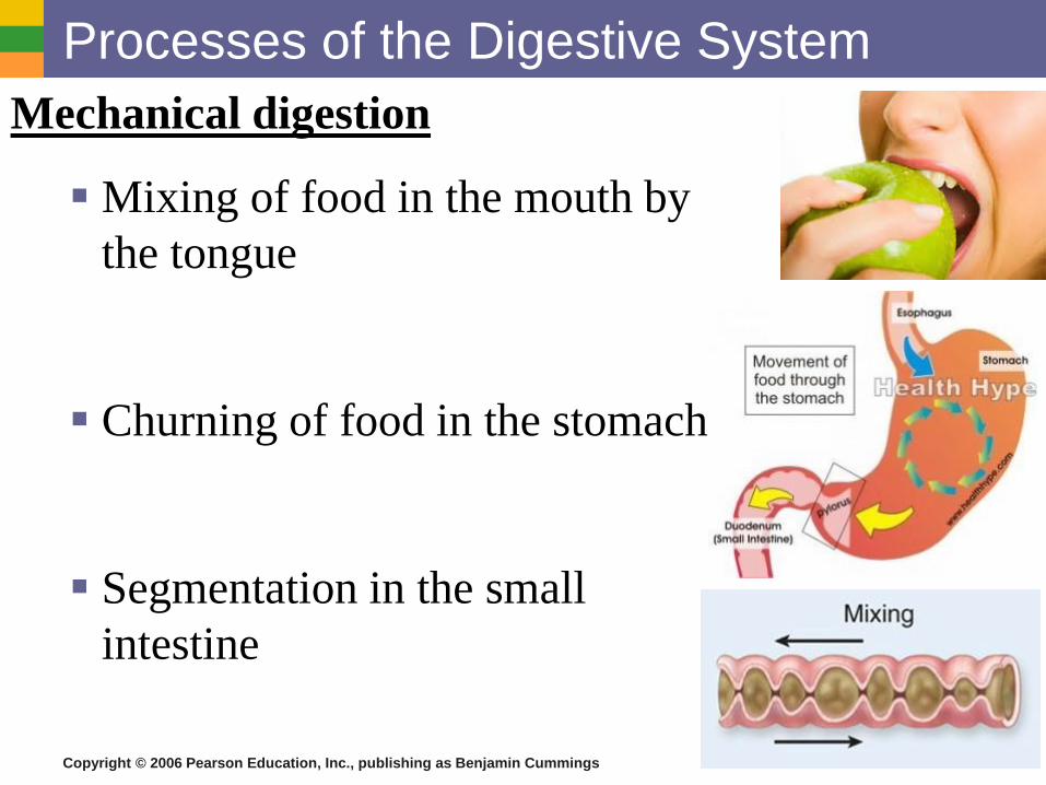
Processes of the Digestive System
Digestive Process
▪ Ingestion
▪ Digestion
▪ Mechanical
▪ Chemical
▪ Absorption
▪ Defecation
Figure 14.11

Copyright © 2006 Pearson Education, Inc., publishing as Benjamin Cummings
Processes of the Digestive System
▪ Ingestion – getting food into the mouth
▪ Propulsion – moving foods from one region of the
digestive system to another

Copyright © 2006 Pearson Education, Inc., publishing as Benjamin Cummings
Processes of the Digestive System
▪ Peristalsis – alternating waves of propulsive contractions that
moves substances from one organ to another
▪ Segmentation – moving materials back and forth to aid in mixing
Figure 14.12

Copyright © 2006 Pearson Education, Inc., publishing as Benjamin Cummings
Processes of the Digestive System
Absorption
▪ End products of digestion are absorbed in the
blood or lymph
▪ Food must enter mucosal cells and then into
blood or lymph capillaries
Defecation
▪ Elimination of indigestible substances as feces

Copyright © 2006 Pearson Education, Inc., publishing as Benjamin Cummings
Copyright © 2006 Pearson Education, Inc., publishing as Benjamin Cummings
Processes of the Digestive System
Mechanical digestion
▪ Mixing of food in the mouth by
the tongue
▪ Churning of food in the stomach
▪ Segmentation in the small
intestine

Copyright © 2006 Pearson Education, Inc., publishing as Benjamin Cummings
Processes of the Digestive System
Chemical Digestion
▪ Enzymes break down food molecules into their building blocks
▪ Each major food group uses different enzymes
▪ Carbohydrates are broken to simple sugars
▪ Proteins are broken to amino acids
▪ Fats are broken to fatty acids and alcohols

Copyright © 2006 Pearson Education, Inc., publishing as Benjamin Cummings
Organs of the Digestive System
▪ Two main groups
▪ Alimentary canal – continuous coiled hollow
tube
▪ Accessory digestive organs – aid in digestion
mechanically or chemically

Copyright © 2006 Pearson Education, Inc., publishing as Benjamin Cummings
Organs of the Alimentary Canal
▪ Mouth
▪ Pharynx
▪ Esophagus
▪ Stomach
▪ Small intestine
▪ Large intestine
▪ Anus
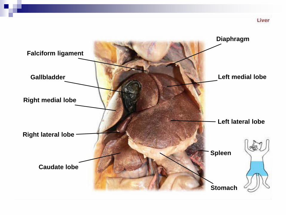
Falciform ligament
Gallbladder
Right medial lobe
Right lateral lobe
Caudate lobe
Diaphragm
Left medial lobe
Left lateral lobe
Spleen
Stomach

Copyright © 2006 Pearson Education, Inc., publishing as Benjamin Cummings
Mouth (Oral Cavity) Anatomy▪ Lips (labia) – protect
the anterior opening
▪ Cheeks – form the lateral walls
▪ Hard palate – forms the anterior roof
▪ Soft palate – forms the posterior roof
▪ Uvula – fleshy projection of the soft palate
Figure 14.2a

Copyright © 2006 Pearson Education, Inc., publishing as Benjamin Cummings
Mouth (Oral Cavity) Anatomy
▪ Vestibule – space between lips externally and teeth and gums internally
▪ Oral cavity – area contained by the teeth
▪ Tongue – attached at hyoid and styloid processes of the skull, and by the lingual frenulum
Figure 14.2a

Copyright © 2006 Pearson Education, Inc., publishing as Benjamin Cummings
Mouth (Oral Cavity) Anatomy
Tonsils
▪ Palatine tonsils
▪ Lingual tonsil
Figure 14.2a

Copyright © 2006 Pearson Education, Inc., publishing as Benjamin Cummings
Processes of the Mouth
▪ Mastication (chewing) of food (mechanical digestion)
▪ Mixing masticated food with saliva (chemical digestion)
▪ Initiation of swallowing by the tongue
▪ Allowing for the sense of taste

Copyright © 2006 Pearson Education, Inc., publishing as Benjamin Cummings
Digestive Activities of the Mouth
Mechanical breakdown
▪ Food is physically broken down by chewing
Chemical digestion
▪ Food is mixed with saliva
▪ Breaking of starch into maltose by salivary
amylase

Copyright © 2006 Pearson Education, Inc., publishing as Benjamin Cummings
Pharynx Anatomy
▪ Nasopharynx –not part of the digestive system
▪ Oropharynx – posterior to oral cavity
▪ Laryngopharynx – below the oropharynx and connected to the esophagus
Figure 14.2a

Copyright © 2006 Pearson Education, Inc., publishing as Benjamin Cummings
Pharynx Function
▪ Serves as a passageway for air and food
▪ Food is propelled to the esophagus by
two muscle layers
▪ Longitudinal inner layer
▪ Circular outer layer
▪ Food movement is by alternating
contractions of the muscle layers
(peristalsis)
▪ Has no digestive function

Copyright © 2006 Pearson Education, Inc., publishing as Benjamin Cummings
Deglutition (Swallowing)
Buccal phase
▪ Voluntary
▪ Occurs in the mouth
▪ Food is formed into
a bolus
▪ The bolus is forced
into the pharynx by
the tongue

Copyright © 2006 Pearson Education, Inc., publishing as Benjamin Cummings
Deglutition (Swallowing)
Pharyngeal-esophageal phase
▪ Involuntary transport of the bolus
▪ All passageways except to the stomach are blocked
▪ Tongue blocks off the mouth
▪ Soft palate (uvula) blocks the nasopharynx
▪ Epiglottis blocks the larynx

Copyright © 2006 Pearson Education, Inc., publishing as Benjamin Cummings
Deglutition (Swallowing)
Pharyngeal-esophogeal phase (continued)
▪ Peristalsis moves the bolus toward the
stomach
▪ The cardioesophageal sphincter is opened
when food presses against it

Copyright © 2006 Pearson Education, Inc., publishing as Benjamin Cummings
Esophagus
▪ Runs from pharynx to stomach
through the diaphragm
▪ Passageway for food only
(respiratory system branches off
after the pharynx)
▪ Has no digestive function
▪ Conducts food by peristalsis
(slow rhythmic squeezing to
move from one point to another)

Copyright © 2006 Pearson Education, Inc., publishing as Benjamin Cummings
Layers of Alimentary Canal Organs
Mucosa
▪ Innermost layer
▪ Moist membrane
▪ Surface epithelium
▪ Small amount of connective
tissue
(lamina propria)
▪ Small smooth muscle layer

Copyright © 2006 Pearson Education, Inc., publishing as Benjamin Cummings
Layers of Alimentary Canal Organs
Submucosa
▪ Just beneath the mucosa
▪ Soft connective tissue with blood vessels,
nerve endings, and lymphatics

Copyright © 2006 Pearson Education, Inc., publishing as Benjamin Cummings
Layers of Alimentary Canal Organs
Muscularis externa – smooth
muscle
▪ Inner circular layer
▪ Outer longitudinal layer
Serosa
▪ Outermost layer –
visceral peritoneum
▪ Layer of serous fluid-
producing cells

Copyright © 2006 Pearson Education, Inc., publishing as Benjamin Cummings
Layers of Alimentary Canal Organs
Figure 14.3
Copyright © 2006 Pearson Education, Inc., publishing as Benjamin Cummings
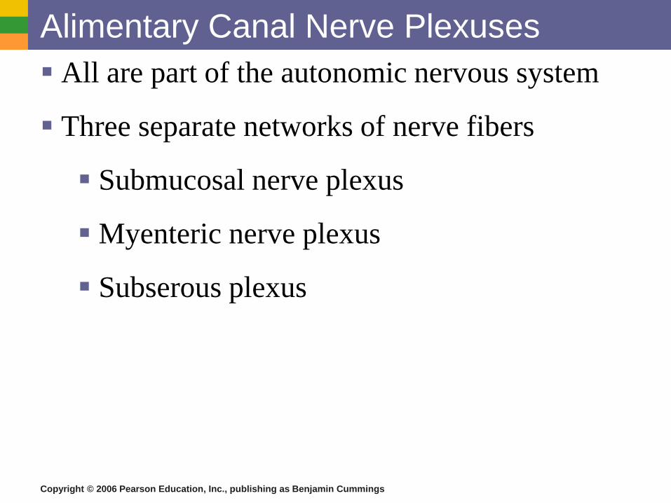
Alimentary Canal Nerve Plexuses
▪ All are part of the autonomic nervous system
▪ Three separate networks of nerve fibers
▪ Submucosal nerve plexus
▪ Myenteric nerve plexus
▪ Subserous plexus

Copyright © 2006 Pearson Education, Inc., publishing as Benjamin Cummings
Stomach Functions
▪ Acts as a storage tank for food
▪ Site of food breakdown
▪ Chemical breakdown of protein begins
▪ Delivers chyme (processed food) to the small
intestine

Copyright © 2006 Pearson Education, Inc., publishing as Benjamin Cummings
Stomach Anatomy
▪ Located on the left side of the abdominal cavity
▪ Food enters at the cardioesophageal sphincter
(also called the cardiac or lower esophageal sphincter)

Copyright © 2006 Pearson Education, Inc., publishing as Benjamin Cummings
Stomach Anatomy▪ Regions of the stomach
▪ Cardiac region – near the heart
▪ Fundus
▪ Body
▪ Phylorus – funnel-shaped terminal end
▪ Food empties into the small intestine at the pyloric sphincter

Copyright © 2006 Pearson Education, Inc., publishing as Benjamin Cummings
Stomach Anatomy
▪ Rugae – internal folds of the mucosa
▪ External regions
▪ Lesser curvature
▪ Greater curvature

Copyright © 2006 Pearson Education, Inc., publishing as Benjamin Cummings
Stomach Anatomy▪ Layers of peritoneum attached to the stomach
▪ Lesser omentum – attaches the liver to the lesser curvature
▪ Greater omentum – attaches the greater curvature to the posterior
body wall
▪ Contains fat to insulate, cushion, and protect abdominal organs

Copyright © 2006 Pearson Education, Inc., publishing as Benjamin Cummings
Specialized Mucosa of the Stomach
▪ Simple columnar epithelium
▪ Mucous neck cells – produce a sticky alkaline mucus
▪ Gastric glands – secrete gastric juice
▪ Chief cells – produce protein-digesting enzymes
(pepsinogens)
▪ Parietal cells – produce hydrochloric acid
▪ Endocrine cells – produce gastrin
Copyright © 2006 Pearson Education, Inc., publishing as Benjamin Cummings
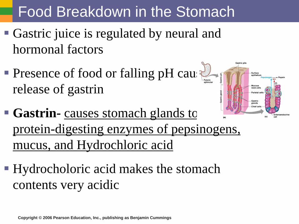
Food Breakdown in the Stomach
▪ Gastric juice is regulated by neural and
hormonal factors
▪ Presence of food or falling pH causes the
release of gastrin
▪ Gastrin- causes stomach glands to produce
protein-digesting enzymes of pepsinogens,
mucus, and Hydrochloric acid
▪ Hydrocholoric acid makes the stomach
contents very acidic

Copyright © 2006 Pearson Education, Inc., publishing as Benjamin Cummings
Structure of the Stomach Mucosa
▪ Gastric pits formed by folded mucosa
▪ Glands and specialized cells are in the gastric gland region

Copyright © 2006 Pearson Education, Inc., publishing as Benjamin Cummings
Necessity of an Extremely Acid
Environment in the Stomach
▪ Activates pepsinogen to pepsin for protein digestion
▪ Provides a hostile environment for microorganisms

Copyright © 2006 Pearson Education, Inc., publishing as Benjamin Cummings
Digestion and Absorption in the Stomach
▪ Protein digestion enzymes
▪ Pepsin – an active protein digesting enzyme
▪ Rennin – works on digesting milk protein
▪ The only absorption that occurs in the stomach is of
alcohol and aspirin

Copyright © 2006 Pearson Education, Inc., publishing as Benjamin Cummings
Propulsion in the Stomach
▪ Food must first be well mixed
▪ Rippling peristalsis occurs in the lower stomach
Figure 14.15

Copyright © 2006 Pearson Education, Inc., publishing as Benjamin Cummings
Propulsion in the Stomach
▪ The pylorus meters out chyme into the small
intestine (30 ml at a time)
▪ The stomach empties in four to six hours
Figure 14.15

Copyright © 2006 Pearson Education, Inc., publishing as Benjamin Cummings

Copyright © 2006 Pearson Education, Inc., publishing as Benjamin Cummings
Small Intestine
▪ The body’s major digestive organ
▪ Site of nutrient absorption into the blood
▪ Muscular tube extending form the pyloric
sphincter to the ileocecal valve
▪ Suspended from the posterior abdominal wall
by the mesentery

Copyright © 2006 Pearson Education, Inc., publishing as Benjamin Cummings
Subdivisions of the Small IntestineDuodenum
▪ Attached to the stomach
▪ Curves around the head
of the pancreas
▪ Location where pancreatic
enzymes and bile get released
Jejunum
▪ Attaches anteriorly to the
duodenum
Ileum
▪ Extends from jejunum to large
intestine

Copyright © 2006 Pearson Education, Inc., publishing as Benjamin Cummings
Propulsion in the Small Intestine
▪ Peristalsis is the major means of moving food
▪ Segmental movements
▪ Mix chyme with digestive juices
▪ Aid in propelling food

Copyright © 2006 Pearson Education, Inc., publishing as Benjamin Cummings
Villi of the Small Intestine
▪ Fingerlike structures formed by the
mucosa
▪ Give the small intestine more surface
area
Figure 14.7a

Copyright © 2006 Pearson Education, Inc., publishing as Benjamin Cummings
Microvilli of the Small Intestine
▪ Small projections of the plasma membrane
▪ Found on absorptive cells
Figure 14.7c

Copyright © 2006 Pearson Education, Inc., publishing as Benjamin Cummings
Structures Involved in Absorption of
Nutrients
▪ Absorptive cells
▪ Blood capillaries
▪ Lacteals (specialized
lymphatic capillaries)
Figure 14.7b

Copyright © 2006 Pearson Education, Inc., publishing as Benjamin Cummings
Folds of the Small Intestine
▪ Called circular folds or plicae circulares
▪ Deep folds of the mucosa and submucosa
▪ Do not disappear when filled with food
▪ The submucosa has Peyer’s patches (collections of
lymphatic tissue)

Copyright © 2006 Pearson Education, Inc., publishing as Benjamin Cummings
Digestion in the Small Intestine
▪ Enzymes from the brush border
▪ Break double sugars into simple sugars
▪ Complete some protein digestion
▪ Pancreatic enzymes play the major digestive function
▪ Help complete digestion of starch (pancreatic amylase)
▪ Carry out about half of all protein digestion (trypsin, etc.)
Copyright © 2006 Pearson Education, Inc., publishing as Benjamin Cummings
Digestion in the Small Intestine
▪ Pancreatic enzymes play the major digestive
function (continued)
▪ Responsible for fat digestion (lipase)
▪ Digest nucleic acids (nucleases)
▪ Alkaline content neutralizes acidic chyme

Copyright © 2006 Pearson Education, Inc., publishing as Benjamin Cummings
Chemical Digestion in the Small Intestine▪ Source of enzymes that are mixed with chyme
▪ Intestinal cells
▪ Pancreas
▪ Bile enters from the gall bladder
▪ All get released into the duodenum section of the small intestines

Copyright © 2006 Pearson Education, Inc., publishing as Benjamin Cummings
Regulation of Pancreas and Liver
Cholecystokinin and Secretin
▪ Two hormones that are produced by the mucosa cells
which influence the release of pancreatic juice and
bile in response to chyme entering the small intestine

Copyright © 2006 Pearson Education, Inc., publishing as Benjamin Cummings
Absorption in the Small Intestine
▪ Water is absorbed along the length of the small
intestine
▪ End products of digestion
▪ Most substances are absorbed by active transport
through cell membranes
▪ Lipids are absorbed by diffusion
▪ Substances are transported to the liver by the hepatic
portal vein or lymph

Copyright © 2006 Pearson Education, Inc., publishing as Benjamin Cummings
Large Intestine
▪ Larger in diameter, but
shorter than the small
intestine

Copyright © 2006 Pearson Education, Inc., publishing as Benjamin Cummings
Functions of the Large Intestine
▪ Absorption of water, ions, and vitamin K
▪ Eliminates indigestible food from the body as
feces
▪ Does not participate in digestion of food
▪ Goblet cells produce mucus to act as a
lubricant

Copyright © 2006 Pearson Education, Inc., publishing as Benjamin Cummings
Food Breakdown and Absorption in the
Large Intestine
▪ No digestive enzymes are produced
▪ Resident bacteria digest remaining nutrients
▪ Produce some vitamin K and B
▪ Release gases
▪ Water, ions, and vitamins K and B are absorbed
▪ Remaining materials are eliminated via feces

Copyright © 2006 Pearson Education, Inc., publishing as Benjamin Cummings
Structures of the Large Intestine▪ Cecum – saclike first part of the large intestine
▪ Appendix
▪ Accumulation of lymphatic tissue that sometimes becomes
inflamed (appendicitis)
▪ Hangs from the cecum

Copyright © 2006 Pearson Education, Inc., publishing as Benjamin Cummings
Structures of the Large Intestine
▪ Colon
▪ Ascending
▪ Transverse
▪ Descending
▪ S-shaped sigmoidal
▪ Rectum – holding site of waste
▪ Anus – external body opening of the large intestines

Copyright © 2006 Pearson Education, Inc., publishing as Benjamin Cummings
Propulsion in the Large Intestine
▪ Sluggish peristalsis
▪ Mass movements
▪ Slow, powerful movements
▪ Occur three to four times per day
▪ Presence of feces in the rectum causes a defecation
reflex
▪ Internal anal sphincter is relaxed
▪ Defecation occurs with relaxation of the
voluntary (external) anal sphincter

Falciform ligament
Gallbladder
Right medial lobe
Right lateral lobe
Caudate lobe
Diaphragm
Left medial lobe
Left lateral lobe
Spleen
Stomach

Copyright © 2006 Pearson Education, Inc., publishing as Benjamin Cummings
▪ End of Alimentary Canal…on to Accessory
Structures.

Copyright © 2006 Pearson Education, Inc., publishing as Benjamin Cummings
Accessory Digestive Organs
▪ Salivary glands
▪ Teeth
▪ Pancreas
▪ Liver
▪ Gall bladder

Copyright © 2006 Pearson Education, Inc., publishing as Benjamin Cummings
Salivary Glands
Saliva-producing glands
▪ Parotid glands – located anterior to ears
▪ Submandibular glands
▪ Sublingual glands

Copyright © 2006 Pearson Education, Inc., publishing as Benjamin Cummings
Saliva
▪ Mixture of mucus and serous fluids
▪ Helps to form a food bolus
▪ Contains salivary amylase to begin starch digestion
▪ Dissolves chemicals so they can be tasted

Copyright © 2006 Pearson Education, Inc., publishing as Benjamin Cummings
Teeth
▪ The role is to masticate (chew) food
▪ Humans have two sets of teeth
▪ Deciduous (baby or milk) teeth
▪ 20 teeth are fully formed by age two

Copyright © 2006 Pearson Education, Inc., publishing as Benjamin Cummings
Teeth
Permanent teeth
▪ Replace deciduous teeth beginning between the ages of 6
to 12
▪ A full set is 32 teeth, but some people do not have
wisdom teeth

Copyright © 2006 Pearson Education, Inc., publishing as Benjamin Cummings
Classification of Teeth
▪ Incisors
▪ Canines
▪ Premolars
▪ Molars
▪ Wisdom Teeth
(3rd Molar)

Copyright © 2006 Pearson Education, Inc., publishing as Benjamin Cummings
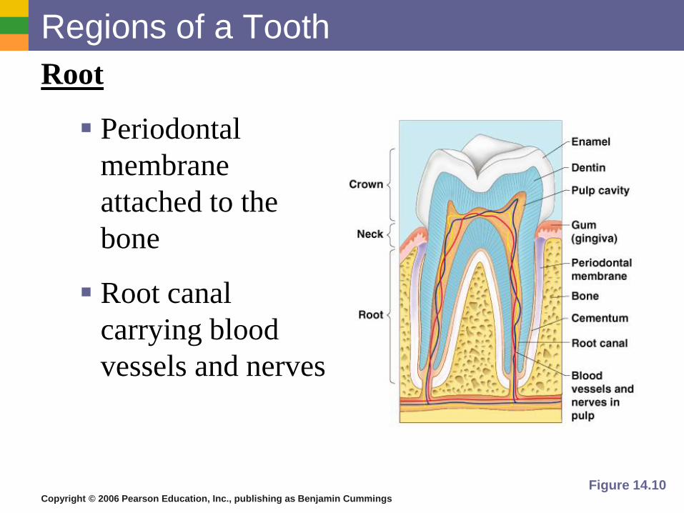
Regions of a Tooth
Crown – exposed part
▪ Outer enamel
▪ Dentin
▪ Pulp cavity
Neck
▪ Region in contact with
the gum
▪ Connects crown to root
Figure 14.10
Copyright © 2006 Pearson Education, Inc., publishing as Benjamin Cummings
Regions of a Tooth
Root
▪ Periodontal
membrane
attached to the
bone
▪ Root canal
carrying blood
vessels and nerves
Figure 14.10

Copyright © 2006 Pearson Education, Inc., publishing as Benjamin Cummings
Pancreas▪ Produces a wide spectrum of digestive enzymes that break
down all categories of food
▪ Enzymes are secreted into the duodenum
▪ Alkaline fluid introduced with enzymes neutralizes acidic chyme
▪ Endocrine products of pancreas
▪ Insulin
▪ Glucagons

Copyright © 2006 Pearson Education, Inc., publishing as Benjamin Cummings
Liver▪ Functions: makes bile, detoxify drugs & alcohol, degrades hormones,
make cholestrerol, and plays a central role in metabolism
▪ Largest gland in the body
▪ Located on the right side of the body under the diaphragm
▪ Consists of four lobes
▪ Connected to the gall bladder via the common hepatic duct

Liver
Gall bladder
Pancreas
Duodenum
Stomach
Bile duct

Copyright © 2006 Pearson Education, Inc., publishing as Benjamin Cummings
Liver
Hepatic Portal Circulation
▪ A unique circulation, brings
nutrient rich blood draining
from the digestive viscera
directly to the liver
▪ This is done so that the liver’s
nutrient needs are met first since
it’s the body’s major metabolic
organ

Copyright © 2006 Pearson Education, Inc., publishing as Benjamin Cummings
Metabolism
▪ Chemical reactions necessary to maintain life
▪ Catabolism – substances are broken down to
simpler substances
▪ Anabolism – larger molecules are built from
smaller ones
▪ Energy is released during catabolism

Copyright © 2006 Pearson Education, Inc., publishing as Benjamin Cummings
Congenital defects
Phenylketonuria
▪ Hereditary inability of
tissue cells to metabolize
the amino acid
phenylalanine, which can
result in brain damage and
retardation unless a
special diet low in
phenylalanine is followed

Copyright © 2006 Pearson Education, Inc., publishing as Benjamin Cummings
Bile▪ Produced by cells in the liver, stored in the gall bladder
▪ Composition
▪ Bile salts
▪ Bile pigment (mostly bilirubin from the breakdown of
hemoglobin)
▪ Cholesterol
▪ Phospholipids
▪ Electrolytes

Copyright © 2006 Pearson Education, Inc., publishing as Benjamin Cummings
Gall Bladder
▪ Sac found in hollow fossa of liver
▪ Stores bile from the liver by way of the cystic duct
▪ Bile is introduced into the duodenum in the presence of
fatty food
▪ Gallstones can cause blockages

Copyright © 2006 Pearson Education, Inc., publishing as Benjamin Cummings
Stimulation of the Release of Pancreatic
Juice
▪ Vagus nerve
▪ Local hormones
▪ Secretin
▪ Cholecystokinin
Figure 14.16

Copyright © 2006 Pearson Education, Inc., publishing as Benjamin Cummings
Regulation of Pancreas and Liver
Cholecystokinin and Secretin
▪ Two hormones that are produced by the mucosa cells
which influence the release of pancreatic juice and
bile in response to chyme entering the small intestine

Copyright © 2006 Pearson Education, Inc., publishing as Benjamin Cummings
Acidosis (Ketoacidosis)
▪ “Acidic blood,” can occur when someone is on a
“no-carbohydrate” diet, has uncontrolled
diabetes mellitus, and starvation when the body
is forced to rely almost totally on fats to fuel its
energy needs

Copyright © 2006 Pearson Education, Inc., publishing as Benjamin Cummings
Developmental Issues of the Liver
Jaundice
▪ A condition where body tissues turn yellow due
to bile buildup within the liver causing bile
pigments to circulate through the body

Copyright © 2006 Pearson Education, Inc., publishing as Benjamin Cummings
Control of Digestive Activity
▪ Mostly controlled by reflexes via the
parasympathetic division
▪ Chemical and mechanical receptors are
located in organ walls that trigger reflexes

Copyright © 2006 Pearson Education, Inc., publishing as Benjamin Cummings
Control of Digestive Activity
▪ Stimuli include:
▪ Stretch of the organ
▪ pH of the contents
▪ Presence of breakdown products
▪ Reflexes include:
▪ Activation or inhibition of glandular secretions
▪ Smooth muscle activity
▪ Sucking Reflex- helps infants to hold onto thenipple and swallow