Embed Size (px)
Citation preview
The Digestive System and Body Metabolism
Chapter 14
• Ingestion— taking in food
• Digestion— breaking food down both physically and chemically
• Absorption— movement of nutrients into the bloodstream
• Defecation— rids the body of indigestible waste
The Digestive System Functions
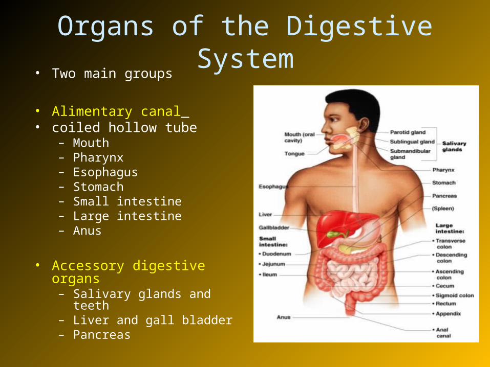
Organs of the Digestive System• Two main groups
• Alimentary canal • coiled hollow tube
– Mouth– Pharynx– Esophagus– Stomach– Small intestine– Large intestine– Anus
• Accessory digestive organs– Salivary glands and teeth– Liver and gall bladder– Pancreas
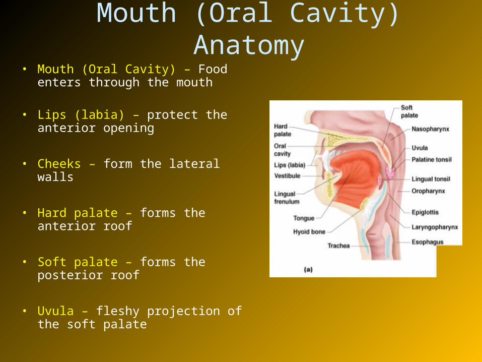
Mouth (Oral Cavity) Anatomy• Mouth (Oral Cavity) – Food enters
through the mouth
• Lips (labia) – protect the anterior opening
• Cheeks – form the lateral walls
• Hard palate – forms the anterior roof
• Soft palate – forms the posterior roof
• Uvula – fleshy projection of the soft palate
Mouth (Oral Cavity) Anatomy
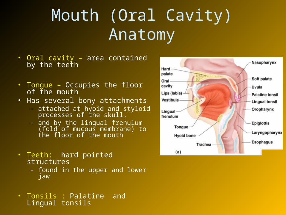
• Oral cavity – area contained by the teeth
• Tongue – Occupies the floor of the mouth
• Has several bony attachments– attached at hyoid and styloid
processes of the skull, – and by the lingual frenulum (fold of
mucous membrane) to the floor of the mouth
• Teeth: hard pointed structures – found in the upper and lower jaw
• Tonsils : Palatine and Lingual tonsils
• Food enters through mouth, mixed with saliva, and masticated (chewed)
• And then swallowed by the tongue
• Taste buds are present on tongue which allows for the sense of taste
Mouth Physiology
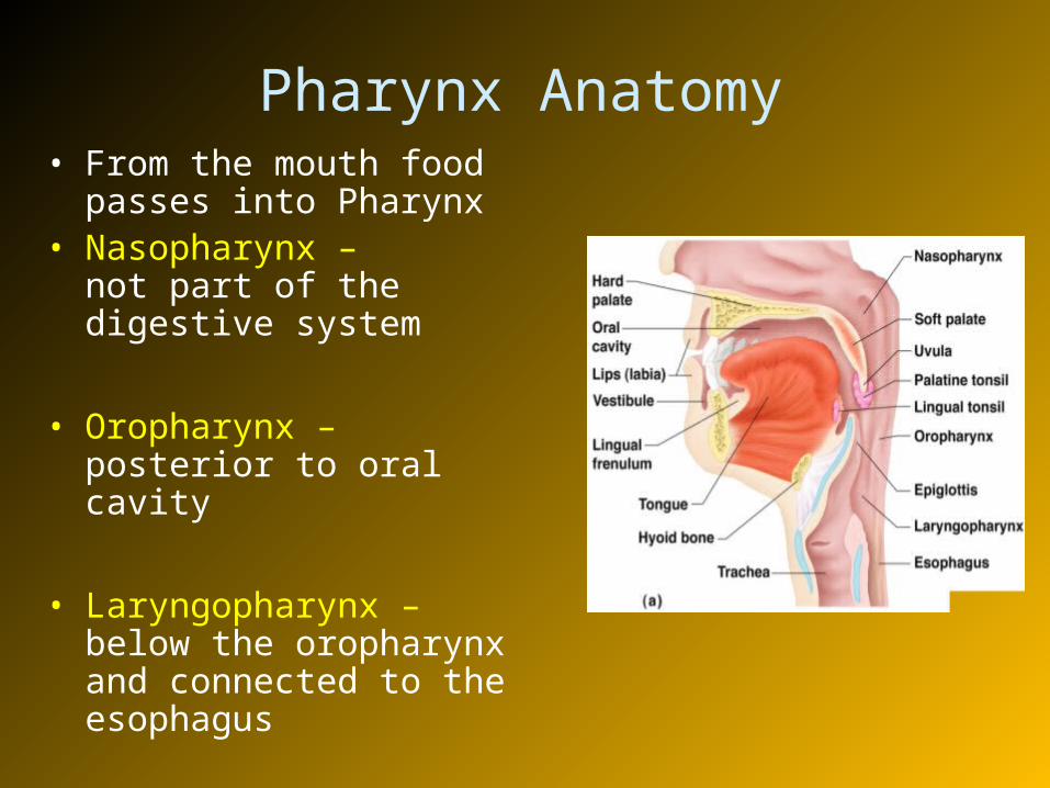
Pharynx Anatomy• From the mouth food passes
into Pharynx• Nasopharynx –
not part of the digestive system
• Oropharynx – posterior to oral cavity
• Laryngopharynx – below the oropharynx and connected to the esophagus
• Serves as a passageway for air and food
• Food is propelled to the esophagus by two muscle layers
– Longitudinal inner layer
– Circular outer layer
• Food movement is by alternating contractions of the muscle layers (peristalsis)
Pharynx Physiology
Esophagus Anatomy and Physiology
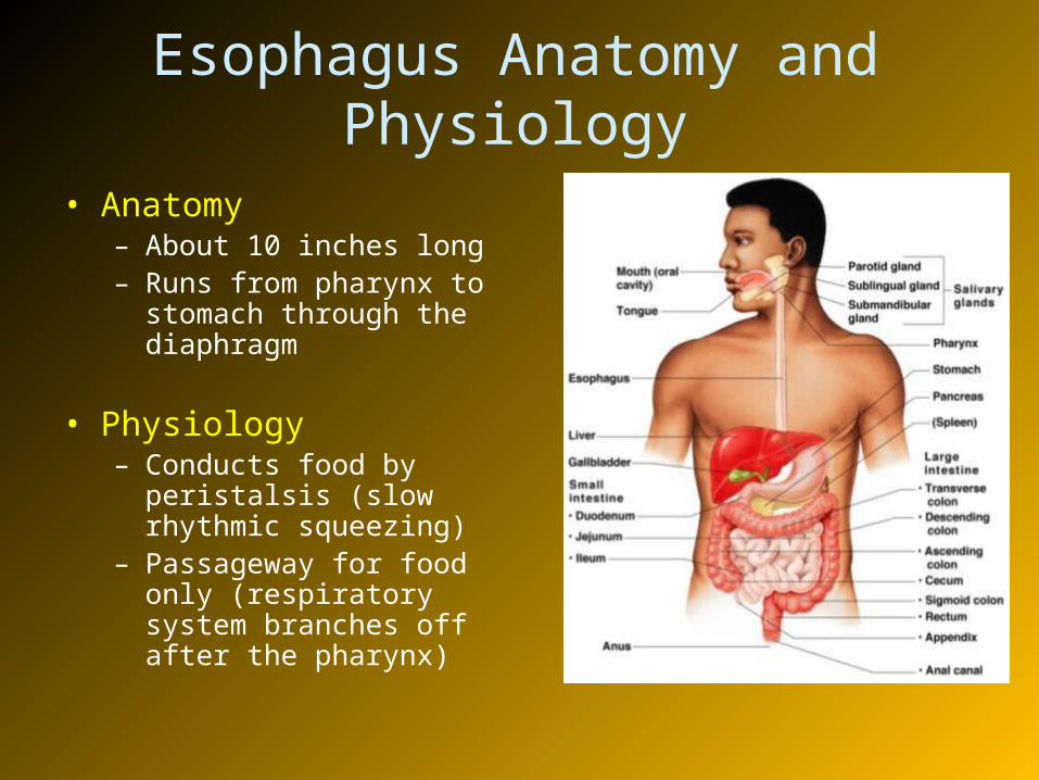
• Anatomy– About 10 inches long– Runs from pharynx to
stomach through the diaphragm
• Physiology– Conducts food by
peristalsis (slow rhythmic squeezing)
– Passageway for food only (respiratory system branches off after the pharynx)
• Walls of alimentary canal from esophagus to the large intestine are made up of four tissue layers:
– Mucosa– Submucosa– Muscularis externa– Serosa
Layers of Alimentary Canal Organs

Layers of Alimentary Canal Organs• Mucosa
– Innermost layer, moist membrane that lines the cavity or lumen of the organ– consists of
• Surface epithelium• Small amount of connective tissue (lamina propria)• Small smooth muscle layer
• Submucosa– Just beneath the mucosa– Soft connective tissue with blood vessels, nerve endings, lymph nodules,
lymphatic vessels

Layers of Alimentary Canal Organs• Muscularis externa – Made up of smooth muscle
– Inner circular layer– Outer longitudinal layer
• Serosa- outermost layer of the wall contains – Visceral peritoneum— Single layer of flat serous fluid
producing cells– Parietal peritoneum— lines the abdominopelvic cavity
• Two important nerve plexuses serve the alimentary canal
• Both are part of the autonomic nervous system– Submucosal nerve plexus– Myenteric nerve plexus
• Function is to regulate mobility and secretory activity of the GI tract organs
Alimentary Canal Nerve Plexuses

Stomach Anatomy
• Located on the left side of the abdominal cavity
• Food enters at the – cardio-esophageal
sphincter
• Food empties into the small intestine – at the pyloric sphincter (valve)
Stomach Anatomy
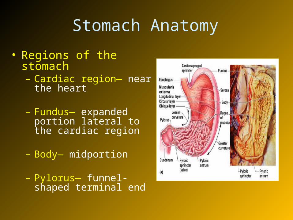
• Regions of the stomach– Cardiac region— near the
heart
– Fundus— expanded portion lateral to the cardiac region
– Body— midportion
– Pylorus— funnel-shaped terminal end
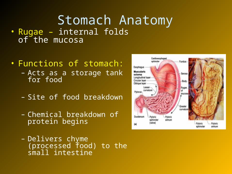
Stomach Anatomy• Rugae – internal folds of
the mucosa
• Functions of stomach:– Acts as a storage tank for
food
– Site of food breakdown
– Chemical breakdown of protein begins
– Delivers chyme (processed food) to the small intestine
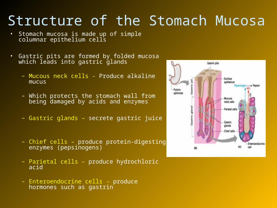
Structure of the Stomach Mucosa• Stomach mucosa is made up of simple columnar
epithelium cells
• Gastric pits are formed by folded mucosa which leads into gastric glands
– Mucous neck cells - Produce alkaline mucus
– Which protects the stomach wall from being damaged by acids and enzymes
– Gastric glands – secrete gastric juice
– Chief cells – produce protein-digesting enzymes (pepsinogens)
– Parietal cells – produce hydrochloric acid
– Enteroendocrine cells – produce hormones such as gastrin
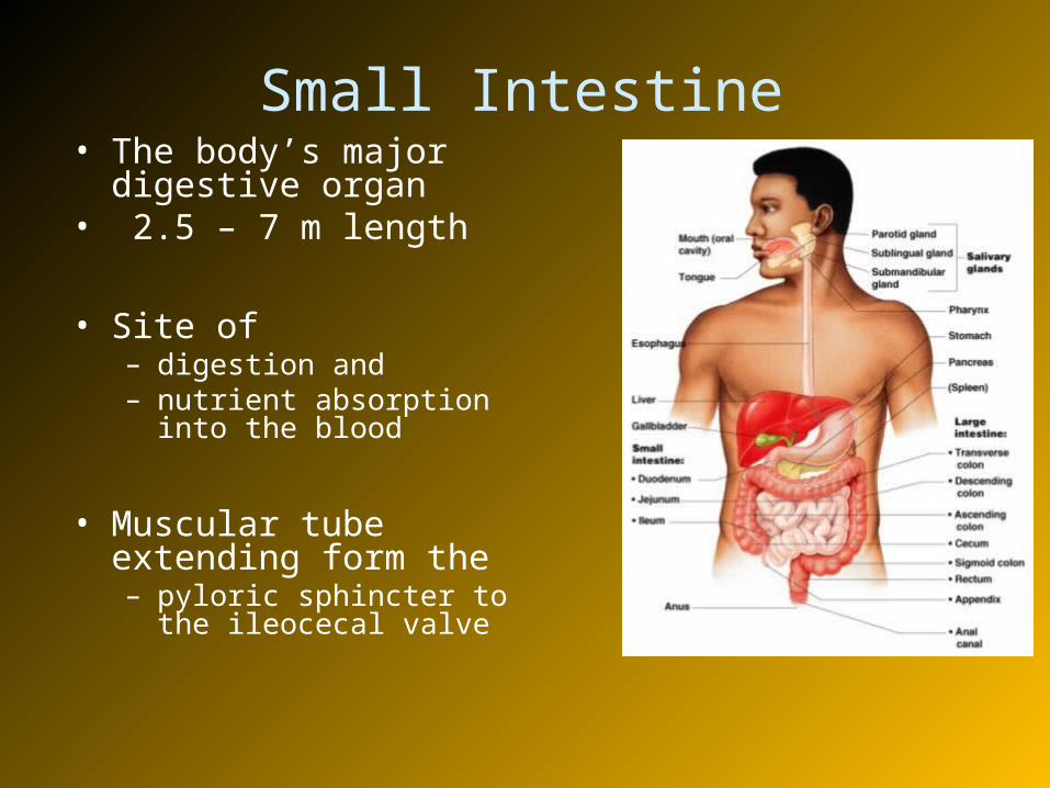
Small Intestine• The body’s major digestive
organ• 2.5 – 7 m length
• Site of – digestion and – nutrient absorption into the
blood
• Muscular tube extending form the – pyloric sphincter to the ileocecal
valve
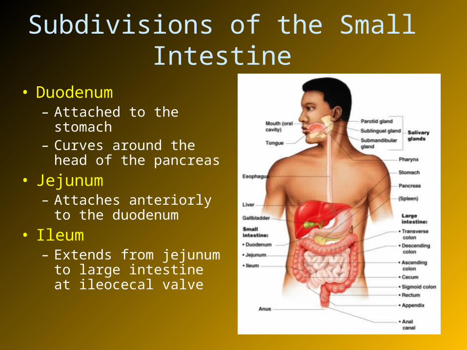
Subdivisions of the Small Intestine
• Duodenum– Attached to the stomach– Curves around the head
of the pancreas
• Jejunum– Attaches anteriorly to
the duodenum
• Ileum– Extends from jejunum to
large intestine at ileocecal valve
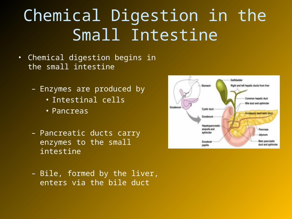
Chemical Digestion in the Small Intestine
• Chemical digestion begins in the small intestine
– Enzymes are produced by• Intestinal cells• Pancreas
– Pancreatic ducts carry enzymes to the small intestine
– Bile, formed by the liver, enters via the bile duct
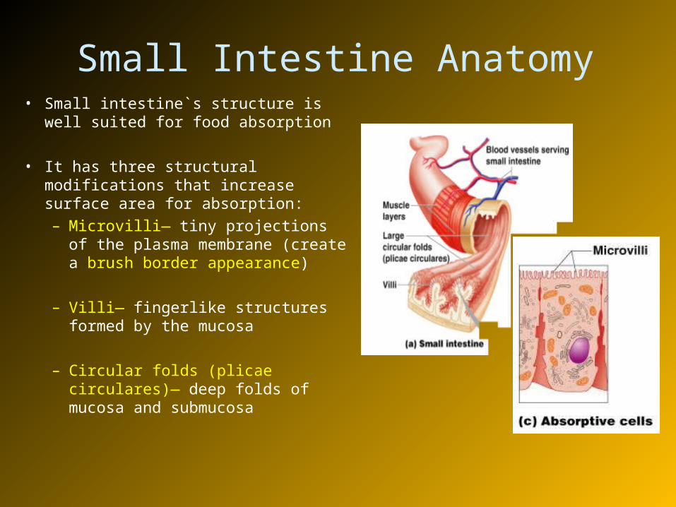
• Small intestine`s structure is well suited for food absorption
• It has three structural modifications that increase surface area for absorption:
– Microvilli— tiny projections of the plasma membrane (create a brush border appearance)
– Villi— fingerlike structures formed by the mucosa
– Circular folds (plicae circulares)— deep folds of mucosa and submucosa
Small Intestine Anatomy
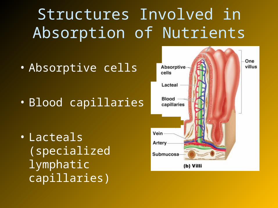
Structures Involved in Absorption of Nutrients
• Absorptive cells
• Blood capillaries
• Lacteals (specialized lymphatic capillaries)

Large Intestine
• Larger in diameter, but - shorter in length
than the small intestine
- 1.5 m long
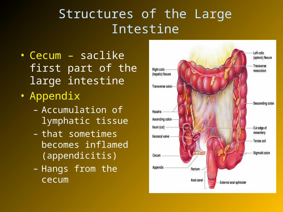
Structures of the Large Intestine
• Cecum – saclike first part of the large intestine
• Appendix– Accumulation of
lymphatic tissue – that sometimes
becomes inflamed (appendicitis)
– Hangs from the cecum
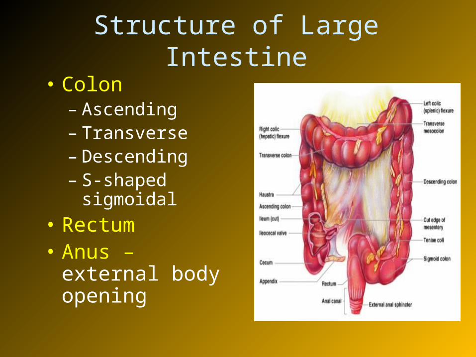
Structure of Large Intestine
• Colon– Ascending– Transverse– Descending– S-shaped
sigmoidal
• Rectum• Anus – external
body opening

Functions of the Large Intestine
• Dry out the indigestible food residue by absorbing water
• Eliminates indigestible food from the body as feces
• Goblet cells present on mucosa, produce alkaline mucus which lubricates the passage of feces
Accessory Digestive Organs
• Teeth
• Salivary glands
• Pancreas
• Liver
• Gall bladder
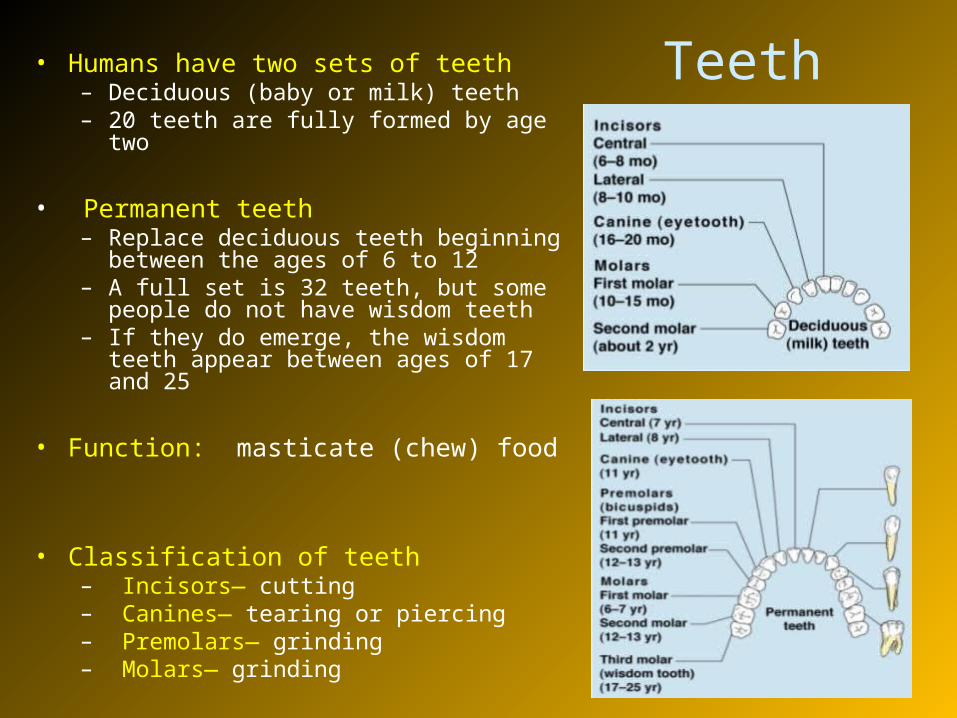
Teeth• Humans have two sets of teeth– Deciduous (baby or milk) teeth– 20 teeth are fully formed by age two
• Permanent teeth– Replace deciduous teeth beginning between
the ages of 6 to 12– A full set is 32 teeth, but some people do
not have wisdom teeth– If they do emerge, the wisdom teeth appear
between ages of 17 and 25
• Function: masticate (chew) food
• Classification of teeth– Incisors— cutting– Canines— tearing or piercing– Premolars— grinding– Molars— grinding
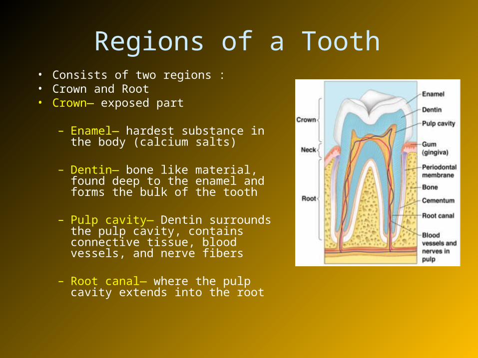
Regions of a Tooth• Consists of two regions :• Crown and Root• Crown— exposed part
– Enamel— hardest substance in the body (calcium salts)
– Dentin— bone like material, found deep to the enamel and forms the bulk of the tooth
– Pulp cavity— Dentin surrounds the pulp cavity, contains connective tissue, blood vessels, and nerve fibers
– Root canal— where the pulp cavity extends into the root
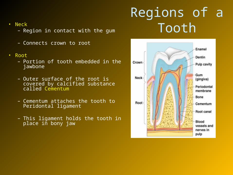
Regions of a Tooth• Neck
– Region in contact with the gum
– Connects crown to root
• Root– Portion of tooth embedded in the
jawbone
– Outer surface of the root is covered by calcified substance called Cementum
– Cementum attaches the tooth to Peridontal ligament
– This ligament holds the tooth in place in bony jaw

Salivary Glands• Saliva-producing glands• Three pairs of salivary glands empty
secretions into the mouth
– Parotid glands – located anterior to ears
– Submandibular glands– Sublingual glands
• Function of saliva– Saliva is a mixture of mucus and
serous fluids
– Moisten and binds the food together and forms a food bolus
– Contains salivary amylase to begin starch digestion
– Dissolves chemicals so they can be tasted
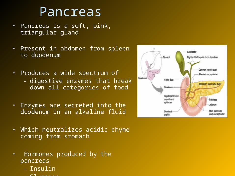
Pancreas• Pancreas is a soft, pink, triangular gland
• Present in abdomen from spleen to duodenum
• Produces a wide spectrum of – digestive enzymes that break down
all categories of food
• Enzymes are secreted into the duodenum in an alkaline fluid
• Which neutralizes acidic chyme coming from stomach
• Hormones produced by the pancreas– Insulin– Glucagon
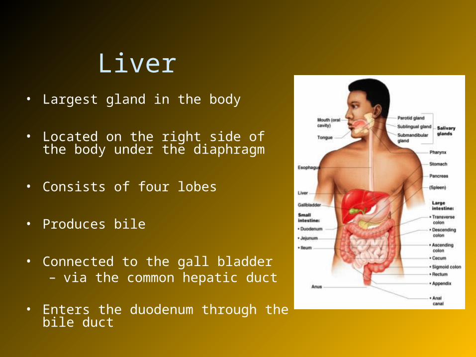
Liver• Largest gland in the body
• Located on the right side of the body under the diaphragm
• Consists of four lobes
• Produces bile
• Connected to the gall bladder – via the common hepatic duct
• Enters the duodenum through the bile duct
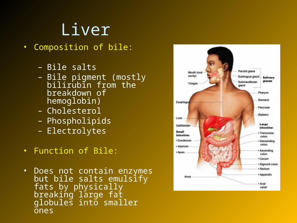
Liver• Composition of bile:
– Bile salts– Bile pigment (mostly bilirubin
from the breakdown of hemoglobin)
– Cholesterol– Phospholipids– Electrolytes
• Function of Bile:
• Does not contain enzymes but bile salts emulsify fats by physically breaking large fat globules into smaller ones
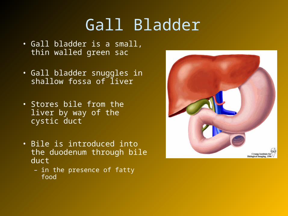
Gall Bladder• Gall bladder is a small, thin
walled green sac • Gall bladder snuggles in
shallow fossa of liver
• Stores bile from the liver by way of the cystic duct
• Bile is introduced into the duodenum through bile duct – in the presence of fatty food
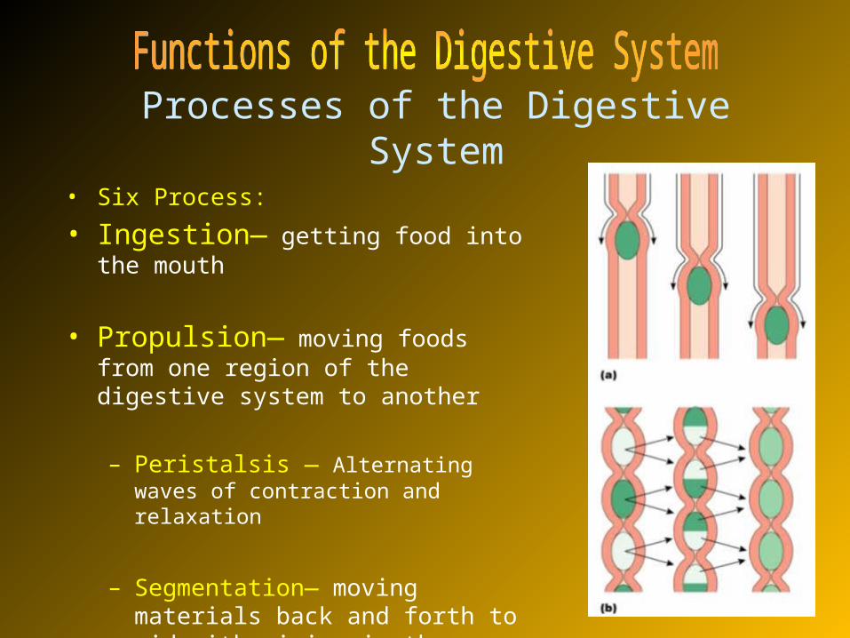
Processes of the Digestive System • Six Process:
• Ingestion— getting food into the mouth
• Propulsion— moving foods from one region of the digestive system to another
– Peristalsis — Alternating waves of contraction and relaxation
– Segmentation— moving materials back and forth to aid with mixing in the small intestine
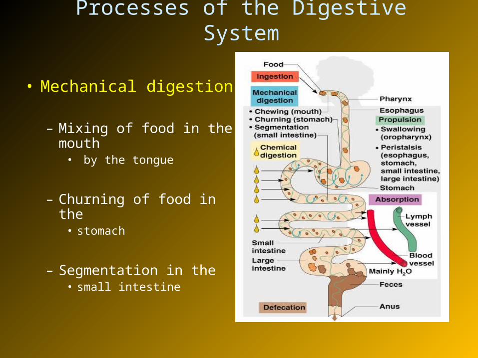
Processes of the Digestive System
• Mechanical digestion
– Mixing of food in the mouth
• by the tongue
– Churning of food in the • stomach
– Segmentation in the • small intestine
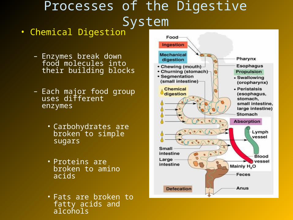
Processes of the Digestive System• Chemical Digestion
– Enzymes break down food molecules into their building blocks
– Each major food group uses different enzymes
• Carbohydrates are broken to simple sugars
• Proteins are broken to amino acids
• Fats are broken to fatty acids and alcohols

Processes of the Digestive System
• Absorption
– End products of digestion are absorbed
• to the blood or lymph
• Small intestine is the major absorptive site
– Defecation– Elimination of indigestible
substances
• as feces
• Mostly controlled by reflexes via parasympathetic division of ANS
• Chemical and mechanical receptors are located in alimentary canal organ walls that trigger reflexes
• By responding to no. of stimuli, eg.– Stretch of the organ by food– pH of the contents– Presence of breakdown products of digestion
• Reflexes include activation or inhibition – of glandular secretions– Smooth muscles that mix and propel the foods along the GI
tract
Control of Digestive Activity
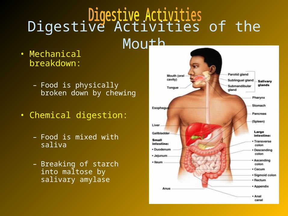
Digestive Activities of the Mouth• Mechanical breakdown:
– Food is physically broken down by chewing
• Chemical digestion:
– Food is mixed with saliva
– Breaking of starch into maltose by salivary amylase

• These organs have no digestive function
• Serve as passageways to the stomach
Activities of the Pharynx and Esophagus
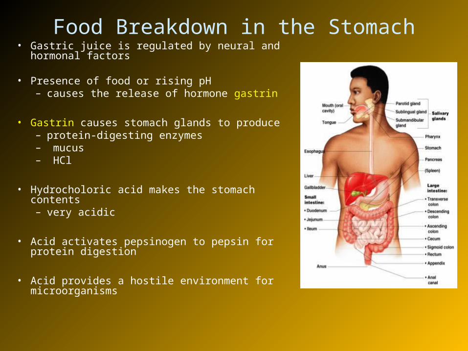
Food Breakdown in the Stomach• Gastric juice is regulated by neural and hormonal
factors
• Presence of food or rising pH – causes the release of hormone gastrin
• Gastrin causes stomach glands to produce – protein-digesting enzymes– mucus– HCl
• Hydrocholoric acid makes the stomach contents – very acidic
• Acid activates pepsinogen to pepsin for protein digestion
• Acid provides a hostile environment for microorganisms

Digestion and Absorption in the Stomach
• Protein digestion enzymes– Pepsin – an active protein digesting enzyme– Rennin – works on digesting milk protein
• The only absorption that occurs in the stomach is – alcohol and aspirin

Propulsion in the Stomach• Food must first be well mixed• Rippling peristalsis occurs in the upper half of stomach• The pylorus meters out chyme into the small intestine
(3 ml at a time)• The stomach empties in four to six hours
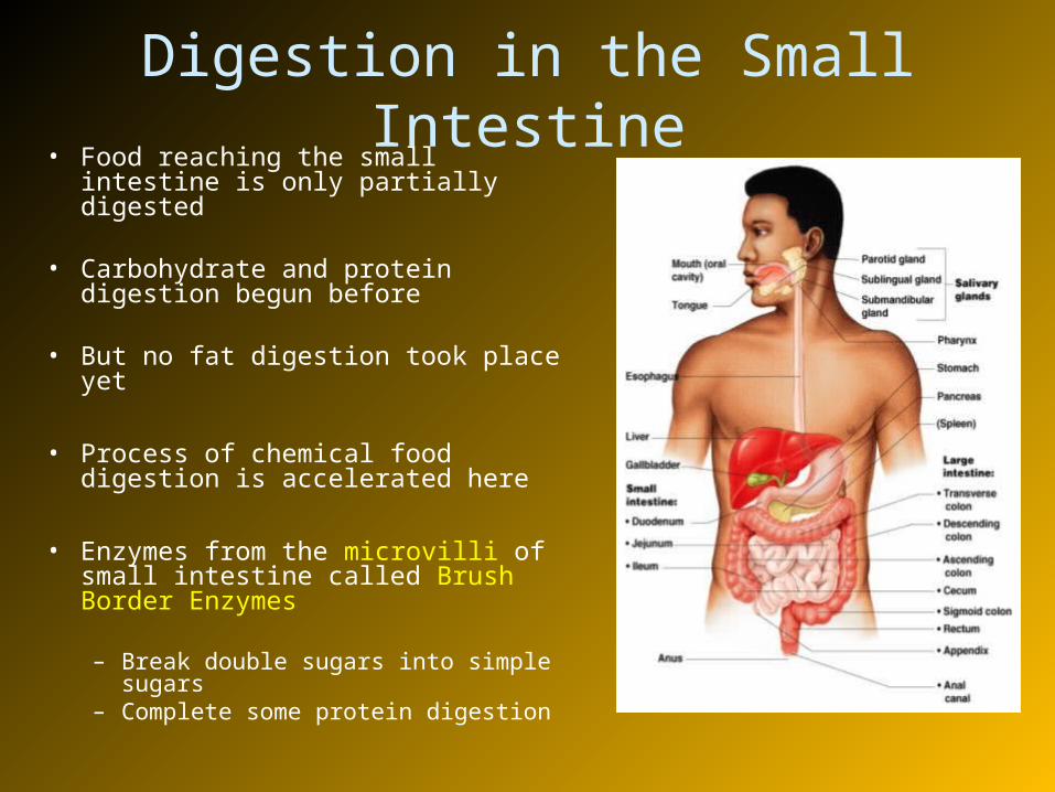
Digestion in the Small Intestine• Food reaching the small intestine is
only partially digested
• Carbohydrate and protein digestion begun before
• But no fat digestion took place yet
• Process of chemical food digestion is accelerated here
• Enzymes from the microvilli of small intestine called Brush Border Enzymes
– Break double sugars into simple sugars
– Complete some protein digestion
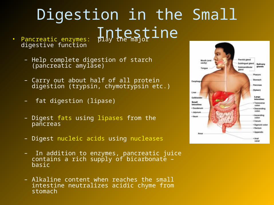
Digestion in the Small Intestine• Pancreatic enzymes: play the major digestive
function
– Help complete digestion of starch (pancreatic amylase)
– Carry out about half of all protein digestion (trypsin, chymotrypsin etc.)
– fat digestion (lipase)
– Digest fats using lipases from the pancreas
– Digest nucleic acids using nucleases
– In addition to enzymes, pancreatic juice contains a rich supply of bicarbonate – basic
– Alkaline content when reaches the small intestine neutralizes acidic chyme from stomach
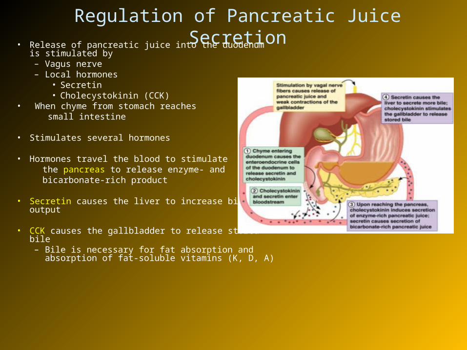
Regulation of Pancreatic Juice Secretion• Release of pancreatic juice into the duodenum is
stimulated by– Vagus nerve– Local hormones
• Secretin• Cholecystokinin (CCK)
• When chyme from stomach reaches small intestine
• Stimulates several hormones
• Hormones travel the blood to stimulate the pancreas to release enzyme- and bicarbonate-rich product
• Secretin causes the liver to increase bile output
• CCK causes the gallbladder to release stored bile– Bile is necessary for fat absorption and absorption
of fat-soluble vitamins (K, D, A)
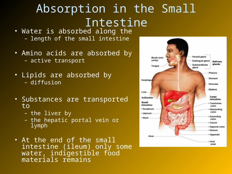
Absorption in the Small Intestine• Water is absorbed along the
– length of the small intestine
• Amino acids are absorbed by – active transport
• Lipids are absorbed by – diffusion
• Substances are transported to – the liver by – the hepatic portal vein or lymph
• At the end of the small intestine (ileum) only some water, indigestible food materials remains
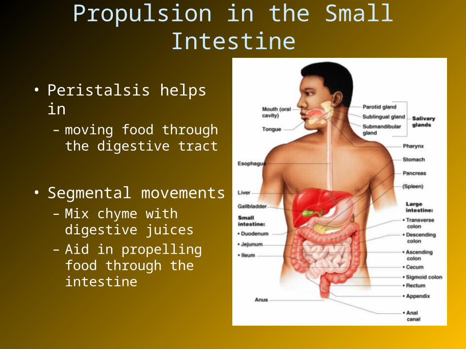
Propulsion in the Small Intestine
• Peristalsis helps in – moving food through
the digestive tract
• Segmental movements– Mix chyme with
digestive juices– Aid in propelling food
through the intestine
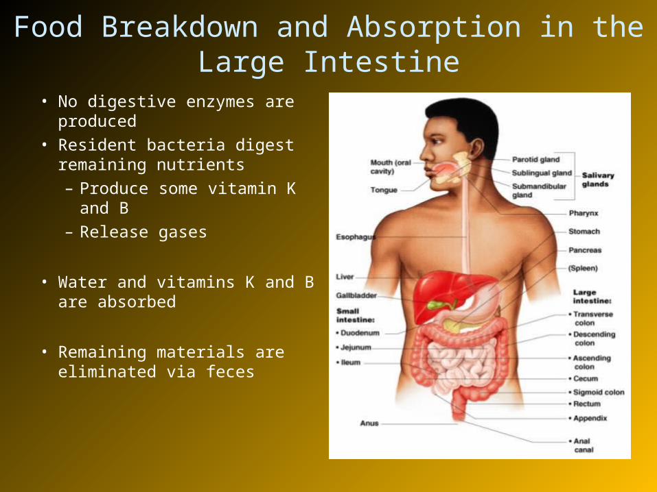
Food Breakdown and Absorption in the Large Intestine
• No digestive enzymes are produced
• Resident bacteria digest remaining nutrients– Produce some vitamin K and
B– Release gases
• Water and vitamins K and B are absorbed
• Remaining materials are eliminated via feces
• Feces contains– Undigested food residues– Mucus– Bacteria– Water
Food Breakdown and Absorption in the Large Intestine

Propulsion in the Large Intestine• Sluggish peristalsis• Mass movements
– Long, Slow, powerful contractile waves
– Occur three to four times per day
– Move the contents toward rectum
• Presence of feces in the rectum – causes a defecation reflex– Defecation reflex – Spinal reflex– Causes the wall of the rectum to
contract– Anal sphincter
• is relaxed– And Defecation occurs
Nutrition• Nutrient— substance used by the body for
growth, maintenance, and repair
• Major nutrients– Carbohydrates
– Lipids
– Proteins
– Water
• Minor nutrients– Vitamins
– Minerals
Dietary Sources of Major Nutrients
• Carbohydrates– Most are derived from plants– Exceptions: lactose from milk and small amounts of glycogens
from meats
• Lipids– Saturated fats from animal products– Unsaturated fats from nuts, seeds, and vegetable oils– Cholesterol from egg yolk, meats, and milk products
• Proteins– Complete proteins – contain all essential amino acids
• Most are from animal products
– Legumes and beans also have proteins, but are incomplete
Dietary Sources of Major Nutrients
• Vitamins– Most vitamins are used as cofactors and act with
enzymes– Found in all major food groups
• Minerals– Play many roles in the body– Most mineral-rich foods are vegetables, legumes,
milk, and some meats
Metabolism
• Chemical reactions necessary to maintain life
• Catabolism – substances are broken down to simpler substances– Energy is released during catabolism
• Anabolism – larger molecules are built from smaller ones– Energy is stored in the body for the future use
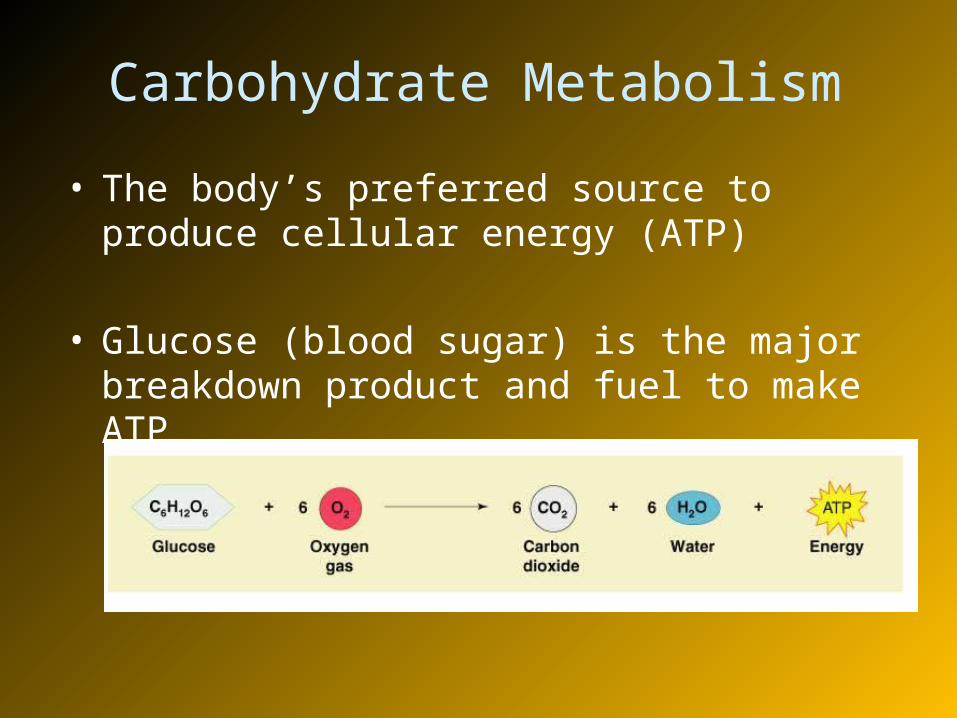
Carbohydrate Metabolism
• The body’s preferred source to produce cellular energy (ATP)
• Glucose (blood sugar) is the major breakdown product and fuel to make ATP

Cellular Respiration• Oxygen-using events take place within the cell to
create ATP from ADP• Carbon leaves cells as carbon dioxide (CO2)• Hydrogen atoms are combined with oxygen to form
water• Energy produced by these reactions adds a
phosphorus to ADP to produce ATP• ATP can be broken down to release energy for cellular
use
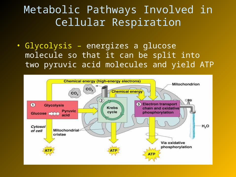
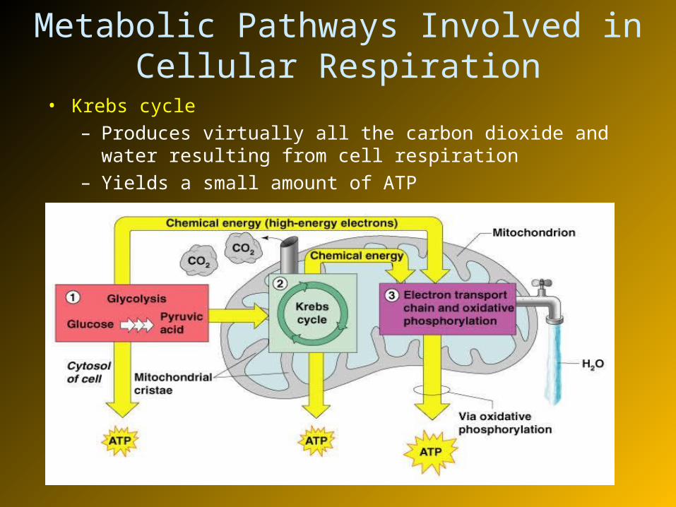
Metabolic Pathways Involved in Cellular Respiration
• Glycolysis – energizes a glucose molecule so that it can be split into two pyruvic acid molecules and yield ATP
Metabolic Pathways Involved in Cellular Respiration
• Krebs cycle– Produces virtually all the carbon dioxide and water resulting
from cell respiration– Yields a small amount of ATP
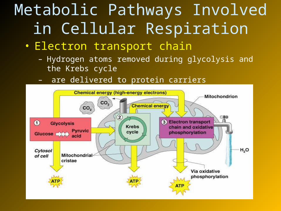
Metabolic Pathways Involved in Cellular Respiration
• Electron transport chain– Hydrogen atoms removed during glycolysis and the Krebs cycle– are delivered to protein carriers
Metabolic Pathways Involved in Cellular Respiration
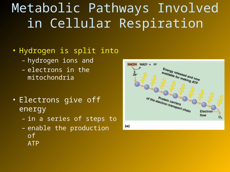
• Hydrogen is split into – hydrogen ions and – electrons in the
mitochondria
• Electrons give off energy – in a series of steps to – enable the production of
ATP
• Hyperglycemia— excessively high levels of glucose in the blood
• Excess glucose is stored in body cells as glycogen
• If blood glucose levels are still too high, excesses are converted to fat
• Hypoglycemia— low levels of glucose in the blood
• Liver breaks down stored glycogen and releases glucose into the blood
Metabolism of Carbohydrates
Fat Metabolism
• Handled mostly by the liver
– Use some fats to make ATP– Synthesize lipoproteins, thromboplastin (clotting
protein), and cholesterol– Release small fat-breakdown products to the blood
• Body cells remove – fat and cholesterol from the blood to build
membranes and steroid hormones as needed
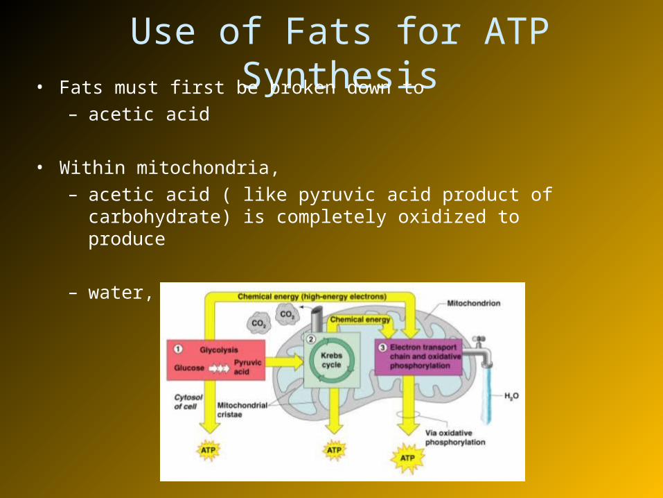
Use of Fats for ATP Synthesis• Fats must first be broken down to
– acetic acid
• Within mitochondria, – acetic acid ( like pyruvic acid product of carbohydrate) is
completely oxidized to produce
– water, carbon dioxide, and ATP
• When there is not enough glucose for fuel
• Then larger amounts of fats are used for ATP production • In such conditions fat production is fast and incomplete
• Acidosis (ketoacidosis) results from incomplete fat oxidation in which acetoacetic acid and acetone accumulate in the blood
– Common with• “No carbohydrate” diets• Uncontrolled diabetes mellitus• Starvation
Fat Metabolism
Protein Metabolism
• Proteins are conserved by body cells because – they are used for most cellular structures
• Ingested proteins are broken down to – amino acids
• Cells remove amino acids to build proteins– Synthesized proteins are actively transported across
cell membranes
Production of ATP from Protein
• Amino acids are used to make ATP only when – proteins are overabundant or there is a shortage of
other sources
• Amine groups are removed from proteins as – Ammonia
• The rest of the protein molecule enters – the Krebs cycle in mitochondria
• The liver converts harmful – ammonia to urea – which can be eliminated in urine
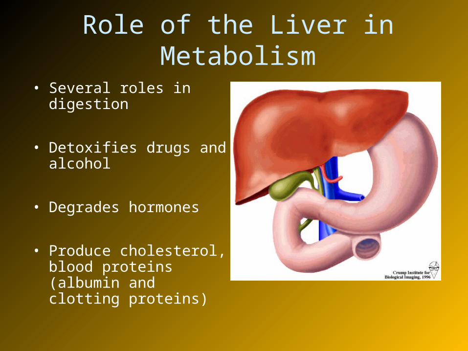
Role of the Liver in Metabolism
• Several roles in digestion
• Detoxifies drugs and alcohol
• Degrades hormones
• Produce cholesterol, blood proteins (albumin and clotting proteins)
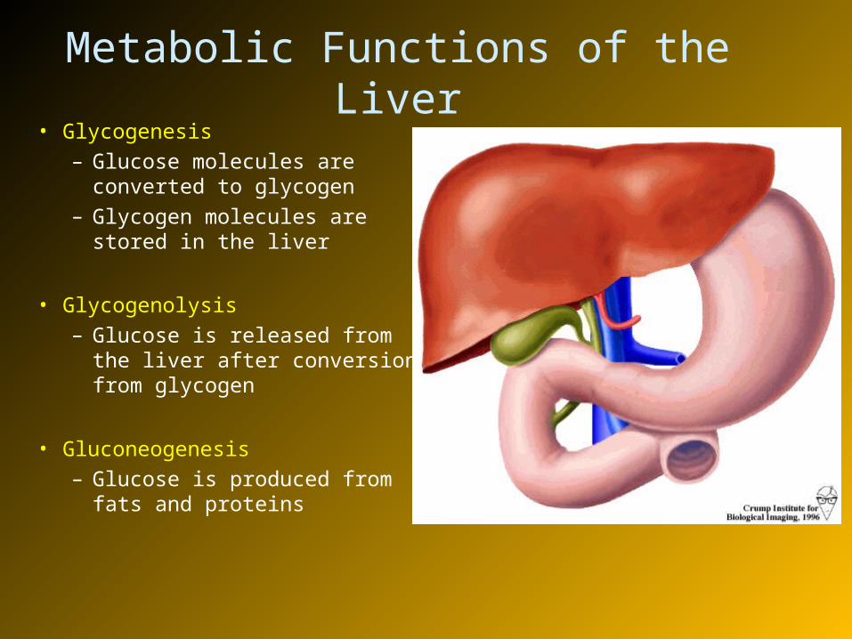
Metabolic Functions of the Liver• Glycogenesis
– Glucose molecules are converted to glycogen
– Glycogen molecules are stored in the liver
• Glycogenolysis– Glucose is released from the
liver after conversion from glycogen
• Gluconeogenesis– Glucose is produced from fats
and proteins
• Fats and fatty acids are picked up by the liver– Some are oxidized to provide energy for liver
cells– The rest are broken down into
• simpler compounds and released into the blood
Metabolic Functions of the Liver
Cholesterol Metabolism• Cholesterol is not used to make ATP
• Functions of cholesterol– Serves as a structural basis of steroid hormones
and vitamin D
– Is a major building block of plasma membranes
• Most cholesterol is produced in the liver – and is not from diet
Cholesterol Transport
• Cholesterol and fatty acids – cannot freely circulate in the bloodstream because they are
not water soluble
• They are transported by – lipoproteins (lipid-protein complexes)
– Low-density lipoproteins (LDLs) transport cholesterol to body cells
– High-density lilpoproteins (HDLs) transport cholesterol from body cells to the liver

Body Energy Balance
• Energy intake = total energy output (heat + work + energy storage)
– Energy intake is energy liberated during food oxidation
– Energy output: Energy lost as• Heat : usually about 60%• plus that used to do work (driven by ATP)• Plus energy that is stored in the form of fat or glycogen
Regulation of Food Intake
• Body weight is usually relatively stable– Energy intake and output remain about equal
• Mechanisms that may regulate food intake– Levels of nutrients in the blood (glucose and amino
acids)– Hormones (insulin, glucagon)– Body temperature (low or high)– Psychological factors
Metabolic Rate and Body Heat Production
• Basic metabolic rate (BMR) – amount of heat produced by the body per unit of time at rest
• Factors that influence BMR
– Surface area – small body usually has higher BMR– Gender – males tend to have higher BMR– Age – children and adolescents have a higher BMR– The amount of thyroxine produced is the most important
control factor• More thyroxine means higher metabolic rate
Total Metabolic Rate (TMR)
• Total amount of kilocalories – the body must consume to fuel ongoing activities
• TMR increases with an increase in body activity
• TMR must equal– calories consumed– to maintain homeostasis and maintain a constant
weight
Changes in the metabolic activity with age
• Problems of the digestive system– Gastroenteritis—inflammation of the gastrointestinal tract– Appendicitis—inflammation of the appendix
• Middle age digestive problems– Ulcers– Gall bladder problems
• Metabolism decreases with old age
• Activity of digestive tract in old age– Fewer digestive juices– Peristalsis slows– Cancer are more common