Embed Size (px)
Citation preview

Essentials of Human Anatomy & Physiology
Copyright © 2003 Pearson Education, Inc. publishing as Benjamin Cummings
Slides 14.1 – 14.14
Seventh Edition
Elaine N. Marieb
Chapter 14
The Digestive System and
Body Metabolism
Lecture Slides in PowerPoint by Jerry L. Cook

The Digestive System and Body The Digestive System and Body
MetabolismMetabolism
Slide 14.1Copyright © 2003 Pearson Education, Inc. publishing as Benjamin Cummings
• Digestion
• Breakdown of ingested food
• Absorption of nutrients into the blood
• Metabolism
• Production of cellular energy (ATP)
• Constructive and degradative cellular activities

Organs of the Digestive SystemOrgans of the Digestive System
Slide 14.2aCopyright © 2003 Pearson Education, Inc. publishing as Benjamin Cummings
• Two main groups
• Alimentary canal – continuous coiled hollow tube
• Accessory digestive organs

Organs of the Digestive SystemOrgans of the Digestive System
Slide 14.2bCopyright © 2003 Pearson Education, Inc. publishing as Benjamin Cummings
Figure 14.1
Organs of the Alimentary CanalOrgans of the Alimentary Canal
Slide 14.3Copyright © 2003 Pearson Education, Inc. publishing as Benjamin Cummings
• Mouth
• Pharynx
• Esophagus
• Stomach
• Small intestine
• Large intestine
• Anus

Mouth (Oral Cavity) AnatomyMouth (Oral Cavity) Anatomy
Slide 14.4Copyright © 2003 Pearson Education, Inc. publishing as Benjamin Cummings
• Lips (labia) – protect the anterior opening
• Cheeks – form the lateral walls
• Hard palate – forms the anterior roof
• Soft palate – forms the posterior roof
• Uvula – fleshy projection of the soft palate
Figure 14.2a

Mouth (Oral Cavity) AnatomyMouth (Oral Cavity) Anatomy
Slide 14.5Copyright © 2003 Pearson Education, Inc. publishing as Benjamin Cummings
• Vestibule – space between lips externally and teeth and gums internally
• Oral cavity – area contained by the teeth
• Tongue – attached at hyoid and styloidprocesses of the skull, and by the lingual frenulum Figure 14.2a

Mouth (Oral Cavity) AnatomyMouth (Oral Cavity) Anatomy
Slide 14.6Copyright © 2003 Pearson Education, Inc. publishing as Benjamin Cummings
• Tonsils
• Palatine tonsils
• Lingual tonsil
Figure 14.2a

Processes of the MouthProcesses of the Mouth
Slide 14.7Copyright © 2003 Pearson Education, Inc. publishing as Benjamin Cummings
• Mastication (chewing) of food
• Mixing masticated food with saliva
• Initiation of swallowing by the tongue
• Allowing for the sense of taste
Pharynx AnatomyPharynx Anatomy
Slide 14.8Copyright © 2003 Pearson Education, Inc. publishing as Benjamin Cummings
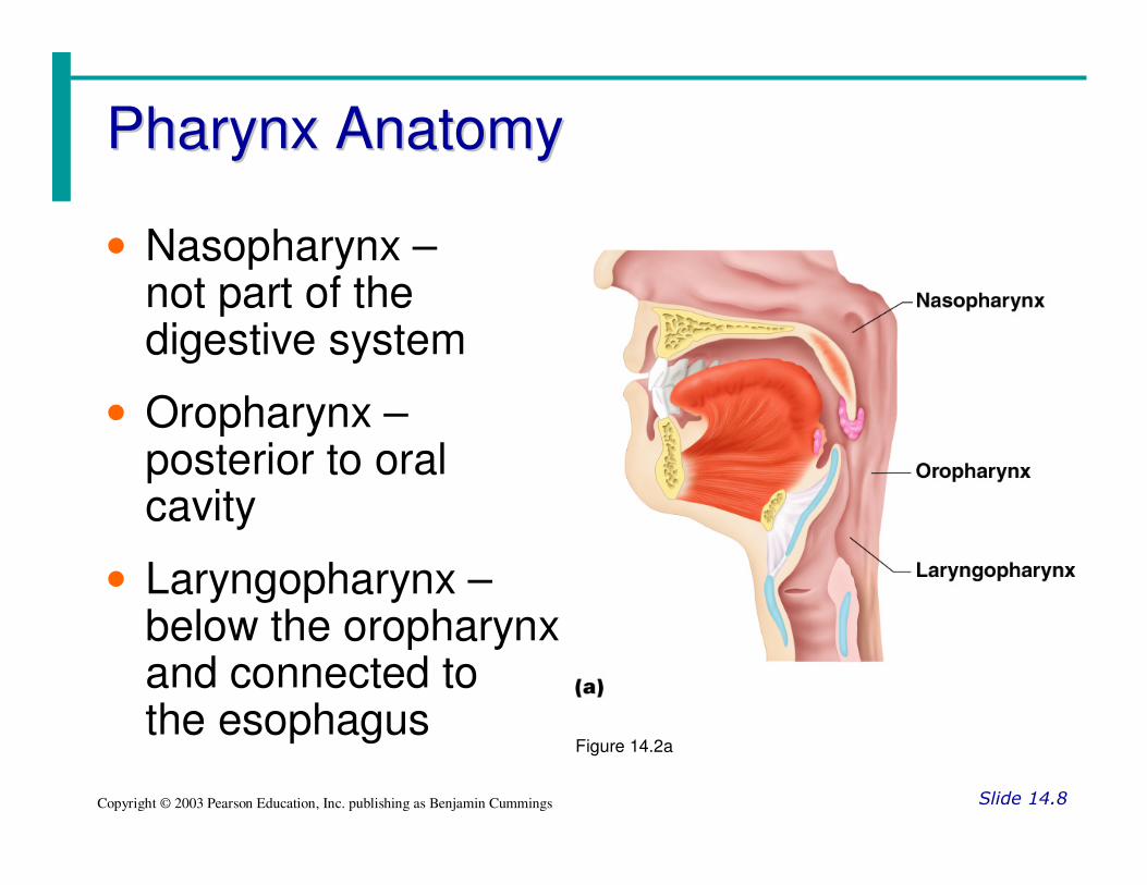
• Nasopharynx –not part of the digestive system
• Oropharynx –posterior to oral cavity
• Laryngopharynx –below the oropharynxand connected to the esophagus
Figure 14.2a
Pharynx FunctionPharynx Function
Slide 14.9Copyright © 2003 Pearson Education, Inc. publishing as Benjamin Cummings
• Serves as a passageway for air and food
• Food is propelled to the esophagus by two muscle layers
• Longitudinal inner layer
• Circular outer layer
• Food movement is by alternating contractions of the muscle layers (peristalsis)
EsophagusEsophagus
Slide 14.10Copyright © 2003 Pearson Education, Inc. publishing as Benjamin Cummings
• Runs from pharynx to stomach through the diaphragm
• Conducts food by peristalsis (slow rhythmic squeezing)
• Passageway for food only (respiratory system branches off after the pharynx)
Layers of Alimentary Canal OrgansLayers of Alimentary Canal Organs
Slide 14.11aCopyright © 2003 Pearson Education, Inc. publishing as Benjamin Cummings
• Mucosa
• Innermost layer
• Moist membrane
• Surface epithelium
• Small amount of connective tissue (lamina propria)
• Small smooth muscle layer
Slide 14.11bCopyright © 2003 Pearson Education, Inc. publishing as Benjamin Cummings
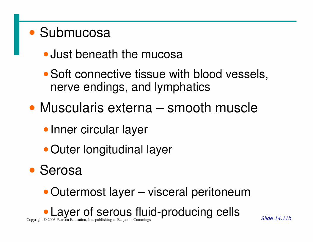
• Submucosa
• Just beneath the mucosa
• Soft connective tissue with blood vessels, nerve endings, and lymphatics
• Muscularis externa – smooth muscle
• Inner circular layer
• Outer longitudinal layer
• Serosa
• Outermost layer – visceral peritoneum
• Layer of serous fluid-producing cells

Layers of Alimentary Canal OrgansLayers of Alimentary Canal Organs
Slide 14.13Copyright © 2003 Pearson Education, Inc. publishing as Benjamin Cummings
Figure 14.3

Alimentary Canal Nerve PlexusesAlimentary Canal Nerve Plexuses
Slide 14.14Copyright © 2003 Pearson Education, Inc. publishing as Benjamin Cummings
• All are part of the autonomic nervous system
• Three separate networks of nerve fibers
• Submucosal nerve plexus
• Myenteric nerve plexus
• Subserous plexus
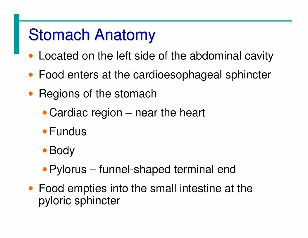
Stomach AnatomyStomach Anatomy
• Located on the left side of the abdominal cavity
• Food enters at the cardioesophageal sphincter
• Regions of the stomach
• Cardiac region – near the heart
• Fundus
• Body
• Pylorus – funnel-shaped terminal end
• Food empties into the small intestine at the pyloric sphincter

Stomach AnatomyStomach Anatomy
Slide 14.16aCopyright © 2003 Pearson Education, Inc. publishing as Benjamin Cummings
• Rugae –internal folds of the mucosa
• External regions
• Lesser curvature
• Greater curvature

Stomach AnatomyStomach Anatomy
Slide 14.16bCopyright © 2003 Pearson Education, Inc. publishing as Benjamin Cummings
• Layers of peritoneum attached to the stomach
• Lesser omentum – attaches the liver to the lesser curvature
• Greater omentum – attaches the greater curvature to the posterior body wall
• Contains fat to insulate, cushion, and protect abdominal organs

Stomach FunctionsStomach Functions
Slide 14.18Copyright © 2003 Pearson Education, Inc. publishing as Benjamin Cummings
• Acts as a storage tank for food
• Site of food breakdown
• Chemical breakdown of protein begins
• Delivers chyme (processed food) to the small intestine

Specialized Mucosa of the StomachSpecialized Mucosa of the Stomach
Slide 14.19Copyright © 2003 Pearson Education, Inc. publishing as Benjamin Cummings
• Simple columnar epithelium
• Mucous neck cells – produce a sticky alkaline mucus
• Gastric glands – secrete gastric juice
• Chief cells – produce protein-digesting enzymes (pepsinogens)
• Parietal cells – produce hydrochloric acid
• Endocrine cells – produce gastrin

Structure of the Stomach MucosaStructure of the Stomach Mucosa
Slide 14.20aCopyright © 2003 Pearson Education, Inc. publishing as Benjamin Cummings
• Gastric pits formed by folded mucosa
• Glands and specialized cells are in the gastric gland region

Small IntestineSmall Intestine
Slide 14.21Copyright © 2003 Pearson Education, Inc. publishing as Benjamin Cummings
• The body’s major digestive organ
• Site of nutrient absorption into the blood
• Muscular tube extending form the pyloric sphincter to the ileocecal valve
• Suspended from the posterior abdominal wall by the mesentery
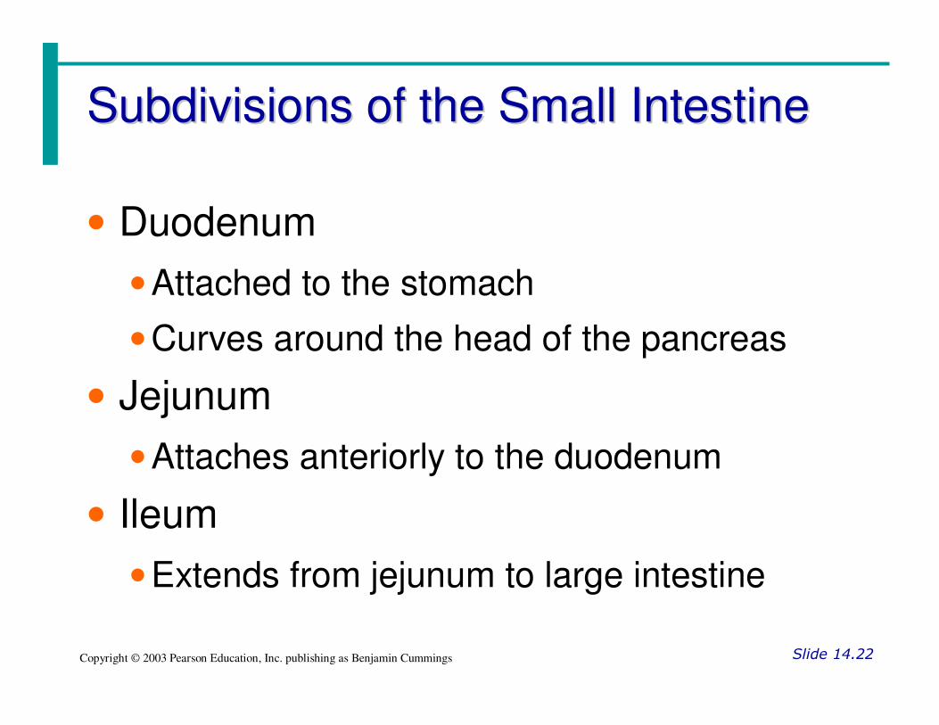
Subdivisions of the Small IntestineSubdivisions of the Small Intestine
Slide 14.22Copyright © 2003 Pearson Education, Inc. publishing as Benjamin Cummings
• Duodenum
• Attached to the stomach
• Curves around the head of the pancreas
• Jejunum
• Attaches anteriorly to the duodenum
• Ileum
• Extends from jejunum to large intestine
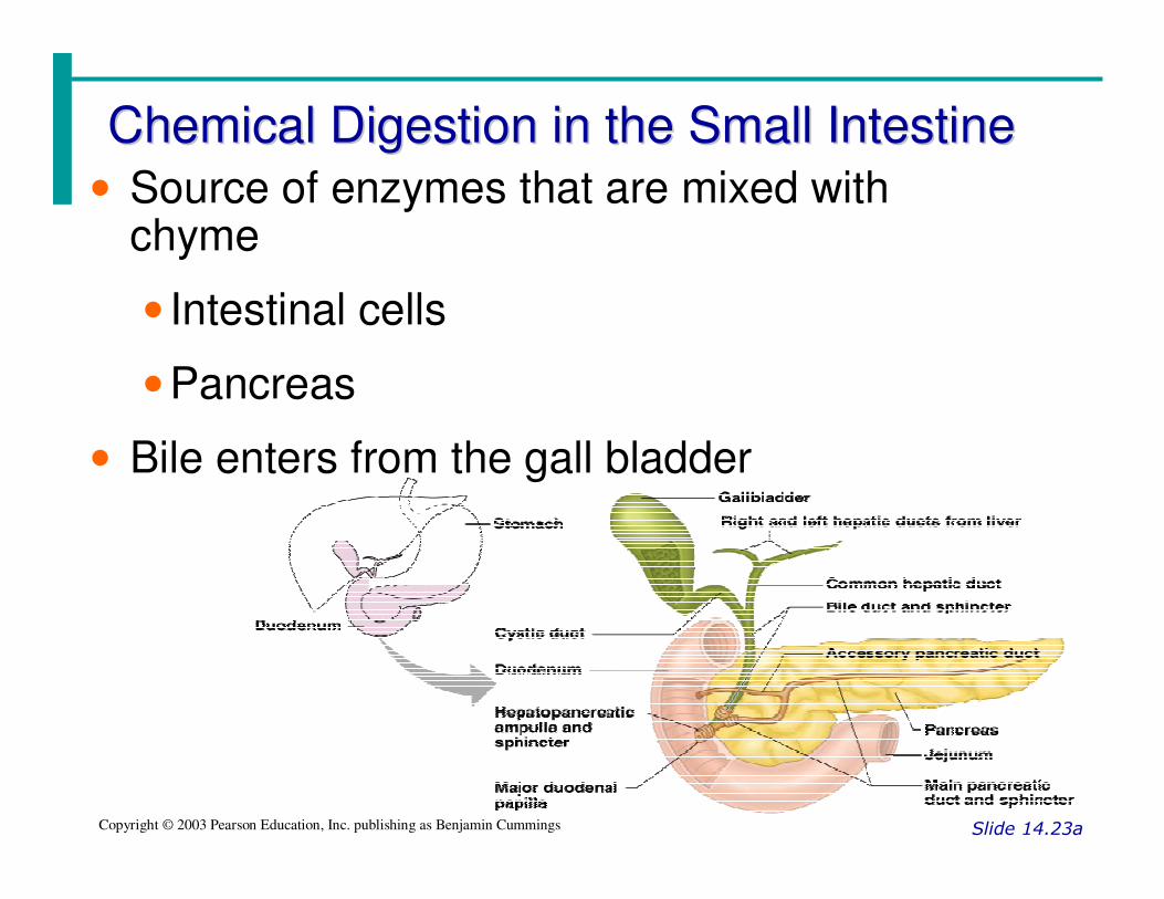
Chemical Digestion in the Small IntestineChemical Digestion in the Small Intestine
Slide 14.23aCopyright © 2003 Pearson Education, Inc. publishing as Benjamin Cummings
• Source of enzymes that are mixed with chyme
• Intestinal cells
• Pancreas
• Bile enters from the gall bladder

Projections of the Small IntestineProjections of the Small Intestine
Slide 14.24Copyright © 2003 Pearson Education, Inc. publishing as Benjamin Cummings
• Villi
• Fingerlike structures formed by the mucosa
• Give the small intestine more surface area
• Microvilli
• Small projections of the plasma membrane
• Found on absorptive cells

Structures Involved in Absorption of NutrientsStructures Involved in Absorption of Nutrients
Slide 14.26Copyright © 2003 Pearson Education, Inc. publishing as Benjamin Cummings
• Absorptive cells
• Blood capillaries
• Lacteals (specialized lymphatic capillaries)
Figure 14.7b

Folds of the Small IntestineFolds of the Small Intestine
Slide 14.27Copyright © 2003 Pearson Education, Inc. publishing as Benjamin Cummings
• Called circular folds or plicae circulares
• Deep folds of the mucosa & submucosa
• Do not disappear when filled with food
• The submucosa has Peyer’s patches (collections of lymphatic tissue like tonsils)
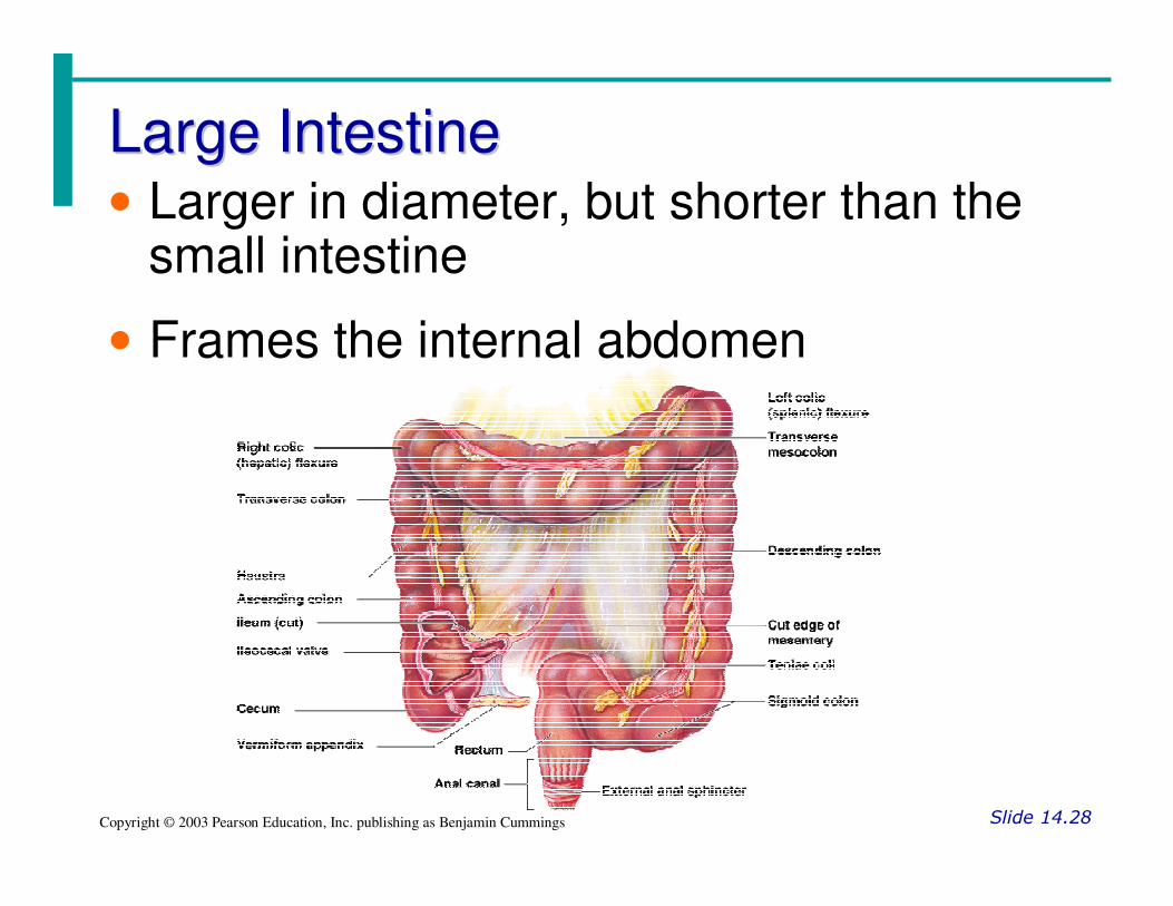
Large IntestineLarge Intestine
Slide 14.28Copyright © 2003 Pearson Education, Inc. publishing as Benjamin Cummings
• Larger in diameter, but shorter than the small intestine
• Frames the internal abdomen

Functions of the Large IntestineFunctions of the Large Intestine
Slide 14.29Copyright © 2003 Pearson Education, Inc. publishing as Benjamin Cummings
• Absorption of water
• Eliminates indigestible food from the body as feces
• Does not participate in digestion of food
• Goblet cells produce mucus to act as a lubricant
Structures of the Large IntestineStructures of the Large Intestine
Slide 14.30aCopyright © 2003 Pearson Education, Inc. publishing as Benjamin Cummings
• Cecum – saclike first part of the large intestine
• Appendix
•Accumulation of lymphatic tissue that sometimes becomes inflamed (appendicitis)
•Hangs from the cecum
•Scientists believe it used to help filter soil and excess bacteria from food

Structures of the Large IntestineStructures of the Large Intestine
Slide 14.30bCopyright © 2003 Pearson Education, Inc. publishing as Benjamin Cummings
• Colon
• Ascending
• Transverse
• Descending
• S-shaped sigmoidal
• Rectum
• Anus – external body opening

Accessory Digestive OrgansAccessory Digestive Organs
Slide 14.32Copyright © 2003 Pearson Education, Inc. publishing as Benjamin Cummings
•• Salivary GlandsSalivary Glands
• Produce saliva
• Mixture of mucus and serous fluids
• Helps to form a food bolus
• Contains salivary amylase to begin starch digestion
• Dissolves chemicals so they can be tasted
• Parotid glands – located anterior to ears
• Submandibular glands
• Sublingual glands

TeethTeeth
Slide 14.35aCopyright © 2003 Pearson Education, Inc. publishing as Benjamin Cummings
• The role is to masticate (chew) food
• Humans have two sets of teeth
• Deciduous (baby or milk) teeth
• 20 teeth are fully formed by age two
• Permanent teeth
• Replace deciduous teeth beginning between the ages of 6 to 12
• A full set is 32 teeth, but some people do not have wisdom teeth

Classification of TeethClassification of Teeth
Slide 14.36aCopyright © 2003 Pearson Education, Inc. publishing as Benjamin Cummings
• Incisors – to cut
• Canines – to pierce
• Premolars & Molars – to grind
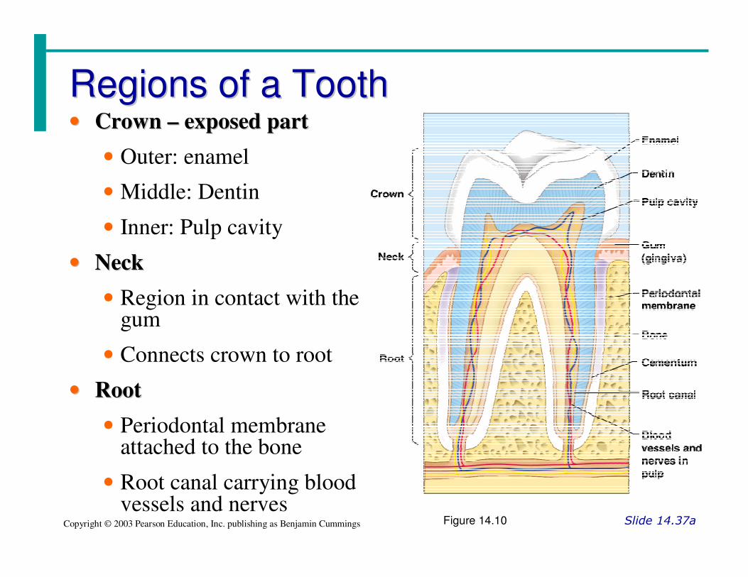
Regions of a ToothRegions of a Tooth
Slide 14.37aCopyright © 2003 Pearson Education, Inc. publishing as Benjamin Cummings
•• Crown Crown –– exposed partexposed part
• Outer: enamel
• Middle: Dentin
• Inner: Pulp cavity
•• NeckNeck
• Region in contact with the gum
• Connects crown to root
•• RootRoot
• Periodontal membrane attached to the bone
• Root canal carrying blood vessels and nerves
Figure 14.10

PancreasPancreas
Slide 14.38Copyright © 2003 Pearson Education, Inc. publishing as Benjamin Cummings
• Produces a wide spectrum of digestive enzymes that break down all categories of food
• Enzymes are secreted into the duodenum
• Alkaline fluid introduced with enzymes neutralizes acidic chyme
• Endocrine products of pancreas
• Insulin
• Glucagons

LiverLiver
Slide 14.39Copyright © 2003 Pearson Education, Inc. publishing as Benjamin Cummings
• Largest gland in the body
• Located on the right side of the body under the diaphragm
• Consists of four lobes suspended from the diaphragm and abdominal wall by the falciform ligament
• Connected to the gall bladder via the common hepatic duct

BileBile
Slide 14.40Copyright © 2003 Pearson Education, Inc. publishing as Benjamin Cummings
• Produced by cells in the liver
• Composition
• Bile salts
• Bile pigment (mostly bilirubin from the breakdown of hemoglobin)
• Cholesterol
• Phospholipids
• Electrolytes

Gall BladderGall Bladder
Slide 14.41Copyright © 2003 Pearson Education, Inc. publishing as Benjamin Cummings
• Sac found in hollow fossa of liver
• Stores bile from the liver by way of the cystic duct
• Bile is introduced into the duodenum in the presence of fatty food
• Gallstones can cause blockages

Processes of the Digestive SystemProcesses of the Digestive System
Slide 14.42aCopyright © 2003 Pearson Education, Inc. publishing as Benjamin Cummings
• Ingestion – getting food into the mouth
• Propulsion – moving foods from one region of the digestive system to another
• Peristalsis – alternating waves of contraction
• Segmentation – moving materials back and forth to aid in mixing
• Mechanical digestion
• Mixing of food in the mouth by the tongue
• Churning of food in the stomach
• Segmentation in the small intestine
Processes of the Digestive SystemProcesses of the Digestive System
Slide 14.44Copyright © 2003 Pearson Education, Inc. publishing as Benjamin Cummings
• Chemical Digestion
• Enzymes break down food molecules into their building blocks
• Each major food group uses different enzymes
• Carbohydrates are broken to simple sugars
• Proteins are broken to amino acids
• Fats are broken to fatty acids and alcohols

Processes of the Digestive SystemProcesses of the Digestive System
Slide 14.45Copyright © 2003 Pearson Education, Inc. publishing as Benjamin Cummings
• Absorption
• End products of digestion are absorbed in the blood or lymph
• Food must enter mucosal cells and then into blood or lymph capillaries
• Defecation
• Elimination of indigestible substances as feces
• Mostly controlled by reflexes via the parasympathetic division
• Chemical and mechanical receptors are located in organ walls that trigger reflexes
• Stimuli include:
• Stretch of the organ
• pH of the contents
• Presence of breakdown products
• Reflexes include:
• Activation or inhibition of glandular secretions
• Smooth muscle activity
Control of Digestive ActivityControl of Digestive Activity
Slide 14.47a

NutritionNutrition
Slide 14.63Copyright © 2003 Pearson Education, Inc. publishing as Benjamin Cummings
• Nutrient – substance used by the body for growth, maintenance, and repair
• Categories of nutrients
• Carbohydrates
• Lipids
• Proteins
• Vitamins
• Mineral
• Water

Dietary Sources of Major NutrientsDietary Sources of Major Nutrients
Slide 14.64Copyright © 2003 Pearson Education, Inc. publishing as Benjamin Cummings
• Carbohydrates
• Most are derived from plants
• Exceptions: lactose from milk and small amounts of glycogens from meats
• Lipids
• Saturated fats from animal products
• Unsaturated fats from nuts, seeds, and vegetable oils
• Cholesterol from egg yolk, meats, and milk products

Dietary Sources of Major NutrientsDietary Sources of Major Nutrients
Slide 14.65Copyright © 2003 Pearson Education, Inc. publishing as Benjamin Cummings
• Proteins
• Complete proteins – contain all essential amino acids
• Most are from animal products
• Legumes and beans also have proteins, but are incomplete
• Vitamins
• Most vitamins are used as cofactors and act with enzymes
• Found in all major food groups

Dietary Sources of Major NutrientsDietary Sources of Major Nutrients
Slide 14.66Copyright © 2003 Pearson Education, Inc. publishing as Benjamin Cummings
• Minerals
• Play many roles in the body
• Most mineral-rich foods are vegetables, legumes, milk, and some meats

MetabolismMetabolism
Slide 14.67Copyright © 2003 Pearson Education, Inc. publishing as Benjamin Cummings
• Chemical reactions necessary to maintain life
• Catabolism – substances are broken down to simpler substances
• Anabolism – larger molecules are built from smaller ones
• Energy is released during catabolism

Carbohydrate MetabolismCarbohydrate Metabolism
Slide 14.68Copyright © 2003 Pearson Education, Inc. publishing as Benjamin Cummings
• The body’s preferred source to produce cellular energy (ATP)
• Glucose (blood sugar) is the major breakdown product and fuel to make ATP
Figure 14.16

Fat MetabolismFat Metabolism
Slide 14.73Copyright © 2003 Pearson Education, Inc. publishing as Benjamin Cummings
• Handled mostly by the liver
• Use some fats to make ATP
• Synthesize lipoproteins, thromboplastin, and cholesterol
• Release breakdown products to the blood
• Body cells remove fat and cholesterol to build membranes and steroid hormones

Protein MetabolismProtein Metabolism
Slide 14.75aCopyright © 2003 Pearson Education, Inc. publishing as Benjamin Cummings
• Proteins are conserved by body cells because they are used for most cellular structures
• Ingested proteins are broken down to amino acids
• Cells remove amino acids to build proteins
• Synthesized proteins are actively transported across cell membranes
• Amino acids are used to make ATP only when proteins are overabundant or there is a shortage of other sources
Role of the Liver in MetabolismRole of the Liver in Metabolism
Slide 14.77Copyright © 2003 Pearson Education, Inc. publishing as Benjamin Cummings
• Several roles in digestion
• Detoxifies drugs and alcohol
• Degrades hormones
• Produce cholesterol, blood proteins (albumin and clotting proteins)
• Plays a central role in metabolism

Metabolic Functions of the LiverMetabolic Functions of the Liver
Slide 14.78Copyright © 2003 Pearson Education, Inc. publishing as Benjamin Cummings
• Glycogenesis
• Glucose molecules are converted to glycogen
• Glycogen molecules are stored in the liver
• Glycogenolysis
• Glucose is released from the liver after conversion from glycogen
• Gluconeogenesis
• Glucose is produced from fats and proteins

Cholesterol MetabolismCholesterol Metabolism
Slide 14.81Copyright © 2003 Pearson Education, Inc. publishing as Benjamin Cummings
• Functions of cholesterol
• Serves as a structural basis of steroid hormones and vitamin D
• Is a major building block of plasma membranes
• Most cholesterol is produced in the liver and is not from diet

Cholesterol TransportCholesterol Transport
Slide 14.82Copyright © 2003 Pearson Education, Inc. publishing as Benjamin Cummings
• Cholesterol and fatty acids cannot freely circulate in the bloodstream
• They are transported by lipoproteins (lipid-protein complexes)
• Low-density lipoproteins (LDLs) transport to body cells
• High-density lilpoproteins (HDLs) transport from body cells to the liver

Body Energy BalanceBody Energy Balance
Slide 14.83Copyright © 2003 Pearson Education, Inc. publishing as Benjamin Cummings
• Energy intake = total energy output (heat + work + energy storage)
• Energy intake is liberated during food oxidation
• Energy output
• Heat is usually about 60%
• Storage energy is in the form of fat or glycogen
Regulation of Food IntakeRegulation of Food Intake
Slide 14.84Copyright © 2003 Pearson Education, Inc. publishing as Benjamin Cummings
• Body weight is usually relatively stable
• Energy intake and output remain about equal
• Mechanisms that may regulate food intake
• Levels of nutrients in the blood
• Hormones
• Body temperature
• Psychological factors

Metabolic Rate and Body Heat Metabolic Rate and Body Heat
ProductionProduction
Slide 14.85aCopyright © 2003 Pearson Education, Inc. publishing as Benjamin Cummings
• Basic metabolic rate (BMR) – amount of heat produced by the body per unit of time at rest
• Factors that influence BMR
• Surface area – small body usually has higher BMR
• Gender – males tend to have higher BMR

Metabolic Rate and Body Heat Metabolic Rate and Body Heat
ProductionProduction
Slide 14.85bCopyright © 2003 Pearson Education, Inc. publishing as Benjamin Cummings
• Factors that influence BMR (continued)
• Age – children and adolescents have a higher BMR
• The amount of thyroxine produced is the most important control factor
• More thyroxine means higher metabolic rate

Total Metabolic Rate (TMR)Total Metabolic Rate (TMR)
Slide 14.86Copyright © 2003 Pearson Education, Inc. publishing as Benjamin Cummings
• Total amount of kilocalories the body must consume to fuel ongoing activities
• TMR increases with an increase in body activity
• TMR must equal calories consumed to maintain homeostasis and maintain a constant weight

Body Temperature RegulationBody Temperature Regulation
Slide 14.87aCopyright © 2003 Pearson Education, Inc. publishing as Benjamin Cummings
• Most energy is released as foods are oxidized
• Most energy escapes as heat
• The body has a narrow range of homeostatic temperature
• Must remain between 35.6° to 37.8°C (96° to 100° F)
• The body’s thermostat is in the hypothalamus
• Initiates heat-loss or heat-promoting mechanisms

Developmental Aspects of the Digestive SystemDevelopmental Aspects of the Digestive System
Slide 14.91Copyright © 2003 Pearson Education, Inc. publishing as Benjamin Cummings
• The alimentary canal is a continuous tube by the 5th week of development
• Digestive glands bud from the mucosa of the alimentary tube
• The developing fetus receives ALL nutrients through the placenta
• In newborns:
• feeding must be frequent
• peristalsis is inefficient
• vomiting is common

Developmental Aspects of the Digestive SystemDevelopmental Aspects of the Digestive System
Slide 14.92aCopyright © 2003 Pearson Education, Inc. publishing as Benjamin Cummings
• Teething begins at around six months
• Metabolism decreases with age
• Middle age digestive problems
• Ulcers
• Gall bladder problems
• Activity of digestive tract in old age
• Fewer digestive juices
• Peristalsis slows
• Diverticulosis and cancer are more common