Embed Size (px)
Citation preview

10/02/08Biochemistry: Nucleic Acid Chem&Struct
Nucleic AcidChemistry & Structure
Andy HowardIntroductory Biochemistry
2 October 2008

10/02/08 Biochemistry: Nucleic Acid Chem&Struct p. 2 of 43
What we’ll discuss Syn, anti revisited Nucleotides Oligo- and polynucleotides DNA duplexes and helicity RNA: structure & types
10/02/08 Biochemistry: Nucleic Acid Chem&Struct p. 3 of 43
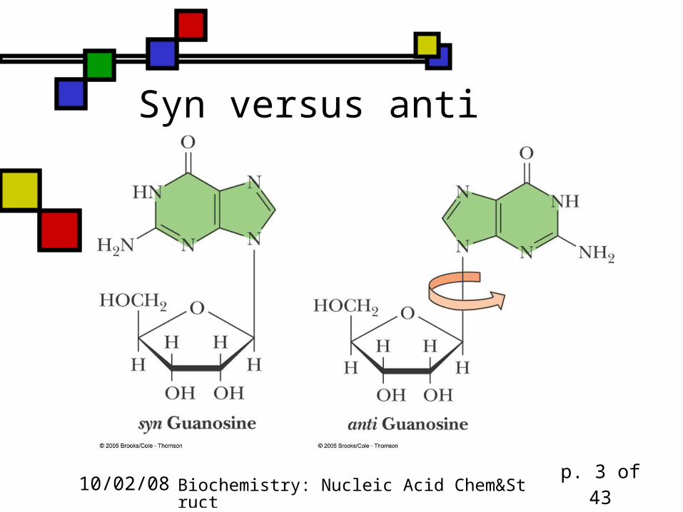
Syn versus anti

10/02/08 Biochemistry: Nucleic Acid Chem&Struct p. 4 of 43
Mono-phosphorylated nucleosides
We have specialized names for the 5’-phospho derivatives of the nucleosides, i.e. the nucleoside monophosphates:
They are nucleotides Adenosine 5’-monophosphate =
AMP = adenylate GMP = guanylate CMP = cytidylate UMP = uridylate
P
O
O-
O-O
HO
HO
O
N
N
NH2
N
N
adenylate

10/02/08 Biochemistry: Nucleic Acid Chem&Struct p. 5 of 43
pKa’s for base N’s and PO4’s
Nucleotide pKa base-N pK1 of PO4 pK2 of PO4
5’-AMP 3.8(N-1) 0.9 6.1
5’-GMP 9.4 (N-1) 0.7 6.1
2.4 (N-7)
5’-CMP 4.5 (N-3) 0.8 6.3
5’-UMP 9.5 (N-3) 1.0 6.4

10/02/08 Biochemistry: Nucleic Acid Chem&Struct p. 6 of 43
UV absorbance These aromatic rings absorb around 260
10/02/08 Biochemistry: Nucleic Acid Chem&Struct p. 7 of 43
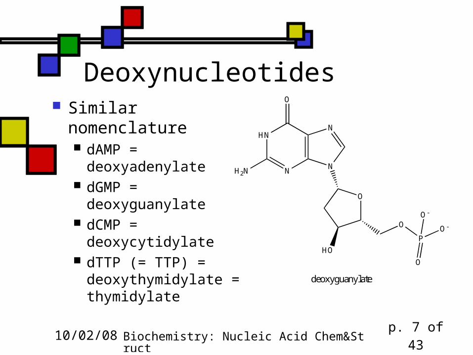
Deoxynucleotides Similar nomenclature
dAMP = deoxyadenylate
dGMP = deoxyguanylate
dCMP = deoxycytidylate
dTTP (= TTP) = deoxythymidylate = thymidylate
P
O
O-
O-O
HO
O
N
N
O
HN
H2N N
deoxyguanylate

10/02/08 Biochemistry: Nucleic Acid Chem&Struct p. 8 of 43
Cyclic phospho-diesters
3’ and 5’ hydroxyls are both involvedin -O-P-O bonds, forming a 6-membered ring (-C5’-C4’-C3’-O-P-O-)
cAMP and cGMP are the important ones(see previous lecture!)

10/02/08 Biochemistry: Nucleic Acid Chem&Struct p. 9 of 43
Di- and triphosphates
Phosphoanhydride bonds link second and perhaps third phosphates to the 5’-OH on the ribose moiety
OHHO
O
N
O
N
H2NP
O
O
O-O-
O
P
O
O-
O
P
O
OH
cytidine triphosphate
Mg2+

10/02/08 Biochemistry: Nucleic Acid Chem&Struct p. 10 of 43
These are polyprotic acids They can dissociate 3 protons (XDP) or 4
protons (XTP) from their phosphoric acid groups
The ionized forms are frequently associated with divalent cations (Mg2+, Mn2+, others)
The -O-P-O bonds beyond the first one are actually phosphoric anhydride linkages
Phosphoanhydrides are acid-labile: quantitative liberation of Pi in 1N HCl for 7 minutes @100ºC

10/02/08 Biochemistry: Nucleic Acid Chem&Struct p. 11 of 43
NTPs: carriers of chemical energy ATP is the energy currency GTP is important in protein synthesis CTP used in phospholipid synthesis UTP forms activated intermediates with
sugars (e.g. UDP-glucose) … and, of course, they’re substrates to
build up RNA and DNA

10/02/08 Biochemistry: Nucleic Acid Chem&Struct p. 12 of 43
Bases are information symbols Base and sugar aren’t directly involved in
metabolic roles of the XTPs But different XTPs do different things, so
there are recognition components to the relevant enzymatic systems that notice whether X is A, U, C, or G
Even in polynucleotides the bases play an informational role

10/02/08 Biochemistry: Nucleic Acid Chem&Struct p. 13 of 43
Oligomers and Polymers
Monomers are nucleotides or deoxynucleotides
Linkages are phosphodiester linkages between 3’ of one ribose and 5’ of the next ribose
It’s logical to start from the 5’ end for synthetic reasons

10/02/08 Biochemistry: Nucleic Acid Chem&Struct p. 14 of 43
Typical DNA dinucleotide Various notations: this is pdApdCp Leave out the p’s if there’s a lot of them!
P
O
-O O-
O
O
NN
O
HN
NH2
N P
O
-O
O
O
O
ON
O N NH2
P
O--O
O

10/02/08 Biochemistry: Nucleic Acid Chem&Struct p. 15 of 43
DNA structure
Many years of careful experimental work enabled fabrication of double-helical model of double-stranded DNA
Explained [A]=[T], [C]=[G] Specific H-bonds stabilize
double-helical structure: see fig. 10.20

10/02/08 Biochemistry: Nucleic Acid Chem&Struct p. 16 of 43
What does double-stranded DNA really look like? Picture on previous slide emphasizes
only the H-bond interactions; it ignores the orientation of the sugars, which are actually tilted relative to the helix axis
Planes of the bases are almost perpendicular to the helical axes on both sides of the double helix
10/02/08 Biochemistry: Nucleic Acid Chem&Struct p. 17 of 43
Sizes (cf fig. 10.20, 11.7)
Diameter of the double helix: 2.37nm Length along one full turn:
10.4 base pairs = pitch = 3.40nm Distance between stacked base pairs =
rise = 0.33 nm Major groove is wider and shallower;
minor groove is narrower and deeper

10/02/08 Biochemistry: Nucleic Acid Chem&Struct p. 18 of 43
What stabilizes this? Variety of stabilizing
interactions Stacking of base pairs Hydrogen bonding between
base pairs Hydrophobic effects (burying
bases, which are less polar) Charge-charge interactions:
phosphates with Mg2+ and cationic proteins
Courtesy dnareplication.info

10/02/08 Biochemistry: Nucleic Acid Chem&Struct p. 19 of 43
How close to instability is it? Pretty close. Heating DNA makes it melt: fig. 11.14 The more GC pairs, the harder it is to
melt Weaker stacking interactions in A-T One more H-bond per GC than per AT
We’ll get into DNA structure a lot more later in this lecture

10/02/08 Biochemistry: Nucleic Acid Chem&Struct p. 20 of 43
iClicker quiz
1. What positions of a pair of aromatic rings leads to stabilizing interactions? (a) Parallel to one another (b) Perpendicular to one another (c) At a 45º angle to one another (d) Both (a) and (b) (e) All three: (a), (b), and ( c)
10/02/08 Biochemistry: Nucleic Acid Chem&Struct p. 21 of 43
Second iClicker question
2. Which has the highest molecular mass among the compounds listed? (a) cytidylate (b) thymidylate (c) adenylate (d) adenosine triphosphate (e) they’re all the same MW

10/02/08 Biochemistry: Nucleic Acid Chem&Struct p. 22 of 43
Base composition for DNA
As noted, [A]=[T], [C]=[G] because of base pairing
[A]/[C] etc. not governed by base pairing Can vary considerably (table 10.3) E.coli : [A], [C] about equal Mycobacterium tuberculosis: [C] > 2*[A] Mammals: [C] < 0.74*[A]

10/02/08 Biochemistry: Nucleic Acid Chem&Struct p. 23 of 43
Molar ratios for various organisms’ DNA (table 10.3)
Source A/G T/C A/T G/C Pur/Pyr
Ox 1.29 1.43 1.04 1.00 1.1Human 1.56 1.75 1.0 1.0 1.0Hen 1.45 1.29 1.06 0.91 0.99Salmon 1.43 1.43 1.02 1.02 1.02Wheat 1.22 1.18 1.00 0.97 0.99Yeast 1.67 1.92 1.03 1.20 1.0H.influenzae 1.74 1.54 1.07 0.91 1.0E.coli K-12 1.05 0.95 1.09 0.99 1.0B. schatz 0.7 0.6 1.12 0.89 1.0

10/02/08 Biochemistry: Nucleic Acid Chem&Struct p. 24 of 43
What did this mean in 1950?
[A]=[T] and [C]=[G] suggested that if the molecule involved two strands, there should be complementarity between them, i.e., if there’s an A on one strand, there will be a T on the other one
Unfortunately it wasn’t entirely clear that the molecule was two-stranded!

10/02/08 Biochemistry: Nucleic Acid Chem&Struct p. 25 of 43
The Watson-Crick contribution
Interpreting the X-ray fiber diffraction photographs taken by Rosalind Franklin and Maurice Wilkins, W&C built a ball-and-stick model for a two-stranded form of DNA
They were able to show that their model was consistent with Franklin’s data
QuickTime™ and aTIFF (Uncompressed) decompressor
are needed to see this picture.
QuickTime™ and aTIFF (Uncompressed) decompressor
are needed to see this picture.

10/02/08 Biochemistry: Nucleic Acid Chem&Struct p. 26 of 43
So how is DNA organized?
Linear sequence is simple to describe: Two strands, each very long and
containing 105 - 108 bases Each base has a complementary base on
the other strand Specific hydrogen bonding patterns
define the complementarity

10/02/08 Biochemistry: Nucleic Acid Chem&Struct p. 27 of 43
Higher levels of organization Just as with protein tertiary structure, DNA
structure has higher levels beyond the base-pairing, beginning with coiling into a double helix
Eukaryotes: Organization of double helix into loop structures
of ~200 base pairs coiled around a protein complex called the histone octamer
Further organization of those loops into larger structures culminating in formation of chromosomes
Prokaryotes: similar but simpler higher-level structures culminating in (often circular) chromosomes

10/02/08 Biochemistry: Nucleic Acid Chem&Struct p. 28 of 43
Supercoiling Refers to levels of organization of DNA
beyond the immediate double-helix We describe circular DNA as relaxed if
the closed double helix could lie flat It’s underwound or overwound if the ends
are broken, twisted, and rejoined. Supercoils restore 10.4 bp/turn relation
upon rejoining

10/02/08 Biochemistry: Nucleic Acid Chem&Struct p. 29 of 43
Supercoiling and flat DNA
Diagram courtesy SIU Carbondale

10/02/08 Biochemistry: Nucleic Acid Chem&Struct p. 30 of 43
Ribonucleic acid We’re done with DNA for the moment. Let’s discuss RNA. RNA is generally, but not always, single-
stranded The regions where localized base-pairing
occurs (local double-stranded regions) often are of functional significance
10/02/08 Biochemistry: Nucleic Acid Chem&Struct p. 31 of 43
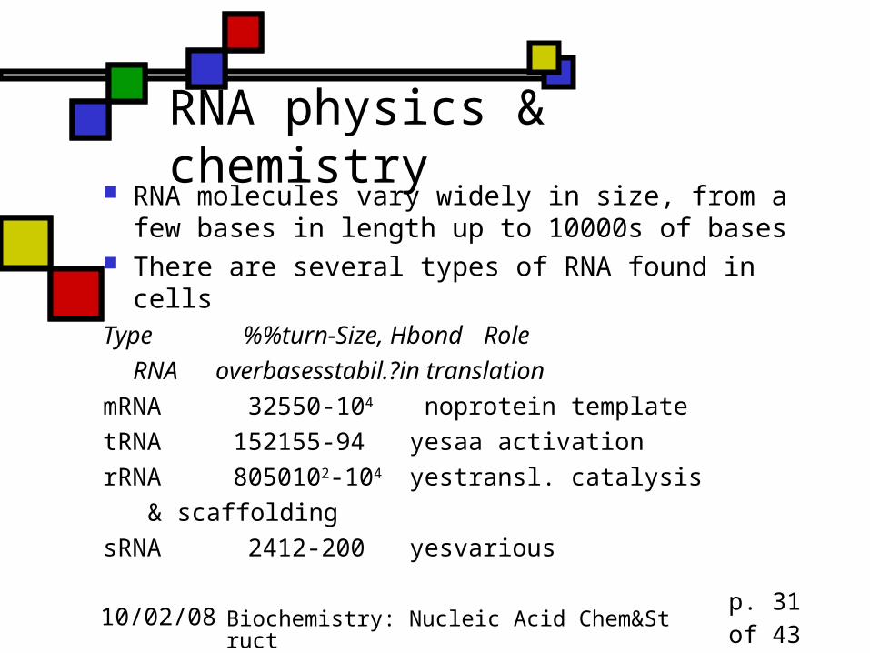
RNA physics & chemistry RNA molecules vary widely in size, from a few
bases in length up to 10000s of bases There are several types of RNA found in cellsType % %turn- Size, Hbond Role
RNA over bases stabil.? in translation
mRNA 3 25 50-104 no protein template
tRNA 15 21 55-94 yes aa activation
rRNA 80 50 102-104 yes transl. catalysis
& scaffolding
sRNA 2 4 12-200 yes various

10/02/08 Biochemistry: Nucleic Acid Chem&Struct p. 32 of 43
Unusual bases in RNA mRNA, sRNA mostly A,C,G,U rRNA, tRNA have some odd ones
10/02/08 Biochemistry: Nucleic Acid Chem&Struct p. 33 of 43
Messenger RNA Contains the codons that define protein
sequence Each codon (3 bases) codes for 1 amino acid Synthesized during transcription, like all other
types of RNA Relatively small % of RNA mass in the cell; but
short-lived, so: Higher % of RNA synthesis devoted to mRNA

10/02/08 Biochemistry: Nucleic Acid Chem&Struct p. 34 of 43
Prokaryotic mRNA One mRNA with a single promoter will
contain coding information for several proteins, i.e., 1 promoter, several genes
Defined stop codons show the ribosome where to put in the breaks
Translation closely coupled to transcription, unlike eukaryotic systems, where they’re separated in space & time

10/02/08 Biochemistry: Nucleic Acid Chem&Struct p. 35 of 43
Eukaryotic mRNA One mRNA per protein But the mRNA will be initially synthesized with
noncoding segments (introns) interspersed between the coding segments (exons):heterogeneous nuclear RNA, hnRNA
snRNPs (q.v.) in nucleus splice out the introns, tying together the exons to make the mature transcript
Each mRNA will end with a poly(A) tail, added after transcription

10/02/08 Biochemistry: Nucleic Acid Chem&Struct p. 36 of 43
Ribosomes and rRNA Ribosome is 65% RNA, rest protein Lots of intrastrand H-bonds Ribosomes characterized by
sedimentation coefficients E.coli: 50S piece+30S piece 70S total Eukaryotes 60S + 40S 80S total
rRNA has pseudouridine, ribothymidine, methylated bases

10/02/08 Biochemistry: Nucleic Acid Chem&Struct p. 37 of 43
Prokaryotic ribosomes (fig.10.25a)

10/02/08 Biochemistry: Nucleic Acid Chem&Struct p. 38 of 43
Eukaryotic ribosomes (fig. 10.25b)

10/02/08 Biochemistry: Nucleic Acid Chem&Struct p. 39 of 43
Transfer RNA Each tRNA carries a specific
amino acid to the ribosomal protein synthesis machine
One full set of tRNA at each cellular site of protein synthesis (cytoplasm, mitochondrion, chloroplast)
These are small molecules: 55-94 bases
QuickTime™ and aTIFF (Uncompressed) decompressor
are needed to see this picture.
A/T sitetRNA model based on cryoEM complexPDB 1QZA

10/02/08 Biochemistry: Nucleic Acid Chem&Struct p. 40 of 43
tRNA contents Many modified bases CCA on the 3’-end is
attached to the amino acid Catalytic attachment of
amino acid to protein is catalyzed by an adenine in one of the 50S rRNAs
QuickTime™ and aTIFF (Uncompressed) decompressor
are needed to see this picture.
Dieter Söll
QuickTime™ and aTIFF (Uncompressed) decompressor
are needed to see this picture.

10/02/08 Biochemistry: Nucleic Acid Chem&Struct p. 41 of 43
Small nuclear RNAs snRNA found mostly
in nucleus 100-200 nucleotides closely associated with proteins
& with other RNA molecules Mostly in ribonucleoprotein particles
(snRNPs), which are involved in mRNA processing, converting full-length transcript into smaller transcript in which introns have been removed, leaving only the exons
QuickTime™ and aTIFF (Uncompressed) decompressor
are needed to see this picture.
Image courtesy Richard Lührmann,
Göttingen

10/02/08 Biochemistry: Nucleic Acid Chem&Struct p. 42 of 43
Other small RNAs 21-28 nucleotides Target RNA or DNA through
complementary base-pairing Several types, based on function:
Small interfering RNAs (q.v.) microRNA: control developmental timing Small nucleolar RNA: catalysts that (among
other things) create the oddball basesQuickTime™ and a
TIFF (Uncompressed) decompressorare needed to see this picture.snoRNA77
courtesy Wikipedia

10/02/08 Biochemistry: Nucleic Acid Chem&Struct p. 43 of 43
iClicker question 3 Suppose you isolate an RNA molecule
that consists of 1500 bases. It is probably:
(a) tRNA (b) mRNA (c) rRNA (d) either mRNA or rRNA (e) none of the above.