Embed Size (px)
Citation preview

1
PART ONE Foundations of Orthodontics
Craniofacial Growth and Development: Developing a Perspective
David S. Carlson and Peter H. Buschang
INTRODUCTIONAn appreciation of the biological principles associated with growth and development, especially of the structures composing the craniofacial complex, is essential for attaining competency within the field of orthodontics. Particular emphasis for the advanced practice of orthodontics is placed on the hard tissues comprising the craniofacial regions, i.e., the skeletal structures and the teeth, because these are the primary components of the craniofacial complex that the orthodontist addresses during treatment. Development, growth, and function of other cranio-facial structures and tissues, such as muscles, neural tissues, and pharyngeal structures, as well as spaces such as the airway, are also of major interest to orthodontists. However, those elements are important primarily in terms of their influence—structurally, functionally, and developmentally—on the growth, size, and form of the skeletal elements of the face and jaws.
This chapter emphasizes postnatal growth, principally of the skeletal structures of the craniofacial complex, because of its importance in orthodontic treatment. Considerable atten-tion is also given to prenatal development of craniofacial tissues and structures because it is critical for understanding postnatal growth. The reader is referred to a number of excellent refer-ences on developmental biology and human embryology for comprehensive reviews of early craniofacial development.1,2
SOMATIC GROWTHThe size and form of the craniofacial complex are major com-ponents of an individual’s overall body structure. Moreover, the growth and maturation of the body as a whole, referred to gen-erally as somatic growth, are highly correlated with those of the craniofacial complex. Therefore, clinical evaluation of the sta-tus and potential for craniofacial growth, and thus of treatment
O U T L I N EIntroduction, 1Somatic Growth, 1
Differential Development and Maturation, 2Variation in Rates of Growth during Maturation, 2
Craniofacial Complex, 3Structural Units, 3
Desmocranium, 3Chondrocranium, 4Viscerocranium, 4Dentition, 4
Functional Units, 4Neurocranium, 4Face, 4Oral Apparatus, 4
Molecular Basis of Craniofacial Development and Growth, 5Cranial Vault, 5
Development of the Cranial Vault, 5Mechanisms of Suture Growth, 6Postnatal Growth of the Cranial Vault, 7
Cranial Base, 8Development of the Cranial Base, 8Mechanism of Synchondrosal Growth, 8Postnatal Growth of the Cranial Base, 10
Midface/Nasomaxillary Complex, 12Development of the Midface, 12Postnatal Growth of the Midface, 13
Mandible, 16Development of the Mandible, 16Growth of the Mandibular Condyle, 18
Histomorphology of the Growing Condyle, 18Age-Related Changes in the Mandibular Condyle, 19Mechanisms of Condylar Growth, 19
Postnatal Growth of the Mandible, 20Arch Development, Tooth Migration, and Eruption, 23Adult Changes in Craniofacial Form, 25Postnatal Interrelationships during Craniofacial Growth, 25Significance of Understanding Craniofacial Growth for
Orthodontics, 27
1
This chapter is enhanced with the following electronic assets at www.expertconsult.com: 2 tables.
2 CHAPTER 1 Craniofacial Growth and Development: Developing a Perspective
planning in orthodontic patients, is highly dependent on an understanding of the somatic growth process.3
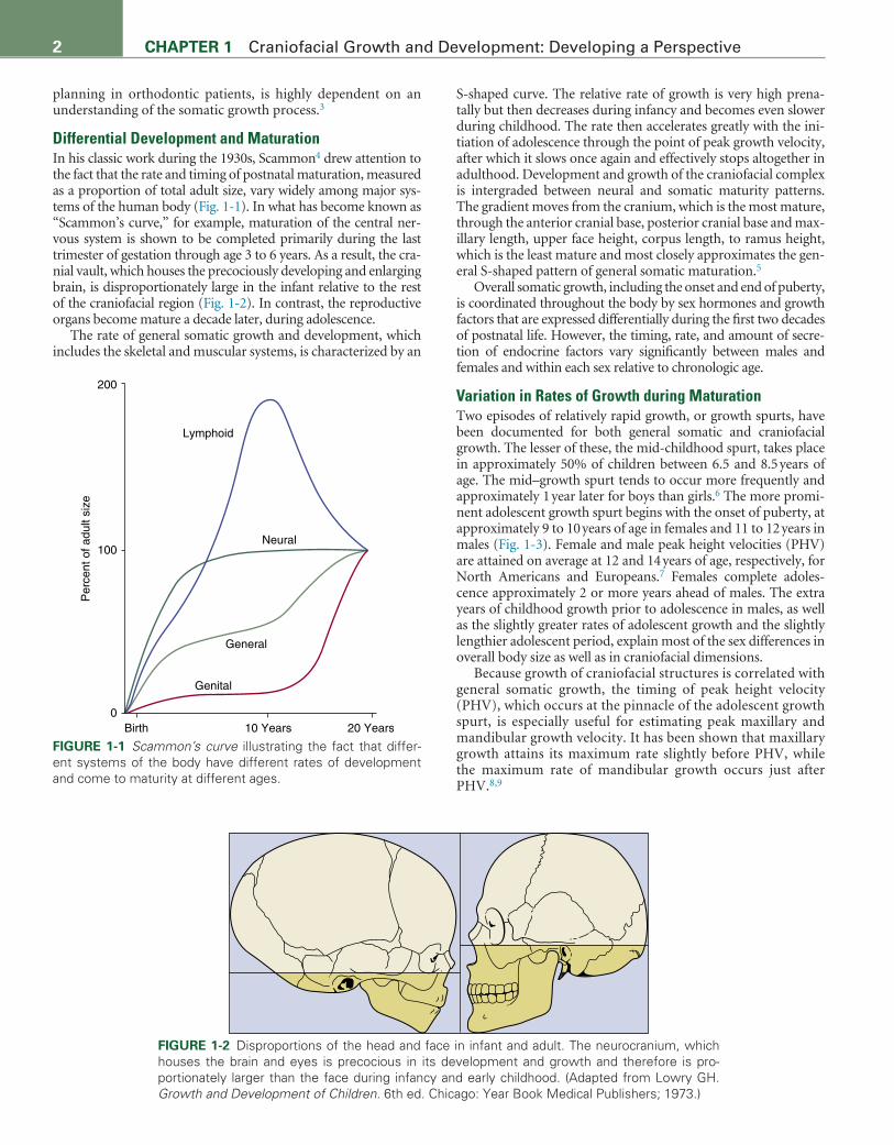
Differential Development and MaturationIn his classic work during the 1930s, Scammon4 drew attention to the fact that the rate and timing of postnatal maturation, measured as a proportion of total adult size, vary widely among major sys-tems of the human body (Fig. 1-1). In what has become known as “Scammon’s curve,” for example, maturation of the central ner-vous system is shown to be completed primarily during the last trimester of gestation through age 3 to 6 years. As a result, the cra-nial vault, which houses the precociously developing and enlarging brain, is disproportionately large in the infant relative to the rest of the craniofacial region (Fig. 1-2). In contrast, the reproductive organs become mature a decade later, during adolescence.
The rate of general somatic growth and development, which includes the skeletal and muscular systems, is characterized by an
S-shaped curve. The relative rate of growth is very high prena-tally but then decreases during infancy and becomes even slower during childhood. The rate then accelerates greatly with the ini-tiation of adolescence through the point of peak growth velocity, after which it slows once again and effectively stops altogether in adulthood. Development and growth of the craniofacial complex is intergraded between neural and somatic maturity patterns. The gradient moves from the cranium, which is the most mature, through the anterior cranial base, posterior cranial base and max-illary length, upper face height, corpus length, to ramus height, which is the least mature and most closely approximates the gen-eral S-shaped pattern of general somatic maturation.5
Overall somatic growth, including the onset and end of puberty, is coordinated throughout the body by sex hormones and growth factors that are expressed differentially during the first two decades of postnatal life. However, the timing, rate, and amount of secre-tion of endocrine factors vary significantly between males and females and within each sex relative to chronologic age.
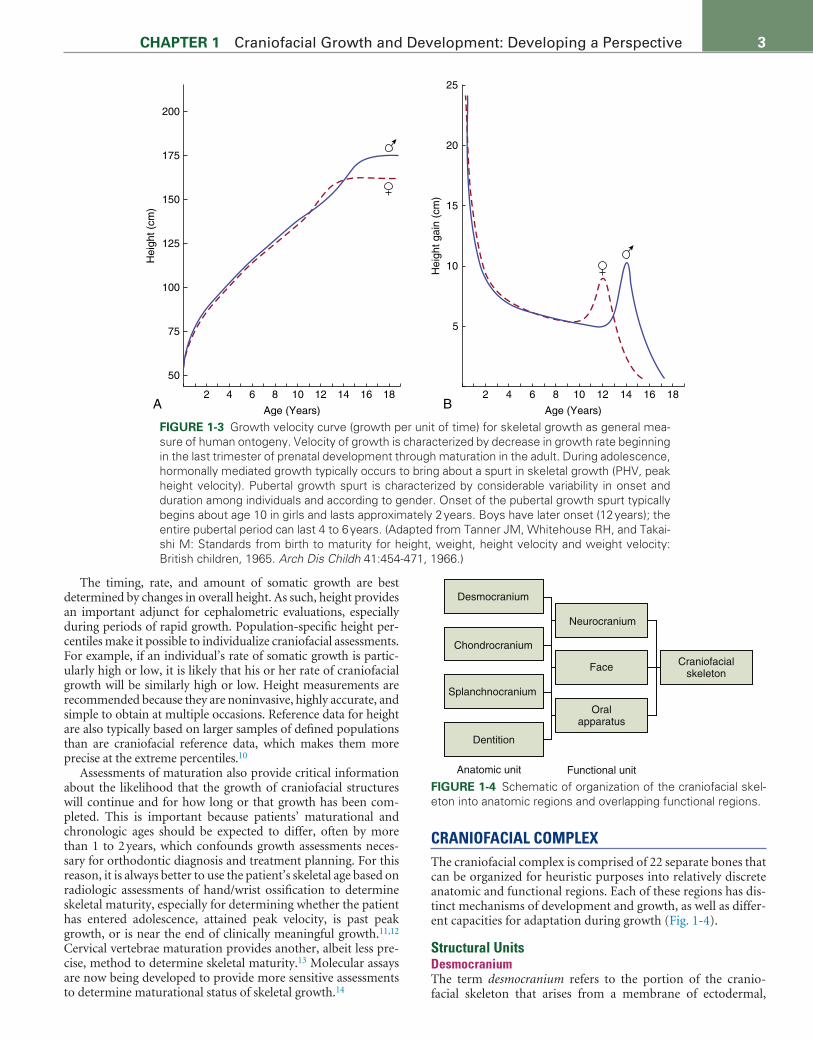
Variation in Rates of Growth during MaturationTwo episodes of relatively rapid growth, or growth spurts, have been documented for both general somatic and craniofacial growth. The lesser of these, the mid-childhood spurt, takes place in approximately 50% of children between 6.5 and 8.5 years of age. The mid–growth spurt tends to occur more frequently and approximately 1 year later for boys than girls.6 The more promi-nent adolescent growth spurt begins with the onset of puberty, at approximately 9 to 10 years of age in females and 11 to 12 years in males (Fig. 1-3). Female and male peak height velocities (PHV) are attained on average at 12 and 14 years of age, respectively, for North Americans and Europeans.7 Females complete adoles-cence approximately 2 or more years ahead of males. The extra years of childhood growth prior to adolescence in males, as well as the slightly greater rates of adolescent growth and the slightly lengthier adolescent period, explain most of the sex differences in overall body size as well as in craniofacial dimensions.
Because growth of craniofacial structures is correlated with general somatic growth, the timing of peak height velocity (PHV), which occurs at the pinnacle of the adolescent growth spurt, is especially useful for estimating peak maxillary and mandibular growth velocity. It has been shown that maxillary growth attains its maximum rate slightly before PHV, while the maximum rate of mandibular growth occurs just after PHV.8,9
200
Lymphoid
Neural
General
Genital
Birth 10 Years 20 Years
100
Per
cent
of a
dult
size
0
FIGURE 1-1 Scammon’s curve illustrating the fact that differ-ent systems of the body have different rates of development and come to maturity at different ages.
FIGURE 1-2 Disproportions of the head and face in infant and adult. The neurocranium, which houses the brain and eyes is precocious in its development and growth and therefore is pro-portionately larger than the face during infancy and early childhood. (Adapted from Lowry GH. Growth and Development of Children. 6th ed. Chicago: Year Book Medical Publishers; 1973.)
3CHAPTER 1 Craniofacial Growth and Development: Developing a Perspective
The timing, rate, and amount of somatic growth are best determined by changes in overall height. As such, height provides an important adjunct for cephalometric evaluations, especially during periods of rapid growth. Population-specific height per-centiles make it possible to individualize craniofacial assessments. For example, if an individual’s rate of somatic growth is partic-ularly high or low, it is likely that his or her rate of craniofacial growth will be similarly high or low. Height measurements are recommended because they are noninvasive, highly accurate, and simple to obtain at multiple occasions. Reference data for height are also typically based on larger samples of defined populations than are craniofacial reference data, which makes them more precise at the extreme percentiles.10
Assessments of maturation also provide critical information about the likelihood that the growth of craniofacial structures will continue and for how long or that growth has been com-pleted. This is important because patients’ maturational and chronologic ages should be expected to differ, often by more than 1 to 2 years, which confounds growth assessments neces-sary for orthodontic diagnosis and treatment planning. For this reason, it is always better to use the patient’s skeletal age based on radiologic assessments of hand/wrist ossification to determine skeletal maturity, especially for determining whether the patient has entered adolescence, attained peak velocity, is past peak growth, or is near the end of clinically meaningful growth.11,12 Cervical vertebrae maturation provides another, albeit less pre-cise, method to determine skeletal maturity.13 Molecular assays are now being developed to provide more sensitive assessments to determine maturational status of skeletal growth.14
CRANIOFACIAL COMPLEXThe craniofacial complex is comprised of 22 separate bones that can be organized for heuristic purposes into relatively discrete anatomic and functional regions. Each of these regions has dis-tinct mechanisms of development and growth, as well as differ-ent capacities for adaptation during growth (Fig. 1-4).
Structural UnitsDesmocraniumThe term desmocranium refers to the portion of the cranio-facial skeleton that arises from a membrane of ectodermal,
Hei
ght g
ain
(cm
)
Hei
ght (
cm)
Age (Years)
50
2 4 6 8 10 12 14 16 18
5
10
15
20
25
2 4 6 8 10 12 14 16 18
75
100
125
150
175
200
Age (Years)A B
FIGURE 1-3 Growth velocity curve (growth per unit of time) for skeletal growth as general mea-sure of human ontogeny. Velocity of growth is characterized by decrease in growth rate beginning in the last trimester of prenatal development through maturation in the adult. During adolescence, hormonally mediated growth typically occurs to bring about a spurt in skeletal growth (PHV, peak height velocity). Pubertal growth spurt is characterized by considerable variability in onset and duration among individuals and according to gender. Onset of the pubertal growth spurt typically begins about age 10 in girls and lasts approximately 2 years. Boys have later onset (12 years); the entire pubertal period can last 4 to 6 years. (Adapted from Tanner JM, Whitehouse RH, and Takai-shi M: Standards from birth to maturity for height, weight, height velocity and weight velocity: British children, 1965. Arch Dis Childh 41:454-471, 1966.)
Desmocranium
Chondrocranium
Splanchnocranium
Anatomic unit
Neurocranium
Face
Oralapparatus
Craniofacialskeleton
Functional unit
Dentition
FIGURE 1-4 Schematic of organization of the craniofacial skel-eton into anatomic regions and overlapping functional regions.

4 CHAPTER 1 Craniofacial Growth and Development: Developing a Perspective
mesodermal, and neural crest origin that surrounds the prox-imal end of the notochord very early in development. As the brain develops and expands in utero, the desmocranium devel-ops initially as a fibrous membrane covering of the brain that eventually will give rise to the bones of the cranial vault and fibrous joints, or sutures, as well as the dura mater over the brain and the periosteum overlying the bones of the cranial vault. In fact, in the absence of a brain, as with anencephaly, the desmocranial bones will fail to develop at all. Because the skel-etal derivatives of the desmocranium have exclusively a mem-branous precursor, initial morphogenesis and subsequent bone growth take place completely via intramembranous ossification.
ChondrocraniumThe chondrocranium forms initially as part of the embryonic anlagen of primary cartilage that will become the cranial base, nasal septum, and nasal capsule. Like the desmocranium, the chondrocranium is also a derivative of the embryonic mem-brane surrounding the developing central nervous structures. However, the chondrocranium is significantly less dependent on the presence of the brain for its initial formation and sub-sequent development. Growth associated with the derivative bones of the cranial base occurs by means of endochondral ossification.
ViscerocraniumThe viscerocranium, also referred to as the splanchnocranium, is composed of all those elements of the craniofacial complex that are derived from the first branchial arch and thus is of neu-ral crest origin. These elements primarily include the bones of the midfacial complex and the mandible. Because the skeletal elements of the viscerocranium have no primary cartilaginous precursors, development and growth of its skeletal derivatives take place via intramembranous ossification that is also char-acterized by the presence of sutures and a specialized form of membrane-derived (secondary) cartilage at the mandibular condyles.
DentitionThe deciduous and permanent teeth are specialized ana-tomic components of the craniofacial complex that are com-posed of unique tissues and undergo a unique mechanism of
development characterized by the interaction between ectoder-mal and mesenchymal tissues.
Functional UnitsThese four anatomic components can be combined organiza-tionally into three overlapping and very broad functional units comprising the craniofacial complex (Fig. 1-5).
NeurocraniumThe neurocranium houses the brain and other elements of the central nervous system, such as the olfactory apparatus and auditory apparatus. As the brain rests on the cranial base and is covered by the cranial vault, development and growth of the neurocranium are characterized by a combination of membra-nous (desmocranium) and cartilaginous (chondrocranium) bone growth.
FaceThe upper face may be defined as the region of the orbits of the eye. The midface, comprised primarily of the max-illae and zygomatic bones, is the region between the orbits and the upper dentition. Ectocranially, the bones of the face are composed externally of the intramembranously formed bones of the viscerocranium. However, the face also receives contributions from the chondrocranium as the cartilaginous nasal capsule and nasal septum. The lower face, comprised of the mandible, develops entirely from the first branchial arch and thus is derived entirely as part of the viscerocranium. The mandible develops and grows via a specialized form of intramembranous formation of both bone and secondary cartilage.
Oral ApparatusThe oral apparatus is composed of the dentition and support-ing structures within the upper and lower jaws. Thus, the oral apparatus also is characterized by a unique morphogenesis of the teeth as well as a specialized form of intramembranous bone growth of the alveolar processes of the maxilla and mandible (viscerocranium). Development and growth of the skeletal structures comprising the oral apparatus are greatly influenced by the muscles of mastication and other soft tissues associated with mastication.
Neurocranium(Desmocranium)
Midface(Splanchnocranium)
Oral apparatus(Dentition)
Lower face(Splanchnocranium)
(Chondrocranium)
FIGURE 1-5 Major components of the craniofacial skeletal complex.

5CHAPTER 1 Craniofacial Growth and Development: Developing a Perspective
MOLECULAR BASIS OF CRANIOFACIAL DEVELOPMENT AND GROWTHPatterning and subsequent formation of craniofacial tissues and structures have a complex, polygenic basis. For example, it has been shown that there are over 90 specific genes in which muta-tions will result in major disruptions of development leading to severe craniofacial malformations.15 Moreover, variations in craniofacial development and growth, from dysmorphologies to malocclusions, are multifactorial as a result of epigenetic mechanisms.16,17 No genes are unique to the craniofacial com-plex. However, certain genes, especially those associated with developmental patterning of the head region and growth of car-tilage, bone, and teeth, are of particular relevance for craniofa-cial development and growth and thus are of special importance for orthodontics. In addition, a number of genes of interest include those responsible for specific craniofacial deformities, such as craniosynostosis and facial clefts. The reader is referred to Hartsfield and Morford (see Chapter 2) for a comprehensive review of genetic mechanisms in the craniofacial region that are most important to orthodontics. A summary of the key genes associated with the patterning, development, and growth of the craniofacial region can be found in E-Table 1-1.
The key genes associated with craniofacial development may be organized informally into two broad yet overlapping groups based on their timing and patterns of expression and also their primary target tissues. First are those highly conserved genes, such as homeobox genes and transcriptions factors, that are responsible primarily for early pattern formation and differen-tiation of primary embryonic tissues and structures, including neural crest cells and head mesoderm. Mutation of those genes typically has a profound role in craniofacial dysmorphogenesis. The second group is comprised of genes such as growth factors and signaling molecules that are also responsible for mediat-ing development, growth, and maintenance of the tissues and structures associated with the craniofacial complex both during embryogenesis and throughout postnatal development. While mutations in this latter group of genes also are associated with craniofacial malformation syndromes, minor variants appear to be more common and may play a role in the development of more minor variations in growth. In addition, genes from both groups may be expressed reiteratively during development and growth, producing a highly complex matrix of interactions required for normal craniofacial morphogenesis. Adding to the
complexity are the issues of wound healing, tissue regeneration, and repair—all processes important during orthodontic treat-ment—that can reinitiate the expression of genes required for early morphogenesis as well as postnatal growth.
Molecular research historically has focused on the role of specific genes critical for craniofacial morphogenesis during embryogenesis. The initial focus in that research typically has been on three areas: (1) naturally occurring genetic mutations associated with craniofacial dysmorphogenesis in humans; (2) development of genetically engineered animal models, typically the mouse, to produce loss of function of selected genes; and (3) mapping of gene expression in experimental animals through in situ hybridization and other biomarker approaches. More recently, significant progress has been made in the identifica-tion of gene variants (polymorphisms) that may be important for the origin of minor variations in craniofacial growth of potential relevance to orthodontic diagnosis and treatment. These genes and their variants could be significant for diagno-sis and response to treatment of dentofacial deformities and minor malocclusions.18 Significant advances in the genetic and epigenetic basis of craniofacial development, including the role of key genes in normal growth and orthodontic treatment, are expected to continue at a rapid pace.19,20
CRANIAL VAULTDevelopment of the Cranial VaultThe most prominent feature of the embryonic cephalic region at 6 to 7 weeks’ gestation is the frontonasal prominence. The frontonasal prominence is a nonpaired structure that forms a dense desmocranial membrane, which covers the entire fore-brain and extends laterally and inferiorly on each side of the developing head to meet the developing maxillary processes. The inner portion of the membrane contains neural crest cells and gives rise to the dura mater covering the brain. The outer portion of the desmocranial membrane, the ectomeninx, is comprised of surface ectoderm deep to which is the paraxial mesoderm. Patterning of the frontonasal prominence to form the cranial vault and elements of the nasal region is induced by expression of sonic hedgehog (Shh) and FGF-8.
By 8 weeks’ gestation, initial blastemas of bone become apparent within the ectomeninx, first for the frontal bone and the squamous temporal bone and subsequently for the parietal bones and squamous portion of the occipital bone (Fig. 1-6).
A B C
FIGURE 1-6 Cleared and stained human fetuses indicating craniofacial skeletal structures at approximately 8 weeks’ gestation (A), 15 weeks’ gestation (B), and 18 weeks’ gestation (C).

5.e1CHAPTER 1 Craniofacial Growth and Development: Developing a Perspective
TABLE 1-1 Summary of Key Genes Associated with the Development and Growth of the Tissues and Structures Comprising the Craniofacial Complex
Gene/Protein General Role and FunctionSignificance for Craniofacial Development and Growth References
Bmp-1 to Bmp-9 Bone morphogenetic protein 1-9
Signaling molecule: Skeletal differentiation, growth, repair
NCC and CF mesenchyme patterning; suture development; odontogenesis; nsCL/P
1-6
Dlx-1 to Dlx-6 Distal-less 1-6 Homeobox: Limb development; chondrogenesis; osteogenesis
Orofacial clefting 7-9
Efnb1 Ephrin B1 Protein coding: Cell division, adhesion Craniofrontonasal syndrome; candidate for role in class III malocclusion
1, 10-12
Fgf-1 to Fgf-18 Fibroblast growth factor 1-18
Growth factors: Differentiation and growth of multiple tissues and structures
CF ectoderm, NCC patterning; suture development; MCC growth; tooth induction; CL/P
1, 3, 4, 13-15
Fgfr-1 to Fgfr-3 Fibroblast growth factor receptor 1-3
Transmembrane receptors: Fgf receptor Anterior cranial base growth; MCC growth; syndromic, nonsyndromic C-SYN; MX hypoplasia; CL/P
1, 3, 4, 15, 16, 17
Gh Growth hormone Peptide hormone-mitogen: Cell growth and tissue regeneration
Growth of multiple CF tissues, structures; variations in MD growth, dentofacial treatment
13, 18
Ghr Growth hormone receptor Transmembrane receptor: Receptor for GH Polymorphisms associated with MD growth and MCC response to dentofacial treatment
19-21
Gli2 to Gli3 Zinc finger protein Gli2-3 Transcription factor: Regulates Ihh and Shh signaling
C-SYN; Greig cephalopolysyndactyly syndrome
1, 10, 22
Gsc Goosecoid Transcription factor: Dorsal–ventral patterning of NCC, head formation; rib fusion
Inner ear, cranial base, MX/MD anom-alies
1, 8, 13, 23, 24
Hoxa1 to Hoxa3 Homeobox A1, A2, A3 Homeobox: Patterning of hindbrain rhombomeres and pharyngeal arches
Neural tube closure, 1st-2nd arch deformities
25, 26
Igf-1 Insulin-like growth factor 1 Growth factor: Mediator of Gh; muscle, cartilage, and bone growth
MX/MD growth; suture development/growth; mediation of MCC to dento-facial treatment
3, 8, 13, 27-30
Ihh Indian hedgehog Signaling molecule: Endochondral and intramembranous ossification
Cranial base development; mediation of MCC growth during dentofacial treatment
31-33
L-Sox5 Long-form of Sox5 Transcription factor: Neurogenesis; chondrogenesis; type II collagen
Mediation of MCC growth during dentofacial treatment
34
Msx1 to Msx2 Muscle segment homeo-box 1-2
Homeobox: Limb development; ectodermal organs
NCC proliferation, migration; odontogenesis; MD development; nsCL/P; Boston-type C-SYN
1, 3, 4, 8, 10, 35
Myo1H and-Myo1C
Myosin 1H, Myosin 1C Protein coding: Cell motility, phagocytosis, vesicle transport
Polymorphisms associated with MD prognathism
36, 37
Nog Noggin Signaling molecule: Patterning of the neural tube and somites
Head formation; neural tube fusion 4, 25, 26
Notch Transmembrane receptor: Neuronal development; cardiac development; osteogenesis
MCC development 38
Osx Osterix Transcription factor: Osteoblast differentiation, mineralization; chondrogenesis
MCC differentiation, endochondral ossification; mediation of MCC growth during dentofacial treatment
39
Pitx1-2 Paired-like homeodomain 1-2
Homeobox: Left–right axis; left lateral mesoderm; skeletal development; myogenesis
MD development; role in Treacher- Collins syndrome; CL/P; odontogenesis
8, 13
Prx-1Prx-2 Homeobox: Epithelial development in limbs and face
NCC patterning; malformations of 1st-2nd arch structures
8, 40, 41
PTHrP Parathyroid-related protein Protein coding: Endochondral bone formation
Development/growth of cranial base, MD, dental arches
42, 43
Runx2 Runt-related transcription factor
Transcription factor: Osteoblast differentiation; intramembranous and endochondral bone growth
Closure of fontanelles and sutures; ossification of cranial base, MX, and MCC; cleidocranial dysplasia
32, 43-46
Shh Sonic hedgehog Transcription factor: Development of limbs, midline brain, neural tube; osteoblastic differentiation; skeletal morphogenesis
Induction of frontonasal ectoderm; cranial base; fusion of facial processes; palatogenesis; odontogenesis; holoprosencephaly
1, 9, 33
Sho2 Signaling molecule: Development of digits; organization of brain, CF mesenchyme
Palatogenesis; TMJ development 6, 9, 38
Continued

5.e2 CHAPTER 1 Craniofacial Growth and Development: Developing a Perspective
TABLE 1-1 Summary of Key Genes Associated with the Development and Growth of the Tissues and Structures Comprising the Craniofacial Complex—cont’d
Gene/Protein General Role and FunctionSignificance for Craniofacial Development and Growth References
Sox9 Transcription factors: Chondrogenesis; type II collagen; male sexual development
Cranial base; MCC growth; CL/P; Pierre-Robin sequence
38, 46-48
Spry 1-2 Sprouty Protein coding: Mediates FGF signaling MD/TMJ development 38, 48Tcof1 Treacle Protein coding: Early embryonic nucleo-
lar-cytoplasmic transportNCC proliferation, migration, survival;
Treacher-Collins syndrome38, 49
Tgf-ß1 to Tgf-ß3 Transforming growth factor-beta 1-3
Growth factor: Proliferation, differentiation, growth, function of multiple tissues
Palatogenesis; MD growth; suture development, maintenance, fusion; sCL/P
3, 24
Twist-1 Twist-related protein 1 Transcription factor: Skeletal development; syndactyly
MCC development; suture fusion; Saethre-Chotzen syndrome; facial asymmetry
9, 35, 38, 50, 51
Vegf Vascular endothelial growth factor
Growth factor: Ingrowth of blood vessels Chondrogenesis in cranial base, MCC 38, 45, 52
Wnt-1 Proto-oncogene protein Wnt 1
Signaling molecule: Cell fate, patterning during embryogenesis
MCC development/growth; MCC growth during dentofacial treatment
6, 32, 38, 53
CF, Craniofacial; CPO, cleft palate only; CL/P, cleft lip and palate; C-SYN, craniosynostosis; MCC, mandibular condylar cartilage; MD, mandible; MX, maxilla; NCC, neural crest cells; nsCL/P, non-syndromal cleft lip and palate; sCL/P, syndromal cleft lip and palate; TMJ, temporomandibular joint.References1. Rice DPC. Craniofacial anomalies: from development to molecular pathogenesis. Curr Mol Med. 2005;5:699–722.2. Zheng L, Yamashiro T, Fukunaga T, Balam TA, Takano-Yamamoto T. Bone morphogenetic protein 3 expression pattern in rat condylar cartilage, femoral cartilage and mandibular
fracture callus. Eur J Oral Sci. 2005;113:318–325.3. Opperman LA, Gakunga PT, Carlson DS. Genetic factors influencing morphogenesis and growth of sutures and synchondroses in the craniofacial complex. In: Carlson DS, ed. Control
mechanisms of craniofacial development and growth. Semin Orthod. 2005;11(4):199–208.4. Chai Y, Maxson RE Jr. Recent advances in craniofacial morphogenesis. Dev Dyn. 2006;235:2353–2375.5. Nie X, Luukko K, Kettunen P. BMP signaling in craniofacial development. Int J Dev Biol. 2006;50:511–521.6. Greene RM, Pisano MM. Palate morphogenesis: current understanding and future directions. Birth Defects Res (Part C). 2010;90:133–154.7. Robledo RE, Rajan L, Li X, Lufkin T. The Dlx5 and Dlx6 homeobox genes are essential for craniofacial, axial, and appendicular skeletal development. Genes Develop. 2002;16:1089–1101.8. Doshi RR, Patil AS. A role of genes in craniofacial growth. IIOBA. 2012;3(2):19–36.9. Hinton RJ. Genes that regulate morphogenesis and growth of the temporomandibular joint: a review. Devel Dyn. 2014;243:864–874.10. Melville H, Wang Y, Taub PJ, Jabs EW. Genetic basis of potential therapeutic strategies for craniosynostosis. Am J Med Genet Part A. 2010;152A:3007–3015.11. Xue F, Wong RWK, Rabie ABM. Genes, genetics, and Class III malocclusion. Orthod Craniofac Res. 2010;13:69–74.12. Xue F, Wong RWK, Rabie ABM. Identification of SNP markers on 1p36 and association analysis of EPB41 with mandibular prognathism in a Chinese population. Arch Oral Biol.
2010;55:867–872.13. Hinton RJ, Carlson DS. Regulation of growth in the mandibular condylar cartilage. In: Carlson DS, ed. Control mechanisms of craniofacial development and growth. Semin Orthod.
2005;11(4):209–218.14. Hatch NE. FGF signaling in craniofacial biological control and pathological craniofacial development. Crit Rev Eukaryot Gene Expr. 2010;20(4):295–311.15. Martinez-Abadias N, Heuze Y, Wang Y, Jabs EW, Aldridge K, Richtmeier J. FGF/FGFR signaling coordinates skull development by modulating magnitude of morphological integration:
evidence from Apert syndrome mouse models. PLoS One. 2011;6(10).16. Rice DPC, Rice R, Thesleff I. Fgfr mRNA isoforms in craniofacial bone development. Bone. 2003;33(1):14–27.17. Heuze Y, Martinez-Abadias N, Stella JM, et al. Quantification of facial skeletal shape variation in fibroblast growth factor receptor-related craniosynostosis syndromes. Birth Def Res
(A). 2014;100:250–259.18. Buschang PH, Hinton RJ. A Gradient of Potential for Modifying Craniofacial Growth. In: Carlson DS, ed. Control mechanisms of craniofacial development and growth. Semin Orthod.
2005;11(4):219–226.19. Zhou J, Lu Y, Gai XH, et al. The growth hormone receptor gene is associated with mandibular height in a Chinese population. J Dent Res. 2005;84:1052–1056.20. Kang EH, Yamaguchi T, Tajima A, et al. Association of the growth of the growth hormone receptor gene polymorphisms with mandibular height in a Korean population. Arch Oral
Biol. 2009;45:556–562.21. Sasaki Y, Satoh K, Hayasaki H, Fukumoto S, Fujiwara T, Nonaka K. The P561T polymorphism of the growth hormone receptor gene has an inhibitory effect on mandibular growth in
young children. Europ J Orthod. 2009;31:536–541.22. Veistinen L, Takatolo M, Tanimoto Y, Kesper D, Vortkamp A, Rice DPC. Loss-of-function of Gli3 in mice causes abnormal frontal bone morphology and premature synostosis of the
interfrontal suture. Front Physiol. 2012;3:1–6.23. Sharpe PT. Homeobox genes and orofacial development. Conn Tiss Res. 1995;32:17–25.24. Spears, Svoboda In: Carlson DS, ed. Growth factors and signaling proteins in craniofacial development. Control mechanisms of craniofacial development and growth. Semin Orthod.
2005;11(4):184–199.25. Carlson B. Human Embryology and Developmental Biology. Philadelphia: Elsevier; 2014.26. Trainor PA, Krumlauf R. Patterning the neural crest: hindbrain segmentation and hox gene plasticity. Nat Rev Neuro. 2000;1:116–124.27. Hajjar D, Santos MF, Kimura ET. Mandibular repositioning modulates IGFBP-3, -4, -5, and -6 expression in the mandibular condyle of young rats. Biorheology. 2006;43(3–4):311–321.28. Marques MR, Hajjar D, Franchini KG, Moriscot AS, Santos MF. Mandibular appliance modulates condylar growth through integrins. J Dent Res. 2008;87(2):153–158.29. Patil AS, Sable RB, Kothari RM. Role of insulin-like growth factors (IGFs), their receptors and genetic regulation in the chondrogenesis and growth of the mandibular condylar cartilage.
J Cell Physiol. 2011;227:1796–1804.30. Frazier-Bowers S, Rincon-Rodriguez R, Zhou J, Alexander K, Lange E. Evidence of linkage in a Hispanic cohort with a class III dentofacial phenotype. J Dent Res. 2009;88:56–60.31. Tang GH, Rabie ABM. Runx2 regulates endochondral ossification in condyle during mandibular advancement. J Dent Res. 2005;84(2):166–171.32. Carreira AC, Lojudice FH, Halcsik E, Navarro RD, Sogayar MC, Granjeiro JM. Bone morphogenetic proteins: facts, challenges, and future perspectives. J Dent Res. 2014;93(4):335–345.33. Balczerski B, Zakaria S, Tucker AS, et al. Distinct spatiotemporal roles of hedgehog signaling during chick and mouse cranial base and axial skeletal development. Dev Biol.
2012;371:203–214.34. Chu FT, Tang GH, Hu Z, Qian YF, Shen G. Mandibular functional positioning only in vertical dimension contributes to condylar adaptation evidenced by concomitant expressions of
L-Sox5 and type II collagen. Arch Oral Biol. 2008;53:567–574.35. Bonaventure J, El-Ghouzzi V. Molecular and cellular basis of syndromic craniosynostosis. Exp Rev Mol Med. 2003;5(29):1–17.36. Tassopoulou-Fishell M, Deeley K, Harvey EM, Sciote JJ, Viera AR. Genetic variation in Myosin 1H contributes to mandibular prognathism. Am J Orthod Dentofac Orthoped.
2012;141(1):51–59.37. Desh H, Gray SL, Horton MJ, et al. Molecular motor MYO1C, acetyltransferase KAT6B and osteogenetic transcription factor RUNX2 expression in human masseter muscle contrib-
utes to development of malocclusion. Arch Oral Biol. 2014;59:601–607.38. Hinton RJ, Serrano M, So S. Differential gene expression in the perichondrium and cartilage of the neonatal mouse temporomandibular joint. Orthod Craniofac Res. 2009;12:168–177.

5.e3CHAPTER 1 Craniofacial Growth and Development: Developing a Perspective
TABLE 1-1 Summary of Key Genes Associated with the Development and Growth of the Tissues and Structures Comprising the Craniofacial Complex—cont’d
39. Jing J, Hinton RJ, Jing Y, Liu Y, Zhou X, Feng JQ. Osterix couples chondrogenesis and osteogenesis in post-natal condylar growth. J Dent Res. 2014;93(10):1014–1021.40. ten Berge D, Brouwer A, Korving J, et al. Prx1 and Prx2 are upstream regulators of sonic hedgehog and control cell proliferation during mandibular arch morphogenesis. Devel. 2001.41. Martin JF, Bradley A, Olsen EN. The paired-like homeobox gene Mhox is required for early events of skeletogenesis in multiple lineages. Genes Develop. 1995;9:1237–1249.42. Kyrkanides S, Kambylafkas P, Miller JH, Tallents RH, Puzas JE. The cranial base in craniofacial development: a gene therapy study. J Dent Res. 2007;86(10):956–961.43. Hinton RJ. Genes that regulate morphogenesis and growth of the temporomandibular joint: a review. Devel Dyn. 2014;243:864–874.44. Rabie ABM, Tang GH, Hägg U. Cbfa1 couples chondrocytes maturation and endochondral ossification in rat mandibular condylar cartilage. Arch Oral Biol. 2004;49(2):109–118.45. Lei WY, Wong RWK, Rabie ABM. Factors regulating endochondral ossification in the spheno-occipital synchondrosis. Angle Orthod. 2008;78(2):215–220.46. Nie X, Luukko K, Kvinnsland IH, Kettunen P. Developmentally regulated expression of Shh and Ihh in the developing mouse cranial base: comparison the Sox9 expression. Anat Rec
A Discov Mol Cell Evol Biol. 2005;286(2):891–898.47. Cendekiawan T, Wong RWK, Rabie ABM. Temporal expression of SOX9 and type II collagen in spheno-occipital synchondrosis of mice after mechanical tension stimuli. Angle Orthod.
2008;78(1):83–88.48. Rabie ABM, She TT, Hägg U. Functional appliance therapy accelerates and enhances condylar growth. Am J Orthod Dentofacial Orthop. 2003;123(1).49. Su P-H, Liu Y-F, Chen J-Y, Lai Y-J, Facial asymmetry and clinical manifestations in patients with novel insertion of the TCOF1 gene. Clin Genet. 2012;82:460–465.50. Coussens AK, Wilkinson CR, Hughes IP, et al. Unravelling the molecular control of calvarial suture fusion in children with craniosynostosis. BMC Genomics. 2007;8:458.51. Melville H, Wang Y, Taub PJ, Jabs EW. Genetic basis of potential therapeutic strategies for craniosynostosis. Am J Med Genet Part A. 2010;152A:3007–3015.52. Rabie ABM, Hägg U. Factors regulating mandibular condylar growth. Am J Orthod Dentofacial Orthop. 2002;122:401–409.53. Enomoto A, Watahiki J, Nampo T, et al. Mastication markedly affects mandibular condylar growth, gene expression, and morphology. Am J Orthod Dentfac Orthop. 2014;146(3):365–363.

6 CHAPTER 1 Craniofacial Growth and Development: Developing a Perspective
Over the ensuing 4 weeks, these condensations of bone steadily increase in size by radial expansion of newly differentiated skeletal tissue within the ectomeninx. As the development of new bone exceeds the rate of growth of the brain, the periph-eral bone fronts become located closer and closer to each other, until they approximate each other as single-thickness plates of flat bones by about 12 weeks’ gestation. At this point, the inter-vening fibrous tissue becomes highly cellular, and fibrous artic-ulations, or sutures, are formed between the individual bone elements (Fig. 1-7).
Growth of the cranial vault bones represents a specialized form of intramembranous ossification that begins prenatally as blastemas of bone tissue that arise de novo within the mid-dle layer of the desmocranial membrane covering of the brain. Once the skeletal elements as plates of bone become located close to each other, their fibrous connections become reorga-nized with the periosteum and the dura mater derived from the outer and inner layers of the desmocranial membrane, respec-tively, extending into the sutural articulations. The sutures then continue to support growth of the cranial vault through another specialized form of intramembranous osteogenesis similar to periosteal bone formation.21–23
Mechanisms of Suture GrowthSutural bone growth can best be considered as a specialized form of intramembranous periosteal bone growth. Once formed, the bones of the cranial vault are enveloped, like all bones, in a skel-etogenic membrane. On the external surface, this membrane is the periosteum. On the intracranial surface, the membrane is the dura mater, which is also derived from the embryonic ectomeninx and is skeletogenic. Viewed in cross section, the outer fibrous layer of periosteum (uniting layer) spans over the cranial suture and provides structural support to the suture and its two or more skeletal elements. The inner osteogenic layers of
the periosteum and the dura reflect into the space between the two cranial vault bones and provide a source of new osteogenic cells (Fig. 1-8). As the bones of the cranial vault become sepa-rated because of expansion of the brain and intracranial con-tents, the osteogenic cells form skeletal tissue and thus provide a mechanism for maintaining relatively close contact through the intervening suture.
The molecular basis of the development and growth of the sutures of the cranial vault has received considerable atten-tion, principally because of the number of naturally occurring and engineered genetic mutations characterized by craniosyn-ostosis (see Wilkie and Morriss-Kay,15 Rice,24 and Chai and Maxson25 for comprehensive reviews). Studies have shown a complex pattern of gene expression within the sutural blas-tema associated with the periosteal reflection as well as the intracranial dura mater. Secretion of soluble factors by the
bb
ble
bb
ps
s
s
Periosteum
Dura
Bony overlap
ble
ble
ble
F19
N5 N21
N1
FIGURE 1-7 Photomicrographs of hematoxylin and eosin–stained histologic sections through the coronal suture of normal rats at embryonic day 19 and postnatal days 1, 5, and 21. Bone (b), bone leading edge (ble), presumptive suture mesenchyme (ps), and suture (s). (From Opperman LA, Gakunga PT, Carlson DS. Genetic factors influencing morphogenesis and growth of sutures and synchondroses in the craniofacial complex. Semin Orthod. 2005;11(4):199–208.)
Capsular layer
Middle layer Outer tableDiploëInner table
Uniting layer
Periosteum
Fibrousosteogenic
Cambrial layer
NV
A A
FIGURE 1-8 Schematic representation indicating the relation-ship between the periosteum and dura mater as a mechanism for a specialized of intramembranous growth within the sutures of cranial vault bones. (Adapted from Pritchard JJ, Scott JH, Girgis FG. The structure and development of cranial and facial sutures. J Anat. 1956;90:73–86.)

7CHAPTER 1 Craniofacial Growth and Development: Developing a Perspective
dura mater in response to growth signals from the expanding, underlying brain is essential for normal cranial suture mor-phogenesis as well as for the maintenance of cranial sutures as patent bone- growth sites through complex tissue inter-actions and feedback between dura mater, bone fronts, and sutures. Both sutures and the dura mater also contain growth factors, such as several members of the family of transforming growth factor beta (TGF-β1, TGF-β2, TGF-β3), bone mor-phogenetic protein (BMP2, BMP7), fibroblast growth factor 4 (FGF-4), insulin-like growth factor 1 (IGF-1), and sonic hedge-hog (SHH) (Fig. 1-9).26,27 Overexpression of transcription fac-tors Runx2 and Msx2 and haploinsufficiency of Twist28 and Noggin29 are also associated with suture obliteration, while loss of function of Gli3 results in premature synostosis.30 Genetic analysis of naturally occurring craniosynostosis in humans has shown that mutations of genes for fibroblast growth factor receptors 1, 2, and 3 (FGFR-1, FGFR-2, and FGFR-3) and in MSX231 and TWIST32,33 genes are also associated with prema-ture suture fusion.
Development and growth of the cranial vault as a whole, and development and growth of bone at the sutural articulations, are primarily dependent on the expansion of the brain and other intracranial contents.34 Furthermore, it has been clearly demon-strated that sutures are secondary, compensatory, and adaptive sites of bone growth that normally respond to biomechanical forces. As the brain expands during prenatal development and during the first decade of life postnatally, forces are created within the neurocranium that cause the bones of the cranial vault to expand outward, which tends to separate them from each other at the sutural boundaries (Fig. 1-10). Under normal conditions, the cellular and molecular substrate associated with the dura mater, the periosteum, and the suture respond to this biomechanical displacement in the same manner in which peri-osteum throughout the skeletal system responds—by initiating and maintaining osteogenesis within the sutures to maintain the proximity of the adjoining skeletal structures. When the biological substrate of the suture is abnormal, however, as in the case of many genetic syndromes such as Crouzon syndrome, Apert syndrome, and Jackson-Weiss syndrome, for example, each of which is associated with mutations of FGFR-2, prema-ture craniosynostosis may result.35,36 The opposite condition,
reduced suture growth, and prolonged patency, as seen in clei-docranial dysostosis, may occur with abnormalities associated with growth factors, including in particular Runx2, which are necessary for normal suture fusion.
Postnatal Growth of the Cranial VaultDue to the very precocious nature of prenatal and early post-natal human brain development, the cranial vault is dispropor-tionately large relative to the rest of the face and body. At birth, the cranial vault is initially characterized by the presence of all of the cranial vault bones. At that time, all the major sutural fibrous articulations between the bones of the cranial vault are present, including the metopic suture between the right and left frontal bone. In addition, there typically are four larger remnants, known as fontanels, of the desmocranial membrane in areas where the pace of bone growth has not been sufficient to approximate the bones of the cranial vault to form a suture (Fig. 1-11).
During the first 24 months after birth, growth of the cra-nial vault bones proceeds rapidly enough to close the fonta-nels as each complex of cranial vault bones becomes organized through interlocking sutures. The metopic suture normally fuses to form a single frontal bone within the first year of life, although the suture may appear to persist for up to 8 years of age or even throughout life in a small percentage of individuals. The cranial vault will continue to enlarge primarily as a result of compensatory growth of the sutural bone fronts stimulated by expansion of the brain. By 4 years of age, the brain and the associated cranial vault will have achieved approximately 80% of adult size; by age 10, the brain and cranial vault have attained 95% of their adult size. Throughout this time of very rapid expansion, the remaining sutures of the cranial vault normally remain patent and actively growing to keep pace with the brain as it expands in size.
Osteogenesis at cranial sutural bone fronts may continue for the first two decades of life. However, by the end of the second decade of life, bone growth at cranial sutures has slowed and the potential for growth of cranial sutures has greatly diminished. Also at that time, the sutures will begin the normal process
Periosteum
Bone
Dura
Periosteum
Bone
Dura
A
B
Runx2
Runx2 BMP2
BMP2
Runx2
BMP2Fgfr1
Fgfr1
Fgfr1Fgfr1 Fgfr1
Fgfr2
Fgf2
TGF-�2TGFr2
TGFr1Msx2
Msx2
TGF-�3TGF-�3
TGF-�2
Twist noggin
FIGURE 1-9 Distribution of growth factors and transcription factors active during suture growth (A) and suture synostosis (B). (Adapted from Opperman LA, Gakunga PT, Carlson DS. Genetic factors influencing morphogenesis and growth of sutures and synchondroses in the craniofacial complex. Semin Orthod. 2005;11(4):199–208)
Sagittal suture
Dura mater
EpidermisDiploë
FIGURE 1-10 Schematic diagram indicating the relationship between expansile growth of the brain as a stimulus for com-pensatory growth of sutures of the cranial vault. (Adapted from Moss ML. The functional matrix. In: Kraus B, Reidel R, eds. Vis-tas Orthod. Philadelphia: Lea & Febiger; 1962;85–98.)

8 CHAPTER 1 Craniofacial Growth and Development: Developing a Perspective
of bony closure, or synostosis, when the potential for sutural growth ceases altogether.
The cranial sutures normally lose the capacity for growth by the end of the second decade of life, and virtually all become synostosed during the life span. Normal suture closure is initi-ated along the endocranial surface. Initially, this is characterized by bridging of bone across the suture and eventually through modeling of bone, leading to complete obliteration of the suture. Cessation of growth at cranial sutures typically begins around age 25 for the sagittal suture and may be extended for 2 to 3 additional years for the coronal suture.
Despite the fact that the major cranial sutures stop growing by the third decade of life, some enlargement of the cranial vault overall typically occurs throughout the lifespan as a result of periosteal deposition along the ectocranial surface. Certain spe-cific areas of the cranial vault, such as the glabellar and nuchal regions, may exhibit slightly greater periosteal growth as a sec-ondary sex characteristic in males.
CRANIAL BASEDevelopment of the Cranial BaseThe ectomeningeal membrane that surrounds the developing brain in the cranial base region gives rise to a number of paired cartilaginous elements that form the embryonic chondrocra-nium. The first of the cartilage anlagen to form arises from neural crest cells at about 6 weeks’ gestation as the parachordal cartilages, which surround the proximal end of the notochord and give rise to the anterior cranial base. The posterior compo-nent of the cranial base is derived primarily from mesoderm to form the basioccipital bone.37 Development of the chondrocra-nium then progresses rostrally to the otic capsule, which will form the petrous portion of the temporal bone; the postsphe-noid, presphenoid, alisphenoid, and orbitosphenoid cartilages of the sphenoid bone; and the nasal capsule and mesethmoid, which will form the ethmoid bone, inferior turbinate, and nasal septum. By 8 weeks’ gestation, the separate cartilage elements have merged to form a single plate of primary hyaline cartilage, the basal plate, extending from the foramen magnum rostrally to the tip of the nasal cavity (Fig. 1-12).
More than 110 separate centers of ossification form in the basal plate, beginning with the parachordal cartilages and continuing rostrally through the sphenoid complex around 9 to 16 weeks, to the ethmoid region as late as 36 weeks. As
these centers of ossification arise within the chondrocra-nium, segments of intervening cartilage form synchondroses (Fig. 1-13). The principal cranial base synchondroses that are most relevant for understanding craniofacial growth are the spheno-occipital synchondrosis, between the body of the sphenoid and the basioccipital bone, and the spheno- ethmoidal synchondrosis, between the sphenoid and eth-moid bones. The greater wing of the sphenoid bone and the squamous portion of the occipital bone develop and grow via intramembranous ossification.
Mechanism of Synchondrosal GrowthCranial base synchondroses are temporary cartilaginous joints located between bones of endochondral origin and growth.
Anteriorfontanel
Metopicsuture
Sphenoidfontanel
Mandibularsymphysis
Mastoidfontanel
FIGURE 1-11 Lateral and frontal views of the neonate skull indicating the location of sutures and fontanels. (Adapted from Sicher H, DuBrul EL. Oral Anatomy. 5th ed. St. Louis: Mosby; 1970.)
Trabeculae cranii
Cerebralhemisphere
Parachordalcartilage Otic capsule
Jugularforamen
Occipitalsclerotomes
Cervicalsclerotomes
Orbitosphenoidcartilage
(optic foramen)
Nasalcapsule
Hypophysealpouch Hyoid bone
Ala orbitalis
Ala temporalis
Internal auditorymeatus
Optic capsule
Parachordalcartilage
Hypophysealcartilage
Occipitalsclerotomes
NotochordA
BFIGURE 1-12 Schematic representation of the cartilaginous basal plate comprising the embryonic chondrocranium. A, Dor-soventral view; B, Lateral view.

9CHAPTER 1 Craniofacial Growth and Development: Developing a Perspective
Synchondroses can best be considered as homologous to the epiphyseal growth plates of long bones. Functionally, both pro-vide a mechanism for rapid endochondral growth of bone in a manner that is capable of overcoming biomechanical loads, thus exhibiting tissue-separating capabilities. Developmentally, cranial base synchondroses and epiphyseal plates of long bones synostose and become obliterated when the skeletal element achieves its mature size and shape. This typically occurs at the end of puberty for epiphyseal growth plates but varies from the end of the juvenile period through the end of puberty for the major cranial base synchondroses.
Cranial base synchondroses and epiphyseal growth plates are both derived from the primary hyaline cartilage that arises as part of the embryonic cartilaginous anlagen. Like endochondral bones and growth plates throughout the body, growth of synchondroses is controlled principally by expres-sion of Indian hedgehog gene (Ihh) and sonic hedgehog (Shh).38,39 The significance of FGFR-3 for growth of the ante-rior cranial base is also indicated by mutations associated with achondroplasia.
Histomorphologically, both cranial base synchondroses and epiphyseal growth plates, are characterized by primary chondro-cytes that are distributed into zones that are highly typical for growth plate cartilage (Fig. 1-14). However, a major difference between epiphyseal growth plates in long bones and cranial base synchondroses is that synchondroses are “bidirectional.” Thus, each cranial base synchondrosis effectively has two back-to-back growth plates with a shared region of newly forming cartilage in the center and bone at each end. Growth plates are “unidirectional.”
The primary hyaline cartilage of the cranial base is the same as that found throughout the embryonic cartilaginous anlage that characterizes all the other cartilaginous bones through-out the body. It is well known that growth of tissues derived from the primary embryonic cartilaginous anlagen tends to be relatively resistant to all but very extreme external influences. Growth of cartilage-derived skeletal elements throughout the body tends to be relatively resistant to environmental and other factors and instead is regulated to a large extent by intrinsic, genetically regulated growth factors and cell-signaling mol-ecules.40 The same is true for the cranial base synchondroses.
Hypophysealfossa
Basisphenoid
Exoccipital
Foramenmagnum
Basioccipital
Sphenoccipitalsynchondrosis
Septalcartilage
Cribriform plateof ethmoid
Crista galli
Lateralpart
Occipital squama
Anteriorintraoccipital
synchondrosis Basilarpart
Occipitalcondyle
Posteriorintraoccipitalsynchondrosis
Ala ofvomer
Vomer
FIGURE 1-13 Drawing of sagittal and basal views of the neonatal skull indicating spheno-occipital synchondrosis and intraoccipital synchondroses. The sphenoethmoidal synchondrosis will arise between the sphenoid and ethmoid bones. (Adapted from Bosma JF. Introduction to the sym-posium. In: Bosma JF, ed. Development of the Basicranium. Bethesda, MD: US Department of Health, Education, and Welfare; 1976:3–28.)
R
P
M
H
E
E H M P R P M H E
A B
FIGURE 1-14 Histologic comparison between the cartilages within a growing epiphyseal plate (A) and cranial base synchondrosis (B) (hematoxylin and eosin–stained). R, Resting zone (dashed line); P, proliferating zone; M, maturational zone; H, hypertrophic zone; E, zone of endochondral ossification.

10 CHAPTER 1 Craniofacial Growth and Development: Developing a Perspective
However, it is important to note that the growth of both epiph-yses and synchondroses can be significantly affected by such epigenetic factors as disease, malnutrition, and undernutrition, as well as other conditions that affect production and expres-sion of endocrine factors responsible for bone growth.
The cartilage cells within both epiphyseal growth plates and cranial base synchondroses are characterized by extensive amounts of extracellular matrix that are secreted by and sep-arate the cartilage cells. This matrix makes the cartilage very dense and strong but also flexible relative to bone and thus better able to absorb mechanical forces without directly affect-ing the cells and potentially altering growth. Because there are no vessels within cartilage extracellular matrix, all nutri-ents, growth factors, and cell-signaling molecules must diffuse through the matrix to reach the chondrocytes. The matrix thus “buffers” the chondrocytes from extrinsic mechanical forces and many soluble molecules that might provide infor-mation about the external environment.41 As a result, cartilage growth in general, and endochondral ossification from pri-mary hyaline cartilage in particular, tend to be more rigidly programmed genetically than intramembranous bone growth associated with periosteum, such as occurs in the desmocra-nium and viscerocranium.
This difference in the mechanisms of growth between bone formed by means of intramembranous ossification and bone derived from endochondral ossification can be summarized through the concepts of skeletal growth centers versus skeletal growth sites.42 Development and growth of the skeletal tissues derived from primary cartilage are significantly more intrin-sically regulated and less dependent for their expression on epigenetic factors. In particular, growth centers have what has been described as “tissue-separating capabilities,” emphasiz-ing the capacity to grow and expand despite the presence of mechanical forces that would seem to be capable of inhibiting or restricting skeletal growth. Thus, epiphyseal and synchon-drosal cartilage are referred to as growth centers. In contrast, a growth site is an area of skeletal growth that occurs secondarily and grows in compensatory fashion to growth and function in a separate but proximate location. Growth sites have no tis-sue-separating capabilities but rather respond more readily to factors extrinsic to their specific area. Periosteal bone growth associated with muscle function is one obvious example of a growth site. Sutural bone growth is another example of a class of growth sites because of its association with bones of intram-embranous origin and its clear connection to periosteal bone growth.
Postnatal Growth of the Cranial BaseLate prenatal and overall postnatal growth of the cranial base is related directly to growth of the synchondroses. There are three principal growth-related cranial base synchondroses that sep-arate the bones of the cranial base at birth. The intersphenoid synchondrosis, between the presphenoid and basisphenoid, fuses around the time of birth in humans and thus does not contribute to postnatal growth. The sphenoethmoidal synchon-drosis, which lies between the sphenoid and the ethmoid bones, is most active with respect to growth of the cranial base through approximately 7 to 8 years of age in humans. At that time, the sphenoethmoidal synchondrosis loses its cartilage phenotype and becomes a suture. Once that transition occurs, growth of the anterior cranial base is essentially complete. As a result, the anterior wall of the sella turcica, which is located on the body of
the sphenoid; the greater wing of the sphenoid; the cribriform plate; and the foramen cecum are commonly used after age 7 as stable reference structures for analyses of serial lateral radio-graphic cephalograms.
The spheno-occipital synchondrosis, between the body of the sphenoid and occipital bones, is most prominent through-out the period of active craniofacial growth and fuses shortly after puberty (Fig. 1-15). Once synostosis occurs, growth of the cranial base, especially in the anteroposterior direction, is essentially over. Subsequent changes in the form of the cranial base, such as in the angulation of the basioccipital bone relative to the anterior cranial base, for example, must come about as a result of bone modeling.
During the early postnatal years, the cranial base under-goes a dramatic shift in its growth pattern (Fig. 1-16). Anterior (nasion–sella) and posterior (sella–basion) cranial base lengths, as well as cranial base angulation (nasion–sella–basion), exhibit greater growth changes during the first 2 to 3 postnatal years than any time thereafter. For example, cranial base angulation decreases more than twice as much during the first 2 postnatal years than between 2 and 17 years of age, primarily due to dif-ferential growth of the spheno-occipital synchondrosis. Growth continues after 2 years of age, but the changes are smaller and steadier.
Between birth and 17 years of age, the anterior cranial base grows approximately 36% (males) to 53% (females) more than the posterior cranial base, with most of the differences occur-ring during the first few years.43 It is important to understand that the anterior cranial base grows more and is also more mature (i.e., closer to its adult size) than the posterior cranial base throughout the postnatal growth. Longitudinal analyses have shown that the anterior cranial base has already attained approximately nearly 90% of its adult size by 4.5 years of age, while the posterior cranial base has attained only about 80% of its adult size (Fig. 1-17). The relative maturity differences between the anterior and posterior cranial base lengths are maintained throughout postnatal growth.
Anterior and posterior cranial base lengths increase because of bony deposition, as well as growth at the spheno-occipital and sphenoethmoidal synchondroses. Postnatally, the poste-rior cranial base becomes longer primarily due to growth at the spheno-occipital synchondrosis. Histologic studies have shown that the spheno-occipital synchondrosis fuses at approximately 16 to 17 years in females and 18 to 19 years in males.44 Radio-graphically, the spheno-occipital synchondrosis shows active growth until approximately 10 to 13 years of age, at which time closure starts superiorly and continues inferiorly around 11 to 14 years in females and 13 to 16 years in males.45,46
Because both landmarks are commonly used to describe the growth of the anterior cranial base, it is important to distin-guish the changes that occur at nasion from those that occur at foramen cecum. After fusion of the sphenoethmoidal syn-chondrosis, which occurs at approximately 7 to 8 years of age, increases in the distance between sella and foramen cecum are due primarily to the posterior and inferior drift of the sella tur-cica. The distance sella–nasion, on the other hand, continues to increase primarily due to bony apposition on the outer surface of the frontal bone associated with the development of the fron-tal sinus (the earliest pneumatization of the frontal sinus occurs around 2 years of age). The anterior cranial fossa continues to expand slightly, and the frontal sinus becomes more promi-nent. As a result, the frontal bone and root of the nose become

11CHAPTER 1 Craniofacial Growth and Development: Developing a Perspective
Spheno-occipitalsynchondrosisForamen lacerum
Occipitomastoidsuture
SOS
FIGURE 1-15 Basal view of a juvenile human indicating the spheno-occipital synchondrosis (SOS).
S-N (m) S-B (m) N-S-Ba (m)
S-N (f) S-B (f) N-S-Ba (f )
0 2 4 6 8 10 12 14 16
mm
Chronological age (Years)
25
20
15
105
0
�5�10
30
FIGURE 1-16 Male (m) and female (f) cranial base growth changes from birth through 17 years of age. (Data from Oht-suki F, Mukherjee D, Lewis AB, etc.: A factor analysis of cranial base and vault dimensions in children, Am J Phys Anthropol 58(3):271-9, 1982.)
Males
Females
Per
cent
100
90
80
70
60
Per
cent
100
90
80
70
60
Chronological age (Years)4.5 6.5 8.5 10.5 12.5 14.5
4.5 6.5 8.5 10.5 12.5 14.5
Stature S-N S-B N-ANSANS-PNS Ar-Go Go-Gn
FIGURE 1-17 Craniofacial growth maturity gradient of males and females. (Adapted from Buschang PH, Baume RM, Nass GG. A cra-niofacial growth maturity gradient for males and females between 4 and 16 years of age. Am J Phys Anthrop. 1983;61:373–382.)
12 CHAPTER 1 Craniofacial Growth and Development: Developing a Perspective
more anteriorly located. Ford47 estimated that the frontal bone drifts anteriorly approximately 7 mm between the time that the sphenoethmoidal synchondrosis fuses and adulthood.
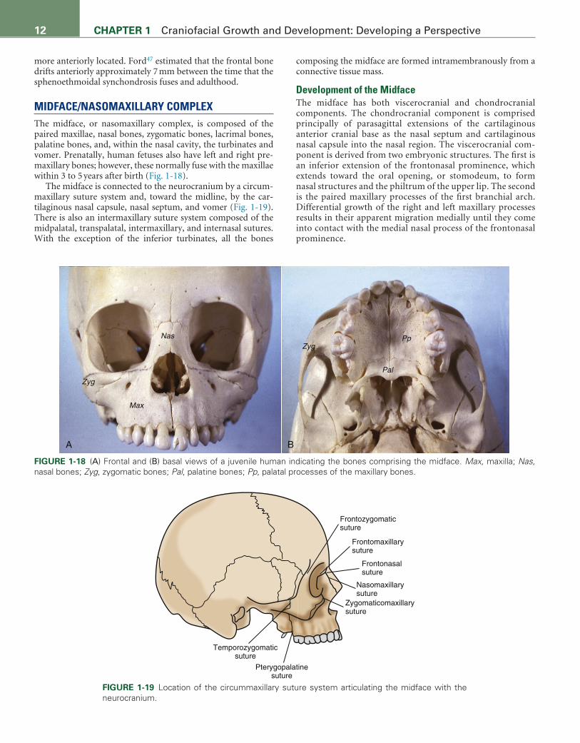
MIDFACE/NASOMAXILLARY COMPLEXThe midface, or nasomaxillary complex, is composed of the paired maxillae, nasal bones, zygomatic bones, lacrimal bones, palatine bones, and, within the nasal cavity, the turbinates and vomer. Prenatally, human fetuses also have left and right pre-maxillary bones; however, these normally fuse with the maxillae within 3 to 5 years after birth (Fig. 1-18).
The midface is connected to the neurocranium by a circum-maxillary suture system and, toward the midline, by the car-tilaginous nasal capsule, nasal septum, and vomer (Fig. 1-19). There is also an intermaxillary suture system composed of the midpalatal, transpalatal, intermaxillary, and internasal sutures. With the exception of the inferior turbinates, all the bones
composing the midface are formed intramembranously from a connective tissue mass.
Development of the MidfaceThe midface has both viscerocranial and chondrocranial components. The chondrocranial component is comprised principally of parasagittal extensions of the cartilaginous anterior cranial base as the nasal septum and cartilaginous nasal capsule into the nasal region. The viscerocranial com-ponent is derived from two embryonic structures. The first is an inferior extension of the frontonasal prominence, which extends toward the oral opening, or stomodeum, to form nasal structures and the philtrum of the upper lip. The second is the paired maxillary processes of the first branchial arch. Differential growth of the right and left maxillary processes results in their apparent migration medially until they come into contact with the medial nasal process of the frontonasal prominence.
Max
Nas
Pal
Zyg
ZygPp
A B
FIGURE 1-18 (A) Frontal and (B) basal views of a juvenile human indicating the bones comprising the midface. Max, maxilla; Nas, nasal bones; Zyg, zygomatic bones; Pal, palatine bones; Pp, palatal processes of the maxillary bones.
Frontozygomaticsuture
Frontomaxillarysuture
Frontonasalsuture
Nasomaxillarysuture
Zygomaticomaxillarysuture
Pterygopalatinesuture
Temporozygomaticsuture
FIGURE 1-19 Location of the circummaxillary suture system articulating the midface with the neurocranium.

13CHAPTER 1 Craniofacial Growth and Development: Developing a Perspective
The skeletal elements comprising the midfacial complex arise almost exclusively from neural crest cells within the maxillary pro-cess of the first branchial arch. The primary palate, which gives rise to the four maxillary incisors, is derived from the frontonasal prominence. Only the facial ethmoid and inferior turbinate are derived from the cartilaginous component of the midface. Like the bones of the cranial vault, since the bones comprising the nasomaxillary complex have no cartilaginous precursors, they rely on intramembranous ossification for their development. How-ever, the exact process by which initial bone formation occurs differs from that of the cranial vault bones. While the bones of the cranial vault arise within a desmocranial membrane, centers of ossification for the nasomaxillary bones develop as blastemas directly within the mesenchyme of the first branchial arch. These blastemas of bone are then surrounded by a periosteum that provides the source of new osteoblastic cells and thus for enlarge-ment of the skeletal element. Molecular signaling mechanisms associated with the development, growth, and maintenance of the facial sutures are dependent on the presence of the nasal capsular cartilage, which appears to play a role similar to the dura mater in sutures of the cranial vault in the expression of TGF-β1, TGF-β2, TGF-β3, and Msx2.48 It has also been shown that Fgf8 plays a sig-nificant role in the integration and coordination of the frontona-sal prominence with the nasal and optic regions.49
Virtually all of the major centers of ossification within the midface can be seen at approximately 7 to 8 weeks’ gestation. At 6 weeks’ gestation, the palatal shelves, which are mesenchymal tissue extensions of the embryonic maxillary processes of the first branchial arches, elevate within the oral cavity, where they will give rise to the hard and soft palates. The palatal shelves begin to ossify at 7 to 8 weeks’ gestation, with the two bone fronts of the palatal processes each extending medially to form the secondary palate, composed of processes from the maxillary bones and from the palatine bones, as they meet in the midline where they form the midpalatal suture.
The molecular mechanisms associated with the development of the palate are among the most studied in all of craniofacial growth and development because of the obvious problem of cleft lip and palate, which is the most common craniofacial deformity (approximately 1:1000 for children of European descent).50,51 Genes that have been identified specifically for a significant role in the genesis of cleft lip and palate now include isoforms of BMP, Dlx, Fgf-8, Msx, Pitx, Sho2, Shh, Sox9, and TGF-β, among others. It is also well documented that epigene-tic factors, such as anoxia due to cigarette smoking and alcohol, have a major impact on nonsyndromal cleft lip and palate.
Development of the nasomaxillary complex proceeds lat-erally and anteroposteriorly with expansion of the brain and cranial cavity and expansion of the oral cavity and oronasal pharynx. Also throughout the fetal period, anterior and infe-rior growth of the nasal septal cartilage, which is an extension of the anterior cranial base, is most prominent. The cartilagi-nous nasal capsule, which envelops the nasal cavity laterally, is primarily structural and contributes little to the overall growth of the nasomaxillary complex other than possible expression of growth factors that support the facial sutures (Fig. 1-20). Thus, the primary factors influencing the growth of the nasomaxillary complex from the late embryonic period and throughout the fetal period and the juvenile period postnatally are an expansion of the brain and cranial vault and growth of the anterior cranial base, including in particular anterior and inferior growth of the nasal septum, as well as expansion of the nasal cavity and oro-nasal pharynx.
Postnatal Growth of the MidfaceAt the time of birth, the midface is well developed but dimin-utive relative to the neurocranium. The circummaxillary and intermaxillary sutures are all present and active as sites of bone growth. The nasal capsule and midline nasal septum are still primarily cartilaginous and continuous with the rest of the
NC
NS
PS
NS
VM
NC
PS
A B
FIGURE 1-20 Frontal histologic sections of human fetuses at approximate ages of 5 weeks’ ges-tation (A) and 11 weeks’ gestation (B) (hematoxylin and eosin–stained). NS, Nasal septal cartilage; NC, nasal capsular cartilage; V, vomer; PS, palatal shelves.

14 CHAPTER 1 Craniofacial Growth and Development: Developing a Perspective
chondrocranium from the anterior cranial base. The septum is also very actively growing by means of interstitial cartilaginous growth, leading to significant anterior and vertical growth of the midface, especially during the first 3 to 4 years of life.
With the exception of the nasal septum, postnatal devel-opment of the nasomaxillary complex occurs via intra-membranous ossification. Growth at the circummaxillary and intermaxillary sutures occurs in response to midfacial displace-ments due principally to growth of the anterior cranial base and nasal septum. Inferior, anterior, and lateral displacements of the midface result in concomitant compensatory sutural growth to account for the majority of vertical, anteroposterior, and trans-verse changes that occur during both childhood and adoles-cence (Fig. 1-21). Along with displacements, extensive surface modeling takes place over the entire nasomaxillary complex, especially along its posterior and superior aspects.
As long as the midface undergoes displacement, sutural growth occurs, with the amounts of bony apposition being related directly to amounts of sutural separation. Growth con-tinues until the sutures are no longer separating. The premaxillary/maxillary suture fuses at approximately 3 to 5 years of age.52 The midpalatal and transpalatal maxillary sutures, which are the major intermaxillary growth sites associated with trans-verse and anteroposterior maxillary growth, have been reported to close between 15 and 18 years of age53 and 20 to 25 years of age,54 depending on the criteria on which closure is based. More recent studies suggest only limited amounts of sutural oblitera-tion (i.e., the development of bony bridges, or spicules, running across the suture after growth has ceased) in adult midpala-tal sutures.55,56 The increasing complexity that characterized sutures during childhood and adolescence appears to be func-tionally, rather than age, related.57 Although data are limited, it appears that closure of the circummaxillary sutures occurs somewhat later than closure of the intermaxillary sutures.
The midface undergoes a complex modeling pattern through-out childhood and adolescence (Fig. 1-22).58 As the midface is displaced anteriorly, compensatory bony deposition occurs
along the posterior margin of the maxillary tuberosity, result-ing in an increase in the length of the entire maxilla and of the dental arches.59 The posterior maxilla is a major modeling site that accounts for most of the increases in maxillary length. The anterior periosteal surface of the maxilla is slightly resorptive, while the buccal surfaces undergo substantial bony deposition. From the sagittal perspective, the area of the anterior nasal spine drifts inferiorly; the A-point also drifts inferiorly and slightly posteriorly. For every 4 mm that the posterior nasal spine drifts posteriorly, it drifts approximately 3 mm inferiorly. Associated
A � 14.6
C � 2.5Re � 4.6
O � 6.4
Su � 11.2x
6.7
3231
9.5
A BFIGURE 1-21 (A) Sutural displacement (Su), apposition of the orbital floor (O), resorption of the nasal floor (Re), apposition at the infrazygomatic crest (C), and dentoalveolar development (A) from 4 years of age through adulthood in nine boys. (B) Width changes (mm) of the maxilla and lateral implants between 3.9 and 17.7 years of age. (From Björk A, Skieller V: Postnatal growth and development of the maxillary complex. IN McNamara JA Jr, ed.: Factors affecting the growth of the midface, Ann Arbor, MI: Center for Human Growth and Development, Michigan Craniofacial Growth Series; 1976:61-100.)
FIGURE 1-22 Maxillary remodeling, with the sizes of the arrows indicating relative amounts of change and with dark and light arrows indicating resorption and apposition, respectively. (Redrawn from Enlow DH, Bang S. Growth and remodeling of the human maxilla. Am J Orthod. 1965;51:446–464.)

15CHAPTER 1 Craniofacial Growth and Development: Developing a Perspective
with inferior displacement of the midfacial complex, bony resorption occurs along the floor of the nasal cavity, whereas apposition occurs on the roof of the oral cavity (i.e., palate) and orbital floor. Implant studies suggest that for every 11 mm of inferior midfacial displacement, the orbital floor drifts supe-riorly 6 mm and the nasal floor drifts inferiorly 5 mm.60 Thus, midfacial height increases due to the combined effects of infe-rior cortical drift and inferior displacement (see Fig. 1-19). The height of the midface is further increased by continued develop-ment of the dentition and alveolar bone. The lack of naturally stable structures on the surface of the midfacial complex makes superimposition difficult.
The width of the midface at the time of birth is proportion-ately large due to the precocious development of the eyes, which are the central features of the neonatal midface. Growth in width during the first 2 to 3 years after birth is associated with expan-sion of the brain laterally and anteroposteriorly, which brings the eyes laterally with it. As this occurs, the sutures separating the two halves of the frontal bone (metopic suture), the two nasal bones (internasal suture), the two maxillae (intermaxil-lary suture), and the two palatine bones (midpalatal suture) are positioned to respond by secondary, compensatory bone for-mation. It has been estimated that the midalveolar and bijugale widths of the maxilla increase approximately 5 mm and 6 mm, respectively, between 7.6 and 16.5 years of age; rates of growth in width diminish slightly with increasing age.61
At the same time that the midface is increasing in width, it is increasing even more dramatically in depth (anteriorly) and height (vertically). The midface increases most in height, next in depth, and least in width. As the brain and eyes grow ante-riorly relative to the middle cranial base, the orbits increase in depth and the anterior cranial base lengthens, primarily as a result of growth at the sphenoethmoidal synchondrosis. Con-comitantly, the nasal septum grows vertically as the midface is displaced inferiorly relative to the anterior cranial base. The combination of these two growth processes—growth in a ver-tical direction associated with interstitial cartilaginous growth within the nasal septum and growth in an anterior direction associated with interstitial cartilage growth within both the nasal septum and synchondroses of the cranial base—results in the typical downward and forward growth of the entire midface relative to the anterior cranial base. Surface deposition cannot account for the downward and forward midfacial growth that occurs during childhood and adolescence.
The age of approximately 7 years is something of a bench-mark for growth of the midface. Growth of the central nervous system—the brain and eyes—is essentially complete at about 7 years of age. Concomitantly, the cartilage of the sphenoeth-moidal synchondrosis ossifies and a suture is formed between the sphenoid and ethmoid bones at about that time. As a result, a relatively stable anterior cranial base is established extend-ing from the sella turcica to the foramen cecum. Also at about 7 years of age, the growth of the cartilages of the nasal capsule and nasal septum changes significantly. The cartilaginous nasal capsule becomes ossified, and the nasal septum, which remains cartilaginous throughout life in humans, decreases significantly in growth activity. Despite these important developmental changes in the growth processes of the midface, downward and forward skeletal growth continues to be significant over the next decade or so, particularly in males during adolescence.
Growth of the nasomaxillary complex continues throughout childhood and adolescence, with substantially greater vertical
than anteroposterior growth potential (Fig. 1-23). By 4.5 years of age, palatal length (anterior nasal spine–posterior nasal spine) and anterior facial height (nasion–anterior nasal spine) have attained approximately 80% and 73% of their adult size, respectively (see Fig. 1-18). In terms of absolute growth, mid-facial heights should be expected to increase 10 to 12 mm in females and 12 to 14 mm in males between 4 and 17 years of age. Palatal length should be expected to increase 8 to 10 mm over the same time period. Because nasion drifts anteriorly at approximately the same rate as the midface is displaced anteri-orly, the sella–nasion–anterior (SNA) nasal spine angle shows little or no change during childhood or adolescence. Although vertical maxillary growth rates peak during adolescence, at approximately the same time as stature, anteroposterior max-illary growth remains more or less constant, with no distinct adolescent spurt.
Because the displacements are not parallel, the midface undergoes varying amounts of vertical and transverse true rotation. True rotation is independent of surface modeling and refers to changes that occur over time in the positions of basal bone; it is commonly assessed with metallic implants placed into the mandibles and maxillae of growing children.62 From the sagittal perspective, most children undergo true forward or counterclockwise (subject facing to the right) rotation of the midface, due to greater inferior displacement of the posterior than anterior maxilla. The true rotation that occurs tends to be covered up or hidden by the resorption that occurs on the nasal floor. For example, true forward rotation is associated with greater resorption in the anterior than posterior aspect of the nasal floor. Due to greater transverse displacements
mm
/deg
141210
86420
�2
16Males
Females
mm
/deg
141210
86420
�2
16
Age (Years)4 6 8 10 12 14 16 18
4 6 8 10 12 14 16 18
ANS-PNS N-ANS SE-PNSNS S-N-ANS
FIGURE 1-23 Maxillary growth changes between 4 and 17 years of age of males and females. (Adapted from data pro-vided by Bhatia SN, Leighton BC. A Manual of Facial Growth: a Computer Analysis of Longitudinal Cephalometric Growth Data. New York: Oxford University Press; 1993.)

16 CHAPTER 1 Craniofacial Growth and Development: Developing a Perspective
posteriorly than anteriorly, the midfacial complex also exhibits transverse rotation around the midpalatal suture (Fig. 1-24). As a result, there is greater sutural growth in the posterior than anterior aspect of the midpalatal suture. Cephalometric analyses using metallic implants have shown that the poste-rior maxilla expands approximately 0.27 to 0.43 mm/yr, with greater expansion occurring during childhood than during adolescence.60
There are definite sex differences in maxillary postnatal growth (Fig. 1-25), with males being larger and growing more than females. Size differences, averaging between 1 mm and 1.5 mm, are small but consistent during infancy and child-hood. Sexual dimorphism increases substantially throughout the midfacial complex during adolescence, with differences of approximately 4 mm in maxillary length (anterior nasal spine to posterior nasal spine [ANS-PNS]) and upper facial height (nasion to anterior nasal spine [N-ANS]) at 17 years of age. Males also have significantly wider midfaces than females, with differences approximating 5 to 7 mm during late adolescence.63 The primary reason that adult males are larger than adult females is the 2 extra years of childhood growth that males have; males enter the adolescence phase of growth at approximately 12 years of age, while females enter around 10 years. Males are also larger than females because they experience a more intense adolescent spurt, but this contributes less to the sex differences observed.
MANDIBLEDevelopment of the MandibleThe mandible develops bilaterally within the mandibular pro-cesses of the first branchial arch. Each embryonic mandibu-lar process contains a rod-like cartilaginous core, Meckel’s cartilage, which is an extension of the chondrocranium into the viscerocranium. Throughout its course, distally Meckel’s cartilage is accompanied by the mandibular division of the tri-geminal nerve (CN V), as well as the inferior alveolar artery and vein. Proximally, Meckel’s cartilage articulates with the cartilaginous cranial base in the petrous region of the tempo-ral bone, where it gives rise to the malleus and incus bones of the inner ear.
By 6 weeks’ gestation, a center of ossification appears in the perichondrial membrane lateral to Meckel’s cartilage. It is criti-cal to note that ossification of the mandible takes place in mem-brane lateral and adjacent to Meckel’s cartilage, and not within Meckel’s cartilage itself (Fig. 1-26). Therefore, it is clear that the mandible develops and subsequently grows by means of intram-embranous ossification and not through endochondral ossifica-tion and replacement of Meckel’s cartilage. The only portion of the developing lower jaw that appears to be derived from endo-chondral ossification of Meckel’s cartilage is the mental ossicles, which are two very small sesamoid bones that are formed in the inferior aspect of the mandibular symphysis.65 These bones are no longer present at the time of birth.
Intramembranous ossification of the body of the mandi-ble proceeds distally toward the mental symphysis and proxi-mally up to the region of the mandibular foramen. As it does so, Meckel’s cartilage begins to degenerate and involute as the inferoalveolar neurovascular bundle becomes progressively enveloped by the intramembranously developing mandibular bone. Meckel’s cartilage completely disappears by approxi-mately 24 weeks’ gestation, remaining in remnant form as the dense sphenomandibular ligament and giving rise to the mal-leus and incus ear ossicles.
Initial evidence of the formation of the temporomandibular joint (TMJ) is seen upon expression of the Barx-1 homeobox gene. By approximately 8 weeks’ gestation, the condylar pro-cess appears as a separate carrot-shaped blastema of cartilage extending from the ramus proximal to the mandibular foramen and extending up to articulate with the squamous (membra-nous) portion of the developing temporal bone. Formation of
Björk and Skieller, 1977
Korn and Baumrind, 1990
Gandini and Buschang, 2000
Iseri and Solow, 2000
Ages (Years)
4-21
8.5-15.5
13.9-16.7
7-12
13-18
Mx
.42
.43
.27
N/A
N/A
Md
N/A
.28
0.19
.22
.13
References
FIGURE 1-24 Transverse expansion (mm/yr) of metallic bone markers inserted into the maxillary (Mx) and mandibular (Md) basal structures.
Diff
eren
ce (
mm
)
2
1
0
3
4
ANS-PNS N-ANS SE-PNS
4 5 6 7 8 9 10 11 12 13 14 15 16 17Age (Years)
FIGURE 1-25 Sex differences (male minus female) in maxillary size. (Adapted from data provided by Bhatia SN, Leighton BC. A Manual of Facial Growth: a Computer Analysis of Longitudi-nal Cephalometric Growth Data. New York: Oxford University Press; 1993.)

17CHAPTER 1 Craniofacial Growth and Development: Developing a Perspective
the joint cavity between the condylar process and the squamous portion of the temporal bone is essentially completed as the TMJ by about 12 weeks’ gestation (Fig. 1-27).
As the cartilage comprising the mandibular condyle arises “secondarily” within a skeletogenic membrane and apart from the primary embryonic cartilaginous anlagen, it is referred to as a secondary cartilage (Fig. 1-28). Secondary cartilage is a unique type of skeletal tissue that has the characteristics of both intra-membranous bone and certain histologic and functional fea-tures of hyaline growth cartilage. Secondary cartilage is formed in areas of precocious stresses and strains within intramembra-nous bones, as well as in areas of rapid development and growth of bone.65,66 Within the craniofacial complex, the angular and the coronoid processes of the mandible also may exhibit the presence of secondary cartilage because these are sites of very
Mental nerve
Mandibular nerve Meckel′scartilage
dorsal end
Meckel′sventral end
Inferioralveolarnerve
Meckel’scartilage
Lingualnerve
Mandibularnerve
Chordatympani
Malleus
Left half ofmandible, cut
MC
MMST
FIGURE 1-26 Drawings of a fetal mandible with lateral (top left) and medial (bottom left) views. Right, Photomicrograph of coronal view of human fetus indicating Meckel’s cartilage medial to the mandible (M). MST, Masseter muscle. (Drawings adapted from Warwick R, Williams PL, eds. Gray’s Anatomy. 35th ed. Philadelphia: WB Saunders; 1973.)
MCC
TMP
AP
CP
FIGURE 1-27 Parasagittal histologic section of human fetus (approximately 12 weeks’ gestation) (hematoxylin and eosin–stained). MCC, Mandibular condylar cartilage; CP, coronoid pro-cess; AP, angular process; TMP, temporalis muscle.
MCC
MC
M
P
FIGURE 1-28 Frontal histologic section of a human fetus (approximately 8 weeks’ gestation) (hematoxylin and eosin–stained). The bone comprising the body and ramus of the mandi-ble (M) originates in the membrane lateral to Meckel’s cartilage (MC). The periosteal membrane enveloping the mandible gives rise secondarily to the mandibular condylar cartilage (MCC).

18 CHAPTER 1 Craniofacial Growth and Development: Developing a Perspective
rapid bone growth associated with the function of the muscles of mastication. In addition, secondary cartilage may be found in areas of sutures that are characterized by rapid intramem-branous bone growth and biomechanical load associated with separation and bending at the articular surfaces.
At birth, the two halves of the mandible are separated in the midline by a fibrous articulation, the mental symphysis, which will fuse by the end of the first year of life. Each half of the mandible is characterized anatomically by (1) a condyle and condylar process, which articulates with the temporal bone to make up the TMJ; (2) a ramus, which extends roughly verti-cally–inferiorly from the condylar process and provides inser-tions for the muscles of mastication; and (3) a corpus, or body, which extends roughly horizontally–anteriorly to provide a base for the mandibular dental arch and house the inferior alveolar– neurovascular bundle. Each of these anatomic structures can also be considered in terms of overlapping functional units (Fig. 1-29). The mandibular condyle and condylar processes obviously are essential for normal articular function of the TMJ and movements of the mandible, while at the same time playing a significant role in mandibular growth for most of the first two decades of life.67 Variation in the function of the TMJ, such as might occur in association with differences in mastication, jaw movements, and jaw position, for example, is highly likely to affect its growth and form. The gonial region of the mandible, at the inferior aspect of the ramus, is related to the function of the masseter and medial pterygoid complex of muscles, while the coronoid process is primarily related to the temporalis muscle. Variation in the growth and form of each of these regions is due in large part to variation in the function of the muscles of mastication. The alveolar process of the mandible functions to provide support for the dentition. Finally, the body of the man-dible, extending from the mandibular foramen to the mental process, provides support and structural connection between the various functional components of the mandible.
Growth of the Mandibular CondyleJust as a suture can be considered to be a specialization of an osteogenic membrane (i.e., periosteum and dura mater), the condylar cartilage can also best be considered to be a specializa-tion of periosteum. As with sutures, growth of the mandibular condyle tends to be relatively highly responsive to mechanical, functional, and hormonal stimuli both at the time of develop-ment and throughout the growth period, similar to intramem-branous bone development elsewhere.
Histomorphology of the Growing CondyleA number of similar but somewhat different terms have been used to describe the histomorphology of the growing mandibu-lar condyle.68 These are summarized according to their equiva-lencies in E-Table 1-2.
The secondary cartilage composing the condyle during growth can be divided into two general layers: an articular layer and a growth layer. The more superficial articular layer is con-tinuous with the outer fibrous layer of the bilaminar periosteum encapsulating the condylar neck and temporal bone, respec-tively. Deep to the articular layer is a subarticular growth layer. The growth layer of the condylar cartilage is organized into an additional series of layers or zones typical of growing cartilage that blend into each other (Fig. 1-30). Each of these zones is present in the neonate and remains in the condyle through
Coronoid process
Alveolar process
Corpus
Gonial region
Condyle
FIGURE 1-29 Major functional units of the mandible.
Articular
Prechondroblastic
Maturational
Hypertrophy
EndochondralOssification
FIGURE 1-30 Histologic section indicating the various layers of the secondary cartilage in a growing mandibular condyle (hema-toxylin and eosin–stained).

18.e1CHAPTER 1 Craniofacial Growth and Development: Developing a Perspective
TABLE 1-2 Comparison of Terminology Used to Describe the Histomorphology of the Condylar Cartilage
Blackwood1 Durkin et al.2 Wright and Moffett3 Petrovic et al.4 Thilander et al.5 Carlson et al.6 Luder7
Articular zone Resting surface articular layer
Articular layer Fibrous capsule Surface articular layer
Fibrous articular tissue
Perichondrium Articular layer
Transitional or prolif-erative layer
Proliferative layer Prechondroblastic layer
Proliferative layer Prechondroblastic (proliferative) layer
Polymorphic cell layer
Flattened cell layer (1 and 2)
Intermediate zone
Hypertrophic cartilage
Hypertrophic cartilage
Erosion zone
Zone of matrix production
Zone of cell hypertrophyZone of calcification and
resorption
Zone of maturationFunctional
chondroblastsHypertrophic
chondroblasts
Hypertrophic zone (nonmineralized)
Hypertrophic zone (mineralized)
Chondroblastic zone (maturation and hypertrophy)
Hyaline cartilageFlattened cell layer
(3)Upper hypertrophic
cell layerLower hypertrophic
cell layerZone of erosionDegenerating
chondroblastsSubchondral bone Zone of
endochondral ossification
Zone of endochon-dral ossification
References1. Blackwood HJJ. Growth of the mandibular condyle of the rat studied with titrated thymidine. Arch Oral Biol. 1966;11:493–500.2. Durkin J, Heeley J, Irving JT. The cartilage of the mandibular condyle. Oral Sci Rev. 1973;2:29–99.3. Wright DM, Moffett BC. The postnatal development of the human temporomandibular joint. Am J Anat. 1974;141:235–250.4. Petrovic A, Stutzmann J, Oudet C. Control processes in the postnatal growth of the condylar cartilage. In: McNamara JA Jr, ed. Determ Mandibular Form Growth. Ann Arbor, MI:
Center for Human Growth and Development, Craniofacial Growth Series; 1975:101–154.5. Thilander B, Carlsson GE, Ingervall B. Postnatal development of the human temporomandibular joint. I. A histological study. Acta Odontol Scand. 1976;34:117–126.6. Carlson DS, McNamara Jr JA, Jaul DH. Histological analysis of the growth of the mandibular condyle in the rhesus monkey (Macaca mulatta). Am J Anat. 1978;151:103–117.7. Luder HU. Postnatal development, aging, and degeneration of the temporomandibular joint in humans, monkeys, and rats, Ann Arbor, MI: Center for Human Growth and Development,
University of Michigan, Craniofacial Growth Series; 1996:32.

19CHAPTER 1 Craniofacial Growth and Development: Developing a Perspective
maturity. However, their absolute and relative size as well as their growth-related activity may vary considerably, depending on the overall rate and amount of condylar growth and on the functional requirements placed on the condyle and TMJ.69,70
Articular layer. The articular layer of the joint surface of the mandibular condyle and temporal portion of the TMJ consist of an avascular dense fibroelastic connective tissue whose collagen fibers are oriented parallel to the articular surface. The articular layer varies in thickness along the condylar head and temporal joint surface, increasing in thickness in the superior aspect of the condyle and on the articular eminence of the glenoid fossa, where compressive forces associated with mastication are greatest.71 The fibrous articular layer of the mandibular condyle and that found in the glenoid fossa and articular eminence are identical functionally to the articular cartilage found in the diarthroideal joints of the postcranial long bones, but their origin and histologic composition are completely different. Articular cartilage is derived from the primary cartilaginous anlagen at the ends of long bones; the articular tissue of the TMJ is a specialization of the fibrous layer of periosteum that covers the mandible and temporal bone.
Growth layer. The growth layer immediately deep to the articular layer is comprised of a series of cellular zones representing the various stages of chondrogenesis in secondary cartilage. The proliferative, or prechondroblastic, zone immediately deep to the articular layer is continuous with the osteogenic layer of the periosteal membrane along the condylar neck.72,73 Its outer portion is composed of undifferentiated mesen-chymal cells that differentiate into skeletoblastic stem cells or prechondroblasts. Morphologically, this zone appears as densely packed with spindle-shaped cells that increase in size and become increasingly separated due to production of intercellular matrix within the inner region of the proliferative zone. The newly formed cartilage cells in the proliferative zone express type I collagen, which is characteristic of bone and underscores the fact that the source of these cells is a periosteal-like membrane. Recent studies of gene expression in the proliferative zone demonstrate that the prechondroblastic layer is also characterized by high expression of FGF-13, FGF-18, TGF-β2, Igf-1, and VEGF.74,75
The zone of maturation contains larger, spherical, maturing chondrocytes arranged in an apparently random fashion. These cartilage cells undergo very few mitoses, which is atypical for cartilage cells found in a growing epiphyseal plate. In addition, there is significantly less extracellular matrix in the mandibular condylar cartilage than is found in the growth plates of devel-oping long bones, which are comprised of primary cartilage. Cartilage cells within the zone of maturation are capable of switching their phenotype to express type II collagen, which is typically expressed by primary cartilage in growing epiphyses in response to biomechanical load.
Cartilage cells in the zone of hypertrophy become progres-sively larger through osmotic activity and absorption of water. Their nuclei become pyknotic and their cytoplasm is increas-ingly evacuated as the cells are about to be encroached upon by the osteoblasts from the endosteal region of the condyle. Genes for procollagen, aggrecan, Sox9, and Ihh are highly expressed in the chondroblastic layer.74
The zone of endochondral ossification is characterized by the initiation of mineralization of the intercellular matrix within the distal-most three to five layers of hypertrophying cells. This matrix is subsequently eroded away by osteoclastic activity and
replaced by bone. The process of endochondral ossification associated with the condylar cartilage is identical to the process that takes place in the primary cartilage of long bone epiphyses.
Age-Related Changes in the Mandibular CondyleDetailed histologic analysis of human autopsy specimens of the human TMJ has demonstrated progressive changes in the thickness and presumed growth activity of the condyle carti-lage throughout development.76–79 These changes appear to be coordinated with functional changes associated with occlusal development.80,81 In general, the combined growth-related lay-ers of the condylar cartilage begin as a relatively thick structure in the neonate (1.25 to 1.5 mm thick) but become much thinner (0.3 mm) by the mixed dentition stage. The cartilage remains generally thin but well defined and actively growing in the per-manent dentition stage until, by age 20 to 30 years, the cartilage essentially disappears and the condyle is capped by a bony plate. Even in adults, however, it is not unusual to see areas of hyaline cartilage (“cartilage islands”) deep to the articular layer in the condyle.
The subarticular region of the temporal component of the TMJ has the same tissue layers as the condyle; however, they are substantially less prominent. Morphologically, the tempo-ral component of the TMJ in the neonate is essentially flat, and the articular disc interposed between the condyle and temporal bone is highly vascular. During the period of the primary denti-tion, at approximately 3 years of age, the temporal surface takes on its characteristic S-shaped contour, and the articular disc becomes avascular in its central region. Thereafter, the tempo-ral surface of the TMJ grows more slowly, with the mandibu-lar fossa becoming deeper as the articular eminence becomes steeper; this happens primarily through the process of bone deposition on the articular eminence and, to a lesser extent, by resorption of bone in the posterosuperior region of the fossa, as well as endosteal deposition in the superior aspect of the fossa. This increase in the contour of the temporal component of the TMJ normally continues until the fourth decade of life.
In summary, the mandibular condylar cartilage is a second-ary cartilage that in subadult individuals serves both as a site of growth and as a place of articulation. As such, it displays functional characteristics of both a growth plate and an articu-lar cartilage, but it differs from both in fundamental aspects of its development and structure throughout ontogeny. Its most superficial layers are not cartilaginous in phenotype but rather are perichondrial in origin. Importantly, the chondrocytes of the mandibular condylar cartilage are derived via mitosis in cells that are themselves not chondrocytes, similar to embry-onic cartilage but not to the growth plate in which the cells that proliferate are chondrocytes. Finally, the prechondrogenic phe-notype of these dividing cells in the mandibular condylar car-tilage can be readily modulated to a preosteogenic phenotype by changes in the periarticular environment. Taken together, these features define a tissue with structural and growth char-acteristics that are consistent with the concept of an adaptive, compensatory growth site and set it apart from primary carti-laginous growth centers.
Mechanisms of Condylar GrowthThe mandibular–condylar cartilage was initially considered to be a growth center with an intrinsic capacity for tissue- separating growth. However, it is now generally understood that growth of the mandibular–condylar cartilage is highly

20 CHAPTER 1 Craniofacial Growth and Development: Developing a Perspective
adaptive and responsive to growth in adjacent regions, par-ticularly the maxilla. Numerous experimental studies were conducted over the past several decades to assess the role that function and jaw position, in particular, might play in influ-encing the postnatal growth of the mandibular condyle. For example, in a number of studies involving anterior postural change of the mandible using rats82,83 and primates84 as experi-mental animals demonstrated significant increases in the over-all length of the mandible. From these experiments, Petrovic and colleagues developed a “cybernetic” model of mandibular growth regulation referred to as the “servosystem hypothesis of mandibular growth” (Fig. 1-31).85,86
There has been a significant expansion of knowledge con-cerning the molecular biology and cellular dynamics associated with growth of the condylar cartilage. It has been shown, for example, that fibroblast growth factor (FGF) and insulin-like growth factor (IGF) are present in the matrix and cell surfaces of the condylar cartilage and that they vary according to their specific location, much like in sutures. Less is known of the presence or importance of transforming growth factor-beta (TGF-β) or other growth factors, and knowledge of hormonal influences on growth of the condylar cartilage is even more rudimentary and somewhat contradictory.87–89
Several studies have begun to explore the molecular basis for the effect of mandibular function and position on mandib-ular growth by using appliances that replicate the effects (e.g., increased mitotic activity, cartilage thickness) reported previ-ously.90–93 Fuentes and coworkers94 used a novel incisor-borne appliance that prompted a crossbite in growing rats and pro-duced a differential change in proliferation and cartilage thick-ness between the crossbite and noncrossbite sides. In animals wearing the appliance, gene expression for IGF-1 and FGF-2
and their receptors in condylar cartilage was altered from that in control rats. The changes in gene expression, which typically preceded the changes in mitotic activity and cartilage thick-ness, were in most instances opposite in direction between the crossbite and noncrossbite sides. Using a similar design, Hajjar and associates95 found that rats fitted with an incisor-borne appliance that prompted anterior displacement of the man-dible exhibited increased expression of both IGF-I and IGF-II mRNA and protein in the MCC. Rabie and colleagues90,96,97 demonstrated that the expression of Sox9, type II collagen, and Indian hedgehog (Ihh) was increased in the condylar car-tilage and glenoid fossa of rats wearing the appliance for 1 to 2 weeks.
In general, these findings parallel the findings discussed previously for development of the sutures of the cranial vault. These similarities between the condylar cartilage and sutures should not be surprising given the periosteal origin of both suture mesenchyme and the secondary cartilage of the mandib-ular condyle.
Postnatal Growth of the MandibleAt birth, the ramus of the mandible is quite short, both in abso-lute terms and in proportion to the mandibular corpus. During postnatal development, the ramus becomes much more promi-nent, particularly in height but also in width. At the same time, the corpus increases in length, providing the necessary space for development and eruption of the mandibular dentition. Associated with these early postnatal changes in the absolute and relative sizes of the mandible are decreases in the gonial angle between the ramus and corpus and increases in the angle between the two corpora.
The mandible has the greatest postnatal growth potential of any component of the craniofacial complex. Growth changes that occur are closely associated with the functional processes that comprise the mandible, including the gonial process, coronoid process, alveolar process, and bony attachments of the suprahyoid muscles, which are all major sites of postnatal modeling. Although condylar growth is often assumed to be the mandible’s primary growth site, it is important to note that the entire superior aspect of the ramus displays approxi-mately the same amount of growth.
Viewed in its lateral projection, the posteroinferior and superior border of the ramus, including the condyle, and the posterosuperior aspect of the coronoid process are depository throughout the period of active growth. The anterior and lower borders (extending approximately to the first molars) of the ramus of the mandible are resorptive. Resorption of bone con-tinues to occur along the anterior border of the ramus, resulting in a longer corpus and increased space for the development and eruption of the mandibular dentition (Fig. 1-32).98 Within the corpus, the greatest growth changes are appositional growth of the alveolar bone associated with dental development and erup-tion. The symphysis, especially the superior aspect, becomes wider due to superior and posterior drift of its posterior aspect (Fig. 1-33).99 There is resorption on the anterior aspect of the symphysis above the bony chin. The cortical region at or just above the chin is the only place on the entire surface of the man-dible that remains stable (i.e., does not model) during postnatal growth, which is why it serves as an important site for superim-posing successive radiographs. The inferior aspect of the ante-rior corpus tends to be depository, but the amounts of bone added are limited and variable.
Hormonalfactors
Condylargrowth
Musclefunction
Centralnervoussystem
Proprioceptors:periodontium-TMJ
Occlusaldeviation
Maxillarygrowth
E
D
A
B
C
F
FIGURE 1-31 Simplified explanation of Petrovic’s “servosys-tem hypothesis of mandibular growth.” Independent growth of the maxilla (A) creates a minor occlusal deviation between the upper and lower dentition (B). This occlusal deviation is perceived by proprioceptors (C), which provide a signal to the muscles responsible for jaw protrusion to be tonically more active (D), which causes the mandibular condyle to become slightly more anteriorly located within the temporomandibular joint, thus stimulating condylar growth (F). Muscle function and the adaptive capacity of the condyle for growth are enhanced by expression of hormonal factors (E), and thus condylar growth may vary depending on the maturational and hormonal status of the individual. (Adapted from Carlson DS. Theories of craniofacial growth in the postgenomic era. Semin Orthod. 2005;11(4):172–183.)

21CHAPTER 1 Craniofacial Growth and Development: Developing a Perspective
Widening of the body of the mandible occurs through depo-sition of bone along the buccal surface and transverse rotation of the right and left corpii. The mandible also widens due to bony deposition along its posterior surface, which, due to its posterolateral orientation, produces a longer and wider body. Growth in width of the superior aspect of the ramus is some-what more complex due to the substantial increases in height that occur. Viewed in a coronal projection, the superior aspect
of the ramus and coronoid process are canted somewhat medi-olaterally. As the mandibular corpus and inferior aspect of the ramus increase in width by deposition along the buccal surface, the buccal surface of bone on the superior aspect of the ramus is resorptive, while the lingual and superior surfaces of bone are depository.
The greatest postnatal changes in mandibular growth also occur during infancy, with overall length (condylion to gna-thion [Co–Gn]) increasing 15 to 18 mm during the first year, 8 to 9 mm during the second year, and then slowing down to increase approximately 5 mm during the third year. During these early years, condylar growth and modeling of the superior aspects of the ramus are directed posteriorly and superiorly, with roughly equal amounts of growth in each direction. This orientation is important because it rapidly increases corpus length to make room for the rapidly developing dentition. After the first few postnatal years, growth of the condyle and supe-rior ramus slows down dramatically and changes orientation toward a predominant superior direction.
By 4.5 years of age, ramus height has attained approximately 64% and 70% of its adult size for males and females, respec-tively (see Fig. 1-18). Corpus length (Go–Gn) closely approx-imates the maturity pattern of midfacial height; it remains more mature than ramus height throughout postnatal growth. This supports the general principle that the vertical aspects of craniofacial growth are less mature and have greater post-natal growth potential than the anteroposterior aspects. Total mandibular length (condylion to menton [Co–Me]) under-goes the greatest increases in length (approximately 25 mm and 30 mm for females and males, respectively) between 4 and 17 years of age, followed by corpus length (gonion to pogon-ion [Go–Pg]; approximately 18 and 22 mm for females and males, respectively) and ramus height (condylion to gonion [Co–Go]; approximately 14 and 17 mm for females and males,
L1p
80%-P
60%-P
40%-P
20%-P
MeGn
20%-APg
40%-A
60%-A
80%-A
B
Inf
Symp
L1p
80%-P
60%-P
40%-P
20%-P
Me
Gn
20%-APg
40%-A
60%-A
80%-A
B
Inf
Symp
T1T2T3
MalesT1T2T3
Females
FIGURE 1-33 Remodeling changes of the symphysis between 6 (T1), 10 (T2), and 15 (T3) years of age. (Adapted from Buschang PH, Julien K, Sachdeva R, et al. Childhood and pubertal growth changes of the human symphysis. Angle Orthod. 1992;62:203–210.)
FIGURE 1-32 Mandibular remodeling, with the sizes of the arrows indicating relative amounts of change and with dark and light arrows indicating resorption and apposition, respectively. (Adapted from Enlow DH, Harris DB. A study of the postnatal growth of the human mandible. Am J Orthod. 1964;50:25.)

22 CHAPTER 1 Craniofacial Growth and Development: Developing a Perspective
respectively) (Fig. 1-34). During later childhood and adoles-cence, the condyle shows substantially greater amounts of supe-rior than posterior growth. For every 1 mm of posterior growth, there is 8 to 9 mm of superior growth. It has been estimated that the condyles of females and males grow 2 to 2.5 and 2.5 to 3.0 mm/yr, respectively, during childhood and adolescence, with the greatest rates occurring during the adolescent spurt (Fig. 1-35). The coronoid process and sigmoid notch follow similar growth patterns. Due to the resorption of bone that nor-mally occurs in the gonial region, ramus height (measured from
gonion to condylion) substantially underestimates the actual amount of growth that occurs at the condyle. There is approxi-mately 1 mm of resorption at gonion for every 3 mm of superior condylar growth.100 Between 7 and 15 years of age, biantegonial and bigonial widths increase approximately 10 mm and 12 mm, respectively.61,63 Importantly, mandibular width continues to increase throughout childhood and adolescence. While an ado-lescent spurt in vertical mandibular growth certainly occurs, a pronounced spurt for the anteroposterior and transverse growth has not been established.
The mandible undergoes substantial amounts of true verti-cal rotation and more limited, but definite, transverse rotation. While the maxilla exhibits more transverse rotation, the mandi-ble exhibits more vertical rotation than the maxilla. The typical pattern of vertical rotation is forward (counterclockwise with the subject facing to the right), due to greater inferior displace-ments of the posterior than anterior aspects of the mandible.101 Rates of vertical mandibular rotation have been estimated to range between 0.4 and 1.3°/yr, with significantly greater rates of rotation during childhood than adolescence (Fig. 1-36). Although relatively few (<10%) children are “true” posterior rotators, up to 25% of adolescents have been reported to be posterior rotators.80 Greater amounts of true mandibular rota-tion occur during the transition to the early mixed dentition than at any time thereafter.102,103
The mandible also rotates transversely due to greater expan-sion of the posterior than of the anterior aspects of the two cor-pii. This type of rotation has been demonstrated repeatedly in subjects with metallic implants and represents expansion of basal bone. It has also been shown that, when viewed from frontal projects, the right and left mandibular nerves are displaced lat-erally throughout growth. Transverse rotation is also age related, with greater amounts occurring during childhood than adoles-cence. The posterior aspect of the mandible expands approxi-mately 65% to 70% as much as the posterior maxilla expands at the posterior aspect of the midpalatal suture (see Fig. 1-20).
As in the rest of the craniofacial complex, sex differences in mandibular growth are evident at the earliest ages and become pronounced during adolescence. At birth, males have signifi-cantly larger mandibles than do females. Sex differences, which are greatest for overall length, followed by corpus length and ramus height, respectively, range from 0 to 2 mm between 1 and 12 years of age, when males initiate their adolescent phase of
4 6 8 10 12 14 16 18
mm
/deg
Age (Years)
25
20
15
10
5
0
30Females
Go-Pg Co-Me Co-GoS-Go N-Me S-N-Pg
4 6 8 10 12 14 16 18
mm
/deg
Age (Years)
25
20
15
10
5
0
30
35Males
FIGURE 1-34 Mandibular growth changes between 4 and 17 years of age of males and females. (Adapted from data pro-vided by Bhatia SN, Leighton BC. A Manual of Facial Growth: a Computer Analysis of Longitudinal Cephalometric Growth Data. New York: Oxford University Press; 1993.)
mm
/yea
r
Age (Years)
Females
90
75
50
25
10
5
6
4
3
2
1
0
�16.5 7.5 8.5 9.5 10.5 11.5 12.5 13.5 14.5 15.5
mm
/yea
r
Age (Years)
Males
90
75
50
25
10
5
6
4
3
2
1
0
�16.5 7.5 8.5 9.5 10.5 11.5 12.5 13.5 14.5 15.5
FIGURE 1-35 Percentile curves for condylar growth of females and males. (Adapted from Buschang PH, Santos Pinto A. Condylar growth and glenoid fossa displacement during childhood and adolescence. Am J Orthod Dentofac Orthop. 1998;113:437–442.)

23CHAPTER 1 Craniofacial Growth and Development: Developing a Perspective
growth. Mandibular dimorphism increases to 4 to 8 mm by the end of the adolescent growth phase (Fig. 1-37). There are no sex differences in vertical rotation during childhood or adolescence.
In summary, the mandible increases in size as a result of the combined processes of proliferation of secondary cartilage at the condyle and differential formation and modeling of bone along the entire surface of the mandible, particularly along its superior and posterior aspects. Growth of the mandible is expressed in a downward and forward direction relative to the cranium and cranial base. The mandible is typically displaced downward more than the maxilla, with the resulting space being taken up by the erupting dentition. Due to the geometry of the craniofacial complex, normal, coordinated growth of the jaws and a normal relationship of the associated occlusal arches require that the relative rate and amount of growth of the max-illa and mandible differ.
ARCH DEVELOPMENT, TOOTH MIGRATION, AND ERUPTIONThe oral apparatus is the region of the craniofacial complex that holds the greatest potential for adaptive changes. Den-tal arch width and perimeter change dramatically, especially
during the transitions to the early mixed and permanent den-titions.104 Maxillary intercanine width increases approximately 3 mm during the transition to the early mixed dentition and an additional 2 mm with the emergence of permanent canines (Fig. 1-38).105 Mandibular intercanine width increases approx-imately 3 mm during initial transition but shows little or no change with the eruption of the permanent canines. Intermo-lar widths progressively increase during childhood and adoles-cence, approximately 4 to 5 mm for the maxilla and 2 to 3 mm for the mandible between 6 and 16 years of age (Fig. 1-39). Max-illary arch depth (incisors to molars) decreases slightly during the transition to the early mixed dentition, increases 1 to 2 mm with the emergence of permanent incisors, and then decreases
References Ages deg/yr
Odegaard 1970 7-14 0.8
Lavergne and Gasson 1977 7-19 0.9
Skieller et al. 1984 Adolescence 1.0
Spady et al. 1992 Childhood 0.9
Adolescence 0.4
Miller and Kerr, 1992 5-10 1.3
10-15 0.8
Karlsen, 1995 6-12 (high angle) 0.7
6-12 (low angle) 1.3
12-15 (high angle) 0.7
12-15 (low angle) 1.3
Wang et al. 2009 5.6-8.5 1.3
8.5-15.5 0.7
Apparentrotation
Angularremodeling
Truerotation
FIGURE 1-36 True mandibular rotation (degrees per year) during childhood and adolescence.
4 5 6 7 8 9 10 11 12 13 14 15 16 17Age (Years)
Diff
eren
ce (
mm
) 76543210
�1
89
Go-Pg Co-Me Co-Go
FIGURE 1-37 Sex differences (male minus female) in mandib-ular size. (Adapted from Bhatia SN, Leighton BC. A Manual of Facial Growth: a Computer Analysis of Longitudinal Cephalo-metric Growth Data. New York: Oxford University Press; 1993.) 6 7 8 9 10 11 12
Age (Years)
Mandible
13 14 15 16 17
mm
30
28
26
24
22
20
6 7 8 9 10 11 12
Maxilla
13 14 15 16 17
mm
35
33
31
29
27
25
Male-d Female-dMale-p Female-p
FIGURE 1-38 Maxillary and mandibular intercanine widths of males and females based on measurements taken from the deciduous (d) and permanent (p) canines. (Data from Moyers RE, van der Linden PGM, Riolo ML, et al. Standards of Human Occlu-sal Development. Ann Arbor, MI: Center for Human Growth and Development; 1976.)
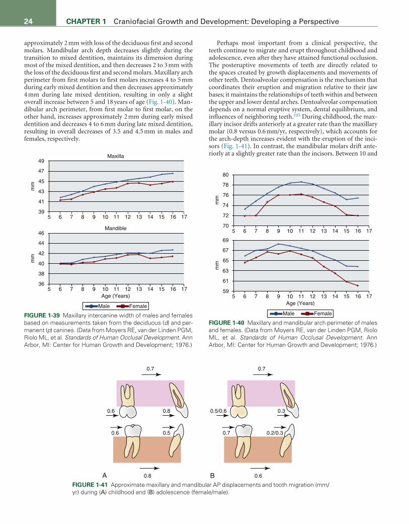
24 CHAPTER 1 Craniofacial Growth and Development: Developing a Perspective
approximately 2 mm with loss of the deciduous first and second molars. Mandibular arch depth decreases slightly during the transition to mixed dentition, maintains its dimension during most of the mixed dentition, and then decreases 2 to 3 mm with the loss of the deciduous first and second molars. Maxillary arch perimeter from first molars to first molars increases 4 to 5 mm during early mixed dentition and then decreases approximately 4 mm during late mixed dentition, resulting in only a slight overall increase between 5 and 18 years of age (Fig. 1-40). Man-dibular arch perimeter, from first molar to first molar, on the other hand, increases approximately 2 mm during early mixed dentition and decreases 4 to 6 mm during late mixed dentition, resulting in overall decreases of 3.5 and 4.5 mm in males and females, respectively.
Perhaps most important from a clinical perspective, the teeth continue to migrate and erupt throughout childhood and adolescence, even after they have attained functional occlusion. The posteruptive movements of teeth are directly related to the spaces created by growth displacements and movements of other teeth. Dentoalveolar compensation is the mechanism that coordinates their eruption and migration relative to their jaw bases; it maintains the relationships of teeth within and between the upper and lower dental arches. Dentoalveolar compensation depends on a normal eruptive system, dental equilibrium, and influences of neighboring teeth.105 During childhood, the max-illary incisor drifts anteriorly at a greater rate than the maxillary molar (0.8 versus 0.6 mm/yr, respectively), which accounts for the arch-depth increases evident with the eruption of the inci-sors (Fig. 1-41). In contrast, the mandibular molars drift ante-riorly at a slightly greater rate than the incisors. Between 10 and
mm
80
78
76
74
72
705 6 7 8 9 10 11 12 13 14 15 16 17
Male Female
Age (Years)
mm
69
67
65
63
61
595 6 7 8 9 10 11 12 13 14 15 16 17
FIGURE 1-40 Maxillary and mandibular arch perimeter of males and females. (Data from Moyers RE, van der Linden PGM, Riolo ML, et al. Standards of Human Occlusal Development. Ann Arbor, MI: Center for Human Growth and Development; 1976.)
0.6 0.8 0.30.5/0.6
0.6 0.5
0.8 0.6
0.7 0.7
0.7 0.2/0.3
A BFIGURE 1-41 Approximate maxillary and mandibular AP displacements and tooth migration (mm/yr) during (A) childhood and (B) adolescence (female/male).
Maxilla
mm
49
47
45
43
41
395 6 7 8 9 10 11 12 13 14 15 16 17
Male Female
Mandible
Age (Years)
mm
46
44
42
40
38
365 6 7 8 9 10 11 12 13 14 15 16 17
FIGURE 1-39 Maxillary intercanine width of males and females based on measurements taken from the deciduous (d) and per-manent (p) canines. (Data from Moyers RE, van der Linden PGM, Riolo ML, et al. Standards of Human Occlusal Development. Ann Arbor, MI: Center for Human Growth and Development; 1976.)

25CHAPTER 1 Craniofacial Growth and Development: Developing a Perspective
15 years of age, the molars (0.5 to 0.7 mm/yr) show significantly greater amounts of anterior drift than the incisors (0.3 mm/yr).
Substantial amounts of eruption occur throughout growth. During childhood, the maxillary first molars and incisors erupt at a rate of approximately 1.0 mm/yr, while their mandibular coun-terparts erupt at a rate of approximately 0.5 mm/yr (Fig. 1-42). During adolescence, the maxillary molars and incisors erupt at rates of 1.2 to 1.4 mm/yr and 0.9 mm/yr, respectively. The man-dibular molars and incisors erupt at a rate of 0.5 to 0.9 mm/yr, with little or no differences between incisor and molar erup-tion. The amounts of eruption that occur are associated closely with the inferior displacements of the midface and, especially, the mandible.
During childhood, there is little or no evidence of sexual dimorphism in the migration and eruption of teeth. In contrast, there is a relatively high degree of dimorphism during adoles-cence in mandibular eruption, with boys showing almost twice as much eruption as girls. The maxillary teeth show only limited sex differences, pertaining primarily to the molars.
ADULT CHANGES IN CRANIOFACIAL FORMThe size and shape of the craniofacial complex continue to change throughout a considerable part of adulthood. Over 90% of the 70 cephalometric distances and 70% of the 69 angles evaluated by Behrents106 showed changes after 17 years of age; 61% of the distances and 28% of the angles showed changes after 35 years of age. In particular, the mandibular plane angle increases in adult females and decreases in adult males, which explains why males 25 to 46 years of age exhibit greater chin projection than females, who undergo increases in NSGn.107
Adult soft tissues undergo the more pronounced changes than the skeletal structures. The nose grows substantially during adulthood, with the tip moving down and forward approx-imately 3 mm after 17 years of age. Males exhibit significantly more nasal growth than females. Upper lip length increases (approximately 2 to 3 mm) in both males and females after 17 years of age, resulting in decreases in upper incisor display over time. Lower lip length also increases, but less than upper lip length. The lips straighten and flatten during adulthood, but the most pronounced changes occur after 50 years of age. The soft tissue profile angle increases over time, with smaller increases when the nose is included than when it is excluded.
Adult profile changes are limited to 2 to 3° and 4 to 6° when the nose is included and excluded, respectively.
POSTNATAL INTERRELATIONSHIPS DURING CRANIOFACIAL GROWTHPostnatal craniofacial growth follows a gradient of relative growth that ranges between the neural and general somatic patterns. Vertical growth and modeling of the viscerocranium, as well as dental eruption, exhibit midchildhood and pubertal growth spurts. Anteroposterior growth and tooth migration, which do not exhibit midchildhood or pubertal growth spurts, change more or less regularly—except for the accelerated migration associated with the loss of teeth—throughout child-hood and adolescence.
Generally, most displacements and rotations of the maxillo–mandibular complex are controlled epigenetically through growth of the chondrocranium, soft tissue growth, and expan-sion of the oronasal capsule. The cartilaginous growth centers play a particularly important role in the primary displacement of the chondrocranium, as well as in the secondary displace-ment of the viscerocranium. The anterior displacement of the midface has been associated with growth of the anterior cranial base and expansion of the anterior cranial fossa; mandibular displacements are more closely associated with growth of the posterior cranial base and middle cranial fossa. Anteroposterior length changes of the anterior cranial base, measured from sella to foramen cecum, coincide closely with expansion of the fron-tal lobes and growth at the sphenoethmoidal synchondrosis. Angular changes of the cranial base have been associated with growth gradients within the synchondroses, complex interac-tions with the growth of the brain, as well as facial growth. The cranial base angle decreases due to greater chondrogenesis in the superior than in the inferior aspects of the sphenoethmoidal and, especially, spheno-occipital synchondroses. Changes in cranial base angulation also appear to be related to changes in brain size, especially to the dramatic changes that occur during the first 2 postnatal years.
Cranial base growth influences the displacement and rota-tion of the viscerocranium. Growth of the posterior cranial base (i.e., spheno-occipital synchondrosis) is directly related to infe-rior and posterior displacements of the glenoid fossa; growth of the anterior cranial base is associated with midfacial dis-placement. Consequently, cranial base growth changes partially
1.0 1.0 0.91.2/1.4
0.5 0.5
2.2 2.1/3.0
0.8 0.7/0.9
0.5/0.9 0.5/0.8
A BFIGURE 1-42 Approximate maxillary and mandibular vertical displacements (mm/yr) and tooth eruption during (A) childhood and (B) adolescence (female/male).
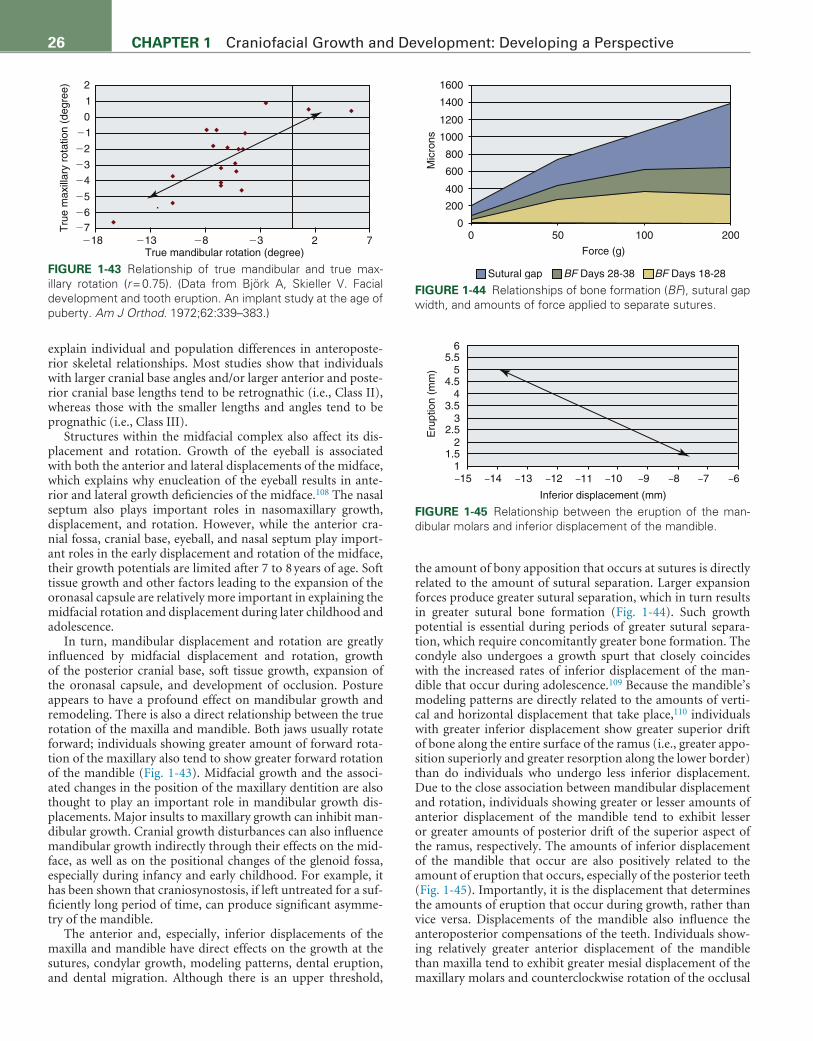
26 CHAPTER 1 Craniofacial Growth and Development: Developing a Perspective
explain individual and population differences in anteroposte-rior skeletal relationships. Most studies show that individuals with larger cranial base angles and/or larger anterior and poste-rior cranial base lengths tend to be retrognathic (i.e., Class II), whereas those with the smaller lengths and angles tend to be prognathic (i.e., Class III).
Structures within the midfacial complex also affect its dis-placement and rotation. Growth of the eyeball is associated with both the anterior and lateral displacements of the midface, which explains why enucleation of the eyeball results in ante-rior and lateral growth deficiencies of the midface.108 The nasal septum also plays important roles in nasomaxillary growth, displacement, and rotation. However, while the anterior cra-nial fossa, cranial base, eyeball, and nasal septum play import-ant roles in the early displacement and rotation of the midface, their growth potentials are limited after 7 to 8 years of age. Soft tissue growth and other factors leading to the expansion of the oronasal capsule are relatively more important in explaining the midfacial rotation and displacement during later childhood and adolescence.
In turn, mandibular displacement and rotation are greatly influenced by midfacial displacement and rotation, growth of the posterior cranial base, soft tissue growth, expansion of the oronasal capsule, and development of occlusion. Posture appears to have a profound effect on mandibular growth and remodeling. There is also a direct relationship between the true rotation of the maxilla and mandible. Both jaws usually rotate forward; individuals showing greater amount of forward rota-tion of the maxillary also tend to show greater forward rotation of the mandible (Fig. 1-43). Midfacial growth and the associ-ated changes in the position of the maxillary dentition are also thought to play an important role in mandibular growth dis-placements. Major insults to maxillary growth can inhibit man-dibular growth. Cranial growth disturbances can also influence mandibular growth indirectly through their effects on the mid-face, as well as on the positional changes of the glenoid fossa, especially during infancy and early childhood. For example, it has been shown that craniosynostosis, if left untreated for a suf-ficiently long period of time, can produce significant asymme-try of the mandible.
The anterior and, especially, inferior displacements of the maxilla and mandible have direct effects on the growth at the sutures, condylar growth, modeling patterns, dental eruption, and dental migration. Although there is an upper threshold,
the amount of bony apposition that occurs at sutures is directly related to the amount of sutural separation. Larger expansion forces produce greater sutural separation, which in turn results in greater sutural bone formation (Fig. 1-44). Such growth potential is essential during periods of greater sutural separa-tion, which require concomitantly greater bone formation. The condyle also undergoes a growth spurt that closely coincides with the increased rates of inferior displacement of the man-dible that occur during adolescence.109 Because the mandible’s modeling patterns are directly related to the amounts of verti-cal and horizontal displacement that take place,110 individuals with greater inferior displacement show greater superior drift of bone along the entire surface of the ramus (i.e., greater appo-sition superiorly and greater resorption along the lower border) than do individuals who undergo less inferior displacement. Due to the close association between mandibular displacement and rotation, individuals showing greater or lesser amounts of anterior displacement of the mandible tend to exhibit lesser or greater amounts of posterior drift of the superior aspect of the ramus, respectively. The amounts of inferior displacement of the mandible that occur are also positively related to the amount of eruption that occurs, especially of the posterior teeth (Fig. 1-45). Importantly, it is the displacement that determines the amounts of eruption that occur during growth, rather than vice versa. Displacements of the mandible also influence the anteroposterior compensations of the teeth. Individuals show-ing relatively greater anterior displacement of the mandible than maxilla tend to exhibit greater mesial displacement of the maxillary molars and counterclockwise rotation of the occlusal
Tru
e m
axill
ary
rota
tion
(deg
ree) 2
1
0
�1
�2
�3
�4
�5
�6�7
�18 �13 �8 �3 2 7True mandibular rotation (degree)
FIGURE 1-43 Relationship of true mandibular and true max-illary rotation (r = 0.75). (Data from Björk A, Skieller V. Facial development and tooth eruption. An implant study at the age of puberty. Am J Orthod. 1972;62:339–383.)
1600
1400
1200
1000
800
600
400
200
00 50 100
Force (g)
Sutural gap BF Days 28-38 BF Days 18-28
Mic
rons
200
FIGURE 1-44 Relationships of bone formation (BF), sutural gap width, and amounts of force applied to separate sutures.
–15
65.5
54.5
43.5
32.5
21.5
1–14 –13 –12 –11 –10
Inferior displacement (mm)
–9 –8 –7 –6
Eru
ptio
n (m
m)
FIGURE 1-45 Relationship between the eruption of the man-dibular molars and inferior displacement of the mandible.

27CHAPTER 1 Craniofacial Growth and Development: Developing a Perspective
plane; those who undergo relatively greater anterior maxillary displacements display greater mesial displacement of the man-dibular molars and minimal mesial displacement of maxillary molars.
The morphologic correlates with true rotation are numerous and hold important clinical implications.111 Vertical rotation has been directly related to changes in tooth position, with true for-ward rotators showing greater amounts of lower incisor procli-nation during eruption; backward rotators show retroclination of the incisors and loss of arch space. Rotation is also related to the modeling pattern that occurs on the lower mandibular bor-der; subjects who undergo the greatest amounts of true forward rotation also exhibit the greatest amounts of posterior resorption and anterior bony deposition (Fig. 1-46). Ramus modeling in general depends on the rotational pattern of the mandible. Indi-viduals who undergo greater amounts of true forward rotation
also exhibit greater amounts of condylar growth, oriented in a more superior–anterior direction (Fig. 1-47). Perhaps the most important clinical correlate is the relationship between true rota-tion and chin position. True rotation of the mandible explains more of the individual variation in chin position than condylar growth or changes in glenoid fossa position.
SIGNIFICANCE OF UNDERSTANDING CRANIOFACIAL GROWTH FOR ORTHODONTICSTo be most effective as clinicians, it is essential that orthodontists understand the development, growth, and adaptive potentials of the craniofacial structures. Along with orthodontic biome-chanics, knowledge of how the craniofacial complex develops and grows provides the foundation for understanding the eti-ology of the various dental and skeletal malocclusions, the best
Forward rotators � 5�
Ar14.2/�1.8
Ar8.3/�3.1
Go7.4/�5.6
Go3.7/�4.8
Co15.7/�0.6
Co10.1/�3.1
SN - 7�
Forward rotators 0-5�
FIGURE 1-46 Vertical/horizontal (mm) remodeling changes of individuals showing greater than 5 degrees of true forward rotation, compared with those showing 0- to 5-mm true forward rotation.
�20
25
20
15
10
5
0�15 �10 �5 0 5 10
Am
ount
(m
m)
�20
140
120
100
80
0�15 �10 �5 0
True rotation (degree)
5 10
Dire
ctio
n (d
eg)
A
BFIGURE 1-47 Relationships of true mandibular rotation and (A) the amount of condylar growth and (B) the direction of condylar growth. (Data from Björk A, Skieller V. Facial development and tooth eruption. An implant study at the age of puberty. Am J Orthod. 1972;62:339–383.)
28 CHAPTER 1 Craniofacial Growth and Development: Developing a Perspective
of all possible treatment approaches, and how patients might be expected to respond after treatment. A thorough under-standing of growth provides the basis for knowing which cra-niofacial components might be expected to respond to a stress and how great the response might be expected to be. Because a structure’s response potential to stress is directly related to its relative growth potential, and the vertical aspects of the man-dible have the greatest relative growth potential, it follows that skeletal malocclusions might be expected to relate to vertical mandibular growth. Class II malocclusion, which is the pre-dominant form of skeletal malocclusion, pertains primarily to the mandible.112,113 These individuals are often retrognathic due to limited true forward rotation of the mandible, which is in turn related to deficient inferior growth displacement of the posterior mandible and/or excessive inferior displacement of the anterior aspect of the mandible.
Knowledge of growth is also important because, whenever possible, orthodontists should try to mimic growth when plan-ning treatment. An understanding of growth provides the bio-logical limits within which treatments can be performed. As previously indicated, the viscerocranium is made up almost entirely of intramembranous bone and is predominantly under epigenetic and environmental control. It is programmed to adapt, and adaptation should be expected whenever it is stressed. The biological system cannot distinguish between stresses imposed by the orthodontist and those imposed during normal growth; it simply responds depending on its growth potential. Continu-ing with the previous example, individuals who exhibit good growth patterns tend to be true forward rotators with condyles that grow in a more anterior direction. Based on this knowledge, hyperdivergent retrognathnic patients would best be served by treatments that focus on rotating the mandible rather than stim-ulating or redirecting condylar growth in a posterior direction.114
Finally, an understanding of growth makes it possible to estimate morphologic changes that might be expected to occur during and after orthodontic treatment. Unless it is intention-ally disrupted, an individual’s growth path before treatment might be expected to continue during and after treatment. Knowing how the maxilla and mandible have been rotated and/or displaced during treatment provides an understanding of the modeling and consequent shape changes that might be expected to occur. Vertical growth after treatment might be problematic in terms of posttreatment crowding, due to its relationship with tooth eruption. It has been shown that the best predictors of crowding of the mandibular incisors, both after treatment and without treatment, are the inferior displacement of the mandi-ble and superior eruption of the incisors.86
As understanding of craniofacial development, growth, and adaptation continues to improve in the future, orthodontists can look forward to even more therapeutic advances that can be used to influence growth and posttreatment stability. This understanding will facilitate greater clinical control of changes in the craniofacial complex due to the normal growth process and in compensatory adaptation of tissues after treatment. Under-standing normal craniofacial growth and especially that of the complex network of underlying molecular factors responsible for craniofacial growth and treatment will also be of immeasur-able benefit in assisting the orthodontist in understanding what may or may not be possible, not only with respect to diagnosing a patient’s underlying abnormality but also in determining the best treatment approach for its correction.17,18,115
REFERENCES 1. Carlson B. Human Embryology and Developmental Biology. Philadelphia:
Elsevier; 2014. 2. Sperber GH. Craniofacial Development. London: BC Decker; 2010. 3. Carlson DS, Ribbens KR. Craniofacial Growth during Adolescence. Cra-
niofacial Growth Series. Ann Arbor, MI: Center for Human Growth and Development; 1987:20.
4. Scammon RE. The measurement of the body in childhood. In: Harris JA, Jackson CM, Scammon RE, eds. The Measurement of the Body in Child-hood. Minneapolis: University of Minneapolis Press; 1930.
5. Buschang PH, Hinton RJ. A gradient of potential for modifying cranio-facial growth. In: Carlson DS, ed. Control Mechanisms of Craniofacial Development and Growth. Semin Orthod; 2005; 11(4):219–226.
6. Buschang PH, Tanguay R, Demirjian A, LaPalme L, Turkewicz J. Math-ematical models of longitudinal mandibular growth for children with normal and untreated Class II, division 1 malocclusion. Eur J Orthod. 1988;10:227–234.
7. Malina RM, Bouchard C, Beunen G. Human growth: selected aspects of current research on well-nourished children. Ann Rev Anthrop. 1988;17:187–219.
8. Lewis AB, Roche AF, Wagner B. Pubertal spurts in cranial base and man-dible. Comparisons within individuals. Angle Orthod. 1985;55:17–30.
9. Buschang PH, Jacob HB, Demirjian A. Female adolescent craniofacial growth spurts: real or fiction? Eur J Orthod. 2013;35:819–825.
10. Tanner JM. Growth at adolescence: an introduction. In: Carlson DS, Ribbens KR, eds. Craniofacial Growth during Adolescence. Craniofacial Growth Series. Ann Arbor, MI: Center for Human Growth and Develop-ment; 1987;20:1–22.
11. Grave KC, Brown T. Skeletal ossification and the adolescent growth spurt. Am J Orthod. 1976;69:611–624.
12. Fishman LS. Radiographic evaluation of skeletal maturation. Angle Orth-od. 1982;52:88–112.
13. Baccetti T, Franchi L, McNamara Jr JA. The cervical vertebral matura-tion (CVM) method for the assessment of optimal treatment timing in dentofacial orthopedics. Semin Orthod. 2005;11:119–129.
14. Massoud MI, Masoud I, Kent Jr RL, Gowharji N, Hassan AH, Cohen L. Relationship between blood-spot insulin-like growth factor 1 levels and hand-wrist assessment of skeletal maturity. Am J Orthod Dentofac Orthop. 2009;136(1):59–64.
15. Wilkie AOM, Morriss-Kay GM. Genetics of craniofacial development and malformation. Nat Rev Genet. 2001;2:458–468.
16. Hefer T, Jaochims HZ, Carlson DS, Finnell RH. Factors associated with the etiology of congenital craniofacial anomalies: an update Part I. J Israeli Med Assoc. 1998;135:209–213, 286–291.
17. Carlson DS. Growth modification: from molecules to mandibles. In: McNamara JA, ed. Growth Modification: What Works, What Doesn’t, and Why. Craniofacial Growth Series. Ann Arbor, MI: Center for Human Growth and Development; 1999:17–65.
18. Carlson DS. Toward a new synthesis for craniofacial biology: a genomic- epigenomic basis for dentofacial orthopedic treatment. In: McNamara JA, ed. The 40th Moyers Symposium. Looking Back…Looking Forward. Craniofacial Growth Series; 50. Ann Arbor, MI: Center for Human Growth and Development; 2014:193–247.
19. D’Souza RN, Dunnvald M, Frazier-Bowers S, et al. Translational genetics: advancing fronts for craniofacial health. J Dent Res. 2013;92(12):1058–1064.
20. Eng G, Chen A, Vess T, Ginsburg GS. Genome technologies and person-alized dental medicine. Oral Dis. 2012;18:223–235.
21. Pritchard JJ, Scott JH, Girgis FG. The structure and development of cranial and facial sutures. J Anat. 1956;90:73–86.
22. Scott JH. The growth of the human face. Proc R Soc Med. 1954;47:91–100. 23. Baer MJ. Patterns of growth of the skull as revealed by vital staining.
Hum Biol. 1954;26:80–126. 24. Rice DPC. Craniofacial anomalies: from development to molecular
pathogenesis. Curr Mol Med. 2005;5:699–722. 25. Chai Y, Maxson Jr RE. Recent advances in craniofacial morphogenesis.
Dev Dyn. 2006;235:2353–2375.
29CHAPTER 1 Craniofacial Growth and Development: Developing a Perspective
26. Kim HJ, et al. FGF-, BMP- and Shh-mediated signalling pathways in the regulation of cranial suture morphogenesis and calvarial bone develop-ment. Devel. 1998;125(7):1241–1251.
27. Rice DP, Kim HJ, Thesleff I. Apoptosis in murine calvarial bone and suture development. Eur J Oral Sci. 1999;107(4):265–275.
28. Carver EA, Oram KF, Gridley T. Craniosynostosis in Twist heterozygous mice: a model for Saethre-Chotzen syndrome. Anat Rec. 2002;268(2):90–92.
29. Warren SM, et al. The BMP antagonist noggin regulates cranial suture fusion. Nature. 2003;422(6932):625–629.
30. Veistinen L, Takatolo M, Tanimoto Y, Kesper D, Vortkamp A, Rice DPC. Loss-of-function of Gli3 in mice causes abnormal frontal bone morphology and premature synostosis of the interfrontal suture. Front Physiol. 2012;3:1–6.
31. Jabs EW, et al. A mutation in the homeodomain of the human MSX2 gene in a family affected with autosomal dominant craniosynostosis. Cell. 1993;75(3):443–450.
32. el Ghouzzi V, et al. Mutations of the TWIST gene in the Saethre-Chotzen syndrome. Nat Genet. 1997;15(1):42–46.
33. Howard TD, et al. Mutations in TWIST, a basic helix-loop- helix transcription factor, in Saethre-Chotzen syndrome. Nat Genet. 1997;15(1):36–41.
34. Moss ML, Salentijn L. The primary role of functional matrices in facial growth. Am J Orthod. 1969;55:566–577.
35. Martinez-Abadias N, Heuze Y, Wang Y, Jabs EW, Aldridge K, Richtmeier J. FGF/FGFR signaling coordinates skull development by modulating magnitude of morphological integration: evidence from Apert syndrome mouse models. PLoS One. 2011;6(10).
36. Heuze Y, Martinez-Abadias N, Stella JM, et al. Quantification of facial skeletal shape variation in fibroblast growth factor receptor-related craniosynostosis syndromes. Birth Def Res (A). 2014;100:250–259.
37. Di Ieva A, Bruner E, Haider T, et al. Skull base embryology: a multidisci-plinary review. Childs Ner Syst. 2014;30:991–1000.
38. Lei WY, Wong RWK, Rabie ABM. Factors regulating endochondral ossification in the spheno-occipital synchondrosis. Angle Orthod. 2008;78(2):215–220.
39. Balczerski B, Zakaria S, Tucker AS, et al. Distinct spatiotemporal roles of hedgehog signaling during chick and mouse cranial base and axial skeletal development. Dev Biol. 2012;371:203–214.
40. Kuhn JL, Ciarelli MJ. The biomechanics of the growth plate. In: Carlson DS, Goldstein SA, eds. Bone Biodynamics in Orthodontic and Orthopedic Treatment. Craniofacial Growth Series. Ann Arbor, MI: Center for Hu-man Growth and Development; 1992:93–110.
41. Opperman LA, Gakunga PT, Carlson DS. Genetic factors influencing morphogenesis and growth of sutures and synchondroses in the cranio-facial complex. In: Carlson DS, ed. Control mechanisms of craniofacial development and growth. Semin Orthod; 2005; 11(4):199–208.
42. Baume LJ. Principles of cephalofacial development revealed by experi-mental biology. Am J Orthod. 1961;47(12):881–901.
43. Buschang PH, Baume RM, Nass GG. A craniofacial growth maturity gradient for males and females between 4 and 16 years of age. Am J Phys Anthrop. 1983;61:373–382.
44. Melsen B. The cranial base: the postnatal development of the cranial base studied histologically on human autopsy material. Acta Orthod Scand Suppl. 1974;62:1–126.
45. Latham RA. The sella point and postnatal growth of the cranial base in the human skull. Am J Orthod. 1972;61:156–162.
46. Powell TV, Brodie AG. Closure of the spheno-occipital synchondrosis. Anat Rec. 1963;147:15–23.
47. Ford HER. Growth of the human cranial base. Am J Orthod. 1958;44:498–506. 48. Adab K, Sayne JR, Carlson DS, Opperman LA. Tgf-β1, Tgf-β2, Tgf-β3
and Msx2 expression is elevated during frontonasal suture morpho-genesis and during active postnatal facial growth. Orthod Craniofac Res. 2002;5(4):227–237.
49. Griffin JN, Compagnucci C, Hu D, et al. Fgf8 dosage determines midfa-cial integration and polarity within the nasal and optic capsules. Dev Biol. 2013;374:185–197.
50. Greene RM, Pisano MM. Palate morphogenesis: current understanding and future directions. Birth Defects Res (Part C). 2010;90:133–154.
51. Bush JO, Jiang R. Palatogenesis: morphological and molecular mecha-nisms of secondary palate development. Devel. 2012;139:231–243.
52. Behrents RG, Harris EF. The premaxillary-maxillary suture and ortho-dontic mechanotherapy. Am J Orthod Dentofac Orthop. 1991;99:1–6.
53. Melsen B. Palatal growth studied on human autopsy material. A histolog-ic microradiographic study. Am J Orthod. 1975;68:42–54.
54. Persson M, Thilander B. Palatal suture closure in man from 15 to 35 years of age. Am J Orthod. 1977;72:42–52.
55. Korbmacher H, Schilling A, Puschel K, Amling M, Kahl-Nieke B. Age- dependent three-dimensional microcomputed tomography analysis of the human midpalatal suture. J Orofac Orthop. 2007;68:364–376.
56. Knaup B, Yildizhan F, Wehrbein H. Age-related changes in the midpalatal suture. A histomorphometric study. J Orofac Orthop. 2004;65:467–474.
57. Herring SW, Mucci RJ. In vivo strain in cranial sutures: the zygomatic arch. J Morphol. 1991;207:225–239.
58. Björk A, Skieller V. Postnatal growth and development of the maxillary complex. In: McNamara Jr JA, ed. Factors Affecting the Growth of the Midface. Craniofacial Growth Series. Ann Arbor, MI: Center for Human Growth and Development; 1976:61–100.
59. Enlow DH, Bang S. Growth and remodeling of the human maxilla. Am J Orthod. 1965;51:446–464.
60. Björk A, Skieller V. Growth of the maxilla in three dimensions as revealed radiographically by the implant method. Br J Orthod. 1977;4:53–64.
61. Hersby RM, Marshall SD, Dawson DV, et al. Transverse skeletal and dentoalveolar changes during growth. Am J Orthod Dentofac Orthop. 2006;130:721–731.
62. Solow B, Houston WJ. Mandibular rotations: concepts and terminology. Eur J Orthod. 1988;10:177–179.
63. Lux CJ, Conradt C, Burden D, et al. Transverse development of the cra-niofacial skeleton and dentition between 7–15 years of age: a longitudinal postero-anterior cephalometric study. Eur J Orthod. 2004;26:31–42.
64. Shibata S, Sakamoto Y, Yokohama-Tamaki T, Murakami G, Cho BH. Distribution of matrix proteins in perichondrium and periosteum during incorporation of Meckel’s cartilage into ossifying mandible: an immuno-histochemical study. Anat Rec. 2014;297:1208–1217.
65. Hall BK. Immobilization and cartilage transformation into bone in the embryonic chick. Anat Rec. 1972;173:391–404.
66. Vinkka H. Secondary cartilages in the facial skeleton of the rat. Proc Finn Dent Soc. 1982;78:36–39.
67. Copray JCVM, Dibbets JMH, Kantomaa T. The role of condylar cartilage in the development of the temporomandibular joint. Angle Orthod. 1988;58:369–380.
68. Carlson DS. Growth of the temporomandibular joint. In: Zarb GA, Carlsson GE, Sessle BJ, Mohl ND, eds. Temporomandibular Joint and Masticatory Muscle Disorders. Copenhagen: Munksgaard; 1994: 128–155.
69. Hinton RJ, Carlson DS. Regulation of growth in the mandibular condylar cartilage. In: Carlson DS, ed. Control mechanisms of craniofacial develop-ment and growth. Semin Orthod; 2005; 11(4):209–218.
70. Enomoto A, Watahiki J, Nampo T, Ichikawa Y, Tachikawa T, Maki K. Mastication markedly affects mandibular condylar cartilage growth, gene expression, and morphology. Am J Orthod Dentofac Orthop. 2014;146(3):355–363.
71. Hinton RJ, Carlson DS. Histological changes in the articular eminence and mandibular fossa during growth of the rhesus monkey (Macaca mulatta). Am J Anat. 1983;166:99–116.
72. Carlson DS, McNamara Jr JA, Jaul DH. Histological analysis of the growth of the mandibular condyle in the rhesus monkey (Macaca mulatta). Am J Anat. 1978;151:103–117.
73. Wright DM, Moffett BC. The postnatal development of the human tem-poromandibular joint. Am J Anat. 1974;141:235–250.
74. Hinton RJ, Serrano M, So S. Differential gene expression in the perichon-drium and cartilage of the neonatal mouse temporomandibular joint. Ortho Craniofac Res. 2009;12:168–177.
75. Hinton RJ. Genes that regulate morphogenesis and growth of the tem-poromandibular joint: a review. Dev Dyn. 2014;243:864–874.

30 CHAPTER 1 Craniofacial Growth and Development: Developing a Perspective
76. Öberg T, Carlsson GE, Fajers CM. The temporomandibular joint. A study on human autopsy material. Acta Odontol Scand. 1971;29: 349–384.
77. Carlsson GE, Öberg T. Remodeling of the temporomandibular joint. Oral Sci Rev. 1974;6:53–86.
78. Lubsen CC, Hansson TL, Nordstrom BB, et al. Histomorphometric anal-ysis of cartilage and subchondral bone in mandibular condyles of young human adults at autopsy. Arch Oral Biol. 1985;30:129–136.
79. Lubsen CC, Hansson TL, Nordstrom BB, et al. Histomorphometry of age and sex changes in mandibular condyles of young human adults. Arch Oral Biol. 1987;32:729–733.
80. Dibbets JMH, Carlson DS. Implications of temporomandibular disorders for facial growth and orthodontic treatment. Semin Orthod Temporoman-dibular Jt Disord: Facts Fallacies. 1995;1(4):258–272.
81. Dechow PD, Carlson DS. Development of mandibular form: phylogeny, ontogeny and function. In: McNeill C, ed. Science and Practice of Occlu-sion. New Malden, UK: Quintessence; 1997:3–22.
82. Petrovic A, Stutzmann J, Oudet C. Control processes in the postnatal growth of the condylar cartilage. In: McNamara Jr JA, ed. Determinants of Mandibular Form and Growth. Craniofacial Growth Series. Ann Arbor, MI: Center for Human Growth and Development; 1975:101–154.
83. Petrovic A. Control of postnatal growth of secondary cartilages of the mandible by mechanisms regulating occlusion. Cybernetic model. Trans Eur Orthod Soc. 1974:69–75.
84. McNamara JA, Carlson DS. Quantitative analysis of temporomandibular joint adaptations to protrusive function. Am J Orthod. 1979;76:593–611.
85. Petrovic A. Experimental and cybernetic approaches to the mechanism of action of functional appliances on mandibular growth. In: McNamara Jr JA, Ribbens KA, eds. Malocclusion and the Periodontium. Craniofacial Growth Series. Ann Arbor, MI: Center for Human Growth and Develop-ment; 1985:213–268.
86. Carlson DS. Theories of craniofacial growth in the postgenomic era. In: Carlson DS, ed. Control Mechanisms of Craniofacial Development and Growth. Semin Orthod; 2005;11(4):172–183.
87. Visnapuu V, Peltomaki T, Ronning O, et al. Distribution of fibroblast growth factors (FGFR-1 and -3) and platelet-derived growth factor receptors (PDGFR) in the rat mandibular condyle during growth. Orthod Craniofac Res. 2002;5:147–153.
88. Ramirez-Yanez GO, Young WG, et al. Influence of growth hormone on the mandibular condylar cartilage of rats. Arch Oral Biol. 2004;49:585–590.
89. Visnapuu V, Peltomaki T, Ronning O, et al. Growth hormone and insulin-like growth factor-I receptors in the temporomandibular joint of the rat. J Dent Res. 2001;80:1903–1907.
90. Rabie ABM, She TT, Hägg U. Functional appliance therapy acceler-ates and enhances condylar growth. Am J Orthod Dentofacial Orthop. 2003;123(1):40–48.
91. Chu FT, Tang GH, Hu Z, Qian YF, Shen G. Mandibular functional positioning only in vertical dimension contributes to condylar adaptation evidenced by concomitant expressions of L-Sox5 and type II collagen. Arch Oral Biol. 2008;53:567–574.
92. Owtad P, Park JH, Shen G, Potres, Darendelier MA. The biology of TMJ growth modification: a review. J Dent Res. 2013;92(4):315–321.
93. Xue F, Wong RWK, Rabie ABM. Genes, genetics, and Class III malocclu-sion. Orthod Craniofac Res. 2010;13:69–74.
94. Fuentes MA, Opperman LA, Buschang P, et al. Lateral functional shift of the mandible: Part II. Effects on gene expression in condylar cartilage. Am J Orthod Dentofac Orthop. 2003;123:160–166.
95. Hajjar D, Santos MF, Kimura ET. Propulsive appliance stimulates the synthesis of insulin-like growth factors I and II in the mandibular condy-lar cartilage of young rats. Arch Oral Biol. 2003;48:635–642.
96. Rabie ABM, Hagg U. Factors regulating mandibular condylar growth. Am J Orthod Dentofac Orthop. 2002;122:401–409.
97. Tang GH, Rabie ABM, Hagg U. Indian hedgehog: a mechanotransduc-tion mediator in condylar cartilage. J Dent Res. 2004;83:434–438.
98. Enlow DH, Harris DB. A study of the postnatal growth of the human mandible. Am J Orthod. 1964;50:25.
99. Buschang PH, Julien K, Sachdeva R, et al. Childhood and pubertal growth changes of the human symphysis. Angle Orthod. 1992;62:203–210.
100. Buschang PH, Santos Pinto A. Condylar growth and glenoid fossa displacement during childhood and adolescence. Am J Orthod Dentofac Orthop. 1998;113:437–442.
101. Björk A, Skieller V. Facial development and tooth eruption. An implant study at the age of puberty. Am J Orthod. 1972;62:339–383.
102. Spady M, Buschang PH, Demirjian A, et al. Mandibular rotation and angular remodeling during childhood and adolescence. Am J Hum Biol. 1992;4:683–689.
103. Wang M, Buschang PH, Behrents R. Mandibular rotation and remodel-ing changes during early childhood. Angle Orthod. 2009;79:271–275.
104. Moorrees CFA, Grøn AM, Lebret LML, et al. Growth studies of the denti-tion: a review. Am J Orthod. 1969;55:600–616.
105. Moyers RE, van der Linden PGM, Riolo ML, et al. Standards of Human Occlusal Development. Ann Arbor, MI: Center for Human Growth and Development; 1976.
106. Behrents RG. Growth in the Aging Craniofacial Skeleton. Craniofacial Growth Series. Ann Arbor, MI: Center for Human Growth and Develop-ment, University of Michigan; 1985. Monograph #17.
107. Bishara SE, Treder JE, Jakobsen JR. Facial and dental changes in adult-hood. Am J Orthod Dentofacial Orthop. 1994;106:175–186.
108. Sarnat BG. Eye and orbital size in the young and adult. Ophthalmologica. 1982;185:74–89.
109. Kim YE, Nanda RS, Sinha PK. Transition of molar relationships in different skeletal growth patterns. Am J Orthod Dentofac Orthop. 2002;121:280–290.
110. Buschang PH, Gandini Jr LG. Mandibular skeletal growth and modeling between 10–15 years of age. Eur J Orthod. 2002;24:69–79.
111. Dibbets JMH. Mandibular rotation and enlargement. Am J Orthod Den-tofac Orthop. 1990;98(1):29–32.
112. McNamara Jr JA. Components of class II malocclusion in children 8–10 years of age. Angle Orthod. 1981;51:177–202.
113. Buschang PH, Martins J. Childhood and adolescent changes of skeletal relationships. Angle Orthod. 1998;68:199–208.
114. Driscoll-Gilliland J, Buschang PH, Behrents RG. An evaluation of growth and stability in untreated and treated subjects. Am J Orthod Dentofac Orthop. 2001;120:588–597.
115. Carlson DS. Biological rationale for early treatment of dentofacial defor-mities. Am J Orthod Dentofac Orthop. 2002;121(6):554–558.