Embed Size (px)
Citation preview
1Acute Complications of Diabetes

2
Diabetic Ketoacidosis
Introduction3
DKA is an acute life threatening complication of DM
¼of hospital admissions for DM
Occurs predominantly in type I though may occur in II
Incidence of DKA in diabetics 15 per 1000 patients
20-30% of cases occur in new-onset diabetes
Mortality less than 5%
Mortality higher in elderly due to underlying renal disease or coexisting infection
Definition4
Exact definition is variable
Most consistent is:Blood glucose level greater than 250 mg/dLBicarbonate less than 15 mEq/L Arterial pH less than 7.3Moderate ketonemia
Pathophysiology5
Body’s response to cellular starvation Brought on by relative insulin deficiency and counter regulatory or
catabolic hormone excessInsulin is responsible for metabolism and storage of carbohydrates, fat
and protein
Lack of insulin and excess counter regulatory hormones (glucagon, catecholamines, cortisol and growth hormone) results
in:Hyperglycemia (due to excess production and underutilization of
glucose)Osmotic diuresisPrerenal azotemiaKetone formationWide anion-gap metabolic acidosis
Clinical manifestations related to hyperglycemia, volume depletion and acidosis
Pathophysiology6
Free fatty acids released in the periphery are bound to albumin and transported to the liver where they
undergo conversion to ketone bodiesThe metabolic acidosis in DKA is due to β-hydroxybutyric acid
and acetoacetic acid which are in equilibriumAcetoacetic acid is metabolized to acetone, another major
ketone bodyDepletion of baseline hepatic glycogen stores tends to favor
ketogenesisLow insulin levels decrease the ability of the brain and cardiac
and skeletal muscle to use ketones as an energy source, also increasing ketonemia
Persistently elevated serum glucose levels eventually causes an osmotic diuresis
Resulting volume depletion worsens hyperglycemia and ketonemia
Electrolytes7
Renal potassium losses already occurring from osmotic diuresis worsen due to renin-angiotensin-aldosterone system activation by volume
depletion
In the kidney, chloride is retained in exchange for the ketoanions being excreted
Loss of ketoanions represents a loss of potential bicarbonate
In face of marked ketonuria, a superimposed hyperchloremic acidosis is also present
Presence of concurrent hyperchloremic metabolic acidosis can be detected by noting a bicarbonate level lower than explainable by the
amount the anion gap has increased
As adipose tissue is broken down, prostaglandins PGI2 and PGE2 are produced
This accounts for the paradoxical vasodilation that occurs despite the profound levels of volume depletion
DKA in Pregnancy8
Physiologic changes in pregnancy makes more prone to DKA
Maternal fasting serum glucose levels are normally lowerLeads to relative insulin deficiency and an increase in baseline
free fatty acid levels in the bloodPregnant patients normally have increased levels of
counter regulatory hormones Chronic respiratory alkalosis
Seen in pregnancy Leads to decreased bicarbonate levels due to a compensatory
renal responseResults in a decrease in buffering capacity
DKA in Pregnancy9
Pregnant patients have increased incidence of vomiting and infections which may precipitate DKA
Maternal acidosis:◦Causes fetal acidosis◦Decreases uterine blood flow and fetal oxygenation◦Shifts the oxygen-hemoglobin dissociation curve to the right
Maternal shifts can lead to fetal dysrhythmia and death
Causes of DKA10
25% have no precipitating causes found
Errors in insulin use, especially in younger population
Omission of daily insulin injections
Stressful events :InfectionStrokeMITraumaPregnancyHyperthyroidismPancreatitisPulmonary embolismSurgerySteroid use
Clinical Features11
Hyperglycemia
Increased osmotic load◦Movement of intracellular water into the vascular compartment◦Ensuing osmotic diuresis gradually leads to volume loss and
renal loss of sodium, chloride, potassium, phosphorus, calcium and magnesium
Patients initially compensate by increasing their fluid intake
Initially polyuria and polydipsia are only symptoms until ketonemia and acidosis develop
Clinical Features12
As acidosis progresses◦Patient develops a compensatory augmented ventilatory
response◦Increased ventilation is stimulated physiologically by
acidemia to diminish PCO2 and counter the metabolic acidosis
Peripheral vasodilation develops from prostaglandins and acidosis
◦Prostaglandins may contribute to unexplained nausea, vomiting and abdominal pain
◦Vomiting exacerbates the potassium losses and contributes to volume depletion, weakness and weight loss
Clinical Features13
Mental confusion or coma may occur with serum osmolarity greater than 340 mosm/L
Abnormal vital signs may be the only significant finding at presentation
Tachycardia with orthostasis or hypotension are usually present
Poor skin turgor
Kussmaul respirations with severe acidemia

Clinical Features14 Acetone presents with odor in some patients
Absence of fever does not exclude infection as a source of the ketoacidosis
Hypothermia may occur due to peripheral vasodilatation
Abdominal pain and tenderness may occur with gastric distension, ileus or pancreatitis
◦Abdominal pain and elevated amylase in those with DKA or pancreatitis may make differentiation difficult
◦Lipase is more specific to pancreatitis
Clinical Suspicion15
If suspect DKA, want immediately:AcucheckUrine dipECGVenous blood gasNormal Saline IV drip
Almost all patients with DKA have glucose
greater than 300 mg/dL
Acidosis16
Elevated serum β-hydroxybutyrate and acetoacetate cause acidosis and ketonuria
Elevated serum ketones may lead to a wide-anion gap metabolic acidosis
Metabolic acidosis may occur due to vomiting, osmotic diuresis and concomitant diuretic use
Some with DKA may present with normal bicarbonate concentration or alkalemia if other alkalotic processes
are severe enough to mask acidosisIn which case the elevated anion gap may be the only clue to
the presence of an underlying metabolic acidosis
ABGs17
Help determine precise acid-base status in order to direct treatment
Venous pH is just as helpfulStudies have shown strong correlation between arterial
and venous pH in patients with DKAVenous pH obtained during routine blood draws can be used
to avoid ABGs
Decreased PCO2 reflects respiratory compensation for metabolic acidosis
Widening of anion gap is superior to pH or bicarbonate concentration alone
Widening is independent of potentially masking effects concurrent with acid base disturbances
Potassium18
Total body potassium is depleted by renal losses
Measured levels usually normal or elevated
Sodium
19
Osmotic diuresis leads to excessive renal losses of NaCl in urine
Hyperglycemia artificially lowers the serum sodium levels
Two corrections:Standard-1.6 mEq added to sodium loss for every 100 mg
of glucose over 100 mg/dLTrue-2.4 mEq added for blood glucose levels greater than
400 mg/dL
Electrolyte Loss:20
Osmotic diuresis contributes to urinary losses and total body depletion of:
PhosphorusCalciumMagnesium
Other values elevated:21
Creatinine◦Some elevation expected due to prerenal azotemia ◦May be factitiously elevated if laboratory assays for Cr and Acetoacetate
interfere
LFTs◦Due to fatty infiltration of the liver which gradually corrects as acidosis is
treated
CPK ◦Due to volume depletion
Amylase
WBCs◦Leukocytosis often present due to hemoconcentration and stress
response◦Absolute band count of 10,000 microL or more reliably predicts infection
in this population
ECG changes22
Underlying rhythm is sinus tachycardia
Changes of hypo/hyperkalemia
Transient changes due to rapidly changing metabolic status
Evaluate for ischemia because MI may precipitate DKA
Differential Diagnosis23
Any entity that causes a high-anion-gap metabolic acidosis
◦Alcoholic or starvation ketoacidosis◦Uremia◦Lactic acidosis◦Ingestions (methanol, ethylene glycol, aspirin)
If ingestion cannot be excluded, serum osmolarity or drug-level testing is required
Patients with hyperosmolar non-ketotic coma tend to:◦Be older◦Have more prolonged course and have prominent mental status
changes◦Serum glucose levels are generally much higher (>600 mg/dL) ◦Have little to no anion-gap metabolic acidosis
Studies24 Diagnosis should be suspected at triage
Aggressive fluid therapy initiated prior to receiving lab results
Place on monitor and have one large bore IV with NS running
Rapid acucheck, urine dip and ECG
CBC
Electrolytes, phosphorus, magnesium, calcium
Blood cultures
ABG optional and required only for monitoring and diagnosis of critically ill
Venous pH (0.03 lower than arterial pH) may be used for critically ill
Treatment Goals:25 Volume repletion
Reversal of metabolic consequences of insulin insufficiency
Correction of electrolyte and acid-base imbalances
Recognition and treatment of precipitating causes
Avoidance of complications
Treatment26 Order of therapeutic priorities is volume first, then
insulin and/or potassium, magnesium and bicarbonate
Monitor glucose, potassium and anion gap, vital signs, level of consciousness, volume input/output
until recovery is well established
Need frequent monitoring of electrolytes (every 1-2 hours) to meet goals of safely replacing deficits and
supplying missing insulin
Resolving hyperglycemia alone is not the end point of therapy
◦Need resolution of the metabolic acidosis or inhibition of ketoacid production to signify resolution of DKA
◦Normalization of anion gap requires 8-16 hours and reflects clearance of ketoacids
Fluid Administration27
Rapid administration is single most important step in treatment
Restores:Intravascular volume Normal tonicityPerfusion of vital organs
Improve glomerular filtration rate
Lower serum glucose and ketone levels
Average adult patient has a 100 ml/Kg (5-10 L) water deficit and a sodium deficit of 7-10 mEq/kg
Normal saline is most frequently recommended fluid for initial volume repletion
Fluid Administration28
Recommended regimen:◦First L of NS within first 30 minutes of presentation◦First 2 L of NS within first 2 hours◦Second 2 L of NS at 2-6 hours◦Third 2 L of NS at 6-12 hours
Above replaces 50% of water deficit within first 12 hours with remaining 50% over next
12 hours
Glucose and ketone concentrations begin to fall with fluids alone
Fluid Administration29
Add D5 to solution when glucose level is between 250-300 mg/dL
Change to hypotonic ½ NS or D5 ½ NS if glucose below 300 mg/dL after initially using
NS
If no extreme volume depletion, may manage with 500 ml/hr for 4 hours
◦May need to monitor CVP or wedge pressure in the elderly or those with heart disease and may risk ARDS
and cerebral edema
Insulin30
Ideal treatment is with continuous IV infusion of small doses of regular insulin
More physiologicProduces linear fall in serum glucose and ketone body
levelsLess associated with severe metabolic complications
such as hypoglycemia, hypokalemia and hypophosphatemia
Insulin31
Recommended dose is 0.1 unit/kg/hr
Effect begins almost immediately after initiation of infusion
Loading dose not necessary and not recommended in children
Insulin32
Need frequent glucose level monitoring
Incidence of non-response to low-dose continuous IV administration is 1-2%
Infection is primary reason for failure
Usually requires 12 hours of insulin infusion or until ketonemia and anion gap is corrected
Potassium33 Patients usually with profound total body hypokalemia
3-5 mEq/kg deficient
Created by insulin deficiency, metabolic acidosis, osmotic diuresis, vomiting
2% of total body potassium is intravascular
Initial serum level is normal or high due to:Intracellular exchange of potassium for hydrogen ions during
acidosisTotal body fluid deficitDiminished renal functionInitial hypokalemia indicates severe total-body potassium depletion
and requires large amounts of potassium within first 24-36 hours
Potassium34
During initial therapy the serum potassium concentration may fall rapidly due to:
◦Action of insulin promoting reentry into cells◦Dilution of extracellular fluid◦Correction of acidosis ◦Increased urinary loss of potassium
Early potassium replacement is a standard modality of care
◦Not given in first L of NS as severe hyperkalemia may precipitate fatal ventricular tachycardia and ventricular
fibrillation
Potassium35
Fluid and insulin therapy alone usually lowers the potassium level rapidly
For each 0.1 change in pH, serum potassium concentration changes by 0.5 mEq/L inversely
Goal is to maintain potassium level within 4-5 mEq/L and avoid life threatening hyper/hypokalemia
Oral potassium is safe and effective and should be used as soon as patient can tolerate po fluids
During first 24 hours, KCl 100-200 mEq usually is required
Phosphate36
Roll of replacement during treatment of DKA is controversial
Recommended not treating until level less than 1 mg/dL
No established roll for initiating IV potassium phosphate in the ED
Magnesium37
Osmotic diuresis may cause significant magnesium depletion
Symptomatic hypomagnesemia in DKA is rare as is need of IV therapy

Bicarbonate38
Role in DKA debated for decades
No clinical study indicates benefit of treating DKA with bicarbonate
Routine use of supplemental bicarbonate in DKA is not recommended
Routine therapy works well without adding bicarbonate
Complications and Mortality39
Complications related to acute diseaseMain contributors to mortality are MI and infectionOld age, severe hypotension, prolonged and severe
coma and underlying renal and cardiovascular diseaseSevere volume depletion leaves elderly at risk for
vascular stasis and DVTAirway protection for critically ill and lethargic patients
at risk for aspiration
Complications related to therapy40
Hypoglycemia
Hypophosphatemia
ARDS
Cerebral edema
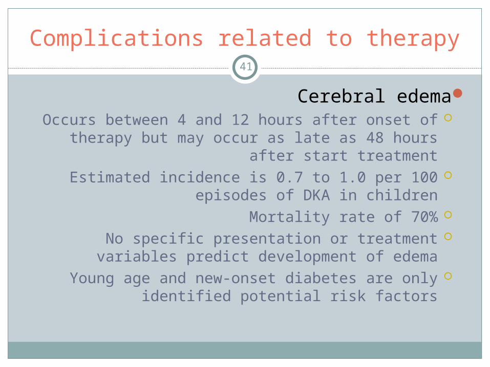
Complications related to therapy41
Cerebral edema Occurs between 4 and 12 hours after onset of therapy
but may occur as late as 48 hours after start treatmentEstimated incidence is 0.7 to 1.0 per 100 episodes of
DKA in childrenMortality rate of 70%No specific presentation or treatment variables predict
development of edemaYoung age and new-onset diabetes are only identified
potential risk factors
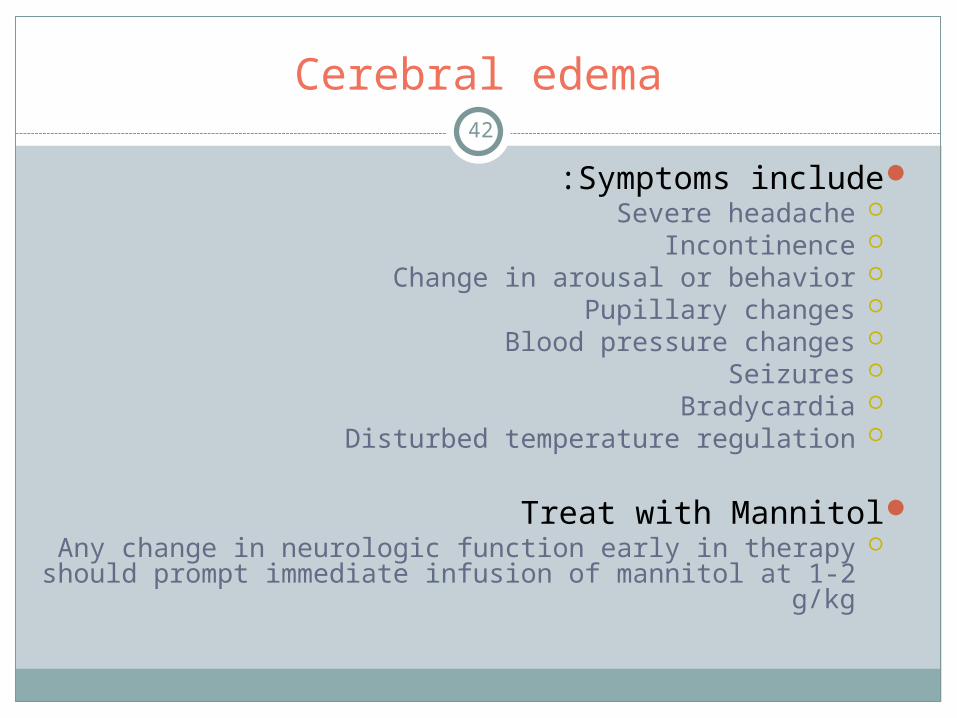
Cerebral edema42
Symptoms include:Severe headacheIncontinenceChange in arousal or behaviorPupillary changesBlood pressure changesSeizuresBradycardiaDisturbed temperature regulation
Treat with MannitolAny change in neurologic function early in therapy should
prompt immediate infusion of mannitol at 1-2 g/kg
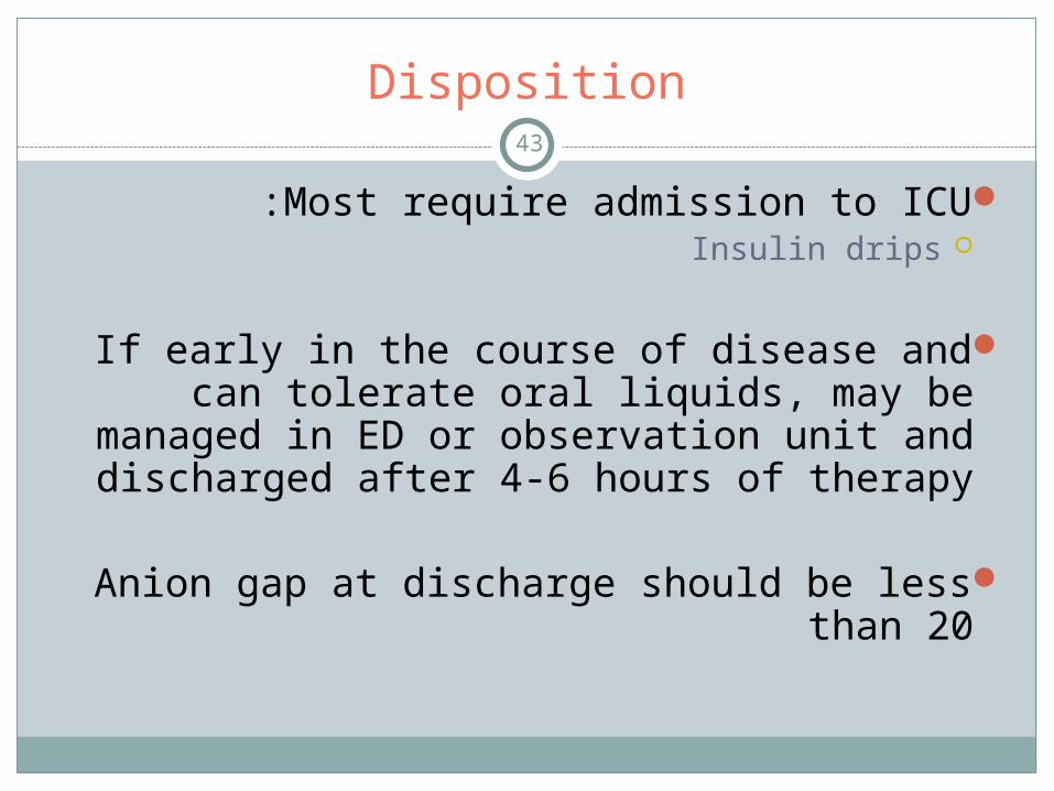
Disposition43
Most require admission to ICU:Insulin drips
If early in the course of disease and can tolerate oral liquids, may be managed in ED or observation unit and discharged after 4-6
hours of therapy
Anion gap at discharge should be less than 20
44
Alcoholic Ketoacidosis
Alcoholic Ketoacidosis45
Wide anion gap acidosis
Most often associated with acute cessation of alcohol consumption after chronic alcohol abuse
Metabolism of alcohol with little or no glucose sources results in elevated levels of ketoacids that typically
produce metabolic acidosis present in the illness
Usually seen in chronic alcoholics but may be seen in first time drinkers who binge drink, especially in those
with volume depletion from poor oral intake and vomiting
Epidemiology46
No gender difference
Usually presents between age 20 to 60
Many with repeated episodes of ketoacidosis
Incidence is unknown but mirrors incidence of alcoholism
Usually self-limited
Poor outcomes may occur
7-25% of deaths of known alcoholics due to AKA
Pathophysiology47
Key featuresIngestion of large quantities of alcoholRelative starvationVolume depletion
Pathophysiology48
Pathophysiologic state occurs with:Depletion of NADAerobic metabolism in the Krebs cycle is inhibitedGlycogen stores are depleted and lipolysis is
stimulated
Occurs in patients with:Recently intoxicatedVolume-contractionPoor nutritionUnderlying liver disease
Pathophysiology49
Insulin secretion is suppressed
Glucagon, catecholamines, and growth hormone are all stimulated
Aerobic metabolism is inhibited and anaerobic metabolism causes lipolysis and ketones are
produced
β-hydroxybutyrate is increased
More ketones are produced with malnourishment and vomiting or with hypophosphatemia
Clinical Features50 Usually occurs after episode of heavy drinking followed by
decrease in alcohol and food intake and vomiting
Nausea, vomiting and abdominal pain of gastritis and pancreatitis may exacerbate progression of illness
With anorexia continuing, symptoms worsen leading to seeking medical help
Symptoms are nonspecific and diagnosis is difficult without labs
No specific physical findings solely with AKAMost commonly tachycardia, tachypnea, diffuse mild to moderate
abdominal tendernessVolume depletion resulting from anorexia, diaphoresis and
vomiting may explain the tachycardia and hypotension
Clinical Features51
Most are alertMental status changes in patients with ketoacidosis
should alert to other causes:Toxic ingestionHypoglycemiaAlcohol-withdrawal seizuresPostictal stateUnrecognized head injury
Labs
52 EtOH levels usually low or undetectableSome may have elevated levels
Elevated anion gap caused by ketones is essential in diagnosis
Since β hydroxybutyrate predominates, degree of ketonemia may not be appreciated
Initial anion gap is 16-33 usually, mean of 21
Frequently mild hypophosphatemia, hyponatremia and/or hypokalemia
Severe derangements are rare
Labs
53Most have elevated bilirubin and liver enzymes
due to liver disease from chronic EtOH use
BUN and creatine kinase are frequently elevated due to relative volume depletion
Serum lactate mildly elevated
Glucose usually mildly elevated Some have hypoglycemiaRarely glucose greater than 200 mg/dL
Acid-Base Balance54
Need to evaluate the anion gap in every patient at risk for AKA
Diagnosis can easily be missed otherwise
Anion gap greater than baseline or 15 signifies a wide-anion-gap acidosis regardless
of bicarbonate concentration or pH, even if alkalemic
ABG not needed to arrive at correct diagnosis
Acid-Base Balance55
Serum pH usually acidemic (55% of time) though may be normal or alkalemic early in
course of disease
Degree of acidosis typically less than in DKA
Since volume loss is virtually always present, some degree of metabolic acidosis is present
Ketones56
Clinical application is variable
Most ketones in AKA are β-hydroxybutyrate The serum and urine nitroprusside test for ketones detects
acetoacetate and may show only mildly elevated ketones
As treatment progresses the acetoacetate will increase and indicates improving condition
Most suggest measuring β-hydroxybutyrate and acetoacetate only if diagnosis is unclear or other
methods are not available to follow patient’s response to therapy
Diagnosis57
May be established with classic presentation of:
The chronic alcoholic with:Recent anorexiaVomitingAbdominal painUnexplained metabolic acidosis with a positive
nitroprusside test, elevated anion gap and a low or mildly elevated serum glucose level
Classic Presentation is Uncommon
58
Difficult to establish diagnosis
Blood alcohol level may be zero
May not provide history of alcohol consumption
Urine nitroprusside testing may be negative or weakly positive despite significant ketoacidosis
pH may vary from significant acidemia to mild alkalemia
Wide anion gap is variable
Initial studies59 Electrolytes
BUNCreatinineLiver enzymesPancreatic enzymesWBC countHematocritUrinalysis Calculate anion gapSerum lactic acid level and serum osmolarity
may be helpful if diagnosis is in doubtABG is unnecessary unless a primary
respiratory acid-base disturbance is suspected
Differential diagnosis60
Very broadSame as for wide-anion-gap metabolic acidosis
Lactic acidosisUremia Ingestions such as:
MethanolEthylene glycol
Methanol and ethylene glycol do not produce ketosis but do have severe acidosis
Absence of urinary ketones cannot exclude diagnosis of AKA if concurrent methanol or ethylene glycol ingestion is suspected
Isopropyl alcohol ingestion Produces ketones and may have mild lactic acidosis
Salicylate poisoningSepsisRenal failureDKAStarvation ketosis
Concurrent Illnesses Promoting Alcohol Cessation and Anorexia61
Need to evaluate for these illnesses:PancreatitisGastritisUpper GI bleedingSeizuresAlcohol withdrawalPneumoniaSepsis Hepatitis
Treatment62
Glucose administration and volume repletionFluid of choice is D5NSGlucose stimulates insulin production, stopping
lipolysis and halts further formation of ketonesGlucose increases oxidation of NADH to NAD and
further limits ketone production
Patients are not hyperosmolar
Cerebral edema is not a concern with large volumes of fluid administration
Treatment63
Insulin No proven benefit May be dangerous as patients have depleted glycogen
stores and normal or low glucose levels
Treatment64
Sodium bicarbonate is not indicated unless patients are severely acidemic with pH 7.1 or
lowerThis level of acidemia not likely explained by AKA aloneVigorous search for alternate explanation must be
undertaken
Treatment65
Hypophosphatemia Frequently seen in alcoholic patientsCan retard resolution of acidosis
Phosphorous is necessary for mitochondrial utilization of glucose to produce NADH oxidation
Phosphate replacement is generally unwarranted in ED unless levels less than 1 are encountered
Oral replenishment is safe and effective
Treatment66 Nitroprusside tests useful because as become more positive
signifies improvement
To prevent theoretical progression to Wernicke’s disease, all patients should receive 50-100 mg of thiamine prior to
administration of glucose
Concomitant administration of magnesium sulfate and multivitamins should be considered and guided by laboratory
results
Acidosis may clear within 12-24 hours
If uncomplicated ED course, may be safely discharged if resolution of acidosis over time and patient able to tolerate oral
fluids
If complicated course, underlying illness or persistent acidosis, admit for further evaluation and treatment
67
Hyperosmolar Hyperglycemic State
Hyperosmolar Hyperglycemic State
68
Syndrome of severe hyperglycemia, hyperosmolarity and relative lack of ketonemia in
patients with poorly uncontrolled DM type II
ADA uses hyperosmolar hyperglycemic state (HHS) and hyperosmolar hyperglycemic non ketotic
syndrome (HHNS)Both commonly used and appropriate
Frequently referred to as non ketotic hyperosmolar coma
Coma should not be used in nomenclatureOnly 10 % present with coma
HHNS: Epidemiology69
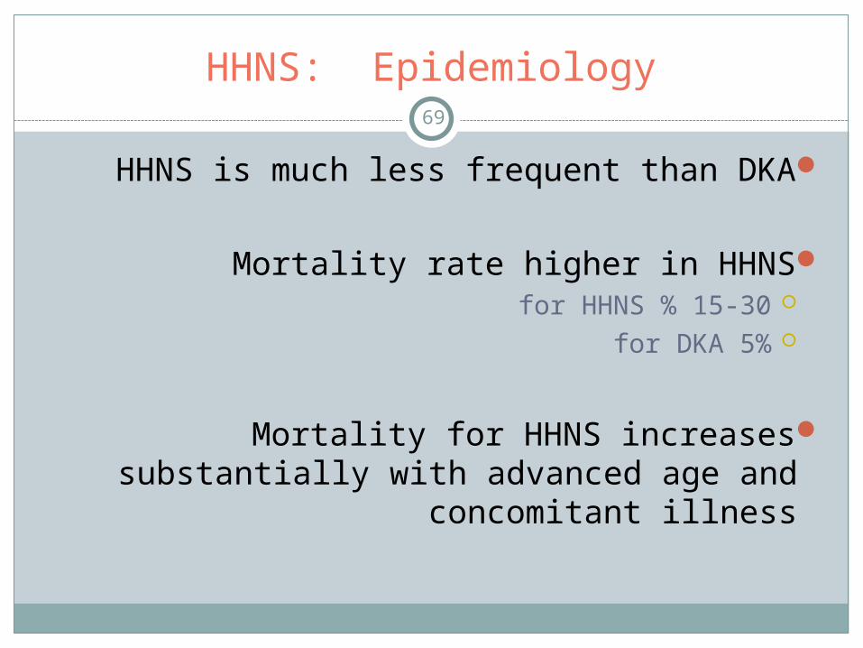
HHNS is much less frequent than DKA
Mortality rate higher in HHNS15-30 % for HHNS5% for DKA
Mortality for HHNS increases substantially with advanced age and concomitant illness
Hyperosmolar Hyperglycemic State
70
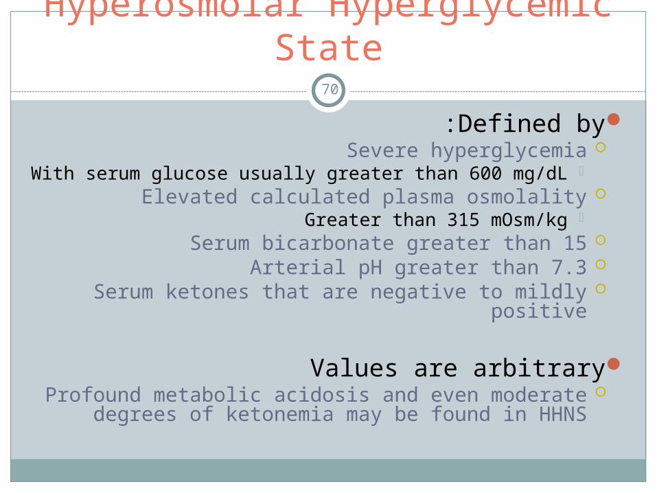
Defined by:Severe hyperglycemia
With serum glucose usually greater than 600 mg/dLElevated calculated plasma osmolality
Greater than 315 mOsm/kgSerum bicarbonate greater than 15Arterial pH greater than 7.3 Serum ketones that are negative to mildly positive
Values are arbitraryProfound metabolic acidosis and even moderate
degrees of ketonemia may be found in HHNS
HHNS and DKA both71
Hyperglycemia
Hyperosmolarity
Severe volume depletion
Electrolyte disturbances
Occasionally acidosis

HHNS72
Acidosis in HHNS more likely due to:Tissue hypoperfusion
Lactic acidosisStarvation ketosis Azotemia
HHNS and DKA Lipolysis73
DKA patients have much higher levels of lipolysis
Release and subsequent oxidation of free fatty acids to ketone bodies
β hydroxybutyrate and AcetoacetateContribute additional anions resulting in a more profound
acidosis
Inhibition of lipolysis and free fatty acid metabolism in HHNS is poorly understood
See table 214-1 on page 1307
HHNS: Pathophysiology74
Three main factors :Decreased utilization of insulinIncreased hepatic gluconeogenesis and glycogenolysisImpaired renal excretion of glucose
Identification early of those at risk for HHNS is most effective means of preventing serious complications
Must be vigilant on helping those who are non-ambulatory with inadequate hydration status
Fundamental risk factor for developing HHNS is impaired access to water
HHNS: Pathophysiology75
With poorly controlled DM II, inadequate utilization of glucose due to insulin resistance results in
hyperglycemia
Absence of adequate tissue response to insulin results in hepatic glycogenolysis and
gluconeogenesis resulting in further hyperglycemia
As serum glucose increases, an osmotic gradient is produced attracting water from the intracellular
space and into the intravenous compartment
HHNS: Pathophysiology76
Initial increase in intravascular volume is accompanied by a temporary increase in the GFR
As serum glucose concentration exceeds 180 mg/dL, capacity of kidneys to reabsorb glucose is exceeded
and glucosuria and a profound osmotic diuresis occurs
Patients with free access to water are often able to prevent profound volume depletion by replacing lost
water with large free water intake
If water requirement is not met, volume depletion occurs
HHNS: Pathophysiology77
During osmotic diuresis, urine produced is markedly hypertonic
Significant loss of sodium and potassium and modest loss of calcium, phosphate, magnesium and urea also
occur
As volume depletion progresses, renal perfusion decreases and GFR is reduced
Renal tubular excretion of glucose is impaired which further worsens the hyperglycemia
A sustained osmotic diuresis may result in total body water losses that often exceeds 20-25% of total body
weight or approximately 8-12 L in a 70 kg person
HHNS: Pathophysiology78
Absence of ketosis in HHNS not clearly understood
Some degree of starvation does occur but a clinically significant ketoacidosis does not occur
Lack of ketoacidosis may be due to:Lower levels of counter regulatory hormonesHigher levels of endogenous insulin that strongly
inhibits lipolysisInhibition of lipolysis by the hyperosmolar state

HHNS: Pathophysiology79
Controversy how counter regulatory hormones glucagons and cortisol, growth hormone and
epinephrine play in HHNSCompared to DKA, glucagon and growth hormone levels are
lower and this may help prevent lipolysis
Compared to DKA, significantly higher levels of insulin are found in peripheral and portal circulation in HHNS
Though insulin levels are insufficient to overcome hyperglycemia, they appear to be sufficient to overcome
lipolysis
Animal studies have shown the hyperosmolar state and severe hyperglycemia inhibit lipolysis in adipose
tissue
HHNS: Clinical Features80
Typical patient is usually elderlyOften referred by a caretaker
Abnormalities in vital signs and or mental status
May complain of:WeaknessAnorexiaFatigueCoughDyspneaAbdominal pain
HHNS81
Many have undiagnosed or poorly controlled type II diabetes
Precipitated by acute illnessPneumonia and urinary tract infections account for 30-
50% of cases
Noncompliance with or under-dosing of insulin has been identified as a common precipitant also
HHNS82
Those predisposed to HHNS often have some level of baseline cognitive impairment such as senile
dementia Self-referral for medical treatment in early stages is rare
Any patient with hyperglycemia, impaired means of communication and limited access to free water is
at major risk for HHNS
Presence of hypertension, renal insufficiency or cardiovascular disease is common in this patient
population and medications commonly used to treat these diseases such as blockers predispose the
development of HHNS
HHNS83
An insidious state goes uncheckedProgressive hyperglycemia Hyperosmolarity Osmotic diuresis
Alterations in vital signs and cognition follow and signal a severity of illness that is often
advanced
HHNS Causes84
A host of metabolic and iatrogenic causes have been identified
DiabetesParental or enteral alimentationGI bleedPEPancreatitisHeat-related illnessMesenteric ischemiaInfectionMI
HHNS Causes85
Severe burnsRenal insufficiencyPeritoneal or hemodialysisCerebrovascular eventsRhabdomyolysisCommonly prescribed drugs that may
predispose to hyperglycemia, volume depletion or other effects leading to HHNS
HHNS may unexpectedly be found in non-diabetics who present with an acute medical
insult such as CVA, severe burns, MI, infection, pancreatitis or other acute illness

HHNS: Physical findings86 Non-specific
Clinical signs of volume depletion:Poor skin turgorDry mucus membranesSunken eyeballsHypotension
Signs correlate with degree of hyperglycemia and hyperosmolality and duration of physiologic imbalance
Wide range of findings such as changes in vital signs and cognition to clear evidence of profound shock and coma may
occur
Normothermia or hypothermia is common due to vasodilation
HHNS: Physical findings87
SeizuresUp to 15% may present with seizuresTypically focalGeneralized seizures that are often resistant to
anticonvulsants may occur
Other CNS symptoms may include:TremorClonusHyperreflexiaHyporeflexiaPositive plantar responseReversible hemiplegia or hemisensory defects
without CVA or structural lesion
HHNS: Physical findings88
Degree of lethargy and coma is proportional to the level of osmolality
Those with coma tend to have:Higher osmolalityHigher hyperglycemia Greater volume contraction
Not surprising that misdiagnosis of stroke or organic brain disease is common in the
elderly
Laboratory tests89
EssentialSerum glucoseElectrolytesCalculated and measured serum osmolalityBUN KetonesCreatinineCBC
Laboratory tests90 Consider
Urinalysis and cultureLiver and pancreatic enzymesCardiac enzymesThyroid functionCoagulation profilesChest x-rayECG
OtherCT of headLPToxicology ABG
Of value only if suspicion of respiratory component to acid-base abnormality
Both PCO2 and pH can be predicted from bicarbonate concentration obtained from venous electrolytes
Electrolyte abnormalities91
Electrolyte abnormalities usually reflect a contraction alkalosis due to profound water deficit
50% of patients with HHNS will have increased anion gap metabolic acidosis
Lactic acidosis, azotemia, starvation ketosis, severe volume contraction
Acute or concurrent illnesses such as ischemic bowel will contribute anions such as lactic acid causing varying degrees of an anion gap
metabolic acidosis
Initial serum electrolyte determinations can be reported as seemingly normal because the concurrent presence of both metabolic alkalosis
and acidosis may result in each canceling out the other’s effect
Lack of careful analysis of serum chemistries may lead to delayed appreciation of the severity of underlying abnormalities, including
volume loss
Sodium92
Serum sodium is suggestive but not a reliable indicator of degree of volume contraction
Though patient is total body sodium depleted, serum sodium (corrected for glucose elevation) may be low, normal or elevated
Measured serum sodium is often reported as factitiously low due to dilutional effect of hyperglycemia
Need to correct the sodium level
Serum sodium decreases by 1.6 mEq for every 100 mg/dL increase in serum glucose above 100 mg/dL
See formula page 1309
Elevated corrected serum sodium during sever hyperglycemia is usually explainable only by profound volume contraction
Normal sodium level or mild hyponatremia usually but not invariably suggests modest dehydration

Osmolarity93
Serum osmolarity has also been shown to correlate with severity of disease as well as neurologic impairment and coma
Calculated effective serum osmolarity excludes osmotically inactive urea that is usually included in laboratory measures of
osmolarity
See formula page 1309
Normal serum osmolarity range is approximately 275 to 295 mOsm/kg
Values above 300 mOsm/kg are indicative of significant hyperosmolarity and those above 320 mOsm are commonly
associated with alterations of cognitive function
Potassium
94 Hypokalemia is most immediate electrolyte based risk and should be anticipated
Total body deficits of 500-700 mEq/l are common
Initial values may be reported as normal during a period of severe volume contraction and with metabolic acidosis when intravascular
hydrogen ions are exchanged for intracellular potassium ions
Presence of acidemia may mask a potentially life-threatening potassium deficit
As intravascular volume is replaced and acidemia is reversed, potassium losses become more apparent
Patients with low serum potassium during the period of severe volume contraction are at greatest risk for dysrhythmia
Importance of potassium replacement during periods of volume repletion and insulin therapy cannot be overemphasized
Labs95
BUN and CrBoth prerenal azotemia and renal azotemia are common
with BUN/Cr ratios often exceeding 30/1
WBCLeukocytosis is variable and a weak clinical indicatorWhen present usually due to infection or
hemoconcentration
Phosphate96
Hypophosphatemia may occur during periods of prolonged hyperglycemia
Acute consequences such as CNS abnormalities, cardiac dysfunction, and rhabdomyolysis are rare and are usually if level is
<1.0 mg/dL
Routine replacement of phosphate and magnesium usually unnecessary unless severe
Both electrolytes tend to normalize as metabolic derangements are addressed
When necessary, gradual replacement minimizes risks of complications such as renal failure or hypocalcemia
Metabolic acidosis is of a wide-anion-gap type, often due to lactic acidosis from poor tissue perfusion, resulting in uremia, mild
starvation ketosis or all three
Treatment97
Improvement in tissue perfusion is the key to effective recovery
Treat hypovolemia, identify and treat precipitating causes, correct electrolyte abnormalities, gradual
correction of hyperglycemia and osmolarity
Cannot overstate importance of judicious therapeutic plans that adjusts for concurrent medical illness such
as LV dysfunction or renal insufficiency
Due to potential complications, rapid therapy should only be reserved for potentially life-threatening
electrolyte abnormalities only
Figure 214-1
Fluid resuscitation98
Initial aim is reestablishing adequate tissue perfusion and decreasing serum glucose
Replacement of intravascular fluid losses alone can account for reductions in serum glucose of 35-70 mg/hr
or up to 80 % of necessary reduction
Average fluid deficit is 20-25% of total body water or 8-12 L
In elderly 50% of body weight is due to total body water
Calculate the water deficit by using patient’s current weight in kilograms and normal total body water

Fluid resuscitation99
One-half of fluid deficits should be replaced over the initial 12 hours and the balance over the next 24 hours when possible
Actual rate of fluid administration should be individualized for each patient based on presence of renal and cardiac
impairment
Initial rates of 500-1500 ml/hr during first 2 hours followed by rates of 250-500 ml per hour are usually well tolerated
Patients with cardiac disease may require a more conservative rate of volume repletion
Renal and cardiovascular function should be carefully monitored
Central venous and urinary tract catheterization should be considered
Fluid resuscitation100
Rate of fluid administration may need to be limited in children
A limited number of reports of cerebral edema occurring during or soon after the resuscitation phase of patients with both DKA and
HHNS have been described
Most cases have occurred in children with DKA and mechanism is unclear
One review showed cerebral edema was found with similar frequency before treatment with replacement fluids
New study shows rehydration of children with DKA during first 4 hours at a rate greater than 50 mL/kg was associated with
increased risk of brain herniation
Little credible data on incidence or clinical indicators that may predispose to cerebral edema in HHNS patients
Fluid resuscitation101
Current recommendations based on available data include limiting rate of volume depletion during first 4 hours to <50 ml/kg of NS
Mental status should be closely monitored during treatment
CT of brain should be obtained with any evidence of cognitive impairment
Most authors agree use of NS is most appropriate initial crystalloid for replacement of intravascular volume
NS is hypotonic to the patient’s serum osmolality and will more rapidly restore plasma volume
Once hypotension, tachycardia and urinary output improve, ½ NS can be used to replace the remaining free water deficit
Potassium102
Potassium deficits are most immediate electrolyte-based risk for a bad outcome
On average potassium losses range from 4-6 mEq/kg though may be as high as 10mEq/kg of body weight
Initial measurements may be normal or even high with acidemia
Patients with levels <3.3 are at highest risk for cardiac dysrhythmia and respiratory arrest and should be
treated with urgency
Insulin therapy precipitously lowers intravascular potassium further and potassium must be vigorously
replaced
Potassium103
When adequate urinary output is assured, potassium replacement should begin
Should replace at 10-20 mEq/hr though if life threatening may require 40 mEq/hr
Central line needed if given more than 20 mEq/hr
Some believe potassium through central line poses risk for conduction defects and should be avoided if
good peripheral line sites are available
Monitoring of serum potassium should occur every hour until a steady state has been achieved
Sodium104
Sodium deficits replenished rapidly since given NS or ½ NS during fluid replacement
Phosphate and Magnesium should be measured
Current guideline recommend giving 1/3 of potassium needed as potassium phosphate to avoid excessive
chloride administration and to prevent hypophosphatemia
Unless severe, alleviation of hypophosphatemia or hypomagnesemia should occur after the patient is
admitted into the ICU setting
Insulin105
Volume repletion should precede insulin therapy
If given before volume repletion, intravascular volume is further depleted due to shifting of osmotically active glucose into the intracellular space bringing free water
with it and this may precipitate vascular collapse
Absorption of insulin by IM or SC route is unreliable in patients with HHNS and continuous infusion of IV
insulin is needed
No proven benefit to bolus of insulin
Continuous infusion of 0.1U/kg/hour is best
Insulin106
Want one unit of regular insulin for every mL of NS in infusion
Steady states utilizing infusion pumps occur within 30 minutes of infusion
Decrease plasma glucose by 50-75 mg/dL per hour along with adequate hydration
If adequate hydration, may double infusion rate until 50-75 mg/dL/hr is achieved
Some patients are insulin resistant and require higher doses
Once level less than 300 mg/dL, should change IV solution to D5 ½ NS and insulin infusion should be reduced to half or
0.05 U/kg/hr.
Disposition107
Need to track pH, vital signs and key lab values in the ED for appropriate management
and disposition of these patients
ICUMost require for initial 24 hours of care
SDUPatients with no significant co morbid conditions and
who demonstrate a good response to initial therapy as evidenced by documented improvement in vital signs,
urine output, electrolyte balance and mentation
Questions108
1 .T/F: The venous pH is just as helpful as arterial pH in patients with DKA and may be obtained during routine blood draws .
2 .T/F: Alcoholic ketoacidosis is usually seen in chronic alcoholics but may be seen in first time drinkers who binge drink, especially in those
with volume depletion from poor oral intake and vomiting .
3 .T/F: In treating DKA, the order of therapeutic priorities is volume first, then insulin and/or potassium, magnesium and bicarbonate .
4 .T/F: DKA patients have much higher levels of lipolysis, resulting in release and subsequent oxidation of free fatty acids to ketone bodies
contributing additional anions resulting in a more profound acidosis than in HHNS .
5 .T/F: Volume repletion should precede insulin therapy in HHNS
Answers: T,T,T,T,T