Embed Size (px)
Citation preview
Our reference: JPB 9631 P-authorquery-v11
AUTHOR QUERY FORM
Journal: JPB Please e-mail or fax your responses and any corrections to:
Article Number: 9631
E-mail: [email protected]
Fax: +31 2048 52799
Dear Author,
Please check your proof carefully and mark all corrections at the appropriate place in the proof (e.g., by using on-screen annotation in the PDF
file) or compile them in a separate list. Note: if you opt to annotate the file with software other than Adobe Reader then please also highlight the appropriate place in the PDF file. To ensure fast publication of your paper please return your corrections within 48 hours.For correction or revision of any artwork, please consult http://www.elsevier.com/artworkinstructions.
Any queries or remarks that have arisen during the processing of your manuscript are listed below and highlighted by flags in the proof. Clickon the ‘Q’ link to go to the location in the proof.
Location inarticle
Query / Remark: click on the Q link to goPlease insert your reply or correction at the corresponding line in the proof
Q1 Please confirm that given name(s) and surname(s) have been identified correctly.
Thank you for your assistance.
Please check this box if you have nocorrections to make to the PDF file
Highlights
JPB 9631 No. of Pages 1, Model 5G
18 December 2013
� Nitrogen doping strongly shifts the absorption edge of titania from UV to visible region. � Sulpur doping decreases the crystallinity of titaniawhereas nitrogen does not affect it. � Nitrogen occupies at both substitutional and interstitial position of titania lattice. � N-TiO2 exhibitssuperior water disinfection property than S-TiO2 and TiO2.
1
1
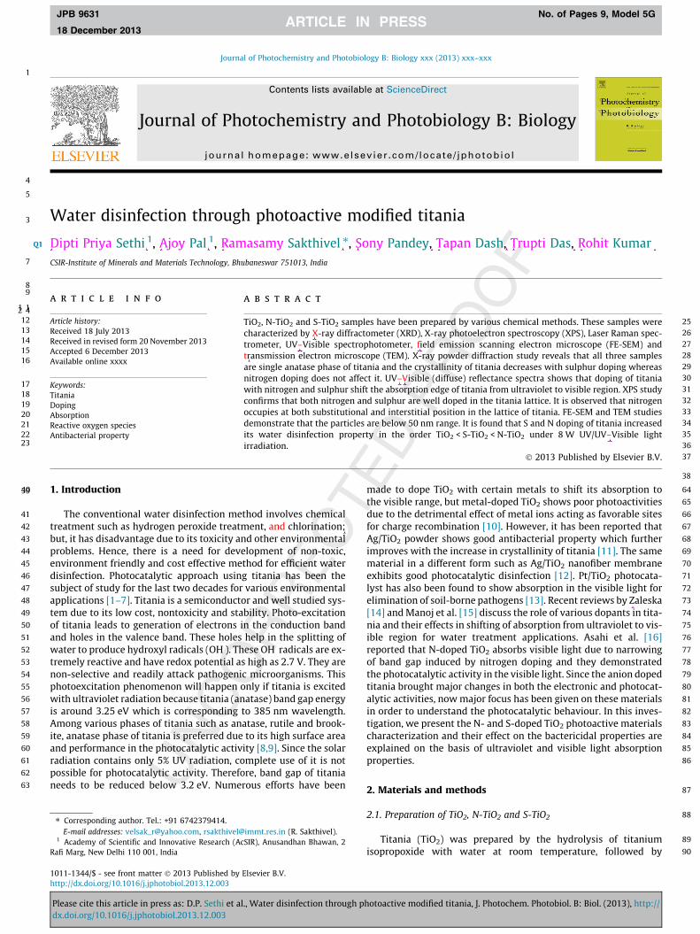
3 Water disinfection through photoactive modified titania
4
5
6 Dipti Priya Sethi 1, Ajoy Pal 1, Ramasamy Sakthivel ⇑, Sony Pandey, Tapan Dash, Trupti Das, Rohit Kumar7 CSIR-Institute of Minerals and Materials Technology, Bhubaneswar 751013, India
89
1 1a r t i c l e i n f o
12 Article history:13 Received 18 July 201314 Received in revised form 20 November 201315 Accepted 6 December 201316 Available online xxxx
17 Keywords:18 Titania19 Doping20 Absorption21 Reactive oxygen species22 Antibacterial property23
2 4a b s t r a c t
25TiO2, N-TiO2 and S-TiO2 samples have been prepared by various chemical methods. These samples were26characterized by X-ray diffractometer (XRD), X-ray photoelectron spectroscopy (XPS), Laser Raman spec-27trometer, UV–Visible spectrophotometer, field emission scanning electron microscope (FE-SEM) and28transmission electron microscope (TEM). X-ray powder diffraction study reveals that all three samples29are single anatase phase of titania and the crystallinity of titania decreases with sulphur doping whereas30nitrogen doping does not affect it. UV–Visible (diffuse) reflectance spectra shows that doping of titania31with nitrogen and sulphur shift the absorption edge of titania from ultraviolet to visible region. XPS study32confirms that both nitrogen and sulphur are well doped in the titania lattice. It is observed that nitrogen33occupies at both substitutional and interstitial position in the lattice of titania. FE-SEM and TEM studies34demonstrate that the particles are below 50 nm range. It is found that S and N doping of titania increased35its water disinfection property in the order TiO2 < S-TiO2 < N-TiO2 under 8 W UV/UV–Visible light36irradiation.37� 2013 Published by Elsevier B.V.
38
3940 1. Introduction
41 The conventional water disinfection method involves chemical42 treatment such as hydrogen peroxide treatment, and chlorination;43 but, it has disadvantage due to its toxicity and other environmental44 problems. Hence, there is a need for development of non-toxic,45 environment friendly and cost effective method for efficient water46 disinfection. Photocatalytic approach using titania has been the47 subject of study for the last two decades for various environmental48 applications [1–7]. Titania is a semiconductor and well studied sys-49 tem due to its low cost, nontoxicity and stability. Photo-excitation50 of titania leads to generation of electrons in the conduction band51 and holes in the valence band. These holes help in the splitting of52 water to produce hydroxyl radicals (OH�). These OH� radicals are ex-53 tremely reactive and have redox potential as high as 2.7 V. They are54 non-selective and readily attack pathogenic microorganisms. This55 photoexcitation phenomenon will happen only if titania is excited56 with ultraviolet radiation because titania (anatase) band gap energy57 is around 3.25 eV which is corresponding to 385 nm wavelength.58 Among various phases of titania such as anatase, rutile and brook-59 ite, anatase phase of titania is preferred due to its high surface area60 and performance in the photocatalytic activity [8,9]. Since the solar61 radiation contains only 5% UV radiation, complete use of it is not62 a63 n
64made to dope TiO2 with certain metals to shift its absorption to65the visible range, but metal-doped TiO2 shows poor photoactivities66due to the detrimental effect of metal ions acting as favorable sites67for charge recombination [10]. However, it has been reported that68Ag/TiO2 powder shows good antibacterial property which further69improves with the increase in crystallinity of titania [11]. The same70material in a different form such as Ag/TiO2 nanofiber membrane71exhibits good photocatalytic disinfection [12]. Pt/TiO2 photocata-72lyst has also been found to show absorption in the visible light for73elimination of soil-borne pathogens [13]. Recent reviews by Zaleska74[14] and Manoj et al. [15] discuss the role of various dopants in tita-75-76]77g78d79d80t-81ls82-83ls84e85n86
87
88
89
90y2
Q1
Journal of Photochemistry and Photobiology B: Biology xxx (2013) xxx–xxx
Contents lists available at ScienceDirect
Journal of Photochemistry and Photobiology B: Biology
journal homepage: www.elsevier .com/locate / jphotobiol
JPB 9631 No. of Pages 9, Model 5G
18 December 2013
possible for photocatalytic activity. Therefore, band gap of titanineeds to be reduced below 3.2 eV. Numerous efforts have bee
1011-1344/$ - see front matter � 2013 Published by Elsevier B.V.http://dx.doi.org/10.1016/j.jphotobiol.2013.12.003
⇑ Corresponding author. Tel.: +91 6742379414.E-mail addresses: [email protected], [email protected] (R. Sakthivel).
1 Academy of Scientific and Innovative Research (AcSIR), Anusandhan Bhawan,Rafi Marg, New Delhi 110 001, India
Please cite this article in press as: D.P. Sethi et al., Water disinfection throughdx.doi.org/10.1016/j.jphotobiol.2013.12.003
nia and their effects in shifting of absorption from ultraviolet to visible region for water treatment applications. Asahi et al. [16reported that N-doped TiO2 absorbs visible light due to narrowinof band gap induced by nitrogen doping and they demonstratethe photocatalytic activity in the visible light. Since the anion dopetitania brought major changes in both the electronic and photocaalytic activities, now major focus has been given on these materiain order to understand the photocatalytic behaviour. In this investigation, we present the N- and S-doped TiO2 photoactive materiacharacterization and their effect on the bactericidal properties arexplained on the basis of ultraviolet and visible light absorptioproperties.
2. Materials and methods
2.1. Preparation of TiO2, N-TiO2 and S-TiO2
Titania (TiO2) was prepared by the hydrolysis of titaniumisopropoxide with water at room temperature, followed b
photoactive modified titania, J. Photochem. Photobiol. B: Biol. (2013), http://
91 se92 at93 to94 (S95 ou96 pe97 fil98 fo99 do
100of101we10213103at104te105ge106
107by108Ne1090.1110we111(FE
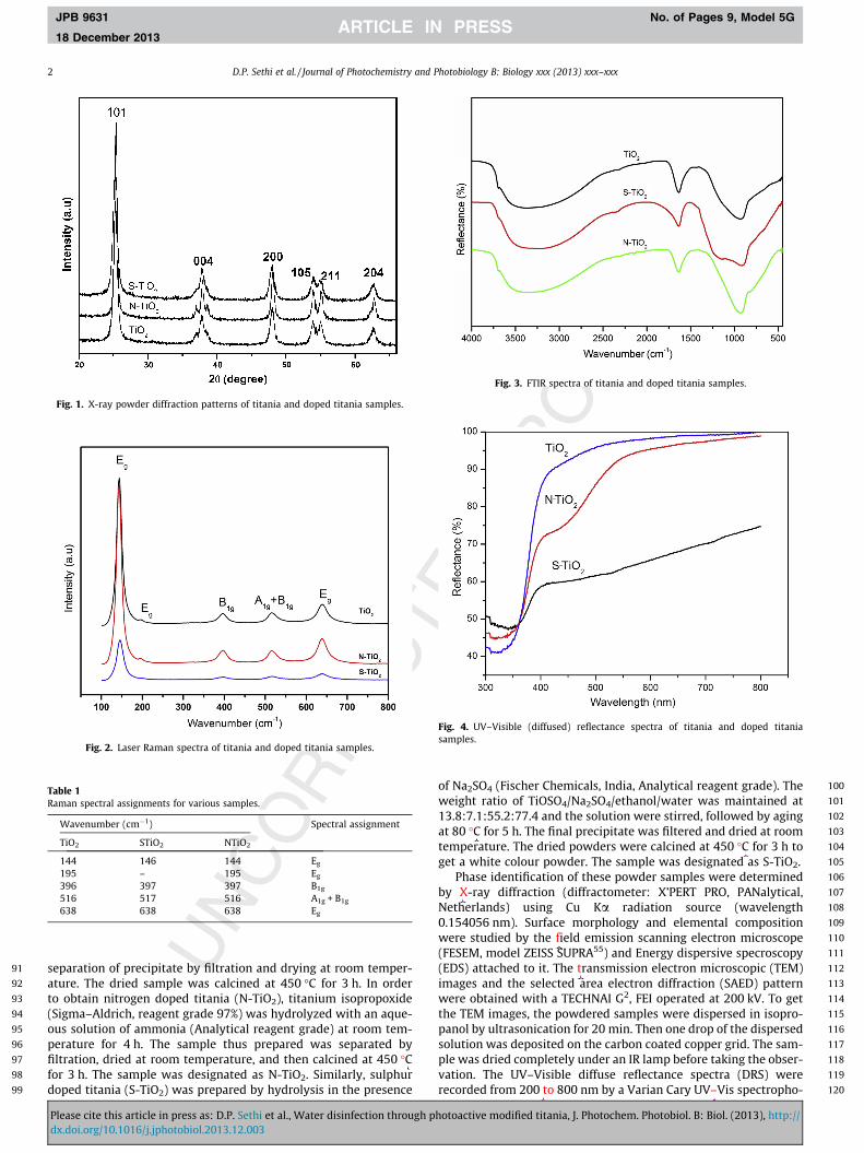
Fig. 1. X-ray powder diffraction patterns of titania and doped titania samples.
Fig. 2. Laser Raman spectra of titania and doped titania samples.
TabRam
Fig. 3. FTIR spectra of titania and doped titania samples.
Fig. 4. UV–Visible (diffused) reflectance spectra of titania and doped titaniasamples.
2 D.P. Sethi et al. / Journal of Photochemistry and Photobiology B: Biology xxx (2013) xxx–xxx
JPB 9631 No. of Pages 9, Model 5G
18 December 2013
Pldx
le 1an spectral assignments for various samples.
Wavenumber (cm�1) Spectral assignment
TiO2 STiO2 NTiO2
144 146 144 Eg
195 – 195 Eg
396 397 397 B1g
516 517 516 A1g + B1g
638 638 638 Eg
112(E113im114we115th116pa117so118pl119va120re
paration of precipitate by filtration and drying at room temper-ure. The dried sample was calcined at 450 �C for 3 h. In orderobtain nitrogen doped titania (N-TiO2), titanium isopropoxide
igma–Aldrich, reagent grade 97%) was hydrolyzed with an aque-s solution of ammonia (Analytical reagent grade) at room tem-rature for 4 h. The sample thus prepared was separated bytration, dried at room temperature, and then calcined at 450 �Cr 3 h. The sample was designated as N-TiO2. Similarly, sulphurped titania (S-TiO2) was prepared by hydrolysis in the presence
ease cite this article in press as: D.P. Sethi et al., Water disinfection through phot.doi.org/10.1016/j.jphotobiol.2013.12.003
Na2SO4 (Fischer Chemicals, India, Analytical reagent grade). Theight ratio of TiOSO4/Na2SO4/ethanol/water was maintained at.8:7.1:55.2:77.4 and the solution were stirred, followed by aging80 �C for 5 h. The final precipitate was filtered and dried at room
mperature. The dried powders were calcined at 450 �C for 3 h tot a white colour powder. The sample was designated as S-TiO2.Phase identification of these powder samples were determinedX-ray diffraction (diffractometer: X’PERT PRO, PANalytical,
therlands) using Cu Ka radiation source (wavelength54056 nm). Surface morphology and elemental compositionre studied by the field emission scanning electron microscopeSEM, model ZEISS SUPRA55) and Energy dispersive specroscopy
DS) attached to it. The transmission electron microscopic (TEM)ages and the selected area electron diffraction (SAED) patternre obtained with a TECHNAI G2, FEI operated at 200 kV. To get
e TEM images, the powdered samples were dispersed in isopro-nol by ultrasonication for 20 min. Then one drop of the dispersedlution was deposited on the carbon coated copper grid. The sam-e was dried completely under an IR lamp before taking the obser-tion. The UV–Visible diffuse reflectance spectra (DRS) werecorded from 200 to 800 nm by a Varian Cary UV–Vis spectropho-
oactive modified titania, J. Photochem. Photobiol. B: Biol. (2013), http://
121 tometer. The Raman spectra were taken in a dispersive type122 micro-Raman spectrometer (Renishaw in Via Reflex, UK). Fourier123 Transform Infrared (FTIR) spectra were recorded in the range of124 400–4000 cm�1 wavenumber on a Fourier Transform Infrared125 spectrophotometer supplied by Perkin Elmer India Pvt Ltd., Kolkata126 -127 G128 n129 e130 f131
132
133 t134 n135 f136 h137 e138 h
139E. coli as mentioned above. For each sample, the biocidal activity140was tested under UV and UV–Visible light. One ml of sample was141drawn from the reaction vessel at various intervals of time and142serially diluted up to 104 times. This was followed by plating1430.1 ml of the sample on Liquid Broth Agar (LBA) plates by adopting144-145s146it147-148149151151
152e153
154
155s156e157n
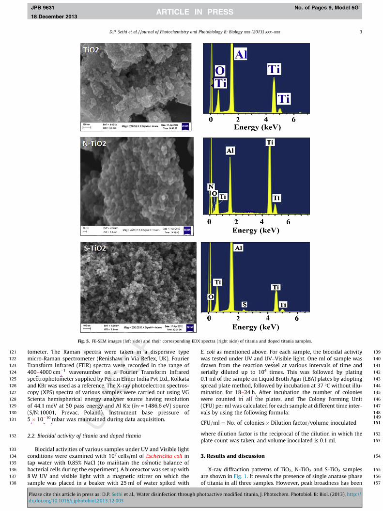
Fig. 5. FE-SEM images (left side) and their corresponding EDX spectra (right side) of titania and doped titania samples.
D.P. Sethi et al. / Journal of Photochemistry and Photobiology B: Biology xxx (2013) xxx–xxx 3
JPB 9631 No. of Pages 9, Model 5G
18 December 2013
and KBr was used as a reference. The X-ray photoelectron spectroscopy (XPS) spectra of various samples were carried out using VScienta hemispherical energy analyser source having resolutioof 44.1 meV at 50 pass energy and Al Ka (hm = 1486.6 eV) sourc(S/N:10001, Prevac, Poland). Instrument base pressure o5 � 10�10 mbar was maintained during data acquisition.
2.2. Biocidal activity of titania and doped titania
Biocidal activities of various samples under UV and Visible lighconditions were examined with 107 cells/ml of Escherichia coli itap water with 0.85% NaCl (to maintain the osmotic balance obacterial cells during the experiment). A bioreactor was set up wit8 W UV and visible light with a magnetic stirrer on which thsample was placed in a beaker with 25 ml of water spiked wit
Please cite this article in press as: D.P. Sethi et al., Water disinfection throughdx.doi.org/10.1016/j.jphotobiol.2013.12.003
spread plate method, followed by incubation at 37 �C without illumination for 18–24 h. After incubation the number of coloniewere counted in all the plates, and The Colony Forming Un(CFU) per ml was calculated for each sample at different time intervals by using the following formula:
CFU=ml ¼ No: of colonies� Dilution factor=volume inoculated
where dilution factor is the reciprocal of the dilution in which thplate count was taken, and volume inoculated is 0.1 ml.
3. Results and discussion
X-ray diffraction patterns of TiO2, N-TiO2 and S-TiO2 sampleare shown in Fig. 1. It reveals the presence of single anatase phasof titania in all three samples. However, peak broadness has bee
photoactive modified titania, J. Photochem. Photobiol. B: Biol. (2013), http://
158 observed in case of sulphur doped titania which indicates that sul-159 phur brings down the crystallinity as in case of SO4/ZrO2 [17]. The160 peak corresponding to h101i plane shows highest intensity among161 others peaks corresponding to 004, 200, 105, 211, 204 planes.162 The observed d values are matched with standard powder diffrac-163 tion data of anatase phase of titania (JCPDS File No. 01-089-4921).164 Laser Raman spectra of these samples are shown in Fig. 2. It shows165 bands around 144, 195, 396, 516, 638 cm�1 and these are ascribed166 fo167 sp168 in169 dr170 po171 tit172 cry
173XRD results. The change in crystallinity of the samples also affects174the bactericidal properties as discussed below. FTIR spectra of all175three samples (Fig. 3) show stretching vibration band for hydroxyl176group of adsorbed water molecules at 3000–3550 cm�1 and bend-177ing vibration band at 1630 cm�1. The bands observed at 3695 and178800 cm�1 are corresponding to respective stretching and bending179vib180[1181
182ha183TiO184an185ni186en187[2
Fig. 6. FE-SEM images (left side) and their corresponding EDX spectra (right side) of titania and doped titania samples.
4 D.P. Sethi et al. / Journal of Photochemistry and Photobiology B: Biology xxx (2013) xxx–xxx
JPB 9631 No. of Pages 9, Model 5G
18 December 2013
Pldx
r the Eg, B1g, A1g + B1g, modes of vibrations [18]. The observedectral bands position of all three samples and their correspond-g vibrations are given in Table 1. The intensity of spectral bandsastically reduced when sulphur is doped in titania indicatingor crystallinity of sample, however, when nitrogen is doped inania the intensity of these bands increases due to increase installinity of sample. This observation is in accordance with the
ease cite this article in press as: D.P. Sethi et al., Water disinfection through phot.doi.org/10.1016/j.jphotobiol.2013.12.003
ration of surface hydroxyl group present in all three samples9,20].Diffused (UV–Visible) reflectance spectra of all three samples
ve been shown in Fig. 4. It shows that the spectra of TiO2, N-2 and S-TiO2 samples are differing from each other. Nitrogen
d sulphur doping brought drastic change in the spectrum of tita-a. It has been reported that N or S doping decreases the band gapergy of titania leading to red shift in UV–Vis absorption band1,22]. Titania shows absorption around 390 nm wavelength
oactive modified titania, J. Photochem. Photobiol. B: Biol. (2013), http://
188 where as N-TiO2 shows very strong absorption around 450 and189 390 nm wavelength region. S-TiO2 sample shows a broad absorp-190 tion in the visible range between 400 and 800 nm wavelength191 and narrow absorption (390 nm) in the UV region. FE-SEM images192 and their corresponding EDS spectra of all three samples are shown193 in Fig. 5. It reveals that all these samples have particles size below194 50 nm, and they are distributed in very narrow range. This
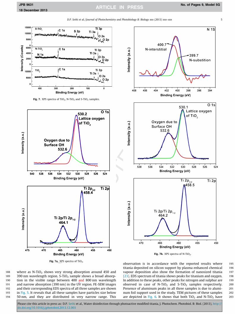
Fig. 7. XPS spectra of TiO2, N-TiO2 and S-TiO2 samples.
Fig. 7a. XPS spectra of TiO2.
D.P. Sethi et al. / Journal of Photochemistry and Photobiology B: Biology xxx (2013) xxx–xxx 5
JPB 9631 No. of Pages 9, Model 5G
18 December 2013
Please cite this article in press as: D.P. Sethi et al., Water disinfection throughdx.doi.org/10.1016/j.jphotobiol.2013.12.003
Fig. 7b. XPS spectra of N-TiO2.
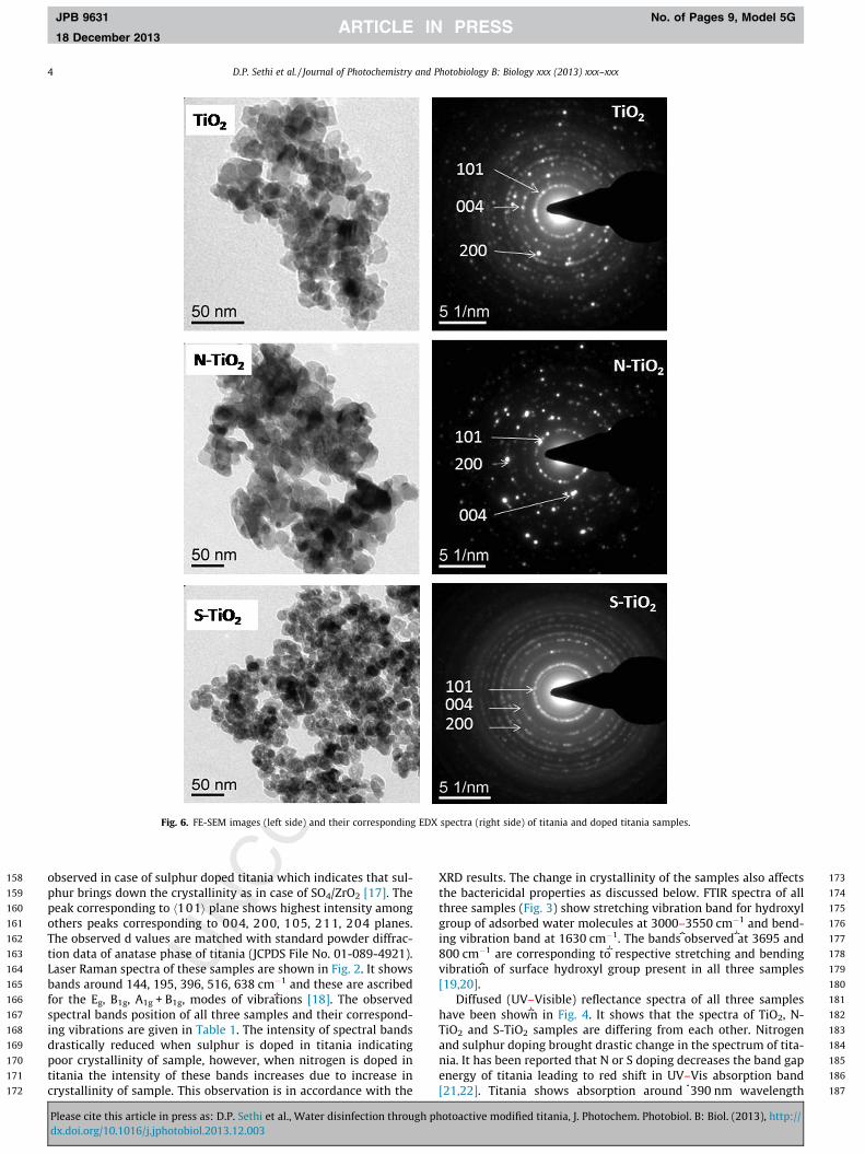
195observation is in accordance with the reported results where196titania deposited on silicon support by plasma enhanced chemical197vapour deposition also show the formation of nanosized titania198[23]. EDS spectrum of titania shows peaks for titanium and oxygen.199In addition to these peaks, other peaks for nitrogen and sulphur are200observed in case of N-TiO2 and S-TiO2 samples respectively.201Presence of aluminum peaks in all three samples is due to alumi-202num foil support used in the study. TEM pictures of these samples203are depicted in Fig. 6. It shows that both TiO2 and N-TiO2 have
photoactive modified titania, J. Photochem. Photobiol. B: Biol. (2013), http://
204sim205sp206in207an208pl209in210lin211m212
213Fig214be215sa216er217Ti
TabXP
e OH
Figsou
Figvis
6 D.P. Sethi et al. / Journal of Photochemistry and Photobiology B: Biology xxx (2013) xxx–xxx
JPB 9631 No. of Pages 9, Model 5G
18 December 2013
Pldx
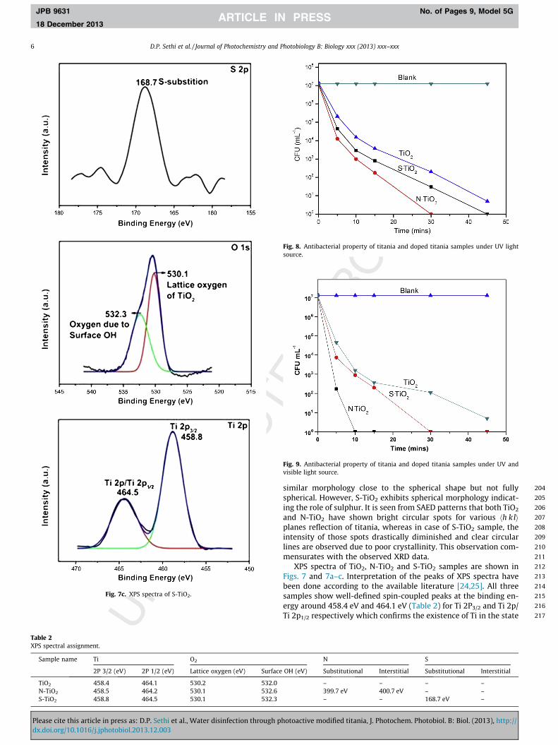
Fig. 7c. XPS spectra of S-TiO2.
le 2S spectral assignment.
Sample name Ti O2
2P 3/2 (eV) 2P 1/2 (eV) Lattice oxygen (eV) Surfac
TiO2 458.4 464.1 530.2 532.0N-TiO2 458.5 464.2 530.1 532.6S-TiO2 458.8 464.5 530.1 532.3
ease cite this article in press as: D.P. Sethi et al., Water disinfection through phot.doi.org/10.1016/j.jphotobiol.2013.12.003
ilar morphology close to the spherical shape but not fullyherical. However, S-TiO2 exhibits spherical morphology indicat-g the role of sulphur. It is seen from SAED patterns that both TiO2
d N-TiO2 have shown bright circular spots for various hh klianes reflection of titania, whereas in case of S-TiO2 sample, thetensity of those spots drastically diminished and clear circulares are observed due to poor crystallinity. This observation com-
ensurates with the observed XRD data.XPS spectra of TiO2, N-TiO2 and S-TiO2 samples are shown ins. 7 and 7a–c. Interpretation of the peaks of XPS spectra have
en done according to the available literature [24,25]. All threemples show well-defined spin-coupled peaks at the binding en-gy around 458.4 eV and 464.1 eV (Table 2) for Ti 2P3/2 and Ti 2p/2p1/2 respectively which confirms the existence of Ti in the state
N S
(eV) Substitutional Interstitial Substitutional Interstitial
– – – –399.7 eV 400.7 eV – –– – 168.7 eV –
. 8. Antibacterial property of titania and doped titania samples under UV lightrce.
. 9. Antibacterial property of titania and doped titania samples under UV andible light source.
oactive modified titania, J. Photochem. Photobiol. B: Biol. (2013), http://
218 is219 -220 -221 e222 s,223 y224 it225 r226
227
228
229
230
231
232
233
234
235
236
237
238
239
240
241
242
243
244
245
246
247
248
249
250
251
252
253
254
255
256
257
258
259
260
261
262
263
264
265
266
267
268
269
270
271
272
273
274
275
276
277
278
279
280
281
282f283d284e285t286s287-288g289does not affect it. XPS study confirms that nitrogen occupies the
D.P. Sethi et al. / Journal of Photochemistry and Photobiology B: Biology xxx (2013) xxx–xxx 7
JPB 9631 No. of Pages 9, Model 5G
18 December 2013
of Ti4+. Oxygen peak at around 530 eV is observed for O 1S and thpeak is de-convoluted to give two peaks, one observed at the binding energy of 530.2 eV and the other at 532.6 eV which are assigned to the lattice oxygen of TiO2 and oxygen due to surfachydroxyl (OH) group respectively. In addition to the above peakthe N-TiO2 sample shows a peak for nitrogen at the binding energof 400 eV (Fig. 7b), when this peak was further de-convolutedhas given two peaks; one at 399.7 eV and another at 400.7 eV fo
the substitutional N-doping (substitution of N by O) and interstitialN-doping in the lattice of TiO2 respectively. In this study, it is inter-esting to observe that both types of N-doping (substitution andinterstitial) take place in the titania lattice structure. However,the peak intensity of 399.7 eV is much stronger than the otherone observed at 400.7 eV peak indicating that more amount ofnitrogen occupy at the interstitial position rather than substitu-tional position in the lattice of TiO2. In the case of S-TiO2(Fig. 7c), a peak for S 2p is observed at the binding energy of168.7 eV which might be due to the partial substitution of S4+ forthe Ti4+ in the TiO2 lattice.
Antibacterial property of TiO2, N-TiO2 and S-TiO2 samplesexamined separately under ultraviolet (UV) alone and UV–Visiblelights are shown in Figs. 8 and 9 respectively. These figures illus-trate that with increasing light exposure time the CFU counts ofbacteria decreases. There is a drastic decrease in viable cell countsof the E. coli during the initial period of light exposure because thereactive oxygen species (ROS) generated due to photocatalysisprobably plays a significant role in arresting the bacterial defensesystem, whereas at a later period of time it decreases very slowly.This could possibly be due to the competition between the residualactive bacterial cells and cell materials (released during thephotocatalytic process) for ROS [26]. However, antibacterial prop-erty differs from sample to sample, it shows the following trend:TiO2 < S-TiO2 < N-TiO2, irrespective of the light source used in thisstudy, either UV light alone or UV–Visible light. However, it is veryinteresting to note that N-TiO2 and S-TiO2 samples show betterantibacterial activity than TiO2 when both UV and Visible lightsare used compared to UV light alone and these changes are dueto change in their optical absorption properties. A comparativestudy of the antibacterial property of N-TiO2 and S-TiO2 samples,revealed that N-TiO2 shows better antibacterial activity due to itshigh crystallinity. This observation very well supports the resultsobtained by Kedziora et al. [11], where they reported that theantibacterial activity of Ag/TiO2 improves with the increase incrystallinity. Further, N-TiO2 shifts the absorption edge of titaniarelatively stronger than S-TiO2 from UV to visible region whichhelps in improving the photocatalytic activity to yield highantibacterial activity. N-TiO2 synthesized by Liu et al. [26] usingethylediamine as nitrogen source shows complete killing of109 CFU/mL of E. coli cells in 120 min with photocatalyst concen-tration of 0.1 mg/mL. But, in this study, it has been observed thatcatalyst concentration of 1 mg/mL has taken 10 min for completekilling of 107 CFU/mL of E. coli cells. So our results are in goodagreement with the literature [26]. Mechanism involved in anti-bacterial property of these materials is based on the following;when light interact with titania based materials present in thewater system, it produces �OH radicals, hydrogen peroxide (H2O2)and superoxide ions (O2�). These are known as reactive oxygenspecies (ROS) which attacks the phospholipids of the cellmembrane of bacteria in suspension [27]. As a consequence cellmembrane gets ruptured due to lipid-peroxidation which subse-quently leads to cell death [28].
4. Conclusions
Among TiO2, N-TiO2 and S-TiO2 samples, N-TiO2 shows goodabsorption in the visible in addition to ultraviolet region therefore,
Please cite this article in press as: D.P. Sethi et al., Water disinfection throughdx.doi.org/10.1016/j.jphotobiol.2013.12.003
it yields high antibacterial property. Nitrogen/sulphur doping otitania significantly improved the visible light absorption to yielhigh antibacterial activity and particularly this effect is seen morin case of N-TiO2. FE-SEM and TEM characterization revealed thathe particles are below 50 nm range. XRD observation confirmthe formation of anatase phase in all three samples. Sulphur doping decreases the crystallinity of titania whereas nitrogen dopin
290both substitutional and interstitial position in the lattice of titania.291On the basis of the above observation, it is suggested that N-TiO2
292could be a better choice material for water disinfection under293combined UV and visible light exposure condition.
294Acknowledgments
295We acknowledge the Ministry of Environment and Forest, New296Delhi for financial support and thanks to the Director of CSIR-297Institute of Minerals and Materials Technology, Bhubaneswar for298his encouragement. One of the authors Dipti Priya Sethi wishes299to acknowledge the Rajiv Gandhi National Fellowship funded by300Ministry of Social Justice and Empowerment Government of India.
301References
302[1] H.A. Foster, I.B. Ditta, S. Varghese, A. Steele, Photocatalytic disinfection using303titanium dioxide: spectrum and mechanism of antimicrobial activity, Appl.304Microbiol. Biotechnol. 90 (2011) 1847–1868.305[2] D. Zhang, G. Li, J.C. Yu, Inorganic materials for photocatalytic water306disinfection, J. Mater. Chem. 20 (2010) 4529–4536.307[3] S. Sakthivel, M. Janczarek, H. Kisch, Visible light activity and308photoelectrochemical properties of nitrogen doped TiO2, J. Phys. Chem. B309108 (2004) 19384–19387.310[4] Y. Wang, Solar photocatalytic degradation of eight commercial dyes in TiO2311suspension, Water Res. 34 (2000) 990–994.312[5] B. Naik, K.M. Parida, C.S. Gopinath, Facile synthesis of N-and S-incorporated313nanocrystalline TiO2 and direct solar light driven photocatalytic activity, J.314Phys. Chem. C 114 (2010) 19473–19482.315[6] A. Mills, S.L. Hunte, An overview of semiconductor photocatalysis, J.316Photochem. Photobiol. A 108 (1997) 1–35.317[7] S. Devipriya, S. Yesodharan, Photocatalytic degradation of pesticide318contaminants in water, Sol. Energy Mater. Sol. C 86 (2005) 309–348.319[8] J. Zhu, J. Yang, Z.F. Bian, J. Ren, Y.M. Liu, Y. Cao, H.X. Li, H.Y. He, K.N. Fan,320Nanocrystalline anatase TiO2 photocatalysts prepared via a facile low321temperature nonhydrolytic sol–gel reaction of TiCl4 and benzyl alcohol, Appl.322Catal. B: Environ. 76 (2007) 82–91.323[9] F.D. Hardcastle, H. Ishihara, R. Sharma, A.S. Biris, Photoelectroactivity and324Raman spectroscopy of anodized titania (TiO2) photoactive water-splitting325catalysts as a function of oxygen-annealing temperature, J. Mater. Chem. 21326(2011) 6337–6345.327[10] O. Carp, C.L. Huisman, A. Reller, Photoinduced reactivity of titanium dioxide,328Progr. Solid State Chem. 32 (2004) 33–177.329[11] A. Kedziora, W. Strek, L. Kepinski, G.B. Ploskonska, W. Doroszkiewicz,330Synthesis and antibacterial activity of novel titanium dioxide doped with331silver, J. Sol–Gel Sci. Technol. 62 (2012) 79–86.332[12] L. Liu, Z. Liu, H. Bai, D.D. Sun, Concurrent filteration and solar photocatalytic333disinfection/degradation using high-performance Ag/TiO2 nanofiber334membrane, Water Res. 46 (2012) 1101–1112.335[13] Y.L. Chen, Y.S. Chen, H. Chan, Y.H. Tseng, S.R. Yang, H.Y. Tsai, H.Y. Liu, D.S. Sun,336H.H. Chang, The use of nanoscale visible light-responsive photocatalyst TiO2–337Pt for the elimination of soil-borne pathogens, PLos One 7 (2012) e31212.338[14] A. Zaleska, Doped-TiO2: a review, Recent Patents Eng. 2 (2008) 157–164.339[15] M.A. Lazar, S. Varghese, S.S. Nair, Photocatalytic water treatment by titanium340dioxide: recent updates, Catalysts 2 (2012) 572–601.341[16] R. Asahi, T. Morikawa, T. Ohwaki, K. Aoki, Y. Taga, Visible-light photocatalysis342in nitrogen-doped titanium oxides, Science 293 (2001) 269–271.343[17] R. Sakthivel, H.A. Prescotta, J. Deutsch, H. Lieske, E. Kemnitz, Synthesis,344characterization, and catalytic activity of SO4/Zr1�xSnxO2, Appl. Catal. A: Gen.345253 (2003) 237–247.346[18] L. Dong, G.X. Cao, Y. Ma, X.L. Jia, G.T. Ye, K.S.K. Guan, Enhanced photocatalytic347degradation properties of nitrogen-doped titania nanotube arrays, Trancs.348Nonferrous Met. Soc. China 19 (2009) 1583–1587.349[19] P.V. Kamath, G.N. Subbana, Electroless nickel hydroxide: synthesis and350characterization, J. Appl. Electrochem. 22 (1992) 478–482.351[20] F. Portemer, A.D. Vidal, M. Figlarz, Characterization of active material352deposited at the nickel hydroxide electrode by electrochemical353impregnation, J. Electrochem. Soc. 139 (1992) 671–678.354[21] M. Xing, J. Zhang, F. Chen, New approaches to prepare nitrogen-doped TiO2355photocatalysts and study on their photocatalytic activities in visible light,356Appl. Catal. B: Environ. 89 (2009) 563–569.
photoactive modified titania, J. Photochem. Photobiol. B: Biol. (2013), http://
357 [22] T. Umebayashi, T. Yamaki, H. Itoh, K. Asai, Band gap narrowing of titanium358 dioxide by sulfur doping, Appl. Phys. Lett. 81 (2002) 454–456.359 [23] K.M.K. Srivatsa, D. Chhikara, M.S. Kumar, Synthesis of anatase titania360 nanostructures at room temperature by PECVD technique, J. Mater. Sci.361 Technol. 27 (2011) 696–700.362 [24] Y. Xie, J. Kum, X. Zhao, S.O. Cho, Enhanced photocatalytic activity of363 mesoporous S-N-codoped TiO2 loaded with Ag nanoparticles, Semicond. Sci.364 Technol. 26 (2011) 085037. 6pp.365 [25] J.A.R. Herrera, E. Mielczarski, J. Mielczarski, N.C. Castilo, J. Kiwi, C. Pulgarin,366 Escherichia coli inactivation by N, S co-doped commercial TiO2 powders under367 UV and visible light, Appl. Catal. Environ. 84 (2008) 448–456.
368[26] Y. Liu, J. Li, X. Qiu, C. Burda, Bactericidal activity of nitrogen-doped metal oxide369nanocatalysts and the influence of bacterial extracellular polymeric370substances (EPS), J. Photochem. Photobiol. A: Chem. 190 (2007) 94–100.371[27] J.M. Slauch, How does the oxidative burst of macrophages kill bacteria? Still an372open question, Mol. Microbiol. 80 (2011) 580–583.373[28] P.C. Maness, S. Smolinski, D.M. Blake, Z. Huang, E.J. Wolfrum, W.A. Jacoby,374Bactericidal activity of photocatalytic TiO2 reaction: toward an understanding375of it killing mechanism, Appl. Environ. Microbiol. 65 (9) (1999) 4094–4098.
376
8 D.P. Sethi et al. / Journal of Photochemistry and Photobiology B: Biology xxx (2013) xxx–xxx
JPB 9631 No. of Pages 9, Model 5G
18 December 2013
Please cite this article in press as: D.P. Sethi et al., Water disinfection through photoactive modified titania, J. Photochem. Photobiol. B: Biol. (2013), http://dx.doi.org/10.1016/j.jphotobiol.2013.12.003