Embed Size (px)
Citation preview

Accepted Manuscript
Title: Azobenzene based polymers as photoactive supportsand micellar structures for applications in biology
Author: Licinio Rocha Cristina-Maria Paius AlinaLuca-Raicu Elena Resmerita Anca Rusu Ioana-AndreeaMoleavin Matthieu Hamel Norica Branza-Nichita NicolaeHurduc
PII: S1010-6030(14)00282-2DOI: http://dx.doi.org/doi:10.1016/j.jphotochem.2014.06.018Reference: JPC 9700
To appear in: Journal of Photochemistry and Photobiology A: Chemistry
Received date: 24-2-2014Revised date: 27-6-2014Accepted date: 28-6-2014
Please cite this article as: L. Rocha, C.-M. Paius, A. Luca-Raicu, E. Resmerita,A. Rusu, I.-A. Moleavin, M. Hamel, N. Branza-Nichita, N. Hurduc, Azobenzenebased polymers as photoactive supports and micellar structures for applicationsin biology, Journal of Photochemistry and Photobiology A: Chemistry (2014),http://dx.doi.org/10.1016/j.jphotochem.2014.06.018
This is a PDF file of an unedited manuscript that has been accepted for publication.As a service to our customers we are providing this early version of the manuscript.The manuscript will undergo copyediting, typesetting, and review of the resulting proofbefore it is published in its final form. Please note that during the production processerrors may be discovered which could affect the content, and all legal disclaimers thatapply to the journal pertain.

Page 1 of 36
Accep
ted
Man
uscr
ipt
1
Azo-polysiloxanes as photosensitive biomaterials.
Photoactive surfaces with adjustable topographic properties for cell culture.
Photoactive micellar structures in aqueous solvents for drug delivery applications.

Page 2 of 36
Accep
ted
Man
uscr
ipt
2
Azobenzene based polymers as photoactive supports and micellar
structures for applications in biology
Licinio Rochaa*, Cristina-Maria Păiuşb, Alina Luca-Raicub, Elena Resmeritaa,b, Anca Rusua, Ioana-
Andreea Moleavinc, Matthieu Hamela, Norica Branza-Nichitad**, Nicolae Hurducb***
aCEA, LIST Saclay, Laboratoire Capteurs et Architectures Électroniques,
91191 Gif-sur-Yvette Cedex, France
b“Gheorghe Asachi” Technical University of Iasi, Department of Natural and Synthetic Polymers,
73, Prof. Dimitrie. Mangeron Street, 700050 Iasi, Romania
cAdvanced Research Center for Bionanoconjugates and Biopolymers, Institute of Macromolecular
Chemistry “Petru Poni” 700050 Iasi, Romania
dInstitute of Biochemistry of the Romanian Academy, Splaiul Independentei 296,
Bucharest, Romania
* [email protected]** [email protected]*** [email protected]

Page 3 of 36
Accep
ted
Man
uscr
ipt
3
ABSTRACT
Azo-polymers have been investigated for the large structural modifications occurring under light
excitation. Photo induced isomerizations of the azobenzene molecular units can provide cooperative
forces able to affect self-assembling processes in the liquid state or leading to an efficient mass
transport in the solid state, which results in large deformations of film surfaces.
We introduce here our studies on azopolymers based on a polysiloxane matrix bearing specific
chemical functions, whose composition can be finely tuned for applications in the biological field.
Depending on their chemical structure, these materials are able to form photoactive surfaces with
adjustable topographic properties or photosensitive micellar architectures in aqueous solvents . In the
first case, the ability to control the surface shape at the optical wavelength scale aims to provide
photoactive cell growth supports with tuneable properties, for the investigation of the environment
influence on cell development. The optical properties of the materials are presented, as well as the
preliminary studies showing the materials potential to modify the cell response to the surface . The
stability of the films surface in contact with biological media and the implications on the cell
behaviour are also addressed. The second property involving formation of micellar structures is
demonstrated by showing the ability of the materials to encapsulate and provide controlled release of
small molecules pointing to their potential use in drug delivery applications.
Keywords: azo-polysiloxanes; surface grating; cell culture; micelles

Page 4 of 36
Accep
ted
Man
uscr
ipt
4
1. INTRODUCTION
Polymers that are able to experience large structural changes under external stimulation, due to
modifications of their chemical and physical properties, have been the subject of intense research in
the past two decades. Processed as solutions, films or bulk materials, these responsive polymers can
generate original functionalities with interesting prospects for a wide range of applications. Besides
the sensing field, where implementation of responsive materials appears to be a straightforward
approach as demonstrated with research on biological and chemical detection [1], biological
applications represent another predilection area for those polymers. An illustration is given through
the developments performed on polymers able to self assemble as 3 dimensional micelle like
structures in aqueous media. In particular the ability to control the assemblies’ organization and
stability with temperature, pH, magnetic or light fields has attracted a strong interest for applications
involving the encapsulation, transport and triggered release of active molecules. Studies on a large
variety of polymeric architectures were performed in this direction as reported in different article
reviews [2-4].
Responsive materials implemented as bulk or thin films provide as well original tools for biological
applications. For example, developments on such materials became of particular interest in the field
of tissue engineering. The ability to control the surface’s chemical and physical properties of cell
culture supports using specific stimulations reveals to be a versatile mean to investigate the
interactions of biological cells with the extra cellular matrix (ECM). The ECM can be described as a
scaffold of physical and chemical signals regulating the cells processes of differentiation, growth and
Page 5 of 36
Accep
ted
Man
uscr
ipt
5
organization in 3 dimensional architectures [5]. As well, cells can emit their own signals through the
secretion of adhesion proteins leading to a remodeling of their environment [6-8], an interplay
reflecting the complexity of the dynamical mechanisms involved in tissues construction.
To investigate the interaction of cells with the ECM, nanostructuration tools have been implemented
for the accurate control they provide on the cell environment properties, from sub-cellular down to
the nanometer scale, . Cells adhesion, growth, migration and proliferation properties were
demonstrated to be affected by nano to micrometer scale topographic modifications of their growth
support depending on the surface structure characteristic parameters [9-12]. For example
micropatterns have been used to orient the cellular growth in specific directions tailored by the
surface pattern properties [13-15]. Mainly produced through conventional optical lithographic
techniques, the resulting cell support properties present however the limitation to be static. A clear
advantage of responsive materials relies on the ability to modulate dynamically the support
properties opening further opportunities in understanding the dynamical relationship between cells
and their environment.
In particular, azoic materials appear as promising ECM to investigate the cell-environment
interactions. Discovered in 1995 by two independent groups [16,17], the efficient mass transport
optically induced in azo-polymer films can be implemented to generate large and reversible
topographic modifications at the optical wavelength scale in correlation with the intensity pattern.
Azo-polymer films behave as a photographic plate in which variations of an image intensity can be
recorded as a film thickness modulation enabling a variety of structures to be produced. A very
important specificity of these photographic plates relies on the possibility to reinitialize the plate
surface through additional thermal or optical treatment of the films [18,19]. While the complete
mechanism of the deformation is still debated, the observed mass transport is established to originate

Page 6 of 36
Accep
ted
Man
uscr
ipt
6
on molecular motions resulting from the azobenzene photo-induced isomerizations. Noteworthy, the
photo-induced motion produced at the molecular scale were demonstrated to generate a force able to
bend or deform amorphous or liquid crystalline polymer films exposed to light with specific
polarization states [20,21]. Widely investigated in the optical and optoelectronic areas [22-28], the
interest on such materials for cell growth applications is more recent as reported with few studies
performed on azo-polymethacrylate materials [29-31].
Here we present a novel class of biomaterials incorporating azobenzene molecules. Flexible
polysiloxane chains are employed as a matrix incorporating the azobenzene units covalently bound
in the side chain. An originality of the materials proposed relies herein on the possibility to finely
tune the chemical composition adding specific chemical groups to the azo-polysiloxane chains.
Besides enabling the dynamic modulation of the film surfaces through optical excitation of the
azobenzene molecules, the effect of the chemical signal transmitted to the biological cells can as
well be investigated depending on the chemical functions added.
First results concerning the ability of the developed azopolysiloxanes to support cell adhesion and
growth are presented and influence of the chemical structures and topography is evidenced.
Depending on the chemical groups added to the azo-polysiloxane chains, materials able to self-
assemble as micelar structures in solution can also be obtained. Besides providing materials suitable
for drug delivery applications, nanostructured films can also be produced from related solutions.
2. AZO-POLYMER FILMS OPTICAL PROPERTIES
Optical induction of surface deformations has been demonstrated in a large variety of azo-materials,
ranging from single crystals to amorphous molecular and polymer films [32-35]. The reported

Page 7 of 36
Accep
ted
Man
uscr
ipt
7
deformations are based on the motions of azobenzene molecular units distributed inside the material
matrix activated under illumination. The azobenzene molecules can be excited from their trans form
to their cis form following absorption of a photon and can relax back to the fundamental trans state
either optically or thermally as depicted in figure 1. Cooperative trans-cis-trans isomerizations can
be efficiently converted to polymer chains conformational changes by covalent bounding of the
azobenzene molecules to the polymer backbone producing then a force able to drive an efficient
mass transport in the films.
hn
kT,hn
250 300 350 400 450 500 550 6000,0
0,2
0,4
0,6
0,8
1,0
Ab
sorb
ance
Wavelength (nm)
Figure 1: azobenzene isomerization and absorption spectrum.
Several attempts to model the conversion process of the molecular photo-induced isomerization into
the observed macroscopic changes have not led yet to a comprehensive understanding of the
processes involved [36-44]. In particular the mechanism enabling a mass transport to occur well
below the transition temperature of the materials studied is still debated as demonstrated by
contradictory experimental results [45,46]. Studies were performed under different optical

Page 8 of 36
Accep
ted
Man
uscr
ipt
8
configurations [47-50] and with materials showing diverse architectures [18,51-55] evidencing a
strong relationship between the topographic changes and the materials structural properties.
3. CHARACTERISATION OF THE AZO-MATERIALS AS PHOTOACTIVE
SUPPORTS
The samples investigated are based on polysiloxanic chains grafted with azobenzene molecules in
the side chain and incorporating additional chemical units as depicted in figure 2. The synthesis
procedure has been previously detailed (see for example [56]).
CH3
Si O Si O
CH3
(CH2)2 (CH2)2
Si O
CH3
(CH2)2
x y z
CH2Cl H2C H2COR1
O
R2
R1:
NN
NN
NO2
R2:
NO2
N
N
NN
NH2
Figure 2: Chemical structure of azo-polysiloxanes.
Mass transport properties have been widely investigated using an interference pattern characterized
by a sinusoidal modulation of its intensity following an axis. As a result, a sinusoidal modulation of
the film thickness is obtained revealing a mass transport correlated to the intensity pattern.
Figure 3 schematizes the experimental set-up employed for the studies reported in the present article.
A frequency doubled laser diode delivering a continuous 488 nm wavelength beam is used to
illuminate the films in the azobenzene absorption band. The laser beam is divided with a beam
splitter and the two resulting beams are superposed onto the film surface after reflexion onto

Page 9 of 36
Accep
ted
Man
uscr
ipt
9
symmetrically oriented mirrors with respect to the normal of the film surface. The result is a
sinusoidaly modulated interference pattern with a modulation axis given by the intersection of the
beams incident plane and the film surface. The typical image obtained with an atomic force
microscope (AFM) of a polymer film after illumination is also given.
Azopolymer film
Substrate
Laser beam488nm
Laser beam488nm
beam
0 µm 20 µm
Figure 3: Optical configuration of the azopolymer film illumination and typical induced surface
relief pattern.
Besides enabling the generation of optical gratings with direct applications in optoelectronic devices,
this configuration appears also to give valuable information on the mass transport process. While the
period corresponds to the fringes spacing of the interference pattern and is controlled by the incident
angle (see fig. 3), the modulation amplitude can be related to the mass transport efficiency.
The modulation dynamics is controlled for a given intensity exposure by the illumination time, i.e.
the total energy absorbed by the film, and its efficiency depends also on the laser polarization state
[57,58] excluding purely thermal effects to be the cause of the induced deformations. The curves of
figure 4 are obtained by monitoring the evolution of a 632 nm Helium-Neon 1st order diffraction
intensity during the polymer illumination and represents the formation of the surface grating in time.
The experiment was performed on a 400 nm thick film of a polymer (azo90-Psi) represented in the

Page 10 of 36
Accep
ted
Man
uscr
ipt
10
figure incorporating the azobenzene molecule with a 90% substitution degree . The mean
illumination intensity was 180 mW.cm-2 and the polarization was set perpendicular to the
interference pattern fringes (p-polarization) to favor a mass transport from the high intensity to the
low intensity areas. Also represented on the graph the result obtained with a beam polarisation along
the interference pattern fringes (s-polarization), evidencing a lower modulation efficiency.
0 5 10 15 20 25 300,0
2,0x10⁻⁴
4,0x10⁻⁴
6,0x10⁻⁴
8,0x10⁻⁴
Diff
ract
ion
inte
nsi
ty (
W)
Time (min)
p-polarisations-polarisation
X=0.1y=0.9(CH2)2
CH2Cl
Si
CH3
O Si
(CH2)2
CH3
O
H2C
x y
O
NN
Figure 4: Time evolution of the 1rst order diffraction efficiency and chemical structure of the azo90-
Psi polymer.
Incorporation of additional chemical functions to the azo-polysiloxane backbone can lead to strong
modifications of physical properties and thus of the mass transport efficiency. A particularity of the
polymers investigated here relies on their low glass transition temperatures (Tg) which can be
modulated between less than 20°C up to 70°C depending on the chemical groups and the substitution
degree on the polysiloxane chains. The polymer film Tg appears as a critical parameter in particular
when it is close to the operating temperature, the film viscosity impacting the deformation

Page 11 of 36
Accep
ted
Man
uscr
ipt
11
mechanism and surface stability [54,59-62]. As well, a difference in the chemical structure of
materials with similar Tg can reveal remarkable and unexpected properties when the operating
temperature is approaching the Tg. An example is given in the figure 5 where the behavior of 2
materials, azo46NO46 and azo50Naph20, exhibiting only a slight difference in their Tg is evidenced.
The figure represents the modulation dynamics of the surface for both materials under illumination
as well as the stability of the induced deformation with time when the illumination is stopped. Along
with the diffraction curves, the topography evolution of the azo46NO46 film after the laser is
stopped is shown in the AFM image evidencing the relaxation of the surface modulation.
The materials reported herein present equivalent substitution degrees on the polysiloxane chains as
concerns the azobenzene molecules, but incorporate different additional chemical units, Nitrophenol
for azo46-NO46-Psi and Naphtol for azo50-Naph20-Psi, with 46% and 20% substitution degree
respectively. The resulting Tg are respectively 27°C and 25°C. The experiments were performed with
an operating temperature of 25°C corresponding to the Tg of azo50-Naph20-Psi.

Page 12 of 36
Accep
ted
Man
uscr
ipt
12
0 20 40 60 80 100 1200,0
2,0x10⁻²
4,0x10⁻²
6,0x10⁻²
8,0x10⁻²
1,0x10⁻¹
1,2x10⁻¹
1,4x10⁻¹
Laser OFFD
iffra
ctio
n ef
ficie
ncy
Time (min)
Laser OFF
Azo50-Naph20-Psi
Azo46-NO46-Psi
Azo50-Naph20-PsiAzo46-NO46-Psi
X = 0.3Y = 0.5z = 0.2
X = 0.08Y = 0.46z = 0.46
(CH2)2
CH2Cl
Si
CH3
O Si
(CH2)2
CH3
O
H2C
Si
(CH2)2
H2C
O
CH3
x y z
O
NN
O
NO2
(CH2)2
CH2Cl
Si
CH3
O Si
(CH2)2
CH3
O
H2C
Si
(CH2)2
H2C
O
CH3
x y z
O
NN
O
Scan direction
Figure 5: azo46-NO46-Psi and azo50-Naph20-Psi polymer films modulation dynamics and stability.
AFM image showing the evolution in time (i.e. scan direction) of a modulated azo46-NO46-Psi film
surface.
While the Tg is slightly higher for azo46NO46, the modulation process is faster and the stability of
the induced modulations at the film surface appears weaker than for azo50Naph20. The observed
phenomena cannot be explained only by differences in the polymer chains flexibilities and the
competition occurring between the photoinduced directed mass transport and the random thermal
redistribution of the polymer chains within the film. Besides a modification of the polymer chains
flexibility, physical inter- or intra-chains interactions provided by the additional chemical units on

Page 13 of 36
Accep
ted
Man
uscr
ipt
13
the azo-polysiloxane chains have to be taken into account to explain the higher stability of the photo-
induced topographic modifications for the lowest Tg material [59].
Stable surfaces can be obtained in those polymers by increasing the substitution degree on the
polysiloxane chains. For example an increase of the Naphthol substitution ratio to 27% increases the
material’s Tg to 40°C and as a result leads to a strong decrease of the modulation dynamics but
produces stable surface gratings with time [59].
The possibility to modulate the stability of the induced topographic modifications enables the
production of surfaces whose properties can be modified dynamically during the cell growth process,
providing a seducing tool for cell culture applications.
4. SUBSTRATES FOR CELL GROWTH APPLICATIONS
Preliminary studies were performed to evaluate the compatibility with cellular adhesion and
proliferation of the synthesized azo-polysiloxanes incorporating different azo-molecules as well as
additional chemical functions.
The studies were performed on a human hepatoma-derived cell line, HepaRG, using confocal
microscopy and fluorescent markers to reveal the structural details of the cells, as a direct indication
of their attachment and adherence to the substrates [63].
Figure 6 shows the typical response of hepatic cells to 3 different azo-polysiloxane films compared
with a standard tissue culture coverglass as a control surface. The nuclei appear in blue and the
distribution of the actin and tubulin cytoskeleton elements is shown in green and red respectively.
The first film, azo90-Psi, is a polysiloxane substituted by the 4-phenylazophenol molecule with an

Page 14 of 36
Accep
ted
Man
uscr
ipt
14
85 ratio. The second one, azo63-Ad18-Psi, incorporates the same azo-molecule at a smaller ratio
enabling the incorporation of adenine nucleobase as additional chemical units with a 18%
substitution degree. The last material, azoNO283-Psi, is a polysiloxane modified by a more polar
azo-molecule, the 4-((4’-nitrophenyl) azo)phenol, with a similar substitution degree as the first
material. The film thickness is close to 1 µm for azo90-Psi and azoNO283-Psi, and 700 nm for
azo63-Ad18-Psi. The chemical structures and substitution degrees of the materials are shown in
table 1.
Glass azo90-Psi azo63-Ad18-Psi azoNO283-Psi
Figure 6: immunofluorescence microscopy images of cells grown on a standard control substrate and
on azo-polymer films with the different chemical structures introduced in table 1. The cell nuclei
are stained in blue and the cytoskeleton element organization is shown in green and red representing
the actin filaments and the microtubules . The scale bar is 100 μm (top line) and 50 μm (middle line).

Page 15 of 36
Accep
ted
Man
uscr
ipt
15
The figure evidences clearly the influence of the polymer support onto the cell culture behaviors.
Compared to the control substrate and other polymer films, the azo90-Psi film demonstrates poorer
cell adhesion properties as revealed by disorganized actin filaments and microtubules, and a low
number of cells attached to the surface in 24 hours after seeding. The incorporation of adenine
nucleobase onto the azo-polysiloxane chains with the sample azo63-Ad18-Psi appears to improve
the affinity of the polymer for the cells as evidenced by the increased number of cells attached onto
the film surface. However, the cell adhesion properties of this polymer are still weaker than those of
the control surface showing a higher number of attached cells . The polymer with the best properties
for cell development is the azoNO283-Psi. This material promoted adhesion of a larger number of
cells compared to the other samples; moreover it appeared to induce formation of cell clusters, as
judged from the high cellular density and the disposal of the actin filaments towards the cell plasma
membrane. This process, which is highly relevant for the HepaRG differentiation [63] will be
investigated in molecular detail in the near future.
The cells grow also well on the azo63-Ad18-Psi and azoNO283-Psi films, taking into consideration
the appearance and distribution of the actin filaments and microtubules ressembling those of the
cells grown on the control surface.
Table 1: Chemical structure of the polymers presented for the cell culture experiments
azo90-Psi azo63-Ad18-Psi azoNO283-Psi

Page 16 of 36
Accep
ted
Man
uscr
ipt
16
X=0.1y=0.9
(CH2)2
CH2Cl
Si
CH3
O Si
(CH2)2
CH3
O
H2C
x y
O
NN
X=0.19y=0.63Z=0.18
(CH2)2
CH2Cl
Si
CH3
O Si
(CH2)2
CH3
O
H2C
Si
(CH2)2
H2C
O
CH3
x y z
O
NN
NN
N
NNH2 X=0.17
y=0.83
(CH2)2
CH2Cl
Si
CH3
O Si
(CH2)2
CH3
O
H2C
x y
O
NN
NO2
While the chemical structure and the rheological properties of the films have a direct effect on the
cell behavior, the polymer surface re-organization during exposure to the aqueous culture medium is
also a critical parameter to consider for cell culture applications.
The consequences of the film exposure to water were thus investigated further for the 3 materials
presented above. The surface evolution was imaged with the AFM operated in the acoustically
driven mode (tapping) in a liquid cell to enable direct measurements of the films surface while
exposed to water. The figure 7 shows the topography obtained for the polymer films with an
equivalent thickness close to 1µm laid onto a glass substrate and exposed to water for 3 hours.

Page 17 of 36
Accep
ted
Man
uscr
ipt
17
Horizontal distance 9.38 µmHeight distance 0.749 µm
0 10 20 30 40 50 60 70 80 90 µm
µm
0,40,30,20,1
0-0,1-0,2-0,3
Horizontal distance 2.58 µmHeight distance 1.27 µm
0 10 20 30 40 50 60 70 80 90 µm
µm
0,6
0,4
0,2
0
-0,2
-0,4
Horizontal distance 8.78 µmHeight distance 0.236 µm
0 10 20 30 40 50 60 70 80 µm
µm
0,150,125
0,10,0750,0500,025
0-0,025-0,050-0,075
Figure 7: AFM images of azo-polymer films surfaces in contact with water : Azo90-Psi (left),
Azo63-Ad18-Psi (middle) and AzoNO283-Psi (right). The films were realized onto a glass substrate.
A very different evolution of the films, showing initially flat surfaces with roughness ranging from
0.6 nm for the Azo63-Ad18-Psi film to 33 nm for the AzoNO283-Psi film, is observed after
exposure to water. Following 3 hours of contact with water the films display an “island” like surface
with different shapes. While the azoNO283-Psi exhibits a relatively good stability of its surface with
modulations reaching 300 nm in height for 10 to 30 µm width, the induced structures for two other
films present higher spatial frequencies. In the case of azo90-Psi the amplitudes of the islands can
reach more than 800 nm for lateral sizes around 10 µm, and for the azo63-Ad18-Psi amplitudes of 1
µm for lateral sizes around 3µm can be obtained. The films were obtained using the same
experimental conditions, which suggests a direct connection of the observed modifications with the
chemical structure of the materials. Depending on the material polarity and rigidity, the films may
re-organize differently their structure in response to the contact with water. In the case shown here,
the more stable film is the one incorporating the azo-molecules with the higher polarity and rigidity,
with a Tg of 48°C as compared, for example, to 36°C Tg of the azo90-Psi.
A first conclusion can be drawn from these observations when correlated to cell behavior described
above. While the relatively flat surface of the azoNO283-Psi film promotes cell adhesion and

Page 18 of 36
Accep
ted
Man
uscr
ipt
18
proliferation, the higher spatial modulation frequencies shown for azo63-Ad18-Psi appear also to
favor the cell development, as compared to the intermediate spatial surface modifications undergone
by the azo90-Psi film. This confirms the importance of the polymer physical and chemical structure
on the cell growth and explains the differences observed between various polymer samples .
To dissociate the effect of chemical and rheological signals sent to cells from the film topography
influence, surfaces presenting a good stability in the presence of water have to be produced. The
stability of polymer films in aqueous media is a critical parameter requiring the engineering of
specific substrates, a main problem being the exfoliation of the films following water infiltration
between the polymer and the substrate surface. A solution employed by Barillé et al. consists in
increasing the substrate hydrophobicity through silanisation of its surface [31]. Another strategy
employed here relies on the use of polymethyl methacarylate (PMMA) as substrates for the films
presenting a higher hydrophobicity than the glass substrates employed.
The influence of water on the surface properties of an azo90-Psi film realized onto a PMMA
substrate is represented in figure 8.
02468
10121416
µm
0 8 16 µm110
90
70
50
30
10
-10
nm
0 8 16 µm02468
1012141618
µm
40
20
0
-20
-40
-60
-80
nm

Page 19 of 36
Accep
ted
Man
uscr
ipt
19
Figure 8: AFM image of an azo90-Psi film surface before (left) and after 20 h exposition to water
(right).
The film was realized onto a PMMA substrate.
The films have similar roughness of 9.2 nm before water exposure and 8.3 nm following 20 hours
exposure to water. The thin structures visible on the pictures correspond to the substrate topography
which is transferred to the film surface. The use of this hydrophobic PMMA substrate stabilizes the
surface. In the present case we expect also the solubilization of a superficial layer of the substrate to
occur during the film coating (realized by spin coating from a trichloro-1,1,2-ethane solution)
resulting in an improved adhesion of the polymer film to the substrate.
The influence on the cells development of the substrate employed to layer the films is observed on
figure 9, showing the azo90-Psi and azo63-Ad18-Psi films with thicknesses between 700 to 850
nm.
Azo90-Psi Azo63-Ad18-Psi

Page 20 of 36
Accep
ted
Man
uscr
ipt
20
Figure 9: immunofluorescence microscopy images of cells grown on azo90-Psi and azo63-Ad18-Psi
films. Each film was laid on either glass (left) or PMMA (right) substrates. The cell nuclei appear
in blue and the cytoskeleton elements in green for actin and red for tubulin.
For the azo90-Psi film a more stable surface appears to result in a lower compatibility of the film
with the cells, since a reduced number of cells were evidenced on the film laid on PMMA. This
indicates that the modulation of the film surface may improve the cell adhesion properties of this
polymer . An opposite behavior was observed for the azo63-Ad18-Psi film , an increased cell
adhesion and proliferation rate being obtained for the film on PMMA.
This experiment cleary shows the possibility to influence the cell culture development by
modifying the film topography of a material with a certain chemical structure. The next step will be
the investigation of controlled topographic modifications induced optically with the aim to
demonstrate the possibility to influence the cell development. In this respect the spontaneous
modulation undergone by the films when exposed to water gives a first indication of surface
modulation parameters favoring the cell adhesion and proliferation.
The possibility to modulate dynamically the surface properties with an optical excitation along with
the chemical and physical properties through chemical engineering, is an original feature of this class
of biomaterials, opening new opportunities for the investigation of the dynamics of the cell-
environment interactions.
An important parameter to be taken into account in further experiments concerns the effect of the
film thickness. For the films investigated here with thicknesses ranging from a few hundreds of

Page 21 of 36
Accep
ted
Man
uscr
ipt
21
nanometers to the micrometer range, the mechanical properties, i.e. the film elasticity and viscosity,
are strongly influenced by the substrate and are different from the bulk properties. We have recently
evidenced important modifications of the elasticity modulus magnitude of azo-polysiloxane films
with different chemical compositions as a function of the film thickness. While the films exhibit an
elastic behavior with a modulus in the MPa domain for thickness of several hundreds of
micrometers, they are acting like rigid bodies with an elasticity modulus reaching the GPa domain
when the thickness is reduced to several hundreds of nanometers [64].
Besides showing different surface behaviors when exposed to water, polymer films with different
thickness are thus expected to influence the cellular adhesion and migration differently. As well, the
effect of the materials elasticity can be investigated through the synthesis of materials presenting
different Tg above or below the 37°C operating temperature. Further experiments are currently under
way.
5. MICELLAR STRUCTURES
Using specific hydrophilic groups as additional chemical function incorporated onto the azo-
polysiloxanes, materials which are able to self-assemble as micellar structures in aqueous
environments can be produced. The aggregation capacity of amphiphilic azo-polysiloxanes have
been evidenced in previous studies [65]. Compared to block copolymers, in the case shown here, the
hydrophilic additional chemical functions and the hydrophobic azobenzene molecules are
statistically distributed along the polysiloxane chains. A possible explanation of the micellar
aggregation capacity can be provided by the flexibility of the polysiloxane main-chain enabling the
arrangement of the hydrophilic and the hydrophobic groups on both sides of the polymeric chain.

Page 22 of 36
Accep
ted
Man
uscr
ipt
22
The incorporation of azo-molecules within such polymeric architectures can provide an optical
triggering mechanism to control the assemblies aggregation/disaggregation process, through the
isomerization induced dipolar changes of the molecules [2,66,67]. Combined with their loading
capacity of small molecular units, these optically responsive polymeric systems offer promising
perspectives for drug delivery applications.
An illustration is given in this section with a copolymer (IM24) based on a polysiloxane
incorporating in the side chain the photo-active 4-phenylazophenol molecule and a triethyl amine as
the hydrophilic group with a 30% and 7% substitution degree respectively. The chemical structure of
the polymer is depicted in figure 10. The synthesis procedure of similar polymers using different
tertiary amines was detailed previously [66,68].
Si
CH3
O
CH2
Si
CH3
O
CH2
Si
CH3
O
CH2
CH2 CH2 CH2
CH2 CH2 H2C
R"
x y z
Cl
R'
R': O N
N
R":
N
H2C
CH2
CH2
CH3
CH3
H3CN
CH2
H3C
H3C
H2C
OH
Cl
azobenzene group
triethil amine dimethylamino ethanolpolysiloxanic main chain triethyl amine dimethylamino ethanolPolysiloxanic main chain
CH3
Si
CH3 CH3 CH3
Si SiO O O
CH2
CH2
CH2
CH2
CH2
CH2
CH2
CH2 H2C Cl
Cl-
O NN
NN
CH3
CH3
H2C
H3CH3C
H3CCH2CH2
CH2
H2C
OH
Polysiloxanic main chain Triethyl amine Dimethylamino ethanol
Azobenzene group
Figure 10: Chemical structure of an amphiphilic azo-polymer (IM24).
The amphiphilic azo-polymers aggregation capacity can be evaluated with the classical method
using pyrene fluorescence spectroscopy [69]. The critical concentration of aggregation (CCA) value
is calculated monitoring the evolution of the first (I1) and the third (I3) vibrational peaks intensity

Page 23 of 36
Accep
ted
Man
uscr
ipt
23
corresponding to the fluorescence emission spectrum of pyrene, which ratio depends on the
environment polarity (figure 11) [70].
350 400 450 500 5500
20406080
100120140160180200220240260280300 I
1
Inte
nsity
(ar
b. u
.)
Wavelength (nm)
B in water, I1/I
3 = 1,7
B in the hydrophobic core of micelles, I
1/I
3 = 1,25
I3
1E-5 1E-4 1E-3 0,01 0,1 1
1,0
1,1
1,2
1,3
1,4
1,5
1,6
1,7
1,8
I 1/I 3
Polymer Concentration
(a) (b)
Figure 11: (a) Fluorescence emission spectra for pyrene in water and in the hydrophobic core of
micelles. The chemical structure of the pyrene is also given. (b) Plot of the I1/I3 ratio evolution as a
function of polymer concentration in water for sample IM 24.
In aqueous solution, the I1/I3 ratio value corresponding to the free pyrene (not incorporated in the
micelles) is located in the range 1.70–1.75.The evolution of the I1/I3 ratio with the polymer
concentration indicates aggregation starts for concentrations higher than 10−3 g.L-1: the value of the
I1/I3 ratio around 1.7 below this concentration is progressively decreasing for increasing
concentrations reflecting the progressive encapsulation of the pyrene molecules into the hydrophobic
core of the forming aggregates.

Page 24 of 36
Accep
ted
Man
uscr
ipt
24
The CCA value is estimated as the first inflexion point of the curves. The CCA is situated in a low
concentration domain between 10-2 and 10-3 g.L-1 what can be explained by the presence in the
polymer side-chain of azobenzenic hydrophobic groups, with a high aggregation tendency [71].
The presence of micellar structures is also confirmed by Dynamic Light Scattering (DLS) and
Scanning Electron Microscopy (SEM) as shown in figure 12.
1 10 100 1000 10000
0
2
4
6
8
10
12
14
16
Num
ber
(%)
Size (nm)
(a) (b)
Figure 12: (a) DLS curves for a 0.25 g.L-1 solution in water of sample IM 24 and (b) SEM image
obtained after evaporation of a droplet from a 0,25 g.L-1 solution of same sample.
For a 0.25 mg.mL-1 polymer concentration, the solution is characterized by a large population with
aggregates sizes centered around 200 nm. The high polydispersity of the solution is also confirmed
by the SEM images showing the presence of both large and small size aggregates exhibiting circular
or more elongated shapes.

Page 25 of 36
Accep
ted
Man
uscr
ipt
25
The polymeric micelles ability to encapsulate and release small molecules when an optical excitation
is applied to the azo-molecules can be monitored in real time, using fluorescent molecules whose
fluorescence intensity is quenched in polar environments. In our experiment, fluorescent Nile Red
was incorporated in the polymeric aggregates and continuously excited at 550 nm wavelength. The
red shifted emission of the molecule centered around 600 nm enables indeed to use an excitation
wavelength outside the absorption range of the azo-molecules.
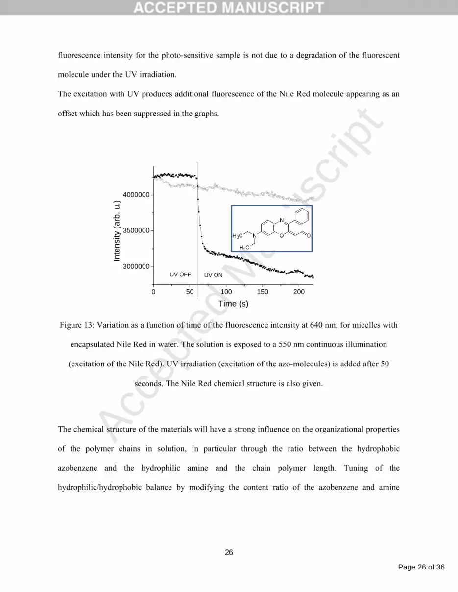
The figure 13 represents the variation of the Nile Red emission at 640 nm under continuous
excitation at 550 nm, as a function of time, in a water solution containing the polymer at 0,015
mg.mL-1 . The evolution of the Nile Red emission was investigated using additional illumination of
the azobenzene molecule in the UV range (full symbols). The evolution of the Nile Red emission
under the same excitation conditions in a non photo-active test sample made of a polysiloxane
incorporating only dimethylamino ethanol units with 17% substitution degree as the hydrophilic
group is also shown (empty symbols).
For the sample with photo-sensitive micelles, a stable signal for the first 60 seconds is observed until
the exposure to UV light leading to a sharp decrease of the Nile Red fluorescence intensity followed
by a slower decrease. This intensity decrease is attributed to the modification of the Nile Red
environment polarity resulting from a release of the Nile Red molecules in the water following
disruption of the micelles. An increase of the micelles core polarity induced by azo-molecules
isomerizations without a destruction of the micelles may as well contribute to the observed decrease
of intensity .
We also observe that the UV illumination of the non-sensitive micelles does not result in a
modification of the Nile Red fluorescence intensity confirming that the observed decrease of the
Page 26 of 36
Accep
ted
Man
uscr
ipt
26
fluorescence intensity for the photo-sensitive sample is not due to a degradation of the fluorescent
molecule under the UV irradiation.
The excitation with UV produces additional fluorescence of the Nile Red molecule appearing as an
offset which has been suppressed in the graphs.
0 50 100 150 200
3000000
3500000
4000000
UV ON
Inte
nsity
(ar
b. u
.)
Time (s)
UV OFF
Figure 13: Variation as a function of time of the fluorescence intensity at 640 nm, for micelles with
encapsulated Nile Red in water. The solution is exposed to a 550 nm continuous illumination
(excitation of the Nile Red). UV irradiation (excitation of the azo-molecules) is added after 50
seconds. The Nile Red chemical structure is also given.
The chemical structure of the materials will have a strong influence on the organizational properties
of the polymer chains in solution, in particular through the ratio between the hydrophobic
azobenzene and the hydrophilic amine and the chain polymer length. Tuning of the
hydrophilic/hydrophobic balance by modifying the content ratio of the azobenzene and amine

Page 27 of 36
Accep
ted
Man
uscr
ipt
27
molecules can impact the size, the CCA and the stability of the nanostructures in solution. Chemical
engineering provides thus an efficient mean to optimize the properties of the materials for drug
delivery applications. For example, increasing the amine content led to an improved stability of the
micelles but with a consequent reduction of their diameter down to 20nm with an expected impact
on the loading capacity. As well, the disruption dynamics and efficiency under optical excitation will
be affected by the chemical structure. More detailed results will be reported in a further publication.
6. CONCLUSION
We have shown here the potential of azo-polysiloxanes for biological applications in at least two
main directions. Depending on their chemical structure, the polymers can be used as thin films or
can self-assemble as nanoparticles when dispersed in aqueous solutions. In the first case, the
polymers can provide optically active supports to facilitate our understanding of the complex
mechanisms involved in the interaction between cells and their environment. In the second case the
aim is to produce nanoparticles able to encapsulate active substances with a perspective for drug
delivery applications.
A specificity of azo-polymers is the real time control of the films topographic properties provided
by the underlying photo-induced mass transport mechanism. Reversible nano and micro-patterns can
be produced on the surface of azo-polymer films through a single step exposure to an interference
pattern enabling a dynamic control on the film surface. The originality of the studies presented here
relies on the fine control over the materials properties, which can be obtained through the
incorporation of azo-molecules and additional chemical functions with controlled substitution

Page 28 of 36
Accep
ted
Man
uscr
ipt
28
degrees. Besides tuning the films opto-mechanical properties and surface modifications stability,
films with different chemical compositions can be used in cell cultures. In this respect we
demonstrated a good compatibility of our materials with biological cells. The chemical structure and
the film topography have shown a strong influence on the cell morphology and development. In
this respect the stability of the polymers surface when exposed to water is also a key parameter for
the cell adhesion and proliferation and can be controlled by the chemical composition of the
material. Stabilization of the film surface under aqueous media can be improved by adapting the
film substrate.
Materials self-assembling in aqueous solvents as nanoparticles, which are able to encapsulate
molecules have also been introduced. The possibility to optically destabilize the polymer chains 3-
dimensional organization resulting in the triggered release of the encapsulated molecules in solution
has been demonstrated. Optimization of the particles properties, i.e. stability, loading capacity and
materials aggregation/disruption dynamical properties will require further investigations to correlate
the materials chemical structure and organizational properties.
REFERENCES
[1] J. Hu and S. Liu, "Responsive Polymers for Detection and Sensing Applications: Current Status
and Future Developments," Macromol. 43, 8315–8330 (2010)
[2] C. J. F. Rijcken et al., "Triggered destabilisation of polymeric micelles and vesicles by changing
polymers polarity: An attractive tool for drug delivery," J. Cont. Rel. 120, 131–148 (2007)
[3] T. O. Kyung et al., "Polymeric nanovehicles for anticancer drugs with triggering release
mechanisms," J. Mater. Chem. 17, 3987–4001 (2007)

Page 29 of 36
Accep
ted
Man
uscr
ipt
29
[4] F. Meng, Z. Zhong and J. Feijen, "Stimuli-Responsive Polymersomes for Programmed Drug
Delivery," Biomacromol. 10, 197-209 (2009)
[5] L. G. Griffith, "Polymeric biomaterials," Acta Mater. 48, 263-277 (2000)
[6] E. Ostuni, L. Yan and G. M. Whitesides, "The interaction of proteins and cells with self-
assembled monolayers of alkanethiolates on gold and silver," Colloids and Surfaces B: Biointerfaces
15, 3–30 (1999)
[7] F. Rosso et al., "From cell-ECM interactions to tissue engineering," J. Cell. Physiol. 199, 174-
180 (2004)
[8] M. Larsen et al., "The matrix reorganized: extracellular matrix remodeling and integrin
signaling," Current Opinion in Cell Biology 18, 463-471 (2006)
[9] Y. W. Fan et al., "Culture of neural cells on silicon wafers with nano-scale surface topograph,”
Journal of Neuroscience Methods 120, 17-23 (2002)
[10] C.-H. Choi et al., "Cell interaction with three-dimensional sharp-tip nanotopography,"
Biomaterials 28, 1672–1679 (2007)
[11] P. P. Girard et al., "Cellular chemomechanics at interfaces: sensing, integration and response,"
Soft Matter 3, 307–326 (2007)
[12] T. Tzvetkova-Chevolleau et al., "The motility of normal and cancer cells in response to the
combined influence of the substrate rigidity and anisotropic microstructure," Biomaterials 29, 1541-
1551 (2008)
[13] A. M. Rajnicek, S. Britland and C. D. McCaig, "Contact guidance of CNS neurites on grooved
quartz: influence of groove dimensions, neuronal age and cell type," J. Cell. Sci. 110, 2905–2913
(1997)

Page 30 of 36
Accep
ted
Man
uscr
ipt
30
[14] E. T. den Braber et al., "Quantitative analysis of cell proliferation and orientation on substrata
with uniform parallel surface microgrooves," Biomaterials 17, 1093–1099 (1996)
[15] I. Nagata, A. Kawana and N. Nakatsuji, "Perpendicular contact guidance of CNS neuroblasts on
artificial microstructures," Development 117, 401-408 (1993).
[16] D. Y. Kim et al., "Laser induced holographic surface relief gratings on nonlinear optical
polymer films," Appl. Phys. Lett. 66, 1166-1168 (1995)
[17] P. Rochon, E. Batalla and A. L. Natansohn, "Optically induced surface gratings on azoaromatic
polymer films," Appl. Phys. Lett. 66, 136-138 (1995)
[18] X. L. Jiang et al., "Unusual polarization dependent optical erasure of surface relief gratings on
azobenzene polymer films," Appl. Phys. Lett. 72, 2502-2504 (1998)
[19] F. Lagugné-Labarthet, T. Buffeteau and C. Sourisseau, "Optical erasures and unusual surface
reliefs of holographic gratings inscribed on thin films of an azobenzene functionalized polymer,"
Phys. Chem. Chem. Phys. 4, 4020-4029 (2002)
[20] D. Bublitz et al., "Photoinduced deformation of azobenzene polyester films," Appl. Phys. B 70,
863–865 (2000)
[21] C. L. van Oosten et al., "Glassy photomechanical liquid-crystal network actuators for
microscale devices," Eur. Phys. J. E 23, 329–336 (2007)
[22] S. J. Zilker et al., "Holographic data storage in amorphous polymers," Adv. Mat. 10, 855-859
(1998)
[23] Ikeda, T., and Tsutsumi, O. "Optical switching and image storage by means of azobenzene
liquid crystal films," Science 268, 1873-1875 (1995)
[24] Y. Shi et al., "Large photoinduced birefringence in an optically nonlinear polyester polymer,"
Appl. Phys. Lett. 59, 2935-2937 (1991)

Page 31 of 36
Accep
ted
Man
uscr
ipt
31
[25] T. Hirose et al., "Azobenzene Liquid-Crystalline Polymer for Optical Switching of Grating
Waveguide Couplers with a Flat Surface," Optics Com. 228, 279–283 (2003)
[26] J. Paterson et al., "Optically inscribed surface relief diffraction gratings on
azobenzenecontaining polymers for coupling light into slab waveguides," Appl. Phys. Lett. 69, 3318-
3320 (1996)
[27] C. Cocoyer et al., "Implementation of submicrometric periodic surface structures : towards
improvement of organic solar cell performances," Appl. Phys. Let. 88, 133108:1-3 (2006)
[28] C. Hubert et al., "Emission properties of an organic light emitting diode patterned by a
photoinduced autostructuration process," Appl. Phys. Lett. 87, 191105-191108 (2005)
[29] H. Baac et al., "Submicron-scale topographical control of cell growth using holographic surface
relief grating," Mater. Sci. Eng. C 24, 209 (2004)
[30] J. K. Lee et al., "The Topographical Guidance of Neurons cultured on Holographic Photo-
Responsive Polymer," Proc. 26th AIC-IEEE, EMBS San Francisco, CA, 4970 (2004)
[31] R. Barillé et al., "Photo-responsive polymer with erasable and reconfigurable micro- and nano-
patterns: An in vitro study for neuron guidance, " Coll. Surfaces B: Biointerfaces 88, 63-71 (2011)

Page 32 of 36
Accep
ted
Man
uscr
ipt
32
[32] P. S. Ramanujam, N. C. Holme and S. Hvilsted, "Atomic force and optical near‐field
microscopic investigations of polarization holographic gratings in a liquid crystalline azobenzene
side‐chain polyester," Appl. Phys. Lett. 68, 1329-1331 (1996)
[33] A. Stracke et al., "Gain effects in optical storage: Thermal induction of a surface relief grating
in a smectic liquid crystal," Adv. Mater. 12, 282–285 (2000)
[34] N. Reinke et al., "Electric field assisted holographic recording of surface relief gratings in an
azo-glass," Appl. Phys. B 78, 205–209 (2004)
[35] H. Nakano, T. Tanino and Y. Shirota, "Surface relief grating formation on a single crystal of 4-
(dimethylamino)azobenzene," Appl. Phys. Lett. 87, 061910:1–3 (2005)
[36] J. Kumar et al., "Gradient force: The mechanism for surface relief grating formation in
azobenzene functionalized polymers," Appl. Phys. Lett. 72, 2096–2098 (1998)
[37] C. J. Barrett, A. L. Natansohn and P. L. Rochon, "Mechanism of optically inscribed high-
efficiency diffraction gratings in azo polymer films," J. Phys. Chem. 100, 8836–8842 (1996)
[38] P. Lefin, C. Fiorini and J.-M. Nunzi, "Anisotropy of the photo-induced translation diffusion of
azobenzene dyes in polymer matrices," Pure Appl. Opt. 7, 71–82 (1998)

Page 33 of 36
Accep
ted
Man
uscr
ipt
33
[39] T. G. Pedersen and P. M. Johansen, "Mean-field theory of photoinduced molecular reorientation
in azobenzene liquid crystalline side-chain polymers," Phys. Rev. Lett. 79, 2470–2473 (1997)
[40] D. Bublitz, B. Fleck and L. Wenke, "A model for surface-relief formation in azobenzene
polymers," Appl. Phys. B 72, 931–936 (2001)
[41] M. L. Juan et al., "Multiscale Model for Photoinduced Molecular Motion in Azo Polymers,"
ACS Nano 3, 1573–1579 (2009)
[42] V. Toshchevikov, M. Saphiannikova and D. Neher, "Microscopic Theory of Light-Induced
Deformation in Amorphous Side-Chain Azobenzene Polymers," J. Phys. Chem. B 113, 5032–5045
(2009)
[43] Y. B. Gaididei, P. L. Christiansen and P. S. Ramanujam, "Theory of photoinduced deformation
of molecular films, " Appl. Phys. B 74, 139–146 (2002)
[44] A. Ambrosio, P. Maddalena and L. Marrucci, "Molecular Model for Light-Driven Spiral Mass
Transport in Azopolymer Films," Phys. Rev. Lett. 110, 146102:1–5 (2013)
[45] N. Mechau, M. Saphiannikova and D. Neher, "Molecular tracer diffusion in thin azobenzene
polymer layers," Appl. Phys. Lett. 89, 251902:1–3 (2006)
[46] P. Karageorgiev et al., "From anisotropic photo-fluidity towards nanomanipulation in the
optical near-field," Nat. Mater. 4, 699–703 (2005)
[47] A. Ambrosio et al., "Light-induced spiral mass transport in azo-polymer films under vortex-
beam illumination," Nature Com. 3, 1-9 (2012)
[48] C. Hubert et al., "Spontaneous patterning of hexagonal structures in an azo-polymer unsing
light-controlled mass transport," Adv. Mat. 14, 729-732 (2002)
Page 34 of 36
Accep
ted
Man
uscr
ipt
34
[49] F. Lagugné-Labarthet, T. Buffeteau and C. Sourisseau, "Properties of birefringence and surface
relief gratings inscribed on azopolymer thin films using various interference patterns," J. Opt.
A:Pure Appl. Opt. 4, S235-S241 (2002)
[50] F. H’dhili et al., "Near-Field optics: direct observation of the field enhancement below an
apertureless probe using a photosensitive polymer," Appl. Phys. Lett. 79, 4019-4021 (2001)
[51] N. K. Viswanathan et al., "Surface relief structures on azo polymer films," J. Mat. Chem. 9,
1941-1955 (1999)
[52] F. Fabbri et al., "Evidence of Two Distinct Mechanisms Driving Photoinduced Matter Motion
in Thin Films Containing Azobenzene Derivatives," J. Phys. Chem. B 115, 1363–1367 (2011)
[53] A. Jacquart et al., "Influence of extrinsic and intrinsic parameters onto the formation of surface
relief gratings in polar azo molecular glasses," Dyes and Pigments 92, 790-797 (2012)
[54] H. Ando et al., "Photoinduced surface relief grating formation using new polymers containing
the same azobenzene chromophore as a photochoromic amorphous molecular material," Mater.
Chem. Phys. 113, 376–381 (2009)
[55] A. Sobolewska and S. Bartkiewicz, "Surface relief grating in azo-polymer obtained for s-s
polarization configuration of the writing beams," Appl. Phys. Lett. 101, 193301:1-3 (2012)
[56] N. Hurduc et al., "Azo-polysiloxanes as new supports for cell cultures," Mat. Sci. and Eng. C
33, 2440–2445 (2013)
[57] X. L. Jiang et al., "Polarization dependent recordings of surface relief gratings in azobenzene
containing polymer films," Appl. Phys. Lett. 68, 2618-2620 (1996)
[58] F. Lagugné-Labarthet, T. Buffeteau and C. Sourisseau, "Inscription of holographic gratings
using circularly polarized light : influence of the optical set-up on the birefringence and surface relief
grating properties," Appl. Phys. B 74, 129-137 (2002)

Page 35 of 36
Accep
ted
Man
uscr
ipt
35
[59] A. Raicu Luca et al., "Mass transport in low Tg azo-polymers: Effect on the surface relief
grating induction and stability of additional side chain groups able to generate physical interactions,"
Appl. Surf. Sci. 290, 172– 179 (2014)
[60] L. Mazaheri, S. Ahmadi-Kandjani and J.-M. Nunzi, "Influence of temperature on the relaxation
kinetics of spontaneous pattern formation in an azo-polymer film," Opt. Commun. 298, 150–153
(2013)
[61] J. Mysliwiec et al., "Dynamics of photoinduced motions in azobenzene grafted polybutadienes,"
Opt. Mater. 33, 1398–1404 (2011)
[62] P. U. Veer, U. Pietsch and M. Saphiannikova, "Time and temperature dependence of surface
relief grating formation in polymers containing azobenzene groups with different dipole moment," J.
Appl. Phys. 106, 014909:1–7 (2009)
[63] C. Petrareanu et al., "Comparative Proteomics Reveals Novel Components at the Plasma
Membrane of Differentiated HepaRG Cells and Different Distribution in Hepatocyte- and Biliary-
Like Cells," PLOS ONE 8, e71859 (2013)
[64] N. Hurduc et al., "Direct observation of athermal photofluidisation in azo-polymer films," Soft
Matter 10, 4640–4647 (2014)
[65] I.-A. Moleavin et al., "Amphiphilic azopolymers capable to generate photo-sensitive micelles,"
Cent. Eur. J. Chem. 9, 1117-1125 (2011)
[66] Q. Duan et al., "Synthesis and Thermoresponsive Property of End-Functionalized Poly(N-
isopropylacrylamide) with Pyrenyl Group," J. Polym. Sci. Part A: Polym. Chem. 44, 1117-1124
(2006)
[67] Y. Einaga et al., "Photo-functional Vesicles Containing Prussian Blue and Azobenzene," J. Am.
Chem. Soc. 121, 3745-3750 (1999)

Page 36 of 36
Accep
ted
Man
uscr
ipt
36
[68] I. Moleavin et al., "Photosensitive micelles based on polysiloxanes containing azobenzene
moieties", Polym. Bull. 65, 69-81 (2010)
[69] Y. Lin and P. Alexandridis, "Self-Assembly of an Amphiphilic Siloxane Graft Copolymer in
Water," J. Phys. Chem. B 106, 10845-10853 (2002)
[70] Grama, S., Moleavin, I.-A., Hodorog-Rusu, A. D., Hurduc, N., Prisacaru, I., and Ibanescu, C.,
"Photosensitive Azo-polysiloxanes for Drug Delivery Applications,", Materiale Plastice 50, 60-64
(2013)
[71] Kuiper, J.-M., Engberts, J. B. F. N., and Poolman, B.,"H-aggregation of azobenzene-substituted
amphiphiles in vesicular membranes," Langmuir 20, 1152-1160 (2004)


