Embed Size (px)
Citation preview
Virtual Reconstruction and Prey Size Preference in theMid Cenozoic Thylacinid, Nimbacinus dicksoni(Thylacinidae, Marsupialia)Marie R. G. Attard1,2*, William C. H. Parr1, Laura A. B. Wilson1, Michael Archer3, Suzanne J. Hand3,
Tracey L. Rogers1, Stephen Wroe2
1 Evolution and Ecology Research Centre, School of Biological, Earth and Environmental Sciences, University of New South Wales, Sydney, New South Wales, Australia,
2 Function, Evolution and Anatomy Research laboratory, Zoology, School of Environmental and Rural Sciences, University of New England, New South Wales, Australia,
3 Evolution of Earth and Life Sciences Research Centre, School of Biological, Earth and Environmental Sciences, University of New South Wales, Sydney, New South Wales,
Australia
Abstract
Thylacinidae is an extinct family of Australian and New Guinean marsupial carnivores, comprizing 12 known species, theoldest of which are late Oligocene (,24 Ma) in age. Except for the recently extinct thylacine (Thylacinus cynocephalus), mostare known from fragmentary craniodental material only, limiting the scope of biomechanical and ecological studies.However, a particularly well-preserved skull of the fossil species Nimbacinus dicksoni, has been recovered from middleMiocene (,16-11.6 Ma) deposits in the Riversleigh World Heritage Area, northwestern Queensland. Here, we ask whether N.dicksoni was more similar to its recently extinct relative or to several large living marsupials in a key aspect of feedingecology, i.e., was N. dicksoni a relatively small or large prey specialist. To address this question we have digitallyreconstructed its skull and applied three-dimensional Finite Element Analysis to compare its mechanical performance withthat of three extant marsupial carnivores and T. cynocephalus. Under loadings adjusted for differences in size that simulatedforces generated by both jaw closing musculature and struggling prey, we found that stress distributions and magnitudesin the skull of N. dicksoni were more similar to those of the living spotted-tailed quoll (Dasyurus maculatus) than to itsrecently extinct relative. Considering the Finite Element Analysis results and dental morphology, we predict that N. dicksonilikely occupied a broadly similar ecological niche to that of D. maculatus, and was likely capable of hunting vertebrate preythat may have exceeded its own body mass.
Citation: Attard MRG, Parr WCH, Wilson LAB, Archer M, Hand SJ, et al. (2014) Virtual Reconstruction and Prey Size Preference in the Mid Cenozoic Thylacinid,Nimbacinus dicksoni (Thylacinidae, Marsupialia). PLoS ONE 9(4): e93088. doi:10.1371/journal.pone.0093088
Editor: Kornelius Kupczik, Friedrich-Schiller-University Jena, Germany
Received September 25, 2013; Accepted March 1, 2014; Published April 9, 2014
Copyright: � 2014 Attard et al. This is an open-access article distributed under the terms of the Creative Commons Attribution License, which permitsunrestricted use, distribution, and reproduction in any medium, provided the original author and source are credited.
Funding: This research was funded by Australian Research Council grants to S. Wroe (DP0666374 and DP0987985), and to M. Archer, and S. J. Hand(LP100200486 and DP1094569). M. Attard is supported by the Postgraduate Writing and Skills Transfer Award sponsored by the Evolution and Ecology ResearchCentre, University of New South Wales. L. Wilson is supported by the Swiss National Science Foundation (PBZHP3_141470). The funders had no role in studydesign, data collection and analysis, decision to publish, or preparation of the manuscript.
Competing Interests: The authors have declared that no competing interests exist.
* E-mail: [email protected]
Introduction
Thylacinids first appear in the Australian fossil record during
the late Oligocene (,24 Ma) and include the largest representa-
tives of the Dasyuromorphia, i.e., families Thylacinidae, Dasyur-
idae and Myrmecobiidae [1–8]. A wide range of feeding ecologies
are known within the order. They include omnivores, insectivores,
small prey specialists, hypercarnivores and osteophageous species
[9]. Variation in the dentition, skull shape and body size (,1–
60 kg) of thylacinids suggests considerable trophic diversity within
the family [10,11]. In addition to the recently extinct thylacine or
Tasmanian ‘tiger’ (Thylacinus cynocephalus), eleven extinct species of
thylacinid have been described [4,12–18]. Up to five species may
have co-existed in the Riversleigh World Heritage Area, north-
western Queensland between the late Oligocene (,24 Ma) to
middle Miocene (16-11.6 Ma) [12,16]. See Table S1 for the
temporal and geographic distribution of all thylacinid species.
The Riversleigh thylacinids inhabited forests [19,20]. These
regions were also occupied by an assortment of other carnivorous/
omnivorous taxa, including ‘giant’ carnivorous rat-kangaroos
(Ekaltadeta spp.), crocodiles (Mekosuchinae, e.g. Baru darrowi and
Trilophosuchus rackhami), flightless dromornithid birds, marsupial
lions (Thylacoleonidae), bandicoots (Peramelemorphia), dasyurids
(Dasyuridae), pythons (Pythonidae), madtsoiid snakes and the
world’s oldest known venomous snakes [21–23]. Subsequent
drying of the Australian continent from the late Miocene (11.6–
5.3 Ma) led to the gradual replacement of forest environments
with open woodlands, shrublands and grasslands [19–21]. These
changes appear to broadly correlate with declining thylacinid
diversity [24].
To date, interpretations of the ecology and feeding behavior of
fossil thylacinids have been largely qualitative. This is, at least in
part, because most extinct species are known only from jaw
fragments and teeth. The near-complete skull of Nimbacinus dicksoni
[4,14,18], a medium-sized thylacinid, provides an opportunity to
more fully investigate feeding ecology in an extinct thylacinid.
Nimbacinus dicksoni was approximately 5 kg in body mass [10].
Fossils of N. dicksoni have been recovered from Oligocene-Miocene
PLOS ONE | www.plosone.org 1 April 2014 | Volume 9 | Issue 4 | e93088
(,24–5.3 Ma) deposits in the Riversleigh World Heritage Area,
northwestern Queensland and Bullock Creek, Northern Territory
[12,14,16,18]. Its dentition is less specialized than that of the
species of Thylacinus, but broadly similar to that of the living
dasyurid, the spotted-tailed quoll (Dasyurus maculatus) in the
arrangement and geometry of molar shearing crests typically
associated with carnivory [18,25].
To date conflicting evidence has been presented regarding the
body size of prey N. dicksoni may have hunted. Predictions of bite
force adjusted for body mass, based on application of 2D beam
theory, have suggested that N. dicksoni may have taken relatively
large prey, as does the slightly smaller D. maculatus [26]. However,
shape analysis of the cranium has suggested that the species may
have been restricted to smaller prey and/or included a higher
proportion of invertebrate food in its diet [11].
The loads imposed on an animal during prey acquisition and
feeding play an important role in the evolution of its skull
morphology [27]. Testing hypotheses regarding the relationship
between the form and function of skulls from extinct species
requires an understanding of this relationship in living animals
[28]. A comparative biomechanics approach involving living
analogues has increasingly been applied to predict the feeding
ecology and predatory behavior of extinct species [29–34]. To
gain further insight in the feeding ecology of N. dicksoni, here we
perform a biomechanical analysis of the skull of N. dicksoni to
predict its mechanical behavior in response to loads simulating the
capture and processing of prey.
Finite Element Analysis (FEA) is a computer modeling approach
now commonly used by biologists and paleontologists to examine
and compare mechanical performance in biological structures in
comparative contexts [35–41]. In FEA, continuous structures,
such as the skull, are divided into discrete, finite numbers of
elements, allowing the prediction of mechanical behavior for
complex geometric shapes. The structure is analyzed in the form
of a matrix algebra problem that is solved with the aid of a
computer [42].
Studies of feeding ecology for thylacinids have primarily focused
on the most recently extinct member of the family, T. cynocephalus,
which survived in Tasmania until 1936 [43]. Our understanding
of the ecology of T. cynocephalus is chiefly based on morphological
comparisons and 2D beam theory [11,26,44,45], as well as
anecdotal accounts of their behavior in the wild [46,47]. Elbow
joint morphology of T. cynocephalus evidently most closely resembles
that of extant ambush predators, a compromise between efficient
distance locomotion and the ability to manipulate and grapple
with prey [48]. Three-dimensional biomechanical modeling of the
skull of T. cynocephalus, extant dasyurids and an introduced
Australian predator (Canis lupus dingo) have suggested potential
limitations in prey body size [32,35].
Morphological and biomechanical comparisons including sym-
patric native predators in Tasmania indicate that the diet of T.
cynocephalus may have overlapped considerably with the two largest
extant marsupial carnivores, the Tasmanian devil (Sarcophilus
harrisii) and spotted-tailed quoll (D. maculatus) [35,44,49]. These
species represented the three largest marsupial carnivores in
Tasmania at the time of European settlement.
Sarcophilus harrisii is the largest living marsupial carnivore (mean
adult male weight 8.7 kg, female 6.1 kg) [50]. They are the only
specialized scavengers among living marsupials, filling a broadly
similar ecological niche to that of osteophagous hyenas [44]. They
are also opportunistic hunters and are known to prey on mammals
that may exceed their body mass [51,52]. Relative to its body size,
its predicted bite force is greater than that of any other extant
mammal studied to date [26].
Quolls are represented by four extant species in Australia and
two in New Guinea [53]. The largest quoll, Dasyurus maculatus
(maximum weight 7 kg) has a broad diet mainly consisting of
mammals and insects, but will occasionally feed on birds and
reptiles [54,55]. Larger prey species constitute a higher proportion
of the diet of adult male D. maculatus, while females and immature
D. maculatus more frequently feed on smaller bodied mammals and
invertebrates [56,57]. The northern quoll (Dasyurus hallucatus) is the
smallest and most arboreal of the four Australian quolls, weighing
up to 1.2 kg [58,59]. Although primarily insectivorous, this active
hunter can feed on a variety of foods: fruits, small mammals, birds,
reptiles, frogs and carrion [60–62].
In this study we aim to determine whether N. dicksoni was
capable of killing large prey relative to their body size, or was
restricted to catching relatively small bodied species. By digital
reconstruction of its skull and applied 3D FEA, we compare its
mechanical performance with that of three extant marsupial
carnivores; S. harrisii, D. maculatus and D. hallucatus. We use
previously applied scaling procedures [31] to account for
differences in body mass, allowing for comparison of results
between species. We predict that N. dicksoni will show similar
distributions and magnitudes of craniomandibular stress to that of
D. maculatus due to similarities in their dental morphology and size
[18]. We also include T. cynocephalus to establish whether the
biomechanical performance of N. dicksoni more closely resembles
that of this larger, more derived thylacinid than dasyurids. A
general assessment of the phylogenetic relationships of dasyur-
omophians, including taxa examined in this study is shown in
Figure S1. We test if the biomechanical patterns and the inferred
feeding aspects in T. cynocephalus in a recent study [35] are derived.
We hypothesize that the relatively long rostrum of T. cynocephalus
will result in higher stresses in the skull during biting and prey
procurement than N. dicksoni and other dasyuromorphians.
Materials and Methods
SpecimensThe skull of Nimbacinus dicksoni (QMF36357) was recovered from
AL90 Site on the Gag Plateau of the Riversleigh World Heritage
area in northwestern Queensland. Precise locality details can be
provided on application to the Queensland Museum. This
specimen was collected under permits issued by Queensland
Department of Environment and Heritage and Environment
Australia, and registered in the paleontological collections of the
Queensland Museum, Brisbane, Australia. All necessary permits
were obtained for the described study, which complied with all
relevant regulations.
The biomechanical performance of the Nimbacinus dicksoni skull
was compared with that of four dasyuromorphian species covering
a range of craniodental morphologies and feeding ecologies. These
comprized three extant dasyurids [Dasyurus hallucatus TMM M-
6921; D. maculatus UNSW Z20; Sarcophilus harrisii AM10756] and
one thylacinid (Thylacinus cynocephalus AM1821). Institutional
abbreviations are QMF (Queensland Museum Fossil, Queens-
land), TMM (Texas Memorial Museum, Austin), UNSW (Uni-
versity of New South Wales, Sydney) and AM (Australian
Museum, Sydney).
We generated 3D finite element models (FEMs) of each skull on
the basis of computed tomography X-ray (CT) scan data.
Digimorph (University of Texas; http://www.digimorph.org) was
the source of CT data of a D. hallucatus skull (0.0784 mm slice
thickness, 0.0784 mm inter-slice distance). Permission to use
Digimorph derived CT scan data was granted by Dr. Timothy
Rowe, Project Director of Digimorph. Other skulls were scanned
Prey Size Preference in Nimbacinus dicksoni
PLOS ONE | www.plosone.org 2 April 2014 | Volume 9 | Issue 4 | e93088
in a Toshiba Aquillon 16 scanner (ToshibaMedical Systems
Corporation, Otawara, Tachigi, Japan) at the Mater Hospital,
Newcastle, NSW (1 mm slice thickness, 0.8 mm inter-slice
distance, 240 mm field of view). The Australian Museum,
Queensland Museum and University of New South Wales granted
the loan of specimens to obtain CT data for this study.
We used the same specimen CT scans of T. cynocephalus, D.
maculatus and S. harrisii as examined by Attard et al. [35], but
constructed the FEMs again using a higher resolution mesh than
that used by Attard et al. [35]. More specifically, the 3D surface
meshes which formed the bases of the FEMs were computed using
the High Quality as opposed to Medium Quality option in Mimics
(ver. 13.2). The generated surface meshes were then converted to
FEMs in STRAND7 (ver. 2.4) following previously established
protocols [31,35,63]. Additionally, 3D objects of T. cynocephalus
and all dasyurid skulls were exported as separate stl files for the
creation of interactive 3D pdf documents that can be viewed using
Adobe Reader (Figure S2, S3, S4, S5).
Digital Reconstruction of Nimbacinus dicksoniFor detailed descriptions of N. dicksoni see Muirhead and Archer
[14] and Wroe and Musser [18]. Previous analysis has suggested
that, within the family, the dentition of N. dicksoni is less derived
than the recent Thylacinus cynocephalus for at least 12 features, but
that relatively few cranial specializations in T. cynocephalus
distinguish the two species. These two taxa share at least three
cranial features not present in the most generalized thylacinid
known from significant cranial material, the late Oligocene
Badjcinus turnbulli [14,18].
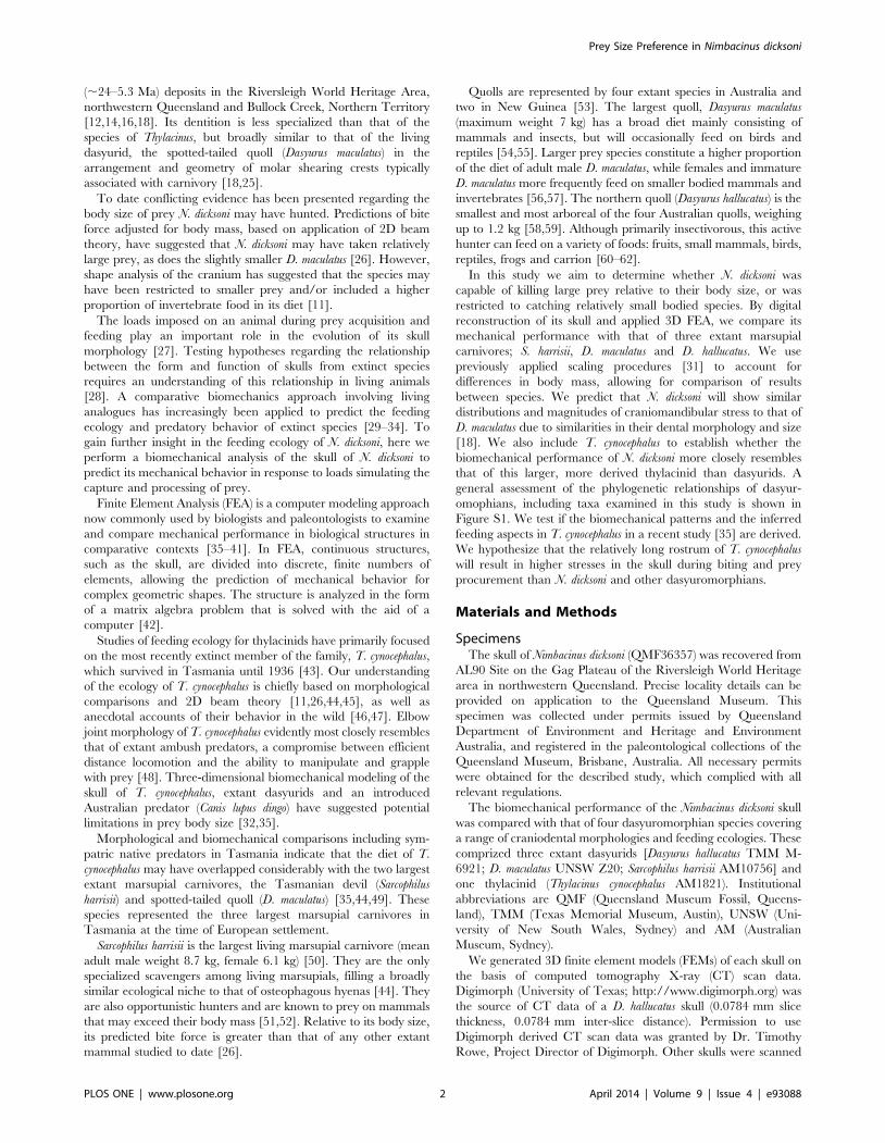
The skull of N. dicksoni is well preserved, although some regions
are absent or damaged. Specifically, some damage/deformation is
present at the postorbital processes, frontal, maxillary and nasal
bones, which are compressed dorsoventrally (Figure 1). These
damaged regions were reconstructed according to the morphology
of surrounding bone regions once the damaged areas had been
isolated and deleted [64]. Regions of bone that showed only minor
damage were smoothed to create a coherent surface mesh for later
solid meshing.
The right and left dentaries were largely intact but missing the
superior regions of the coronoid processes, the temporomandib-
ular joints (TMJ), condyles and angular processes (Figure 1). The
anterior of the mandible is broken, separating both dentaries. We
used the right dentary as a basis for reconstruction because its
dentition was more complete, with only the incisors missing
(Figure 1). We used a surface mesh of the right dentary of D.
maculatus to reconstruct posterior regions of the right dentary of N.
dicksoni. Dasyurus maculatus was chosen as its mandible was most
similar in shape to that of N. dicksoni [11], thereby minimizing the
extent of warping needed (and see below).
Reconstruction involved scaling the dentary of D. maculatus to
the same size as that of N. dicksoni on the basis of skull length
(condylo-basal). The missing posterior region of the N. dicksoni
specimen was then isolated on the D. maculatus specimen and the
mesh fitted to the existing structure in the mesh of N. dicksoni using
Iterative Closest Point (ICP) registration. ICP is an algorithm that
revises the transformation needed to minimize the distance
between the points of two partially overlapping meshes. This
process re-oriented the D. maculatus dentary in accordance with the
morphology of the N. dicksoni dentary [65]. The anterior region of
the D. maculatus dentary was deleted and the posterior region
‘warped’ so that overlapping regions of the coronoid process and
angular process from the D. maculatus mesh matched the existing
morphology of N. dicksoni. The manual warping method was used
as much of the target (fossil) morphology was missing, making it
impossible to apply homologous landmarks on both complete (D.
maculatus) and incomplete fossil (N. dicksoni) specimens. Procedures
to warp overlapping skull regions followed established protocols
used by Oldfield et al. and Parr et al. [38,66]. Manual warping
works by establishing a grid of control points around the complete
model (note that at this stage the incomplete fossil model has been
ICP registered with the scaled complete model by matching the
orientations of the regions of the jaw that are present in both
models). These control points are then manipulated so that the
surface morphology of the complete model matches that of the
Figure 1. Digital reconstruction of Nimbacinus dicksoni. Original(grey) and reconstructed 3D (yellow) in (A) lateral view and (B) dorsalview. (C) Pre-processed Finite Element model of N. dicksoni, showingjaw musculature represented by trusses.doi:10.1371/journal.pone.0093088.g001
Prey Size Preference in Nimbacinus dicksoni
PLOS ONE | www.plosone.org 3 April 2014 | Volume 9 | Issue 4 | e93088
target (fossil). This is another variation of Template Mesh
Deformation [66], but with the template points being the grid
control points around the complete model rather than homologous
anatomical points on both models.
Similarly, the TMJ of the complete (D. maculatus) model was
warped so that the condyle articulated with and fitted the N.
dicksoni cotyle of the cranium, again by using the manual template
mesh deformation warping method. The left dentary was created
by mirroring the reconstructed right dentary. These were
positioned so that the condyles articulated with the cranium, the
outer surfaces of the lower molars made contact with the inner
surface of the upper molars, and the tips of the lower canines
aligned with their ‘sockets’ in the cranium (see Figure 1).
It is important to note that the shape of the warp was
determined by the existing regions of the N. dicksoni dentary; the
need for the condyle to articulate with the cotyle to form the TMJ
and for the coronoid process to fit between the cranium and the
zygomatic arch. These requirements act as restraints on the warp
such that the shape of the starting mesh (D. maculatus in this case) is
not important in the sense that the warping process would always
end with a similarly shaped posterior region of the mandible
regardless of which taxon was used. We reiterate that D. maculatus
was used because it was the most similar in shape [11] and
therefore required less ‘warping’.
The N. dicksoni cranium was missing the following teeth: left I1-
4, right I1, 3-4, both right and left C1 and right LDP2. The
existing I2 and LDP2 on N. dicksoni were mirrored. All incisors
were missing from the mandible. Incisors from D. maculatus were
isolated, scaled and fitted into the empty tooth sockets on N.
dicksoni. Figure 1 displays the completed reconstruction of N.
dicksoni.
Finite Element ModelsThe assembly of FEMs largely follows previously published
procedures [30,31,35]. As the skull of N. dicksoni was not fully
preserved, we were unable to assign multiple material properties to
the digital reconstruction without introducing additional assump-
tions. Consequently, as in most FEA incorporating fossil material
[30,39,67], all FEMs were homogeneous and assigned a single
material property for cortical bone (E = 13.7 GPa, v = 0.3, where E
is Young’s modulus of elasticity and v is Poisson’s ratio) [68] to
enable direct comparisons between species. Poisson’s ratio and
Young’s modulus are fundamental metrics in the comparison of
stress or deformation for any material when strained elastically,
including homogeneous materials [69]. Young’s modulus is a
measure of stiffness in the material, whereas Poisson’s ratio is the
negative ratio between transverse strain and longitudinal strain in
an elastic material subjected to uniaxial stress [70].
Each homogeneous model was comprized of four-noded
tetrahedral elements or ‘bricks’ (tet4). FEMs for QMF36357,
AM1821, AM10756, UNSW Z20 and TMM M-6921 were
comprized of 1564048, 1429714, 1799292, 1402103 and 1956942
bricks respectively. Tet4 models are theoretically less accurate than
models comprized of tet10 elements. However, the models used in
this study are large and any difference in accuracy between results
from tet4 versus tet10 models will diminish as the number of
elements is increased. Comparable analyses comparing tet4 and
tet10 based models much smaller than those used here (,252000
elements) found differences of ,10% [27].
Modeling Masticatory Muscle ForcesJaw elevators were modeled as seven muscle subdivisions:
temporalis superficialis, temporalis profundus, masseter superficialis, masseter
profundus, zygomatico mandibularis, pterygoideus internus and pterygoideus
externus [32]. Proportions used for each jaw muscle division were
based on muscle mass proportions from a dissected Virginia
opossum (Didelphis virginiana) [71]. Muscle forces were predicted on
the basis of maximum cross-sectional areas (CSA) using the ‘dry
skull’ method [72]. To improve the accuracy of our CSA
measurements, we used our FEMs to record the co-ordinates of
,100 nodes at the perimeter of each muscle cross sectional area
[73]. The FEM was moved to the correct orientation described by
Thomason [70] to select nodes outlining the CSA. The node co-
ordinates were then plotted in plane geometry software, GEUP 5
(version 5.0.3) and connected to form a multi-sided polygon. The
area of the polygon was measured to estimate the CSA of each
major jaw closing muscle. To minimize the incidence of artefacts
at bite points and muscle origin and insertion areas, surface
regions at these sites were tessellated using a network of stiff beam
elements [74].
Restraints, Loading Conditions and ScalingDasyurids frequently use a penetrating canine bite to kill prey
[44,75–78] which involves the application of a bending load [79].
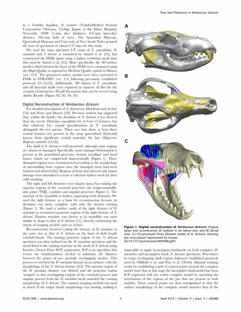
Figure 2. Position of nodes selected on each model to measure von Mises stress. Nodes were selected at equidistant points along the (A)mid-sagittal plane, (B) zygomatic arch and (C) mandible to measure the distribution of von Mises stress for each loading case.doi:10.1371/journal.pone.0093088.g002
Prey Size Preference in Nimbacinus dicksoni
PLOS ONE | www.plosone.org 4 April 2014 | Volume 9 | Issue 4 | e93088
We simulated bilateral canine biting (intrinsic load) and four
extrinsic loads to simulate loads generated by struggling prey (axial
twist, lateral shake, pullback and dorsoventral) for all models using
protocols described by Attard et al. [35] and following McHenry et
al. [31]. Extrinsic loads were modeled without applying bite forces
so as to clearly reveal the different influences of each separate
loading [31]. A gape angle of 35u was applied in all linear static
load cases.
Table 1. Predicted body mass and masticatory muscle forces for modeled dasyuromorphians.
Species Predicted body mass (kg) Temporalis muscle force (N) Masseteric muscle force (N) Total muscle force (N)
Dasyurus hallucatus 0.78 67.60 55.89 123.49
Dasyurus maculatus 2.88 211.01 178.67 389.67
Nimbacinus dicksoni 5.25 282.38 368.33 650.71
Sarcophilus harrisii 14.20 300.46 384.73 685.19
Thylacinus cynocephalus 32.49 706.64 843.21 1,549.85
Predicted body mass (kg) calculated using the regression equation for dasyuromorphians provided by Myers [76] based on lower molar row length. Temporalis andmasseteric muscle forces (N) were calculated based on cross-sectional area [67].doi:10.1371/journal.pone.0093088.t001
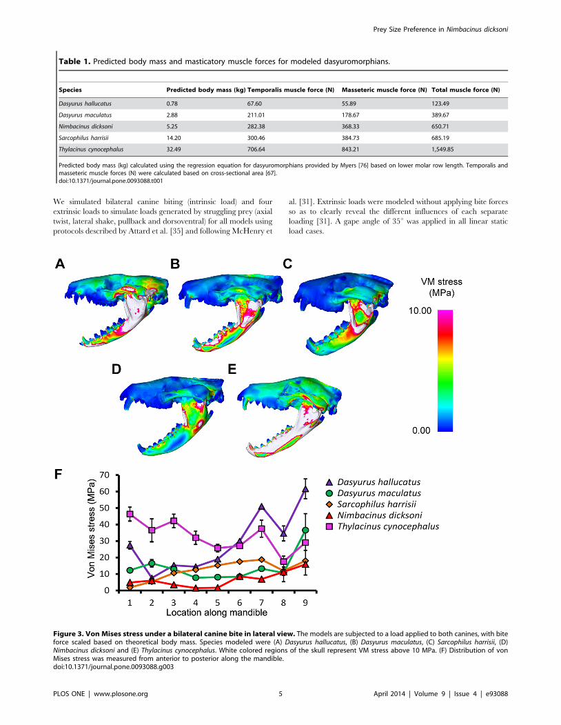
Figure 3. Von Mises stress under a bilateral canine bite in lateral view. The models are subjected to a load applied to both canines, with biteforce scaled based on theoretical body mass. Species modeled were (A) Dasyurus hallucatus, (B) Dasyurus maculatus, (C) Sarcophilus harrisii, (D)Nimbacinus dicksoni and (E) Thylacinus cynocephalus. White colored regions of the skull represent VM stress above 10 MPa. (F) Distribution of vonMises stress was measured from anterior to posterior along the mandible.doi:10.1371/journal.pone.0093088.g003
Prey Size Preference in Nimbacinus dicksoni
PLOS ONE | www.plosone.org 5 April 2014 | Volume 9 | Issue 4 | e93088
A considerable size range exists between specimens considered
in the present study. The relationship between bite force and body
mass is negatively allometric [26,80]. To account for differences in
body mass, a second series of load cases were solved following the
scaling procedures of McHenry et al. [31]. Here, for each model,
an estimate of bite force was made based on regression of body
mass to predicted bite force for dasyuromorphians [z = 0.6998 (log
y)+1.8735, where and y = mass (g) and z = bite force at canines (N)]
[26], with body mass for each specimen predicted using the
equation based on lower molar row length [log y = 21.075+3.209(log x), where x = lower molar length (mm), and y = mass (g)]
as presented by Myers [81]. Muscle forces were then scaled for
each specimen to achieve bite forces predicted on the basis of body
mass. FEMs were solved using these scaled muscle forces.
Prediction of bite force based on body mass using the regression
equation provided in Wroe et al. [26] is close to that which would
be expected following a 2/3 power relationship, whereby muscle
force is proportional to area while body mass is proportional to
volume [82]. The maximum bite force measured in Newtons (N)
was also estimated for intrinsic loads (Table S2) using FEMs with
un-scaled, specimen-specific estimated muscle forces (Table S3).
Three dimensional approaches are likely to be more accurate than
2D based approaches [83].
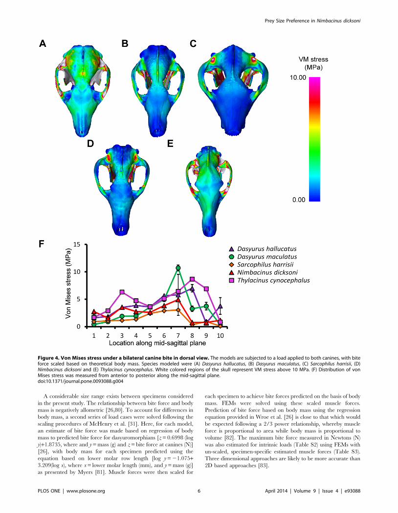
Figure 4. Von Mises stress under a bilateral canine bite in dorsal view. The models are subjected to a load applied to both canines, with biteforce scaled based on theoretical body mass. Species modeled were (A) Dasyurus hallucatus, (B) Dasyurus maculatus, (C) Sarcophilus harrisii, (D)Nimbacinus dicksoni and (E) Thylacinus cynocephalus. White colored regions of the skull represent VM stress above 10 MPa. (F) Distribution of vonMises stress was measured from anterior to posterior along the mid-sagittal plane.doi:10.1371/journal.pone.0093088.g004
Prey Size Preference in Nimbacinus dicksoni
PLOS ONE | www.plosone.org 6 April 2014 | Volume 9 | Issue 4 | e93088
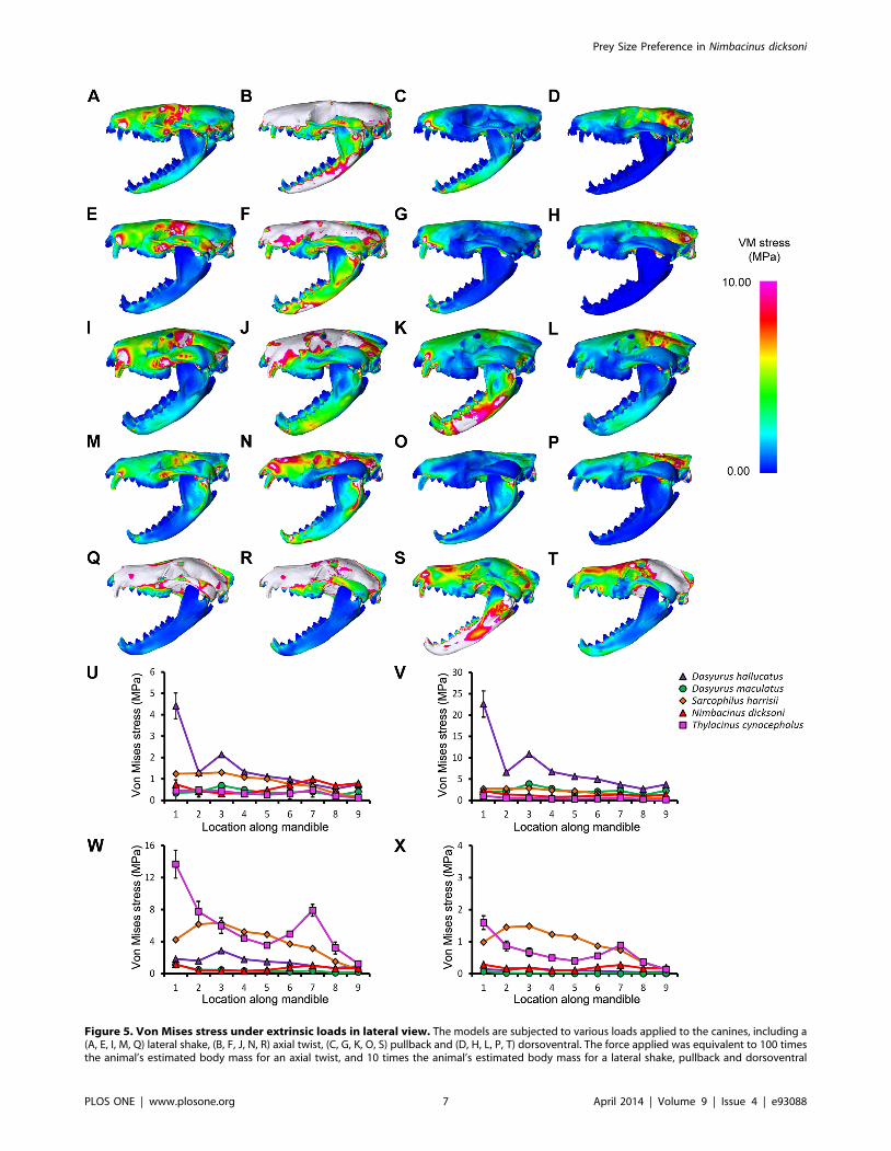
Figure 5. Von Mises stress under extrinsic loads in lateral view. The models are subjected to various loads applied to the canines, including a(A, E, I, M, Q) lateral shake, (B, F, J, N, R) axial twist, (C, G, K, O, S) pullback and (D, H, L, P, T) dorsoventral. The force applied was equivalent to 100 timesthe animal’s estimated body mass for an axial twist, and 10 times the animal’s estimated body mass for a lateral shake, pullback and dorsoventral
Prey Size Preference in Nimbacinus dicksoni
PLOS ONE | www.plosone.org 7 April 2014 | Volume 9 | Issue 4 | e93088
A H-frame connecting the canines of the upper and lower jaws
was used to apply extrinsic forces, with forces applied at the center
of the frame [32,35]. The force (N) applied to extrinsic loads was
an arbitrary figure, applied for strictly comparative purposes,
equivalent to 100 times the animal’s estimated body mass for an
axial twist, and 10 times the animal’s estimated body mass for a
lateral shake, pullback and dorsoventral shake [81]. Each
simulation in which forces are applied with the anterior teeth
(canines) restrained is a test for the hypothesis that stress will be
highest for species with the longest rostrum.
Von Mises (VM) stress is a good predictor of failure in ductile
materials such as bone [84,85] and VM stress is used here as a
metric for comparison between models following Attard et al. [35].
Nodes were selected at equidistant points along the mid-sagittal
plane, zygomatic arch and mandible (Figure 2) following Attard et
al. [35] and at each node values were calculated by averaging VM
stress recorded in the surrounding elements to assess changes in
stress magnitudes and distributions under different loadings.
Principal component analysis (PCA) was used to visualize
differences between species in average VM stress values for
equidistant nodes along the mid-sagittal plane (N = 10). PCA is an
ordination technique that summarizes the maximum variation
among a set of variables on few, uncorrelated axes (principal
components) [86]. PCA was performed separately on VM stress
values for each extrinsic load (axial twist, lateral shake, pullback
and dorsoventral) and for a bilateral canine bite. All VM stress
values were log transformed prior to PCA.
Differences in VM stress values between species were compared
using a Kruskal-Wallis test, which is regarded as a multiple-group
extension of the Man-Whitney test [87]. Significance values were
corrected for multiple comparisons using Bonferroni corrections,
as a conservative approach.
Results
The predicted body mass (kg) of each species was generally
within the expected range for each of the extant species (Table 1).
Body mass estimates ranged from 0.78 kg for D. hallucatus, up to
32.49 kg for T. cynocephalus. However, the body mass estimated for
S. harrisii of 14.20 kg was slightly above the upper limit observed
for males (13 kg) [88], possibly because the teeth and skull are
relatively large in this species. The robust craniodental morphol-
ogy and relatively large teeth in S. harrisii are probably related to its
habitual osteophagy, as has been observed in bone-cracking
carnivorans [89]. To obtain body mass estimates for these taxa
using simple or multiple regressions adjusted from cranidoental
variables may lead to overestimates of body mass. Predicted
maximum muscle forces for N. dicksoni (651 N) were relatively high,
approaching those of the larger S. harrisii (685 N) (Table 1).
Thylacinus cynocephalus displayed comparatively high levels of VM
stress in the cranium and mandible for most simulations (Figure 3–
6, S3). This is consistent with results of Attard et.al. [35]. Dasyurus
hallucatus showed relatively high levels of stress in the posterior of
the mandible for a canine bite (Figure 3A), and along the ventral
surface of the ramus for most extrinsic loads (Figure 5A–C).
The regions of highest stress along the dentary of N. dicksoni were
located at the coronoid fossa and condylar process (Figure 3D).
These regions of peak stress may be in part artifacts of
reconstruction. Otherwise the dentary of N. dicksoni revealed
similar stress patterns for a bilateral bite to D. maculatus (Figure 3).
The distribution of stress for N. dicksoni in the cranium in response
to a bilateral bite was intermediate between S. harrisii and D.
maculatus (Figure 4). The magnitudes of stress along the mid-
sagittal plane of N. dicksoni were slightly higher than for S. harrisii
and lower than for D. maculatus (Figure 4F).
The highest stress in the cranium occurred at the zygomatic
arch for all species in response to a bilateral canine bite (Figure 4).
Von Mises stress along the zygomatic arch during a bilateral
canine bite gradually increased posteriorly in S. harrisii, while stress
peaked at node 3 in T. cynocephalus followed by a gradual decrease
posteriorly (Figure S6A). The three other species displayed two
peaks in stress along the zygomatic arch during a bilateral canine
bite; one at the middle and the other at the posterior region of the
zygomatic arch. Thylacinus cynocephalus was the only species to show
two distinct peaks in stress for a bilateral bite along the mid-sagittal
crest (Figure 4F). These stress points occurred at the temporal
ridge and at the most narrowed region of the nasal (Figure 4E).
Von Mises stress measured along the mid-sagittal crest for a
bilateral bite revealed one point of peak stress halfway along the
frontal of D. maculatus, S. harrisii and N. dicksoni and at the temporal
ridge for D. hallucatus (Figure 4).
Stress was quite evenly distributed along the dentaries for all
species in response to lateral shaking and axial twisting, with the
exception of D. hallucatus, wherein stresses peaked anteriorly
(Figure 5U–V) resulting in significantly higher VM stress values for
that species compared to all others (x2 = 32.87, P,0.0001). An
axial twist resulted in much higher levels of stress along the mid-
sagittal crest for D. hallucatus compared to all other species, and
peaked at the anterior of the nasal and at the frontal (Figure 6B).
Sarcophilus harrisii and T. cynocephalus showed higher levels of stress
along the mandible for a pullback and dorsoventral shake than
other species included in this study (Figure 5W–X). Comparisons
of mandible VM stress values revealed significant differences
between species after Bonferroni correction for both pullback
(x2 = 33.28, P,0.001) and dorsoventral shake (x2 = 35.61, P,
0.0001), with the exception of T. cynocephalus and S. harrisii. Two
points of peak stress were apparent along the dentary for T.
cynocephalus in these two simulations; one at the most anterior
point, and the second at the coronoid fossa. Stress distribution
along the dentary of S. harrisii followed a similar trend for a
pullback and dorsoventral shake; peaking at the ramus inferior to
M1 then gradually decreasing posteriorly.
PCA results for mid-sagittal node VM stress values (Figure 7)
showed that a high proportion of variance could be explained in
all cases by two Principal Component (PC) axes (.85%). These
plots provide an appreciation of interspecific differences across all
10 mid-sagittal nodes and bite simulations. PCA results indicate
that the main axes of interspecific variance for all bites were
explained by either nodes 1 and/or 7–10. Thylacinus cynocephalus
and S. harrisii differed significantly for VM stress values under a
bilateral bite at the canines (x2 = 12.95, P = 0.04) and PCA results
indicated separation of those two species along PC1 (60.6%),
which largely explained variance in node 8 and node 10
(Figure 7A). PC1 for a bilateral canine bite revealed close
similarities between N. dicksoni and D. maculatus, whereas PC2
(28.9%) clearly separated N. dicksoni from D. maculatus and reflected
differences in node 7 (as seen in Figure 4).
shake. Species compared were (A–D) Dasyurus hallucatus, (E–H) Dasyurus maculatus, (I–L) Sarcophilus harrisii, (M–P) Nimbacinus dicksoni and (Q–T)Thylacinus cynocephalus. White colored regions of the skull represent VM stress above 10 MPa. Distribution of von Mises (VM) stress was measuredfrom anterior to posterior along the mandible for a (U) lateral shake, (V) axial twist, (W) pullback and (X) dorsoventral.doi:10.1371/journal.pone.0093088.g005
Prey Size Preference in Nimbacinus dicksoni
PLOS ONE | www.plosone.org 8 April 2014 | Volume 9 | Issue 4 | e93088
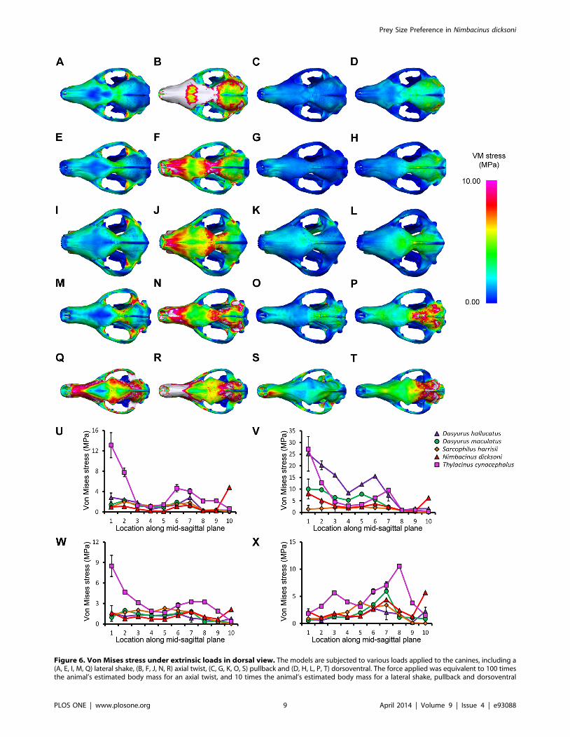
Figure 6. Von Mises stress under extrinsic loads in dorsal view. The models are subjected to various loads applied to the canines, including a(A, E, I, M, Q) lateral shake, (B, F, J, N, R) axial twist, (C, G, K, O, S) pullback and (D, H, L, P, T) dorsoventral. The force applied was equivalent to 100 timesthe animal’s estimated body mass for an axial twist, and 10 times the animal’s estimated body mass for a lateral shake, pullback and dorsoventral
Prey Size Preference in Nimbacinus dicksoni
PLOS ONE | www.plosone.org 9 April 2014 | Volume 9 | Issue 4 | e93088
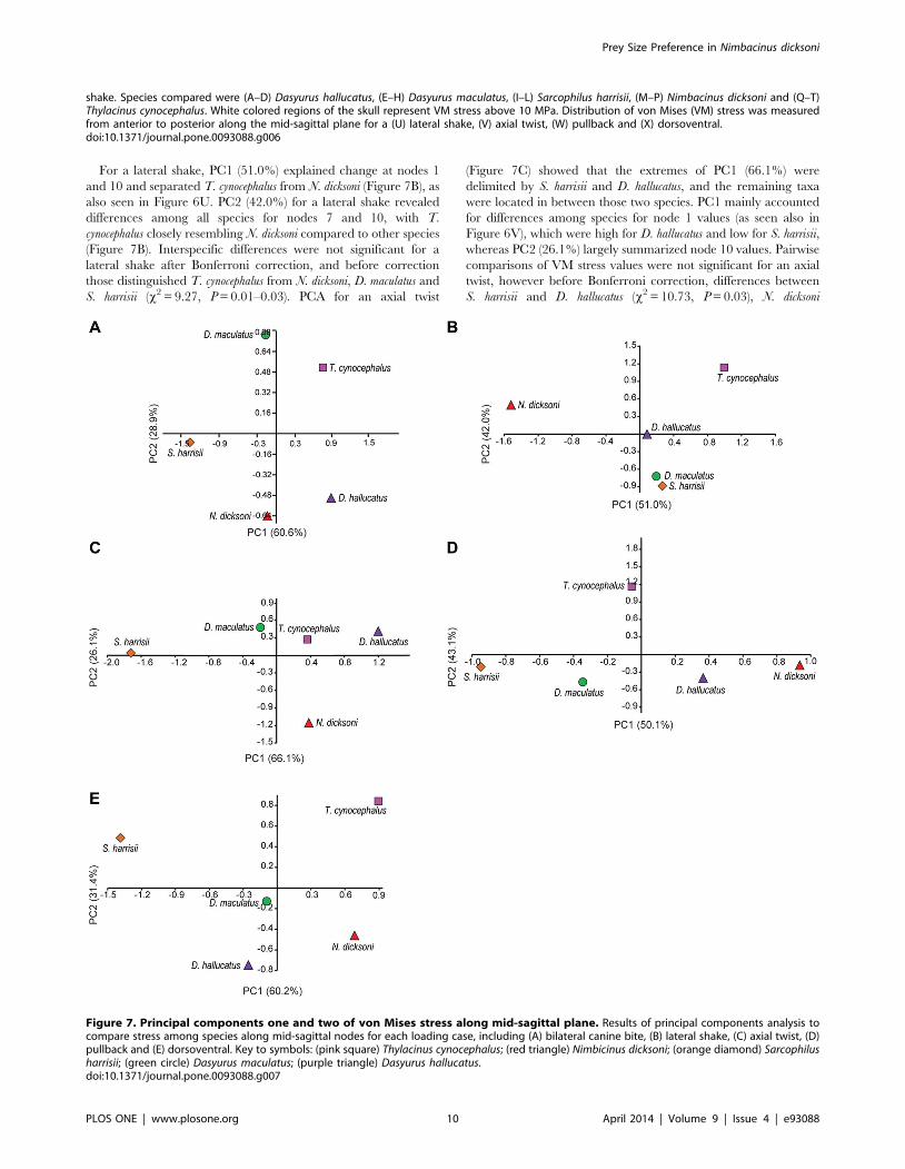
For a lateral shake, PC1 (51.0%) explained change at nodes 1
and 10 and separated T. cynocephalus from N. dicksoni (Figure 7B), as
also seen in Figure 6U. PC2 (42.0%) for a lateral shake revealed
differences among all species for nodes 7 and 10, with T.
cynocephalus closely resembling N. dicksoni compared to other species
(Figure 7B). Interspecific differences were not significant for a
lateral shake after Bonferroni correction, and before correction
those distinguished T. cynocephalus from N. dicksoni, D. maculatus and
S. harrisii (x2 = 9.27, P = 0.01–0.03). PCA for an axial twist
(Figure 7C) showed that the extremes of PC1 (66.1%) were
delimited by S. harrisii and D. hallucatus, and the remaining taxa
were located in between those two species. PC1 mainly accounted
for differences among species for node 1 values (as seen also in
Figure 6V), which were high for D. hallucatus and low for S. harrisii,
whereas PC2 (26.1%) largely summarized node 10 values. Pairwise
comparisons of VM stress values were not significant for an axial
twist, however before Bonferroni correction, differences between
S. harrisii and D. hallucatus (x2 = 10.73, P = 0.03), N. dicksoni
shake. Species compared were (A–D) Dasyurus hallucatus, (E–H) Dasyurus maculatus, (I–L) Sarcophilus harrisii, (M–P) Nimbacinus dicksoni and (Q–T)Thylacinus cynocephalus. White colored regions of the skull represent VM stress above 10 MPa. Distribution of von Mises (VM) stress was measuredfrom anterior to posterior along the mid-sagittal plane for a (U) lateral shake, (V) axial twist, (W) pullback and (X) dorsoventral.doi:10.1371/journal.pone.0093088.g006
Figure 7. Principal components one and two of von Mises stress along mid-sagittal plane. Results of principal components analysis tocompare stress among species along mid-sagittal nodes for each loading case, including (A) bilateral canine bite, (B) lateral shake, (C) axial twist, (D)pullback and (E) dorsoventral. Key to symbols: (pink square) Thylacinus cynocephalus; (red triangle) Nimbicinus dicksoni; (orange diamond) Sarcophilusharrisii; (green circle) Dasyurus maculatus; (purple triangle) Dasyurus hallucatus.doi:10.1371/journal.pone.0093088.g007
Prey Size Preference in Nimbacinus dicksoni
PLOS ONE | www.plosone.org 10 April 2014 | Volume 9 | Issue 4 | e93088
(x2 = 10.73, P = 0.01) and T. cynocephalus (x2 = 10.73, P = 0.04)
could be distinguished from one another. For a pullback bite, PC1
(50.1%) mainly explained interspecific differences for node 10, and
PC2 (43.1%) explained change at nodes 1 and 8 (Figure 7D). VM
stress values for T. cynocephalus were different from those for D.
hallucatus (x2 = 14.37, P = 0.02) and D. maculatus (x2 = 14.37,
P = 0.04) for a pullback bite. For the dorsoventral shake, PC1
explained 60.2% of variance and reflected differences between T.
cynocephalus and S. harrisii, whereas values across nodes were more
similar among the remaining three species, located toward the
middle of PC1 (Figure 7E). PC2 (31.4%) separated D. hallucatus
from T. cynocephalus, and pairwise comparisons revealed node
values to be different between those two species (x2 = 14.78,
P = 0.01).
Discussion
Differences in biomechanical performance between the three
extant dasyurids included in this study appear consistent with their
respective known feeding behaviors. Dasyurus hallucatus showed
comparatively higher levels of stress in most simulations than S.
harrisii and D. maculatus. Dasyurus hallucatus eats invertebrates and
other relatively small prey [60–62], which may not require
adaptation to sustain the full range of extrinsic loads simulated
here. This species shows particularly high VM stress in axial
twisting, especially in contrast to S. harrisii However, it performs
relatively well under pull-back loading, which may be linked to a
capacity for pulling invertebrates from the ground. Observational
studies on wild D. hallucatus will be required to confirm the
functional role of their skull in prey acquisition. Future work on
the comparative musculoskeletal anatomy and collection of in vivo
or ex vivo biomechanical data of the extant species would likely
improve the predictive power of current bite force and muscle
force estimations. Overall consistencies found between known prey
size and biomechanical performance for extant dasyuromorphians
underscore the potential value of projections based on compar-
ative FEA for extinct/fossil taxa.
Our comparative biomechanical modeling of dasyuromorphian
skulls suggests considerable differences in predatory behaviors
between the two thylacinids considered here. Our 3D based results
indicate that the Oligocene to Miocene N. dicksoni had a high bite
force for its size, comparable to that of extant dasyurids known to
take relatively large prey, D. maculatus and S. harrisii [48,60]. In
light of similar levels of ‘carnassialization’ (development of
relatively long, high amplitude vertical shearing crests) in the
cheektooth dentition with D. maculatus, and a lack of obvious dental
specialization consistent with regular bone-cracking, our results
suggest a predominantly carnivorous diet for N. dicksoni that may
have included relatively large prey. Dasyurus maculatus are
opportunistic hunters, varying their diet in response to environ-
mental disturbances and short-term fluctuations in prey abun-
dance [54,56]. They will prey on vertebrate species up to and
sometimes exceeding their own body mass. Prey includes
bandicoots, smaller dasyurids, possums, smaller macropodoids,
snakes, lizards, birds and frogs, as well as invertebrates. Potential
prey for a fox-sized thylacinid living in the closed forest
communities of Riversleigh likely included many small to
medium-size birds, frogs, lizards and snakes, as well as a wide
range of marsupials, including bandicoots (peramelemorphians),
dasyurids (dasyuromorphians), kangaroos (macropodoids), thingo-
dontans (yalkiparidontians), marsupial moles (notoryctemorphians)
and wombats (vombatoids) [12].
Although our FEA results for N. dicksoni suggest a capacity to kill
prey approaching or exceeding its own body mass, its prey range
may have been limited by competition with sympatric carnivores.
The extent of niche overlap and competition within this ancient,
medium-large sized carnivore community may have been partially
alleviated by occupying different habitats and specializing in
different hunting strategies. The recovery of a near complete
skeleton of N. dicksoni [14] will provide further information on the
locomotion and predatory behavior based on postcranial material;
for example, was N. dicksoni as arboreal as the extant D. maculatus?
Differences in mechanical performance suggest that T.
cynocephalus is unusual relative to other dasyuromorphians,
including, N. dicksoni, as indicated by distinctly higher VM stresses
than all other species in response to each loading case. Thylacinus
cynocephalus, in contrast to N. dicksoni, has completely lost the
metaconid on the lower molars and has a proportionately much
larger postmetacrista on the upper molars. On the basis of
traditional beam theory we predicted that taxa with longer rostra
would exhibit higher stress [90], as evident in the long-snouted T.
cynocephalus relative to shorter-snouted dasyuromorphians. Differ-
ences between T. cynocephalus and other species were also
significant for three out of five simulations examined after
conservative Bonferroni correction for multiple testing. These
results further support the contention by Attard et al. [35] that
niche breadth in T. cynocephalus may have been more limited and
that it likely preyed on relatively small to medium-sized vertebrates
such as wallabies, possums and bandicoots.
Although measures of skull performance in response to forces
imposed by struggling prey revealed closer similarity between the
fossil thylacinid N. dicksoni and large extant carnivorous dasyurids,
than with T. cynocephalus, there were differences. Our reconstruc-
tion suggests that the TMJ was more elevated in N. dicksoni than in
D. maculatus, and higher relative to the height of the cheektooth
row. The TMJ is a complex joint and is important for occlusion
and mastication [91,92]. The position of the TMJ can influence
bite strength and muscle activation [93]. The position of the TMJ
along the anterior-posterior axis tends to lie closer to the plane of
the tooth row in carnivorous taxa [11]. Conclusive determination
of the precise position and morphology of the TMJ in N. dicksoni
must await the discovery of more complete cranial material.
Morphological evidence from past studies further demonstrates
diversity within this family. The smallest thylacinid, Muribacinus
gadiyuli, is believed to have fed on relatively small vertebrates and
invertebrates because it lacks a number of dental features present
in large prey specialists (e.g., robust protoconids and brachyce-
phalization) such as similarly sized D. maculatus [17]. The variety of
feeding behaviors among thylacinids may have helped facilitate
their co-existence within different ecological niches that were later
filled by diversifying carnivorous dasyurids.
Supporting Information
Figure S1 Phylogenetic tree of dasyuromophians inves-tigated in this study. One of several recent assessments of the
phylogenetic relationships of dasyuromorphians, including taxa
that have been examined in this study (Wroe & Musser 2001).
(TIF)
Figure S2 Interactive 3D pdf showing the digitallysegmented cranium and mandible of Thylacinus cyno-cephalus.
(PDF)
Figure S3 Interactive 3D pdf showing the digitallysegmented cranium and mandible of Sarcophilus harri-sii.
(PDF)
Prey Size Preference in Nimbacinus dicksoni
PLOS ONE | www.plosone.org 11 April 2014 | Volume 9 | Issue 4 | e93088
Figure S4 Interactive 3D pdf showing the digitallysegmented cranium and mandible of Dasyurus macula-tus.(PDF)
Figure S5 Interactive 3D pdf showing the digitallysegmented cranium and mandible of Dasyurus halluca-tus.(PDF)
Figure S6 Von Mises stress along zygomatic arch for allloading cases. Distribution of von Mises (VM) stress was
measured from anterior to posterior along the zygomatic arch for
a (A) bilateral canine bite, (B) lateral shake, (C) axial twist, (D)
pullback and (E) dorsoventral.
(TIF)
Table S1 Temporal and geographic distribution ofthylacinid species. Abbreviations: Aust, Australian mainland;
E., Early; L., Late; M., Middle; Mio, Miocene; NG, New Guinea;
NT, Northern Territory; Oligo, Oligocene; Plio, Pliocene; Qld,
Queensland; Tas, Tasmania.
(PDF)
Table S2 Maximum bite forces (N) for un-scaledhomogeneous models for a bilateral canine bite.(PDF)
Table S3 Muscle forces used for each jaw muscledivision in un-scaled intrinsic models. Species studied were
Dasyurus hallucatus, Dasyurus maculatus, Sarcophilus harrisii, Nimbacinus
dicksoni and Thylacinus cynocephalus. These were calculated using
muscle mass proportions from dissected Didelphis virginiana (Turn-
bull 1970). Muscle forces were scaled for a bilateral canine bite by
multiplying the muscle force by the ratio between bite force
estimated using body mass regressions and maximum bite force
estimated from the un-scaled model.
(PDF)
References S1 Supporting Information references.
(PDF)
Acknowledgments
We thank Sandy Ingleby from the Australian museum for providing several
comparative specimens and the makers of Digimorph for access to CT scan
data.
Author Contributions
Conceived and designed the experiments: MRGA SW. Performed the
experiments: MRGA WCHP. Analyzed the data: MRGA LABW WCHP.
Contributed reagents/materials/analysis tools: SW. Wrote the paper:
MRGA WCHP LABW MA SJH TLR SW.
References
1. Krajewski C, Buckley L, Westerman M (1997) DNA phylogeny of the marsupialwolf resolved. Proc R Soc Lond, Ser B: Biol Sci 264: 911–917.
2. Krajewski C, Driskell AC, Baverstock PR, Braun MJ (1992) Phylogeneticrelationships of the thylacine (Mammalia: Thylacinidae) among dasyuroid
marsupials: evidence from cytochrome b DNA sequences. Proc R Soc Lond,
Ser B: Biol Sci 250: 19–27.
3. Lowenstein JM, Sarich VM, Richardson BJ (1981) Albumin systematics of the
extinct mammoth and Tasmanian wolf. Nature 291: 409–411.
4. Muirhead J, Wroe S (1998) A new genus and species, Badjcinus turnbulli
(Thylacinidae: Marsupialia), from the late Oligocene of Riversleigh, northern
Australia, and an investigation of thylacinid phylogeny. J Vert Paleontol 18:612–626.
5. Sarich V, Lowenstein JM, Richardson BJ (1982) Phylogenetic relationships of
the thylacine (Thylacinus cynocephalus, Marsupialia) as reflected in comparativeserology. In: Archer M, editor. Carnivorous Marsupials. Sydney: Royal
Zoological Society of New South Wales. pp. 445–476.
6. Szalay FS (1982) A new appraisal of marsupial phylogeny and classification. In:
Archer M, editor. Carnivorous Marsupials. Sydney: Royal Zoological Society of
New South Wales. pp. 621–640.
7. Thomas RH, Schaffner W, Wilson AC, Paabo S (1989) DNA phylogeny of the
extinct marsupial wolf. Nature 340: 465–467.
8. Wroe S, Archer M (2006) Origins and early radiations of marsupials. In: MerrickJR, Archer M, Hickey GM, Lee MSY, editors. Evolution and Biogeography of
Australasian Vertebrates. Sydney: Australian Scientific Publishing. pp. 517–540.
9. Goswami A, Milne N, Wroe S (2011) Biting through constraints: cranial
morphology, disparity and convergence across living and fossil carnivorous
mammals. Proceedings of the Royal Society B: Biological Sciences 278: 1831–1839.
10. Wroe S (2001) Maximucinus muirheadae, gen. et sp. nov. (Thylacinidae:Marsupialia), from the Miocene of Riversleigh, north-western Queensland,
with estimates of body weights for fossil thylacinids. Aust J Zool 49: 603–614.
11. Wroe S, Milne N (2007) Convergence and remarkably consistent constraint inthe evolution of mammalian carnivore skull shape. Evolution 61: 1251–1260.
12. Archer M, Arena DA, Bassarova M, Beck RMD, Black K, et al. (2006) Current
status of species-level representation in faunas from selected fossil localities in theRiversleigh World Heritage Area, northwestern Queensland. Alcheringa 30: 1–
17.
13. Muirhead J (1997) Two new thylacines (Marsupialia: Thylacinidae) from early
Miocene sediments of Riversleigh, northwestern Queensland and a revision of
the family Thylacinidae. Mem Queensl Mus 41: 367–377.
14. Muirhead J, Archer M (1990) Nimbacinus dicksoni, a plesiomorphic thylacine
(Marsupialia: Thylacinidae) from Tertiary deposits of Queensland and theNorthern Territory. Mem Queensl Mus 28: 203–221.
15. Murray PF (1997) Thylacinus megiriani, a new species of thylacine (Marsupialia:
Thylacinidae) from the Ongeva local fauna of central Australia. Rec S Aust Mus30: 43–61.
16. Murray PF, Megirian D (2000) Two new genera and three new species of
Thylacinidae (Marsupialia) from the Miocene of the Northern Territory,
Australia. The Beagle, Records of the Museums and Art Galleries of the
Northern Territory 16: 145–162.
17. Wroe S (1996) Muribacinus gadiyuli, (Thylacinidae: Marsupialia), a very
plesiomorphic thylacinid from the Miocene of Riversleigh, northwestern
Queensland, and the problem of paraphyly for the Dasyuridae. J Paleontol
70: 1032–1044.
18. Wroe S, Musser A (2001) The skull of Nimbacinus dicksoni (Thylacinidae:
Marsupialia). Aust J Zool 49: 487–514.
19. Archer M, Hand S, Godthelp H (1991) Riversleigh: The Story of Animals in
Ancient Rainforests of Inland Australia. Sydney: Reed Books. 264 p.
20. Travouillon KJ, Legendre S, Archer M, Hand SJ (2009) Palaeoecological
analyses of Riversleigh’s Oligo-Miocene sites: implications for Oligo-Miocene
climate change in Australia. Palaeogeogr, Palaeoclimatol, Palaeoecol 276: 24–
37.
21. Archer M, Brammal J, Field J, Hand SJ, Hook C (2002) The Evolution of
Australia: 110 million years of change. Sydney: Australian Museum. 91 p.
22. Wroe S (2002) A review of terrestrial mammalian and reptilian carnivore
ecology in Australian fossil faunas and factors influencing their diversity: The
myth of reptilian domination and its broader ramifications. Aust J Zool 49: 603–
614.
23. Wroe S, Myers TJ, Wells RT, Gillespie A (1999) Estimating the weight of the
Pleistocene marsupial lion, Thylacoleo carnifex (Thylacoleonidae: Marsupialia):
implications for the ecomorphology of a marsupial super-predator and
hypotheses of impoverishment of Australian marsupial carnivore faunas.
Aust J Zool 47: 489–498.
24. Wroe S (2003) Australian marsupial carnivores: an overview of recent advances
in palaeontology. In: Jones M, Dickman C, Archer M, editors. Predators with
Pouches: The Biology of Carnivorous Marsupials. Collingwood: CSIRO
Publishing. pp. 102–123.
25. Wroe S, Brammall J, Cooke BN (1998) The skull of Ekaltadeta ima (Marsupialia,
Hypsiprymnodontidae?): an analysis of some marsupial cranial features and a re-
investigation of propleopine phylogeny, with notes on the inference of carnivory
in mammals. J Paleontol 72: 738–751.
26. Wroe S, McHenry C, Thomason J (2005) Bite club: comparative bite force in big
biting mammals and the prediction of predatory behaviour in fossil taxa.
Proc R Soc Lond, Ser B: Biol Sci 272: 619–625.
27. Dumont ER, Piccirillo J, Grosse IR (2005) Finite element analysis of biting
behavior and bone stress in the facial skeletons of bats. Anat Rec 283: 319–330.
28. Ross CF (2005) Finite element analysis in vertebrate biomechanics. Anat
Rec A Discov Mol Cell Evol Biol 283A: 253–258.
29. Tseng ZJ (2009) Cranial function in a late Miocene Dinocrocuta gigantea
(Mammalia: Carnivora) revealed by comparative finite element analysis.
Biol J Linn Soc 96: 51–67.
30. Wroe S, Ferrara TL, McHenry CR, Curnoe D, Chamoli U (2010) The
craniomandibular mechanics of being human. Proc R Soc Lond, Ser B: Biol Sci
277: 3579–3586.
Prey Size Preference in Nimbacinus dicksoni
PLOS ONE | www.plosone.org 12 April 2014 | Volume 9 | Issue 4 | e93088
31. McHenry CR, Wroe S, Clausen PD, Moreno K, Cunningham E (2007)
Supermodeled sabercat, predatory behavior in Smilodon fatalis revealed by high-resolution 3D computer simulation. Proc Natl Acad Sci USA 104: 16010–16015.
32. Wroe S, Clausen P, McHenry C, Moreno K, Cunningham E (2007) Computer
simulation of feeding behaviour in the thylacine and dingo as a novel test forconvergence and niche overlap. Proc R Soc Lond, Ser B: Biol Sci 274: 2819–
2828.33. Young MT, Rayfield EJ, Holliday CM, Witmer LM, Button DJ, et al. (2012)
Cranial biomechanics of Diplodocus (Dinosauria, Sauropoda): testing hypotheses
of feeding behaviour in an extinct megaherbivore. Naturwissenschaften 99: 637–643.
34. Bell PR, Snively E, Shychoski L (2009) A comparison of the jaw mechanics inhadrosaurid and ceratopsid dinosaurs using finite element analysis. Anat Rec
(Hoboken) 292: 1338–1351.35. Attard MRG, Chamoli U, Ferrara TL, Rogers TL, Wroe S (2011) Skull
mechanics and implications for feeding behaviour in a large marsupial carnivore
guild: the thylacine, Tasmanian devil and spotted-tailed quoll. J Zool 285: 292–300.
36. Chamoli U, Wroe S (2011) Allometry in the distribution of material propertiesand geometry of the felid skull: Why larger species may need to change and how
they may achieve it. J Theor Biol 283: 217–226.
37. Moazen M, Curtis N, Evans SE, O’Higgins P, Fagan MJ (2008) Combined finiteelement and multibody dynamics analysis of biting in a Uromastyx hardwickii lizard
skull. J Anat 213: 499–508.38. Oldfield CC, McHenry CR, Clausen PD, Chamoli U, Parr WCH, et al. (2012)
Finite element analysis of ursid cranial mechanics and the prediction of feedingbehaviour in the extinct giant Agriotherium africanum. J Zool 286: 163–170.
39. Rayfield EJ, Norman DB, Horner CC, Horner JR, Smith PM, et al. (2001)
Cranial design and function in a large theropod dinosaur. Nature 409: 1033–1037.
40. Slater GJ, Figueirido B, Louis L, Yang P, Van Valkenburgh B (2010)Biomechanical Consequences of Rapid Evolution in the Polar Bear Lineage.
PLoS ONE 5: e13870.
41. Strait DS, Grosse IR, Dechow PC, Smith AL, Wang Q, et al. (2010) Thestructural rigidity of the cranium of Australopithecus africanus: implications for diet,
dietary adaptations, and the allometry of feeding biomechanics. Anat Rec(Hoboken) 293: 583–593.
42. Thresher RW, Saito GE (1973) The stress analysis of human teeth. J Biomech 6:443–449.
43. Paddle R (2000) The Last Tasmanian tiger: the History and Extinction of the
Thylacine. Oakleigh, VIC: Cambridge University Press. 273 p.44. Jones ME, Stoddart DM (1998) Reconstruction of the predatory behaviour of
the extinct marsupial thylacine (Thylacinus cynocephalus). J Zool 246: 239–246.45. Jones ME (2003) Convergence in ecomorphology and guild structure among
marsupial and placental carnivores. In: Jones ME, Dickman C, Archer M,
editors. Predators with Pouches: The Biology of Carnivorous Marsupials.Collingwood: CSIRO Publishing. pp. 285–269.
46. Guiler ER (1985) Thylacine: The tragedy of the Tasmanian tiger. Melbourne:Oxford University Press. 207 p.
47. Bailey C (2003) Tiger Tales: Stories of the Tasmanian Tiger. Sydney:HarperCollins Publishers. 164 p.
48. Figueirido B, Janis CM (2011) The predatory behaviour of the thylacine:
Tasmanian tiger or marsupial wolf? Biol Lett 7: 937–940.49. Jones ME, Barmuta LA (1998) Diet overlap and abundance of sympatric
dasyurid carnivores: a hypothesis of competition? J Anim Ecol 67: 410–421.50. Bradshaw CJA, Brook BW (2005) Disease and the devil: density-dependent
epidemiological processes explain historical population fluctuations in the
Tasmanian devil. Ecography 28: 181–190.51. Guiler E (1970) Obsevations on the Tasmanian devil, Sarcophilus harrisii
(Marsupialia : Dasyuridae) I. Numbers, home, range, movements and food intwo populations. Aust J Zool 18: 49–62.
52. Taylor RJ (1986) Notes on the diet of the carnivorous mammals of the upper
Henry River region, Western Tasmania. Pap Proc R Soc Tasman 120: 7–10.53. Groves CP (2005) Order Dasyuromorphia. In: Wilson DE, Reeder DM, editors.
Mammal Species of the World: A Taxonomic and Geographic Reference. 3rded. Baltimore: Johns Hopkins University Press. pp. 23–37.
54. Edgar R, Belcher C (1995) Spotted-tailed quoll, Dasyurus maculatus. In: StrahanR, editor. The Mammals of Australia. Sydney: Reed. pp. 67–69.
55. Glen AS, Dickman CR (2006) Diet of the spotted-tailed quoll (Dasyurus maculatus)
in eastern Australia: effects of season, sex and size. J Zool 269: 241–248.56. Jones ME (1997) Character displacement in Australian dasyurid carnivores: size
relationships and prey size patterns. Ecology 78: 2569–2587.57. Dawson JP, Claridge AW, Triggs B, Paull DJ (2007) Diet of a native carnivore,
the spotted-tailed quoll (Dasyurus maculatus), before and after an intense wildfire.
Wildl Res 34: 342–351.58. Braithwaite RW, Begg RJ (1995) Northern quoll Dasyurus hallucatus Gould, 1842.
In: Strahan R, editor. The Mammals of Australia: National Photographic Indexof Australian Wildlife. Sydney: Reed Books. pp. 65–66.
59. Strahan R (1995) The Mammals of Australia. Sydney: New Holland PublishingPty Ltd.
60. Belcher CA (1995) Diet of the Tiger quoll (Dasyurus maculatus) in East Gippsland,
Victoria. Wildl Res 22: 341–357.61. Oakwood M (1997) The ecology of the northern quoll, Dasyurus hallucatus [PhD
thesis]. Canberra: Australian National University. 556 p.
62. Pollock AB (1999) Notes on status, distribution and diet of northern quollDasyurus hallucatus in the Mackay-Bowen area, mideastern Queensland. Aust
Zool 31: 388–395.63. Wroe S (2008) Cranial mechanics compared in extinct marsupial and extant
African lions using a finite-element approach. J Zool 274: 332–339.
64. Benazzi S, Bookstein FL, Strait DS, Weber GW (2011) A new OH5reconstruction with an assessment of its uncertainty. J Hum Evol 61: 75–88.
65. Besl PJ, McKay ND (1992) A method for registration of 3D shapes. IEEETransactions on Pattern Analysis and Machine Intelligence. pp. 239–256.
66. Parr WCH, Wroe S, Chamoli U, Richards HS, McCurry MR, et al. (2012)Toward integration of geometric morphometrics and computational biome-
chanics: New methods for 3D virtual reconstruction and quantitative analysis of
Finite Element Models. J Theor Biol 301: 1–14.67. Rayfield EJ (2007) Finite element analysis and understanding the biomechanics
and evolution of living and fossil organisms. Annu Rev Earth Planet Sci 35: 541–576.
68. Cook SD, Weinstein AH, Klawitter JJ (1982) A three-dimensional finite element
analysis of a porous rooted Co–Cr–Mo alloy dental implant. J Dent Res 61: 25–29.
69. Dumont ER, Grosse IR, Slater GJ (2009) Requirements for comparing theperformance of finite element models of biological structures. J Theor Biol 256:
96–103.70. Greaves G, Greer A, Lakes R, Rouxel T (2011) Poisson’s ratio and modern
materials. Nat Mater 10: 823–837.
71. Turnbull WD (1970) Mammalian masticatory apparatus. Fieldiana: Geology 18:149–356.
72. Thomason JJ (1991) Cranial strength in relation to estimated biting forces insome mammals. Can J Zool 69: 2326–2333.
73. Chamoli U (2011) Biomechanics of the felid skulls: A comparative study using
finite element approach [Masters thesis]. Sydney: University of New SouthWales. 144 p.
74. Clausen P, Wroe S, McHenry C, Moreno K, Bourke J (2008) The vector of jawmuscle force as determined by computer-generated three dimensional
simulation: a test of Greaves’ model. J Biomech 41: 3184–3188.75. Jones ME (1995) Guild structure of the large marsupial carnivores in Tasmania
[PhD thesis]. Hobart: University of Tasmania. 176 p.
76. Pellis SM, Nelson A (1984) Some aspects of predatory behaviour of the quollDasyurus viverrinus (Marsupialia: Dasyuridae). Aust Mammal 7: 5–15.
77. Pellis SM, Officer RCE (1987) An analysis of some predatory behaviour patternsin four species of carnivorous marsupials (Dasyuridae), with comparative notes
on the Eutherian cat Felis catus. Ethology 75: 177.
78. Fleay D (1932) The rare dasyures (native cats). Vic Nat 49: 63–69.79. Dumont ER, Herrel A (2003) The effect of gape angle and bite point on bite
force in bats. J Exp Biol 206: 2117–2123.80. Christiansen P, Wroe S (2007) Bite forces and evolutionary adaptations to
feeding ecology in carnivores. Ecology 88: 347–358.81. Myers TJ (2001) Marsupial body mass prediction. Aust J Zool 49: 99–118.
82. Wroe S, Chamoli U, Parr WCH, Clausen P, Ridgely R, et al. (2013)
Comparative biomechanical Modeling of Metatherian and Placental Saber-Tooths: A Different Kind of Bite for an Extreme Pouched Predator. PLoS ONE
8: e66888.83. Ellis JL, Thomason JJ, Kebreab E, France J (2008) Calibration of estimated
biting forces in domestic canids: comparison of post-mortem and in vivo
measurements. J Anat 212: 769–780.84. Nalla RK, Kinney JH, Ritchie RO (2003) Mechanistic fracture criteria for the
failure of human cortical bone. Nat Mater 2: 164–168.85. Tsafnat N, Wroe S (2010) An experimentally validated micromechanical model
of a rat vertebra under compressive loading. J Anat 218: 40–46.
86. Mitteroecker P, Gunz P (2009) Advances in geometric morphometrics. Evol Biol36: 235–247.
87. Zar JH (1996) Multiple regression and correlation. Biostatistical Analysis 3rd edUpper Saddle River, NJ: Prentice Hall: 353–360.
88. Owen D, Pemberton D (2005) Tasmanian Devil: A Unique and ThreatenedAnimal. Crows Nest, NSW: Allen and Unwin. 225 p.
89. Figueirido B, Tseng ZJ, Martı́n-Serra A (2013) Skull shape evolution in
durophagous carnivorans. Evolution 67: 1975–1993.90. Walmsley CW, Smits PD, Quayle MR, McCurry MR, Richards HS, et al.
(2013) Why the long face? The mechanics of mandibular symphysis proportionsin crocodiles. PLoS ONE 8: e53873.
91. Hylander WL (1979) An experimental analysis of temporomandibular joint
reaction force in macaques. Am J Phys Anthropol 51: 433–456.92. Breul R, Mall G, Landgraf J, Scheck R (1999) Biomechanical analysis of stress
distribution in the human temporomandibular-joint. Ann Anat 181: 55–60.93. Hickman DM, Cramer R (1998) The effect of different condylar positions on
masticatory muscle electromyographic activity in humans. Oral Surg Oral MedOral Pathol Oral Radiol Endod 85: 18–23.
Prey Size Preference in Nimbacinus dicksoni
PLOS ONE | www.plosone.org 13 April 2014 | Volume 9 | Issue 4 | e93088


