Embed Size (px)
Citation preview
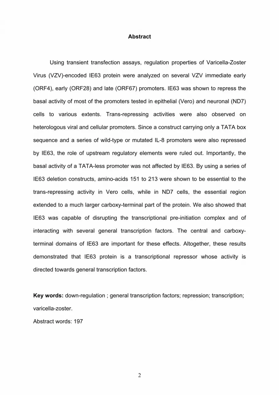
1
Varicella-zoster-virus IE63 protein represses the basal transcription
machinery by disorganizing the pre-initiation complex.
Emmanuel Di Valentin1, Sébastien Bontems1, Lionel Habran1, Olivier Jolois3, Nicolas
Markine-Goriaynoff2, Alain Vanderplasschen2, Catherine Sadzot-Delvaux1 and
Jacques Piette1*
1 Laboratory of Virology and Immunology, University of Liège, B-4000 Liège, Belgium
2 Laboratory of Immunology and Vaccinology, University of Liège, B-4000 Liège,
Belgium
3 Laboratory of Histology, University of Liège, B-4000 Liège, Belgium
*To whom correspondence should be addressed: Jacques Piette, Laboratory of
Virology and Immunology, Institute of Pathology B23, University of Liège, B-4000
Liège, Belgium.
Tel: 32-4-366 2442, Fax: 32-4-366 9933, E-mail: [email protected]
Short title: VZV IE63 regulatory properties
2
Abstract
Using transient transfection assays, regulation properties of Varicella-Zoster
Virus (VZV)-encoded IE63 protein were analyzed on several VZV immediate early
(ORF4), early (ORF28) and late (ORF67) promoters. IE63 was shown to repress the
basal activity of most of the promoters tested in epithelial (Vero) and neuronal (ND7)
cells to various extents. Trans-repressing activities were also observed on
heterologous viral and cellular promoters. Since a construct carrying only a TATA box
sequence and a series of wild-type or mutated IL-8 promoters were also repressed
by IE63, the role of upstream regulatory elements were ruled out. Importantly, the
basal activity of a TATA-less promoter was not affected by IE63. By using a series of
IE63 deletion constructs, amino-acids 151 to 213 were shown to be essential to the
trans-repressing activity in Vero cells, while in ND7 cells, the essential region
extended to a much larger carboxy-terminal part of the protein. We also showed that
IE63 was capable of disrupting the transcriptional pre-initiation complex and of
interacting with several general transcription factors. The central and carboxy-
terminal domains of IE63 are important for these effects. Altogether, these results
demonstrated that IE63 protein is a transcriptional repressor whose activity is
directed towards general transcription factors.
Key words: down-regulation ; general transcription factors; repression; transcription;
varicella-zoster.
Abstract words: 197

3
Introduction
Varicella-Zoster-Virus (VZV) is a human alphaherpesvirus that causes
chickenpox (varicella) and becomes latent in dorsal root ganglia (DRG). Upon
reactivation from latency, VZV is responsible for zoster (shingles). The VZV genome
is a double-stranded DNA molecule composed of 71 open reading frames (ORFs)
encoding 68 proteins that seem to be regulated in a manner similar to other
alphaherpesviruses. After entry into the cells, VZV genes are expressed in a
temporal cascade. The immediate early (IE) genes are expressed first. They
stimulate early (E) gene expression, providing most of the proteins necessary for viral
DNA replication. After DNA synthesis has occurred, genes of the late (L) class
encoding structural proteins are expressed. In contrast to Herpes Simplex Virus type
1 (HSV-1), the VZV latency is characterized by the expression of several IE (ORFs 4,
62 and 63) and E (ORFs 21, 29, 66) genes (Cohrs et al., 1996, Cohrs et al., 2003,
Grinfeld and Kennedy, 2004, Mahalingam et al., 1996, Meier et al., 1993). However,
the biochemical and molecular processes controlling latency and reactivation of VZV
are still largely unknown.
VZV open reading frames 63 and 70 (ORF63/70) encode a 278-amino-acid
long protein with a predicted molecular mass of 30.5 kDa but produce an extensively
modified 45 kDa protein in infected cells (Debrus et al., 1995). ORF63 is expressed
as an IE protein (IE63) and exhibits a limited homology with the HSV-1 IE protein
ICP22 (Davison and McGeoch, 1986, Davison and Scott, 1986) and the EHV-1
EICP22 protein (Derbigny et al., 2000). IE63 most closely resembles HSV-1 Us1.5,
which is a 274-amino-acid protein encoded by a gene that is co-linear with the HSV-1
ICP22 (Baiker et al., 2004).
4
IE63 is of particular interest in VZV pathogenesis since it is abundantly
expressed during acute infection as well as during latency (Debrus et al., 1995,
Grinfeld and Kennedy, 2004, Kennedy et al., 2000, Mahalingam et al., 1996). During
lytic infection, its localization is mostly nuclear but it can also be faintly detected in the
cytoplasm. However, during latency, IE63 is almost exclusively localized in the
cytoplasm of infected neuronal cells (Grinfeld and Kennedy, 2004, Lungu et al., 1998,
Mahalingam et al., 1996). IE63 protein interacts with VZV IE62 protein (Baiker et al.,
2004, Lynch et al., 2002) and is essential for VZV replication (Baiker et al., 2004,
Sommer et al., 2001) and latency (Cohen et al., 2004).
Transcription of viral genes during a productive infection is mediated by
interaction between viral activator or repressor proteins and various components of
the cellular transcription machinery. This interaction is important for increasing or
decreasing the assembly of the preinitiation complex (PIC) necessary for
transcription of genes by RNA polymerase II (RNA POL II). Activators or repressors
may act directly or indirectly on the PIC formation. The PIC is composed of the RNA
POL II and general transcription factors (GTFs) (for recent review, see (Hahn, 2004)
that can be attached on promoter templates in vitro (Buratowski, 1994, Grondin and
DeLuca, 2000, Hampsey, 1998). In most cases, the recognition of promoters is
mediated by TFIID through the binding of the TATA binding protein (TBP) subunit to
TATA box elements. The action of several viral proteins has been shown to be
directed toward the PIC assembly (Choy and Green, 1993, Grondin and DeLuca,
2000, Gu et al., 1995, Huang and McCance, 2002, Jang et al., 2001, Kim et al.,
2003, Long et al., 1999, Manet et al., 1993).
The gene regulatory properties of IE63 protein are not very well understood
and remain controversial. Some authors have reported that IE63 protein could

5
repress the VZV IE62 promoter, stimulate the VZV thymidine kinase promoter but
could not affect expression of late genes in Vero cells (Jackers et al., 1992). Another
team suggested that expressed under the control of its own promoter, IE63 did not
exert any regulatory properties on IE, E and L genes in the same cell line (Kost et al.,
1995). Conversely, IE63 was shown to up-regulate IE62 effect on VZV gI promoter in
A3.01 cells, a CD4+ T-cell line and to interact with VZV IE62 protein (Lynch et al.,
2002). Recently, IE63 has been shown to exert important trans-repressive properties
on VZV DNA polymerase gene (ORF28) promoter both in Vero and ND7 cells
(Bontems et al., 2002). Therefore, an extensive series of transient transfection
studies were performed in order to better understand IE63 gene regulatory properties
and its mechanism of action. From the data presented in this paper, it appears that
IE63 is capable of repressing the basal activity of several promoters and is able to
interfere with the components of the pre-initiation complex.
6
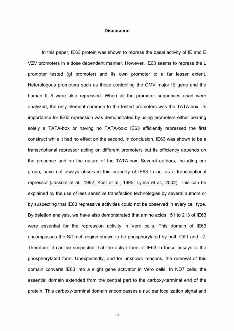
Results
IE63 is a transcriptional repressor targeting the TATA-box. For many
years, the transcriptional regulatory properties of IE63 remained unclear, being
described either as a transcriptional repressor or activator depending on the promoter
tested or without any transcriptional effect (Jackers et al., 1992, Kost et al., 1995,
Lynch et al., 2002). Recently, it was unambiguously shown that IE63 represses
transcription initiated from the promoter controlling the VZV DNA polymerase gene
(Bontems et al., 2002). In order to better characterize IE63 regulatory properties, we
decided to extend this important observation to other VZV promoters and to compare
IE63 activity in two cell lines where VZV leads to either a productive (monkey kidney
epithelial cells, Vero) or non-productive (immortalized rat sensory neurons, ND7)
cycle (Bontems et al., 2002). Vero and ND7 cells were transfected with a plasmid
expressing IE63 together with various constructs (Table 1) where the reporter gene
was under the control of autologous (Figure 1) as schematically represented in
Figure 1A or heterologous viral and cellular promoters (Figures 2 and 3) as
schematically represented in Figures 2A and 3A. Figure 1B and 1C show that in both
Vero and ND7 cells, IE63 exerts transcriptional repression properties towards
autologous VZV promoters belonging to two classes: ORF4 (p4-Luc) which encodes
a gene regulator expressed as an IE protein, ORF 28 (pPOL-Luc) which encodes the
viral DNA polymerase and is an E protein. The basal activity of these promoters was
dose-dependently decreased with IE63 expression (pcDNA-IE63). IE63 did not show
any repressive activity on the promoter belonging to the L class (p67-Luc) that
controls expression of the glycoprotein I (Figure 1B and C). The presence of IE63
7
expression in the two cell lines was detected by western blot analysis (Figure 1B and
C, lower part).
In order to identify elements of these promoters that could help us to clarify the
mechanism of repression mediated by IE63, sequence alignment analysis was
carried out. The only motives shared by these promoters were a TATA-box and the
Initiator element (InR) suggesting that IE63 could exert its repression on basal
transcription initiation. Then, computer search for TATA-boxes was carried out on the
promoter sequences using three different programs (Table 1). The scores were
arbitrarily set on the same scale from 0 to 1; 1 being the best score. TATA-boxes of
ORF4 (p4-Luc), ORF28 (pPOL-Luc), and ORF67 (p67-Luc) were previously mapped
(Kinchington et al., 1994, Kost et al., 1995, Ling et al., 1992, Meier et al., 1994). As
expected, sequence analysis of these 3 TATA-boxes revealed that ORF4 and 28
obtained good scores, while the score of the ORF67 TATA box was much lower and
could be classified as an atypical TATA-box (Table 1). We then extended the
sequence analysis to other VZV promoters (Table 1). Strikingly, one of the lowest
scores was obtained for a TATA-box from the ORF63 promoter (p63-Luc) which
apparently had also an atypical TATA-box (Table 1). Transient transfection of Vero
cells was therefore carried out with this promoter (Figure 1D). As shown in Figure 1D,
IE63 exhibited a lesser repressing activity against its own promoter either under an
extended or a short (163 bp) version (p63(163bp)-Luc) (Figure 1A). A similar
behaviour was observed in ND7 cells transfected with the extended version of the
ORF63 promoter (data not shown).
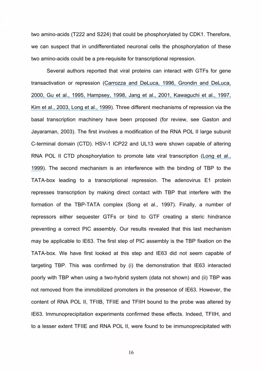
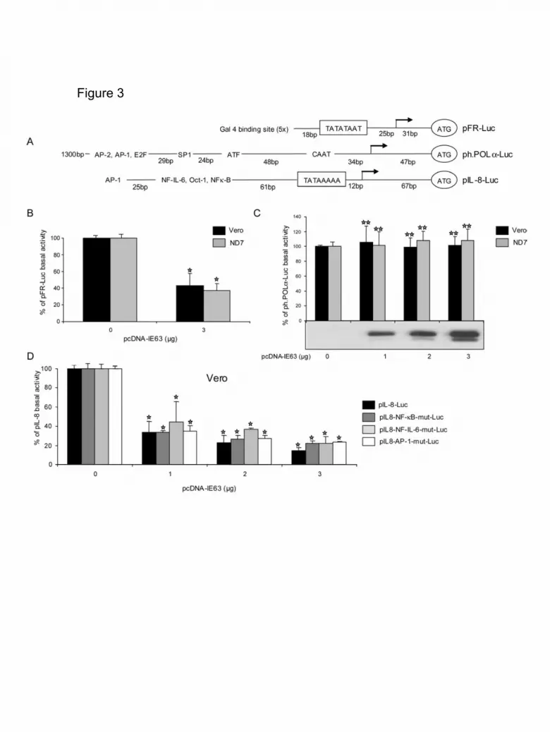
We also analyzed different heterologous viral and cellular promoters known to
have good TATA-boxes (Table 1). These were: the major IE promoter from human
cytomegalovirus (CMV, pCMV-Luc) (Foecking and Hofstetter, 1986) and the HSV-1
8
UL44 (gC, pEL-Pgc-Luc) promoters (Lium and Silverstein, 1997). As expected, the
scores obtained with the various programs were very close to 1 (Table 1). IE63
repression property of these promoters was also examined (Figure 2). In that respect,
these two promoters also had their basal activities down-regulated by IE63, both in
Vero and ND7 cells (Figure 2B, C). The observation that both VZV and heterologous
promoters could have their basal activity repressed by IE63 in a similar extend and
independently of the cell type used is an interesting information that clarifies the
regulatory properties of this viral protein.
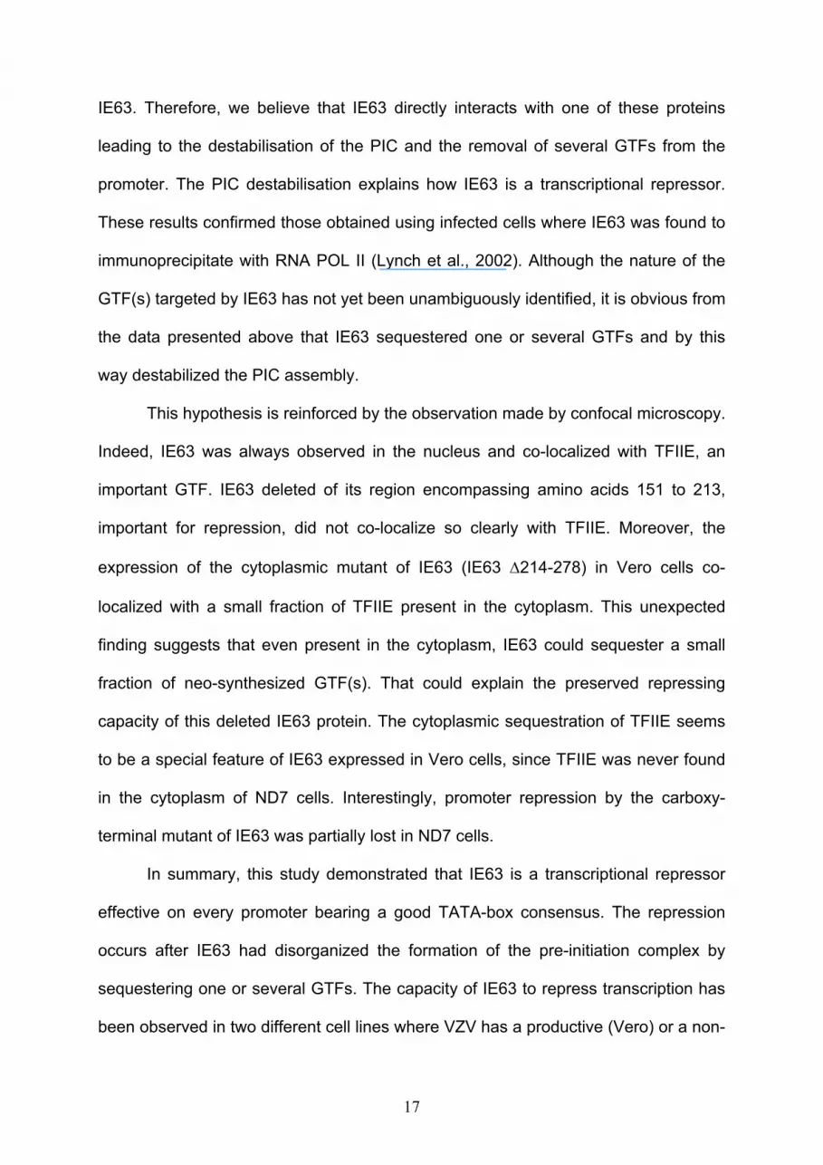
To further clarify the importance of TATA-box and/or InR elements in the
repression mediated by IE63, we used a reporter plasmid whose transcription was
under the control of a TATA box (pFR-Luc). The scores obtained by this sequence
were quite good (Table 1). Figure 3B shows that IE63 reduced the activity of this
promoter by about 60% both in Vero and ND7 cells, reinforcing the idea that IE63
targeted TATA-boxes and/or InR. In order to confirm that IE63 targets only TATA-box
motif, we have analyzed the effect of IE63 on the human polymerase alpha promoter
(ph.POLα-Luc) which lacks this motive but bears an InR (Figure 3A and C). As
expected, no TATA-box motive within this promoter was detectable by computer
analysis (Table 1). Even without any detectable TATA-box, this promoter exhibited a
high basal activity. Although IE63 protein was dose-dependently expressed (Fig 3c,
lower part), the basal activity of the human polymerase alpha promoter was not
decreased both in Vero and ND7 cells (Fig 3c). These results confirmed, on the one
hand, that several upstream regulatory elements such as AP-2, AP-1, EF2, SP1, ATF
and CAAT box were not involved in IE63 repression and, on the other hand, IE63
protein targets only the TATA-box sequence. This experiment also ruled out the role
of the InR sequence in the IE63-mediated repression. The lack of importance of

9
several upstream regulatory elements was further confirmed by using various
constructs of the cellular IL-8 promoter which has been very well-characterized
(Roebuck, 1999). The wild-type version of the promoter was dose-dependently
repressed in both cell lines (Figure 3D, Table 1) and this repression was independent
on the mutations of various upstream regulatory elements. This demonstrated that
AP-1, NF-IL-6 and NF-κB elements were not required for IE63 activity.
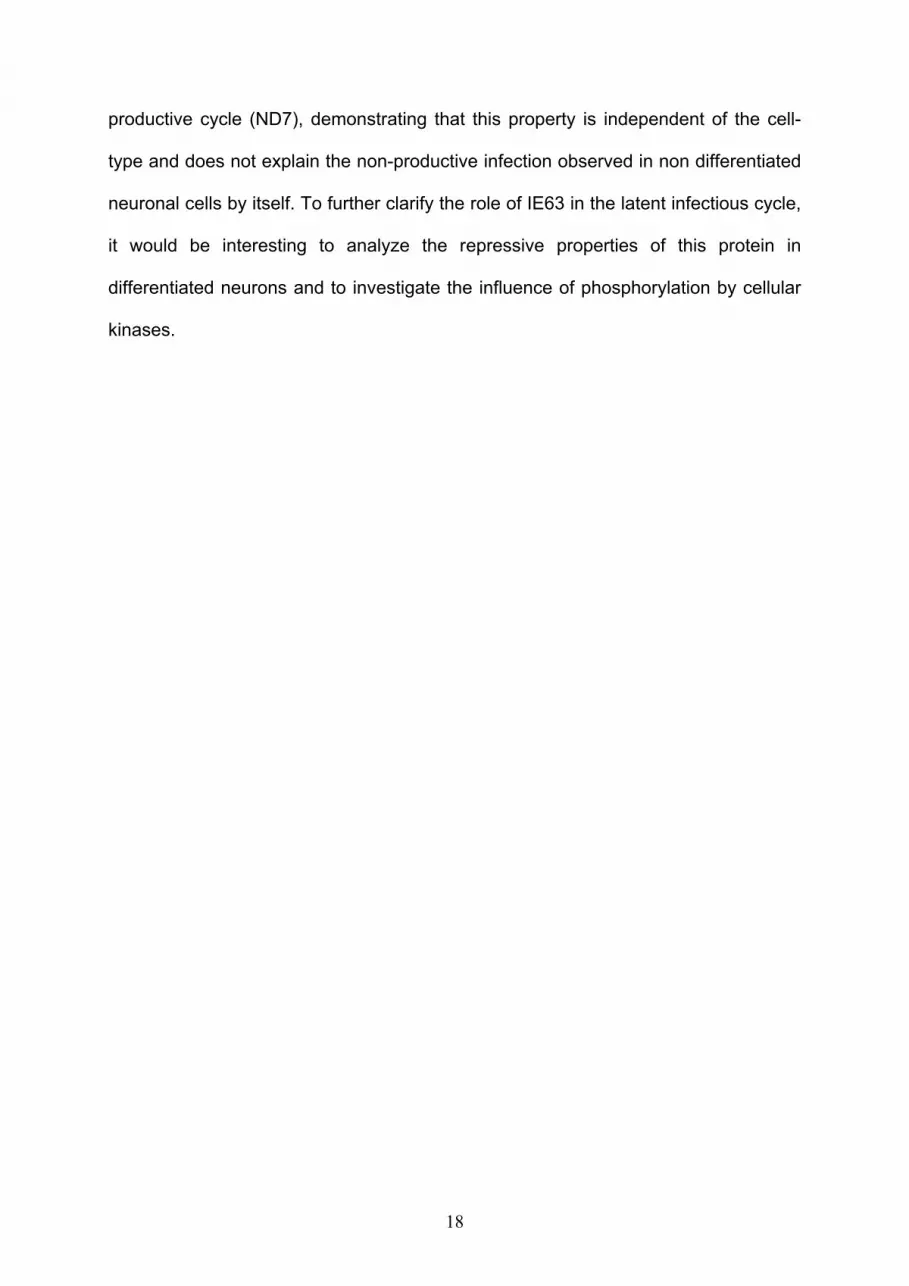
IE63 central and carboxy-terminal domains are important for repression.
Recently, we showed that IE63 is a highly phosphorylated protein and the
phosphorylation sites situated in the carboxy-terminal regions are necessary for
repression (Bontems et al., 2002). In order to better characterize IE63 domains that
are important for repression, IE63 deletion mutants were constructed: (i) IE63 ∆1-75
with the amino-terminal region removed, (ii) IE63 ∆76-150 with the central region
partly deleted, (iii) IE63 ∆151-213 with the region rich in CK1-CK2 phosphorylation
sites was deleted and (iv) IE63 ∆214-278 where the carboxy-terminal domain
containing two putative phosphorylation sites for CDK1 (Baiker et al., 2004, Bontems
et al., 2002) and a nuclear localization signal were removed (Figure 4A). Western blot
analysis demonstrated that these mutant proteins were stably expressed after
transfection of Vero (Figure 4B) and ND7 (Figure 4C) with 1 to 3 µg of expression
vectors. It should be noted that the removal of domain 151-213 of IE63 modified the
electrophoretic migration of the protein which appeared somewhat smaller than
expected (25 kDa instead of 34 kDa) (Figure 4B). This is likely due to the removal of
a domain which is known to be highly phosphorylated in the wild-type protein
(Bontems et al. 2002). Co-transfection of cells with IE63 wild-type or the deleted
constructs and VZV DNA polymerase reporter vector (pPOL-Luc) led to unexpected
10
results (Figure 4D and E). Indeed, in Vero cells, the removal of either the amino-
terminal (IE63 ∆1-75), the second domain (IE63 ∆76-150) or the carboxy-terminal
domain (IE63 ∆214-278) of IE63 did not alter its transrepression activity on the
ORF28 (pPOL-Luc) promoter (Figure 4D) in comparison with IE63 wild-type.
However, IE63 domain 3 was found essential for repression since its removal led to
the loss of the repressive activity of the protein. Surprisingly, even the basal activity
of the promoter was increased with this construct. In ND7 cells, the pattern of
repression obtained with these mutant proteins was not identical (Figure 4E). The
proteins lacking domain 3 (IE63 ∆150-213) or domain 4 (IE63 ∆214-278) turned out
to be less efficient in repressing the ORF28 promoter, demonstrating the importance
of the carboxy-terminal part of IE63 in this cell type (Figure 4E). Similar results were
obtained with the IL-8 promoter (data not shown).
IE63 co-localizes in the cell nucleus with TFIIE. As it is likely that IE63
targeted TATA-boxes, we decided to characterize the cellular localization of IE63 with
a component of the basal transcription machinery. In the classical model of
transcriptional pre-initiation complex (PIC) assembly, the transcription factor TFIIE
(composed of two subunits, TFIIEα and TFIIEβ) binds the RNA POL II at a late stage
of initiation. Therefore, TFIIE was found to be a good marker for PIC localization in
the cell nucleus. Confocal microscopy analysis was carried out on Vero and ND7
cells transfected with the various constructs leading to wild-type or mutant IE63
expression (Figure 5). Co-localization between IE63 and endogenous TFIIEα subunit
was analyzed by the use of secondary antibodies linked to Fluorescein (TFIIE, in
green) and Texas red (IE63, in red) (Figure 5A to 5F). IE63 wild- type protein was
mainly observed in the nucleus of Vero and ND7 cells. As shown in Figure 5A and
11
5B, both IE63 and TFIIE are perfectly localized in the nucleus of the two cell types
except in the nucleolus where IE63 was absent. Even if these results did not
demonstrate on their own the interaction between IE63 and TFIIE, they showed that
these proteins exhibited a very similar distribution in the nucleus. A similar co-
localization pattern was observed in cells expressing IE63 ∆1-75 and IE63 ∆76-150
(data not shown).
Analysis of the cells transfected with pcDNA-IE63 ∆151-213 revealed that the
mutant protein expressed was mainly localised in the nucleus (Figure 5C and 5D).
However, significantly less IE63 co-localization with TFIIE was observed compared
with the wild-type protein. In the case of the IE63 ∆214-278, due to the removal of the
nuclear localisation signal, this protein was exclusively localised in the cytoplasm of
the two cell lines (Figure 5E and 5F). Surprisingly, although in all conditions TFIIE
was exclusively located in the nucleus, a minor fraction of TFIIE could be detected in
the cytoplasm of Vero cells expressing IE63 ∆214-278 (Figure 5E). In that case, the
two proteins were shown to co-localize. This observation suggested that IE63 could
partly sequester TFIIE or other associated GTFs in this cellular compartment. In ND7
cells, IE63 was also mainly localized in the cytoplasm but not TFIIE (Figure 5F).
Importantly, IE63∆214-278 and TFIIE were never co-localizing in the nucleus of ND7
cells (Figure 5F). A positive control of nuclear co-localization in identical experimental
conditions was carried out by co-transfecting IE63 and IE62 expression vectors in the
two cell lines. IE62 was chosen since this protein was previously shown capable of
interacting with IE63 (Lynch et al., 2002) (Baiker et al., 2004). As shown in Figures
5G and 5H, IE62 (in green) and IE63 (in red) were detected in the cell nucleus and
perfectly co-localized.

12
IE63 disorganizes the transcriptional pre-initiation complex. We then
decided to examine whether IE63 could influence the PIC assembly when formed on
a DNA probe. In that respect, a PIC assembly assay (Grondin and DeLuca, 2000)
was performed involving a probe encompassing well-described transcription initiation
sites (from HSV-1 gC or IL-8 promoters) bound to magnetic beads and extracts from
Vero and ND7 cells either transfected or not with an IE63 expression vector. After
PIC formation, the DNA-protein complexes were extensively washed and analyzed
by SDS PAGE and silver-stained. Due to a weak binding efficiency of the Vero cell
extracts to the gC or IL-8 probes, these experiments were only carried out with ND7
cells. As shown in Figure 6A, SDS-PAGE analysis reveals several bands with a
diminished intensity after IE63 expression (shown by the arrows), whereas others
were totally unchanged (shown by stars). Examples of putative GTFs that could
correspond to proteins having a reduced band intensity are listed in Figure 6A: TFIIE
subunits: α (56 kDa), β (34 kDa), RNA POL II subunits: POLR2A (220 kDa), POLR2C
(33 kDa), TAFs: TAF1 (250 kDa), TAF7 (55 kDa), TAF10 (30 kDa), TFIIH:
polypeptide 2 (44 kDa), polypeptide 3 (34 kDa) and TFIIB (33 kDa). The mean
reduction in the amount of putative transcription factors by IE63 expression was
calculated after gel photodensitometry: TAF1: 39 %, POLR2A: 41 %, TFIIEα: 18 %,
TFIIH2: 25 %, TFIIEβ: 50%. It should be noted that no increase in the band intensity
was observed after IE63 expression in ND7 cells. In order to identify the factors
incorporated within the pre-initiation complex whose concentration was lowered by
IE63 on the gC probe, western blot analysis was carried out on several GTFs [RNA
POL II, TFIIE, TFIIH, TFIID (TBP subunit) and TFIIB] and IE63. PIC assembly assays
were performed in parallel on nuclear proteins bound to the probe and taken from
cells transfected either with an empty vector (pcDNA3.1-) or with an IE63 expression
13
vector (pcDNA-IE63) (Figure 6B). Positive control was the protein input unbound to
the probe (“not bound”). The first important information obtained by Western blot
analysis was that only a very marginal fraction of IE63 could be found associated to
the basal transcription machinery. The vast majority of IE63 was found in the flow
through. Among the factors found to be associated with the pre-initiation complex,
three of them (TFIIEα, TFIIH and TFIIB) had their concentration lowered when IE63
was expressed (Figure 6B, central lane). The content in the RNA POL II large subunit
(POLR2A) was also lowered but to a lesser extent. Finally, TBP was found to be
unaffected by the presence of IE63. This last result could be explained by the weak
interaction between these two proteins observed with a two-hybrid system (data not
shown) and by the fact that TBP is buried inside the pre-initiation complex with no
external exposure. We have also carried out a PIC assembly assay on the IL-8 probe
and similar results were obtained (data not shown). Overall, these data demonstrated
that IE63 is capable of interfering with several important factors of the transcriptional
pre-initiation complex and therefore explained why IE63 represses transcription.
In order to correlate the data obtained in transient transfection assays with
transcriptional pre-initiation complex assembly, PIC assays were also carried out on
extracts from ND7 cells transfected with plasmids expressing IE63 where the
domains 3 or 4 were removed and compared to wild-type IE63 or un-transfected cells
(Figure 6C). Western blot analysis of the PIC content showed that TFIIE and TFIIB
were not affected when domain 4 of IE63 was deleted (Figure 6C, lane 4). A slight
decrease in the content of TFIIE was also observed with IE63 lacking the domain 3
(Figure 6C). This demonstrates that the region of IE63 that encompassed most of the
phosphorylation sites was crucial for removing these factors. Again, the IE63 deletion
mutants were found stable and well-expressed in these conditions and the level of
14
TFIIE expressed was similar in cells that either expressed IE63 (wild-type or deletion
mutants) or not (Figure 6D).
To further confirm IE63 interaction with several GTFs, immunoprecipitation assays of
IE63 were carried out (Figure 6E). ND7 cells were transfected with pcDNA-IE63-
IRES2-EGFP (allowing the expression of IE63 and GFP) and lysed 24 hours later.
Western blot analyses of the proteins immunoprecipated with IE63 revealed the
presence of TFIIH (Figure 6E). The presence of RNA POL II (POLR2A) and TFIIE in
the immunoprecipitates could also be faintly detected (Figure 6E). As a control, IE63
was also revealed by western blotting among the immunoprecipated proteins.
Conversely, TFIIH immunoprecipitation also allowed the detection of IE63 in the
transfected cells (Figure 6F).
From these data, we can conclude that IE63 disorganizes the basal transcription
initiation complex by interfering with the binding of several important GTFs on
promoters through interaction with its phosphorylated domain leading to a repression
of gene transcription.
15
Discussion
In this paper, IE63 protein was shown to repress the basal activity of IE and E
VZV promoters in a dose dependent manner. However, IE63 seems to repress the L
promoter tested (gI promoter) and its own promoter to a far lesser extent.
Heterologous promoters such as those controlling the CMV major IE gene and the
human IL-8 were also repressed. When all the promoter sequences used were
analyzed, the only element common to the tested promoters was the TATA-box. Its
importance for IE63 repression was demonstrated by using promoters either bearing
solely a TATA-box or having no TATA-box: IE63 efficiently repressed the first
construct while it had no effect on the second. In conclusion, IE63 was shown to be a
transcriptional repressor acting on different promoters but its efficiency depends on
the presence and on the nature of the TATA-box. Several authors, including our
group, have not always observed this property of IE63 to act as a transcriptional
repressor (Jackers et al., 1992; Kost et al., 1995; Lynch et al., 2002). This can be
explained by the use of less sensitive transfection technologies by several authors or
by suspecting that IE63 repressive activities could not be observed in every cell type.
By deletion analysis, we have also demonstrated that amino acids 151 to 213 of IE63
were essential for the repression activity in Vero cells. This domain of IE63
encompasses the S/T-rich region shown to be phosphorylated by both CK1 and –2.
Therefore, it can be suspected that the active form of IE63 in these assays is the
phosphorylated form. Unexpectedly, and for unknown reasons, the removal of this
domain converts IE63 into a slight gene activator in Vero cells. In ND7 cells, the
essential domain extended from the central part to the carboxy-terminal end of the
protein. This carboxy-terminal domain encompasses a nuclear localization signal and
16
two amino-acids (T222 and S224) that could be phosphorylated by CDK1. Therefore,
we can suspect that in undifferentiated neuronal cells the phosphorylation of these
two amino-acids could be a pre-requisite for transcriptional repression.
Several authors reported that viral proteins can interact with GTFs for gene
transactivation or repression (Carrozza and DeLuca, 1996, Grondin and DeLuca,
2000, Gu et al., 1995, Hampsey, 1998, Jang et al., 2001, Kawaguchi et al., 1997,
Kim et al., 2003, Long et al., 1999). Three different mechanisms of repression via the
basal transcription machinery have been proposed (for review, see Gaston and
Jayaraman, 2003). The first involves a modification of the RNA POL II large subunit
C-terminal domain (CTD). HSV-1 ICP22 and UL13 were shown capable of altering
RNA POL II CTD phosphorylation to promote late viral transcription (Long et al.,
1999). The second mechanism is an interference with the binding of TBP to the
TATA-box leading to a transcriptional repression. The adenovirus E1 protein
represses transcription by making direct contact with TBP that interfere with the
formation of the TBP-TATA complex (Song et al., 1997). Finally, a number of
repressors either sequester GTFs or bind to GTF creating a steric hindrance
preventing a correct PIC assembly. Our results revealed that this last mechanism
may be applicable to IE63. The first step of PIC assembly is the TBP fixation on the
TATA-box. We have first looked at this step and IE63 did not seem capable of
targeting TBP. This was confirmed by (i) the demonstration that IE63 interacted
poorly with TBP when using a two-hybrid system (data not shown) and (ii) TBP was
not removed from the immobilized promoters in the presence of IE63. However, the
content of RNA POL II, TFIIB, TFIIE and TFIIH bound to the probe was altered by
IE63. Immunoprecipitation experiments confirmed these effects. Indeed, TFIIH, and
to a lesser extent TFIIE and RNA POL II, were found to be immunoprecipitated with
17
IE63. Therefore, we believe that IE63 directly interacts with one of these proteins
leading to the destabilisation of the PIC and the removal of several GTFs from the
promoter. The PIC destabilisation explains how IE63 is a transcriptional repressor.
These results confirmed those obtained using infected cells where IE63 was found to
immunoprecipitate with RNA POL II (Lynch et al., 2002). Although the nature of the
GTF(s) targeted by IE63 has not yet been unambiguously identified, it is obvious from
the data presented above that IE63 sequestered one or several GTFs and by this
way destabilized the PIC assembly.
This hypothesis is reinforced by the observation made by confocal microscopy.
Indeed, IE63 was always observed in the nucleus and co-localized with TFIIE, an
important GTF. IE63 deleted of its region encompassing amino acids 151 to 213,
important for repression, did not co-localize so clearly with TFIIE. Moreover, the
expression of the cytoplasmic mutant of IE63 (IE63 ∆214-278) in Vero cells co-
localized with a small fraction of TFIIE present in the cytoplasm. This unexpected
finding suggests that even present in the cytoplasm, IE63 could sequester a small
fraction of neo-synthesized GTF(s). That could explain the preserved repressing
capacity of this deleted IE63 protein. The cytoplasmic sequestration of TFIIE seems
to be a special feature of IE63 expressed in Vero cells, since TFIIE was never found
in the cytoplasm of ND7 cells. Interestingly, promoter repression by the carboxy-
terminal mutant of IE63 was partially lost in ND7 cells.
In summary, this study demonstrated that IE63 is a transcriptional repressor
effective on every promoter bearing a good TATA-box consensus. The repression
occurs after IE63 had disorganized the formation of the pre-initiation complex by
sequestering one or several GTFs. The capacity of IE63 to repress transcription has
been observed in two different cell lines where VZV has a productive (Vero) or a non-
18
productive cycle (ND7), demonstrating that this property is independent of the cell-
type and does not explain the non-productive infection observed in non differentiated
neuronal cells by itself. To further clarify the role of IE63 in the latent infectious cycle,
it would be interesting to analyze the repressive properties of this protein in
differentiated neurons and to investigate the influence of phosphorylation by cellular
kinases.
19
Materials and methods
Plasmids- IE63 activity on different promoters was performed by transient
transfection assays. Reporter plasmids with the luciferase (Luc) gene are under the
control of VZV or heterologous promoters using the pGL3-Basic vector (Promega):
p4-Luc (VZV ORF4 promoter), pPOL-Luc (VZV ORF28), p67-Luc (VZV ORF67), p63-
Luc (VZV ORF63). To construct the p4-Luc, the VZV ORF4 promoter was amplified
by PCR from the p4-CAT (Defechereux et al., 1993) and then cloned in the Sma I
and Bgl II sites. The pPOL-Luc was constructed as described previously (Bontems et
al., 2002). To obtain the p67-Luc, pgI-CAT plasmid (Ling et al., 1992) was digested
by Bgl II to excise the VZV ORF67 promoter and cloned in the pGL3-Basic vector into
the Bgl II sites. The p63-Luc plasmid was generated by inserting the intergenic region
between ORF62 and ORF63 in the pGL3-basic vector. A shorter version containing
163bp of IE63 promoter driving luciferase gene was also performed; p63(163bp)-Luc.
The pFR-Luc contains the luciferase gene and a TATA box sequence (Stratagene,
USA). Plasmid pIL-8-Luc, a human IL-8 promoter fragment of 133bp and plasmid pIL-
8-NF-κB-mut-Luc, a human IL-8 promoter fragment of 133bp with the NF-κB site
GGAATTTCCT (-80 to -71bp), mutated to TAACTTTCCT. Plasmid pIL-8-AP-1-mut-
Luc contained a human IL-8 promoter fragment of 133 bp with the AP-1 site
TGACTCA (-126 to -120bp) mutated to TATCTCA. Plasmid pIL-8-NF-IL-6-mut-Luc
[containing a human IL-8 promoter fragment of 133bp with the NF-IL6 site
CAGTTGCAAATCGT (-94 to -81bp) mutated into AGCTTGCAAATCGT]. All these IL-
8-Luc plasmids were kindly provided by Dr. W. Vandenberghe and Dr. G. Haegeman
(Ghent University, Ghent, Belgium). The pEL-Pgc-Luc contains the promoter of HSV-
1 UL44 (gC) gene and the luciferase reporter gene (Lium and Silverstein, 1997).
20
Plasmid ph.POLα-Luc contains the human DNA polymerase alpha promoter and was
described elsewhere (Moon et al., 2001, Truscott et al., 2003). For the expression of
IE63 or IE62, we cloned the ORF63 or ORF62 (under the control of the CMV
promoter) in the pcDNA3.1- (InVitrogen) in order to obtain pcDNA-IE63 (Bontems et
al., 2002) or the pcDNA-IE62. ORF63 was also cloned in the pcDNA-IRES2-EGFP
(BD Biosciences, Clonetech) for dual expression of IE63 and EGFP (pcDNA-IE63-
IRES2-EGFP). Four plasmids expressing IE63 deletion mutants were constructed by
PCR from pcDNA-IE63. The first, pcDNA-IE63∆1-75, led to the expression of IE63
lacking, the first 75 amino acids. pcDNA-∆75-150, pcDNA∆151-213, pcDNA∆214-278
are plasmids that express IE63 deleted of amino acids 75 to 150, 151 to 213 and
214-278, respectively. The ORF63∆214-278 was also cloned in frame with the amino
c-myc tag in the pCMV Tag3 (Stratagene) to obtain the pmyc-IE63∆214-278. All
these expression constructions were sequenced.
TATA-box analysis – The promoter sequences tested were submitted to
three different software that search for the core promoter and/or the TATA-box (the
score was reported on a scale between 0.5 and 1.0):
http://www.fruitfly.org/seq_tools/promoter.html (Reese, 1996), http://tfbind.ims.u-
tokyo.ac.jp (Heinemeyer et al., 1998) , http://motif.genome.ad.jp/ (Heinemeyer et al.,
1999).
Antibodies – The antibodies used in this work are against -IE63 (monoclonal
and polyclonal, (Bontems et al., 2002, Debrus et al., 1995, Kennedy et al., 2001)), -
TBP (polyclonal, Santa Cruz Biotechnology, SC-273), -TFIIB (monoclonal,
Transduction Laboratories, T41520), -TFIIEα (polyclonal, Santa Cruz Biotechnology,
SC-237), TFIIH –CDK7 (monoclonal, Santa Cruz Biotechnology, SC-7344) and RNA
POL II largest subunit (monoclonal, Santa Cruz Biotechnology, sc-17798), -c-Myc
21
epitope (monoclonal, Santa Cruz Biotechnology, sc-40), -IE62 (polyclonal, (Baudoux
et al., 1995)).
Cells - Vero cells (a monkey kidney cell line, ATCC CCL-81) were grown in
EMEM medium (Biowhittaker) supplemented with L-glutamine and 10% foetal bovine
serum (Biowhittaker). ND7 cells (ECACC n° 92090903) were obtained from a fusion
of murine neuroblastoma cells with primary nerve cells from rat dorsal root ganglia.
They were grown in RPMI-1640 (Biowhittaker) supplemented with L-glutamine and
5% foetal bovine serum (Invitrogen) (Wood et al., 1990).
Cytoplasmic and nuclear protein extracts - Briefly, cells from confluent 175
cm2 dishes were harvested, washed in cold PBS (137 mM NaCl, 8 mM Na2HPO4, 1.5
mM KH2PO4, 2.7 mM KCl, pH7.4), pelleted and resuspended in 180 µL of cold
hypotonic buffer (10mM HEPES-KOH pH7.9, 0.1 mM EDTA 0.1 mM, 10 mM KCl, 2
mM MgCl2, 1 mM DTT, 0.5 mM PMSF, 0.5% IGEPAL and protease inhibitors
[Complete Protease Inhibitors, Roche Molecular Biochemicals]). The cells were then
incubated for 15 min on ice, vortexed 10 s and centrifuged 30 s at 14.000 rpm. The
supernatants corresponding to cytoplasmice extract were stored at –80°C. The
pellets were resuspended in 120 µL of hypertonic buffer containing 50 mM HEPES-
KOH pH 7.9, 50 mM KCl, 300 mM NaCl, 1 mM DTT, 0.5 mM PMSF and protease
inhibitors [Complete Protease Inhibitors, Roche Molecular Biochemicals]). Cells were
allowed to swell on ice for 30 min. After centrifugation (15 min at 20,000 g at 4 °C),
the supernatants containing the nuclear proteins were stored at –80 °C. Protein
concentrations were measured by the Bradford method (reagent from Biorad).
Total cellular protein extracts (RIPA lysis) and Western blot analysis -
Total cellular extracts from Vero cells were obtained by RIPA lyses. Briefly, cells from
35-mm diameter six-wells cluster dishes were harvested, washed in PBS (137 mM
22
NaCl, 8 mM Na2HPO4, 1.5 mM KH2PO4, 2.7 mM KCl, pH7.4), pelleted and
resuspended in 200 µL of RIPA lyses buffer (PBS supplemented with 1% Nonited P-
40, 0.5% Tween 20, 0.1% (w/v) SDS, 5 mM EDTA, 1 mM dithiothreitol, and protease
inhibitors [Complete protease inhibitors, Roche Molecular Biochemicals]). Cells were
allowed to swell on ice for 30 min, vortexed, then transferred on ice for an additional
30 min, and centrifuged 20 min at 20,000 x g at 4 °C. Protein concentrations of the
supernatants were measured by the Bradford method (reagent from Biorad) and
analyzed by western blotting (15 µg of protein was loaded).
Transient transfection assays - Transfections were carried out with cells
(Vero or ND7) seeded into 35-mm diameter six-wells cluster dishes using the
FUGENE 6 transfectant reagent according to the manufacturer’s prescriptions
(Roche Molecular Biochemicals). For each experiment, cells were co-transfected with
2 µg of luciferase reporter plasmids and 0 to 3 µg of IE63 plasmid expression
(pcDNA-IE63) The amounts of DNA were adjusted with herring sperm DNA. Special
attention was made to obtain an equimolar ratio of CMV promoters in each
independent experiment. Twenty-four hours post-transfection, cells were harvested
and Luciferase assays were performed using the “Luciferase Reporter Gene Assay,
high sensitivity” kit (Roche Molecular Biochemicals), according to the instructions of
the manufacturer. For each experiment, the concentration of proteins in each sample
was measured in order to normalize the results. Data from luciferase assays were
collected from at least three independent transfection experiments. P values were
calculated using graphpad quickcalcs software (www.graphpad.com). To compare
observed and expected means, one sample t-test was used. For comparison of two
means, unpaired t-test was chosen. For western-blot analysis, 15 µg of protein
extracts (RIPA lysis) were loaded on a 10% SDS-PAGE gel. After migration and
23
transfer, detection of IE63 was made using a monoclonal antibody as previously
described (Bontems et al., 2002, Kennedy et al., 2001).
Confocal microscopy – Cells, transfected by pcDNA-IE63 grown on
coverslips were rinsed with warmed PBS and fixed with 4% (w/v)
paraformaldehyde/PBS for 10 min at room temperature and 20 min at 37 °C. After
washing with PBS, the cells were permeabilized with PBS containing 0.1 % Triton X-
100 for 10 min at room temperature and 20 min at 37 °C. Cells were then incubated
with a rabbit serum anti-TFIIE endogenous and a monoclonal antibody anti-IE63
(9A12) (Debrus et al., 1995) (to detect IE63wt, IE63 ∆1-75, IE63 ∆76-150 and IE63
∆151-213) or monoclonal antibody anti c-myc (for detection of IE63∆214-278) in PBS
+ 1% fetal bovine serum (FBS) for 1 hour at 37 °C. After washing with PBS + FBS
1%, coverslips were incubated with FITC-conjugated anti-rabbit secondary antibodies
(DAKO A/S, Denmark) and a Texas Red-conjugated anti-mouse secondary antibody
(Molecular Probes, Leiden, The Netherlands) for 1 hour at 37 °C. For IE62 and IE63
staining, cells were first co-transfected with the same amount of expression plasmids
pcDNA-IE62 and pcDNA-IE63. To detect IE62, polyclonal antibody was used and
then FITC-conjugated anti-rabbit secondary antibodies (DAKO A/S, Denmark). The
staining for IE63 was the same as described above. Following a PBS rinse,
coverslips were mounted with SlowFade Light Anti-fade reagent (Molecular Probes,
Leiden, The Netherlands). Confocal microscopy analyses were performed with a TCS
SP confocal microscope (Leica) as described previously (Vanderplasschen et al.,
2000). Pictures were collected using a 63x0.9HCX APO L objective and electronic
amplifications giving rise to square pictures corresponding to 39.7 x 39.7 µm of the
specimen. Cross-talking was avoided by sequential acquisition of the green and the
red signals and by setting appropriate spectral windows. The absence of cross-
24
talking between channels was demonstrated by the analysis of single positive
specimens (data not shown).
PIC assembly assays - A 70 bp of HSV-1 gC (UL44) promoter and a 100 bp
IL-8 promoter were synthesized and biotinylated (Eurogentec, Belgium) on the 5'
end. The purified biotinylated promoters were then immobilized on a magnetic resin
conjugated with streptavidin (Dynal) at 10 µg of DNA biotinylated per 200 µg of beads
by assay. A 175 cm2 T-flask containing cells was transfected with 24 µg of plasmids
(pcDNA3.1-, pcDNA-IE63wt). Cells were treated for a period of 36 hours with G418
(1mg ml-1) (In Vitrogen) in order to select only the transfected cells. About 500 µg of
the total cell extracts (estimated by the Bradford assay) either containing IE63 protein
or not was added to 50 µg of immobilized promoters in 400 µL of binding buffer as
described by (Grondin and DeLuca, 2000). The samples were then incubated for 1
hour at room temperature on a rotating mixer. After incubation, the immobilized
templates with the proteins were concentrated with a magnetic concentrator (Dynal),
resuspended and washed 5 times in 400 µL of binding buffer. The whole samples
were finally analyzed by western blotting and silver staining (Invitrogen, SilverXpress)
after SDS-PAGE 10 or 12% gels. Silver-stained gels densitometry was done using
ImageQuant software.
Immunoprecipitations – Mouse monoclonal antibody against IE63 (9A12,
30 µl) and 30 µl of protein A-Sepharose (Amersham Pharmacia Biotech) were
incubated in buffer C (Yamamoto et al., 2001) (20 mM Tris-HCl [pH 7.9 at 4°C],
0.5 mM EDTA, 20% [vol/vol] glycerol, 0.5 mM PMSF, 10 mM 2-mercaptoethanol,
0.002% [vol/vol] Nonidet P-40) containing 100 mM KCl (BC100) and 200 µg of bovine
serum albumin (BSA)/ml for 2 h at 4°C with rotation. The protein A-Sepharose beads
were precipitated and washed twice with 1 mL of buffer C containing 0.5 M KCl
25
(BC500) and twice again with 1 ml of BC100. ND7 cells transfected with pcDNA-
IE63-IRES-EGFP or with pcDNA-IRES-EFGP were lysed with protocole lysis using
Buffer I and buffer II (described above, extract) containing 20% of glycerol. About 400
µg of cellular extracts were then incubated with the prepared anti-IE63 antibody-
protein A beads in a 1.5 mL of BC500 overnight at 4°C with rotation. The beads were
washed three times with 1 mL of BC500 and boiled in SDS sample buffer. Then the
immunoprecipitated proteins released from the beads were analyzed by Western
blotting. The membranes were either cut into several pieces before being incubated
with the various antibodies or reprobed.
26
Acknowledgments. We thank Dr. S. Silverstein (Columbia University, New York,
USA), Dr. Truscott and Dr. M. Nepveu (McGill University, Montreal, Quebec) and Dr.
W. Vandenberghe and Dr. G. Haegeman (Ghent University, Ghent, Belgium) for
kindly providing us plasmids pEL-Pgc-Luc, ph.POLα-Luc and pIL-8-Luc, respectively.
This work was supported by grants from the Belgian National Fund for Scientific
Research (F.N.R.S.). Sébastien Bontems and Emmanuel Di Valentin are supported
by Télévie (F.N.R.S., Brussels). Lionel Habran is supported by “Fonds pour la
Recherche en Industrie et Agriculture" (F.R.I.A., Brussels). Jacques Piette is
Research Director from the F.N.R.S. (Brussels, Belgium). We also thank N. Renotte
for technical assistance.

27
References
Baiker, A., Bagowski, C., Ito, H., Sommer, M., Zerboni, L., Fabel, K., Hay, J.,
Ruyechan, W. and Arvin, A. M. (2004). The Immediate-Early 63 Protein of Varicella-
Zoster Virus: Analysis of Functional Domains Required for Replication In Vitro and for
T-Cell and Skin Tropism in the SCIDhu Model In Vivo. J. Virol. 78, 1181-1194.
Baudoux, L., Defechereux, P., Schoonbroodt, S., Merville, M. P., Rentier, B. and
Piette, J. (1995). Mutational analysis of varicella-zoster virus major immediate-early
protein IE62. Nucleic Acids Res. 23, 1341-1349.
Bontems, S., Di Valentin, E., Baudoux, L., Rentier, B., Sadzot-Delvaux, C. and Piette,
J. (2002). Phosphorylation of varicella-zoster virus IE63 protein by casein kinases
influences its cellular localization and gene regulation activity. J. Biol. Chem. 277,
21050-21060.
Buratowski, S. (1994). The basics of basal transcription by RNA polymerase II. Cell
77, 1-3.
Carrozza, M. J. and DeLuca, N. A. (1996). Interaction of the viral activator protein
ICP4 with TFIID through TAF250. Mol. Cell. Biol. 16, 3085-3093.
Choy, B. and Green, M. R. (1993). Eukaryotic activators function during multiple
steps of preinitiation complex assembly. Nature 366, 531-536.
Cohen, J. I., Cox, E., Pesnicak, L., Srinivas, S. and Krogmann, T. (2004). The
varicella-zoster virus open reading frame 63 latency-associated protein is critical for
establishment of latency. J. Virol. 78, 11833-11840.
28
Cohrs, R. J., Barbour, M. and Gilden, D. H. (1996). Varicella-zoster virus (VZV)
transcription during latency in human ganglia: detection of transcripts mapping to
genes 21, 29, 62, and 63 in a cDNA library enriched for VZV RNA. J. Virol. 70, 2789-
2796.
Cohrs, R. J., Gilden, D. H., Kinchington, P. R., Grinfeld, E. and Kennedy, P. G.
(2003). Varicella-zoster virus gene 66 transcription and translation in latently infected
human Ganglia. J. Virol. 77, 6660-6665.
Davison, A. J. and McGeoch, D. J. (1986). Evolutionary comparisons of the S
segments in the genomes of herpes simplex virus type 1 and varicella-zoster virus. J.
Gen. Virol. 67 (Pt 4), 597-611.
Davison, A. J. and Scott, J. E. (1986). The complete DNA sequence of varicella-
zoster virus. J. Gen. Virol. 67 ( Pt 9), 1759-1816.
Debrus, S., Sadzot-Delvaux, C., Nikkels, A. F., Piette, J. and Rentier, B. (1995).
Varicella-zoster virus gene 63 encodes an immediate-early protein that is abundantly
expressed during latency. J.Virol. 69, 3240-3245.
Defechereux, P., Melen, L., Baudoux, L., Merville-Louis, M. P., Rentier, B. and Piette,
J. (1993). Characterization of the regulatory functions of varicella-zoster virus open
reading frame 4 gene product. J.Virol. 67, 4379-4385.
Derbigny, W. A., Kim, S. K., Caughman, G. B. and O'Callaghan, D. J. (2000). The
EICP22 protein of equine herpesvirus 1 physically interacts with the immediate-early
protein and with itself to form dimers and higher-order complexes. J. Virol. 74, 1425-
1435.

29
Foecking, M. K. and Hofstetter, H. (1986). Powerful and versatile enhancer-promoter
unit for mammalian expression vectors. Gene 45, 101-105.
Grinfeld, E. and Kennedy, P. (2004). Translation of varicella-zoster virus genes
during human ganglionic latency. Virus Genes 29, 317-319.
Grondin, B. and DeLuca, N. (2000). Herpes simplex virus type 1 ICP4 promotes
transcription preinitiation complex formation by enhancing the binding of TFIID to
DNA. J. Virol. 74, 11504-11510.
Gu, B., Kuddus, R. and DeLuca, N. A. (1995). Repression of activator-mediated
transcription by herpes simplex virus ICP4 via a mechanism involving interactions
with the basal transcription factors TATA-binding protein and TFIIB. Mol. Cell. Biol.
15, 3618-3626.
Hahn, S. (2004). Structure and mechanism of the RNA polymerase II transcription
machinery. Nat. Struct. Mol. Biol. 11, 394-403.
Hampsey, M. (1998). Molecular genetics of the RNA polymerase II general
transcriptional machinery. Microbiol. Mol. Biol. Rev. 62, 465-503.
Heinemeyer, T., Chen, X., Karas, H., Kel, A. E., Kel, O. V., Liebich, I., Meinhardt, T.,
Reuter, I., Schacherer, F. and Wingender, E. (1999). Expanding the TRANSFAC
database towards an expert system of regulatory molecular mechanisms. Nucleic
Acids Res. 27, 318-322.
Heinemeyer, T., Wingender, E., Reuter, I., Hermjakob, H., Kel, A. E., Kel, O. V.,
Ignatieva, E. V., Ananko, E. A., Podkolodnaya, O. A., Kolpakov, F. A., Podkolodny,
30
N. L. and Kolchanov, N. A. (1998). Databases on transcriptional regulation:
TRANSFAC, TRRD and COMPEL. Nucleic Acids Res. 26, 362-367.
Huang, S. M. and McCance, D. J. (2002). Down Regulation of the Interleukin-8
Promoter by Human Papillomavirus Type 16 E6 and E7 through Effects on CREB
Binding Protein/p300 and P/CAF. J. Virol. 76, 8710-8721.
Jackers, P., Defechereux, P., Baudoux, L., Lambert, C., Massaer, M., Merville-Louis,
M. P., Rentier, B. and Piette, J. (1992). Characterization of regulatory functions of the
varicella-zoster virus gene 63-encoded protein. J. Virol. 66, 3899-3903.
Jang, H. K., Albrecht, R. A., Buczynski, K. A., Kim, S. K., Derbigny, W. A. and
O'Callaghan, D. J. (2001). Mapping the sequences that mediate interaction of the
equine herpesvirus 1 immediate-early protein and human TFIIB. J. Virol. 75, 10219-
10230.
Kawaguchi, Y., Bruni, R. and Roizman, B. (1997). Interaction of herpes simplex virus
1 alpha regulatory protein ICP0 with elongation factor 1delta: ICP0 affects
translational machinery. J. Virol. 71, 1019-1024.
Kennedy, P. G., Grinfeld, E. and Bell, J. E. (2000). Varicella-zoster virus gene
expression in latently infected and explanted human ganglia. J. Virol. 74, 11893-
11898.
Kennedy, P. G., Grinfeld, E., Bontems, S. and Sadzot-Delvaux, C. (2001). Varicella-
Zoster virus gene expression in latently infected rat dorsal root ganglia. Virology 289,
218-223.
31
Kim, S. K., Jang, H. K., Albrecht, R. A., Derbigny, W. A., Zhang, Y. and O'Callaghan,
D. J. (2003). Interaction of the equine herpesvirus 1 EICP0 protein with the
immediate-early (IE) protein, TFIIB, and TBP may mediate the antagonism between
the IE and EICP0 proteins. J. Virol. 77, 2675-2685.
Kinchington, P. R., Vergnes, J. P., Defechereux, P., Piette, J. and Turse, S. E.
(1994). Transcriptional mapping of the varicella-zoster virus regulatory genes
encoding open reading frames 4 and 63. J. Virol. 68, 3570-3581.
Kinchington, P. R., Vergnes, J. P. and Turse, S. E. (1995). Transcriptional mapping
of varicella-zoster virus regulatory proteins. Neurology 45, S33-35.
Kost, R. G., Kupinsky, H. and Straus, S. E. (1995). Varicella-zoster virus gene 63:
transcript mapping and regulatory activity. Virology 209, 218-224.
Ling, P., Kinchington, P. R., Sadeghi-Zadeh, M., Ruyechan, W. T. and Hay, J. (1992).
Transcription from varicella-zoster virus gene 67 (glycoprotein IV). J. Virol. 66, 3690-
3698.
Lium, E. K. and Silverstein, S. (1997). Mutational analysis of the herpes simplex virus
type 1 ICP0 C3HC4 zinc ring finger reveals a requirement for ICP0 in the expression
of the essential alpha27 gene. J. Virol. 71, 8602-8614.
Long, M. C., Leong, V., Schaffer, P. A., Spencer, C. A. and Rice, S. A. (1999). ICP22
and the UL13 protein kinase are both required for herpes simplex virus-induced
modification of the large subunit of RNA polymerase II. J. Virol. 73, 5593-5604.
32
Lungu, O., Panagiotidis, C. A., Annunziato, P. W., Gershon, A. A. and Silverstein, S.
J. (1998). Aberrant intracellular localization of Varicella-Zoster virus regulatory
proteins during latency. Proc. Natl. Acad. Sci. U. S. A. 95, 7080-7085.
Lynch, J. M., Kenyon, T. K., Grose, C., Hay, J. and Ruyechan, W. T. (2002). Physical
and functional interaction between the varicella zoster virus IE63 and IE62 proteins.
Virology 302, 71-82.
Mahalingam, R., Wellish, M., Cohrs, R., Debrus, S., Piette, J., Rentier, B. and Gilden,
D. H. (1996). Expression of protein encoded by varicella-zoster virus open reading
frame 63 in latently infected human ganglionic neurons. Proc. Natl. Acad. Sci. U. S.
A. 93, 2122-2124.
Manet, E., Allera, C., Gruffat, H., Mikaelian, I., Rigolet, A. and Sergeant, A. (1993).
The acidic activation domain of the Epstein-Barr virus transcription factor R interacts
in vitro with both TBP and TFIIB and is cell-specifically potentiated by a proline-rich
region. Gene. Expr. 3, 49-59.
Meier, J. L., Holman, R. P., Croen, K. D., Smialek, J. E. and Straus, S. E. (1993).
Varicella-zoster virus transcription in human trigeminal ganglia. Virology 193, 193-
200.
Meier, J. L., Luo, X., Sawadogo, M. and Straus, S. E. (1994). The cellular
transcription factor USF cooperates with varicella-zoster virus immediate-early
protein 62 to symmetrically activate a bidirectional viral promoter. Mol. Cell. Biol. 14,
6896-6906.
33
Moon, N. S., Premdas, P., Truscott, M., Leduy, L., Berube, G. and Nepveu, A.
(2001). S phase-specific proteolytic cleavage is required to activate stable DNA
binding by the CDP/Cut homeodomain protein. Mol. Cell. Biol. 21, 6332-6345.
Reese, M. G., N. L. Harris, and F. H. Eeckman. (1996). Large scale sequencing
specific neural networks for promoter and splice site recognition. L. Hunter and T. E.
Klein (ed.), Biocomputing. Proceedings of the 1996 Pacific Symposium. World
Scientific Publishing Co., Singapore.
Roebuck, K. A. (1999). Regulation of interleukin-8 gene expression. J Interferon
Cytokine Res. 19, 429-438.
Sommer, M. H., Zagha, E., Serrano, O. K., Ku, C. C., Zerboni, L., Baiker, A., Santos,
R., Spengler, M., Lynch, J., Grose, C., Ruyechan, W., Hay, J. and Arvin, A. M.
(2001). Mutational analysis of the repeated open reading frames, ORFs 63 and 70
and ORFs 64 and 69, of varicella-zoster virus. J. Virol. 75, 8224-8239.
Song, C. Z., Loewenstein, P. M., Toth, K., Tang, Q., Nishikawa, A. and Green, M.
(1997). The adenovirus E1A repression domain disrupts the interaction between the
TATA binding protein and the TATA box in a manner reversible by TFIIB. Mol. Cell.
Biol. 17, 2186-2193.
Truscott, M., Raynal, L., Premdas, P., Goulet, B., Leduy, L., Berube, G. and Nepveu,
A. (2003). CDP/Cux stimulates transcription from the DNA polymerase alpha gene
promoter. Mol. Cell. Biol. 23, 3013-3028.
Vanderplasschen, A., Markine-Goriaynoff, N., Lomonte, P., Suzuki, M., Hiraoka, N.,
Yeh, J. C., Bureau, F., Willems, L., Thiry, E., Fukuda, M. and Pastoret, P. P. (2000).
34
A multipotential beta -1,6-N-acetylglucosaminyl-transferase is encoded by bovine
herpesvirus type 4. Proc. Natl. Acad. Sci. U. S. A. 97, 5756-5761.
Wood, J. N., Bevan, S. J., Coote, P. R., Dunn, P. M., Harmar, A., Hogan, P.,
Latchman, D. S., Morrison, C., Rougon, G., Theveniau, M. and . (1990). Novel cell
lines display properties of nociceptive sensory neurons. Proc. R. Soc. Lond. B Biol.
Sci. 241, 187-194.
Yamamoto, S., Watanabe, Y., van der Spek, P. J., Watanabe, T., Fujimoto, H.,
Hanaoka, F. and Ohkuma, Y. (2001). Studies of nematode TFIIE function reveal a
link between Ser-5 phosphorylation of RNA polymerase II and the transition from
transcription initiation to elongation. Mol. Cell. Biol. 21, 1-15.
35
Table legend
Table 1. Synthesis of VZV IE63 effect on all promoters tested.
A. Schematic representation of VZV (IE4, p4-Luc; DNA polymerase, pPOL-Luc;
gC, p67-Luc; IE63, p63-Luc and p63(163bp)-Luc; ORF66, p66-Luc) and
heterologous (HSV-1 gC, pEL-Pgc-Luc; CMV IE, pCMV-Luc, pFR-Luc, human DNA
polymerase alpha, ph.POLα-Luc; human IL-8, pIL-8-Luc) promoters tested.
B. Computer analysis of TATA-boxes mapped for all promoters using three
different programs: Fruitfly.org (http://www.fruitfly.org/seq_tools/promoter.html),
TFIIbind (http://tfbind.ims.u-tokyo.ac.jp) and Transfac (http://motif.genome.jp). For
p63-Luc, the computer analysis score corresponds to the first TATA-box
(TTATTCATAGA). The percentage of IE63 repression obtained on each promoter in
Vero or ND7 cells transfected with 3 µg of pcDNA-IE63 and 2 µg of reporter plasmid
is shown (right part). The results shown are the average obtained with a minimum of
three independent experiments. nd = not done.
36
Figure legends
Figure 1. Effect of VZV IE63 protein on the basal activity of VZV promoters.
(A) Schematic sequence organization of VZV ORF4 (p4-Luc), ORF28 (pPOL-Luc),
ORF67 (p67-Luc) and ORF63 (p63-Luc and p63(163bp)-Luc) promoters used as
reporter plasmids. The arrows indicate the transcription start sites and the previously
mapped TATA-box are framed. Luciferase gene ATG position is circled. The reporter
plasmid p63-Luc contains IE63 full length promoter. ORF63 promoter contains two
potential transcription start sites (cloned into p63-Luc) located at positions –88 and –
157 relative to the ORF63 ATG. These two potential TATA-boxes, proposed by these
authors are TATAAA and TTATTCATAGA. p63(163bp)-Luc vector encompasses
only the first TATA-box (TTATTCATAGA) and contains 163 bp relative to the ORF63
ATG. Known upstream regulatory elements (Kinchington et al., 1995) are shown, on
IE4 promoter: SP1 binding site (GC box) and CAAT (CAAT box) binding site.
(B), (C) and (D). Effect of VZV IE63 protein on VZV IE4 promoter (p4-Luc), VZV DNA
polymerase promoter (ORF28, pPOL-Luc), VZV gI promoter (p67-Luc) and VZV
ORF63 promoter (p63-Luc and p63(163bp)-Luc). Vero (B, D) or ND7 (C) cells were
co-transfected with luciferase reporter plasmids and increasing concentrations of
plasmids expressing IE63 (pcDNA-IE63) as described in the material and methods
section. Results are presented in percentage of stimulation with respect to the basal
expression of the promoter used (100%). At least three independent experiments
were conducted for each condition and standard errors of the mean are shown as
error bars. The expression of IE63 protein was confirmed by western blotting (Figure
1B, 1C and 1D, lower part). =p value<0.002; = p value≥0.002.

37
Figure 2. Effect of IE63 protein on heterologous promoters.
(A) The sequence organization of CMV (IE) and HSV-1 UL44 promoters used as
reporter plasmids are shown. The arrows indicate the transcription start sites and the
previously mapped TATA-box.
(B) Effect of VZV IE63 protein on CMV (major IE) and HSV-1 UL44 (gC) promoters.
Vero (B) or ND7 (C) cells were co-transfected with reporter plasmids (pCMV-Luc or
pEL-Pgc-Luc) and an increasing concentration of plasmid expressing IE63 (pcDNA-
IE63). Results are presented in percentage of stimulation with respect to the basal
expression of the promoter used (100%). At least three independent experiments
were conducted for each condition. Standard errors of the mean are shown as error
bars. =p value<0.002; = p value≥0.002.
Figure 3. Importance of the TATA-box and upstream elements for IE63 repressive
activity.
(A). Schematic sequence organization of a promoter containing only a TATA-Box
element (pFR-Luc), a TATA-box-less promoter (the human DNA polymerase alpha,
ph. POLα-Luc) and the human interleukin-8 promoter (pIL-8-Luc) reporter plasmids.
The arrows indicate the transcription start sites and the previously mapped TATA-
box. Several known upstream regulatory elements of human DNA polymerase alpha
and IL-8 promoters are shown: AP-1, AP-2, E2F, SP1 binding site (GC box), ATF,
CAAT (CAAT box) binding sites and AP-1, NF-IL-6, Oct-1, NF-κB binding sites,
respectively.
(B) and (C). VZV IE63 protein regulation on the basal activity of the pFR-Luc and
ph.POLα-Luc. Vero or ND7 cells were co-transfected with reporter plasmids: pFR-
38
Luc (B) or ph.POLα-Luc (C) and with 1 to 3 µg of plasmids pcDNA-IE63. Results are
presented as percentage of stimulation with respect to the basal expression of the
promoter used (100%). At least three different experiments were conducted and
standard errors of the mean are shown as error bars.
(D). IE63 effects on the pIL-8-Luc. Vero cells were co-transfected with 2 µg of
reporter plasmids (pIL-8-Luc, pIL-8-NF-κB-mut-Luc, pIL-8-NF-IL-6-mut-Luc or pIL-8-
AP-1-mut-Luc) and 1, 2, and 3 µg of pcDNA-IE63. 24 hours post-transfection, cells
were harvested and luciferase assays were performed. Results are presented as
percentage of stimulation with respect to the basal expression of the promoter used
(100%). At least three different experiments were conducted. =p value<0.002;
= p value≥0.002.
Figure 4. Mapping of VZV IE63 protein repressive properties.
(A). Schematic representations of IE63 wild-type and four IE63 deletion constructs
(IE63 ∆1-75, IE63 ∆76-150, IE63 ∆151-213 and IE63 ∆214-218). Two prediction
phosphorylation sites for CDK1 and the NLS (black box) are shown. Amino-acids 151
to 278 contain potential sites for cellular kinases (CK1, CK2 and CDK1).
(B) and (C). Expression of wild type and mutated IE63 in Vero (B) or ND7 (C) cells
was analyzed by western blotting. Vero or ND7 cells were co-transfected with 2 µg of
reporter plasmids (pPOL-Luc) with 1, 2, and 3 µg of plasmids pcDNA-IE63 wild-type
or deletion constructs. Cells were harvested and then a RIPA lysis was done. Cellular
protein extracts were loaded on a 10% SDS-PAGE gel. After migration and transfer
on a PVDF membrane, IE63 proteins were detected using a polyclonal antibody.
Secondary antibodies used were coupled to peroxidase. The membranes were then
revealed with ECL kit (Amersham Pharmacia).
39
(D) and (E). Vero and ND7 cells were co-transfected with VZV DNA polymerase
promoter reporter plasmid (pPol-Luc) with 1-3 µg of plasmids pcDNA-IE63 wild type
or deletion constructs (pcDNA-IE63wt, pcDNA-IE63∆1-75, pcDNA-IE63∆76-150,
pcDNA-IE63∆151-213, pcDNA-IE63∆214-278) constructs. Results are presented as
percentage of stimulation with respect to the basal expression of the promoter
(reporter alone=100%). Three different experiments or more were conducted, and
standard errors of the mean are shown as error bars.
Figure 5. Confocal microscopy analysis of wild-type and deleted IE63 proteins in
Vero and ND7 cells.
Vero and ND7 cells were transfected with 2 µg of plasmids expressing either wild-
type (A,B) or mutant IE63 (C, D, E, and F) and then fixed before being incubated with
antibodies against IE63 and TFIIE. Confocal microscopy analysis was performed
after incubation with anti-IE63 or anti-TFIIE antibodies and staining with Texas Red-
or FITC-conjugated secondary antibodies, respectively. Vero (G) and ND7 (H) cells
were co-transfected with the same amount of pcDNA-IE62 and pcDNA-IE63.
Polyclonal antibody directed against IE62 and monoclonal antibody directed against
IE63 were used. Then a staining with anti-mouse Texas Red- (showing IE63 in red)
or anti-rabbit FITC- (showing IE62 in green) conjugated secondary antibody was
performed. These images were merged to study co-localization.
Figure 6. Effect of VZV IE63 on the pre-initiation complex formation in ND7 cells.
Cells were transfected with pcDNA3.1- or with pcDNA-IE63 wild type or mutated.
Nuclear extracts were incubated for 1h with immobilized HSV-1 gC promoters (as
described in the experimental procedures). The assembled PIC was then washed
40
and the proteins bound onto the promoters were analyzed by SDS-PAGE and
subsequent silver-staining (A) or western blotting (B).
(A). Silver stained SDS-PAGE analysis. Molecular weight corresponding to SDS6H-
1VL (Sigma) is indicated, Lane 1: PIC assembly with extracts from ND7 transfected
with pcDNA3.1, Lane 2: PIC assembly with extracts from ND7 transfected with
pcDNA-IE63. Arrow points indicate a decrease in staining when lane 2 was
compared to lane 1. Putative corresponding GTFs are shown next to the arrows.
Stars correspond to proteins whose amounts are unchanged by IE63.
(B). The presence of the RNA POL II largest subunit (POLR2A), TFIIEα, TFIIH, TBP,
TFIIB and IE63 after PIC assembly on the gC probe with extracts from ND7
transfected with pcDNA3.1- (lane 1) and from ND7 transfected with pcDNA-IE63
(lane 2) were assessed by Western blotting analysis. The unbound extracts were
used as a positive control (lane 3).
(C) The presence of TFIIEα, TBP and TFIIB after PIC assembly with extracts from
ND7 transfected with pcDNA3.1 (lane 1), pcDNA-IE63 (lane 2), pcDNA-IE63∆151-
213 (lane 3) or pcDNA-IE63∆214-278 (lane 4) were assessed by Western blotting
analysis. The excess of extracts that were not bound to the gC promoter was used as
positive control (lane 5).
(D). The expression levels of IE63 (wild type or mutated forms) and of TFIIEα were
verified by western blot (12% SDS-PAGE) of extracts from ND7 transfected with
pcDNA-IE63wt (lane 1), pcDNA-IE63∆151-213 (lane 2), pcDNA-IE63∆214-278 (lane
3) and pcDNA3.1- (lane 4).
(E). Immunoprecipitation of IE63 protein. Immunoprecipitation was performed in a
175 cm2 T-flask of ND7 transfected with 12 µg of pcDNA-IE63-IRES2-EGFP (lane 1)
or pcDNA-IRES2-EGFP (lane 2). IE63 protein was immunoprecipitated using an IE63
41
monoclonal antibody (9A12). After washing, immunoprecipitated proteins were
loaded on 10% SDS PAGE and western blotting analysis was carried out with
antibodies directed against POLR2A, TFIIEα, TFIIH and IE63.
(F). Immunoprecipitation of TFIIH from ND7 cells transfected with 12 µg of pcDNA-
IE63-IRES2-EGFP. After washing, immunoprecipitated proteins were analyzed by
western blotting with the IE63 monoclonal antibody (9A12).
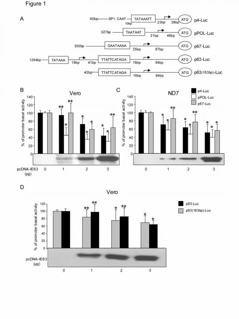
Figure 1

Figure 2
Fi 3Figure 3
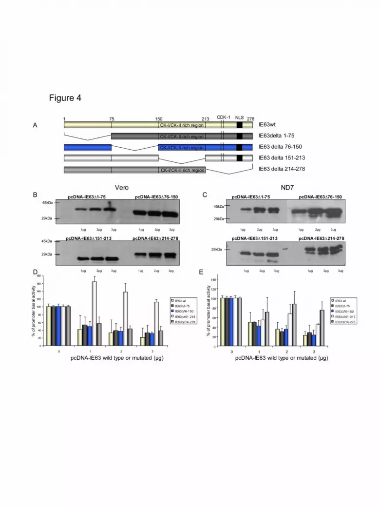
Figure 4
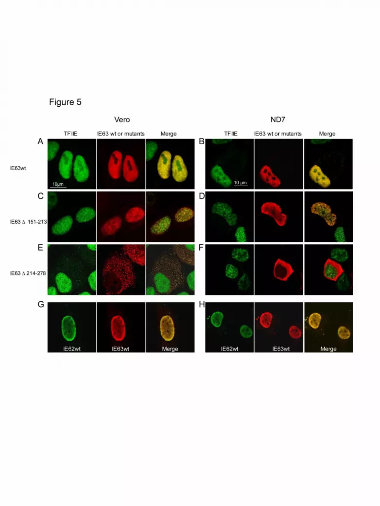
Figure 5
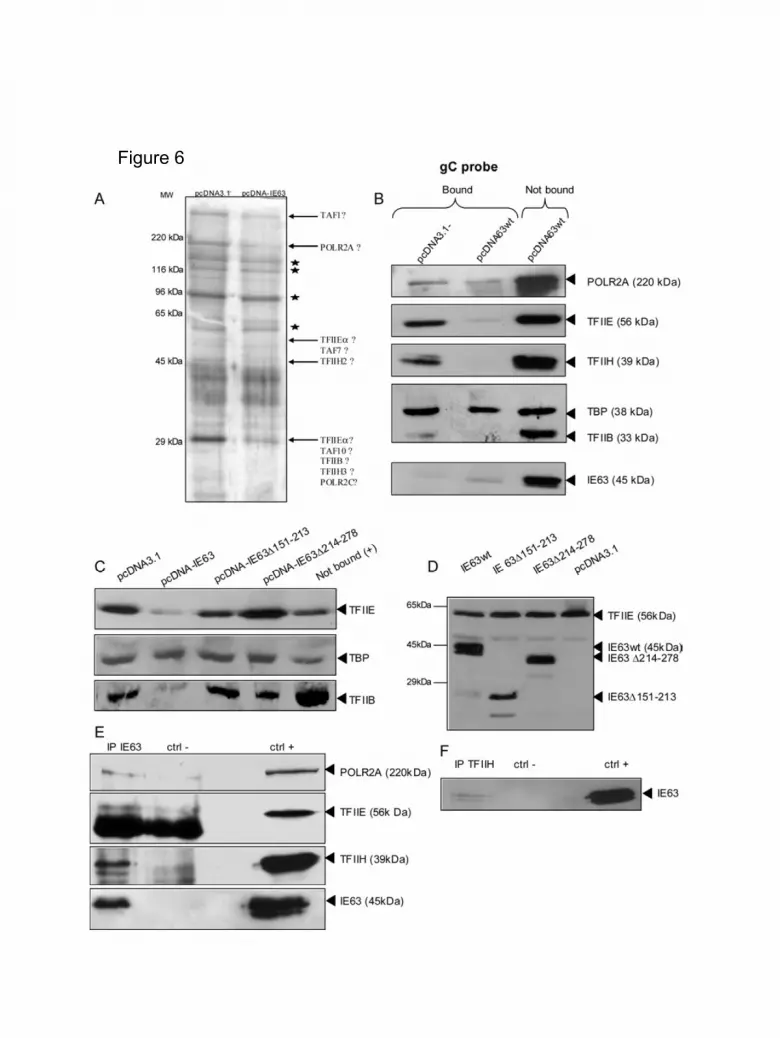
Figure 6
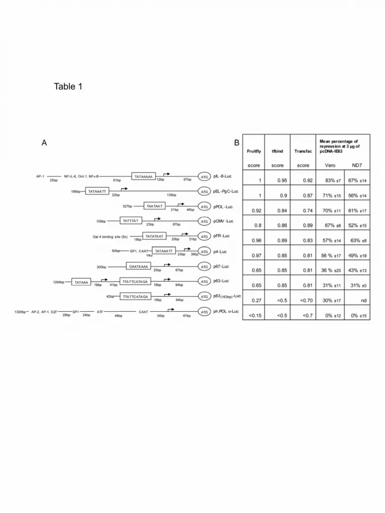
Table 1