Embed Size (px)
Citation preview
NYSGI � 8 � 85�
THE PHYSIOLOGY OFREATH- HOLD DIVING
QSQIjHNE Il'.IppUndersea and Hyperbaric ~, ~�~t g,>,~>Medical Society Workshop
Ckaes E, G. Lendgreaa, M.D., Fh&.and
Massiino I'e~pao. M.D.
IlM ~ ~ I ~ ah I 8 8 % ~SSSO gochwile %Le
8ethesga, Marylaaad f0414U.SA
THE I'H1JSIOLOCft OP' BREATH-HOLI! 1!IVIN6
Undersea and Hyperbaric medical Society Workshop
held at
Buffalo, New York28-29 October 1985
Chaired by
C 1 ae. E. C. Lundgren, M.8., Ph.X!.
Edited by
C'taes E. Cj. Lundgren, Q.D., Ph.&.Mass imo Pe rri gna, .M.8.
Sponsored by
The Neur gork Sea Cjrant Institute
UPS Publication Number 72 WS/BH! 4/15/87
April 1987
CONTENTS
Parti ci pants
Acknowledgments
IntrodUction
Sessi on I: Historical Hates
Vill
ix
Breath-hold Diving; A Brief History
H. Rahn
Von Trieleben's Diving BellA. B. Crai g, Jr.
Session II: Limits of Breath-hold DivingH. Rahn, Chairman
Depth Limits of Breath-hold DivingB. Crai g, Jr.
Depth and Time in Relation to Gas ExchangeA. Olszowka
12
Breath-hold and Ascent BlackoutZ. H. Lanphi er
32
CNS Tolerance to AsphyxiaB, K. Siesta
44
General Discussion 62
Sessi on III: Breaking Point AnalysisB. Crai g, Jr., Chai rman
The Diving Response and Breath-holding Capacity in ManC. B. G. Lundgren
64
Effects of 02 and CO2 Breath-hold Breaking PointY. C. Lin
75
Respiratory Neuromuscular output in BreathholdingW. A. Whi tel aw
88
Session IV: Thermal Regular i on and Metaboli c AspectsR. Bl sner, Chai rman
Decrease in Body Insulation With Exercise in Cool WaterY. S. Park, D. R. Pendergast, and D. W. Rennie
98
Thermoregulation in Wet Suit DiversY. S. Park, H. J. Ki m, D. W. Renni e, and S. K. Hong
l24
Energetics of Breath-hold DivingD. R. Pendergast
111
Diving Pattern and Thermoregulatory Responses of Male and FemaleWet Suit Divers
K. Shi raki, H. Konda, S. Sagawa, and Y. S. Park
Session V: Cardiovascular AspectsS. K. Hong, Chairman
148The Diving Response: A Comparison of Animals and NanR. Slsner
158Breath-hold Bradycardia in Nan: An OverviewS. K. Hong
174
Nodifications in the Vasomotor Arm of the Human Diving ResponseR. fV. Arnold
188
The Cardiac Response to Breath-hold Diving at Rest and whileExercising A Comparison Between Nan and Diving Homeotherms!P, J. Bvtler and A. J. doakes
200
Cardiac Arrhythmias as a Precursor to Drowning AccidentsJ. R. PfcDonovgh, J. P. Barvtt, and J. C. Saffron
212
General Discussion 230
Session VI: Safety Aspects of Breath-hold &ivingC. E. G. Lvndgren, Chairman
Pathophysiology of Near-drowning: Aspiration vs. Non-aspirationP. G. Boqsen
233
Hyperventilation: hn Unpredictable Danger to the Sports Diverp. C, tandsberg
256
Breath-hold DiVing ACCidents in the Nediterranean AreaD. Zanni ni
268
Mss of Consciousness During Breath-hold DivingP. C. Data
274
General Discussion 284
Alveolar Gas Composition and PerformanceH. Rahn
287
Depth Records: Practical ConsiderationsB. Hai orca
291
Cardiac Performance During Breath-hold Diving in Nan: An OverviewM. Ferri gno, Pf. H. Liner, and C. S. G. Lundgren
THE PHYSIOLOGY OF BREATH-HOL 0 DI V ING
PARTICIPANTS
I~~~+
Cl aes E.G. Lundgreni M.D. > Ph.D.De pt. of Phy si ol ogySUNY at Buffalo124 Sherman HallBuffal o> NY 14214
Robert W. Arnol d~ M. D.Mayo Graduate School of MedicineDept. of OphthalmologyRochester> Minnesota 55 905
Dr. P. G. Boy se nDept. of Anesthes iol ogyJ HMHCBox J-254Gainesvf1 1 e> Fl ori da 32610
Professor P. J. B utl erDept. of Zoology and Comparative PhysiologyThe Univer sity of BirminghamP 0 Box 363B i rmf ngh amB15 2TT U. K.
Dr. Al bert Crai aBox 642The University of Rochester Medical Center601 El mwood AvenueRochesteri NY 14642
Professor Pi er Giorgio DataIsti tuto Di F$ siol ogi a UmanaFacolta' di Medicina e ChirurgfaV ia Del Vesti ni66100 Chieti
Ital y
Or. Robert El snerProfessor of Marine ScienceInsti tute of Marine ScienceUniversity of AlaskaFai rbanks> AK 99775-1080
Mass] mo Fer rf gno> M. O.Oe pt. of Phys $ ol ogySUNY at Buffalo124 Sherman HallBuffal o, NY 14214
Suk Kt Hong> M. D. ~ Ph. D.Dept. of PhysiologySUNY at Buffal o124 Sherman Hal 1Buf f al o, NY 14214
Or, Preter G. Landsberg1 Geneva StreetLakef gael dBenon1South Afr1ca 1501
The Rev. Edward H. Lanphfer> M.D,Untversfty of Wfscons> n 8 fotron2115 Observatory Or! veMadison> WI 53706
Yu- Chon g L in > Ph. O.Dept. of PhysiologyUn1versf ty of Hawaii fSchool of Medh cfne1960 East West RoadHonolulu, HI 96822
Mr. Enzo MaforcaVia Larga 2496100 S< racusaItaly
J ohn R. McDonough. M.D.8-6007 All enmore Medical CenterTacoma> Washington 98405
Albert 01 szowkai M.O.Dept. of Phy sf ol ogySUNY at 8uffalo124 Sherman Hal 1Bu f f al o. NY 14214
Ya.g Saeng Park> Ph.D.Kos<n Medfcal CollegeDept. of physiology34 Amnam-Dong, Suh-KuBusan 600Kor ea
David Pendergast> Ed. D.Dept. of PhysiologySUN Y at Buf f al o124 She rm an H al 1Buf f al o> NY 1421.4
Hermann Rahn. Ph. D.
Dept. of Physi ol ogySUNY at Buffalo124 Sherman HallBuf f al o, NY 14214
Donal d W, Rennie> M. D.Dept. of PhysfologyState Unfversfty of NY at Buf fal o124 She rm an H al 1Buf f al o> NY 14214
Professor Kefzo Shf rakf
Dept. of Appl fed Physi ol ogyUniversity of Occupational and Environmental Heal thSchool of Medf cf ne1-1 Isef gaoka> Yah atanf shi-ku,807 Kf taky ushuJ apan
Prof essor Bo Sf esj oLunds UniversftetFor skn. Av d. 4. i E-Bl oc ketLasarettetS-221 85 L un dSweden
W fl 1 I am A. Whf tel aw i M. D.Dfv I sIan of Pulmonary Medi cf neThe Unfversfty of Ca'I gary3330 Hosp f tal Drive N.W.Calgary> AlbertaCanada T2N 4N1
Prof essor Dam f ano Zannf nfCorso Europa 1342/b16166 Genova! taly
A.CKNOWLE'D4MENTS
On behal.f of all the participants, the Workshop Chairman thanksthe New York Sea Grant Institute for its support.
Special thanks go to Miss Pamela Caron for her great effortsin transcribing the discussions from tape recordings. Thanksare also due to Ms. Ann Barker of the Undersea and HyperbaricMedical Society for helping to prepare these proceedings forpublication.
Reproduction in whale or in part is permi tted for any purpose.2'he opinions, conclusions, and recommendations contained i nthi s report are not to be construed as of'ficial or necessari lyreflecting the views of the Undersea and Hyperbari c Wedi calSoci ety Incorporated.
XNTRODUCI'ION
This publication brings together the presentations and discussionsthat were part of the two-day workshop "The Physiology of Breath-holdDiving". The workshop was held at the State University of New York atBuf falo on October 28-29, 1985, almost exactly 20 years af ter thelast major meeting on the same subject. That was the Symposium on thePhysiology of Breath-Hold Diving and the Ama of Japan, which tookplace in Tokyo, Japan, in conjunction with the XXIII InternationalCongress of Physiological Sciences.
In 1965 it was probably felt that the diving women of Japan andKorea - the only maj or group of people resor ting to breath-holddiving for a living- were soon to disappear. It is surprising that atthis time, 20 years later, these remarkable divers are holding theirown and even probably incr easing in number, as the market value oftheir catches continues to rise. Furthermore, in the last 40 years themaximal depths reached by men and women holding their breath haveincreased dramatically to more than l00 meters. While the small groupof these record-setting divers is highly skilled and trained, the verynature of its pursuit puts these persons at the limit of what isphysiologically feasible and medically safe. Indeed, there isevidence that sometimes the limit has been surpassed. Nonetheless,like many other hazardous sporting activities, including spear-fishingcompetitions, these record attempts continue.
While we have included a presentation and a film featuring deepbreath-hold diving in the workshop program, we do not endorse thisactivity. However, as scientist, one is obliged to try to understandthe mechanisms that determine the limits, and also to identify therisks, of deep breath-hold diving. For this reason, at this workshopwe appreciated the opportunity to draw on the experience of Mr.Maiorca, who is one of the record-holding breath-hold divers.
As public's interest in diving has expanded over the years fmillions of people around the world have enjoyed the underwaterscenery, either breath-hold diving or simply snorkeling at thesurface. Recreational divers span a wide spectrum of divingproficiency< from the highly skilled spear � f ishermen in South Africanwaters to the snorkelers on the beaches of temperate and tropicalcoasts. Add to these the many persons who become involuntary breath-hold divers in drowning incidents, and the number of people affectedby by our knowledge, or lack thereof, becomes impressive.
The purpose of this workshop was to revi ew the current status ofbreath-hold diving physiology. Generous support for this project wasprovided by the New York Sea Grant Institute Contract no.NA85AADSG021! . The costs for printing of these Proceedings werecovered by the Undersea and Hyperbaric Medical Society, Inc. Thepresentations and discussions are presented as they occurred in thecourse of the workshop. However, two additional contributions havebeen included because they contain information that was frequentlyreferred to in the discussions and presentations. One is a reprint ofan article by Y.S. Park, D.R. Pendergast and D.W. Rennie: Decrease in
body insulation with exercise in cool water. Undersea Biomed. Res.3.1:159-168. The other one was kindly written by Dr. H. Rahnspecifically for this publication and it is entitled: Alveolar GasComposition and Perf ormance. All presentations were printed f romcamera-ready manuscripts provided by the authors and the discussionswere transcribed from tape recordings. These transcripts were subjectto some editing and then reviewed by the speakers.
We hope the reader will enjoy these Proceedings as much as weenjoyed the workshop< and we thank all the participants whocontributed with their interesting presentations and livelydiscussions.
Claes Lundgren, N, D., Ph.D. Nassimo Ferrigno, N.D.
Breath � hold Diving: A Brief History
Hermann Rahn
Department of PhysiologyState University of New York at Buffalo
Buffalo, New York 14214
Historical accounts of breath-hold divers can be traced back more than
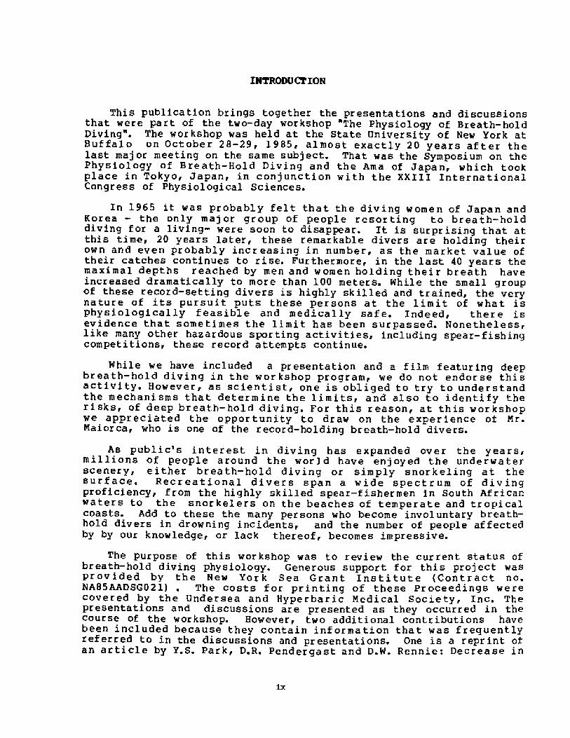
2000 years, descri.bi.ng the activities among the sponge divers of Greece,pearl divers in the Persi.an Gulf and India, and the seafood divers ofsouthern Korea and Japan Fig. 1!. ! ~3
� 0
Letitude range ce 30 t0cJuly water tenporeture ce 30' � 30 C
In each region the skills developed independently but are basicallysimilar, namely, head-first descent in shallow or deep dives, the latterbeing assisted by a weight. Ascent from deep dives was also assisted bypulling up the diver with a rope. As Davis �934! wrote, breath-hold divingwas also a recognized profession in naval warfare, divers being employed todestroy boom defenses in harbors during the siege of Tyre �33 B.C.! andSyracuse �15 B.C.! According to Chris Lambertsen pers. commun.! theFrogmen of the U.S. Navy's Underwater Demolition Teams operated as breath-hold swimmers throughout World War II. Even today breath-hold diving isstill part of the training of underwater demolition teams.
The modern scientific era of breath-hold diving might be said to havebegun in the l920's when Gito Teruoka, Director of the Institute forScience of Labor in Japan, Fig. 2! became concerned about the occupationalhealth, hazards, and strenuous physical exertion of some 25,000 women whoearned their living by diving, not for pearls, but for various types ofseafood, to depths of up to 20 meters. His meticulous observations ofdiving times, patterns, depth, and alveolar gas concentration following
these dives were published in 1932 in Arbeitsphysiologie. I thi.nk it isfair to say that his work was promptly forgotten and only rediscovered 25years later when Dr. S. K. Hong began his studies of the Korean Ama.
Interest in breath � hold divinq re-emerged i.n the 1940's and 1950'swith increased public awareness in swimmi.ng, snorkeling, and scuba divingas well as their associated hazards. Scientific interest began to focus onthe physiologi.cal consequences of voluntary apnea, particularly whensubmersed, the changes in lung gases, chest mechanics, and cardiovascularsystems as weIl as the role played by the 02 and C02 stores of the body.In 1965 the first International Breath-hold Symposium was held in Japan anddedicated to Professor Teruoka Bahn and Yokoyama, 1965!. It provided thefirst comprehensive account of diving patterns and physiology of the Amain Japan and Korea, the first estimations of the work of diving, the firsthints that repeated dives could result in bends, and a new focus on divingbradycardia and the importance of water temperature and body insulation.
In the intervening years much attenti.on has been focused on thecardiovascular changes during submersion and lung compression, depthrecords, and associated changes in lung mechanics and gas stores, divingpatterns, energetics and thermoregulation, and a better understanding ofthe circumstances that lead to fatal accidents in competitive sports.Unfortunately for the physiologist the breath-hold diver during descent orascent becomes rather inaccessible to simple measurements of hiscardiovascular and pulmonary system. In other words, he becomes a blackbox. However, the more recent techniques pioneered by Kooyman �981! andZapol �986! on free diving Weddell seals in Antarctica promise a new toolwhich could also be applied to man in obtaining blood samples, heart rate,and continuous records of diving velocities during repeated dives as wellas at the surface. The other new tool is that of treating the diver as aclosed black box and using the computer to calculate the 02, C02, andN2 changes in the lunq, blood, and tissues during descent and ascent. Such
results will be di.scussed in this Symposium.
Today marks the second International Symposium devoted to breath-holddiving, and thi.s will bring us up to date on all the new insights that havebeen gained during the last twenty years.
References
Davis, R. H. 1934. Deep diving and underwater rescue. J. Roy. Soc. Arts82: 1032-1047.
Kooyman, G. L. 1981. Weddel 1 Seal: Consummate Diver. CambridgeUniversity Press, Cambridge.
Rahn, H., and T. Yokoyama. 1965. Physiology of Breath-Hold Diving and theAma of Japan. Public. 1341. Nat. Acad. Sci., Nat. Res. Counc.,Washington, DC.
Teruoka, H. 1932. Die Ama und ihre Arbeit. Eine Untersuchung uber diejapanischen Taucherinnen!. Arbeitsphysiol. 5: 239-251.
Zapol,W. M. 1986. Diving physiology of the Antarctic Weddell Seal. In:Environmental Physiology L. E. Farhi and C. V. Paganelli, eds.!Springer Verlag, NY in presss! .
YON TRIELEBEN'S OBEYING BELL
*Ibert B. craig. JrDeportment of Physiology, UniversitM ofRochester School of tiedicine. Rochester, NY l4642
Fig. l. rhe Warship Vasa �!.
At the beginning of the l7thcentury 5~eden controlled most of thef!altic Sea and occupied much of thesurrounding land. King Gustav 11 Adolfof Sweden expressed the view that Nextto God the welfare of Sweden depends onher Navy. Building small ships is onlya wast.e of young trees.' with theseconcepts in mind he ordered many ships,one of which was the Vase. This ship,vhich was launched in 1627, was 5U�65 feet! long and displaced 1300 tons Fig. 1! . After being fitted, she setsail on August 10, 1628 against a
light breese from the southsouth-vest. In a few hundred meters asudden squall forced her to heel overso far to port that the water floodedin through the lover cannon ports...The Vase went down with sails up andflags flying �!. In 1956 AndersPransen located the remains of theship. Its restoration is an interestingstory, in itself, and a visit to thespecially constructed Vase museum inStockholm is a rewarding experience.
At the time of the sinking and forseveral years ther'eafter the locationthe Vase was well known. In 1664, 36yearS after the Cataatrophe, SaneAlbreckt von Treileben of Sweden andAndreas Peckell, a German salvageexpert, recovered most of the 64 cannonon board. The heavier cannon weighedbetween one and tvo tons. An Italianprieat, PranCeSCo Neqri, traVelingthrough the area on his vay to theNorth Cape, described the procedure inhis book, Viagqio Settentrionalepublished in 1700 at Psdua. The diversvore leather' suits and descended in adiving bel.l Fiq. 2!. They use& varioustools on the ends of 2 e poles to freethe cannon and to secure lines vithwhich they were hauled to the surface Piq. 3!. The usual diving time was 15minutes.
A reproduction of the diving bellconstructed from descriptions given bymarten Trievald in his book Kosten attlefwa under vatn �! is on display atthe vase Nuseum in Stockholm. It isconical, measures 1.22 m in height, andhas a bottom circumference of 1.06 m.
Fig. 2. von Treileben's diving bell �!
Pig. 3. implements oaed for salvage work �!
total air volume of c.he tel 1 is 533If it were assumed t.hat the diver
was l. 5 m tall, the top of his headwould be gust below the top of thebell. As he was lowered to the ~reckwhich lay at 30 m, the level of water~ould rise to just over half the heightof the bell Fig, 4! . It is dif f icultto imagine how the divers did anyuse f ul work vi th the tools depicted. Inaddition there is lic.tie or no light atthe bottom of the silted StockholmHarbor, and t.he temperature is aconstant 4 C throughout the year.Revert.hei.ess, almost all of the cannonwere recovered.
From the physiologic view thistype of diving can be consideredbreath-hold diving. The usualbreath-hold diver makes an inspiration,suspends ventilat.ion, and goes belowthe surface. In effect, the diver takesa limited supply of air for use duringthe period of apnea. Although thesupply of air is larger for the workerin the bell than that for the usualbreath-hald diver, it is limited.Assuming that the worker's oxygenconsumption was about 1 1/min, theexchange ratio was .8, and that all ofthe CO2 produced would be expired, itcan be calculated that the p CO2 in thebell would rise to about 80 torr at theend of 8 15 minute work period at 30 m.However, as the P CO2 increased some ofthe C02 would bf' stored and vould notappear in the air space of the divingbell. Therefore, it is unlikely thatthe P C02 would rise to 80 torr.
It might be interesting to repeatthis 250 year old type of breath-holddiving and to utilise some of ourmodern capacities for gas analysis andsimulated diving. Varying the depthsand the external volumes Should provideresults vith which to test some of thereCent eathematiCal eadela whiChpredict gas exchanges and partialpressures.
It is tempting the conclude thatthe increase of the CO2 might have beenthe major factor limiting the divingtime under these conditions. On theother hand, in 4 C water the divervould lose considerable body heat evenin 15 minutes. Kang et al �! foundthat the Korean Ama's working timeduring the ~inter months when the waterwas 10 C averaged 16 minutes. A,t alLtimes of the year the working timerelated directly to the watertemperature. FOr the Ama the limits ofthe working time were dictated by theLoss of body heat, and the same mighthaVe been true fOr the SalVage wOrkereon the Vase.
kvef f2'30 3ylZOm
Fig. 4. Calculated level of the water inside thediving bell at different depths.
Heing a diving bell tO salvage thecannon from the Vase was a tremendousfeat. In the Ama Symposium in 1965 andthe cur rent meeting there have beenmany papers concerned with gasexchanges during breath hold diving.Ho~ever, most discussions have beenlimited to the diver who makes a largeinspiration and has this air supply forthe dive. Reconsideration of the oldertechniques vhich employed the divingbell might be an interesting extensionof breath-hold diving to explore.
KEFEKEHCESFranren, h. The icarahip Vaea �th ed.i
stockholm: Horstedts/sonniers I966.2. Triewald. M. Konsten atc, lefwa under vatn.Stockholm L13 ~ .3. Kang, D.H., S. H. Song, C. S. Sun, S. K. HongChanges in body temperature and basal metabolicrate af the arne. J. hppl. Physiol. l8: 483-488,L963
Discussion following Dr. Craig's presentation
You had about 100 liters of oxygen in that bell at thesurface and you were using it up at l liter per minute according toyour protocol> so there was more than enough oxygen at 4 atmospheresThey were actually breathing inspired oxygen tensions equivalent to 8Opercent~ and so it seems to me there was no problem in terms ofoxygen.
Dr. Craig: I didn't see any problem.
Dr. Rahn: And if I make similar calculations for CO2< there reallyisn't much of a CO2 problem.
Dr. Craig: Well> if you assume that all the C02 s expired in theatmosphere', i"ght get up to 70 torr, but you have to partition theCO between two compartments: the body and the atmosphere, and I thinkthis is an interesting problem.Dr. Elsner: There is also a third compartment, that is the CO2dissolved in the water.
Dr. Rahn: So you think the problem was body temperature.
Dr. Craig: I don't know. I just opened the problem.
Dr, Rabn: Any indication of the lights that they used'P
Dr. Craig: No indications of any lights. In the Stockholm harborwhen you stir up the silt you get zero visibility. I just don't seehow they did the job. But they brought. up almost all of the cannon.During the salvage procedures they never used lights for anything.They did everything by feel.
Hong: It is inconceivable that they could stay 15 minutes in 4 Cwater. The Ama tolerate 10 C water for 16 minutes.
Dr. Craig: They had some clothing protection.
Siesjo: Was there any special reason why they worked in water ofC? Even in Sweden there are a f ew weeks when the temperature gets
higher'
Dr. Craig: Not at 30 meters of depth.
Dr ~ Elsner: How did they actually do the work? Did they take abreat" inside the bell and go outside and work, then go back inside
or did they manage to stay inside the bell and stillperform the outside works
re Craig'- I don't know. The book that describes this is in oldwedish and this information may be in that book.
Discussion following Dr. Craig's presentation
Dr. Perrigno: Dr. Craig, you showed an elephant seal.. Does this divingmammal have a sphincter in his inferior vena cavan
Dr Elsner: There is a very well def ined muscular sphincter in theinferior vena cava just above the diaphragm and from some otherstudies we did in a different species we have some idea of theoccurrence of a partial constriction of that sphincter during diving.It appears that the flow of blood from the inferior vena cava passingthrough that sphincter takes place at a very sluggish rate during thedive.
Dr. Lanphier: In preparing for this workshop I went through my oldfiles on the subject and I discovered among many other things a letterthat I had wri tten to Dr. Craig shor tly af ter publication of his"Fennology" paper, tr'ying desperately to call his attention to thefact that residual volumes as ordinarily measured are essentiallyirrelevant with regard to the depth of breath-hold diving. I don' tthink that there is time to discuss the matter at the moment, so wewill let it stand as an apple of discord on the table and hope we canget back to it. Because I think it is awfully important.
Dr. Craig: May I make a comment about that'P I do remember theletter. The residual volume that we used was measured by thetechnique Dr. Rahn et al. and I think that it was a pretty goodtechnique. In addition, we tried to express f urther air af terexpiring to minimal volume. One method of doing it was taking one ofthe surgica3. abdominal binders and putting it around the thorax, andthen having two strong students stand on either side and pull untilthe subject raised his hand to stop. He could get out about 200 mlmore. An alternative method was to breath out. to residual volume andhold a tube in the mouth that was connected to a water/ai r system.With this you could suddenly exert a negative pressure of 20 cm 820 atthe mouth and see if you got out anymore volume. Now I know you aregoing to say that the airways were closed, but we could under thoseconditions get out only 200 ml. So we made an attempt to see if wewere at residual volume. Now of course I realize what you would say<"those techniques are not good because of the airways closure and youcan't get the air volume out but you can still compress it".
Dr Xanphier: Yes, that is exactly it. I admire your attempt to getaround it, but there isn't a way to get around it, except by measuringit by a method involving compression.
Dr. Craig: Which is the body box.
Dr I anphier: Yes, under increased pressure, and I don't thinkanybody has done that.
Dr. Craig: Wel3., with the body plethysmograph the defined residualvolume is the same as with other methods.
Dr. Lanphier: You do that by going to FRC and substracting what youcan exhale, but that is limited by airway c3.osure and so on. I think
it is an area af discord and we don't have time to talk about it.
Dr. Hang: What is the maximal amount of blood that can be shi fteQinto the thorax during diving? It seems to increase every yearindicated by the increase in diving depths. Also, can we train diver>to increase the volume of blood shift into the thorax?
Dr. Craig: I would like ta think that they haven't yet reached thelimit af the amount of blood that can be shifted. Some technique>can be useful. For instance, if I were going to attempt a recordbreath-hald dive, I would start out of the water, not in water.also remember that at those depths the changes in volume becom~relatively smaller. I think the limit will be reached when one ofthese record breath-hold divers comes up coughing blood.
Dr. hrnold: If, as Dr. Craig suggested, we have a blood exchange i~the thoracic cavity explaining the ability to tolerate the greatdepths, the blood in a seal would be in the inferior vena cava/ whicgis well adapted ta have a quick venous return. However, in a humanthere may be the bronchial arterial blood which is not as readi]yavailable to venous return. If that's true and if thoracic pressure,increases quickly, venous return may be substantially compromised,
Dr. Lundgren: I wauld like to draw your att.ention to the fact thatthe work that Dr. Ferrigno will present addresses preciselyproblem, that is, the importance of intrathoracic pressure changeswith depth for cardiac function.
it is an area of discord and we don't have time to talk about it.
Dr. Bong: What is the maximal amount of blood that can be shif tedinto the thorax during diving? It seems to increase every year asindicated by the increase in diving depths. Also, can we train diversto increase the volume of blood shift into the thorax?
Dr. Craig: I would like to think that they haven't yet reached thelimit of the amount of blood that can be shif ted. Some techniquescan be usef ul. For instance, if I were going to attempt a recordbreath-hold dive, I would start out of the water, not in water. Andalso remember that at those depths the changes in volume becomerelatively smaller. I think the limit will be reached when one ofthese record breath-hold divers comes up coughing blood.
Dr. Arnold: If, as Dr. Craig suggested, we have a blood exchange inthe thoracic cavity explaining the ability to tolerate the greatdepths, the blood in a seal would be in the inferior vena cava, whichis well adapted to have a quick venous return. However, in a humanthere may be the bronchial arterial blood which is not as readilyavailable to venous return. If that's true and if thoracic pressureincreases quickly, venous return may be substantially compromised.
Dr. I undgren: I would like to draw your attention to the fact thatthe work that Dr. Ferrigno will present addresses precisely thisproblem, that is, the importance of intrathoracic pressure changeswith depth for cardiac function.
DEPTH LIMITS of BREATH-HOLD Dl VIN6
41bert 5. Craig. JrDepartment of Physiology, University ofRochester School of He0icine, Rochester, NY l 4642
L rlrP rn!aneTLC L
r .L!4r~l5
DP65-a7/e7sr5Pv li 7U
60-
4 !-
2 I
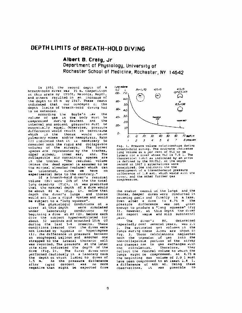
In 1951 r.he record depth of abreath-hold diver was 35 m. Competitionin thi- arena oy Crott, ! aiorca, hayol,and others resulted in an increase oft.he depth to 65 m by 1961. These ieatsindicated that our concepts or. thedepth limits of breath-hold diving hadt.o ae extended.
ACCOrding the f!oyle's Law thevolume ot gas in the body must becompressed during descent and theinternal and ambient, pressures must beessent.ially equal. �therwise, pres-uredifferences ~ould result in bacotcaumawhich in the thorax would causepulmonary edema and/oc hemoptysis. Rahn�! indicated chat it is nece -sary toconsider both the rigid and collapsiblevolumes oi the airways. The formerspaces are represented by the t.rachea,upper aicway, inner ear, etc. Thecollapsible air concaining spaces arein the thocax,. "The residual volume minus the dead space! is assumed ro bethe mioimal alveolar volurae! which canbe tol.crated, since we have uoexperiment.al data to the contrary."
If a breath-hold diver's residualvolume RV! were 20% of the the totallung capacity TLC!, it was predictedchat the maximal depth of a dive woul.dbe about 40 m. Fig. 1! . !claw thatdepth the diver's lungs and thoraxwould act. like a rigid systeis and wouldbe sub!ect to a lung squeeze
The physioiogic conditions of adiver at t.hie depth were simulatedunder laboratory conditions bybeginning a dive at RV �! . !efore eachdive the subject hyperventi la ted torabout 30 seconds and breathed 100% 02during the last 3-4 breaths. Theseconditions insured that che div s werenot limit.ed by hypoxia or hypercapnea 1!. 1'he difference in pressure betweenan esophageal balloon and another onestrapped to the lateral thoracic wallwas recorded. The pressure at the latersite also indicateo the depth of t.hedive Fig. 2!, The first dives wereperformed cautiously in a labocary tarikthe depth or which limted to dives ofL.5 m. As the pressure differenceaf ter capid descent was no morenegative chan might be expected t rom
0- ! < B7 30 4r! SO 6 ! W rape, rn/ 2 9 0 5 6 t 8 pmsrmn
Pig. 1. Presure volume relatianships duringareath-hold aiving. The ordinate indicateslung volume as a per cent of TLC or asliters for a diver whose Ti. is 7.0 I, Thethearetical limit as indicated by an artowis defined ay the RviTLC. At che depthrecord in 1967 2 alcernatives werecansidired. One indicates the theaevelopment at a transtharacic pressureuiiterence oi 1.8 amt. which would kill thedriver, and the Other further gaacompression.
the static recoil of che lungs and thet.horax, deeper dives were conducted inswinuning pools and f inally in a lake.Even atter a dive to 4.15 m thepressure dii terence was not greatenough to produce a "lung squeeze" r ig3! . However, at. this depth the diverdid report vague and mild substernalpain.
The diver's RV, determinedrepeatedly over .everal years, was 2.0l. I'he estimated gas volumes in thelungs d~ring these dives are sho~n inFig. 3. These calculations neglectedboch the transfer of gas into thenon-collapsible portion of the airwayand changes due to gas exchanges withthe c i ccula t ion. Therefore, theyreflect the maximal volume to which tnelungs might be compressed. At 4.75 mthe beginning gas vol~me of 2.0 1 musthave been compressed to at least 1.4 1 ~a difference of 60 ! ml. Osing t.neseobservations, it. was possible to
ii!Prg. 2. Photographs oi 2 records duringdives. The upper half of each shows thepressure, expressed as depth, in theballoon strayed tro the lateral tharacic«sll. The lo~er part is the record of thepressure difference bet~san the outer andtha esophageal balloon.
f ~ eI 0 0 r
a
� 20 so0ewr n m94Nv
40 d0v. 20
10
reCOnetruct a dive in «hich a subjectwit.h a TU at the surface of 7.0 1 anda RV of 1.5 1 would descend to 65 m,which was the record in 1967. Underthese conditions the lung volume wouldhave to be compresseo to 570 ml lessthan the RV. Lt «as suggested thaC ifthe RV represented the minimalanatomical volume of the thorax, thetransfer of blood from the exr.ernal tathe internal thoracic regions «auld bea mechanism by which the lung gasvolume mighC be further compressed FigI!.
The current depth record or oreathlrolu diving held by Maiorca is 100 m.as there was no evidence of barotraumain that dive, it is apparenC that thegas volume at that depth must have been9I of the surface volume. Using thefbeg inning condi t iona mentioned abo veor a 65 m dive, a plunge to 100 m
~ould require a minimal qas compressionto 870 ml less than the RV.
Lanphier has indicated personalcommunicat ron! that ineasurement ot theRV by any of the exisr.ir g techniquesonly measures tne minimal lung volumeto which a subject can expire using ainaximal muscle force. ne suggested thatduring breath-hold diving t.he alv6olarvolume could be compressed tartherwithout supposing tne transfer oibLOod. after disCussing ChiSpossibility, we atremptecl to compressour sub;ect's tnoracic volome to 10 ess
by applying the maxi,ha 1e~ternal farce which the subject. couldtolerate. Under these conair.ions it. «aspossible to express about 200 ml fthe a m roma i r«ay s into a spirometer . 0thttempts to remove gas fr h ' yer
om r. e airwaysatter expiring maximalL y were made oy
rapidly applying a negative pressure ot20 cm h20 at the mouth far 10 SeCOndswith care to iteep the glottis open. Intive subjects an av rage of 250 ml waswithdrawn from Che air~ay. From ourobservations adding an external or aninternal force ta that produced by themuscles did not change the RVappreciably.
auring a dive the COmpreSSiOn ofthe air in the lungs must be associatedwith a physical change in the size ofthe thorax. When capacity of thismechanism is reached, some other meansot air compression such as an increasein the bload volume within the thoraxmust occur. Trarisposition of blood in
Fig. 3. Observed transthoracic pressuredifferences during three difterent seriesOi diVee are ShO«n in the upper part. Thecircles indicate dives per formed in twodifterent pooLs, t.he crosses in a Lare. Inthe lowe lower part the caLculated change in gasvolume in this sub;ect «hose lung volume atthe beginning of rhe dive was RV and equalro 2,0 l is indicated.
Qwz! esrtltrfc IZ5-
�-i0
40 50w s 6
!'0 W ml K' s me,m7 8 9 /0 rrnst m n
Fig. 4. The cross harched area indicatesthe blood volume which would be required tomain t.ain pressure equalization assumingthat the diver started Ei'om the surracealter inspiring maxiinally. Changes involume related to gas exchanges with thecircular.ian have been neglectea.
and out of the t.horax is well known inSOme other SituaCions, Buring pOSitivepressure breathing as such as 83 ! ml ofblood could be displaced crom t.het.horax to the lo~er parts of che bodyin one subject identi t'ied only as Rahn�!. IC is also known during negativepressure breathing the t.horacic bloodvolume is increased 7! .
Such a shift of olooo in otherparts of the body has been postulat.ed.The tranrfer of blood into the venouschannel and sinuses in the middle earof sea lions and seals has oeensuggested as a way ot providingpreeaure Campenaation across t.hetympanic membrane �,6!. It was notedthat the elephant seal has a largeinterior vena cava which extends rro nbelow the diaphrais to the distalportion of the abdominal cavity �!. Inone large male t.his vessel co itained 201 blood which was about 25% of thetotal blood volu ne as measured oyexsanguination. During thi s arne s tudyanother male seal was contained in acargo net, lowered to 3UO m, andbrought to the surface without apparentill effects. At that. depth the gas int.he air containing spaces .aust havebeen Compreeeed to 3n ot r.he .urEaCeVolume. Even though the elephant seal'sr ibs are completely carti lagenous,is ditf icult to conceive ot amechanical change to 3% of r.ne surfacevolum~. lt was suggesteo that t.nevenous resevoir might represent areadily available source of blood whichcould be transfered to the thoracicregion and ~auld provide adoitional gascoinpression.
This proposed shift in bloodvolume as a mechanism which enableshuman br ath-hold divers to achievedepths beyond that accounted for oyreduction in the size of the thorax,poses some practical considerations. Itis known that immersion up to the neckresults in negative pressure oreathingwhich causes a shif t oE blood into ther.horax 7! . If a diver at tempts arecord dive, it would seem reasonablethat he should remain out of the waterprior to diving and also be in theupright pcs i cion. Such conditionswould minimize t.he thoracic bloodvolume and naximize the blood in theperiphery. Theoretically, thispreparati on would optimize the amountof blood ~bien could be shif ted intothe thoracic regions to providepressure compensation.
If the depth limit of breath holddiving is associated with suchtransfers of blood, it ~ould bepredi.cted chat the ultimate record forsuch diving will be signaled by t.hedevelopment of hempotysis and/orpulmonary edema. I hope that such aperson has minimal and t.ransientdisorders which will indicate to othersthat the physiolgic limits ofbreath-hold diving have been reached.
permission to reprint. F ig. 1-4 was obt.ainedr rom the pu il isners of Resp. Physiol.
i EFEBFsCES1, craig, A, B., Jr. causes of loss ofconsciousness during underwater swimming.J. Appl. Pnys in 1. 16: 583-586, 1961.2. Craig, A. B., Jr. Oepth limits of oroathbold diving An example oE Pennology! .Resp. Physial. 5: 14-22, 1966.3. Elsner, R.W,, P.F. Scnolander, A.B.craig, E,G, Jiamond, L. Irving, m. pilson,K. Johansen, and E. Bradst.reer.. A venousalood oxygen resevoir in t.he divi.ngelephant seal. p lysiologist 7:124, 1964.4. Penn, 'A.Q., A.B Qtis, H, Rahn, L. E.Chadwick, and A,H. Begnauer. Oisplacementof blood trom the lungs by pressureoreathing, Am, J. Physiol. 151:258-269,1947.5, �ohl, B. Seal ears. Science 157:99,1967,6. Odend'nal, S. and T.C.A, Paul er.Pressure regulation in the middle earcavity ot sea lions: a possiale mechanism.Science 153:764-769, 1966.7, Rahn, d. 'rhe phyeialgiC Streeses of theAha. In: PhyeiOIOgy Of Breath-Bald Divingand the Ama af Japan. Ed. B. Rahn. Bat.Acad. Sci. and Aat. Res. Council U.S.A!,Punlicar.ion ao. 1341, 1965.
11
Depth and Time In Relation to Gas Exchange
A.Olszowka
As an explanation for the occurrence of accidental drowning in
breath-hold dives Craig has proposed that the hyperventilation prior to1
such dives could. reduce C02 sensitivity and thus allow a profoundhypoxia to develop leading to unconsciousness. On the other hand Paulev
has reported on cases of decompression sickness as well evidence of2
increased CO tensions in some tissues following repeated breath-hold3
2
dives.
In order to explore in more detail the changes of 02, C02, and N2
that occur in a breath-hold dive a computer model was developed. which
permits one to simulate the changes in the tensions of all 3 gases in
the lungs, blood, and tissues.
MODEL DESCRIPTION.
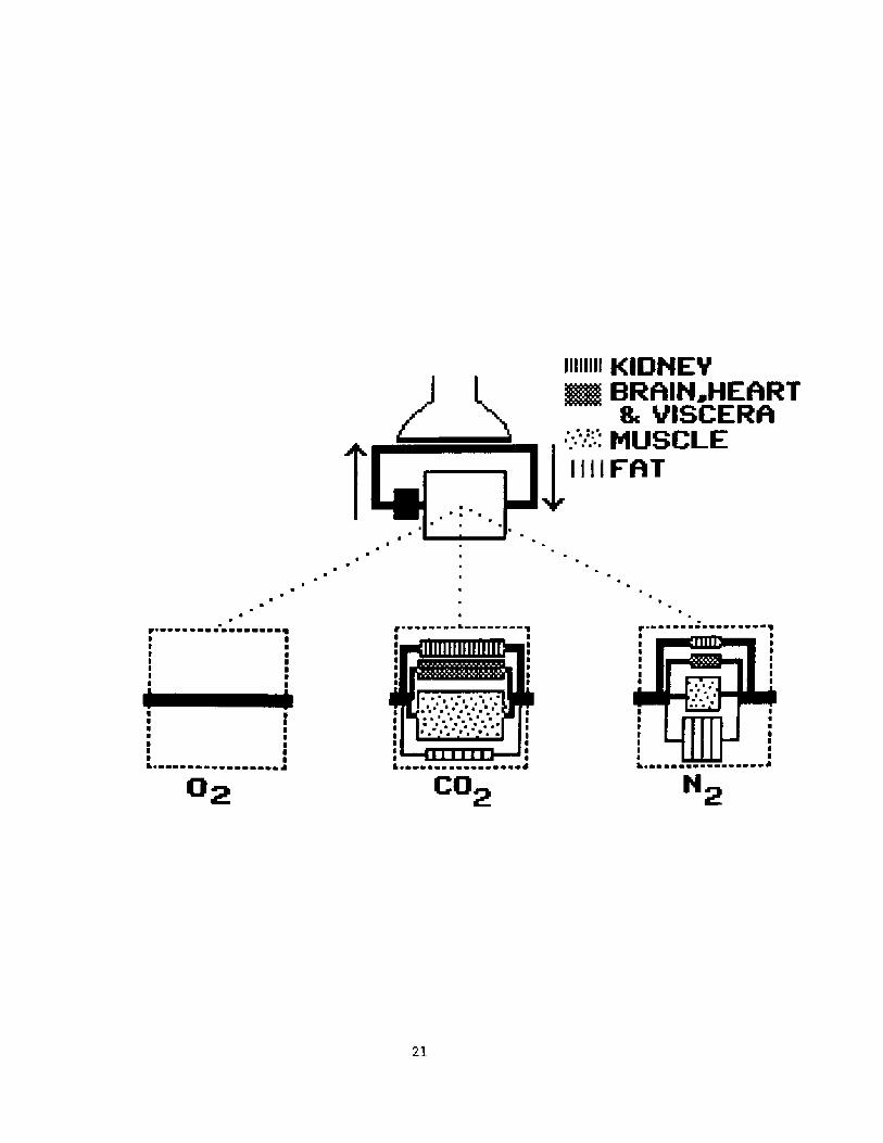
Figure l shows the outlines of the model used to analyze gas
exchange during a breath-hold,. It consist of a series of stores forming
a closed loop.
Lung stores
Both oxygen and nitrogen stores consist of a gas phase only while
carbon dioxide is assumed to have a tissue component which has an
effective volume ELTV! of 1.5 liter.
Blood stores
The blood stores are broken into an arterial, a venous, and an
"effective" reserve volume. The size of the arterial and. venous volumes
are set to the prebreath-hold cardiac output multiplied by the
respective transit times, which are assumed to be 5 liters/min ,6
seconds ,and 12 seconds respectively. These volumes are each subdivided
12
into smaller volumes of equal magnitude in each of which perfect mixing
is assumed to exist and changes in concentration are proportional to the
difference between the mass of gas entering the volume minus the amount
leaving it.
The size of the "effective" reserve volume was assigned a value
which yielded an alveolar Po2 versus time profile which best fitted the
breath-hold data of Hong et als 4
Tissue stores
Since oxygen is poorly soluble and the venous oxygen tensions in
the analysis is substantially above the P50 of myoglobin,oxygen tissue
stores are assumed to be nonexistent.
Nitrogen stores were assumed to be made up of four compartments:
l!Compartment A having a volume of 0.3 liters receiving 24% of the
resting cardiac output and representing kidney.
2!Compartment B having a volume of 5.5 liters receiving 48'% of the
cardiac output and representing brain, heart, liver and the G.I. tract.
3!Compartment C having a volume of 48.1 liters receiving 18% of the
resting cardiac output and representing muscle, skin and the connective
tissue. Xn those simulations where the cardiac output increased
during the dive it was assumed that all the increase in flow went to
this compartment. Similarly when the oxygen consumption increased
during the dive it was assumed that all the increase also occurred in
this tissue.
4!Compartment D having a volume of 50 liters receiving 10% of the
resting cardiac output and representing fat. Factored into its large
volume is an assumed nitrogen fat tissue blood partition coefficient.
of 5.
13
total CO store was assigned a volume which yielded an alveolar Pc02
versus time relationship which best fitted the breath-hold data of
et als4
Hong
COMPUTATIONS.
Alveolar gas composition prior to the breath-hold was computed
Pulmonary end capillary gasusing the classic alveolar equations.
concentrations are then computed. by means of algorithms described
elsewhere assuming alveolar and end capillary equilibrium. These5
concentrations are then used to compute the arterial and. mixed venous
ones assuming respectively the values 0.2 and 5.0 L/min for the right to
left shunt fraction and pre-breathhold cardiac output. For all gases
tissue partial pressures are assumed to be in equilibrium with the
venous blood draining the tissue and at the beginning of the breath-hold
the composition of the blood reserve volume is set equal to that of the
venous blood.
When exchange in the unsteady states is analysed a set of mass
balance equations are used to quantify the gas transport in each
compartment during a small time interval DT. For the lung, blood, and N2
tissue stores this is expressed as as an equality between the change in
the amount of gas in the store and the amount of gas removed from or
added to the blood perfusing that store during the interval DT. A
similar expression is used for the 02 and CO2 tissue stores except that
14
Changes in the tissue Carbon Dioxide stores were computed by
assuming that end-capillary Pco represented tissue Pco and that the
CO2 volume of a compartment equals the total CO2 store times thefraction of the total body volume representd by that compartment. The
the amount of 02 removed or C02 added by metabolism is added to one sideof the equation.
Starting with the lung, and then proceeding in turn to thee arterial, tissue, blood reserve and. venous compartments one can by
solving the resulting set of equations use the concentrations at time Tto obtain the concentrations at time T+DT. Setting T = T + DT ,theprocess can be repeated starting again with gas exchange in the lungstores.
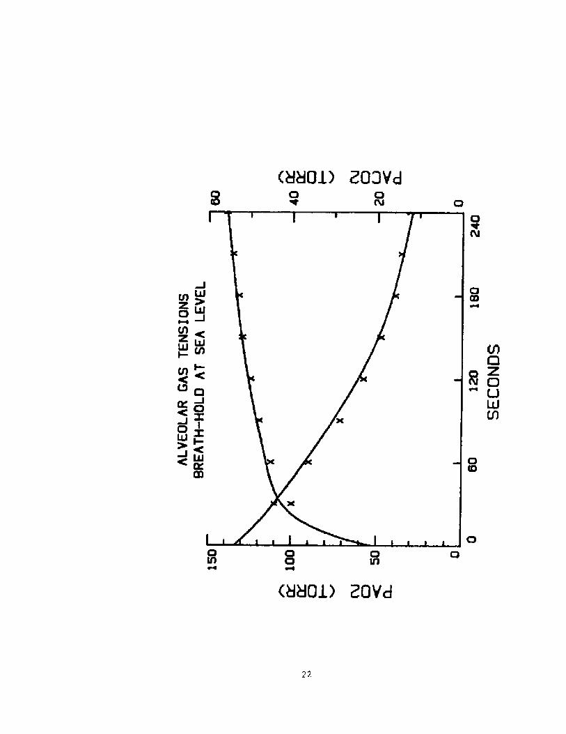
Model TestincS. Before applying this modeL to a breath-hoLd dive itsimulated the alveolar 0 and CO tensions during a four-minute breath-2 2
hold at the surface using the lung volumes and oxygen consumption ratesrecorded by Hong and associates for 9 subjects, average age 304
years,TLC BTPS! = 54SO, R.V. = l470, and 02 consumption = 247 ml/min,who held their breath after a full inspiration without priorhyperventilation. Every 30 seconds they expired into a small bag toobtain an alveolar gas sample and reinspired.
Zn those experiments a significant increase in uptake of oxygenfrom the lung was noted in the first minute of the breath-hold which isconsistent with an increase in cardiac output. Therefore in addition toadjusting the blood reserve volume we also adjusted the magnitude ofthis cardiac output increase so as to produce an anveolar Po2 vs timerelationship which best fitted the above data. In figure 2 are plottedth changes in alveolar Po and Pco during a 240 sec breathold assumingt e
2 2
blood reserve volume of 3.2 liters,a tissue CO2 space of 40 liters andan increase in cardiac output of 1 liter per minute- The alveolar 02 andCO tensions are shown by crosses. The continuous line represents the2
integration of the alveolar o and CO tensions presented by the model.2 2
15
Except for the starting values at zero time not shown! the agreement
appears to be reasonable.
lt is worth noting in this figure that except for the first 30
seconds, the change in alveolar Co2 is somewhat less than the change inalveolar 0 . The final alveolar Co attained is highly depended on the
'size of the Co2 stores. Using an earlier version of the model in whichonly one Co2 compartment was present the resulting size of the Co2 storethat produced the best fit to the data was about half that required
using the present model. This difference is due to the presence in the
present model of 2 large compartments muscle and fat! that receive
relatively small fractions of the cardiac output and consequently have
proportionately less impact on the changes in Co concentrations of the
blood than the other campartments. Indeed Farhi has pointed out that in.6
experiments performed to estimate the size of the body Co2 stores, the
reported size of the store tends to be related to the duration of the
experiment -a reflection of the fact that compartments with large "time
constants" have more of an effect on alveolar gas composition in longer
expirements than shorter ones.
Having been tested against data obtained during breath-holds at sea
level, the model was then used to simulate the changes in lung, blood,
and tissue gas concentrations that would occur in a brathhold dive to a
depth of 10 ATM � a feat that has been performed by E. Maiorca one of
the participants in this workshop. In these simulaions we assigned to
the various blood and tissue compartments the same size as that used in
the sea-level simulation. For the TLC we used 7.2 liters a value
appropriate for Mr. Maiorca, this information having kindly given to us
by him through correspondence with Dr. Ferrigno. Furthermore since Kr.
16
Maiorca hyperventilates before he dives we arbitrarilay set the predive
alveolar Co2 to 20 Torr.
Xn such deep dives the compression of the lung can so encrease the
partial pressure of the gases that are present, that a significant
transfer of gas from lung to the blood and tissue can occur during the
descent.ln figure 3 are plotted the changes vs time of the volume of 02in the lung and blood as well as their sum. The duration of the dive was
set to 220 seconds half of which was used for descent and half for
ascent back to the surface. Both the rate of descent and the rate of
ascent were assumed to be constant. Oxygen consumtion was assumed to be
300 ml per minute.
The slope of the line representing total 02 stores represents theoxygen consumption. Note that the transfer of 0 from lung to blood
during the descent is of such magnitude that the volume of 02 in theblood rises during the descent despite the fact that the tissue is
absorbing O2 for metabolism. However at the end of the dive the blood 02volume falls at a rate faster than that due to metabolism and during
that period the lung 02 volume is increasing. This implies that there is
then a transfer of 02 back from the blood to the lung.
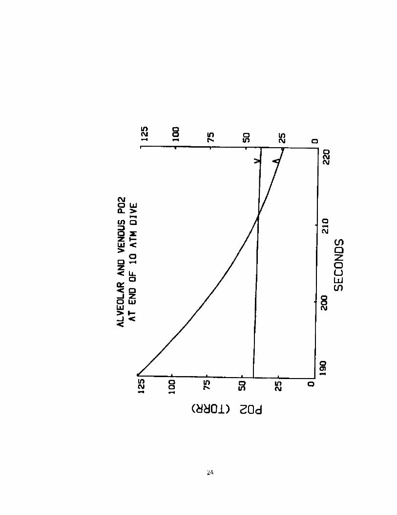
The reverse in the transfer of 02 that occurs at the end of thedive is better illustrated in figure 4 in which is plotted the alveolar
and mixed venous 02 partial pressures during the last 30 seconds of the
dive. Note that near the end of the dive the mixed venous Po2 is higherthan the alveolar Po2.
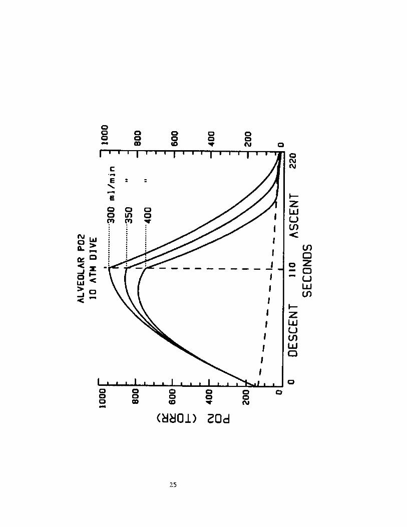
The profile of the 02 changes that occur in a dive will obviously
be affected by 0 consumption and the Lung volume at the beginning of
the dive.ln figure 5 are plotted the alveolar po2 profiles during such adive when the 02 consumtion is 300,350, and 400 ml/min respectively. TLC
17
is again set at 7.2 L.
ln figure 6 are plotted the alveolar Po2 profiles in such a dive
when the TLC is set to 6.0, 6.6, and 7.2 L respectively. 0 in all three
cases is set at 300 ml.
In such deep dives one can also get significant transfers of
nitrogen between lung and tissues. In figure 7 are plotted the changes
in the combined nitrogen volumes stored in the blood and tissues as well
as that stored. in the lung. Note that at the end of the dive, not all
the nitrogen that was transferred away from the lung is returned ta it,
resulting in a net increase of 700 ml in the amount of N2 in the blood
and tissues.
While breath-hold dives to 10 ATM are interesting to simulate, of
more practical interest is the gas transport in working breath-hold
dives to much shallower depths. Zn figure 8 are plotted the computed
alveolar and mixed venous Po2 during a 1.5 ATM dive. 02 consumtion
during the dive was assumed to be 1 L/min and cardiac output 10 L/min.
Descent, bottom, and ascent times were 15, 30, and 15 seconds
respectively. Note that in this relatively prosaic dive the possibility
of a reverse alveolar-mixed venous gradient exists.
In its present form the model can be used. to analyze gas exchange
during a single breath-hold dive. During such a single dive it predicts
the accumulation of nitrogen in the blood and tissues as well as a
reversal of the alveolar-mixed venous 02 gradient. Of more interest is
the accumulated effects in a repeated series of breath-hold dives. To do
this the model will be modified to include the gas exchange between the
lung and the environment that occurs in the interval between dives.
18
REFERENCES
1. Craig, A. B. and Harley, A. D. �961!. Underwater swimming and
loss of consciousness. Jour. Amer. Med. Assoc. 176: 255-259.
2. Paulev, P. �965!. Decompression sickness following repeated
breath-hold dives. J. Appl. Physiol, 20: 1028-1031.
3. Paulev, P. and N. Naeraa �967!. Hypoxia and carbon dioxide
retention following breath-hold diving. J. Appl. Physiol. 22: 436-440.
4. Hong, S. K., Y. C. LIN, D. A. Lally, B. J. Yim, N. Kominami, P. W.
Hong, and T. 0. Moore �971!. Alveolar gas exchange and cardiovascular
functions during breath holding with air. J. Appl. Physiol. 30: 540-547
5. Olszowka, A. J., and L. E. Farhi �968!. A system of digital
computer subroutines for blood gas calculations. Respir. Physiol. 4:270-
280.
6. Farhi, L. E. �964!. Gas stores in the body. Handbook of
Respiration Volume IZ 873-885.
19
FEGURE LEGENDS
Fig, 1. Out].ine of model used to analyze exchange in lung,
tissues during a breath-hold. See text for details.
Fig. 2. Alveo3.ar Po2 and Pco2 during breath-hold of 240 seconds at
level. Solid lines are the values produced by
the crosses represent average va3.ues of 9 breath-holding
subjects reported by Hong et als4
Fig. 3 Behavior of oxygen stores during a breath-hold dive to 10 ATM.
Lower 2 lines represent stores in lung and blood. Tissue stores
are assumed to be negligible. Top line represents the total of
the oxygen stores.
Fig. 4 Alveolar A! and mixed venous V! Po during the last 302
seconds of a breath-hold dive to 10 ATM.
Fig. 6 Alveolar ar Po2 during a breath-hold dive to 10 ATM whe> TL
6.0, 6.6, and 7.2 Liters.
Fig. 7 Behavior of nitrogen stores during a breath-hold d
Fig. 8 Alveolar P o during a breath-hold diveconsumption is 1000 ML per min.
20
Fig. 5 Alveolar Po> during a breath-hold dive to 10 ATM when oxygenconsumption is 300, 350, and 400 m3. per min. The dashed 3.ine
shows the alveolar Po2 during a breath-hold at
Illlllll K!og+Y
~ BRAIN HEART
t
ssseseaeear ~ ~ r r rpearse
II
as asser
H~hrseesar aarasreeae4
Co~
22
C!D
M M UJ0 ~i c!CO
Z I-hJ CC3
C7X ~C3 O a
O
23
VA UJ
Vl 0
C! &
4$ C
C3
C 4o
lKC Q4 ZW tULal> I-
C o a HBOJ.! ZQd24
C!C!C3
C!
u C!
27
O OO lAPl PJ
0 OO NN Al
28
CA
A ZUJ ~
~ CICl
!C
g I-
X
D4/
O 0 Oa e aM
Discussion following Dr. Olszowka's presentation
Dr. Craig: Dr. Olszowka, you didn't mention C02. Would it be a factorduring the descent7
Dr. Olszowka: In terms of the effect on oxygen exchange any rise inC02 would shift the oxygen dissociation curve a bit and thereforewould facilitate the transfer of oxygen between blood and tissues andalso -near the end of the dive � between venous blood and the alveoli.Dr. Hong has stated that in his experiments arterial PCO2 was in thelow 50's or high 40's. In the 10 Atm breath-hold dive simulationar'ter'ial PCp2 did not go up signif icantly. In computing CO~ exchangeper se I have assumed that the sizes and flows assigned to Ehe storesto f it the br eath-hold data at sea level also apply to the divingsituation. This might not be valid in the diving situation because ofshifts in blood flow not accounted for in the model.
Dr. Craig: But because of the rate of descent and the depth, doesn' tthe C02 go up as the gas volume of the lung decreases?
Dr. Olszowka: The Pc<2 in the lung does go up and then as you' recoming towards the surface, the PCp2 droPs in a way that is Parallelto the drop in P02. While the alveolar PC~2 does get up to the 60'sin the exercising diver at the bottom of the 1.5 Atm dive it isprevented from getting too high by the presence of tissue stores.Then as you are ascending the lungs expand at a rate faster than therate at WhiCh CO2 Can get baCk into the lungS. AS a result alveOlarPppp fal ls to a value that is not substantial ly higher than the val ueat Ehe beginning of the dive.
Dr. Craig: The reason I mention the C02 is that when we were divingin Hawaii we were going only 10 meters but going down very fast, andwhen we reached the bottom -where we calculated the P 02 to be veryhigh- we had the sensation that we had reached the breakPng point anddidn't know how we were going to stay there. Dives to this depth�00 meters! and at this speed must have resulted in values of PCp2that were fairly high.
Dr. Olszowka: I recall that in the 1.5 Atm simulation the values arein the 60's and thus ar'e consistent with the experience you describe.I know, however, that during rebreathing experiments one can easilytolerate PC02 values in the 60's. However, if one could not expandone's lungs, that is to perform respiratory movements, such levelswould probably be very uncomfortable.
Dr. Rabn: I would like to interject something about the interestingthing I noticed as I looked over some of Dr. Olszowka's calculations.When you come up af ter 3 o 4 minutes there has been almost no changein the C02 content of the lungs, which did not surprise me because ofthe big rniddle in Teruoka's publications 50 years ago. He had thesewomen go down to 20 meters for approximately 1 minute and when theycame to the surface he would get their very f irst expiration asalveolar samples, which had almost normal CO2 values, Presumably ftheir excess C02 was all in the tissues. So the amazing thing to me isthat even with the simulation of the 100 meter dive with the Naiorca
29
pattern, the C02 tension at the end of the dive is not very high-Dr Lundgren: One comment as far as the practical execution of thesedives goes, and Mr. Maiorca can tell you more about it, and perhapsthe most impressive illustration will be the excerpts of films thathave been taken of Nr. Maiorca's deep dives. They clearly show thatthese very deep record-breaking dives are always preceded by rathervigorous hyperventilation. The tendency for the alveolar CO2 to riseto high levels during the dive is obviously minimized by thatmaneuver.
Dr. Olsxowka: I should have mentioned that. We simulated thehyperventilation of Mr. Maiorca by assuming that the alveolar CO2 wasonly 20 torr before the start of the dive. I now recall that, as aresult of this, the highest alveolar PCO2 reached during thesimulation of this dive was in the low 40's.
Dr. Hong: In your calculations and model, how do you deal with thelung volume during the dive to LOO m?
Dr. Olszowka: I assumed that the gas in the lung was compressible andthe resulting volume changes were adjusted only by the net transfer ofgas between the alveoli and the blood. I did not address the problemof whether or not the lungs could be compressed. I just assumed theycould be.
Dr. Rabn: The program shows that at a depth of 100 m the total lungvolume is 400 cc and if you take another 150 for dead space, youhaven't got much lef t. I would like to point out something that Ifound interesting. You didn't mention anything about the dashed linein f igure 5, where you had platted alveolar PO2 during the 100 meterdive. The dashed line is based on what Dr. Hong did on divers breath-holding at the surface. You can see that at 30 seconds you are below100 torr and you keep on gradually becoming hypoxic and ending upapproximately at 20-25 torr � that' s where unconsciousness sets in.That's why I find this dashed line particularly interesting because inthe computations based on Mr. Maiorca's lung volume, going to depth of100 meters, at similar times you always have a higher alveolar P<2except during the Last f ew seconds, compared to the surf ace br eat5-hold.
Dr. Lanpbier: What was the oxygen consumption on the dashed line?
Dr. Rahn: 247 ml per minute compared to 300 ml per minute for thel00 meter dive.
Dr. Lanpbier: In other words, it was lower than that associated withthe continuous l ines.
Dr. Rabn: It is very interesting what you can do with pressure tosaturate your hemoglobin.
Dr. Lundgren: If I can make a brief comment while we have this slideon, it's true that while Dr. Hong said that the dashed line representsa non dive situation, I would be comfortable in making the
30
extrapolation to the situation of an underwater swimmer who swims thelength or perhaps several lengths of a swimming pool. Although he isnow working admittedly at a higher oxygen consumption than thatassociated with the figure, he represents the surface breath-holdsituation more than the diving one because he does not have thebenefit of compression. Ef I may briefly shift to another aspect ofDr, olszowka's presentation, that of nitrogen uptake, I do not recallthe magnitudes of the shifts in nit.rogen volumes. But I tried tocatch the numbers as they flashed by and it seemed to me that althoughthe total amount of nitrogen taken up isn't terribly large, the factthat a lot of that nitrogen might be in a tissue phase, where thenitrogen pressure is pretty high, could be of consequence. It seemedto me that you would have a tissue phase where, if I did mycalculations correctly, you might have a nitrogen pressure of between4 and 4.5 times the atmospheric pressure. That is an oversaturationin the order of 4 to l and the question inevitably comes up whetherthat is enough to cause a threat of bubble formation.
Dr, Olssovka: At the end of the dive the compartment representingheart, brain and viscera has the highest oversaturation ratio, andi t' s about 4 to l.
Dr. Lanphier: I haven't really looked into this question on a singledeep dive, but I have no indication that single breath-hold dives haveever caused decompr es sion sickness.
Dr. Olsxovka: We do plan to simulate the ef f ect of repeated diveswhere eventually the additional nitrogen accumulated after each divemight produce tissue N2 pressures at the surface large enough toproduce decompression sickness.
Dr. Lundgren: Certainly we don't know of decompression sicknessoccurring in these deep dives but the clinical diagnosis ofdecompression sickness with all its imperfections may not catch bubbleformation that actually can do damage. Although it isn't immediatelyobvious as the diver emerges, it could be something that will show upyear s later on a cumulative basi s.
31
BREATHED 4ND ASCENT BL4CKOUT
Department of Preventive Medicine and The Biotran,University of Wisconsin, Madison, Wisconsin %706.
The physiology of breath � hold BH! diving gainsimportance from associated deaths. The more we can understandabout the course of events in these deaths, the more specificand credible our educational efforts will be in helping toprevent them.
Even mamentary loss or impairment of consciousness in orunder water is enough to result in drowning. Cerebral hypoxiass presumably the cause of impaired consciousness in virtuallyevery death in breath � hold diving. Hypoxia may result fromevents such as cardiac arrhythmia, but the predominant causeis respiratory: simply staying underwater too long. 4 basicquestion is; "Why didn't air hunger forre the diver to come tothe surface to resume breathing � � or why didn't it do sasooner 7"
We need to recognize a somewhat arbitrary distinctionamong respiratory cases: those in which an attempt to reachthe surface from some depth appears to be involved and tho~ein which ascent was probably not a factor. I suggest adoptingbreath � hold bl ackout BB! as an overall term and ascentblackout AB! f or those cases where ascent seems to have beenimportant. "Shallow water blackout" is inappropriate becausethat term belongs to an entirely different condition describedby Barlaw and MacIntosh �! in 1944 and because BB canprobably occur at any depth.
BREATH � HOLD BLACKOUT.
BH bl acLout BB! i s most commonly recognized in mishapsthat occur in swimming pool s and other relatively shallowwater. Gf ten, the swimmer or di ver made no attempt at al I toreach the surface. The same sequence can presumably occur atany depth, but few cases other than those observed from thesurface would be recognized and attributed to BB. Al Craig �!deserves gratitude for describing BB and bringing it ta theattention of physicians and the aquatic public.
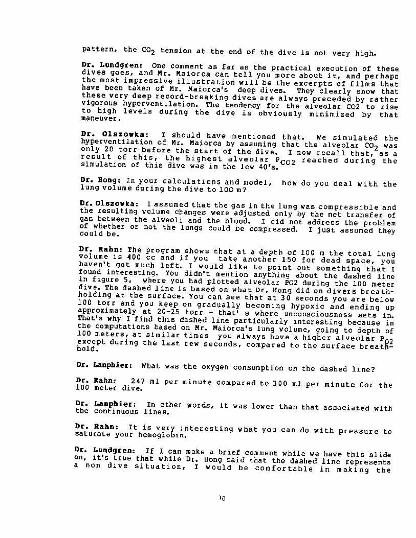
I like Hermann Rahn 's way of depicting the physiologiralbasis of BB. Fig 1 was borrowed from him. It is based on anO~ � CG> diagram. The "CG~ reservoir" an the r ight includes a
32
small column representinq the lunq. The blood-and � tissuereservoir is much larger; but it can be depleted byhyperventi 1 at i on, and the input from CO~ production during aBH may not bring it back even to a normal level. As the lowerarrow on the graph suggests, subsequent ga» exchange may carrythe PQ~ wel 1 into the hypo:: i c zone bef ore the cambined ef f ectof falling PO~ and rising PCO~ would praduce significant airhunger.
/cogHypercarbi a
$0
Anox
Fig. }. The course af PGm and PLO~ dur inq breath hol ding af ternormal breathing and two degrees of hyperventilation. FromHermann Rahn.!
It xs unrealistic to suqgest avoiding hyperventilati anentirely. Sciarli ~! proposed a method of keepinq it withinsaf e 1 i mi t s, but Hi 1 1 4! seems to have torpedoed thatappraach. Lertainly, hyperventilation should not be carriedto the point of symptomatic hypocapnxa.
ASCENT BLACKOUT.
Ascent blackout AB! is an insidious hazard. A diver whostarts ascent in good condition and at a seemingly appropriatetime may lose consciousness and drown before he can resumebreathing.
33
0 20 40 60 Hit'I imc Seconds
20 40 00 80'I'inc. Sccosd»
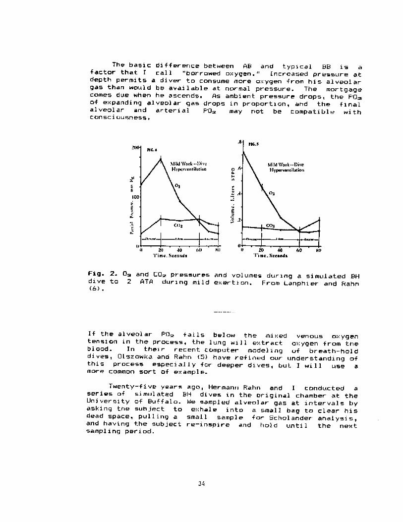
Fig. 2. O~ and CQ~ pressures and volumes during a simulated &Hdive to 2 ATA during mild exerti on. From Lanphier and Rahn�> .
lf the alveolar PO> falls below the mixed venous oxygentension in the process, the lung wi1 1 extract oxygen from theblood. In their r ecent computer model ing of breath � holddives, Dlszowka and Rahn �! have ref ined our understanding ofthis process especial ly for deeper dives, but I will use amore common sort of example.
Twenty � five years ago, Hermann Rahn and I conducted aseries of simulated BH dives in the original chamber at theUni versity of Buf f alo. Ne sampled alveolar gas at intervals byasking the sub ject to exhal e into a small bag to clear hi sdead space, pull ing a small sample for Scholander analysis,and having the subject re-inspire and hold until the nextsampl inq period.
The basicfartor that I
depth permitsgas than wouldcomes due whenof expanding alalveolar andCOnsciousnesS ~
difference between 48 and typ>cal BB is acall "borrowed oxygen." increased pressure at
diver to consume more oxygen from his alveolarbe available at normal pressure. The mortgagehe ascends. As ambient pressure drops, the F'G~veolar gas drops in proportion, and the finalarterial F'O> may not. be compatible with
Fiq ~ is taken fram that. study, which we reported in 196:.�! . The graph at. the 1 ef t indi cate» the values af al veal arI' j~ and F'CD~. 1he graph at the r i ght shows cal cul ated $1 RDvolumes af G~ and CD~ in the lunq at correspanding point~.
These values ar e averages from twa almost � identical divesby sub ject DC: Deputy Donald Chamber 1 in of the Eri e CountyBher i f f ' s Underwater Di vi si an. We chose Don ' s val ues, and I am
using them now, because he was clase to the limits oftolerance f or air hunger at depth, and hi s surf acing valuesmiqht well have resulted in drowning had he been in the
water. Upon surfacing, he was cyanotic and very clearlyimpaired.
Since these were "dry" dives, we must assume that. theeffect af immersion on CD~ storaqe described by Lundqren �!would, if. anything, have made the CG~ values lawer.
After mild work during 20 sec of descent and Cu sec at 2ATA, DC 's F'AG> was sl i ght 1 y below 5<3 torr gust before he beganascent. It. dropped to about. hal f that value upon hi s returnto I ATA. The volume plot indicates that al though exertioncontinued, uptake af oxygen I.rom the lunq was decreased evenbef ore ascent. and ceased in the f irst. part of ascent,.Final ly, according to aur cal oui at ians, there was a smal 1reversed transfer of D fr am the bload to the alveoli.
Ouch greater reversal s of G~ tr ansf er are predi cted f ordeeper di ves by Gl sz owka and Rahn �!, and thi s may occurrelatively early in asrent. Reversed D> flux is notnecessari 1 y indi cat i ve of the ri sk of. hypax i a. It. of tenr ef lects a 1arqe amount af D~ transf erred f rom the lung tablood . torage early in the dive.
Rough calculations indicate that DC borrowed and consumedabout 175 ml mare G~ than would have been avail able at F'0>50 torr at I ATA. Thi s ~mount waul d have met hi s needs f aronly about k5 sec, but it is mainly responsible for his closeapproach to AB.
When ascent began, the F'CG> started dropping at once andhovered slightly abave its normal value. Subjects in thisstudy of ten commented on the relief af air hunger that theyexperienced upon ascent. We wondered how many divers haveresponded to this relief by dallying on ascent and have diedas a result. Thi s appears ta be a seri ous hazard. Anothercritical period is the time at the surface immediatelyfollowing the first breath. } in et al. iB! have calledattent.x on to t.he importance of the 5 sec or so in whi ch the
PD~ of blood reachinq the brazen wi1 l continue to f al l. Albana 9! di scussed these hazards in graphic terms.
Excessive hyperventilation before a dive can probablylead to quiet lass of consciousness at depth Just as it doesclose to the surf ace. Perhaps lesser degrees afhyperventilation allow a diver to averstay has time at depthbut st x 1 1 try to return to the sur f ace. I f the surf ace i sclose, nei ther b8 nor AB f al laws. Deputy Chamber lin hadhyperventx lated for one minute befar e his dive, and that wasapparently enough to place him at the verge of catastrophe�Deeper dept.h would have encouraged ham to "barraw" even moreUm. and harder work, more time at depth, or more time xnascent al 1 cauld have pushed ham over the edge. It zs trulysurpr sxnq that there are not more fatalities fram 4& and deepBB than apparently accur.
BREAKPOINTS AT DEPTH
If Dan Chamber I in had nat been constrained by aurprotocc1, he would have started his ascent a litt 1.e soaner.If the dx ve had been a real one xn open water, that would havebeen none too soon. What f actors gave DC appropri ate airhunger7 What f actors beside hyper vent!. latian mi ght havecaused him to go beyand the point of no return?
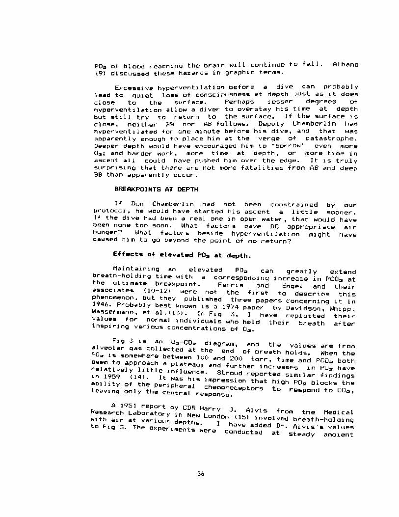
Effects of elevated PO> at depth.
brsath-hMaintaxnzng an elevated PO~ can gre tl 't da y ex en
rsa � hold>ng time with a corresponding incr ease in PCG~ at.the ultimate breakpoint. Ferris and Engel and theirassoc>ates �D-12! were nat t,he f!.rst to describe thisphenomenan but the y published three papers cancerninq it in1946. Probably best known is a 1974 paper by Davidson, WhxWassermann, et al . �.5! . avl 'son r I pp ~
In Fig ", I have replatted theireir reath af tervalues for normal individuals who held the' b
inspiring various concentrations of Om.
diagram, and the values are framalveolar gas collected at the end of breath holds. When thePQ> xs, somewhere between 1VO and 200 torr txseem to approach 1 t orr, >me and PCO~ both
r clat 1 1 ttlp a eau: and furth
ave y i e influence. Bt er increases in PQ~ haveBtroud reported similar findings
abzlity of the peripheral chemorecesn u �4! ~ It was his impression that hi h Piq D~ blocks the
chemoreceptors ta respond toy e central response.
1951 report by CDR Harr J.Research Laborator arry J. Alvis from the Medicalwith air at vari
ary an New London �5!ous depths. I n u involved breath-holding
to Fag ~. The exxperiments were c ave added Dr. Al vi s 's valuesconducted at steady ambient
36
pressures in a recompression chamber, so they were notsz mul ated dive~; but they offer sever al impor tant lessons.
80
70
PCO2 ~mmH ! 400 0 IOO 200 500 400 SOO
PO2, mmHg
Fig.3. The influence of final PD> upon alveolar PCG> at 9Hbr eakpoint. Values from Davidson et al.�~!, Alvis l5!, andLanphier and F ahn �!.
Alvis's subjects were "experienced divers," and perhaps thatis why they seem to be a breed apart from Davidson's. Evenamong the divers, one individual stands out. Presumably, theability of such divers to tolerate high PCG> would put them atexceptional risk of AB.
Alvis's two highest PCU> values do not line up very well,but each represents the mean of 6 breath holds, and the S.D. 'sare both less than 2 torr. Alvis achieved remarkableconsistency as well as remarkable values, and how he did thatis part of the stary.
CDF Alviss had observed that when involuntary contractionsof the diaphragm began, they could be quelled temporarily byswallowing. So he instructed his subjects to hold theirbreath as long as possible with the help of at least onepet xod of swallowing. Some of his subjects used swallowing
37
repeatedly, but apparently in a consistent way.
Papers by both 4gostoni �6! and Lin, et al. 8! callattention to the considerable time that may elapse between thefirst involuntary ef for ts and the ul timate breakpoint. 4diver who really wanted to be safe could probably achieve thatby starting up when the first involuntary contractions began.By the same token, maneuvers that effectively suppressinvol untary activity and air hunger may be responsible forsome fatalit.ies. Rigg, Rebuck, and Campbell �7! found thateven a strong inspiratory effort or an isovolume maneuvercould be as effective in extending time as an actualrespiratory excursion.
We see what combinations of PO and PCG~ causedDavidson's and 41vis s subjects to break their breathholds.Where were Deputy Chamberlin's values in relation to these7 Ihave added two large asterisks to Fig 3 to show them. Theupper one indicates Don 's PO~ and PCO~ just before startingascent: the lower one indicates his final values uponsurfacing.
Even at depth, DC reached a lower POm than any ofDavidson 's subjects, and hi s PCO~ was higher than that of anyof 41 vi s's divers when they held their breath wi th air atnormal pressure. The 1 ower asteri sk shows t.he drop in PCO~with ascent. DC's surfacing PO> is the lowest on the graph.That supports the impression that such a level is not likelyto be reached voluntarily, at least not without very excessivehyperventilation.
What would Deputy Chamberlin's values have beerI if he hadmade a real. dive but had not hyperventilated at all7 We can 'tbe sure. His F'CQ~'s might have been just. about what we seehere; certainly, such values would have been reached sooner,and the PO~ would not have fallen to this extent.
Even though DC's F'O> was high early in the dive, elevatedPCI> is not likely to have made much difference in hisbreakpoint. By that time, his PO~ was unusually low. Butthat observation does not rule out an unfavorable effect ofelevated PQ~ in some di ves: a diver who goes to 100 ft �0 m!or deeper ought to begin ascent whi le hi s PO~ i s st i 1 1 wel 1above 100 torr. At such levels, his "CO~ tolerance" probablywi1 1 be increased, and we may wonder what sensati ons couldpossibly tel 1 him that the time for ascent has arrived.
Lung volus!e.
I have f ound onl y one respiratory f actor that mightreduce comfortable breath � holding time at depth. That is the
38
reduction of lung volume by compressian. Unfortunately, thereseems ta be no study that directly app!ies.
Starting a breathhold at a lower lung volume certainlyleads ta shorter time. With oxygen, low lung volume willincrease the rate of rise of PCO~. W>,th air, it will hastenboth the rise of PCO~ and the drop in PO>. The~e changes mayexplain most of the differences; but the mechanism that isassociated with respiratory excursion is probably involvedalso.
Breath holding with oxygen at normal pressure is probablymost nearly comparable ta breath-hold dives with air in thesense that falling PO> is not a major factor. Accordinq toNithoefer lB>, f j,nal PACO~ i s sti 1 1 more than 80/ ofthe maximum even at a final volume close to RV.
In breath holdinq after hyperventilation with oxygen,Klocke and Rahn l9> reported such prodiqious figures asHermann Rahn's 14-min time and another subject's final PACO~of 9l torr. Although they all started at maximum inspiratian,some subjects encroached upon their residual volume beforebreak~nq. In terms of lung valume, Klocke and Rahn's subjertswere certainly comparable to divers approachinq maximumf easible depth. It i s di f f ]cult to support the view that lawlung valume, per se, wauld cause much earlier air hungerexcept perhaps at an ex tr erne.
SLNtVRY AND CONCLUSIONS.
Breath � hol d bl ackout BB> i s proposed as the overal 1term for loss of consciousness due ta *pnei c hypoxia in BHdiving. Ascent blackaut AB> would then distinquish cases inwhich ascent appears to be an important factor
2. Typical BB is adequately explaj.ned by excessivepre � dive hyperventilation. The diver 's PG~ falls to a hypoviclevel, and consciousness may be impaired or lost beforesignificant air hunger develops.
4. Hyperventilation is probably involved in most cases ofAB a!so, but other factars are also implicated. Increasedambient pressure permits oxygen to be "borrowed" from alveolargas, and subsequent ascent may result in PaO>'s j.ncompatiblewith consciousness.
5. Alveolar PCO> drops on ascent, and this may conferrelief f.rom air hunqer. If relief causes the diver to tarry,his chance of survival is reduced. Another critical periodoccurs immediately after surfacing.
39
6. Reversed transfer of 0>, from the blood to thealveo!i, may occur upon ascent especially from deep BH dives.
7. Elevated PUm can markedly evtend BH time, and this maybe a f actor in AB at greater depth».
8. Lertain individuals and groups, especially those withreduced sensitivity ta CG~, may be at unusual risk of BB andAB.
9. Maneuvers such as swal lowing and inspiratory effortmay suppress involuntary diaphragmatic contractions orotherwise encourage 1 anger BH times. They may be respansibl ef or same cases of BB or AB.
l l. Reduction of lung volume by compressxan of al veolargas at depth may tend to shorten BH time, but its influence isprobably small compared to that of factors tending to increasethe duration of &W.
11. Actions or circumstances that appear to invite BB orAB can be summarized:
a. Excessive hyperventilat.ion
b. Unusual depth
c. Failure to heed air hunger
d. Maneuvers to forestall contractions or air hungere. Delays during ascent
f . Carel essness upon sur f ac x nq
Acknowled emen
Dr. Lanphxer's recent worl:. has been funded bv the Llniver 'ity ofNxsconsin Sea Grant Instit.ute under grants from the National SeaGrant College Program, hlational Qceanic and AtmosphericAdministratian, U.S. Department of Commerce, and from the Stateof Wx sconsin. Federal grant hlA84AA � Cr-<3<!C>65, pro!ect RiNA � l l.
REFERENCES
i. Barlow HB, Macintosh FC. Shallow water black-out.Royal Naval Physiological Laboratory Report R.N.P. 44/125 UPS48 a!, 1944-
Crai g AB Jr. Summar y of 58 cases of lass ofconsciousness durl,ng underwater swimming and diving. Med SciSports 1976: 8: 1 71 � 175.
Sciarli R. Un test simple de controle deI 'hyperventilation pour le plongeur en apnee. Bull Med SubHyp 1970; 2: ~ 6.
4. Hi l I P McN. Hyperventilation, breath holding andal veal ar oxygen tensions at the breaking paint. Resp Physxol1. 97; 19: 201-209.
5. Ol szawka AJ, Rahn H. Breath � hol d di ving. In: SuttonJR, Houston CS, Coates 9, eds. Hypoxia and cold. Praeger,1 995.
6. Lanphier EH, Rahn W. Alveolar gas exchange duringbreath � hald diving. J Appl Physi ol 196'i; 18: 471 � 477.
7. Lundgren CEG. Breath hol d di vying mammals. In:Hypoxia, Exercise, and Altitude: Proceedings of the thirdBanff international hypaxia symposium. New York: Alan R.Li ss, 198.~".:65-=81.
8. Lin YC, Lal-ly DA, Moore TA, Hong Sk. Physiological andconventional breath � hold breaking parents. J Appl Physiol1974; :-7:291 � 96.
9. Albano, G. Principles and observations on thephysiology af the scuba diver. QNR Report DR � 150. Arlington:Off ice af Naval Research, 1970'.i01 � ~23.
10. Stevens CD, Ferris EB, webb JP, Engel GL, Logan M.Valuntary breathhalding. I. Pulmonary gas exchange duringbreathhol di ng. J Cl in Invest 1946; 25: 723-729.
Engel BL, Ferris EB, webb JP, Stevens CD. Voluntarybreathhalding. I I. The relation of the max@mum time ofbreathhalding to the oxygen tension of the inspired air.Cl i n Invest 194*: 25: 729 � 7 ' ' ~
41
12. Ferri s EB, Engel 6L, Stevens CD, Webb J. Voluntarybreathhalding. I l I. The relation of the ma>:imum time ofbreathhal ding to the oxygen and carbon di ox i de tensi ons ofar terial blood, with a note on its clinical and physiologicalsigni f i cance. J Cl in Invest 1946; ~ 5: 134-74.:".
1 . Davidson JT, Whipp BJ, Wasser man I;, I.'.oyal 5N,Luqliani R. Role of the carotid bodies in breath-hal ding. NEng J Med 1974; 290: 819-822.
14. Stroud RC. Combined ventilatory and breath � holdingevaluation af sensitivity to respiratory gases. J ApplPhysi ol 1959" ,14: 35.'-.-~56.
15. Al vis HJ. Breathhalding breakpoint at variousincr eased pressures. Medical Research Laboratory, USNSubmar]ne Base, thew Landon, Report Na. 1!7, Vol I >: 11.A � 12>>,1951.
16. Agastoni E. Diaphragm activity during breath holding:factors related to its onset. J Appl Physiol 196'-; 18:. 0 � 36.
17. Rigg JRA, Rebuck AS, Campbell EJM. 4 study of factorsinfluenring relief of discomfort in breath-holding in normalsub jects. Cl i n Sci Mal Med 1974; 47: 193-199.
18. Mithoefer JC. Breath holding. ln: Fenn WO, Rahn H,eds. Handbook of Physiology Section 3: Respiration Vo! I I.Nashi ngton: Amer i can Physi al og ical Sac i ety, 1965" .1O1 1 -iu25.
19. I locke FJ, Rahn H. Breath holding af ter breathing afa" ygen. J Appl Physial 1959; 14:689-693.
42
Discussion follmring Dr. Lanphier's presentation
Dr. Kannini: Is it better to exhale during the last few meters of theascents
Dr. Lanphier: I have heard that but I can't think of any reason whythat would be desirable. I consider that dangerous.
Dr. Rahn: Aren't you throwing oxygen away if you expire7
Dr. Lanphier: Yes.
Dr Lundgren: I think the idea of getting some relief f rom expi ringis probably a correct one. Blowing out would be something that,because of sheer movement of the chest and lungs, might give somerelief. You may recall the experimental background for this notion inFowler's old work Breaking Point of Breath-Holding. J. Appl. Physiol.6:539-541, 1954.! where he showed that you could hold your breathlonger if you rebreathed your own foul lung air into a rubber bag. Sowhat it could provide, I presume, is a relief from the urge to breathwhich would otherwise make you drown. Whether that would help you interms of gas exchange is very questionable because, as you say, youwould throw away oxygen and reduce the space in which you coulddeliver CO2. But by allowing you not to inhale water it would help.In the same vein, the relief that Dr. Lanphier referred to duringascent may perhaps come from the mere expansion of the chest and thelungs.
Dr. Craig: Dr. Lanphier, you mentioned excessive hyperventilation asan undesirable factor that would increase the chances of losingconsciousness. I think we should point out that we think of avigorous hyperventilation as being more dangerous than mildhyperventilation, but we should also consider that the time course ofhyperventilation is very important. In fact, if you werehyperventilating moderately for a long period of time, you would bemuch worse off than with a short period of vigorous hyperventilationbecause of the time flow-limiting aspects of the C02 elimination.
Dr Lanphier: The only thing is important here is how much youdeplete the CO> reservoir, and I think it is more effectively done bylong periods of mild hyperventilation. I think we are going to hearlater that if you try to overdo it, you may increase your CO2production enough to offset elimination.
CNS TOLERANCE TO ASPHYXIA
Bo K. Siesjo, Laboratory for Experimental Brain Research, University ofLund, Lund, Sweden
Although aaphyxia leada to both a reduCtiOn in arterial OXygen tenaiOnand to carbon diOxide retention it is the hypoxic component which usuallycauses CNS dysfunction. In fact, some results demonstrare that at certaindegrees of hypoxia, hypercapnia improves the f unction of the brain. Tneobjective of this artiCle ia to provide an overview of the tolerance of thebrain to asphyxia. I will begin by discussing functional etf ects ofarrerial nypOxia and proCeed by COnsidering the modulating influenCe oihypercapnia. Following this, I will enquire into the cellular and molecularmer.haniams by WhiCh hypoXia di.arupta nOrmal integrated brain tunCtiona ~Finally, I shall discuss mechanisms of brain damage as this evolves aftersevere hypoxiaiischemia . Kore derailed accounts of rhese subjects can befound in recent review articles Siesjo 1978, l981, biesjo and Wieloch1985a, Siesjo and Ingvar 1985!.
l. Brain d SfunCtipn in h Oxia
Extensive information exists on signs and symptoms oi cerebral oxygendefiCienCy, whether due tO mild, mOderate, Or SeVere arrerial hypnxia ~ Thisis because the subject has been of considerable inreresr. ro researchworkers in the fields of high altitude physiology and in aviation medicine see ~e ~ - Lute 1965!. Fig. 1 lists some signs and symptoms oi arterialhypoxia, as tnese have been observed at different simulated altitudes, andgives tne equivalent inspired oxygen concentrations. The figure shOws thatcertain visual functions, notably the acuity oi the dark adapted eye, failar. very mild degrees oi hypoxia, while it takes slightly more severehypoxia tO af feet short term memory, or the accomplishment of complextasks ~ Serious symptoms, such as loss of critical judgement, usually occurfirst when Pa02 ialls below about 40 mmHg, and consciousness is lost firstwhen Pa02 falls to 30 mmHg, or lower.
Observations in man during exposure to hypoxia revealed rhat evenmoderate hypoxia elicits a compensatory increase in ventilation wirh areduction in paCV2 see Fig. I!. If occurring in isolation, suchventilatory hypocapnia would be expected to reduce cerebral blood flow CBF!. however, early measurements in man demonstrated an increase in CBF Kety et al. 1948, Cohen et al. 1967 ! . Obviously, r he reduced oxygendelivery must cause cerebral vasodilation. The possibility remai~s, though,that the hypocapnia accompanying mild to moderate degrees of hypoxiacurtails the rise in CBF for a given reduction in Ya07 see below!.
AlthOugh animal eXperimentS CannOt giVe COmparable infOrmation on thefunctional effects oi' mild to moderate hypoxia, they allow precisemeasurements of physiological variables. In this context, ir. is of interes'tthat when Pa02 is reduced in steps from normal values about 90 mmHg! CBFis graaually increased unr.il, at a Pa02 of about 50 mmHg, the rate ofincrease is suddenly enhanced HcDowaLL 1966, Borgstrom et al. 1975.J6hannsson and Siesjo 1975!. We conclude from this that cerebral resistance
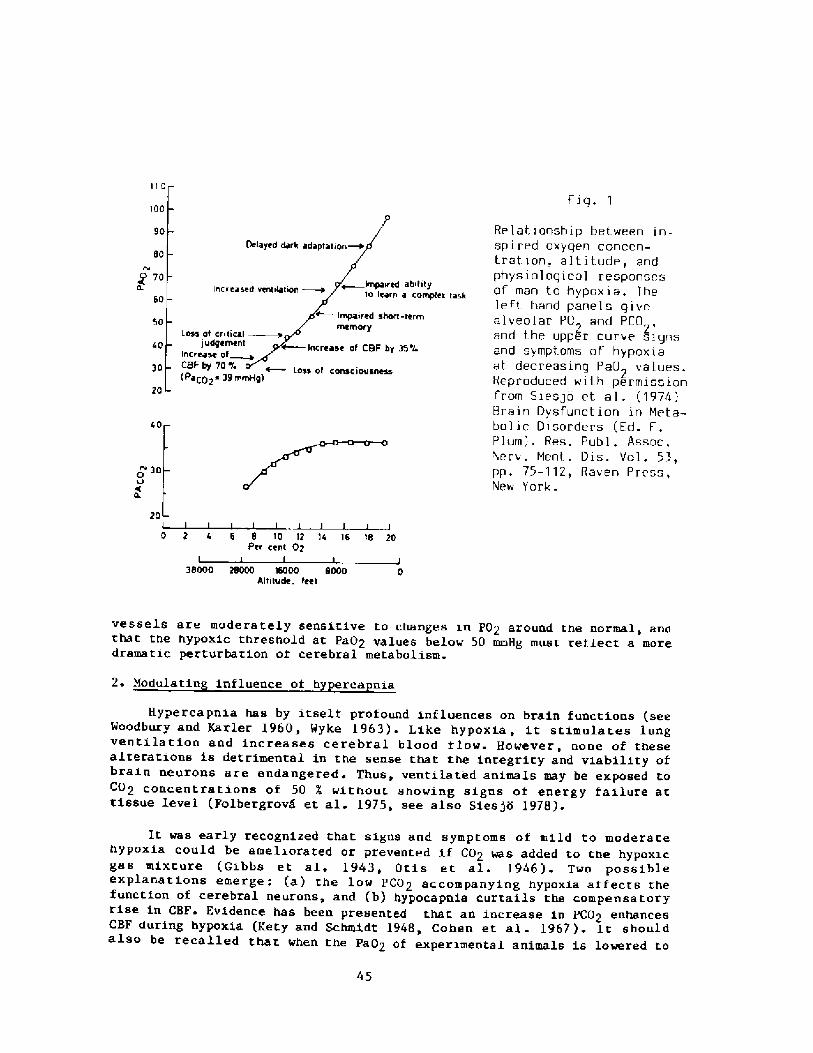
I I PPiq. 1
IPP
Relationship bet.ween in-spired oxyqen concen-t.ration, altitude, andphysioloqical responsesof man to hypoxta. lheleft. hand panels qivfalveolar PD and PCO,,and t.he upper curve signsand symptoms of hypoxiaal decreasing PaD> values.Reproduced wit.h permissionfrom Siesjo et al. �974!Brain Dysfunction in Meta-bolir Disorders Ed.Plum!. Res. Publ. Assoc.Nerv. Ment.. Dis. Vol. 53,pp. 75-112, Haven Press,New York.
90
ap
o 70
60ed abitltyrh a complex taskIn Crea 5
-term
LO55 of crltlcal1UOgemeh't
Increase of~CSF by 70% isatp2* 39 mH9!
Sp
30 55 of cohSc 1ovsne5s
20
cp
IV go0
20
0 2 6 6 6 10 12 'Is 16 16 20Per cent 02
3eooo 2SOOO mooo SO00Attitude. feet
vessels are moderately sensitive to changes in PO2 around the normal, andthat the hypoxic threshold at pa02 values below 50 mmHg must reilect a moredramatic perturbation oi cerebral metabolism.
2. Modulatin influence of h erca nia
Hypercapnia has by itself protound influences on brain functions seetxtoodbury and Karler 1960, Hyke 1963! . Like hypoxia, it stimulates lungventilation and increases cerebral blood f Low. However, none of thesealterations is detrimental in the sense that the integrity and viability ofbrain neurons are endangered. Thus, ventilated animals may be exposed toCO2 concentrations of 50 X without showing signs of energy failure attissue level Folbergrov5 et al. 1975, see also Sies jo 1978!.
45
It was early recognized that signs and symptoms of mild to moderatehypoxia could be ameliorated or prevented if CO2 was added to the hypoxicgas mixture Gibbs et al. 1943, Otis et al. l946!. Two possibleexplanations emerge: a! the low pCO2 accompanying hypoxia affects thefunction of cerebral neurons, and b! hypocapnia curtails the compensatoryrise in CBF. Evidence has been presented that an increase in pC02 enhancesCBF during hypoxia Kety and Schmidt 1948, Cohen et al. 1967!. It shouldalao be recalled that when the Pa02 of experimental animals is lowered to
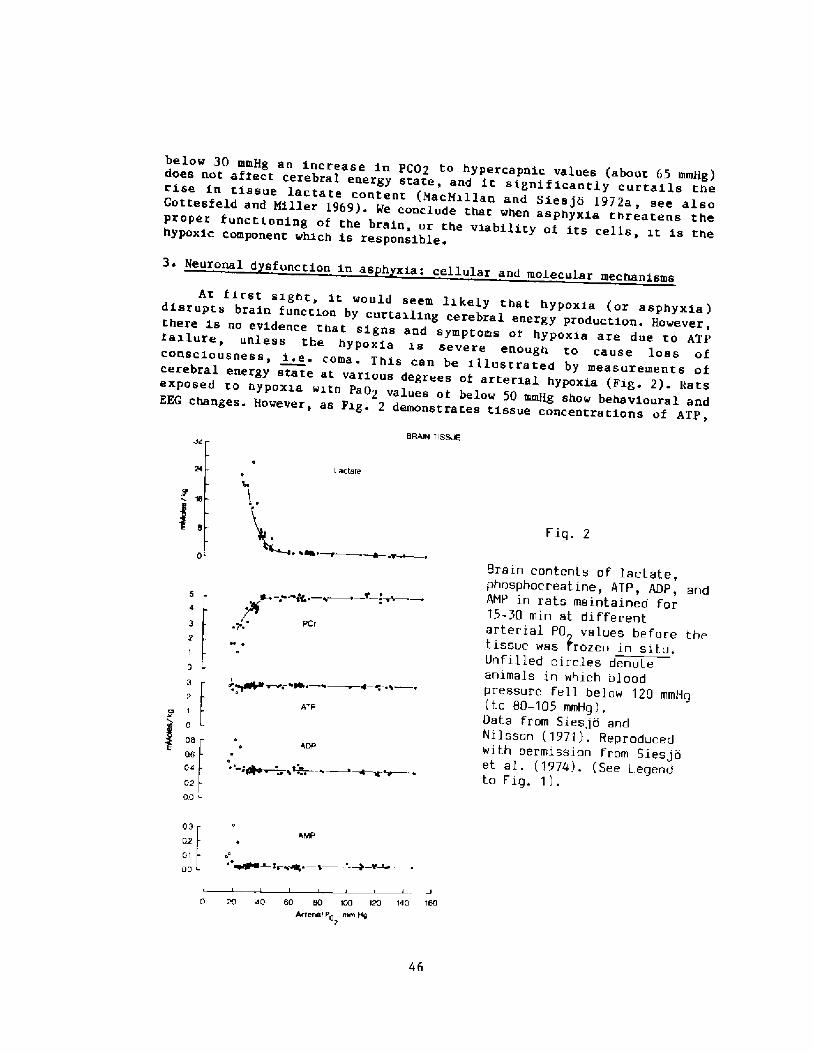
below 30 mmHg an increase in PCp2 to hypercapnic values about 65 000Hg!does not affect cerebral energy state, and it significantly curtails therise in tissue lactate content MacMillan and Siesjb l972a, see alsoGottesfeld and Millet 1969! ~ We conclude that when asphyxia threatens theproper f unctioning of the brain, or the viability of its cells 1t is thehypoxic component which is responsible.
3 ~ Neuronal d sfunction in as h xia: cellular and molecular mechanismsAt f irst sight, it would seem likely that hypoxia or asphyxia!disrupts brain function by curtailing cerebral energy production. However,
there is no evidence that signs and symptoms oi hypoxia are due to ATPfailure, unless the hypoxia is severe enough to cause loss ofconsciousness, i.e. coma. This can be illustrated by measurements ofcerebral energy state at various degrees of arterial hypoxia Fig 2!. katsexposed to hypoxia with Pa02 values of below 50 mmHg show behavioural andEEG changes ~ However, as Fig. 2 demonstrates tissue concentrations of ATP,
8RAIN TISR%
Leetere
Fig. 2
Brain contents of lactate,phosphocreatine, RTP, RDP, andRMP in rats maintained for15-30 min at differentarterial PO values before thet.issue was rozen in situ.Unfilled circles denoteanimals in which bloodpressure fell below 120 mmHg t o 80-105 mmHg ! .Data from Siesjo andNilsson 1971!. Reproducedwith permission from Siesjoet al. �974!. See Legendto fig. 1! .
gt. � A
rr~~ � ~e8r � + � ~ �,� L�I4
ATP
0
060402pp
08 P
0.2 4 AMP
01 I~ ~OO
40 80 80 rPP 120 WP 160Arterrel Pp mm Hg
2
46
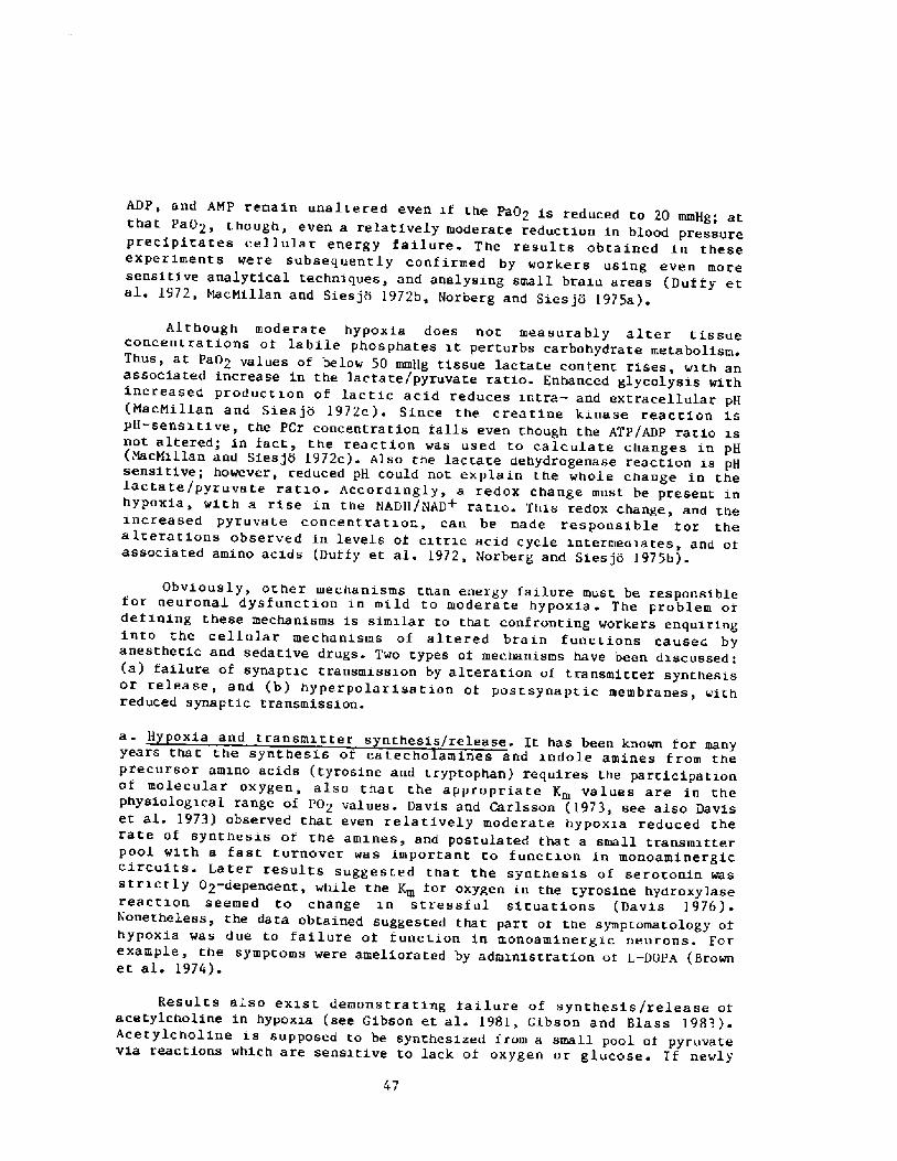
ADp, and ANp remain unaltered even if the Pa02 is reduced to 20 mmHg; atthat PaO2, though, even a relatively moderate reduction in blood pressureprecipitates cellular energy failure The results obtained in theseexperiments were subsequently confirmed by workers using even moresensitive analytical techniques, and analysing small brain areas Duf fy etal. 1972, NacMillan and Siesjo 1972b, Norberg and Siesjo 1975a!.
Although moderate hypoxia does not measurably alter tissueconcent ra t iona o f la bile phos phates i.t perturbs carbohydrate metabolism.Thus> «PaO2 values of below 50 mmHg tissue lactate content. rises, with anassociated increase in the lactate/pyruvate ratio. Enhanced glycolysis withincreased production of lactic acid reduces intra- and extracellular pH MacNillan and Siesjo 1972c!. Since the creatine kinase reaction ispH-sensiti.ve, the PCr concentration ialls even though the ATP/ADP ratio isnot altered; in fact, the reaction was used to calculate changes in pH NacNillan and Siesjo 1972c!. Also the lactate dehydrogenase reaction i.s pHsensitive; however, reduced pH could not explain the whole change in thelactate/pyruvate ratio. Accordingly, a redox change must be presenr. inhyPoXia, With a riae in the NADH/NAD+ ratiO. ThiS redOX Change, and rheincreased pyruvate concentration, can be made responsible f or thealterations observed in levels oi citric acid cycle intermediates, and ofassociated amino acids Duify et al. 1972, Norberg and Siesjo 1975b!.
Obviously, other mechanisms than energy failure must be responsiblefor neuronal dysfunction in mild to moderate hypoxia. The problem ofdef ining these mechanisms is similar to that confronting workers enquiringinto the cellular mechanisms of altered brain functions caused byanesthetic and sedative drugs. Two rypes of mechani.sms have been discussed: a! failure of synaptic r ransmi.ssion by alteration of transmitter synthesisor release, and b! hyperpolarisation of postsynaptic membranes, withreduced synaptic transmission.
a. H oxia and transmit ter s nthesis/release. It has been known for manyyears that the synthesis of catecho amines and indole amines f rom theprecursor amino acids tyrosine and tryptophan! requires the participationof molecular oxygen, also that the appropriate Km values are in thephysiological range of PO2 values ~ Davis and Carlsson �973, see also Daviser. al. 1973! observed that even relatively moderate hypoxia reduced therate of synthesis of the amines, and postulated that a small transmitterpool with a fast turnover was important to function in monoaminergiccircuita. Later reaults suggesred that the synthesis of seroronin wasstrictly 02-dePendent, while the Km ior oxygen in rhe tyrosine hydroxylasereaction seemed to change in stressiul situations Davis 1976!-Nonetheless, the data obtained suggested that part oi the sympromatology ofhypoxia was due to failure oi f unction in monoaminergic neurons. Forexample, the symptoms were ameliorated by administration oi L-DOPA Brownet al. 1974!.
Results also exist demonstrating iailure of synthesis/release ofacetylcholine in hypoxia see Gibson et al. 1981, Gibson and Blass 19S3 !.Acer.ylcholine is supposed to be synthesized from a small pool oi pyruvatevia reactions which are sensitive to lack of oxygen or glucose If newly
47
SyntheaiZed acetylcholine iS Whar. 1S pret erentially releaSed One COuldargue that hyPOXia leadS tO failure Of ChOlinerg1C tranSmiSS1On. Insupport, results demonstrate that mild-to-moderate hypoxia reduces the rateof acetylcholine synthesis Gibson and Dufiy 1981!. However, even morerecent results hint that tne iall in tne rate oi synthesis could be theresult rather rhan the cauee of the reduced synaptic activity Peterson andGibson 1982 ! . These new results suggest that mild hypoxia retards Ca 2+influx into pre-synaptic terminals, thereby reducing release oi transmitte»and, secondarily, its synthesis. We reCognize that such an ef feet, ifexisting, could possibly explain failure of transmission at other synapsesas well.
b ~ H OXia and neurpnal h er olariaation HypOXia, hypercapnia andmetabolic inhibitors such as 2,4-dinitrophenol cause hyperpolarisation oineuronS and decrease the resistance of their membranea, probably by an1ncrease in K+ conductance Speckmann and Caspers 1974, Krnjevic 1975,Krnjevic et al. 1978, Hansen et al. 1982!. As discussed by Hansen �985!theSe eVentS wOuld be expected to blOCk SynaptiC tranamiasiOn by clampingthe postsynaptic membranes at. hyperpolarised values, thereby rendering r hemembranes less prone to depolarisation.
It th1S Were the meChaniSm Oi hypoXic neurOnal dySf unCtiOn, whatcauses the hyperpolarising efflux of K+ along conductance channels? K+ChannelS are uSually COnaidered tO be VOltage-dependent or SenSiti Ve tOchemical perturbation, notably to a raised Ca2 concentration Thus, ifCa2+ enters the cell or is released 1 rom intracellular binding orSequeStratiOn. Sitea, K+ COnduCtanCe ShOuld inCreaSe. There are nO reSultSdemOnatrating that mOderate hypOXia cauaeS Ca2+ influx inrO neurOns SinCeSuCh influx 1S a 1ate PhenOmenOn, OCCurring at very 1Ow PO2 Or blOOd flOwvalues Hansen and Zeuthen 1981, Harris et al. 1981!. In theory, though.Ca2+ COuld be relea Sed f rom inr ra Cellular binding/SequeatratiOn Sites,either by a lnwering Ot pHi ~e. Neeeh and ThomaS 1980! Or byagonist-receptor interactions at the membrane Surface, activatingphospholipase C {Berridge 1984!.
Whether or not Ca 2+ ir a second messenger it is conceivable that aninCreased V+ COnduCtanCe during 1SChemia, Or indeed any permeab111ty Cha~gecontributing to the functional "block", occurs in response tophosphorylatiOn/dephOSphOrylation Oi membrane prOteinS by the appropriatekinases and phosphatases Neary and Alkon 1983, Nestler and Greengaard1983, see also Hansen 1985!. This is a speculative deduction, though, andwe neither know the first nor the second messenger 1n this hypotheticalchain of events.
4. Ischemia
If the arterial hypoxia iS SuffiCiently severe CardiOVaSCular funCtiOnusual1,y suffers, with a fall in cerebral perfusion pressure. Since hypoxiaabOliaheS CerebrovaSCular autOregulat1On, hypOtenS1On iS SynOnymOuS with areduCtiOn in CBF. AS lOng aS CBF doeS not fall belOw nOrmal remember thatit is inCreaSed during hypoXla! the conditinn iS One Oi hypOxia withrelative iSchemia Or reStriCted hyperemia!. AS we have Seen, Cerebral
48
energy state may then deteriorate see Fig. 2 above!. Such energy failuremust endanger the viability of brain cells, especially neurons. For thesereasons, a reduction of blood pressure during severe arterial hypoxia canhave devastating consequences.
Under normoxic circumstances, CBF can be reduced to about 18 ml'100g 1 min 1 beiore spontaneous or evoked electrical activity fail, and toabout 12 ml 100 g l.min 1 bef ore membrane iailure ensues with massiverelease of K+ from cells, and uptake oi Ca2+ Branston et al. 1977, Astrupet al. 1981, Harris et al. 1981! ~ This depolarisarion, and theinflux/release of Ca2+, cause an avalance series of metabolic reactionswith enhanced lipolysis, proteolysis, disassembly of microtubuli andprotei.n phosphorylations, reactions which may be responsible for the finalcell damage, once the ischemia has surpassed the revival time, i.e theshortest ischemic period compatible with full restitution see Siesjo 1981,Siesjo and Wieloch 1985a!
Clearly, if arterial oxygen tensions are low the ischemia need not bethat dense to cause membrane failure and cell injury/death Lt seemswarranted to discuss two important issues: ischemic revival times, andselective neuronal vulnerability.
a. Revival times. It is common clinical experience thar. cardiac arrestlasting longer than 4-5 min invariably leads to irreversible brain damage.There may be several reasons for these very short revival times. Hostimportanly, patients with cardiac arrest probably have poor tissueperi usion in the immediate recirculation period, with prolongation oi theiSChemia intO the periOd Of reSuSCitatiOn. EXperimentally, One Can aChieVesquare wave ischemia and, by pressor agents, secure prompt recirculation ofthe iSChemiC tiSSue. Under SuCh CirCumstanCea, SOme COmpleX phySiolOgicaland metabolic functions can return, at least for many hours, followingischemic periods of as long as 60 min Hossmann 1982, 1985!. Long-termrecovery of neurological function, with little or no histopathologicdamage, has been achieved after ischemic periods oi 16 � 20 min forliterature, see Siesjo 1978, Hossmann 1982!.
b. Selective vulnerabilit . While it may be pssible to identify a revivaltime for the function of the brain as a whole, or in terms ofneuropathologically deiined brain damage, the term becomes ambiguous whenone considers recovery oi indivi.dual brain neurons, or groups oi neurons.This is because a selective vulnerability exists, with pronounceddifferences in susceptibility to incur damage following hypoxia/ischemia ~e.. Brierley 1976!. Recent results have provided some challengingconcepts First, neuronal necrosis following short periods of ischemia can"mature" over hours and days, and may appear after a delay during whichfunction is transiently resumed Kirino 1982, Pulsinelli et al. 1982,Pulsinelli and Dufiy 1983, Suzuki et al. 1983! ~ Second, under seeminglyoptimal experimental conditions unequivocal brain damage has been observedaf ter such short ischemic periods as 5 min Kirino 1982, Smith et al.1984! Typically, such damage then affects pyramidal cells in thehippocampus, notably in the subiculum, CA1, and CA4 sectors, and neurons insome other selectively vulnerable areas Kirino 1982, Pulsinelli et al
49
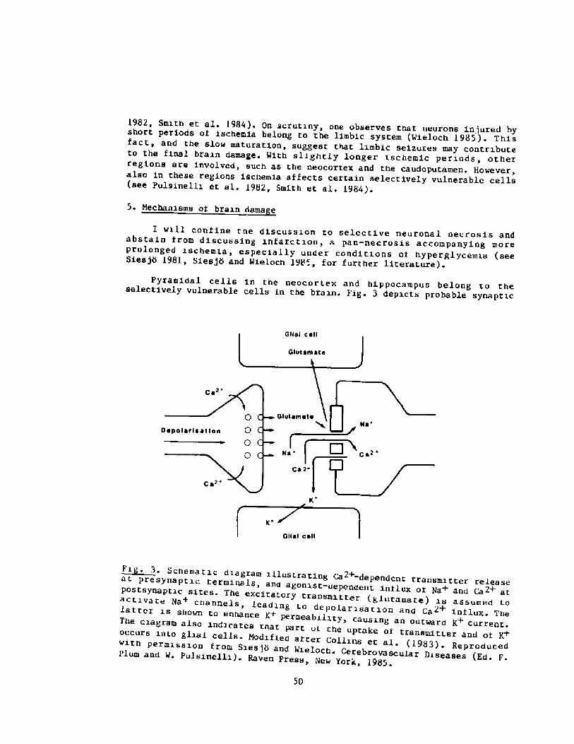
1982, Smith et al. 1984!, On scrutiny, one observes that neurons injured byshort periods oi ischemia belong to the limbic system Wieloch 1985!. Thisfact, and the slow maturation, suggest that limbic seizures may contributeto the final brain damage. With slightly longer ischemic periods, otherregions are involved, such as the neocortex and the caudoputamen. However,also in these regions ischemia affects certain selectively vulnerable cells see Pulsinelli et al. 1982, Smith et al. 1984!.
5. h1echanisms of brain dame e
I will coniine the discussion to selective neuronal necrosis andabstain from discussing infarction, a pan-necrosis accompanying moreprolonged ischemia, especially under conditions of hyperglycemia seeSiesjo 1981, Siesjo and Wieloch 198', for further literature!.
Pyramidal cells in the neocortex and hippocampus belong to theselectively vulnerable cells in the brain Fig 3 depicts probable synapti.c
scnemattc anagram tllnatrarlnt pa2+-depentent trFi . 3 ranssLL t ter re les seostat presynaptic terminals, and agonlst-dependent inilux oi N + a Cag+
a an atp synaptic sites. The excitatory transmit.ter 1 !activate 5a + channe er g utamate is assumedchannels, leading to depolar isation and Ca2+ influx. Thelatter is shown to enhance K+ permeability, causing an outward K+ current ~The diagram alao indicates that part or the uptake oi transmit r
+occurs into i o ransmitter and oi Kwith ermissi.glial cells- Nodified after Collins et al ~ �98 !
a ~ 3! Reproducedpermission from Siesjo and Wieloch. Cerebrovascular Diseases Ed. F.Plum and W. Pulsinelli!. Raven Press, New York, 1985.
events leading to excitation of such cells Collins et aL. 1983! Anexcitatory transmitter glutamate or aspartate! is believed to open Na+channels, the influx of Na+ then depolarising the postsynaptic membrane,this in turn causing Ca 2+ ini'lux. Apart f rom cont ra but ing todepolarisation, the inf lux of Ca2+ can cause Lipolysis, proteolysis, andprotein phosphorylation, i.e. enhance calcium-activated orcalcium-dependent reactions.
The events depicted in Fig. 3 illustrate features of two currenthypothesis on mechanisms of neurona1 necrosis. Most importantly, the f igurefocusses attentio~ on calcium and excitarory amino acids. It had previouslybeen assumed that uncontrolled calcium influx, and/or its release f romintracellular binding or sequestration sites, contributed to cell necrosisin other tissues than the brain. The application of the calcium hypothesisto the brain was inspired by the observation that selectively vulnerableneurons appeared identical to those identified as having high dendriticcalcium conductances, making these cells prone to epileptogenic firing seeSiesjo 1981, Siesjo and Wieloch 1985 a and b!.
It now seems that such a simple calcium hypothesis " vulnerable cellshave high calcium conductances"! does not suffice to explain selectiveneuronal vulnerability. Previous work on the neurotoxicity of excitatoryamino acids led to the hypothesis of excitotoxic neuronal damage, and itsassociation with seizure states OLney 1978, Collins et al. 1983!. ThishypOtheaiS haS nOw been extended tO iSChemia Jgfrgenaen and Diemer 1982,Simon et aL. 1984, Wieloch 1985!. The hypothesis receives strong supportfrom observations that vulnerable neurons are innervated by glutamatergic or aspartergic! afferents, and that lesions of such afferents seem toameliorate ischemic damage Wieloch 1985! ~
According to this hypothesis, neurons innervated by excitatory aminoacids are vulnerable because ischemia leads to massive release oi the aminoacids from presynaptic endings see Benveniste et al. 1984!, and bcauseagonist-receptor interaction then leads to uncontrolled ini lux of Na+and/or Ca 2+ int o post ay na p tie element s. This is, essentially, unbalancedexcitation. However, it has been recognized that the outcome of theischemic insult must depend on inhibitory tonus as well. For example,lesions of the noradrenergic system originating in the locus coeruleus, asyStem COnVeying inhibitiOn, haS been fOund tO aggravate the iSChemiCneuronal damage Blomqvist et al. 1985! ~ Clearly, agonist-receptorinteraction must play an important role in the development oi ischemicneuronal necrosis.
6 ~ Summar and conclusions
The results discussed make it clear that CNS tolerance to asphyxiamust be discussed at two levels. The f irst concerns changes in brainf unction at mild to moderate degrees of hypoxia or asphyxia!. Since suchchanges can cause disturbing and threatening failure of intelLectual andemotional functions ~e.. Loss of visual acuiry, failure to accomplishcomplex tasks, loss of critical judgement! it is justified to explore themechanisms involved. Energy iailure at the cellular level does not seem
5l
responsible ~ Possibly, reduced rates of synthesis of monoamines and ofacetylcholine could contribute. However, it seems even more likely that themetabolic perturbation leads to the appearance oi a second messenger, thisin turn influenCing membrane permeability by phasphnrylatinn Of iOniCchannels Such a sequence of events could underlie an observedhyperpOlariaatiOn Of neuronS, With reduCed reSiStanCe due tO enhanced K+permeability, events that could induce blockade ot synaptic transmission.
The second level of hypoxia is one sufficiently severe to endanger theviability of brain cells. Since these cells are quite resistant to purearterial hypoxia overt cell damage requires that hypoxia is complicated byhypotension, or circulatory collaps Under such conditions hypoxia-ischemia! damage is incurred by certain selectively vulnerablecells, their vulnerability residing in special agonist-receptorinteractions. Specifically, vulnerable cells seem innervated by excitatoryaminO acids whose interaction with post-synaptiC Sites causes influx ofNa+ and Ca~+. Probably, the toxicity of these amino acids is due to suchinilux, and tO enhanCed firing Oi neurOns innerVated by them.REFERENCES
Astrup J, Siesjb B K, and Symon L �981! Thresholds in cerebral zschemiathe ischemic penumbra Stroke 12:723-725-
Benveniste H, Drejer J, Schousboe A, and Diemer N H �984! Elevation oi theextracellular concentrations of glutamate and aspartate in rat hippocampusduring transient cerebral ischemia monitored by intracerebralmicrodialysis. J Neurochem 43:1369-1374
Berrzdge M J �984! Inositol trisphosphate and diacylglycerol as secondmessengers. Biochem J 220:345-360
Blomqvzst P, Lindvall 0, and Wieloch T �985! Lesion oi the noradrenergiclocus coeruleus system aggravate neuronal necrosis after transient cerebralischemia. Neurosci Lett. In press.
Borgstrom L, J6hannsson N, and Siesjo B K �975! The relationship betweenarterial P02 and cerebral blood fin~ in hypoxic hypoxia . Acta Physiol Scand93:423-432
Branston N M, Strong A J, and Symon L �977 ! Extracellular potassiumactivity, evoked potential and tissue blood flow. Relationships duringprogressive ischaemia in baboon cerebral cortex. J Neurol Sci. 32;305-321 '
Brierley J B �977 ! Cerebral hypoxia. In. 'Greeniield s Neuropathology, ed'by W. Blackwood and J.A.N. Corsellis. London: Arnold, p. 43-85.
Brown R M, Davis J N, and Carlsson A �974! Dopa reversal ofhypoxia-induced disruption of rhe conditioned avoidance response. J PharmPharmacol 25:412-414
52
Cohen P J, Alexander S C, Smith T C, Reivich M, and Wollman H �967!Eifects of hypoxia and normocarbia on cerebral blood flow and metabolism inconsci.ous man. J Appl Physiol 23:183-189.
Collins R C, Lothman E W, and Olney J W �983! Status epilepticus in thelimbic system: Biochemical and pathological changes. In: Advances inNeurology ~ Vol. 34: Status Fpilepticus Delgado-Escueta A V, Wasterlain CG, Treiman D M, Porter R J, eds. ! . Raven Press, New York, p. 277-288
Davis J N, and Carlsson A �973! The ef feet oi hypoxia on monoaminesynthesis, levels and metabolism in rat brain. J Neurochem 21:783-790.
Davis J N, Carlsson A, MacMillan V, and Siesjo B K �973! Brain tryptorhanhydroxylation: dependence on arterial oxygen tension. Science 182:72-74.
Davis J K �976! Brain tyrosi.ne hydroxylation: alteration ot oxygenati inity in vivo by immobilization or electroshock in the rat. J Neurochem27-211-215.
Duf 1 y T E, Nelson S R, and Lowry 0 H �972! Cerebral carbohydratemetabolism during acute hypoxia and recovery. J Neurochem 19:925-934.
Folberg rove J, Norberg K, Quistorff B, and Siesjo B K �975! Carbohydrateand actino acid metabolism in rat cerebral cortex in moderate and extremehypercapnia. J Neurochem 25:457-462.
Gibbs F A, Gibbs E L, and Lennox W G �943! The value of carbon dioxide incounteracting the effects of low oxygen. J Aviat Med 14:250 � 261 .
Gibson G E, Pulsinel.li W,Blass J P, and Duffy T E �981! Brain dysfunctionin mild to moderate hypoxia . Am J Med 70:1247-1254.
Gibson G E, and Blass, J P �983! Metabolism and neur'otransmission. In:Handbook of Neurochemistry, edited by A. Lajtha, New York: plenum, vol. 3,p. 633-651.
Gibson G E, and Duffy T f. �981! Impaired synthesis of acetylcholine bymild hypoxic hypoxia or nitrous oxides J Neurochem 36:28-33.
Gottesfeld Z, and Miller A T �969! Metabolic response of rat brain roacute hypoxia: influence oi polycythemia and hypercapnia . Amer J Physiol216:1374-1379.
Hansen A J, and Zeuthen T �98l! Extracellular ion concentrations duringspreading depression and iscnemia in the rat bra in cortex. Acta PhysiolScend 113:437-445
Hansen A J, Hounsgaard J, and Jahnsen H �982! Anoxia increases potassiumconductance in hippocampal nerve cells. Acta Physiol Scand 115:301-310
Hansen A J �985! Effect ot anoxia on ion distribution in the brain.Physiol Rev 65�!:101-148.
53
Harris R J, Symon L, Branston N M, and Bayhan M �981! Changesextracellular calcium activity in cerebral ischemia ~ J Cereb Blood FlowMetabol 1:203-209.
Hossmann K,.-A. �982! Treatment of experimental cerebral ischemia. J CerebBlood Flow Metabol. 2:275-297 ~
Hossmann K-A �985! Post-ischemic resuscitation oi the brain: selectivevulnerability versus global resistance. In: Progress in Brai~ Research,Vol. 63, edited by K. Kogure, K.-A. Hossmann, B.K. Siesjo and F.A Welsh.Blsevier Science Publishers B.V ~, Biomedical Division, Amsterdam, pp. 1-15.
J6hannsson H, and Si,esjo B K �975! Cerebral blood flow and oxygenconsumption in the rat in hypoxic hypoxia. Acta Physiol Scand 93:269-276.
Jgrgensen M B, and Diemer N H �982! Selective neuron loss aiter cerebralischemia in the rat: Possible role of transmitter glutamate. Acta NeurolScand 66:536-546.
Kety S S, and Schmidt C F �948! The eifects of altered arterial tensionsof carbon dioxide and oxygen on cerebral blood flow and cerebral oxygenconsumption or normal young men. J Clin Invest 27:484-491.
Kirino T �982! Delayed neuronal death in the gerbil hippocampus followingischemia . Brain Res 239:57-69.
Krnjevit K �975! Coupling of neuronal metabolism and electrical activiry-Brain Work, edited by D.H. Ingvar and N.A. Lassen. Copenhagen:
Munksgaard, 65-78. Alfred Benzon Symp !
Kznjevid K Puil E and W erman, R �978! Significance of 2,4 � dinitrophenolaction. on spinal motoneurones. J Physiol London 275:225-239.
Luft U C �965! Aviation hp ysiology � the effects of altitude. In'. Handbookoi Physiology. Respiration, eaired by W.O. Fenn and H R h Wan . ahn. Washington,Physiol Soc, sect 3, vol. II chapt. 44, p. 1099-1145.
McDo wall D 0 �966 ! Inter rela tionshcerebral blood flow. In: Ox enonships between blood oxygen tensions
b J.PE Pa nn: xygen Measurements in Blood and Tissues, edited
y J.P- Payne and D.W. Hill ~ London: Churchill. p. 205-219.
MacMillan V, and Sies jo B K �912a! The etiect ot pInvest 30:237-244.
e rain durin arterialg hypoxemia. Scend J Clin Lab
MacMillanacMillan V, and Siesjo B K �972b! Brain ener metaS d J Clin Lab In t 30.126-1366-136
MacMillan V, and Siesjo B K �972c! Intraarterial hypoxemia 1 2c! Intracellular pH of the brainphosphokinase equilibrium. Scend J 2 method and from the creatinr um. Scend J Clin Lab Invest 30:117-125
Meech R W, and Thomas R 0 �980! Ei feet or measured calcium chlorideinjections on the membrane potential and internal pH of snail neurones. JPhysiol London 298:111-129.
Neary J T, and Alkon. D L �983! Protein phosphorylation/dephosphorylationand the transient voltage-dependent porassium conductance in Hermissendacrassicornis. J Biol Chem 258:8979-89S3.
Nestler E J, and Greengaard P �983! Protein phosphorylation in the brain ~Nature London 305:583-5SS.
Nor berg 1', and Sies jo B K �975a! Cerebral metabolism in hypoxic hypoxia.I. Pattern of activation of glycolysis, a reevaluation. Brain Res 86:31-44 ~
Norberg, K, and Sees jo B K �975b! Cerebral metabolism in hypoxic hypoxia.II. Citric acid cycle intermediates and associated amino acids. Brain Res86:45-54.
Olney J �978! Neurotoxicity oi excira tory amino acids. In: Kainic as aTool in Neurobiology E G McGeer, J W Olney, P L McGeer, eds.! Raven Press,New York. pp. 95-121.
otis A B, Rahn H, Epstein M A, and Fenn W 0 �946! Performance as relatedto composition of alveolar air. Am J Physio1. 146:207-221.
Peterson C, and Gibson G E �982! 3,4-diaminopyridine alters acetylcholinemetabolism and behaviour during hypoxia. J Pharmacol Exp Ther 222:576-582.
Pulsinelli W A, Brierley J B, and Plum F �982! Temporal prof ile oineuronal damage in a model of transient f orebrain ischemia. Ann Neurol11:491-498.
Pul sinelli W A, and Duffy T E �983! Regiona1, energy balance in rat brawnafter transient forebrain ischemia. J Neurochem 40;1500-1503.
Schmahl F W, Betz E, Dettinger E, and Hohorst H J �966!Energiestof f wechsel der Grosshirnrinde und Elektroencephalogramm be+Sauerstotfmangel. Piliigers Arch ges Physiol 292:46-59-
Sies jo B K �978! Brain Energy Merabolism. John Wiley 6 Sons, Lrd. ThePitman Press, Bath.
Sies jo B K �981! Cell damage in the brain; A speculative synthesis. JCereb Blood Flow Metabol 1:155-185.
Sees jo B K, and Niisson L �971! The influence of arterial hypoxemia uponlabile phosphates and upon extracellular and intracellular lactate andpyruvate concentrations in the rat brain. Scana J Clin Lab Invest 27:83-96.
55
Sies jo B X, Ji5hannsson H, L j unggren B, and Nor berg K �974 ! Braindysfunction in cerebral hypoxia and ischemia. Res Publ Assoc kes Nerv MentDis, 75-112.
SiesjU B K, and Ingvar H �985! Ventilation and brain metabolism. In:Handbook of Physiology, to be published by Ameri.can Physiological Society.In press.
Siesjo B K, and Wieloch T �985 a! Brain injury: Neurochemical aspects. In.'Central Nervous System Trauma � Status Report Becker D, Povlishock J,eds ~ ! William Byrd Press Inc., Richmond, VA pp. 513-532 ~
Sies jo B K, and Wieloch T �985 b! Molecular mechanisms of ischemic braindamage: Ca2+-related events. In: Cerebrovascular Dieseases, edited by F.Plum and W. Pulsinelli. Raven Press, New York. pp. 187-200
Simon R P, Swan J H, serif f ith T, and Meldrum B S �984! Blockade ofN~ethyl-D-aspartate receptors may protect against ischemic damage in thebrain. Science 226:850-852.
Smith H-L, Auer R N, and Siesjo B K �984! The density and distribution oiischemic brain injury in the rat following two to ten minutes of forebrainischemia. Acta Neuropathol Berl !. 64:319-332.
Speckmann E-J, and Caspers H �974! The effect of 02- and CO2-tensions inthe nervous tissue on neuronal activity and DC potentials. In: Handbook ofElectroencephalography and Clinical Neurophysiology, edited by A kemondAmsterdam: Elsevier, vol . 2, pt. C, p. 71-89 '
Suzuki R, Yamaguchi T, Choh-Luh L, and Klatzo I �983! The ei fects oi5-minute ischemia in mongolian gerbils: II. Changes oi spontaneous neuronalactivity in cerebral cortex and CA 1 sector of hippocampus. ActaNeuropathol Berl! 60:217-222.
Wieloch T �985! Neurochemical correlates to selective neuronalvulnerability Progress in Brain Res. vol. 63. In press.
Woodbury D H, and Karler R � 960! The role of carbon dioxide in the nervoussystem. Anesthesiology 21:686-703.
Wyke B �963! Brain Function and Metabolic Disorders, Butterworths, London.Acknowle ements . 'Work from the author s own laboratory was supported bygrants from the Swedish Medical Research Council, and from USPHS.
Discussion following Dr. Siesjo's presentation
Dr. Arnold: Have you considered the use of calcium channel blockersafter clinical transient ischemic attacks or cardiac arrests andrevivals
Dr. Siesjo: Yes, there are at least three experimental studies whichhave shown that you can ameliorate the damage if you give the calciumentry blockers after transient ischemia. They have also been used inmiddle cerebral artery occlusion and seem to have effect on theassociated damage. Clinical trials are now being initiated< one ofthem in Scandinavia. They are double blind control studies and theirresults are not ready yet.
Dr. Hong: What is the mechanism for the entry of calcium across thepost-synaptic membrane?
Dr. Siesjo: It brings in calcium by opening voltage-dependent calci.umchannels.
Dr. Hong: What about the sodium-calcium exchange mechanisms
Dr. Siesjo: The primary effect of the excitatory transmitter isprobably to open conductance channels for Na+. The depolarizationassociated with sodium influx is supposed to open voltage-dependentcalcium channels.
Dr. Rahn: Voltage dependents
Dr. Siesjo: Yes. And both ions carry a current across the membrane.A depolar izing current.
Dr. Hong: Is it possible that hypoxia-induced increase in cell sodiumcould be responsible for the increase in cell calcium via inhibitionof sod i um-ca l ci um exchange m ech an i sm s?
Dr. Siesjo: Most people would assume that in the majority of cellsyou have both a sodium-calcium exchange< which is not ATP-dependent<and an ATPase which would pump calcium out of the cell.
Dr. Hong: This is another mechanism. Sodium-calcium exchange itselfis not related to calcium ATPase.
Dr. Siesjo: No, but there are probably two mechanisms for calciumextrusion, sodium-calcium exchange being one.
Dr. Hong: You are not eliminating the possibility of the involvementof sodium calcium exchange.
Dr. Siesjo: Oh no, the reason why I am talking about the calciuminflux along conductance channels is that neurophysiologistsconsider many pyramidal cells to f ire repetitively by calcium-dependent mechanisms, that is, the calcium influx contributes toexcitation by depolarizing cells.
57
A ld: I was very interested in your mention of the limbicDr. rno: wsystem being specif ically sensitive to hypoxic damage. i r pW th res ect to
underwater blackout, there are two main possibilities: cardiogenic orjust a purely chronic blood gas change. You didn't mention anyt ingabout watershed areas of the brain being specifically sensitive, whichwould be due to a cardiogenic as opposed to long term gas exchangemechanism.
Dr. Siesjo: I didn't because time didn't allow me to do that, but ify ou have a situation with a reduced perfusion pressure and when youreach the critical levels of perfusion pressure, which would be around30 mm Hg, the first areas to suffer would be the watershed areas,That's quite clear, but I was discussing circumstances where you haveanother type of selective neuronal vulnerability, i.e., those in whichthe decrease in perfusion pressure is to lower values. But clearly inan asphyxtic situation where there would be cardiac involvement, youcould very well have lesions which reflect an inhomogeneous perfusionof the ischemic tissue.
Dr. Rahn: Dr. Lundgren and I have been having discussions beforeyour arrival about warning signs before becoming unconscious underwater. Dr. Craig, a long time ago, pointed out that we do not havewarning signs when we are under water, but we do have warning signswhen our cardiac output is reduced to a point where our brain nolonger gets suff icient blood flow and, in other words, duringhypotension. What is the di f ference between a blackout under waterwith presumably normal blood flow to the brain but low oxygen, versushypotension wherein the oxygen tension falls in your brain due tolower blood pressure?
Dr. Siesjo: I really don't know but I wonder if I could respond witha question. Is anything known about the response to the twosituations in terms of plasma adrenaline concentrations?
Dr. Rahn: I don't know.
Dr. Craig: One of the things we found when we reviewed a number ofcases of loss of consciousness associated with underwater swimming anddiving was that in many situations the subject continues doing what hehas been doing, continues swimming normally and is unconscious. I' venever known whether this is a retrograde amnesia secondary tohypoxia, or there is anything in biochemistry of brain function whichsuggests different levels of consciousness.
Dr. Siesjo: I don't know of any neurochemical sensor � or response�which would tell you why you have this difference.
Dr. Craig: There is also the classical physiology experiment whenyou write a series of numbers and you are asked to remember the lastone and, as a result of hypoxia, the remembered number always precedesthe last one on the list.
Dr. NcDonough: Some observations in clinical medicine, especiallycardiology, might lend some dimension to the question af how much timeone has before losing consciousness when the cardiocirculatory system
58
unde r goe s a s ud den chang e.
pr. Rahn: In terms of flow or are we looking at oxygen delivery?
Dr. McDonough: No, in terms of flow.
Dr. Rahn: There may be a difference.
Dr. Mcponoughr Yes. For instance, in a situation of suddenventricular f ibr illation, when flow becomes almost zero suddenly, andwhen posture is supine having observed this several times in patientsin the coronary care unit!, there is a gradual loss of consciousnessover a period of perhaps 6 or 8 seconds and a patient is aware thatthere is a pending loss of consciousness. In the erect individual whoundergoes ventricular fibrillation and is being monitored, there maybe a duration of 3-6 seconds from onset of ventricular fibrillationuntil sudden collapse and fall. My experience with one such subjectbeing ECG-monitored in a gymnasium exercise program and suddenlycollapsing and successfully resuscitated, was that this individual hadabsolutely no warning of the impending loss of consciousness. So thatposture may be an i mpor tant f actor here.
Dr. Lin: In a tilt table experiment, the subject can always detect animpending fainting, some kind of warning that tells him it is coming,presumably due to the decreased cardiac output. People who arestanding erect and motionless in hot temperature also know theimpending fainting, presumably due to decreased blood flow to thebrain.
Dr. Rahn: So I suppose that there is a dif ference in terms of bloodflow decrease which leads to low oxygen and normal blood flow to thebrain with reduced oxygen.
Dr. Perriqno: I know that Mr. Maiorca has suffered a total of sevenepisodes of syncope, during horizontal underwater swimming and duringdeep breath-hold dives. So I would like to ask him if he experiencedany warning symptoms before losing consciousness.
Mr. Naiorca: No, I never had any warning symptoms before losingconsciousness.
Dr. Blsner: It may be interesting to think about what happens tocertain seals during dives. There are several species that we knowliving in the polar regions of the world, south and north. Theirhabitat f or a long period of the year' is in r egions having acontinuous ice cover, and they move from one breathing hole toanother. In the antarctic the Weddell seal is capable of a breath-hold time of approximately 60 minutes, and we know that they are ableto go f rom a breathing hole to make a long excursion of a couple ofkilometers and come back to the same breathing hole. Not only dothey have to find their way back to the breathing hole but they haveto sense the half time of the stored oxygen to get back.
Sieajo: That's remarkable. One thing which could be very rapid,of course, would be a response of an intracellular messenger system
59
which would reduce synthesis or release of agonists likeacetylcholine. There is evidence that hypoxia reduces syntheeisand/or release of transmitters such as catechole and indolamines, aswell as acetylcholine. Clearly, that could alter synaptic functionand, thereby, mental and motor performance. The data which I showedfrom the literature show that the loss of critical judgement occurs atabout 40 mm Hg of alveolar oxygen tension. I guess that a classicalsymptom of hypoxia is loss of judgement.
Dr. Lundgren: I would like to ask Dr. Siesjo if he has had anopportunity in his type of analysis to look at acclimated animals.Acclimated man may show a remarkably increased tolerance to hypoxiaand the same thing could be true in animals. ls the acclimationunderstandable in terms of energy states and so forth?
Dr. Sieejo: No, i t isn' t. We haven't done it, but i t has been done.The mechanisms are not clear, though.
Dr. Arnold: Dr. Siesjo, while we are on the subject of blackout, andyou' re the brain expert, do you think that seizure could be a possiblemechanism in underwater bl ac ko u t?
Dr Siesjo: Yes I think so. The ideas about what happens to theneurons in terms of their polarization is that first you would get aslight hyperpolarization, which would reduce or block synaptictransmission, and then you would get depolarization. In thedepolarization stage you could easily have synchronous discharge ofneurons which would give you a seizure type of functional activity.Then, of course, you block ordered brain functions. I guess that theCO~ tension attained will greatly influence whether you would getseizure discharge or not.
Dr. Rahn: On that note I would like to ask Dr. Craig whether we havegood evidence for seizure underwater'
Dr. Craig: There has been a number of observations f hseveral year s a o. I w s, one o t em
g . won t mention the source, but he said "we use toput everyone through an underwater swimming". The 1k i i If
uncoor inated and then the sto edu d mm ng. the swimming movements became somewhaty ppe swimming, we pulled them out..e s no ev ence of anyone undergoing seizure. On the other
ere s the well known hypoxic seizure butof movements, and that is differ re u it s just a coupleseizur es. s erent from the epileptic type of
Dr. Siesjo: Can I ask if someone has s pe z f zca ly looked
Dr. Arnold: I have seen very few of them butty i al to ic-clo ic, 1 tic if
seizure. epi eptic if you re looking for a lymbic
Dr. Siesjo: What would you test forseizures occur7 you test for if you suspected that limbic
60
Dr. Arnold: Well, first, one would check for memory def icits. Thereis often emotional lability during that period of time.
Dr. Rannini: I observed a seizure in my buddy while we were breath-hold diving in a swimming pool. He lost consciousness underwater andwe took him out of the water over the pool's rim, where he suf fered atypical tonic-clonic seizure. When he finally regained consciousness,af ter about twenty minutes, he said that two months earlier he hadsuffered a head trauma. An EEG examination revealed fronto-parietalabnormalities.
Dr. Siesjo: I should perhaps, before I forget it, mention that forthose of you who are interested in the symptoms of complete anoxia inthe brain, there is a remarkable and fascinating article by Rossen etal. f rom 1943. Rossen, R., H. Kaba t, and J.P. Ander son. Acutearrest of cerebral circulation in man. Arch. Neurol. Psychiat. ~:510-528, 1943!. The authors took volunteers, I think prisoners whovolunteered to do this, put a cuff around the neck and recorded thesymptoms of these individuals second by second, and describedeverything which happened up to 2.5 minutes of anoxia. That's in man.
GENBRAL DESCOSSZON
Dr. Arnold: Just once again, if we could make our best summary rightnow as to whether seizure is a likely contributor or main candidateof underwater blackout. In your areas that were susceptible, tNereticular activating system in the brain stem seem to be bettmrprotected than the limbic system and a limbic seizure would causeemotional changes, but should not alter consciousness unless itgeneralizes. When it generalizes, then it will look tonic clonicf &+if we' ve observed, as Dr. Craig has observed, lots of different casesand there was no tonic clonic activity other than the one with traumaiI would say it's probably a less likely candida=e for the actualunderwater blackout, although limbic seizures might be possible.
Dr. Siesjo: I should perhaps mention that it is discussed at presentwhether short periods of ischemia give rise to a longlasting state ufi.ncreased excitabili ty at certain synapses. When, for examplarglutamate receptors are bombarded with their agonists, as would occurduring a transient ischemic attack, the ensuing calcium influx issupposed to activate calcium-dependent proteases which break dowacytoskeletal proteins. The result of that is that you increase thenumber of receptors at the synapse so now the post-synaptic receptorsare supposed to emerge with an increased sensitivity to their owntransmitter. Then, even if you release normal amounts of transmitterin the hours or days to come, the response will be brisker than it wasbefore. Conceivably, therefore, one could have a hyperexci table stahelasting for hours and days after a calcium transient influx of thistype-
Dr. Lundgrenr In John West's description of the last AmericanHimalayan expedition, in an issue of Science in 1984 West, J.BScience 223: 784-788, 1984!, he mentiones that 13 out of 16 climber shad measurable signs of cerebellar dysfunction that still persistedone year after the expedition. Can you in any way place that in thecontext of your observations of selective vulnerability'
Dr. Siesjo: Experimentally, one can grade the ischemia to its densit.yand length so that the only area in the brain which would be hit, andeventually more or less completely destroyed, would be the CAl areain the hippocampus. It can be expected that the animals afterwardshave a defect in short term memory. There are patients described withsimilar lesions and, as far as I know, they have memory deficits. Arecent case illustrates the point. That patient had had a few minuteaof cardiac arrest during an anesthesia accident and survived and livedwith a surprisingly good intellectual capacity and died for some otherreason two or three years afterwards. On pathology, it turned out tha.t.the only damage in the brain was a complete destruction of the CA3.sector in the hippocampus. There are patients of this type who cancome into his or her physician, start the interview and appearabsolutely intact in their intellectual functions, then the doctorcan leave the room, come back in, and the patient will stand up andintroduce himself or herself. Presumably, such patients havehippocampal damage.
Dr. Arnold: Siebke Siebke, et al. Lancet 1: 1275-1277i 1975!
62
reported a case of a five-year-old in Norway, who probably has therecord of survival af ter being for ty minutes under the ice. Hegradually had a return of memory function which was slower than hismotor function and it appeared as if his limbic system had the slowestrecovery. He was also starting f rom a f ive-year-old system which wasmor e adaptable to recover ing than an adul t.
Dr. Rahn: I'd like to ask Dr. Lanphier to reflect upon theobservations reported by Cross on the divers in the TuamotuArchipelago.
Dr. Lanphier: We' re talking here about the condition called Taravanaand it was described classically, I think, in an article in Skin Diverin 1962 by E.R. Cross, wbo also gave a gaper at the l 965 Tokyosymposium and it's a real puzzle because these people apparentlysurface successfully, but then they undergo some drastic occurrenceand fall back into the water. Hypoxia could be an explanation, wewere particularly intrigued with the possibility that it representeddecompression sickness involving probably the fastest tissues, theblood, perhaps! with bubbles that immediately obstructed cerebralvessels. I don't have the answer to what's happening to these people.Tbe way to weasel it out would be to say that sometimes it' sdecompression sickness and sometimes it's hypoxia> but I think that isvery unlikely. It's got to be one or the other and, it would befascinating to find out which.
Dr. Rahm: These were dives to 30 or 40 meters
Dr. Lanphier: They were as deep and long as you could do; and I thinkthat the observations by Dr. Lin and his associates point out thatthere's about a five second delay between the first breath and thebrain actually seeing additional oxygen, and that would almost beenough to explain these cases.
Dr. Rahn: But would it explain what looked like spinal hits whichresolved in most cases af ter two or f ive days. Was there severe spinaldamage7
Dr. Lanphier: I had forgotten about that one � that of course wouldmake it look more like decompression sickness.
Dr. Craig: Wasn't there also some type of impairment and some peopleexperienced residual damage?
Dr. Rahu: Yes, in terms of vision and in terms of hearing.
Dr. Lanphier: After what we' ve beard just now, I think it could belate hypoxia. But I prefer to have decompression sickness as thee xplana ti on.
The Diving Response and Breath-holding Capacity in ManClaes ED G ~ Lundgren
Department of Physiology, State University of New YorkBuffalo, New York l4214
The diving response has traditionally been looked upon as animportant oxygen conserving mechanism in natural divers such asseals, whales, and ducks. Peripheral vasoconstriction combinedwith bradycardia and lowered cardiac output may reserve theoxygen content of the blood primarily for the heart and CNSallowing the animals to perform long duration breath-hold diveslasting as much as 30-60 minutes in the large diving mammals.This mechanism has also been suggested to have contributed to thesurvival of persons who have been accidentally submerged in icewater for up to about 40 minutes l!. Not surprisingly, there isno experimental support for this notion. Nore remarkable is thatthere seems to be very little in terms of animal experimentationto directly test the importance of the diving response or thedive duration in diving animals.
In recent years there has even been a de-emphasis of theimportance of the diving response since it was found that theresponse, at least in terms of bradycardia, often is absent infreely diving birds and seals. However, its presence under morestrenuous conditions such as forced diving in the laboratory orvery deep dives in the Weddell seal suggest that it is ofbiological value �!.
In conjunction with studying the circulatory responses inducks whose beaks were forcibly submersed under water, Hollenbergand Uvnas �! found that chemoreceptor denervation abolished thecirculatory response. Although the study was not aimed atrecording maximal tolerance to asphyxia, one of three denervatedducks unexpectedly succumbed af ter 2 to 3 min of asphyxia whichvas much less than non-denervated ducks vere able to tolerate�! ~
The diving response may be reliably produced in manperforming voluntary breath-holds while exposed to cool water.The possibility that the diving response may be of importance forman's ability to breath-haLd has recently been explored in acouple of studies �,5!. In our laboratory a series ofexperiments vere performed with subjects submerged in water at.different temperatures �!. They made repetitive breath-holdsof maximal duration at a standardized lung volume of 85% of vitalcapacity< the breath-holds being separated by 4 minute rest.intervals. In Fig l are shown the results of such series ofbreath-holds performed by one of the subjects while submergedidif ferent days, in water at temperatures ranging f rom 15 C to35 C. Each series began with several breath-holds while the
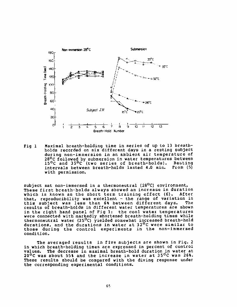
Nort-'rortterwm 28'0 $11bmersiort180
55 CO. -- -W
I 6I Cly I
t60
140
120
'13.Q
a32 C
II!
.IIc~ 100'D
X 80
60lh i 28'C
40
20
1 2 5 4 6 6 7 8 9 10 11 12 15Breath- Hold hlumber
Maximal breath-holding time in series of up to 13 breath-holds recorded on six different days in a resting subjectduring non-immersion in an ambient air temperature of28 C followed by submersion in water temperatures between15 C and 35 c two series of breath-holds!. Restingintervals between breath-holds lasted 4.0 min. Prom �!with pe r m i s s i on.
Fig 1
subject sat non-immersed in a thermoneutral �8 C! environment.These first breath-holds always showed an increase in durationwhich is known as the short term training effect �!. Afterthat, reproducibility was excellent - the range of variation inthis subject was less than 6% between different days. Theresults of breath-holds in different water temperatures are shownin the right hand panel of Pig l: the cool water temperatureswere connected with markedly shortened breath-holding times whilethermoneutral water �50C! yielded somewhat increased breath-holddurations, and the durations in water at 32 C were similar tothose during the control experiments in the non-immersedcondi tion.
65
The averaged results in five subjects are shown in Fig. 2in which breath-holding times are expressed in percent of controlvalues. The decrease in maximal breath-hold duration in water at20 C was about 55% and the increase in water at 35 C was 26%.These results should be compared with the diving response underthe corresponding experimental conditions.
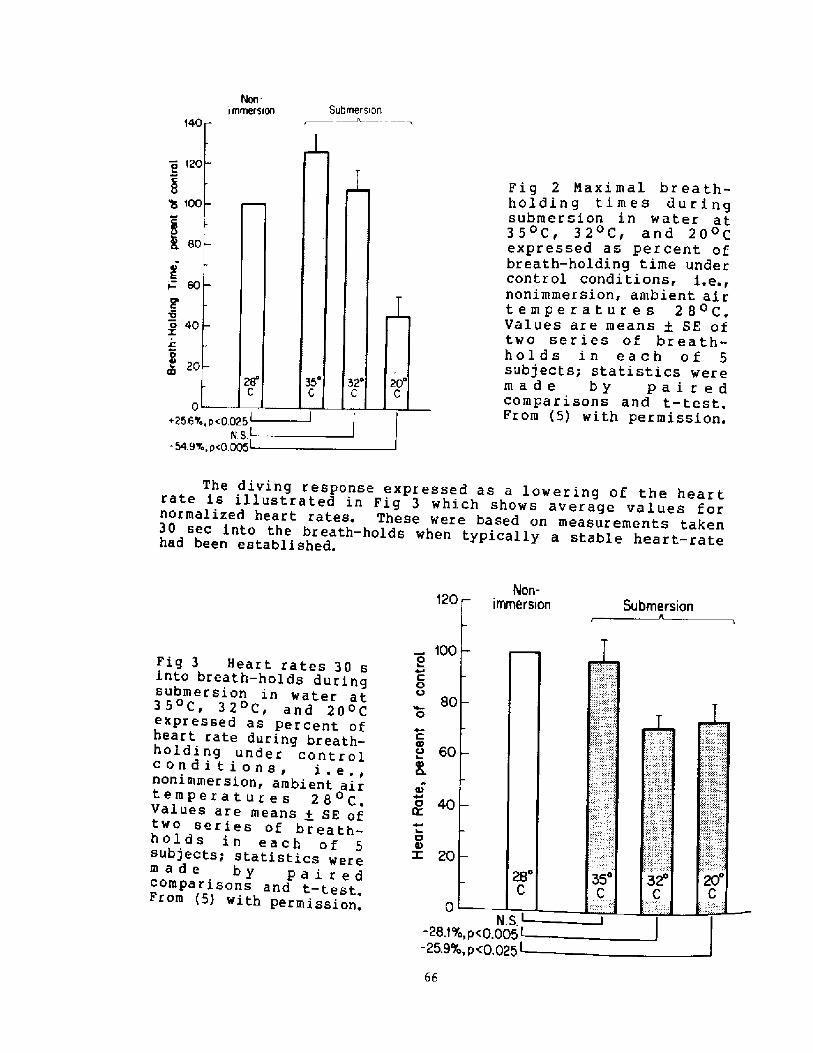
SubmersionI IYlmer5 1ori14
a I2
Q 10
I60
~ 40XI
020
0+256%
-54.9
The diving response expressed as a lowering of the heartrate is illustrated in Fig 3 which shows average values fornormalized heart rates. These were based on measurements taken30 sec into the breath-holds when typically a stable heart-ratehad been established.
f20
f008CQ
80O
-28.f
-25.9
Fig 3 Heart rates 30 sinto breath-holds duringsubmersion in water at35OC» 32 Cr and 20 Cexpressed as percent ofheart rate during breath-holding under controlcondi ti ons, i.e.,nonimmersion, ambient airtemperatures 28 C.Values are means + SE oftwo series of breath-holds in each of 5subjects; statistics weremade by pai redcompa r i sons and t- test.From �! with permission.
C
u 60
O.
o 40
o
z 20
Fig 2 Maxi mal br ea th-holdin g tim es du r ingsubmersion in ~ater at350Cr 32 C, and 20OCexpressed as percent ofbreath-holding time undercontr ol condi ti ons, i.e.,nonimmersion, ambient airtemperatur es 280C.Values are means + SE oftwo series of breath-holds in each of 5subjects; statistics weremade by pai r edcompa r i sons and t- test.From �! with permission.
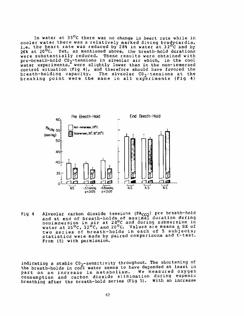
In water at 35 C there was no change in heart rate while in0
cooler water there was a relatively marked diving bradycardia,i.e. the heart rate was reduced by 28% in water at 32 C and by
at 20 C. Yet, as mentioned above, the breath � hold durations0
were substantially reduced. These results were obtained withpre-breath-hold C02-tensions in alveolar air which, in the coolwater experiments, were slightly lower than in the non-immersedcontrol situation Fig 4!, and therefore should have favored thebreath-holding capacity. The alveolar C02-tensions at thebreaking point were the same in all experiments Fig 4!
Pre Breath- Hold End Breath-Hold
Non-irrmersen, 2S'CU�9 Subrneratn,35,32,20 Cc+ 55
mnHg!50
45
40
N S -3, I rrrnHg -3.6mmHg N.S N.S. N.Sp<0.05 p <0.05
Alveolar carbon dioxide tensions PACp2! pre breath-holdand at end of breath-holds of maximaI duration duringnonimmersion in air at 28 C and during submersion inwater at 35 C< 32 C, and 20 C. Values are means + SE oftwo ser ies of breath � holds in each of 5 subj ects;statistics were made by paired comparisons and t-test.From �! with permission.
Fig 4
67
indicating a stable CO -sensitivity throughout. The shortening ofthe breath-holds in cool water seems to have depended at least inPart on an increase in metabolism. We measured oxygenconsumption and carbon dioxide elimination during eupenicbreathing after the breath-hold series Fig 5!. With an increase
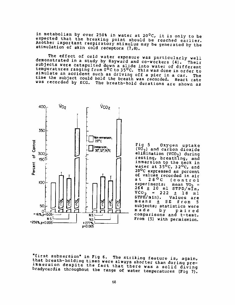
The effect of cold water exposure was particularly welldemonstrated in a study by Hayward and co-workers �!. Theirsubjects were catapulted down a slide into water of differenttemperatures ranging from 0 C to 35 C. This was done in order tosimulate an accident such as driving off a pier in a car. Thetime the subject could hold the breath was recorded. Heart ratewas recorded by ECG. The breath-hold durations are shown as
VCO2V02
fkn-imersen,
ErrmerSen,35',S2',20'C
Fig 5 Oxygen uptake VO2! and carbon dioxideelimination VCO2! duringresting, breathing, andirnmer sion to the neck i nwater at 35 C, 32 C, and20 C expressed as percentof values recorded in airat 28 C controle xpe r i ment s: mean V02264 + 20 ml STPD/msn,VCO2 = 222 + l8 mlSTPD/min!. Values aremeans + SE f rom 5
subjects; statistics weremade by pairedcomparisons and t-test.From �! with permission.
300
150
4JO Ol
CL
�0
+ 16%,p<0.05N,S.
~256'L, p<0.005
N.S.N.S.
+277%,p<0.005
"first submersion" in Fig 6. The striking feature is, again,that breath-holding times were always shorter than during pre-immersion despite the fact that there was a solid divingbradycardia throughout the range of water temperatures Fig 7!.
68
in metabolisra by over 250% in water' at 20 C, it is only to beexpected that the breaking point should be reached earlier.Another important respiratory stimulus may be generated by thestimulation of skin cold receptors �,8! ~
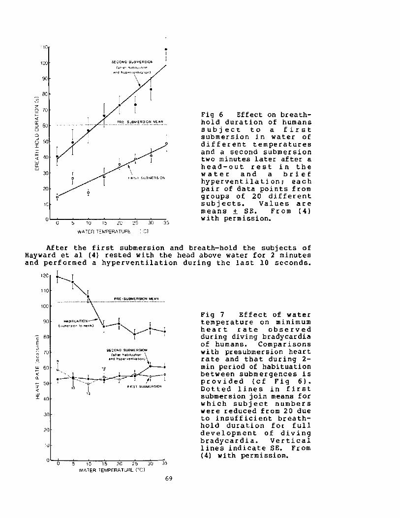
80VlZCI 70
X60
C
o 50T.Z
40
Fig 6 Effect on breath-hold duration of humanssubject to a firstsubmersion in water ofdif f er ent temperaturesand a second submer siontwo minutes later af ter ahead-out rest in thewater and a briefhyper vent i la t i on; eachpair of data points fromgroups of 20 differentsubjects. Values aremeans + SE. From �!with permission.
30
20
10
0 0 5 10 15 20 25 30 35WATER TEMPERATURE CI
110
100
Fig 7 Effect of watertemperature on minimumheart rate observedduring diving bradycardiaof humans. Comparisonswith presubmersion heartrate and that during 2-min period of habituationbetween submergences isprovided cf Fig 6!.Dotted lines in firstsubmersion join means forwhich subject numberswere reduced from 20 dueto insuf f icient breath-hold duration f or f ulldevelopment of divingbradycardia. Verticallines indicate SE. From�! with permissios.
50E
70Cl
60
K50
lZUJ
40
30
20
:0
00 5 10 15 20 25 30 35
wATER TEIUIPERATURE I C>
69
Af ter the first submersion and breath � hold the subjects ofHayward et al �! rested with the head above water for 2 minutesand perf ormed a hyperventilation during the last l0 seconds.
120
This was followed by a second breath-hold. This phase of theexperiment was designed to simulate a situation in which a personin a submerged car is able to breathe from an air pocket befareattempting to escape. Again, the subjects developed a markeddiving bradycardia across the range of water temperatures Fig7!. That they did so as much in water at 35 C as at coldertemperatures is at odds with many other investigators' findings e.g. 9, 10, ll, 12, 13, 14, 15, 16, 17!, including our own �!.At any rate, the duration of the breath-holds Fig 6! af terhabituation were still below control levels in waters colder than15 C and the increase, above control, in breath-holding time inwater that was warmer than 15 C �! may have been due to a numberof factors including hyperventilation.
Because of the possibility that cold receptor stimulationand/or increased metabalism may mask any effect of the divingresponse in man who is not a natural diver we designedexperiments in which the diving response was elicited by breath-holding and wettinq only the face with cold water �8!. Thediving response was further strengthened by performing thebreath-holds in combination with light exercise on a bicycleergometer. In these experiments the diving response wasmonitored in terms of cardiac output, heart rate and limb bloodf low.
The strengthening of the diving response that we were ableto achieve by flushing the face with cold water consisted of aroughly 25% depressian of cardiac output, mostly due tobradycardia and an almast 50% reduction of forearm blood flow.Despite these relatively pronounced circulatory changes, allbeing part of the diving response, there was no measurable changein the breath-hold durations.
Our concLusion from these various observations is that thediving response in man, at least when it consists of reducinglimb blood flow by half and cardiac output by up to 308, whetherelicited in conjunction with whole body submersion or faceimmersion only, is of no consequence for man's breath holdingabili ty.
As for our observation in subjects submersed inthermoneutral �5 C! water that the breath-holding time maysubstantially exceed that observed in the non-immersed controlsituation, we have observations indicating that their ability tostore C02 is enhanced in this situation �9!. Purely as aspeculation, we have considered that, if the increase in cardiacoutput during immersion were to lead to an increase in perfusionof the chemoreceptors, this could perhaps slow the buildup ofrespiratory drive. Further detaiLs are being worked out inongoing experimentation.
70
REFERENCES
Nemi rof f, M.J., Salz, G.R., Weg, J.G. Surv ival af ter cold-water near-drowning: the protective effect of the divingreflex. Am. Rev. Respir. Dis. 115 suppl!:145, 1977.
Kooyman, G.L., Campbell, W.B. Heart rates in freely divingWeddell seals. Leptonychotes wedelli. Comp. Biochem.Physiol. 43A:31-36, 1972.
Hollenberg, N.K., Uvnas, B. The role of the cardiovascularresponse in the resistance to asphyxia. of avian divers.Acta Physiol. Scan. 58:150-161, 1963.
Hayward, J.S., Hay, C., Mathews, B.R., Overw eel, C.H.,Radford, D.D. Temperature effect on the human dive responsein relation to cold water near-drowning. J. Appl. Physiol.:Respirat. Environ. Exercise Physiol. 56:202-206, 1984.
Sterba, J.A,, Lundgren, C.E.G. Diving bradycardia andbreath-ho3.ding time in man. Undersea Biomed. Res. 12:139-150, 1985.
Heath, J.R., Irwin, C.J. An increase in breath-hold timeappearing after breath-holding. Respir. Physiol. 4:73-77,1968.
Keatinge, W.R., Nadel, J.A. Immediate respiratory responsesto sudden cooling of the skin. J. Appl. Physio3.. 20:65-69,1965.
Cooper, K., Martin, S., Ribea, P. Respiratory and otherresponses in subjects immersed in cold water. J. Appl.Physiol. 40:903-910, 1976.
Craig, A.B.,Jr. Heart rate responses to apneic underwaterdiving and to breath holding in man. J. Appl. Physiol.18:854-862 1963.
Hong, S.K,, Moore, T.O,, Seto, G., Park H.K fBernauer, E.M. Lung volumes and apneic bradycardia indivers. J. App3.. Physiol. 29:172-176, 1970.
Kawakami, Y., Natelson, B.H., DuBois, A.B. Cardiovasculareffects of face immersion and factors affecting divingreflex in man. J. App3.. Physiol. 23:964-970, 1967.
Kobayasi, S., and Ogawa, T. Effect of water temperature onbradycardia during nonapneic facial immersion in man. Jpn-J. Physiol. 23:613-624, 1973,
Moore, T ~ 0., Lin, Y C., Lally, D A., Hong, S K. Ef fects oftemperature, immersion, and ambient pressure on human apneicbradycardia. J. Appl. Physiol. 33:36-41, 1972.
14. Paulev< P.E. Cardiac rhythm during breath-holding and waterimmersion in man. Acta Physiol. Scand. 73:139-3.50, 1968,
15. Song, S.H., Lee, W.K., Chung, Y.A., Hong, S.K. Mechanism ofapneic bradycardia in man. J. Appl. Physiol. 27:323-327,1969 ~
16. Specky D F g Bruce, D.S. Ef f ects of varying thermal andapneic conditions on the human diving reflex. UndersaaBi omed. Res. 5: 9-14, 1 97 8.
17. Whayne, T.F.,Jr., and Killip, T.< III. Simulated diving inman: comparison of facial stimuli and response inarrhythmia. J. Appl. Physi ol. 22: 800-807, 1967.
18. Sterba, J.A., Lundgren, C.E.G. Breath-hold duration in manand the diving response induced by face immersion.Unpublished.
19. Sterba, J.A., Lundgren, C.E.G. Unpublished observations.
72
Discussion following Dr. Xundgren's presentation
Dr. Hong: What is the relationship between the breath-holding time andthe oxygen consumption' In general, there is an inverse relationshipbetween the V02 and the breath-holding time. Have you considered thisrelationship as a factor for determining the breath-holding time inyour experiments?
Dr. Lundgren: The best person to give you a quantitative answer isDr. Lin. We have not correlated oxygen consumption and breath-holding time in a quantitative manner.
Dr. Hong: The oxygen consumption is expected to be high during coldwater immersion, which should cut down the breath-holding time.
Dr. I undgren: Yes, I'm saying that that is probably the explanation,plus cold receptor stimulation. Those are the most likely candidatesto explain the shorter breath-holding time in the cold water. In35 C water, which I didn't discuss much today, we observed apr olonged br ea th-holding time.
Dr. Craig: Along the same lines about the 35 C water and the otherwater temperatures, how long were your subjects immersed before youmade measurements'
Dr. Lundqren: Well, the first immersed breath-hold was immediatelyupon immersion, and then they sat for six to seven breath-holds inthe water with four minute rest periods in between, so say that eachbreath-hold took two minutes and a rest period of four, we' re talkingsomething like half an hour for the last breath-hold.
Dr. Craig: We had subjects in 35 C water for about an hour and theresponse to the breath-hold was that the heart rate changed over thewhole hour, first from a slight bradycardia to tachycardia withinthe hour, so the time period does make a difference.
Dr. Siesjo: Dr. Lundgren, I was wondering about one thing you said.Normally we would believe that brain blood flow depends on twofactors, perfusion pressure and vascular resistance. There isnormally a very efficient vascular autoregulation. You stated that anincrease in cardiac output would lead to an increase in blood flow,what is the evidence f or that?
Dr. Lundgren: Well< I cannot speak for the brain, I can speak forthe rest of the body in that, in our catheter ized subjects whom weput in an identical situation, we did indeed see a rather drastic fallin peripheral resistance in conjunction with head-out immersion Arborelius, N., Jr., U.I. Balldin, B. Lilja, and C.E.G. Lundgren.Aerospace Ned. 43�!: 592-598, 1972!. It almost completely balancedthe increase in cardiac output.
73
Dr. Lundgren: Itof these series,have comparablebreath-holds, so
might well make a difference, however, for any onewhether it's in 32, 20, or 35 degree C water, we
conditions between the eleventh and the twelfthin each series, they are comparable in that phase.
Or. Siesjo: Was there any evidence that that happened to the brain/
Or. Lundgren: We did not measure brain blood flow.
Dr. Siesjo: I would be surprised if there were a change. There is a�article by Scheinberg and Stead from 1949. Scheinberg, P.Stead �949!. J. Clin. Invest. ?,Q,. 1163-1171!. They looked at tg<effect of head-up tilting on cerebral blood flows using Scheinberg~~modification of the Kety-Schmidt technique. There were no changes i�blood flow.
EFFECT OF 0. AND CO? ON BREATH-HOLD BREAKING POINT"
Y. C. Lin
Depar tment of PhysiologyUniversity of Haaaii John A. Burns School of Medicine
Honolulu, Haaaii 9b622
Horkers in the field of breath-hold BH! studies recognized from thestart the complexity of BH breaking point and the inadequacy of chemicalstimuli in predicting BH breaking point. Simple observations sarrant suchconclusion in that BH terminates at a variety of alveolar gas compositions;rebreathing can be tolerated much better than BH at a similar alveolar gascomposition; and that BH time varies greatly from person to person and fromtime to time in the same individual under an identical condition.
A var'iety of factors, including subjective ones, contribute to bringabout the conventional BH breaking point, that is, to terminate BH shenone feels BH can no longer be continued. This subjectiveness contributesto the great variability of BH time ahich defies prediction based on chemi-cal and mechanical factors, On the other hand, chemical stimuli alonedetermine a physiologically definable BH end point. At such time during acourse of BH, specific physiological responses occur', signif'ying desire toresume breathing, but BH may still be continued by conscious effort. This"breaking point" is termed physiological breaking point ahich, in con-trast to the conventional breaking point, varies aithzn a narroa range.This reviea examines the effect of P02 and PC02 on BH breaking points anddrass attention to the multitude of factors controlling them.
TIME COURSE OF A BREATH-HOLD
The time course of' breath-hold BH! can be characterised by 1! volun-tary inhibition of respiratory muscular activity, aith the glottis closedthe intrathoracic pressure is stable and slightly above the ambient; 2! theonset of involuntary inspiratory activity aith the glottis closed, account-ing for the subatmospheric and cyclic intrathoracic pressure ahich intensi-fies as BH continues; and, finally, 3! opening of the air say. Thissequence of events corresponds, respectively, to the event markings A, B,and C Fig. 1!.
PHYSIOLOGICAL BREAKING POINT
At event B, involuntary ventilatory activit.ies IVA! occur ahileinhibition of glottis opening is still possible, It signals the end of the
easy-going phase" �2!; and identifies the onset of "desire to breathe"
Suppot ted in par't by Sea Grant NAB1AADQO070, Haaaii Heart Association,and Pioneer Imin Cultural foundation.
75
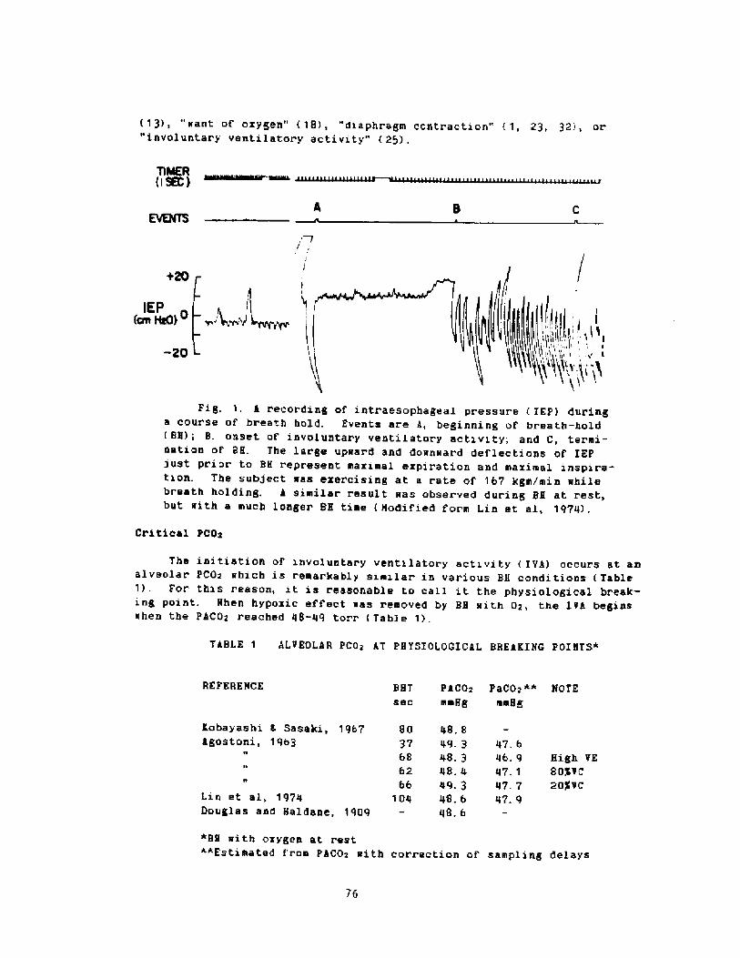
�3!, "want of oxygen" �8>, "diaphragm contraction" �, 23, 32!, or"involuntary ventilatory activity" �5!.
TIMER ISK!
I Ital > i
Fig. 1. A recording of intraesophageal pressure IEP! duringa course of breath hold. Events ar.e A, beginning of breath-hold BH!; B. onset of involuntary ventilator'y actxvxty; and C, ter'mi-nation of BH. The large upward and downward deflections of IEPjust prior to BH represent maximal ezpiration and mazimal inspira-tion. The subject was ezerci sing at a rate of 167 kgm/min whilebreath holding. A similar result was obser'ved during BH at rest,but with a much longer BH time Nodified form Lin et al, 1974! .
Critical PCOx
The initiation of involuntary ventilatory activity IVA! occurs at axralveolar pCO~ whxch is remarkably similar in various BH conditions Table1!. For this reason, it is reasonable to call it the physiologxcal break-ing point. Nhen hypozic effect was removed by BB with Or, the IVA beginswhen the PACOr reached 48-49 torr Table 1!.
TABLE 1 ALVEOLAR PCOr AT PHYSIOLOGICAL BREAKING POINTS*
REFERENCE PaCO~**
mmHg
BHT PACOxsec mmHg
Kobayashi L Sasaki, 1967Agostoni, 1963
~ IHigh VE8ogvc20$VC
Lin et al, 1974Douglas and Haldane, 1909
*BH with oxygen at rest~*Estimated from PACOx with correction of sampling delays
76
IEP cm HAO!
-20
8O 48. 8
37 49. 368 48. 362 48. 4
66 49. 3104 48. 6
48. 6
47. 646. 947. 147. 747. '9
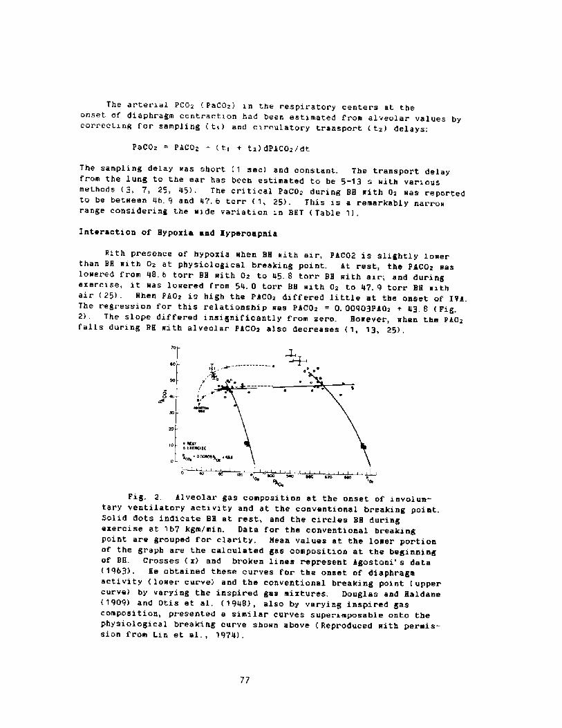
The ar ter'ial PCOz PaCOz! in the respirator y center s at theonset of diaphragm contraction had been estimated from alveolar' values bycorrecting f or sampling tr ! and circulatory t.r ansport tz! delays;
PaCOz = PACOz � tr + tz! dPiCOz/dt
The sampling delay was shor't 1 sec! and constant. The transport delayfrom the lung to the ear has been estimated to be 5-13 s with variousmethods 3, 7, 25, 45!. The critical PaCOz dur.ing BH with Oz was reportedto be between 46. 9 and 47, 6 torr 1, 25!, This is a remarkably narrowr'ange considering the wide variation in BHT Table 1!.
Interaction of Hypoxia and Hypercapnia
Hith presence of hypoxia when BH with air, PLC02 is slightly lowerthan BH with Oz at physiological breaking point. at rest, the PkCOz waslowered from 48.6 torr BH with Oz to 45. 8 torr BH with air; and duringexercise, it was lower'ed from 54. 0 torr BH with Oz to 47. 9 torr BH withair 25!. Hhen PBOz is high the PSCOz diff'ered little at the onset of IVk.The regression for this relationship was PkCOz = 0, 00903PXOz + 43. 8 Fig.2!. The slope differed insignificantly from zero. However, when the Pk0zfalls during BH with alveolar PaCOz also decreases 1, 13, 25!.
M 4D
0 ~ K.tr0 KmllCmE
0 OMOS0
Fig, 2. Alveolar gas composition at the onset of involun-tar y ventilatory activity and at the conventional br eeking poiatSolid dots indicate BH at rest, and the circles BH duringexercise at 'I67 kgm/min. Data for the conventional breakingpoint are grouped for clarity, Mean values at the lower portionof the graph are the calculated gas composition at the beginningof BH Crosses x! and br'oken lines r.epresent kgostoni' s data�963!, He obtained these cur ves f' or the onset of diaphragmactivity lower curve! and the conventional breaking point uppercurve! by varying the inspired gas mixtures. Douglas and Haldane�909! and Otis et al, �948!, also by varying inspired gascomposition, presented a similar curves super imposable onto thephysiological breaking curve sho~n above Reproduced with permis-sion from Lin et al., 1974!.
77
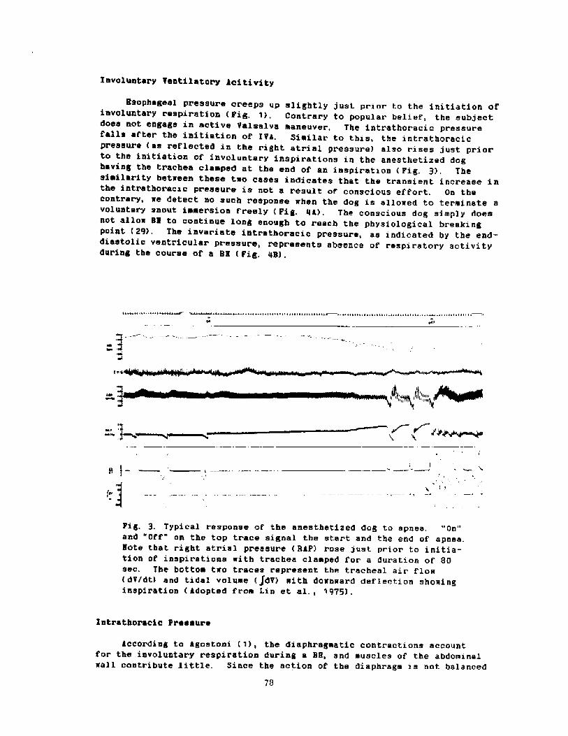
? nvoluntary Ventilatox'y ici ti vi ty
Bsophageal pressure creeps up slightly just prior to the initiation ofinvoluntary respiration Fig, 1!, Contrary to popular belief, the subjectdoes not engage in active Valealva maneuvex, The intrathoxacic pressuref'alla af'tex' the initiation of IVk. Similar to this, the intratboracicpx'essure as x'eflected in the right atrial pressure! also rises just prior'to the initiation of involuntary inspirations in the anesthetized doghaving the trachea clamped at the end of an inspiration Fig, 3!. Thesimilar'ity between these t«o cases indicates that the transient increase inthe intr'athoracic px'essure is not a result of' conscious ef f'ort. On thecontrary, we detect no such response when the dog is allowed to terminate avoluntary snout immersion f'xeely Fig. 44!. The conscious dog simply doesnot allo«BB to continue long enough to reach the physiological breakingpoint �9!. The invaxiate intxatboracic pressure, as indicated by the end-diastolic ventri.euler pxessux'e, represents absence of respiratory activityduring tbe course of a BH Fig. 48! .
s l-
F' j Pig. 3 Typical response of' the anesthetized dog to apnea. "On"and "Off" on the top trace signal the start and the end of apnea.Note that right atrial px.essure RaP! rose just priox to initia-tion of inspirations «ith trachea clamped f' or a duration of 80sec. The bottom t«o traces represent tbe tracheal air flow dV/dt! and tidal volume jdV! with down«ard deflection showinginspiration gdopted from Lin et al., 9975!.
Intrathoracic Pressure
Recording to kgostoni !, the diaphragmatic contractions accountfor the involuntary respiration during a BB, and muscles of the abdominal«all contribute little. Since the action of the diaphragm is not balanced
78
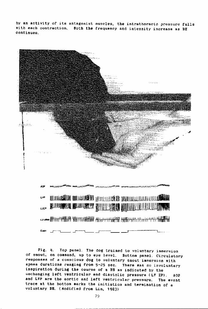

In a series of experiments in 5 males, tin et al, �5! recorded esopha-geal pressure during BH either with air or oxygen both at rest and dut ingexercise �-3 times resting VOz!. Esophageal pressure star ts at a levelslightly above the ambient, but falls continuously after the onset of IVA tobetween 10-15 torr below the ambient at the conventional breaking point.Exercise
Elevation of rate of oxygen consumption shortens duration to bothphysiological and conventional breaking poi nts, This is not surprising,since the rate of rise of alveolar PACOz increases during exercise, andshould shorten BHT corr'espondingly. Although exercise elevates PACO> at IVAat a given PAO2, it is possible that part of the PACOz elevation could berelated to exaggerated sampling delay and transport delay 7!.
CONVENTIONAL BREAKING POINT
Following its onset, the frequency and intensity of' IVA increase withcontinued BH. Nevertheless, conscious suppression of glottis prevails.Eventual opening of the glottis occurs when ventilatory stimulus i s toostrong to be resisted further by conscious eff'ort. The event at C Fig. 1!ends an active BH and is termed variously as the "conventional breakingpoint", the end of the "struggle phase" 12!, or simply the breaking point.The duration between B and C is highly variable, This variability accountsfor BH time ranging f'rom 20 s 37! to 270 s 2O! on an identical command:"Exhale maximally, without prior hyper ventilation, followed by maximalinhalation and hold the br eath as long as possible. " Obviously, it involvesnon-chemical stimuli.
Breath-hold Time
A student af Schneider at the Hesleyan University held his breath for15 mi n 13 sec after a period of hyper ventilation with air f'ollowed by thr eedeep breaths of pure oxygen 37!. Nithoefer 31! mentioned in his reviewthat under the same condition, another student at the same university heldhis breath f' or 20. 1 min. BHT of' this magnitude have also been reportedfollowing hyperventilation with Oz by Klocke and Rahrr 22!. One of theirsubjects, H. R., performed a BH for 14 min, 13. 6 min, and 14. 4 min an thr'eeoccasions. To my knowledge, the longest published record af BH with airwithout prior hyperventilation is 270 sec 20!. BHT of' 240 sec under thesame conditions are routinely demonstr ated by Hong and his colleagues at theUniversity of Hawaii 21!, These unusually long BHTs are i n accord with 02supply and demand relationships. However, the great majority of subjectsdemonstrate ability to BH only a fraction of the BHT cited above. Fig. 5sho~s BHT with air without prior hyperventilation. Such variability in BHTindicates clearly the involvement of non-chemical factor s.
Oxygen Supply and Demand
The f'actors that contribute to the eventual termination of BH have
80
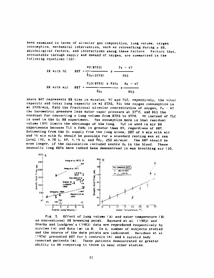
been examined in terms of' alveolar gas composition, lung volume, oxygenconsumption, mechanical intervention, such as rebreathing during a BH,psychological factors, and interactions among these factors Factors that,accountable through supply and demand of' oxygen, are summarized in thef oil otr i ng equa ti ons 22!:
VC BTPS! Py � 47BH mith 02 BHT
V02 STPD! 863
TLC BTPS! x Fi02 Py - 47
xVO2 863
BH mith air BHT
mhere BHT repr'esents BH time in minutes, VC and TLC, repectively, the vitalcapacity and total lung capacity in ml BTPS, VOz the oxygen consumption inml sTPD/min, Fi�2 the fractional alveolar concentration of oxygen, P9 � 47the barometric pressure less mater vapor pressure at 37'C, and 863 theconstant for converting a lung volume from BTPS to STPD, VC instead of TLCis used in the Oz BH experiment. The assumption here is that residualvolume RV! limits the shrinkage of the lung. TLC is used in air BHeXperimentS beCauae TLC X FaN2 is greater than RV, regar'dless Of BHT.Estimating from the 02 supply from the lung alone, BHT of 4 min mith airand 16 min mith Gz should be possible for a standard resting man at sealevel VC, 4. 78 l.; RV, 1. 19 L; and VOz, 250 ml/min!. The BHT should beeven longer, if the calculation included usable Oz in the blood. Theseunusually long BHTs have indeed been demonstraed in man breathing air 20,
500
l50IMony et oL1970 e
+n
Mony e! oi, 1971
Llerll ol1974 ~ ~
pnnntnon nr ol1924 4
Ster 9 nL nnrty ron1995 tre
250
15 Prn- lmrnnrnrnn 9MT nnr 200C
150sZ
o eo r 511501~ I I 1 l4151110
E
0 900X
'to0In
o 100Ia
pL FltCrrv FttCTV VC
50 0 '5 10 13 2C 23Water Temperature, '
3C 32 3 4APPron Lurtt! Volume, L
Fig, 5. Effect of lung volume k! and mater temperature B!on conventional BH breaking point. Haymard et al, 1983! andSter ba and Lundgren' s 1983! data are repr oduced respecti vely byCirClee O! and dots ~ ! in B. In X, number Of Subjeote Studiedand the source of the data points are indicated, Davidson et al.�974! presented BHT for 5 controls a! and 6 carotid bodyresected patients �!, These patients demonstrated no greaterability to BH comparing to those in many other studies.
Bl
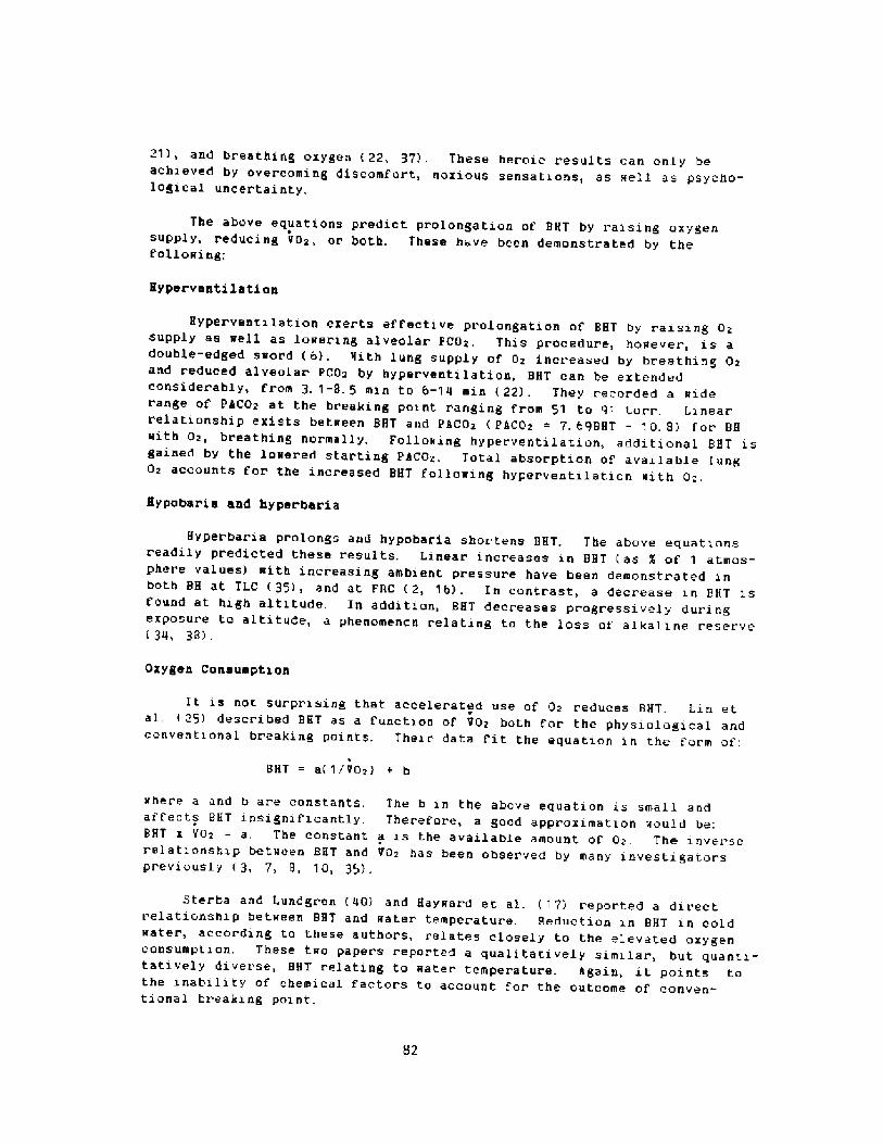
21!, and breathing oxygen 22, 37!, These heroic r esults can only beachieved by overcoming discomfort, noxious sensations, as well as psycho-logical uncertainty,
The above equations predict prolongation of BHT by raising oxygensupply, reducing VOz, or both, These have been demonstrated by thefollowing:
Hyperventilation
Hyperventilation exerts effective prolongation of BHT by raising Ozsupply as well as lowering alveolar PCOz, This procedure, however, is adouble-edged sword �!, Hith lung supply of Oz increased by breathing Ozand reduced alveolar PCOz by hyper ventilation, BHT can be extendedconsiderably, from 3. 1-8. 5 min to 6-14 min 22! . They recor ded a widerange of PkCOz at the br'eeking point ranging from 51 to 91 torr. Linearrelationship exists between BBT and P! COz PACOz = 7. 69BHT � 10. 8! for BHwith Oz, breathing normally, Following hyperventilation, additional BHT isgained by the lowered star ting Pi COz. Total absor'ption of available lungOz accounts for the increased BHT following hyperventilation with Oz.
Hypobari a and hyper'bari a
Hyper bar'ia prolongs and hypobaria shor tens BHT. The above equationsreadily pr edicted these results. Linear increases in BHT as X of 1 atmos-phere values! with increasing ambient pressure have been demonstrated inboth BH at TLC �5!, and at FRC �, 16!. Tn contrast, a decrease in BHT isfound at high altitude. In addition, BHT decreases progressively duringexposure to altitude, a phenomenon relating ta the loss of alkaline reserve�4, 38!.
Oxygen Consumption
It is not surprising that accelerated use of Oz r educes BHT. Lin et.al, 25! described BHT as a function of VOz both for the physiological andconventional breaking points, Their data fit the equation in the form of:
BHT = a�/VOz! + b
The b in the above equation is small andTher efor'e, a good approximation would be:
a is the available amount of Oz. The inverseVOz has been obser ved by many investigators
wher e a and b ar e constants.af 'eats BHT insignificantly.BHT x VOz = a, The constantrelationship between BHT and
previously 3, 7, 8, 10, 35! .
82
Sterba and Lundgren �0! and Hayward et al �7! reported a directrelationship between BHT and water temperatur'e. Reduction in BHT in coldwater, according ta these authors, relates closely to the elevated oxygenconsumption, These two papers reported a qualitatively similar, but quanti-tatively diverse, BHT relating to water temperature. again, it points tothe inabili ty of chemical f actor's to account for the outcome of conven-tional breaki ng point.
Breath-Hold Time and Alveolar' Gas during Diving
Diving involves physical activity which depresses BET. Pr.of'essionalBH divers limit their diving time to 60 sec ox less, Hong et al. 19! re-ported a contest for 20 ama and recorded a maximal, diving time of 82 sec,Diving duration of 9G sec 39!, 118 sec 42!, and 115 ssc 9! are recordedoccasionally. The experienced divers also limit their hyperventilationprior to a dive to a minimum. They know that hyperventilation increasesdiving time, but they also aware of' the danger of extreme hypoxia on ascent.
Alveolar gas composition has been analyzed in the field �, 19, 42!and in the laboratory with simulated dive �4!, The course of alveolar gasexchange dif'fere greatly from that in ordinary BH. Both alveolar POz andPCOz at the breaking point is lower than in comparable BH at the normalambient conditions. Extreme hypoxia could, theref ore, occur dur ing theascent phase,
NON-CHEMICAL FACTORS
Psychological factors have long been recognised as the major determi-nants of conventional breaking point 4, 18, 37, 43!, and undoubtedlycontribute to the wide range of BHT reported in the literature 27, 30!.Fig, 5 demonstrates clearly the involvement of factors other than theavailability of Oz. Prolongation of BHT occux s when respiratory movementsare allowed, such as rebreathing without improving alveolar gases �, 14,23, 44!. Furthermore, reviews show that rigid concepts of chemical andmechanical thresholds are inadequate for interpreting BE breaking point�5, 44!, suggesting the impor tance of subjective factors. Since psycho-logical factor s play such an important role and, since it i.s unxeasonableto assume the coastancy of these factors within and between individuals,caution should be exercised on regulatory mechanisms based on conventionalbreaking point �1!. Physiological breaking point would be suited betterfor such pux pose.
S ! HOARY
Two breath-hold BH! breaking points have been identified: physio-logical and conventional. The former constitutes the initiation ofinvoluntary ventilatory efforts with glottis closed, and the lattercoincides with eventual opening of' glottis. A variety of factors influencethe ability to BH, including psychological ones, which account f' or thegreat variability in BH time. within as well as between individuals. Theinvolvement of' psychological factors render rigid concepts of chemical andmechanical thresholds inadequate in interpreting conventional breakingpoints. However, experimental subjects could reproduce consistently thetime to initiation of involuntary respiration, i, e. the physiologicalbx'asking point which relates closely to arterial levels of PCOx and PO2.Contxol of the Conventional. breaking point is more complex. Oz and CO2 arebut two factors in a set of complex mechanisms that govern the BH breakingpoi nts.
REFERENCE
Agostoai., E. Diaphraga activity during breath holding: factors related toita onset. J. Appl. Phyaiol. 18:30-36, 1963,
2, Alvin, H. J. Bteath-holding break point at various increased pressure.S. Navy Nedical Research Laboratory Report 8177, 10: 110-120, 1951.
3. Astrand, P. O. Breath-holding during and after auscular exercise. J.Appl. Phyaiol. 15: 220-224, 1960.
Bartlett, D., Jr. Rff'acta of Valsslva aad Nueller aaneuvers on breath-holdiag tine. J. Appl. Physiol. 42: 717-721, 1977.
5 Cain, S. N. Breathing point of tao breath holds separated by a singleinspiration. J. Appl. Physiol. 11: 87-90, 1957.
6. Craig, A. B. Jr, Causes of loss of consciousaess during uaderaaterssiaaiag. J. Appl. Physiol. 16:583-586, 1961.
7. Craig, A. B. Jr., aad S. A. Babcock. Alveolar COx during breath holdingaad exercise. J. Appl. Phyaiol. 17: 874-876, 1962.
B. Craig, P. N., and R. G. Cuaaiags. Breath-holding during exercise. J.Appl. Physi ol. 13; 30-34, 1958.
9. Cross, E. R., Taravaaa. Diving syndroae in the Tuaaotu diver In:Physiology of Breath-Hold Diving and the Aaa of Japan, edited by H, Raha,Saahington, D. C.: National Acadeay of Science, Publication 81341,1965, p. 207-219.
10. Cuaaings, R. G. Breath-holdiag at beginning of exercise. J. Appl.Physiol. 17. 221-224, 1962.
11, Davidson, J. T., B. J. Shipp, K. Naeseraan, S. N. Royal and R. Lugliani.Role of' the carotid bodies in breath-holding. N. Rngl. J. Ned.290: 819-822, 1 974.
12. De!ours, P. Hasards of hypoxia during diving. In: "Physiology ofBreath-Hold Diving and the Aaa of Japan", edited by H. Rahn.Nashington, D. C.: Natonal Acadeay of Science, publication 81341,1 965, p. 1 83-1 93.
13. Douglas, C. G., aad J. S. Saldane. The causes of periodic or Cheyne-Stokes breathing. J. Physiol. 38: 401-419, 1909,
14. Poal sr, H. S. Breakiag point of breath-holdiag. J. Appl. Physiol. 6: 539-545, 1954,
Godfrey, S., and E. J. N. Caapbell. The control of breath holding. Resp.Phyaiol, 5: 385-400, 1968.
84
16. Besser, C, N, Breath holding under high pressur'e. In: physiology ofBreath-Hold Diving and the kea of Japan, edited by H. Rahn.Hashington, D. C,: National kcadeey of Science, publication 81341,1965, p. 165-181,
17. Haymard, J. S., C. Hay, B. R. Nat theme, C. 8, Overmeel, and D, D.Redford. Teaperature eff'ect on the huaan dive response in relation tocold mater near -dromning. J. kppl. Physi ol. 56; 202-20b, 1984,
18, Hill, L. and N. Flack. The effect of excess carbon diozide and mant ofoxygen upon the respiration and the circulation. J. Physiol. 37: 77-111, 19G8.
19. Hong, S. K,, H., Rahn, D. H. Rang, S. H. Song, and B, K. Rang, Divingpattern, lung voluaes, and alveolar gas of' the Korean diving moean aaa! . J. appl. Physiol. 18: 457-465, 19b3,
20. Hong, S. K., T. G. Hoore, Q. Seto, H. K. Par k, H. R. Hiatt, and E. N.Bernauer, Lung vol uaes and apneic bradycardi a i n di vers. J. kppl.Physi ol. 29: 1 72-176> 1 970.
21, Hong. S. K., T. 0, Noore, Q. Seto, H. K. Par k, Hiatt, and H H.Bernauer. Lung voluees and apneic bradycardia in divers. J. appl.Physi ol. 29; 1 72-176, 1 97G.
22. Xlocke, P. J., and H. Rahn. Breath-holding after breathing of oxygen,J. appl Physiol. 14: 689-693, 1959.
23. Kobayashi, S, and C. ~y.ki. Breaking-point of breath holding and toler-ance tine in rebreathing. Japn, J. Physiol. 17: 43-56, 19b7.
24. Lanphier, E. H., and H. Rahn. alveclar gas ezchange during breath-hOlddiving. J. Hppl. Physiol. 18: 471-477, 1963.
25. Lin, Y. C,, D. j. Lally, T. G. Hoore, and S. R. Hong. Physiological andconventional breath-hold breaking points, J. kppl. Physiol. 37: 291-29b,1974.
26, Lin, Y. C, T. O. Hoore, J. J. HcHaeara, and S. R. Hong. Ozygen consuep-tion and conservation during apnea in the anesthetized dog. Resp.Physiol. 24: 313"324, 1975.
27. Lin, Y. C. Breath-hold diving in terrestrial eaeeals. In: "Hxer cise andSport Sciences Reviems", Vol, 10. Rdi ted by R. L. Terjung.Philadelphia: Franklin Press, 1982, p. 270-307.
28. Lin, Y. C, Heeodynaeic changes during voluntar y snout ieeersion inchr onically instrueented dogs. Chinese J. Physiol, 26-. 1-10, 1983,
29. Lin, Y. C. E. L. Car leon, H. P. HcCutcbeon, and H. Sandier. Car diovas-cular reactions during voluntary apnea in dogs, ke. J, Physiol, Reg.Intg. Coep. Physiol.! 245: R143-R150, 1983,
Nithoef er, J. C. The breaking point of breath holding. In: Physiologyof Breath-Hold Diving and the kaa of Japan, edited by H, Rahn,Nashington, D. C.: National academy of Science, Publication 81341,1965, p. 195-205,
30.
Nithoefer, J. C. Breath holding. In: Handbook of Physiology, Respira-tion. Nashington, D. C,; am. Physiol, Soc., 1965, 2: 1011-1025.
31.
Noble, H. I, H., J. H. Eisele, H. L Pr ankel, R, Else, and a. Guz. Therole of the diaphragm in the sensation of holding the breath. Clin.Sci. 41: 275-283, 1 971.
32-
33. Otis, g. B., H. Rahn and R. 0, Penn alveolar gas exchanges duringbreath bold. am, J. Physi ol. 152: 674-686, 1948.
34. Rahn, 8,, H. T. Bahyson, J. P, Kuxsorthy and J. M. Hagen. adaptation tohigh altitude: chagnes in breath-holding time. J. kppl. Physiol.6; 154-157, 1953.
36. Schilling, C, R., R. k. Hansen, and J, k. Haskins, The effect ofincreased air pressure on vital capacity, expiratory force and breath-holding ability. gm. J. Physiol. 110: 616-619, 1935,
37. Schneider, E. C. Observation on holding the breaath, km. J. Physiol.94: 464-470, 1 930.
38. Schneider, E. C. Respiration at high altitude. Yale J. Biol. Red.537-550, 1932.
39. Scholander, P. F., H. T. Hammel, H. LeHessurier, E, Hemingsen and N.Carey. CirCulatOr y adjuetment in pearl diverS. J. appl. Phyeiol.17: 1B4-190, 1962.
40, sterba, J. a, and C. E. C. Lundgren. Diving br'adycardia and br eath-holding time in man. Undersea Bioeed, Res. 12: 139-150, 1985.
Stroud, R. C. Combined ventilatory and brath-holding evaluation ofsensitivity to respiratory gases, J. appl. physiol. 14; 353-356, 1959
Teruoka, G. Die arne und ihre arbeit. drbeitsphysiol. 5: 239-251, 1932.
43 Hhite, P. D. Obser vations on some tests of physical fitness. km, J. Red.Sci. 159: 866-874, 1920
Rhitelas, R. a., B, NcBride, J. kr nar, and K. Corbet Respiratory neuro-muscular output during breath-holding. J. k. Physiol. 50: 435-443, 1981.
45. Ril liame, N. H. Jr. g simple method Fo! measuring lung-to-ear circu-lation time aith an ear oximeter. J. appl. Physiol. 16; 15-20, 1961.
86
35. Robard, S. The effect of' oxygen, altitude and exercise on breath-holdingtime. Am. J. Phsyiol. 150; 148-152, 1947.
Discussion follmring Dr. Lin's presentation
Dr. Rahn: I have a question f or Dr. Hong. When you were doing thosefour minute breath-holds mentioned by Dr. Kin, were you alsodelivering alveolar samples?
Dr. Hong: Yes, that was the trickt
Dr. Lundgren: The point has just been made, and yet I think it shouldbe emphasized even more: we may be talking about different things whenwe talk about breath-holding. There is a big difference betweenactually holding your breath and re-breathing into a bladder. Theterminology here is a little hazy, when we talk of breath-holding.
Dr. Rahn: You mentioned also some ways to prolong breath � holding,and it doesn't make any difference whether you exhale into a bag andreinhale or whether you squeeze a tennis ball. It seems to me thereis a big difference.
Dr. Lin: But that just simply affirms that the chemical factor is notan important determinant of breath-hold time. The discomfort,physical and/or psychological, is the major' determinant. If you doanything to divert from discomfort you could prolong the breath-hold.
Dr. Rahn: Yes, but is blowing into a bag and reinhaling the samediversion as squeezing a ball ?
Dr. Lin: No, wait a minute, I didn't say that they prolong a breath-hold in the same way. There are many ways one can prolong a breath-hold, such as, exhale, inhale, swallowing, and even squeez ing a tennisball will help.
Dr. Craig: When you get the involuntary contractions, you accentuatethem by pumping in and out with the involuntary contractions. Whythat helps, I don't know. It seems to be the most common way fordivers to prolong the breath-hold period.
Dr. Kannini: With regard to the breath-hold breaking point, I wouldlike to report some observations on breath-holding times in a group ofthirty-seven subjects during training for SCUBA diving. Theyperformed a total of l800 breath-holds, both before and af ter a monthof training. Those of them who had been training and practicingbreath-hold diving even before the course achieved the longest breath-holding times in every condition, i.e., either dry or in the wateriwith preceding hyperventilation or with resistance to diaphragmaticcontractions, at rest or during work. The only condition where theydid not differ much was in the dry breath-hold at rest. One month ofphysical work-out and underwater training did not allow the subjects>who previous to the course had not been breath-hold divers, to dobetter than the trained ones or to substantially improve their ownbreath-hold times. That could be due to several factors, among whichthe increased resistance to hypercapnia and to neuromuscular stimuli<or it could be due to a stronger breath-hold diving reflex in thepreviously trained subjects.
87
RESPIRATORY NEUROMUSCULAR OUTPUT IN BREATHHOLDING
by
W. A. WhiteiawDepartment of Medicine
Faculty of MedicineUniversity of Calgary
Calgary, Alberta, Canada
88

Although breathholding is commonly thought of as voluntary suppressionof respiratory effort, most subjects studied in the laboratory andexperienced physiologist breathholders, in particular, have involuntaryrhythmic contractions of inspiratory muscles that begin shortly after thebeginning of breathholding and increase in amplitude and frequency until thebreakpoint. To avoid breathing in spite of these contractions, thebreathholder must voluntarily seal off his glottis or pharyngeal airway, andthe breakpoint occurs when some sensation associated with prolongedbreathholding persuades him to open his airway and allow the inspiratoryefforts he has been making all along to actually draw in some air. Suchcontractions were reported by Agostoni in 1960 �! and he and others �!have used the time when contractions first appeared as the true or"physiological" breakpoint of breathholding. Such experiments showed thatthe physiological breakpoint was influenced by oxygen and carbon dioxidetensions in much the same way as the breakpoint defined in the more usualmanner, but of course the "physiological" breathhold times were considerablyshorter--of the order of a third or a quarter the time the subject couldkeep from breathing by sealing his upper airvay.
The act of breathholding itself somehow constitutes a stimulus tobreathe or a source of discomfort over and above the sensation due toaltered blood gas tensions resulting from apnea. This was shown in 1908 byHill and Flack �!, who compared breathholding with rebreathing from a smallbag, which produces the same stimulus of asphyxia. Rebreathing could betolerated several times longer than breathholding. Fowler's experiment in1954 suggested the same thing �!. Near the breakpoint of a voluntarybreathhold he permitted subjects to take two or three br'catha, but from abag containing a gas mixture identical in composition to alveolar gas atthat moment. Without any change in gas tension, these subjects were able toresume breathholding and postpone the breakpoint by many seconds.
Campbell and Godfrey, in a series of papers, developed the hypothesisthat the contractions themselves were the source of the extra discomfortthat finally obliged subjects to take a breath �,6,7 !. The most strikingexperimental evidence they put forward came from an experiment in whichvoluntary breathholding at functional residual capacity FRC! was compared.with apnea induced by curare in the same subjects. When respiratory muscleefforts were abolished by curare, most of the discomfort of breathholdingdisappeared �!.
We have done a series of breathholding experiments to test theCampbell-Godfrey hypothesis and examine mechanisms that govern thegeneration of respiratory muscle contractions and how they might give riseto the discomfort of breathholding. We considered that if the contractionswere directly responsible for making the subject abandon breathholding92there should be a correlation between some measurable feature of thecontractions and the breakpoint.
All the experiments are performed in the same way. Breathhoids areperformed by normal subjects sitting in a chair who begin taking a few deepbreaths from a small bag containing 8X carbon dioxide in oxygen. At thealtitude of our laboratory this mixture has a P close to mixed venous, analveolar plateau is established, and from then on P rises nearly linearlyC02
C02with time at a rate which is nearly constant, independent of minuteventilation and nearly equal to the rate at which it rises during abreathhold �,8!. Respiratory muscle contractions are monitored by
89
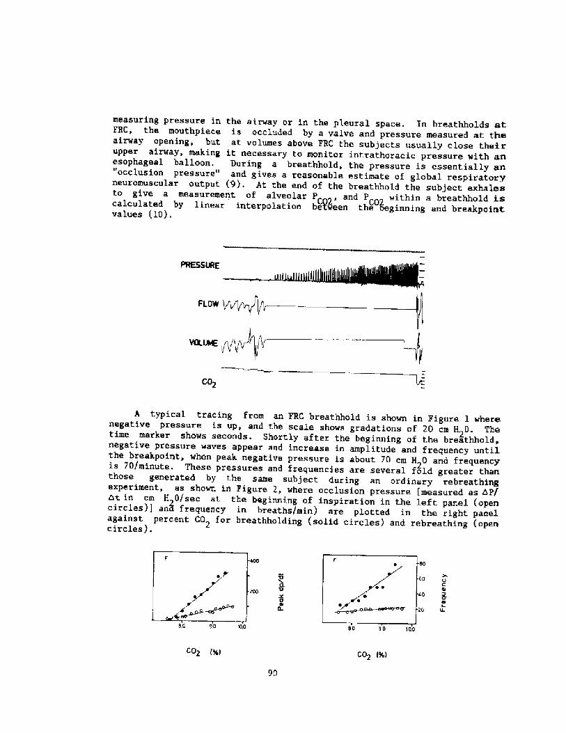
FLOW y~~
VOLVO jQQ f'C02
A typical tracing from an FRC breathhold is shown in Figure 1 wherenegative pressure is up, and the scale shows gradations of 20 cm H20. Thetime marker show~ seconds. Shortly after the beginning of the breathhold,negative pressure waves appear and increase in amplitude and frequency untilthe breakpoint, when peak negative pressure is about 70 cm H 0 and frequencyis 70/minute. These pressures and frequencies are several fold greater thanthose generated by the same subject during an ordinary rebreathingexperiment, as shown in Figure 2, where occlusion pressure [measured as AP/ht. in cm H 0/sec at the beginning of inspiration in the left panel opencircles!] an! frequency in breaths/min! are plotted in the right panelagainst percent CO for breathholding solid circles! and rebreathing open2circles!.
C Ol0'4ILL
0 90 m0S.Q 90 'M
CO~ g C02 Rj
90
measuring pressure in the airway or in the pleural space. En breathholds mtFRC, the mouthpiece is occluded by a valve and pressure measured at theairway opening, but at volumes above FRC the subjects usually close theirupper airway, making it necessary to monitor intrathoracic pressure with anesophageal balloon. During a breathhold, the pressure is essentially an"occlusion pressure" and gives a reasonable estimate of global respiratoryneuromuscular output 9!. At the end of the breathhold the subject exhale+to give a measurement of alveolar P 02, and PC within a breathhold iscalculated by linear interpolation between the oeginning and breakpointvalues �0!.

In a second kind of experiment, subjects began breathholds at FRC witha variety of starting values of P . This was achieved simply by havingthem rebreathe from the small bag for 3ifferent periods before beginning thebreathhold. The results of such an experiment are shown in Figure 3. Peakinspiratory pressure in cm H 0 is plotted against C02. Different symbolsrepresent different breathholls. Means from all the contractions in a 10sec interval are shown.
150
OPV
Z
~ 100
50
9.5 10.0C02 'YojL5
Starting from a low P solid circles! peak negative pressure of theC02contractions rose, quicPj.y at first, then more slowly to breakpoint.
Starting from a higher P 2 open circles and split circles! pressure rosequickly, then slowly again, but at the breakpoint had reached a higherC02
value, at a higher P and at a higher frequency, not shown! than in theprevious breathhold. Pn an experiment like Fowler s, where the subject wasC02 ~ I
permitted to take a few breaths from the bag at intervals, the magnitude andfrequency of contractions dropped immediately each time he did so, then roserapidly again to a high level Fig. 4!.
D E50
Q X
8.0 9.0 9.CQ2 %!
91
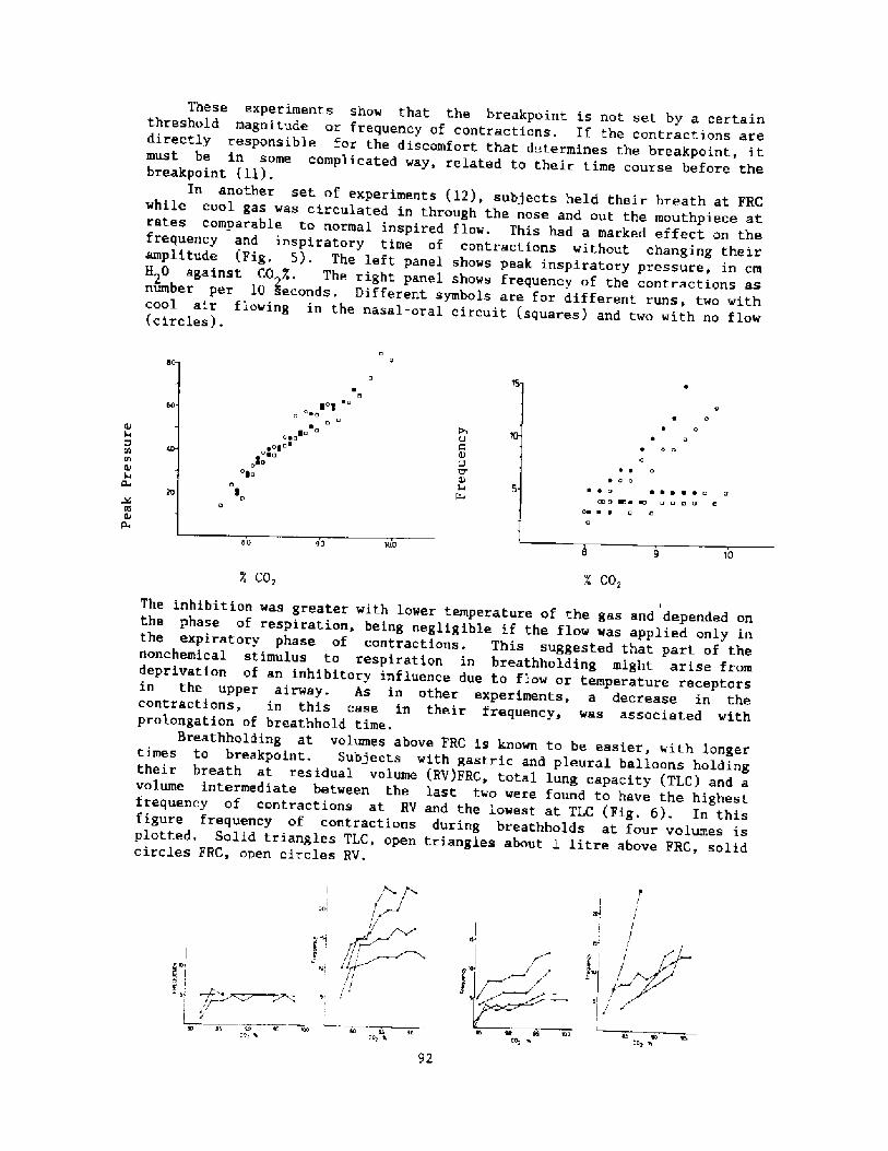
C02CO,
IThe inhibition was greater with lower temperature of the gas and depended onthe phase of respiration, being negligible if the flow was applied only inthe expiratory phase of contractions. This suggested that part of thenonchemical stimu.lus to respiration in breathholding might arise fromdeprivation of an inhibitory influence due to flow or temperature receptorsin the upper airway. As in other experiments, a decrease in thecontractions, in this case in their frequency, was associated withprolongation of breathhold time.
Breathholding at volumes above FRC is known to be easier, with longertimes to breakpoint. Subjects with gastric and pleural balloons holdingtheir breath at residual volume RV!FRC, total lung capacity TLC! and avolume intermediate between the last two were found to have the highestfrequency of contractions at RV and the lowest at TLC Fig. 6!. In thisfigure frequency of contractions during breathholds at four volumes isplotted. Solid triangles TLC, open triangles about 1 litre above PRC, solidcircles FRC, open circles RV.
apl/
SI l6 lieco, ~ 4S so
Cop ll
92
These experiments show that the breakpoint is not set by a certainthreshold magnitude or frequency of contractions. If the contractions aredirectly responsible for the discomfort that determines the breakpoint, itmust be in some complicated way, related to their time course before thebreakpoint ll!.
In another set of experiments 12!, subjects held their breath at FRCwhile cool gas was circulated in through the nose and out the mouthpiece atrates comparable to normal inspired flow. This had a marked effect on thefrequency and inspiratory time of contractions without changing theiramplitude Fig. 5!. The left panel shows peak inspiratory pressure, in cmH 0 against CO %, The right panel shows frequency of the contractions asnumber per 10 seconds. Different symbols are for different runs, two withcool air flowing in the nasal-oral circuit squares! and two with no flow circles!.
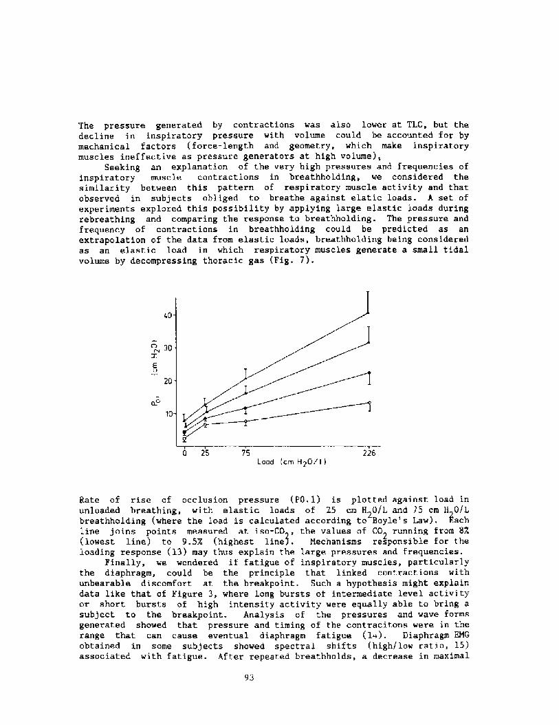
The pressure generated by contractions was also lower at TLC, but thedecline in inspiratory pressure with volume could be accounted for bymechanical factors force-length and geometry, which make inspiratorymuscles ineffective as pressure generators at high volume!,
Seeking an explanation of the very high pressures and frequencies ofinspiratory muscle contractions in breathholding, we considered thesimilarity between this pattern of respiratory muscle activity and thatobserved in subjects obliged to breathe against elatic loads. A set ofexperiments explored this possibility by applying large elastic loads duringrebreathing and comparing the response to breathholding. The pressure andfrequency of contractions in breathholding could be predicted as anextrapolation of the data from elastic loads, breathholding being consideredas an elastic load in which respiratory muscles generate a small tidalvolume by decompressing thoracic gas Fig. 7!.
o 30
20
10
750 25Load crn H20/ I
Rate of rise of occlusion pressure PO. 1! is plotted against load inunloaded breathing, with elastic loads of 25 cm H 0/L and 75 cm H G/Lbreathholding where the load is calculated according to Boyle's Law!. kachline joins points measured at iso-CO , the values of CO running from 8X lowest line! to 9.5X highest line!. Mechanisms responsible for theloading response �3! may thus explain the large pressures and frequencie~.
Finally, we wondered if fatigue of inspiratory muscles, particularlythe diaphragm, could be the principle that linked contractions withunbearable discomfort at the breakpoint. Such a hypothesis might explaindata like that of Figure 3, where long bursts of intermediate level activityor short bursts of high intensity activity were equally able to bring asubject to the breakpoint. Analysis of the pressures and wave formsgener'ated showed that pressure and timing of the contracitons were in therange. that can cause eventual diaphragm fatigue �~!. Diaphragm EMGobtained in some subjects showed spectral shifts high/low ratio, 15!associated with fatigue. After repeated breathholds, a decrease in maximal
93
transdiaphragmatic pressure was noted. It thus seems possible thatcontractions of the magnitude seen in breathholding are capable ofeventually causing diaphragm fatigue, and that the intolerance of longerbreathholds is related to the need to avoid fatigue or to afferentinformation detecting incipient fatigue.
In summary, breathholding studies in the laboratory find a strongassociation between the number, frequency and itnensity of involuntaryinspiratory muscle contractions and the breakpoint. The relation is not asimple one, but the principle of avoiding diaphragm fatigue is one plausiblelink. Unexpected stimuli, the example being cold in the upper airway mayhave an effect on the contractions and prolong breathholding, as can othermaneuvres known to delay the breakpoint.
The application of this kind of experiment to breathhold diving is notcertain. It is not known whether experienced divers suppress thecontractions or whether they occur and constitute an important urge toreturn to the surface. Occasional normal subjects are able to suppresscontractions for a long time, but most find thi.s very difficult and can holdtheir breath much longer by sealing the upper airway while contractionsoccur. Our experiments were conducted after oxygen breathing. Breathholdson air waould likely produce more vigorous contractions, and hypoxiaincreases diaphragm vulnerability to fatigue 16!. If contractions of thisnature are important, such things as this time course of gas tensions in adive, and the ambient water temperature could have an important influence onthe urge to breathe and the tolerable duration of the dive.
REFERENCES
Agostoni, E. Diaphragm activity during breathholding: factors relatedto its onset. J. Appl. Physiol. 18:30-36, 1963.Lin, Y.C., D.A. Lally, T.Q. Moore and S.K. Hong. Physiological andconventional breathhold breaking points. J. Appl. Physiol. 37:291-296,1974.
3. Hill, L., and M. Flack . The effect of excess of carbon dioxide and ofwant of oxygen upon the respiration and the circulation. J. Appl.Physiol. London 37:77-111, 1908.
4. Fowler, M.S. Breaking point of breathholding, J. Appl. Physiol.6."S39-S45, 1954.
5. Campbell, K.J.M., S, Freeman, T.J.H. Clark, J.G. Robson and J. Norman .The effect of muscular paralysis induced by tubocurarine on theduration and sensation of breathholding. Clin. Sci. 32-425-432, 1967.
6. Godfrey, S. and E.J.M. Campbell. Mechanical and chemical control ofbreathholding. Q.J. Exp. Physiol. 54:117-128, 1969.Godfrey, S. and E.J.M. Campbell. The control of breathholding.Respir. Physiol. 5:385-400, 1968.
Read, D.J.C. A clinical method for assessing the ventilatory response
94
to carbon dioxide. Australas. Ann. Med. 16:20-32, 1967.
Whitelaw, W.A., J. P. Derenne and J. Milic-Emili. Occlusion pressure asa measure of respiratory centre output in conscious man. Respir.Physiol. 14:181-199. 1975.
Mithoeffer, J.C. Mechanisms of pulmonary gas exchange and COtransport during breathholding. J. Appl Physiol. 14:706-710, 1910.
Whitelaw, W.A., B. McBride, J. Amar and K. Corbet. Respiratoryneuromuscular output during breathholding. J. Appl. Physiol. 50:435-443, 1981.
10.
13. Cherniack, N.S. and M.D. Altose. Respiratory responses to ventilatoryloading. In: Regulation of Breathing, Part II. ed.! T.F. Hornbein,Marcel Dekker, 1981.
14, Bellemare, F. and A. Grassino. Effect of pressure and timing ofcontraction on human diaphragm fatigue. J. Appl. Physiol. 53:1190-1195, 1982.
15, Gross, D. A. Grassino, W.R.D. Ross et al. Electromyogram pattern ofdiaphragmatic fatigue. J. Appl. Physiol. 46:1, 1979.
16, Roussos, C.S. and P.T. Macklem. Respiratory muscle fatigue: a cause ofrespiratory failure? Clin. Sci. Mol. Med. 53:419, 1977.
95
12. B. McBride and W.A. Whitelaw. A physiological stimulus to upper airwayreceptors in humans. J. Appl. Physiol. 51:1189-1197, 1981.
Discussion following Dr. White].aw's presentation
Dr. Arnold: Are there any calculations of how much energy respiratorymuscles consume while you' re between the onset of involuntarydiaphragmatic contr actions and blackout?
Dr. Nhitelaw: We haven't calculated that but you can make a guess atit because there are measurements now that relate the tension-timeindex to the oxygen consumption of respiratory muscles, thediaphragm, anyway, and these would certainly be consuming lots ofoxygen. Fatigue is probably due to an imbalance between supply ofoxygen and oxygen demand and it's aggravated when cardiac output islow and perfusion is less, when there's hypoxia, and when there'8hypercapnia, and all of these things could actually be going on in abreath-hold as well. So it's not easy to see how they interact indetail.
Dr. Rennie: This may not be an appropriate time to ask this question,but with gastric pressure going up to plus 20 to 30 torr at the sametime as the esophageal pressure is decreasing by 20 or 30 torr, allthe conditions are set for a diaphragmatic circulatory pump that mayaugment somehow the cardiac pump under these conditions. Now> what dowe know about the interrelationship between the diaphragm and theheart during breath-hold as an augmented circulatory pump?
Dr. Nhitelaw: I don't know, don'0 know at all.
Dr. Rahnr You were asking about the work done by the diaphragm.Could that be calculated from the volume and pressure changes, ratherthan from extrapolating to tension of the diaphragm itself?
Dr. Nhitelaw: I agree � I jumped to oxygen consumption and bypassedthe work question, but sure you can calculate the work problem.
Dr. Rahn: Well, all you'd have to know is what the volume was at thepressure point.
Dr. Nhitelaw: Well, only the pressure because the volume iscalculated from the pressure swing and the starting volume FRC!.
Dr. Lundgren: Were you thinking of the expansion of the gas?
Dr. Rahn: Yes. Because that would be easy to do, wouldn't it? Itcould give you some idea of how much energy you are losing.
Dr. Craig: You' re wasting a lot of energy.
Dr. Perrigno: You mentioned the Hering-Breuer reflex. I was thinkingabout the difference between dry and wet breath-hold dives. How doesimmersion affect the Hering-Breuer reflex? Does it affect it throughintr a thoracic blood pooling?
Dr. Whitelaw: It could, but first, of all immersion drops lung volume,so that would have an effect, and thoracic blood volume might have aneffect on a vagal reflex, it would not be the Hering-Breuer reflex. I
96
don't really know the answer to this question at all � we thought wewould do this experiment under water once just to have some data forthe meeting, but we didn't manage to get it.
Dr Arnold: I think the breaking point is very impor tant to peoplewho are going to dive more than once and enjoy it. I think anothermessage f rom the last couple of papers is that people, who have beenable to survive near drowning, have gone through an experiment testingthe physiology and giving us all sor ts of information. I'm sure thebreath-holding time for a given individual is going to be shorter withlower temperature, but near drowning victims seem to survive better ifthe temperature is lower. To a certain extent, I think it's a benefitif a given individual goes under the ice water and goes unconscioussooner without going through a lot of this energy consumption and ifthey' re able to do that without inhaling water, the heart rate isstill maintained.
Dr. Lundgren: I have a comment that's related to the question of howwe deal as breath-holders with these diaphragmatic movements and it iskind of anecdotal but some of you, I'm sure, have made the sameobservation. Those of you who have hyperventilated on oxygen and heldyour breath for a long time, may have noticed how there is a periodof some air hunger, if you will, fairly early in the breath-hold andthen it goes away. The question is: what is this adaptive mechanism?Another, and certainly anecdotal, piece of information that I want tooffer you has to do with some experiments that we did a long tirpe agoin Sweden, when we were interested in ventilation without respiratorymovements. We used a Thunberg barospirator Thunberg, T. DerBarospirator, ein neuer Apparat fur kunsliche Atmung. Skan. Arch.Physiol. 48:80-94, 1926!, a steel cylinder in which you could place asubject and which was connected to a huge piston pump that oscillatedback and forth and gave pressure swings in the barospirator. In thatway you actually achieve pulmonary ventilation without respiratorymovements. Now< we did this, and it was very hard to relax and not tobreathe, despite the fact that our COq-meter indicated that we were/if anything, hyperventilated. With training, we could relaxcompletely and we observed, objectively, lack of movement. There was avery distinct training effect and the reason I'm mentioning this isthat, perhaps with training in breath-holding, you become lesssensitive to the need for respiratory movements as a way of satisfyingyour r espi ratory regulation.
97
Undersea Bi<//n ed/< a/ Resear<'h. Vn/. I I, IV<x 2, June /984
Decrease in body insulation with exercisein cool water
Y. S. PARK, D. R. PENDERGAST, and D. W, RENNIE
Pep«r<n<en<S <ij PhySia/O//V, 5<are Un<'< rnily «/ /Veu Y<>rt «! B«ff«/a, B«ffo/a..h' y /42/4.and Kosin Medi< a/ Co//ree, B«s«n 6//0, Karen
Park YS, Pendergast DR. Rennie DW. Decrease in body insulation with exercise in cool water.Undersea Biomed Res l984: l!�!:�9-168.� Steady-state body insulation was ineasured in 7healthy male subjects during rest and exercise For 3 h in water of 28'C-32'C. At res , maximal bodyinsulation increased as a linear function of mean subcutaneous fat thickness by an ainount approx-irnately 4-fokl what would be predicted froin the physical insulation of fat alone, With arm plus legexercise, body insulation declined as an exponential function of he exercise intensity, reachingapproximately 25% of the resting value at work load~above Vo = l.2 liters min '. During exercisethe relationship between overall body insulation and mean subcutaneous fat thickness was almostidentical to that predicted from fat insulation alone. These results suggest that 75% of maximalbody insulation in resting subject~ is achieved by use of skeletal muscle as an insulative barrier andthat the muscle component is increased with increasing fat thickness. This muscle insulation shellis lost during exercise. As a practical consequence, heat generated by muscular exercise in watercolder than critical water emperalure cannot offset cooling unless the exercise intensity is great.
body insulation
wale r tein pe ra lureexerciseskeletal mu scl e
The indirect evidence seems quite good that he thickness of' subcutaneous fat is only oneof several factors slowing the rate of central body heat loss in cold water, There is generalagree/nent that subcutaneous fat provides an insulative barrier to the toss of body heat duringimmersion in coot water I � 3!. f:arlson et al. �!, Keatinge �!, Sloan and Keatinge �!, Rennie�!, Burton and Edholm 8!, and Pugh and Edholm 9! have shown for subjects resting in coldwater that more obese persons with greater subcutaneous fat thickness cool at slower rates atthe same water temperature. Second, the ratio of surface area to mass must be considered as afactor when comparing cooling rates of individuals having substantially different body shapesand sizes, e.g., men vs. women or aduhs vs. children 9, l0!. For man in water a third factor,and possibly the most important, is the insulative shell beneath the skin and subcutaneous fat.Sloan and Keatinge �! pointed out that lean subjects cool much less rapidly than would bepredicted from their ratio of surface area to mass, "probably because deep tissues providedappreciable insulation to supplement hat of skin and subcutaneous fat." Rennie et al. l 1!reported that the linear regression of maximal body insulation on subcutaneous fat thickness,
Y. S. PARK, D. R. PENDERGAST, AND D. W. RENNIE
when extrapolated to fat thickness of zero, yielded a positive intercept on the insulation axis;therefore they postulated the existence of an in-series insulative inuscle shell based on theconcepts of insulative cold acclimatization developed by Carlson et al. �!. Exercise in watercooler than 25'C has been shown to actually accelerate body cooling above rates at rest �,12!, probably due to increased muscle perfusion and hence reduced insulation. However, therehas been no quantitative assessment of the actual magnitude of muscle insulation duringprogressive increments of exercise, primarily because most studies have been done in waterso cold as to preclude achievement of the thermal balance needed to calculate overall bodyinsulation,
The current study represents an attempt to quantify the relative insulative value of subcu-taneous fat vs. muscle during prolonged immersion in water of 28'C-32'C by measuring thesteady-state body insulation during the last hour of a 3-h immersion of subjects at rest andduring each of several levels of coinbined arm and leg exercise.
METHODS
To study the effects of long-term exercise on body insulation in water, 7 healthy malevolunteers were used with a range of subcutaneous fat thickness from 1.4 to 7.4 mm. None ofthe subjects were in rigorous physical training. Their physical characteristics are summarizedin Table l.
Skinfold thickness was measured with a Lange caliper Cambridge Scientific Inc., Cam-
bridge, MD! at each of 10 positions chin, cheek, back, chest, midlateral thorax, umbilicus,
lateral waist, triceps, knee, and calf!, and the weighted mean subcutaneous fat thickness SFT!was estimated after correction for 4-rnm double-skinfold thickness for each position �1!.
Percentage of total body fat was estimated from the relationship between skinfold thicknessand adiposity as described by Allen et al. �3!.
Establishmerrt of critical water remperarure. Critical water teinperature T, !, defined asthe lowest temperature a resting individual can tolerate for 3 h without shivering, was deter-mined for each subject in a series of preliminary experiments by immersing them to the chinin vigorously stirred water v = 0.25 m s '! as previously described � 1!. Subjects wereclothed in swim suits and seated motionless for up to 3 h. Water temperature was regulated towithin 0.05'C at each selected temperature. The Vo- was measured directly at 20-min intervalsusing a closed-circuit 13L Collins spirometer Warren E. Collins, Inc., Braintree, MA!, lf thethree final readings exceeded the initial hour's Vo by more than i07o, the water temperaturewas considered too cold and was raised 1'C for the subsequent test. Usually 2 but occasionally
3 iminersions were necessary. Table 1 summarizes the T,�. of the subjects.
Exercise in warer of <rirical temperature. A modified Collins bicycle ergorneter was mountedover a water tank 3 rn long, 1.5 rn wide, and 3 m deep. Subjects were seated comfortably onthe ergometer seat with legs extended horizontally to the foot pedals. A rope with hand loopspassed through an adjustable friction ring mounted on the ergoineter frame so that activeflexion of first one arm and then the other could be accomplished over the full range of armflexion in synchrony with leg pedaling. After the subject was strapped comfortably to the seatthe entire frame was lowered into the water to the level of the subject's chin. An exercise
intensity that could be continued for 3 h was established empirically for each subject bymodifying the frequency of simultaneous cycling and arm flexion and the drag. The Vo, was
99
BODY INSUI.ATIOH Ih' COOE WATER
E
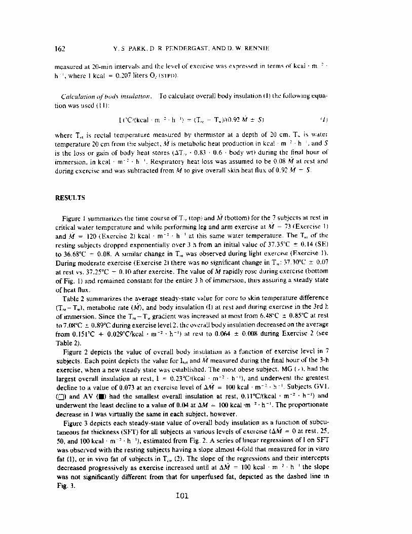
167 Y. S. PARK, D. R. PENDERGAST, AND D. W. REGIA!}E
measured at 20-min intervals and the level ol' excrcrse was expressed in terms of kcal m
h ', where 1 kcal = 0,207 liters O. sir n!.
Calcularion ofbody inswlarion. '1'o calculate overall body insulation �! the following equa-tion was used �1!:
1 C/ kcal m - 'h '! = T�� T.!/�.92 M = S!
where T� is rectal temperature measured by thermistor at a depth of 20 cm. T� is watertemperature 20 cm from the subject, M is metabolic hea production in kca! rn ' h '. and 5is the loss or gain of body heat stores DT�0.83 0.6 body wt! during the final hour ofimmersion. in kcal m ' -h ', Respiratory heat loss was assumed to be 0.08 M at rest andduring exercise and was subtracted from M to give overall skin heat flux of 0.92 M ~ S.
RESULTS
101
Figure 1 summarizes the time course of T� top! and M bottom! for the 7 subjects at rest incritical water temperature and while performing leg and arm exercise at M = 73 Exercise 1!and M = 120 Exercise 2! kcal m ' h ' at this same water temperature. The T�of theresting subjects dropped exponentially over 3 h from an initial value of 37.35'C ~ 0,14 SE!to 36,68'C ~ 0.08. A similar change in T�was observed during light exercise Exercise 1!.During moderate exercise Exercise 2! there was no significant change in T�: 37.30'C ~ 0.07at rest vs, 37.25'C ~ 0.10 after exercise. The value of M rapidly rose during exercise bottomof Fig. 1! and remained constant for the entire 3 h of immersion. thus assuring a steady stateof heat flux.
Table 2 summarizes the average steady-state value for core to skin temperature difference T�� T�!, metabolic rate M!, and body insulation �! at rest and during exercise in the 3rd hof immersion. Since the T�� T gradient was increased at most from 6,48 C = 0.85'C at restto 7.08'C ~ 0.89'C during exercise level 2, the overall body insulation decreased on the averagefrom 0,151'C + 0.029'C/kcal m ' h '! at rest to 0.064 ~ 0.008 during Exercise 2 seeTable 2!.
Figure 2 depicts the value of overall body insulation as a function of exercise level in 7subjects, Each point depicts the value for I��and M measured during the final hour of the 3-hexercise, when a new steady state was established. The most obese subject, lvlG ~ !. had thelargest overall insulation at rest, l = 0.23'C/ kcal m - 'h '!, and underwent the greatestdecline to a value of 0.073 at an exercise level of AM = 100 kcal m '- h '. Subjects GVL Q! and AV �! had the smallest overall insulation at rest, 0.11 C/ kcal m ' -h '! andunderwent the least decline to a value of 0.04 at ~ = 100 kcal m-' h ', The proportionate
decrease in I was virtuaHy the same in each subject, however.Figure 3 depicts each steady-sta e value of overall body insulation as a function of subcu-
taneous fat thickness SFT! for all subjects at various levels of exercise hM = 0 at rest. 25,50, and 100 kcal rn '- h '!, estimated from Fig. 2. A series of linear regressions of 1 on SFTwas observed with the resting subjects having a slope almost 4-fold tha measured for in vitrofat �!, or in vivo fat of subjects in T,. �!. The slope of the regressions and their interceptsdecreased progressively as exercise increased until at AM = 100 kcal m ' h ' the slopewas not significantly different from that for unperfused fat, depicted as the dashed line inFig. 3.
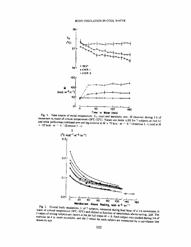
Y. S. PARK, D. R, PENDERGAST, AND D. W, RENN1E
TABLE 2
T�� T... METAnOr iC RArE, ANn BOOV IVSUr ATiON DER>NG 3RD H OE IxtMERSiON
During Exercise'
At Rest
T�� T,� 'C!
M kcal- m-' h-'!
S" kcal m ' h '!
T-- T-0.92M+ S
'C/ kcal m ' h '!
6.48 0,85
47+ 3.597,08 0,89
123+ 6.25
6,37 ~ 0.75
72.04 ~ 2.43
� 3.6 0.76
0.151 w 0.0291.47+ 3.51
0.064 ~ 0,008
� 6.5+ 3.0
0.095 ~ 0.011
'Exercise 1: dM = 25 kcat rn ' h '. Exercise 2: dM = 76. "Change inbodyheat storage.
MSCUSS 101'
103
For many years investigators have intimated that factors in addition to subcutaneous fatthickness must determine body cooling rates or body insulation in water � � 7, 10, I 1!. Thepresent study focuses on the hypothesis that skeletal muscle acts as an insulative barrier inresting subjects. Although we claim no originality for this concept, which dates back at leastto Carlson et al. �!, the design of this study does provide for the first time a quantitativeestimate of the importance of muscle, and hopefully it will be a stimulus for basic studies onthe circulation of skeletal muscle in imniersion hypothermia.
The results depicted in Figs, 2 and 3 indicate that exercise progressively diniinished overallbody insulation to values that can be accounted for by unperfused subcutaneous fat and skinalone �!. Figure 4, which is derived from data in Fig. 2, depicts the decrease of body insulationfor each individual exercise period as a percentage of its value at rest, On the average, theinsulation decreased to approximately 50% of I �at kM = 50 kcal m ' h ', to 35% at hM= 100, and to 27% at hM = 150. Thus skeletal muscle appears to provide as much as 75% oftotal body insulation in subjects resting in water of critical temperature. with subcutaneous fatand skin accounting for the remainder see Fig. 4!. A similar conclusion has been drawn inanother study �!, in which superficial shell fat and skin! insulation was estimated from directmeasurements of fat and skin temperatures and skin heat Ilux. This suggests that for subjectsimmersed in moderately cold water, heat loss is controlled largely by blood liow to skeletalmuscle with unperfused subcutaneous fat and skin providing a less important role than com-monly supposed.
Measurements of skeletal muscle blood low in different parts of the body during immersionhave not been reported, However, resting blood low in the entire forearm is reduced to only10%%u of control when the temperature of water irnrnersion changes from 35 C to 30'C �4, 15!.It therefore seems likely that the blood vessels of muscle constrict in response to whole-bodyimmersion in water that feels tepid, thereby providing an insulative layer underneath thesuperficial tissues. The signal for this severe vasoconstriction deserves to be an object of study,since neither central nor skin temperatures are greatly different from these during comfortableexposure in air. This additional insulation would naturally be removed at least in part byexercise hyperemia.
In the present study, as well as in other studies �, 16, 17!, the maximal body insulationattained during rest at critical water temperature was linearly correlated with the subcutaneous
165BODY IN S U L ATION I 8 COOL WATER
0.3
hr '!
0.2I
'C-kcat 'rn 2 hr !
0.1
0 2 4 6 80.0
Subcutaneaus Fat Thickness, mntFig. 3, Overall body insulation, l, plotted as a function of subcutaneous fat thickness in 7 subjects.
Maximal I values during 3rd h of immersion in water of critical temperature are depicted by the top line,hf = 50 kcal m ' h '. Each of the lower solid lines depicts I during 3rdh of exercise at 75, l00, andl50 kcal m '- h, Dashed lirie depicts physical insulation of fat alone, i.e., 0.058'Cl kcal m -' h '!.See Refs. l and 2,
<axgtt / I ttrsf! x 100
<00
> Imax
60
40
20
104
0 20' 40 60 t 80 ioo ~20 i40 i60Metabolism Above Resting, kcot e hr '
Fig. 4. percentage of maximal insulation at res , % I � is plotted for all 7 subjects as a function ofmetabolism above resting. At i3hf = 50 kcal - in ' h ', % I, = 48. The asymptote for % l, approaches25% when ~ exceeds 150 kcal m-' h- '
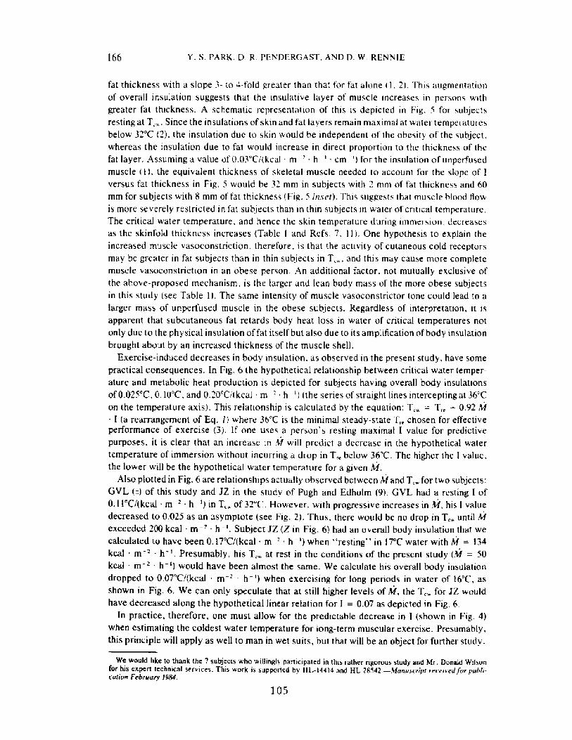
166 Y. S. PARK, D. R. PENDERGAST. AND D. W. REiVNIE
We wou d like to thank the 7 subjects who wiuingly participated in this rather rigorous study and Mr. Donald Wilsonfor his expert technicat services. This work is supported by HL-l44i4 and HL-28S42 �,Manuscript received for pubti-cation February ! 984.
105
fat thickness with a slope 3- to 4-fold greater than that for fat alone I, 2!. 'I'his auginentationof overall insulation suggests that the insulative layer ol muscle increases in person~ withgreater I'at thickness. A schematic representation of this is depicted in Fig. 5 for subjectsresting at T,. Since the insulations of skin and t'at layers remain maxiinal at water temperaturesbelov' 3".C '2!, the insulation due to skin would be independent of the obesity of the subject,whereas the insulation due to fat would increase in direct proportion to the thickness of thefat layer. Assuming a value of 0,03 Ci kcal m ' h ' cin '! for the insulation of unpert'usedmuscle I!, the equivalent thickness of skeletal muscle needed to account for the slope ol' Iversus fat thickness in Fig, 5 would be 32 tnm in subjects with 2 mm of fat thickness and 60mm for subjects with 8 mm of fat thickness Fig. 5inset!. This sugges s that muscle blood liowis more severely restricted in I'it subject» than in thin subject~ in water of critical temperature,The critical water temperature, and hence the skin tempera ure during immersion, decreasesas the skinfold thicknes~ increases Table I and Refs. 7, I I!. One hypothesis to explain theincreased muscle vasoconstriction, therefore. is that the activity of cu aneous cold receptors nay be greater in fat subjects than in thin subjects in T,�, and this may cause more completemuscle vasoconstriction in an obese per~on. An additional factor. not mutually exclusive ofthe above-proposed mechanism, is the larger and lean body mass ol' the more obese subjectsin this study see 'I'able 1!. The same intensity of muscle vasoconstrictor tone could lead to alarger mass of unperfused muscle in the obese subjec s. Regardless of interpretation, it isapparent that subcutaneous fat retards body heat loss in water of critical temperatures notonly due to the physical insulation of fat itself but also due o its amplilication ofbody insulationbrought abo~t by an increased thickness of the tnuscle shell.
Exercise-induced decreases in body insulation, as observed in the present study, have soinepractical consequences. In Fig. 6 the hypothetical relationship between critical water emper-ature and metabolic heat production is depicted for subjects having overall body insulationsof 0.025'C, 0.10'C, and 0.20'C/ kcal m = h '! the series of straight lines intercepting at 36'Con the temperature axis!. This relationship is calculated by the equation: T,�= T�� 0,92 M- I a rearrangernent of Eq, l! where 36'C is the minimal steady-state 'I�chosen for effectiveperformance of exercise �!, If one uses a person's resting maximal I value for predictivepurposes, it is clear that an increase in M will predict a decrease in the hypothetical watertemperature of immersion without incurring a drop in T�below 36'C. The higher the I value,the lower will be the hypothetical water temperature for a given M.
Also plotted in Fig. 6 are relationships actually observed between Mand T,. for two subjects:GVL =! of this study and JZ in the study of Pugh and Edholm 9!. GVL had a resting I ofO.II'C/ kcal in -' h '! in T,. of 32 C, However, with progressive increases in M, his I valuedecreased to 0.025 as an asymptote see Fig. 2!, Thus. there would be no drop in T,�until Mexceeded 200 kcal m ' -h '. Subject JZ Z in Fig. 6! had an overall body insulation that wecalculated to have been 0. l7'Ct kcal m h '! when "resting" in I7'C water with M = I34kcal m ' - h '. Presumably, his T,. at rest in the conditions of the present study M = 50kcal - in ' - h '! would have been altnost the same. We calculate his overall body insulationdropped to 0.07'C/ kcal m ' h '! when exercising for long periods in water of l6'C, asshown in Fig, 6, We can only speculate that at still higher levels of M, the T,. for JZ wouldhave decreased along the hypothetical linear relation for I = 0.07 as depicted in Fig. 6.
In practice, therefore, one must allow for the predictable decrease in I shown in Fig. 4!when estimating the coldest water temperature for iong-term muscular exercise. Presumably,this principle will apply as well to man in wet suits, but that will be an object for further study.
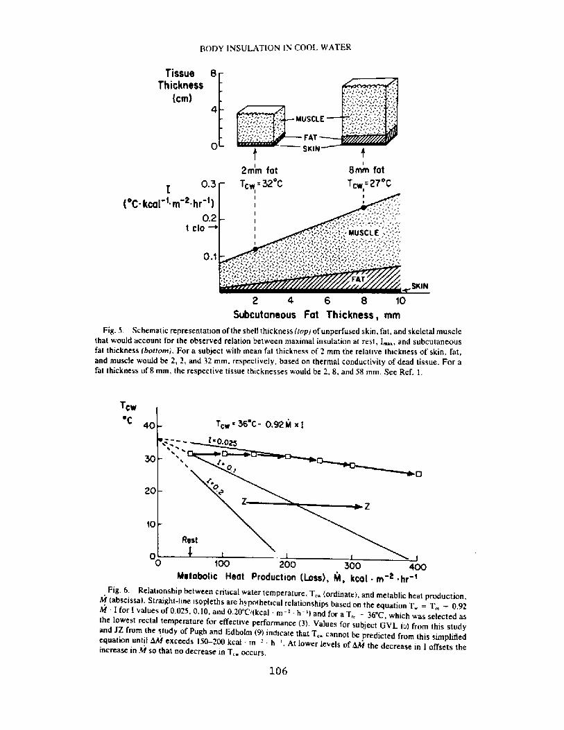
BODY INSULATION IN COOL WATER
Tissue B
Thickness
Ctn!
uSC
F4T
SKIN
2mm fot
T 32'8mm fot
T -27'0.3
'C kcat 'm ~ hr I!0.2
t clo ~
0.$
4 6 B to
Subcutaneous Fat Thickness, mmFig. 5. Schematic representation of the shel! thickness top! of unperfused skin, fat, and skeletal muscle
that would account for the observed relation between maximal insulation at rest, I �and subcutaneousfat thickness botrom!. For a subject with mean fat thickness of 2 mtn the relative thickness of skin, fat,and muscle would be 2, 2, and 32 mm. respectively. based on thermal conductivity of dead tissue. For afat thickness of 8 mm, the respective tissue thicknesses would be 2, 8, and 58 mm. See Ref. l.
TCW'C
30
20
10
106
0 >00 200 300 400Metabotic Heot Production Loss!, NI, kca[ ~ m < ~ hr t
Fig. 6. Relationship between critical water temperature, T, ordinateh and metablic heat production,hf abscissa l. Straight-line isopleths are hypothetical relationships based on the equation T = T�� 0.92hf I for I values of 0.025, 0,10, and 0.20'C/ kcal m ' h-'l and for a T�= 36 C, which was selected asthe lowest rectal temperature for effective performance �!. Values for subject GVL o! from this studyand JZ from the study of Pugh and Edholm 9! indicate that T, cannot be predicted frotn this simplifiedequation until d8f exceeds l50-200kcal m '- h '. At lower levels of4hf th d I ff ht e decrease in I offsets theincrease in M so that no decrease in T, occurs,
leS Y. S. PARK, D, R. PENDERGAST. AND D. W, RENNIE
Park YS, Pendergast DR, Rennie DW. Diminution de I'isolation corporelle durant I'exercice en eaufroide. Undersea Biomed Res 1984; I I�!:159-168.� L'isolation corporelle 5 Id at stable fut mesu-r6e pendan 3 h chez 7 sujets males en san� au repos et 0 I'exercice dans de I'eau 5 28'C-32'C. Aurepos, I'isolation corporelle rnaximale augmente Iinciairemen en fonrtion de I'6paisseur moyennedu tissus adipeux sous-cutane d'une valeur d'approximativemeni 4 fnis celle predite par I'isolationphysique de la graisse seule. Au cours de I'exercice avec les bras e les jambes. I'isolation corporellediminua exponentieilement en fonction de I'intens � du travail, at eignant environ 2S% de la valeurde repos aux charges de travail d6passan une Vo. = 1.2 litres - min '. Duran�'exercice. Ia relationentre I'isolation corporelle dans son ensemble et I'6paisseur moyenne du tissu s adipeux sous-cutan66 ait presque identique h celle predite par I'isolation de Ia graisse seule. Ces r6sultats suggkrent que7S% de I'isolation corporelle max male chez les sujet s au repos est achevee par I'emploi des musclessquelettiqueS COmme barribre isolante, et que la cOmpOsan e muSCulaire est augmentee par I'ac-croissement de I'epaisseur de graisse. Cet e envelope d'isolation musculaire est perdue pendantI'exercice. Comme cons6quence pratique, la chaleur produite par I'exercice musculaire dans deI'eau 5 une temp6rature supdrieure 6 une valeur critique ne peut pas conlrecarrer le refroidissement5 moins d'efkctuer un travail 5 une intensit i plus grande
isolation corporellemuscle squelettique
tempdrature de I 'eauexercice
REFERENCES
3.07
1. Hatfield HS, Pugh LGC. Thermal conductivity ol human fa and muscle. Nature 19SI; 168'918-9192. Veicsteinas A. Ferret i G. Rennie DW. Superficial shell insula ion in res ing and exercising men in
cold water, J Appl Physiol: Respii Environ Exercise Physiol 1982; 52:ISS7-1564.3. Bullard RW, Rapp GM. Problems of body heai Joss in water immersion. Aerosp Med 1970; 41:1269-
1277.4. Carlson LD, Hsieh ACL, Fullington F, Elsner RW. Immersion in cold water and body tissue insulation.
J Aviat Med 1958; 29:14S-152.S. Keatinge WR. The effect of work and clo hing on the maintenance of body temperature in water. Q J
Exp Physiol 1961; 46:69-82,6. Sloan RFG, Keatinge WR. Cooling rates of young people swimming in cold water. J Appl Physiol
1973; 3S:371-37S.7. Rennie DW. Thermal insulation ol Korean diving women and non-divers in water. In: Rahn H, ed.
Physiology of breath-hold diving and the Ama ol Japan. Washing on. DC: National Academy ofScience, National Research Council, 196S:315-324
8. Burton AC, Edholm OG. Man in a cold environment. London: Edward Arnold Publishing Ltd., 19SS.9 Pugh LGC, Edholm OG. The physiology of channel swimmers. Lancet 19SS; October:761-767.
10. Kollias J, Bartle L, Bergsteinova V, Skinner JS, 13uskirk ER, Nicholas WC. Metabolic and thermalresponses of women during cooling in water. J Appl Physiol 1974; 36:S77-580.
I I. Rennie DW, Covino BG, Howell BJ, Song SH, Kang BS, Hong SK. Physical insulation of Koreandiving women. J Appl Physiol 1962; 17:961 � 966.
12. Cannon P, Keatinge WR. The metabolic rate and heat loss of fa and thin men in heat balance in coldand warm water. J Physiol 1960; IS4:329-344.
13. Allen TH, Peng MT, Chon KP, Ruang TF, Chang C, Fang HS. Prediction of total adiposity from skinfolds and curvilinear relationship between external and internal adiposity. Metabolism 19S6; 5:346-3S2.
14, Rennie DW. Body heat loss during immersion in water. [n: Voshimara H, Weiner JS, eds. Humanadaptability and its methodology. Tokyo: Japan Society for the Promotion of Sciences, 1966:36-41.
IS. Bacroft H, Edholm OG, The effect of temperature on blood low and deep temperature in the humanforearm. J Physiol 1943; 102:S-20.
16, Hong SK, Pattern of cold adap ation in women divers of Korea ama 1. Fed Proc 1973: 32:1614-1622.17. Hanna JM, Hong SK. Cr ical water temperature and effective insulation in scuba divers in Hawaii. J
Appl Physio! 1972; 33:770-773.
Discussion related to the ar ticle Deer ease in body insulation w i. thexercise in cool water
Dr. hrnold: Dr. Rennie, in a hypothermic patient who has been out ofthe water or otherwise, when one worries about rewarming the limbsquickly, the thought being that one ends up getting a dumping of verycold blood back f rom the limbs into the ventricles causingventricular fibrillation, it may very well be that the temperature ofthe muscles and the periphery is lower than the rectal temperatureand therefore your whole periphery seems to be a better insulator foryour core.
Dr. Rennie: That's right. If in fact there is a severe sustainedvasoconstriction of skeletal muscle as a consequence of cool waterstress, then you would imagine there could be a progr essive loss ofstored heat f rom a poorly per fused limb, or lengthening of thethermopathway providing a progressively longer physical pathway forthe conduction of heat through that muscle. You'r e quite r ight, thatmuscle would cool below the temperature of the central core providinga heat sink. In the case of rewarming you possibly do run the risk ofcold blood going back into the core and still further contributing tothe drop in core temperature. That's kind of a controversial question.
Dr Blsner: It would seem to suggest f rom what you have done andfrom the earlier discussions that if you were in a survival situationand you are dumped into cold water, you may choose to try to swim tothe shore. Your chances would improve if you are large and fat.However, if you' re in a situation in which there is no hope ofswimming to the shore, it would perhaps be better to be skinny andsmall and loose consciousness and depend upon CNS cooling untilrescued. We have seen recent examples of this response in childrenimmersed in cold water for periods up to 40 min.
Dr. Rahn- To continue this argument in another way, what would be thebest procedure af ter falling overboard in the middle of the Atlantic'P
Dr. Rennie: In the middle of the AtlanticP Stay with the boat, staywith your clothes, remain immobile, go to the fetal position.
Dr. Rahn: In other words, don't exercise
Dr. Rennie: Don't exercise. Now, if this is a skeletal musclevasoconstriction problem and is cold induced, then it is still an openquestion what exercise hyperemia will do under those conditions. Willit break through the cold induced vasoconstriction completely, or onlyin part? And if it's only in part, then this could even af feet yourswimming performance. It would be like swimming with a partialtour niquet on, which is still another reason for remaining immobileror at least not striking out for a distant shore at a high rate ofspeed.
Dr. Rahn: What about the breath-hold diver?
Dr. Rennie: Dr. Elsner has got data from years ago on the effects ofbreath-hold on human calf blood flow. I think he interpreted that as
I08
vasoconstriction in the calf. I don't remember if it was in the muscleor no t.
Dr. Blsner: In some subjects the flow shut right down so it has tobe muscle ischemia as well as skin vasoconstriction.
Dr. Rahn: What about a real situation, for example, Dr. Hong's Ama inten degrees water, diving daily for l5 minutes at the most.
Dr. Rennie: Well, the interpretation that I would have, of course, isthat the functional insulation in the real world is so much less thanthe insulation studied under conditions of critical water temperature,that there is no comparison between the two whatsoever. Combinedshivering and exercise so reduce the insulation, that it is immaterialwhether they develop a slight increase in the insulation of theperiphery in water of critical temperature. They may truly havedeveloped that increase in insulation. But whether it was ofsurvival value, practical value, I would say not anymore than anincrease in basal metabolic rate could reasonably be argued to be asurvival factor in the Eskimo.
Dr. Rahn: You know what the diving endurance is at l0 degrees C andat 25 degrees C in water, Can you on the basis of these timedi fferences, and knowing the rectal temperatures, go back andreinterpret and predict the duration7
Dr Rennie: Yes you can. Everything you gain by increasing metabolicheat. production by exercise, you loose by the decrease in insulation,so that any increased metabolic heat produced by exercising has noef feet on the minimal operational water temperature in which oneworks. That is, there is no ef feet at the levels of metabolic ratethat they use for the 35-45 minutes that they are working< which isonly about three times greater than rest.
Dr. Van Liew: Does still water act as an insulator7
Dr Rennie: All our studies were done with vigorously stirred wateriso that the additional stirring effect of exercise, for example, wouldbe mini miz ed.
Dr Van Lieu: But in the real world, the amount of sti rring is animpor tant complication.
Dr. Rennie: In the real world that's still a further complicationpthat's correc t.
Dr Lundgren: The amount of reduction in blood flow that you saw andthat we saw, in the face immersion studies that I mentioned earlierpwhere the fall in forearm blood flow was 50%, has to involve muscleto a substantial degree, because in order' to cut the blood flow inhalf for the whole limb, given the high proportion of muscle tissuerelative to skin tissue, a lot of the reduction has to be taken out ofm us cl e bl ood f l ow.
Dr Blsner: It's very cur ious that in our studies some years ago one
I09

of the subjects who showed extremely low calf blood flow during faceimmersion is a fellow who, characteristically, complained of legcramps when he was surfing.
110
THERMOREGULATION IN WET SUIT DIVERS
Y.S. Park, H.J. Kim, D.W. Rennie and S.K . Hong
Departmentof Physiology and Diving Science Institute,Kosin Medical College, Pusan 600, Korea;
Ocean and Underwater Medical Research Center, Chinhae, Korea;Department of Physiology, State University of New York at Buffalo,
Buffalo, NY 14214
Maintenance of normal body temperature is a critical problem for diversworking in cold water. Because of the high heat conductivity and heatcapacity of water, body heat is rapidly lost from the skin to the adjacentlayer of water. This drain of body heat is the principal problem of diversand, in fact, the dive time of unprotected divers is primarily determined bythis loss of body heat �,2!.
As reported elsewhere �! the thermal cost of diving in Korean womendivers decreased remarkably since they adopted wet suits probably due toadditional insulation provided by the suit. In breath-hold diving, diversrepeat the cycle of a dive and surface recovery each lasting for 30 - 40 secin the case of Korean women divers �!. During surface recovery, divers areresting, in a head-out water immersion, whereas during dives they exerciseand are exposed to various degrees of hydrostatic pressure. As discussed byRennie in the preceeding paper, insulation of the human body undergoes adramatic reduction during exercise in water. Moreover, physical insulationof the wet suit changes with pressure due to compression of trapped air �!.Therefore, overall thermal insulation and hence heat balance! of breath-hold wet suit divers will be changed cyclically during the course of divingwork.
In the present paper, we will first present data of heat exchangesobtained in Korean women wet suit divers during their natural diving work,and then describe effects of exercise and pressure on the thermal insulationof wet suit divers in cold water.
A. Thermal Balance ~Dnrin Breath-hold ~Divin
In 4 Korean women wet suit divers the heat exchange was studied whilethey were working in the sea � - 5 m depth! �!, In order to evaluate theeffect of wet suits on the thermal balance, the subjects wore wet suits �mm thick neoprene jacket, pants, hood and boots! in one series and cottonsuits traditional diving suits of previous Korean women divers! in theother,
Fig. 1 compares the time course of rectal TR! and the mean skin TS!temperatures of wet suit and cotton suit divers during diving work in summer�2.5 C water! and in winter �0oC water!. In cotton suit divers, the TRdeclined to about 35oC in 30 min in winter and in 1 hr in summer, at whichtime divers did not want to continue diving work any longer. The TS dropped
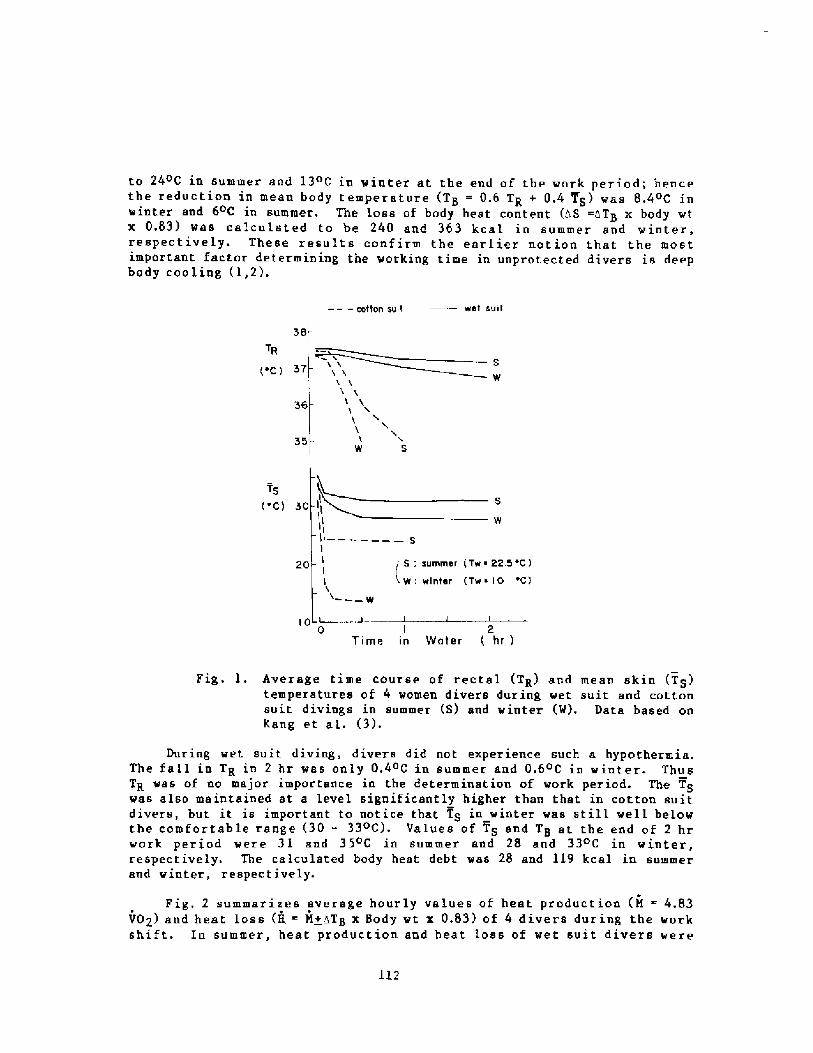
� � � cotton suit wst SU II
38
TR
'C !SW
w s
Ts
'C! 30
III
I ��I
I'I
� � � W
S; sommar Tw ~ 22 5 C!W: wintar Tw ~ I 0 'C!
20
IO0 2
Time in Water hr !
Fig. l. Average time course of rectal TR! and mean skin TS!temperatures of 4 women divers during wet suit and cottonsuit divings in summer S! and winter W!. Data based onKang et al. �!.
During wet suit. diving, divers did not experience such a hypothermia.The fall in TR in 2 br was only 0.4 C in summer and 0.6 C in winter. ThusTR was of no major importance in the determination of work period. The TSwas also maintained at a level significantly higher than that in cotton suitdivers, but it is important to notice that TS in winter was still well belowthe comfortable range �0 � 33 C!. Values of TS and TB at the end of 2 hrwork period were 31 and 35 C in summer and 28 and 33oC in winter,respectively. The calculated body heat debt was 28 and 119 kcal in summerand winter, respectively.
Fig. 2 summarizes average hourly values of heat production M 4.83VOZ! and heat loss H = M+zTB x Body wt x 0.83! of 4 divers during the workshift. In summer, heat production and beat loss of wet suit divers were
112
to 24oC in summer and 13 C in winter at the end of the work period; hencethe reduction in mean body temperature TB = 0.6 TR + 0.4 TS! was 8.4 C inwinter and 6oC in summer. The loss of body heat content AS =hT x body wtBx 0.83! was calculated to be 240 and 363 kcal in summer and winter,respectively. These results conf irm tbe earlier notion that the mostimportant factor determining the working time in unprotected divers ia deepbody cooling �,2!.
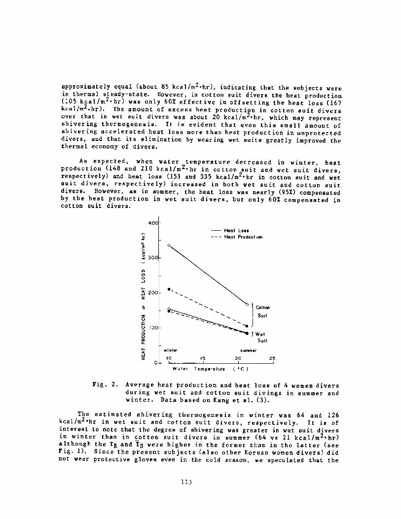
As expected, when water temperature decreased in winter, heatproduction �48 and 210 kcal/m ~ hr in cotton suit and wet suit divers,2
respectively! and heat loss �53 and 335 kcal/m ~ hr in cotton suit and wetsuit divers, respectively! increased in both wet suit and cotton suitdivers. However, as in summer, the heat loss was nearly 95X! compensatedby the heat production in wet suit divers, but only 60% compensated incot ton su it divers.
400Heat Loss
� � � Heat Product ionI
w C o300
rh0
~~ 200Z
'I CottonSuitZ 0
o 100!00 ! Wet
Suit
I- winter
x g 100
summer
20 25I
15
Water Temperature 'C !
Fig. 2. Average heat production and heat loss of 4- women diversdur ing wet suit and cot ton su it d iv ings in s utsmer andwinter. Data based on Kang et al. �!.
The estimated shivering thermogenesis in winter was 64 and 126kcal/m 'hr in wet suit and cotton suit divers, respectively. It is of2 ~
interest to note that the degree of shiver ing was greater in wet suit diversin winter than in cotton suit divers in summer �4 vs 21 kcal/m 'hr!although the TK and T8 were higher in the former than in the lat ter seeFig. 1!. Since the present subjects also other Korean women divers! didnot wear protective gloves even in the cold season, we speculated that the
113
approximately equal about 85 kcal/m -hr!, indicating that the subjects were2
in thermal steady-state. However, in cot ton suit divers the heat production I05 keel/m ~ hr! was only 60X effective in offsetting the heat loss �612.
kcal./m hr! ~ The amount of excess heat product ion in cot ton suit divers2
over that in wet suit divers was about 20 kcal/m ~ hr, which may representshivering thermogenesis- It is evident that even this small amount ofshivering accelerated heat loss more than heat production in unprotecteddivers, and that its elimination by wearing wet suits greatly improved thethermal economy of divers.
exposure of the hands to cold �0oC! water potentiated the shiveringresponse in wet suit divers. In fact, Van Someren et al, �! have observedthat selective cooling of hands and feet stimulates heat product ion insubjects immersed in 29oC water. Rtgardless of the mechanism, theeffectiveness of shivering in maintaining thermal balance in wet suit diversis clearly evident in Pig. 2.
Evidently, the reduction of heat loss by wearing wet suits was due toadditional insulation provided by the suit. As illustrated in Pig. 3, thetotal insulation estimated from the regtal to water temperature differenceand skin heat loss Itotal TH-Tg!/HS! appeared to be 2.1 to 2.8 timesgreater in wet suit divers in summer and winter, respectively �.170 vs0 ~ 193 C/kcal/m ~hr in summer vs winter! than in cotton suit divers �.081 vs0.068 in summer vs winter!. However, the tissue insulation Ibody TH-TS!/HS! was not significantly different between the two conditions approximately 0.07oC/kcal/m ~hr!. Consequently, the additional insulationafforded by wet suits Is�it Itotsl Ibody! was 0.10 � 0.13oC/kcal/m ~ hr.Since the physical insulation of wet suits may vary with the depth of divingand the physiological insulation provided by wet suits decreases withexercise see below!, the values of suit insulation obtained in workingbreath-hold divers, as described above, may only represent the averageinsulation during diving work. In any event, such a value of extrainsulation is equivalent to the insulation that can be provided by the 17 mmfat layer O.loC/kcal/m ihr-:0.006oC/kcal/m i br per mm fat ~ 17 mm fat!.Such an increase in subcutaneous fat insulation would reduce proportionatelythe heat loss of divers for a given temperature difference between thecentral body and water.
o ctCf!
X' summer winter summer winterWET SUITCOTTON SUIT
Pig. 3. Total and body insulations I! during diving work in wetsuit and cotton suit divers in summer and winter. Thestippled portion of the bar represents the insulationprovided by wet suits. Each value represents the mean +SR! of 4 women divers. Data based on Kang et al. �! ~
1 l4
B. Effect of Exercise on Thermal Balance of Wet ~sni ~Di rs
It is well known that exercise in cold water increases heat loss morethan heat production in an unprotected individual �,8!. In order toevaluate the effect of exercise on the thermal balance of wet suit divers wehave measured heat exchanges in 4 Korean women wet suit divers at rest andduring exercise in cold water. Subjects were clad in their personal wetsuits jacket, pan~ and boots of 5 - 6 mm thick! and were immersed up to theneck in a circulating water bath. The subject rested for 3 hr in a seatedposition or exercised for 2 hr at a constant intensity using a bicycleergorneter submersed in the bath. In order to prevent convective heat lossfrom under the suit 9! we taped the suit at the ankles and the wrists. Foreach subject the water temperature was adjusted to the critical temperature Tcw, the lowest water temperature a resting subject could tolerate for 3 hrwithout shivering! in order to evaluate the thermal insulation at themaximal degree of peripheral vasoconstr iction. The average Tcw with wetsuits was 16.5 + 1.2 SE!oC.
eFig. 4 depicts average skin heat loss HS! and rectal temperature TR!
over the final 1 hr at rest or dur ing exerc ise. The exercise intensity wasexpressed as a percentage of the resting metabolic rate. The HS at rest wason the average 45 kcal/m 'hr, but it increased linearly with the exerciseintensity. However, the TH was not different whether the subjects wereresting or were exercising at the various levels tested less than 3.5 Net!.These results indicate that thermal insulation decreased inversely withexercise intensity. As shown in Fig. 5, the calculated overall insulation
200
Tw 1S-5 - 12scHS
< kcaV1TT hr !
1 00
038
TR ec!
36
fh I� x ft x � ~
100 200 300
RE STIRG METABOLISM400
Fig. 4. Steady-state skin heat loss HS! and rectaltemperature TR! of 4 fernale wet suit divers as a functionof exercise intensity X resting metabolism! in water ofcritical temperature Tcw!, Each symbol represents anindividual diver. Dotted line represents TK calculatedusing the following formula: TE Tg+ ltotai x 0.92M!.
115
Itotal Tg-TW!HS! decreased from about 0.5 C/kcal /m ~ hr at rest toapproximately one balf st 2 Met and to one third of the resting value at 3Met. This decrease in Itotal appeared to be due in part to the reduction inbody insulation Ibody! and in Part to the decrease in insulation affordedby wet suits. The Ibod decl ined exPonentially from 0.23OC/keel/m hr at2.
rest to 0.06 at 3 Met. the apparent suit i.nsulation estimated from thedifference between the total and body ins~lation Isuit Itotal Ihody,stippled area! was on the average 0,27OC/kcal/m ~ br at rest, butdecreased gradually with exercise intensity until it reached approximately0.1OC/keel/m wbr at above 3 Met. The latter value of Isuit is similar tothe physical insulation of 5 mm neoprene wet suits obtained using a coppermanikin �0!. Since there was no apparent reason for change in thickness and hence physical insulation! of wet suits between rest and exercise wespeculate that the unexpectedly high functional insulation of wet suits inresting subjects is a consequence of physiological regulations in coldwater.
I H SVLATIOH
'CthCOlrrrI~ hr l
o.s
4»r,w
I jult ~ 0.1~w
k~o '
400IOO 200 300'4 R E ST I HG MET480LIsM
Fig. 5. Steady-State insulations I! of 4 female wet suit diversas a function of exercise intensity in water of criticaltemperature, ln each subject, values of Itota] and Ibodywere normalized against the corresponding mean value atrest. Each symbol represent s an individual diver.Stippled area represents the insulation afforded by wetsuits Isuit! Dashed line represents thecalculated by adding an Is t value of 0.1 to Ib d atsurt bodyvarious exercise levels.
When exposed to cold, the human body increases thermal insulationthrough peripheral vasoconstriction. As schematically illustrated in Fig.6, this regulation of thermal insulat ion is mostly accomplished in theextremities and not in the trunk 8,11,12!. Infrared thermography studiesby Hayward et al.�3! indicated that the highest skin temperatures following
116
prolonged resting in water are lateral thorax, upper chest and groin. Theconstriction of limb blood vessels in cold water may occur also in wetsuited subjects. Since the physical insulation of foamed neoprene willdecrease as the curvature of surface increases �4!, the insulative value ofwet suits will be much smaller in the limb than in the trunk. Furthermore,the design of wet suits is such that most of the r.runk surface is covered bydouble sheets pants and jacket! and the limbs by a single sheet. Thus wetsuits provide good insulation to the trunk surface, but poor insulation tothe limbs. As a consequence, the skin temperature underneath the suit willbecome much lower in the extremities than in the trunk during immersion incold water �1.3oC, chest vs 26oC, leg in the present study!. In other
gCVATLJRE
THICKNESS
Fig. 6. A schematic illustration of the insulative shell stippledarea! of a wet suited subject in cold water. Note thatthe trunk is covered by double sheets pants and jacket!and the limbs by a single sheet of the suit. Insetillustrates schema tie a 1 ly changes in c lo thing in su la t ion.as a function of thickness and curvature after Van Dillaet al. l3!.
words, immersion vith wet suits is analogous to exposing the limb to watercolder than that exposing the trunk. This will lead to strongvasoconstriction in the extremities. Restriction of limb blood flov willgreatly reduce the surface area for heat exchange and most of the heatexchange between the body core and water will take place at the trunksurface where suit insulation is relatively high. For these reasons, wetsuits provide far greater physiological insulation at rest than duringexercise. The exercise hyperemia reduces not only the thermal gradient from
117
the deep tissues to the skin but also thermal insulation down the length ofthe limb. Therefore, much of the heat produced in the skeletal muscle isdissipated through the large surface area of limbs rather than returning tothe body core. Since the insulative value of wet suits is relatively low inthe limbs because of its design single sheet! and the high curvature, theeffect of exercise is to increase the area for heat exchange over a poorlyinsulated region in parallel to the trunk- Added to this is increasedconvective heat loss from the escape of heated water at wrist, ankle andneck seals 9!. Estimation of the heat exchange area A = Isuit x 0 925/ T~!, N is metabolic rate in kcal/hr!, assuming that Isuit is constantat 0.12Wkcal/m2 hr, indicates that the area at rest is only 0.66 m, whichis equivalent to 40% of the total suit surface area Fig. 7!. The heatexchange area increases with exercise and becomes almost identical to theactual suit surface area at M = 200 kcal/hr. This analysis clearlyindicates that the relatively high apparent suit insulation in the restingsubject is due to reduced surface area for heat exchange.
6I
2Z
100
60
20
050 100 I 50
IAETABOLISM k col/kr !200
Fig. 7. The effect of heat exchange area of wet suits A! at restand at two levels of exercise. 'Ihe formula for A is shownin the inset. The value of Isuit was set at0.120C/kcal/m~ hr, which was observed for the 5 mmneoprene wet suit using an electrically heated manikin�0! .
gf ~sure. ~ ~ g~~ ~~~on ~ ~ ~ Qj'~
Three male wet suit divers were immersed in 15 � 16 C water in thewetpot of a hyperbaric chamber up to the neck. The chamber pressure wasmaintained at 1, 2 or 3 ATA air. Subjects rested for 3 hr or exercised for2 hr using bicycle ergometer- The Itotal, Ibody and Isuit were estimated
118
Having established the pattern of thermal exchange in wet suit diversat the surface of the water, we next investigated the effect of hydrostaticpressure on the thermal balance of divers.
as described above vhen the body temperature change wss minimal during thefinal hour of immersion. The actual thickness of suit reduced from 5 mm at1 ATA to 3.5 at 2 ATA and 2.6 at 3 ATA. Thus, the phys ical insulation ofthe suit might be decreased with pressure, and consequently skin surfaceunderneath the suit vas cooled more as the pressure increased, The TS atrest was approximately 27 C at 1 ATA, 24.5 C at 2 ATA and 24.2 C at 3 ATA.Fig. 8 illustrates thermal insulations at rest and during exercise in waterat 1, 2 and 3 ATA air. The degree of exercise vas expressed as themetabolism above resting. At all pressures, insulations declined inverselywith exercise intensity. The Itotal, either at rest or during exercise,decreased as the pressure increased. On the other hand, Ibody increasedslightly at pressure than at the surface l ATA!. Consequently, thedifference between ltotal and Ibody i.e., Is�it! decreased as pressureincreased inset!. The relatively high Ibody at 2 and 3 ATA as comparedvith 1 ATA may be attributed to more intensive peripheral vasoconstrictioninduced by the lower skin temperatures at pressure.
A practical implication of these findings is that if a wet suit diveris in a situation when escape from the cold water is not possible, he isbetter off to move to the surface and hold still than to swim if wasting ofenergy is to be prevented. Even with wet suits, exercise increases heatloss as much as heat production in cold water.
0.2
I N SU LAT I ON 'C/kcal/m* hr!
O. I
L0 20 40 60 BoMetabolism abavo Rastiag03
Tw 15 � 16 C
l~o I GRATA~ 2 ATAa 3 ATA
~ Itatal
0.2
O. I
0 20 40 60 BoME TABOLI SM ABOVE R E STING kaal/ms-hr !
Fig. 8. Insulations I! of wet suit divers at rest and dur ingexercise in 15 � 16oC water at l, 2 and 3 ATA air. Valuesrepresent the mean of 3 male divers.
119
D. ~Ch i~nits ~n Wet Suit Insulation with Surface to Dive Time Ratio inB th h id ~iving
Since the functional insulation of wet suits changes with exercise andpressure, as described above, the protective effect of wet suits in abreath-hold diver will increase as the surface to dive time ratio increases.In fact, a preliminary observation in 2 Korean women divers in summer �3 Cwater! indicated that the apparent Is�it increased from O.l26 to0.153 C/keel/m 'hr as their surface to dive time ratio was changed from 1/1to 2/1. Thus, by adjusting the surface to dive time ratio one can prolongthe working time without increasing heat loss during breath-hold diving incold water. This wisdom of behavioral adjustment of diving pattern depending on water temperature! is actual ly observed in male d ivers ofTsushima Island, Japan, as will be presented by Shiraki in the followingpaper.
120
REFERENCES
Kang, BS, Song SH, Suh CS, Hong SK, Changes in body temperature andbasal metabolic rate of the ama. J Appl Physiol 1963; 18: 483-488.
Kang DH, Kim PK, Kang BS, Song SH, Hong SK. Energy metabolism and bodytemperature of the ama. J Appl Physiol 1965; 20: 40-50.
Kang DH, Park YS, Park YD, Lee IS, Yeon DS, I.ee SH, Hong SY, Rennie DW,Hong SK. Energetics of wet suit diving in Korean women breath-holddivers. J Appl Physiol 1983; 54: 1702-1707.
Park YS, Rahn H, Lee IS, Lee SI, Kang DH, Hong SY, Hong SK. Pattern ofwet suit diving in Korean women breath-hold divers. Undersea BiomedRes 1983; 10: 203-215.
Beckman EL. Thermal protection during immersion in cold water. In:Underwater Physiology Vol II Lambertsen CJ and Greenbaum LJ eds. Pub1181, National Academy of Sciences � National Research Council,Washington, D.C. 1963; pp 247-266.
Van Someren RNM, Coleshaw SRK, Mincer PJ, Keatinge WR. Restoration ofthermoregulatory response to body cooling by cooling hands and feet. JAppl Physiol 1982; 53: 1228-1233.
Crittenden G, Morlock JF, Moore TO. Recovery parameters followingunderwater exerc ise. Aerospace Med 1974; 45: 1225-1260.
Keatinge WR. Survival in Cold Water. Blackwell Scientific Pub.Oxford and Edinburgh, 1969.
Wolf f AH, Coleshaw SRK, Newstead CG, Keatinge WR. Heat exchanges inwet suits. J Appl Physiol 1985; 58: 770-777.
Goldman RF, Breckenridge JR, Reeves E, Beckman EL. "Wet" versus "dry"suit approaches to water immera ion protect ive clothing. Aerospace Med1966; 37: 485-487.
Burton AC, Edholm OG. Man in Cold Environment. Hafner Pub. Co., NewYork and London, 1969.
Cannon P, Keatinge WR. The metabolic rate and heat loss of fat andthin men in heat balance in cold and warm water. J Physiol 1960; 154:329-344.
Hayward JS, Collis M, Eckerson JD. Thermographic evaluation ofrelative heat loss areas of man during cold water immersion. AerospaceMed 1973; 44: 708-711.
Van Dilla M, Day R, Siple PA. Special problem of hands.Physiology of Heat Regulation and the Clothing. Newburgh LH ed-
121
Discussion following Dr. Park's presentation
Dr Craig: In one of your slides you showed the insulation index forsubjects not in the wet suit, and then you showed the increase in theinsulation index with the wet suit. You said the insulation index ofthe body itself was not dif f er ent. This is somewhat dif f er ent f romour point of view, this would imply that your skin temperature withthe wet suit was not different from your skin temperature without thewet sui t.
Dr. Park: During diving work in the sea the average skin temperatureof divers was much higher with the wet suit than without the wet suit by about 0.7 C in summer and by 14 C in winter!, however, the skinheat flux was much lower with the wet suit than without the wet suit.Consequently, the insulation index of the body calculated by dividingthe rectal to skin temperature difference with the skin heat fluxappeared ta be similar between the two conditians.
Dr. Rahn: I want to ask you about the assumption that the insulationof the wet suit itself remains constant, and it seems to me that theboundary layer - you' re going to have a boundary layer around the wetsuit - will make a difference! and as your metabolism increases, yourwork rate increases and therefore your boundary layer is going tobecome reduced.
Dr. Park: It may well be, but probably not much so in vigorouslycirculating water. We tried to eliminate the boundary layer outsideby stirring water very vigorously.
Dr. Rahn: Yes, that's f ine, but when you sit in the suit and you' regoing to have very vigorous stirring, you' re still going to havebaundary layers and now, when you work harder and harder, you willreduce those boundary layers and I was wondering whether that curvethat you showed for the suit insulation figure 5! is not the realstory, because yau had to invent a new kind of insulation. I'mjust wondering whether the difference in the suit insulation betweenresting and exercising subjects represents the changes in the boundarylayer due to excessive work.
Dr. Park: Partly, probably. But like I said, water circulation wasvigorous ~
Dr Rahn: Ah, but I'm telling you that, no matter how vigorous it is,I'm going to have pockets all over here and there that will produce aboundary layer. Then when I exercise I will reduce those boundarylayer s, a t least I think, what do you think?
Dr. Rennie: Well there's also a degree of convective heat loss frominside.
Dr. Park: Inside of the suit, now you' re talking about outside orinside?
Dr. Rabn: I'm talking about outside.
122
Dr. Rennie: The water circulation inside the suit complicates mattersalso.
Dr Rahn: But it seems to me you can never wipe out the boundarylayer no matter how much current you have because man is not asingle beautiful egg.
Dr. Park: Certainly so, but like Dr. Rennie said, convectionunderneath the suit could also have an effect. When we did thisexperiment, we taped the suit at the ankles and the wrists to preventheat convection from under the suit as much as possible. But still weprobably had some convection underneath the suit, which reduces theexternal insulation. One more thing I want to point out is that thevalue of the suit insulation measured during heavy exercise was verysimilar to that obtained by Goldman et al. Aerospace Med. 1966; 37:485-487!. In their study they measured the wet suit insulation usingan electrically heated copper mannequin with and without circulatingwater. The difference was surprisingly small< indicating that theeffect of boundary layer on the physical insulation of the suit israther small.
123
Diving pat tern and thermoregulatory responsesof male and female wet-suit divers
K. Shiraki, N. Konda, S. Sagawa and Y. S, Park
Department of Physiology, School of Medicine, University of Occupationaland Environmental Health, Kitakyushu, Japan, and Kosin Medical College,Pusan, Korea
Along the coast and islands of Japan and South Korea there live morethan 30,000 breath-hold divers who harvest professionally the ocean floor,gathering abalones, snails, sea urchi ns and sea weeds . While thisprofession belongs exclusively to females in Korea Hae-Nyo!, both sexesare engaged in diving work in Japan Ama!. According to the statisti cscompiled in 1977 by the Ministry of Agriculture, Forestry and Fisheries ofthe Japanese Government, there are approximately 13,000 full-time divers inJapan, of whom 8,500 �5X! are males; moreover, the number of male diversremain the same while that of the female decreased by 20X since L965.It is also significant to note that male divers are responsible for 75X ofthe annual harvest by all divers in Japan. Since these divers starttheir profession at the teenage and continue until they reach more than 60years old, they have been the subject of manv physiological studies,Most of these studies have been focused on female divers in whom divingpattern, cardiovascular, respiratory and thermoregulatory functions havebeen extensively investigated l-3!. In contrast, male divers so farreceived little attention from physiologists despite their dominance innumbers and quantity of the harvest over the female counterpart. 'Whilethis review is focused on comparing physiological functions between maleand female divers Ama!, special emphasis is laid on the sex difference ofthermoregulatory mechanisms of breath-hold divers.Investigations of male divers were carri ed out at Tsushima Island, locatedbetween the island of Kyushu. Japan and the Korean peninsula �4 20 N, I2920 E!, and the data for the female counterpart were obtained from earlierstudies on Korean women divers,
Experimental methods were matched in divers of both sexes, and theexperiment was conducted in two seasons, 'once in summer and another inwinter. However there were indispensable differences in the way ofdiving and wet suits between Japanese male and Korean female divers,Male divers in Tsushima Island used boats to reach the area where theydived, on the contrary female divers in Pusan area dived in the sea closeto the shore approximately l0 m from the shore!. All divers of bothsexes wore neoprene wet suits. During winter season, male divers wore athicker wet suit �.5-6.0 mm! and also were protected with a hood, glovesand boots made of foam neoprene, however during the summer season, theywore a thinner wet suits � mm! with cotton gloves and socks and no hood.On the other hand, Korean women divers wore a neoprene wet suit of samethickness regardless of the season, 'hood and boots of neoprene and cotton
124
gloves were used in summer and winter. Thus, the physical insulationexternal to the body was obviously increased in winter for male divers.
Measurement gf diving pagtCpn The depth of diving was recorded on asmall aneroid gauge connected by a 30-m teflon tube to a rubber balloon of500 ml capacity. The balloon was protected by a perforated polyethylenebottle and attached to the diver's belt. During actual divingoperations the pressure changes were monitored either by reading a pressuregauge manually �! or by recording through a pressure transducerautomatically �!. The duration of each dive and the interval betweentwo successive dives, i.e surface time, were measured during their naturalwork shifts.
Meaaurgmgnt oj beat exchange during diving wgl'k. The body temperaturewas measured with thermocouples copper-constantan!. Skin temperatureof 7 points forehead, chest, forearm, hand, thigh, calf, and foot! in malesubjects and 4 points chest, forearm, thigh and calf ! in female diverswere measured by thermocouples taped on the skin and mean skin temperature Tsk! was calculated by the formula of Hardy and Dubois �! for the 7measurements and by Ramanathan �! for the 4 measurements. Rectaltemperature Tre! was measured by a thermocouple probe sealed inpolyethylene tubing inserted 10-15 cm beyond the anal sphincter. Meanbody temperature Tb! was calculated by the following equation: Tb0.6Tre t 0.4Tsk. Oxygen consumption was measured by collecting expiredgas into a meteorological balloon. The gas was collected immediatelyprior to the diving while resting in air and at 5 to 15 min intervals inwater while the suLIject was on the surface between dives. Heatproduction M, kcal/m /h! was calculated indirectly from oxygen consumptionand respiratory quotient. Total heat loss H, kcal/mP/h! was calculatedas follows'. H = M t dTb ~ BW + G.II3!, where dTb represents the change inthe mean body temperature in water C/II!, BW is body weight kg!, and 0.83is the specific heat of the body kcal/ C/kg!.
Mt,'mgremerit of fherlyi insiliatjon The tissue insulation �-tissue!during diving work was estimated by the following formula: J-tissue Tre - Tsk!/Hsk, where Hsk represents the skin heat flux kcal/m /h!, whichwas computed by the following formula: Hsk = H - 0.08 M, where 0.08represents the fractional loss of M through the respiratory tract 8!.The total insulation I-total! provided by the shell and wet suits in thediver was estimated by the following formula' .I-total = Tre � Tw!/Hsk,where Tw represents water temperature.
Results and discussion
Diviog p~a terLL The average rate of ascent and descent for male andfemale divers is summarized in Table i. The average descent rate ofshallow dives was simiLar for both sexes. However, the rate of descentfor deep dives in male divers was almost 2-folds of the shallow dives andof female deep dives. MaLe divers used lighter counterweights thanfemale divers �! and thus the increased rate of descent can not beexplained by differences in buoyancy. Actual underwater observations
125
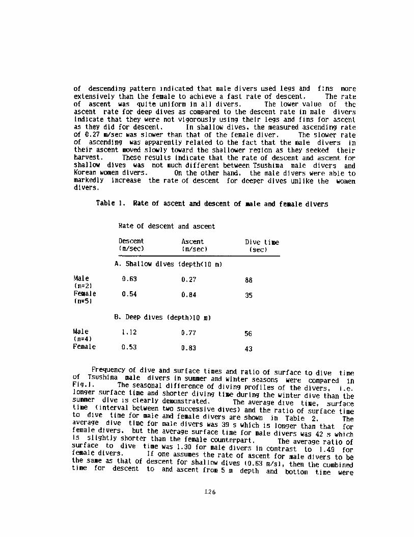
of descending pattern indicated that male divers used legs and fins moreextensively than the female to achieve a fast rate of descent, The rateof ascent was suite uniform in all divers. The lower value of theascent rate for deep dives as compared to the descent rate in male diversindicate that they were not vigorously using their legs and fins for ascentas they did for descent. In shallow dives, the measured ascending rateof 0,27 m/sec was slower than that of the female diver. The slower rateof ascending was apparently related to the fact that the male divers intheir ascent moved slowly toward the shallower region as they seeked theirharvest. These results indicate that the rate of descent and ascent forshallow dives was not much different between Tsushima male divers andKorean women divers. On the other hand, the male divers were able tomarkedly increase the rate of descent for deeper dives unlike the womendivers.
Table l. Rate of ascent and descent of male and female divers
Rate of descent and ascent
Descent m/sec!
Ascent m/sec!
Dive time sec!
A. Shallow dives depth�0 m!
Male n=2!Female n~5!
0.63 0.27 88
0.54 0.84 35
B. Deep dives depth0 m!
Male n=4!Female
i.l2 0.77 56
0.53 0.83 43
Frequency of dive and surface times and ratio of surface to dive timeof Tsushima male divers in summer and winter seasons were compared inFig.i. The seasonal difference of diving profiles of the divers, i.e.longer surface time and shorter diving time during the winter dive than thesummer dive is clearly demonstrated. The average dive time, surfacetime interval between two successive dives! and the ratio of surface timeto dive time for male and female divers are shown in Table 2. Theaverage dive time for male divers was 39 s which is longer than that forfemale divers, but the average surface time for male diver 42 h his sli htls g y shorter than the female counterpart. The average ratio ofsurface to dive time was 1.30 for male divers in contrast to 1.49 forfemale divers. If one assumes the rate of ascent for male divers to bethe same as that of descent for shallow dives �.63 m/s!, then the combinedtime for descent to and ascent from 5 m depth and bottom time were
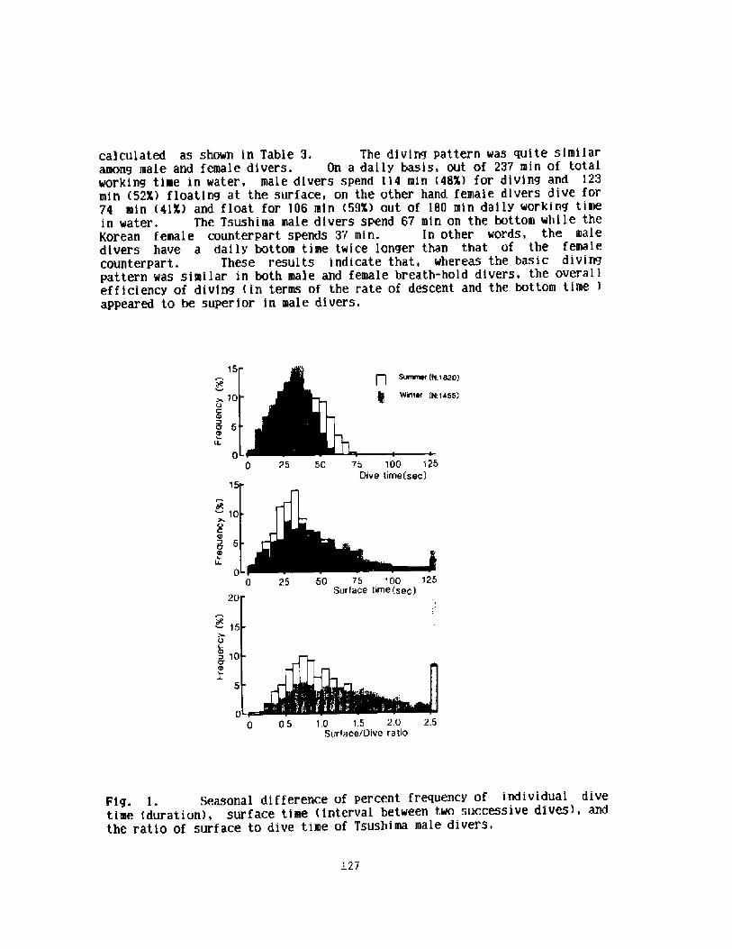
calculated as shown in Table 3. The diving pattern was quite similaramong male and female divers. On a daily basis, out of 237 min of totalworking time in water, make divers spend 114 min �8X! for diving and 123min �2X! fkoating at the surface, on the other hand female divers dive for74 min �1X! and float for 106 min �9X! out of 180 min daily working timein water. The Tsushima male divers spend 67 min on the bottom while theKorean female counterpart spends 37 min. In other words, the maledivers have a daily bottom time twice longer than that of the femalecounterpart. These results indicate that, whereas the basic divingpattern was similar in both make and female breath-hold divers, the overallefficiency of diving in terms of the rate of descent and the bottom time !appeared to be superior in make divers.
15
~ 10o C
50
:1820!
:1455!
0 25 50 75 100 125Dive time sec!
O C
8
p 25 5p 75 100 125
20Surface time sec!
ig15
V Ca 10
00 05 10 15 20 25
Surface/Dive ratio
Fig. i. Seasonal difference of percent frequency of individual divetime duration!, surface time interval between two successive dives!, andthe ratio of surface to dive time of Tsushima male divers,
127
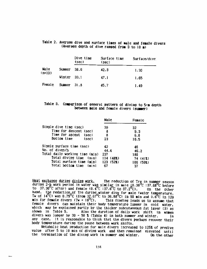
Table 2. Average dive and surface times of male and female divers Average depth of dive ranged from 3 to 10 m!
Surface time Surface/dive sec!
Dive time sec!
Male n=10!
Summer 38.6 42.3 1.30
Winter 33.1
Female Summer 31.8
47.1 1,65
45,7 1.49
Table 3. Comparison of general pattern of diving to 5-m depthbetween male and female divers suer!
Mal e Female
Single dive time sec!Time for descent sec!Time for ascent sec!Bottom time sec!
32 9.36.0
16,5
3988
23
Single surface time sec!No. of dives/hTotal daily working time min!
Total diving time min!Total surface time min!Total bottom time min!
4244.4
237114 �8X!123 �2X!67
4646.2
18074 �!X!
106 �9X!37
128
ggy$ ~g+Dgg dgI jgg Qlyjog ggpjb The reduction of Trs in summsr seasonduring 2-! work period in water wag similg in male $0.38 C :37.68 C beforeto 37.30 C after ! and female �.4 C :37.6 C to 37.2 C!. On the otherhand, t!e reduction of Tre during winter dlvg for male water temperature,Tw of 14 C! was 0.?9 C from 3$.67 C to 36.88 C! in 90 min and 0.6 C in 120min for female divers Tw = 10 C!. This finding leads us to assume thatfemale divers can maintain their body temperature longer in cold water,which may be explained partly by the thicker subcutaneous fat layer �! asshown in Table 5. Also the duration of daily work shift in womendivers was longer by 30 - 5D X Table 4! in both summer and winter. lnany case, it is reasonable to think that the divers perhaps recover theirbody temperature during the pause between work shifts.
Metabolic heat production for male divers increased to 125'X of predivevalue after 5 to 10 min of diving work and then remained eievated untilthe termination of the diving work in summer and winter. On the other
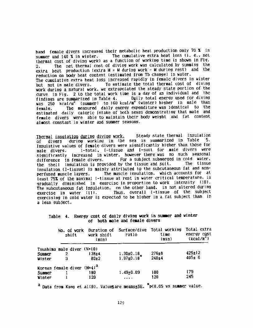
Tbgrmal j~LIjyfjgII ~ring QjvjIIg work. Steady state thermal insulationof divers during working in the sea is summarized in Table 5.Insulative values of female divers were significantly higher than those formale divers. I-total, I-tissue and I-suit for male divers weresignificantly increased in winter, however there was no such seasonaldifference in female divers. For a subject submerged in cold water,the shell insulation is provided by the tissue and suit. The tissueinsulation I-tissue! is mainly attributed to the subcutaneous fat and non-perfused muscle layers. The muscle insulation, which accounts for atleast 75X of the maximal I-tissue at rest in water critical temperature, isgradually diminished in exercise in proportion to work intensity �0!.The subcutaneous fat insulation, on the other hand, is not alter ed duringexercise in water �1!. Thus, overall I-tissue of the subjectexercising in cold water is expected to be higher in a fat subject than ina lean subject,
Table 4. Energy cost of daily diving work in suer and winterof both male and female divers
No. of work Duration of Surface/dive Total working Total extrashift work shift ratio time energy cyst
min! min! kcal/m !
Tsushima male diver N=10!Summer 2 138a4Winter 3 8012
425al2405+ 6
1.30a0.18 276181.97a0.18 240a4
Korean female diver N=4!Summer 1 180Winter 1 120
179245
1.49+0.09 180120
Data from Kang et al 9!. ValueSare means+SE. p<0.05 vs summer value.
I2g
hand female divers increased their metabolic heat production only 70 X insummer and 140 X in winter. The cumulative extra heat loss i. e., netthermal cost of diving work! as a function of working time is shown in Fig.2. The net thermal cost of diving work was calculated by summing theextra heat production extra M = M during work - M during rest! and thereduction on body heat content estimated from Tb change! in water.The cumulative extra heat loss increased rapidly in female divers in winterbut not in male divers. To estimate the total thermal cost of divingwork during a natural work, we extrapolated the steady state portion of thecurve in Fig. 2 to the total work time in a day of an individual and thefindings are su! miarized in Table 4. D~ily total energy used for divingwas 250 kcal/m summer! to 160 kcal/m winter! higher in male thanfemale. The measured daily energy expenditure was identical to theestimated daily caloric intake of both sexes demonstrating that male andfemale divers were able to maintain their body weight and fat contentalmost constant in winter and summer seasons.
r EBALE0 ---.� � 0BALt'. e wet suits summer!
o wet suits {wrriter!300
E
o 25
o 20
0$
> 150Lt!
e 100I
50
O 00 20 40 60 80 100 120
Time in water min!
Fig. 2. Cumulative extra heat loss during the diving work in male andfemale divers. Points and vertical bars represent mean USE. Data ofthe females are replotted by using the report of Kang et al. 9!.
Table 5, Seasonal changes in thermal insulation C. ~ .hlkcal!of male and female divers
Summer Minter
I-total I-tissue i-suit body I-totalfat X!
l-tissue I-suit bodyfat X!
Female 0.170 0.071 0.099 25,8 0.193 0.066 0.127 25.4
p<0.05 vs corresponding summer value.data from Kang et al 9!
l30
Male 0.065 0.028 0.037 14.7 0.135 0.053 0.082 15,3
A signif icantky higher I-tissue during winter p<0,05! in male diversindicates a further vasoconstriction in male divers, since the body fatcontent was identicaL in summer and ~inter. No further increase in I-tissue occurred in female divers indicating that they were in a state ofnear maximum vasoconstriction in summer and winter. The insulativevalue of wet suits I-suit! significantly increased in make divers but notin female divers. The difference would be partly attributed to thethicknss of wet suits and the body area protected with the suit refer themethod section!. Thus, the physical insulation external to the body wasobviously increased in ~inter only in make divers. This may not fullyaccount for the increased change in I-suit, because Goldman et al. �2!have reported that the difference of physical insu$atign of 4.76 and 6. 35mm neoprene wet suits was slight �. 128 vs. 0.139 C.m .h/kcal!. Sincethe insulative value of wet suits was reported to decrease as the workintensity in water increased 8!, then the lower value of I-suit in summermale divers may be accounted for by the higher work intensity in water.Also bypass heat flow due to convective ~ater exchange through the wet suit� 3! could account for the reduction in I-total, especially in male diver~who do not wear the hood in summer. The convective heat fLow from theneck portion of the wet suit would be high in water, keading to anunderestimation of the I-total. Another important reason for the changein I-suit is a change in diving pattern between summer and ~inter in maledivers. Since the diver stayed Longer on the water surface in winter,as indicated by the increased surface/dive time ratio, and a foam neoprenesuit is compressed less, the physical insulation of the wet suit should behigher in winter.ln the laboratory, we conducted an experiment to test whether or not I-tissue increases in high pressure in ~ater and I-suit decreases inproportion to the depth of the water. A clear increase in I-tissue andreduction in I-suit is illustrated in Fig, 3. To test this phenomenonin actual diving in the field, we conducted an experiment to measure theinsulative values on two Korean ~omen divers by fixing the surface/divetime ratio to 1 and 2 and the results are summariz;ed in Table 6. Thedata clearly suggest that when the surface/dive time ratio was incr easedfrom 1 to 2, the insulative values of tissues and wet suits increasedconsiderablly. This finding indicates that divers may maintain theirbody temperature with less increase in metabolic heat production.We therefore conclude that professional male divers of Tsushima Islandincreased I-suit in the cold season by behavioral adjustment of theirdiving pattern, whereas such behavioral adaptation was not observed inKorean female divers.
131
Subject KS
! tissue
2Pressure ATA!
Fig. 3. Relationship between insulative values and water pressures.I tissue and I suit r epresent the average insulative value of the bodesurface and wet suit during the stead> state of water immersion up to theneck, respectivelv, i neoprene is the insulative value of neoprene usedfor the wet suit �mm!.
Table 6. Effect of diving pattern on changes in insulative valuesof diving women
kcal/m /h!
Resting air! ¹i 5i.3«2 56.0
Dive S/D=l! ¹1 86.5 0.2i0 0.067«2 97.8 0.164 0.056 0.143 32.3 32.5
0.108 2?.2 27.4
G.i?6 37.7 75.70.130 31.5 62.5
Dive S/D=2! «i 56,8 0.245 0.070«2 70.9 0.2l4 0.080
l32
~ 0.~0E
o 0.15
~ 0.10
0.05
0C
l-total l-tissue i-suit Dive Surface
C/kcal/m /h!0 2
sec!
1.
2.
3.
4.
6.
7.
8.
9.
10.
11.
12.
13.
References
Hong, S.K., H. Rahn, D. H, Kang, S. H. Song, and B. S. Kang. Divingpattern, lung volumes, and a.lveolar gas of the Korean diving woman ama!. J. Appl. Physiol. 18: 457-465, 1963.Hong, S. K. and H. Rahn. The diving women of Korea and Japan. Sci. Am.216'34-43, 1967.Hong, S. K. Pattern of cold adaptation in women divers of Korea ama!.Fed. Proc. 32: 1614-1622, 1973.Park, Y. S., H. Rahn, I. S. Lee, et al. Patterns of wet suit diving inKorean women breath-hold divers. Undersea Biomed. Res. 10: 203-215,1983.Shiraki, K., N. Konda, S. Sagawa, et al. Diving pattern of Tsushimamale breath-hold divers Katsugi!. Undersea Biomed. Res. 12, 1985 inpress !.Hardy, J. D., and E. F. DuBois, The technique of measuring radiationand convection. J. Nutr. 15:461-475, 1938.Ramanathan, N. L. A new weighting system for mean surface temperature ofthe human body. J. Appl. Physiol, 19:531-533, 1964.Rennie, D. W., B. G. Covino, B. J. Howell, et al. Physical insulation ofKorean diving women. J. Appl. Physiol. 17:961-966, 1966.Kang, S. H., Y. S, Park, Y. D. Park, et al. Energetics of wet-suitdiving in Korean women breath-hold divers. J. Appl, Physiol.:Respirat.Environ. Exercise Physiol. 54:1702-1707, 1983.Rennie, D. W., Y. S. Park, A. Veicsteinas, and D. Pendergast. Metabolicand circulatory adaptation to cold ~ater stress, In: ExerciseBioenergetics and Gas Exchange, edited by P. Cerretelli and B. Whipp.Amsterdam, Netherland: Elsevier/North-Holland, 1980, p. 315-321.Veicsternas, A., G, Ferretti, and D. W. Rennie. Superficial shellinsulation in resting and exercising men in cold ~ater. J. Appl.Physiol.: Respirat. Environ. Exercise Physiol. 52-'1557-1564, 1982.Goldman. R. F., J, R. Breckenridge, E. Reeves, and E. L. Beckman. "Wet"versus "dry" suit approaches to water immersion protective clothing.Aerospace Med. 37:485-487, 1966,Wolff, A. H., S. R. K. Coleshaw, C. G. Newsted, and W. R. Keatinge.Heat exchange in wet suit. J. Appl. Physiol. 58:770-777, 1985.
133
Discussion folloving Dr. Shiraki's presentation
Dr. Craig: If the male has a longer diving time, does the maleproduce more or make more money than the female?
Dr. Shiraki: Well, the situation in Korea and in Japan is quitedifferent, but I can say that the male divers make two or three time+more.
Dr. Hong: You said that the breath-hold divers dive deeper duringwinter< but you did not give the reason why they dive deeper inwinter than in summer. Are there economic reasons for that?
Dr. Shiraki: During the summertime they pick up abalones while duringthe wintertime they pick up sea urchins, and also sea cucumbers, whichlive deeper than the abalones.
Dr. Slsner: I would like to ask a question about the Funado divers<particularly about the two men in the boat, is it possible that theman who gives her the weight is her husband, and the fellow who pullaher up is her boyfriend?
Dr Lundgrea: A question that is related to some earlier discussionawe' ve had. The information that I'm looking for may be in your dataFor those, I think it was the women, who increased their metabolismduring the winter, does that influence breath-hold diving time?
Dr. Hong: If you take the grand average of nearly two thousand dives,single dive times decreased during winter.
Dr. Lundgren: Is it reasonable to relate that to an increase inmetabolism? The male divers did not increase their metabolism duringthe winter< right? Does their winter dive time also change< or does itstay the same?
Dr. Shirmki: The dive time in the winter is shortened.
Dr. Lia: Dr. Park and Dr. Hong's study indicated that insulation isinsuf f icient in Korean women in the winter, because the bodytemperature fell noticeably, but in the summer< the suit providessuf f icient, insulation.
134
Energetics of Breath-Hold Diving
Dav id R. Pendergast, Department of Physiology, SUNYAB, Buf falo, NY 14214
Abstract
The 02 consumed and C02 produced during a breath-hold dive is important
in determining the time of a breath-hold and the depth of the dive. A typical
dive consists of a combination of descent, swimming at the bottom and ascent.
The energy cost K! of movement in water is directly propcrtional to the
external work W! and inversely proportional to the net mechanical efficiency
e!, E ~ W/e. The W is proportional to the total resistance to movement D!
and the distance moved d!. The D during descent and ascent is a combination
of the drag of the body Db! and the difference between weight in water and
lung volume DB. During bottom swimming, D would be equal to Db and would be
described by the equation, Db = KV , with the constant K reflecting primarily
the physical characteristics of the diver and the exponent n the hydrodynamic
characteristics. The n can vary from -1.2 to 1.85 and is dependent on depth
and speed and K can vary from -3 to 9 and is dependent upon equipment,
swimming style, body composition and to a degree, speed. The DB can vary from
+6 to -6 Kg and is dependent on the weight in water, lung volume, and depth of
the diver. Due to the differences in body composition and lung volume, the D
for men and women is different. The D can be altered by the use of weights
during descent and ascent. The value of e can vary from -2 to 1 5X. The
actual value is dependent on the speed, equipment used and swimming style.
The energy supplied would be 50 and 70X aerobic for a .5 and 2 min dive,
respectively, with the balance of the energy being supplied by, primarily,
high energy phosphates in the shorter dives and glycolysis leading to lactic
acid production in the longer dives. As long as there is a 3 min recovery
between shorter dives, minimal net lactic acid build up is expected. Diving
in cold water would increase the E by about 10X due primarily to a decreased e
and body cooling of 1-2oC would result in an additional l0% increase in K.
135
Introduction
Breath-hold div ing can take many forms. These could range from
repetitive diving to 5-20 m to do a job, to diving deeper than 70 m for the
purpose of setting a breath-hold diving record. Although these activities are
quite different, they have many similarities. Most specifically, the time of
the breath-hold is determined to a great extent by the total lung volume prior
to the dive and the rate of oxygen consuraption and CO2 production. Other
presentations in this workshop and previous papers have been devoted to the
role of lung volume �,7,8,14! and CO2 production 8,13!. The purpose of this
presentation is to discuss the factors that influence the rate of consumption
of oxygen during a dive. The VO2 is important not only because it determines
the rate of reduction of alveolar PO2, but also because it influences the PCO2
which is critical in setting diving time.
Tn order to analyze breath-hold diving it must be divided into descent,
swimming at the bottom, and the ascent. This is important because the factors
that determine energy cost of the three parts of a breath-hold dive may be
quite different. The total energy cost E! of a dive is determined by the
external work W! and net mechanical efficiency e!, see equation 1:
E ~ W
e
The W is a product of the water resistance, termed drag D!, and the distance
of progression through the water d! equation 2!:
�!
Combining equations �! and �! we can derive the fol lowing equation �!:
�!E D ' d
e
This equation can be rearranged further to show that the E to swim a given d
is equal to the ratio D to e equation 4!:
E ~ D
d e
136
The above equations can be analyzed quantitatively if one assumes that I I 02
5 Keel and 1 Kca1 ~ 427 Kg m. The ratio of E/d or D/e can be termed
economy. It is important to note that both D and e are independently variab].e
and can be inf 1uenced by the velocity of swimming V!, body composition and
size, sex, swimming style, water depth and water temperature. In order to
understand the energetics of breath-hold diving, the factors that influence
both D and e wi I I be analyzed.
Previous investigators have measured or estimated the oxygen cost of
repetitive diving in the f ield at periodic intervals to estimate the overal I
energy balance �!, while other investigators studied Navy divers, in a
protected environment, making single dives with passive descents and active
ascents �3!. In another study �4!, the authors estimated the cost of diving
from the force-velocity relationship during descent and ascent and
measurements of water resistance. This analysis made many assumptions, most
importantly that e was 3% and was independent of speed. This latter
assumption we know is not correct �,5,9,10,11!.
The best measurements of energy cost of breath-hold diving have been made
by Craig �!. He measured the VO2 and VC02 during and after the dive, for
varying number of repetitive dives, both assisted and unassisted. The dives
ranged from 30 to 60 s in duration and they were to depths of 5-10 m. In
general he found that a 5 m dive required .8 I OZ and a 10 m dive 1.2 I 02 '
In addition to energy derived from V02, lactic acid built up in the blood
after the first dive and decreased thereafter. These data imply that at least.
part of the E for the dive came from glycolysis leading to lactic acid
production anaerobic metabolism! and, in addition, from high energy phosphate
depletion aerobic metabolism! as indicated by the large V02 during recovery
�!. The data from the above studies can be combined and it would appear that
the E of breath-hold diving ranges from .8 to 1.4 1 02 for dives to depths
ranging from 5 to 27 m and lasting between 30 and 90 s. The variability of
137
diving styles and profiles make a quantitative analysis of the data from these
studies impossible. Furthermore, no attempt was made to extrapolate the cost
of these relatively shal low dives to deeper dives. To this end, I will
present data from selected studies concerning factors that could influence E
and, therfore, diving depth.
To determine the E of diving, both D and e have to be considered.
Lnasmuchas they are both variable and influenced by different factors each
wil 1 be considered separately.
The drag that must be overcome by a diver is the total of the water
resistance per se �,3! and the net buoyancy of the body ll!. For example,
to swim at a given speed at the surface, women, who are more buoyant than men,
require about 1/2 of the E �,9!. Specifically in the case of diving, the
lung volume acts as a buoyant force that must be overcome by propulsion and,
therefore, is added to the propulsive force needed to overcome water
resistance. At the beginning of a dive, the buoyant force is large
equivalent to total lung volume 4-8 Xg! and as the diver goes deeper, the
volume decreases as does the buoyant force. Ironical ly, as the lung volume is
increased, to increase breath-hold diving time, the buoyant force increases.
During descent, the propu lsive force can be maintained at a constant level
and, as the depth of the dive increases, the diver s speed would increase.
Once on the bottom, the total lung volume is not. critical; however, the
density diver plus equipment! is important, as the attitude of the diver
affects the drag 9,10,11!. During ascent, the initial period requires the
diver to overcome the water resistance plus the effect of the body s sinking
force, as the lung volume and buoyant force are low. As the diver approaches
the surface, the lung volume increases and the buoyant force assists the
138
ascent.
The drag resulting from propulsion in water increases exponential ly as a
function of velocity in water see equation 5; 1,3,4,5,6,9,10,11,12!, with the
rate constant K ref lecting the characteristics of the diver and the exponent n
the hydrodynamic characteristics:
�!D Kvn
8oth K and n are variable and are related to the drag coefficient CD!, the
density of the water Pg! and the subject s ef feet ive cross sectional area A.
The latter variable is proportional to the subject s mass and inversely
proportional to the subject s body density pN! and height L! ~
Theoretically, the exponent in equation 5 should range from 1 at low speeds,
to 2 at moderate speeds and as high as 4 at high speeds. In fact, the n
ranges from -1.2 to 1.9 with most conditions being around 1.8-1.85, while the
K can vary from 3 to 9. From equation 5 an equation for the coef ficient of
drag CD see equation 6! can be derived and can be used to examine the factors
that affect the K in equation 5:
~x ~ gCD ~ 2K -- � --- ~ L
pw�!
L39
where g is the acceleration due to gravity. Studies in our laboratory have
shown that the CD is about 20K less in women than in men. Furthermore, at the
surface, the CD is 20X greater than it is at 2 and 20K greater at 2 than at
4 . These changes can be directly translated into changes in E, and,
therefore, the depth of water or in fact swimming within 2 of the surface
will affect the E. Another important aspect of D is that, due to the
exponentia 1 increase in D with V, E increases exponential ly with V. As a
further point, the increased E at high speed is not recovered during, a "glide"
due to the high water resistance. This suggests that selecting the correct
swinasing speed is critical for breath-hold diving.
The drag discussed in the paragraph above refers to D without subject
movement. As soon as the arms or legs are moved, the D is increased
�,5,9,10,11!. The depth of kick of the legs, when combined with the kicking
frequency, influences dramatically the D. In studies in our laboratory, D,
during svimming with f ins, was relatively high at low speeds -7 Kg!,
decreased at moderate speeds -5 Kg! and increased again at high speeds -10
Kg!. These changes in D vere a result of the changes in the depth of kicking
at the three speeds. Paradoxical ly, at low and moderate speeds the economy
E/d! of swimming was similar as changes in e offset the changes in D. At
speeds greater than .9 m/s, the increase in e vas less than the increase in D,
and E/d increased dramatical ly. It should be noted that technical skil 1 of
swisssing can affect both D and e �,9,10,11! and swimming with fins can change
E/d by about +20X. Fins alone in surface swimming tend to buoy the legs and
decrease the E/d; however, in a vertical descent, this effect could result in
a increased E.
The net result of these factors on D during breath-hold diving is
obviously very complicated. Being assisted during descent and ascent can
lover the drag, especial ly considering the drag created by swimming. The D is
al so inf luenced by buoyant forces and vater depth. If a subject produces a
propulsive force of 2 Kg during descent, his speed would range from .5 m/s at
5 m to 1.5 m/s at 30 m, vhile, during ascent, even if the propulsive force
vere 4.5 Kg at 30 m, the speed vould be .4 m/s and would only increase to .65
m/s at 5 m with an additional 2 Kg propulsive force.
Efficiency
As described above, the net mechanical efficiency is equal to the ratio
of E to W. Svimming efficiency can vary from 2 to 15X �,3,4,5,6,9,10,11!.
The variability depends, to a great extent, on the technical ability of the
swimmer. Women have a two times better efficiency than men due to their
140
g,rester buoyancy �,9!. In addit ion, f in swimming has a higher e than
swimming without f ins. Most importantly, e increases linearly with speed for
most types of swimming; however, in f in swimming, e is high at low speeds
-8%!, decreases at moderate speeds -4X! and increases again at high speeds
-10X!. As discussed above, these changes are paral leled by changes in D.
The e appears to be related primarily to kicking frequency.
Diving
Combining the data from above we can calculate the E for dives to
different depths, Assuming that the 02 available at the maximum is .21 times
the total lung capacity and knowing E/d, we can calculate the maximal depth of
a dive when swimming without f ins, with f ins and swimming with minimal drag
and maximal efficiency minimal E/d!. Under these circumstances the total
available 1 02 would be between .75 and 1.5 1 02 and be dependent on total
lung, capacity �-7 I!. The maximal diving depth wou1.d range from 5 to 10 m
without fins, from 10 to 20 m with fins and from 15 to 30 m in the case of
maximal efficiency. It is obvious from this discussion that, for dives to
depths greater than 30 m, the diver must be assisted both during ascent and
descent.
Effect of Water Temperature
Most breath-hold diving is performed in water colder than thermoneutral.
Even if there is no shivering, exposure to water below -34oC results in
metabolic alterations to swimming. Investigators have noticed �0-12! that
the E/d is increased during exposure to cold water, even though the exposure
i.s not long enough to decrease core temperature. This increase amounts to 10-
20X additional E at any speed. If the exposure is long enough to cause a
decrease in core temperature, the E is increased an additional 10 to 20X at
speeds. The increase in E is primarily the result of decreased e, as
141
there is either increased metabolic activity in muscles antagonistic to
awisssing or the resistance of the muscles themselves increases when they are
cooled.
8usmary
The energy cost of breath-hold diving has been measured to be between .8
and 1.4 1 02 for dives to depths ranging from 5 to 27 m. Although the minimal
cost .8 1 Og! appears not to be too variable, as the depth increases, there
is a large variabil ity in energy cost. Many factors can influence this cost
as described above. It is clear that more experimental data is needed to
quantify the exact cost of diving to various depths, with special
consideration to water temperature. At any rate, under optimal conditions,
unassisted diving would appear to be limited to -30 m. More investigation is
needed to prov ide the data necessary to calculate the maximal diving depth
during an assisted dive.
l42
References
l. Alley, LE. An analysis of water resistance and propulsion in swimming
the crawl stroke. Res. Quart. 23:253-270, 1952.
2. Craig, A.B., Jr. and W-L. Medd. Oxygen consumption and carbon dioxide
production during breath-hold diving. J. Appl. Physiol. 24:190-202,
1 968.
3, Rarpovich, P.V ~ Water resistance in swimming. Res. Quart. 4:21-28,
1 933,
4. di Prampero, PE., DR. Pendergast, DW. Wilson, and DW. Rennie.
Energetics of swimming in man. J. Appl. Physiol. 37;1-5, 1974.
5, Gof f, L.G., E.F. Brubach, and H. Specht. Measurement of respiratory
responses and work efficiency of under water swimmers util ising improved
instrumentat ion. J. App 1, Physio I. 10:197-202, 1957.
6. Holmer, I. Oxygen uptake during swimming in man. J. App1. Physio 1 ~
33:502-509, 1972.
7. Kang, D.H., P.K, Rim, B.S. Kang, S.H. Hong, and S.K. Hong. Energy
metabolism and body temperature of the ama. J. Appl. Physiol. 20:46-50,
1 965.
8. Lanphier, E.H. and H. Rahu. Alveolar gas exchange during breath-hold
d iv ing. J. App 1. Phys io l. 18:471-477, 1 963,
9. Pendergast, D.R., P.E. di Prampero, A.B. Craig, Jr., D.W. Wil son and D.W.
Rennie. A quantitative analysis of the front crawl in men and women. J.
App1. Physio l. 43:475 � 479, 1977.
10- Pendergast, D.R., and D.W. Rennie, The energy cost of swimming with
SCUBA. Sixth Symposium on Underwater Physiology. Abstract! 1975.
Pendergast, D.R., P.E. di Prampero, A.B. Craig, Jr. and D.W. Rennie. The
influence of selected biomechanical factors on the energy cost of
swimming. In: Swimming Medicine IV. B. Eriksson and B. Furberg, eds.
University Park Press, 1978, pp. 367-378.
l43
12. Push, L.C.G. and O.G. Edho 1m. The physio logy of channel swimmers.
Lancet 2:761-768, 1955.
13. Schaefer, K.E. The role of carbon dioxide in the physiology of human
diving. In: Proc. Underwater Physiology Symposium. L.G. Gof f, ed.
Washington, D.C.: National Academy of Sciences - National Research
Couuc i 1, Pub l. 37 7, 1955, pp. 131-139.
14. Yokoyama, T. and S. Iwa ski. Energy cost of d iv ing ama. In' .The
Physiology of Breath-Hald Diving and the Ama of Japan. H. Rahu, ed.
Washington, D.C.: National Academy of Sciences � National Research
Counc il, 1965, pp. 349-365.
144
Discussion following Dr. Pendergast's presentation
Dr. Boysen: In the study you did there were individuals of differenttechnical skills, from the novice all the way up to experts andprofessionals. Was that a breath-hold swim?
Dr. Pendergast: No, those were SCUBA swims.
Dr. Boysen: Then my question is: there is a big difference betweenthe expert and the professional. The two groups would be expected tobe closer together. You alluded to the fact that you didn't know thedistance they went. I don't know whether they were swimmingaerobically or anaerobically, but you said you thought the bigdifference in their performance was technique. To further elucidatethat, do you know anything else about their degree of fitness' Inother words, their oxygen consumption, their maximum oxygenconsumption, in and especially out of water; were they the same ordi f f er ent'?
Dr. Pendergast: The swims that I showed you were all sub-maximalaerobic swims and that's why they ended at the velocities that theydid. So these were swims these individuals could keep up for six oreight minutes at each speed. So there was no anaerobic component, andsecondly, if there were a change in fitness, it would not affect theener gy cost per unit distance of svimming. I think this is anextremely important point: f itness does not af feet that, whiletechnique does. Now, we did a similar graphical analysis of thosedivers and the big difference in the divers was the stroke frequencyand the depth of the kick and so one can account for the changes inwhole cost based on those two variables.
Dr. Bong: Let us get down to the nitty gritty. Are the valuescalculated by Yokohama in agreement with your data'? What is thebottom line' ?
Dr. Pendergast: Well, I think that Yokohama's estimates were basedon Dr. Craig's data and Dr. Craig actually made the measurements. Atthe two depths the V02 that they calculated or measured agrees withour data. They were right for the wrong reason, and that is that thedrag and efficiency that they used vere too low. The bottom line is,as the energy cost is the ratio of drag and efficiency, it turned outto be the same energy cost.
Dr. hrnold: Dr. Pendergast, at the end of your abstract you mentionedthat. a three minute recovery period between your one minute or sodives was sufficient to take care of the lactic acid build-up.
Dr. Pendergast: Yes, as repeated diving continues, that lactic acidis burned as fuel and no net accumulation ensues, as shown by Craig.Three minutes are sufficient to resynthesize the high energyphosphates.
Dr. Arnold: In l979, I did a study with Dr. Elsner where we comparedfacial immersion breath � holds of 30 second duration to determinewhether the period of time between two breath-holds affected the
14'
amount of bradycardia elicited by the second. We found that a shortperiod of time between the breath-holds did alter the bradycardia inthe second and, in fact, there was more bradycardia or faster onset ofbradycardia in the second breath-hold than in the first. We assumedthat that might very well have had something to do with either bloodgas or lactic acid build-up and that a recovery period of at least twominutes should separate that.
Dr. Pendergast: Ny comments are based on two things - one is thatit's well known now that lactic acid can be burned as a substrate forsubsequent exercises. In your experiments, you were resting whichmakes a considerable difference in the lactate wash-out. We haverecently done a study that shows that if you do a set amount of supermaximal exercise, if you start with resting or some elevated lactate,at the end of that exercise, you wind up with the same lactate. Nowwhat this means is that the balance of the lactate was burned in thatparticulaL exercise if it's an aerobic exercise. So the lactate canin fact be burned off. The other answer would be that, if in fact,there is lactate build-up, which there probably is for other reasons,then in fact the lactic acid would serve to drive the heart ratehigher and counteract in part your bradycardia.Dr. Craig: In regard to the lactate, we used very different types ofdiving patterns. 30 second down then 30 second rest, 5 meters diving,l0 meters diving on different schedules and in some of the moredifficult dives we did lactic acid and we never found a significantelevation in lactic acid in repetitive diving.
Dr. Lnndgren: I don't know if it's a question or a comment, but, Dr.Pendergast, you made the point that swimming harder will allow energyproduction by glycolysis and that would not put a penalty on the diverin terms of C02 production and oxygen consumption, which is kind ofobvious, but I suppose you' re not proposing that you would thereby beable to dive longer � well longer perhaps in distance, is that whatyou imply7 What is perhaps obvious to everybody is that lactacidemia,for one, is a respiratory stimulus itself and it could also increasePC02 by reducing the solubility of any C02 present.
Dr. Pendergast: I guess I would suggest this somewhat tongue-in-cheekbecause I don't think you can predict whether the increase in drive toventilation or the small reduction in P02 would offset the tremendoussavings that you could have in oxygen consumption as far as the costof the swimming is concerned. The other important thing is that thecost per unit distance, over the range of speeds that you can dobreath-hold diving, is constant, and therefore one conceivably, andI'm saying this to an extent tongue-in-cheek, could cover a muchgreater distance under that situation than under the other.
Dr. Van Liev: Is it possible to say that there is a given amount ofoxygen saved, due to the lactic and alactic debts7 That wouldindependent of speed.
Dr. Pendergast: Yes, there is. The alactic, so called, deficit issomewhere between one and two liters of oxygen total, so in the costof a dive you can save that much energy immediately with really no net
146
consequence, as long as there is enough time in between the dives toresynthesize the high energy phosphates. If the time between thedives is too short, then you would have embarrassed your high energyphosphates and you'd begin to produce lactic acid which then perhapswould accumulate over time. The lactic acid component would beequivalent to three milliliters of oxygen per millimole per kilogrambody weight and this can account f or maximally probably about f ourliters, but it would take about two to two and a half minutes to buildthat total deficit and so totally it's estimated that the total amountof oxygen equivalent available from anaerobic sources would besomewhere between four and six liters of oxygen.
147
THE DIVING RESPONSE: A COMPARISON OF ANIMALS AND MAN
Rober t Elsner, Institute of Marine Science, University of Alaska,Fair banks, Alaska 99775-1 080.
I think that it is fair to say that we agr'ee with respect to theexistence of a set of' r eflexes, collectively described as the "divingresponse", in certain marine species of bir ds and mammals duringsubmer gence. The response is an identifiable physiological condition whichoccur s in dives, especially those of long duration, and results in anadaptation of the animal to the cessation of external respiration thuspr oduced. In many species the success of' the r esponse is dependent upon�! enhanced oxygen stor age by large blood volume and high levels ofhemoglobin and myoglobin, �! tol.er ance of elevated concentrations ofmetabolic products, such as CO2, H+ and lactate and �! cerebral andcardiac resistance to hypoxemia. Adaptations which permit seals, ducks andother aquatic diving animals to tolerate long submer sions r esemble generalvertebr ate reactions to asphyxia. Neural. regulatory mechanisms are alsosimilar . Species vary in the quant,itative expression of the responses andin the intensity with which they are r egulated. Apnea., bradycardia andredistribution of car diac output favoring brain and heart are precipitatedin var ying degrees by aquat ic immersion, The display of these eventsdepends upon dive dur ation and its emotional content . In this pr esentationI wish to review some of the evidence r elating to diving r esponses inhumans and animals. Adaptations of marine mammals to high pressure havebeen reviewed by Scholander �940! and Denison and Kooyman �973!,
In his classical work of 1940, Scholander pointed out that a sealf'reely submerging in water sometimes f'ailed to show diving bradycardia,However, seals reacted to startle and loud noise with sudden heart r ateslowing. Experimental diving studies in which animals were trained todive upon a signal resulted in variable responses in the sea lion, harborseal and dolphin Elsner, 1965; Elsner et al. 1966; Elsner, Kenney &Burgess, 1966!, There was usually less decline in hear t rate duringtrained immer sion than during for ced immersion sea lion and harbor seal!,but in dolphins the bradycar dia was intensified dur ing trained dives.Kooyman et al. �980! descr ibed dives of up to 20 min in Weddell seals which ar e capable of' maximum dives exceeding 60 min! in which little orno dependence upon the anaerobic resources r equired for longer dives wasmade, although car diovascular changes, signalled by bradycardia,presumably occurred Kooyman & Campbell, 1972!. Oxygen reserves wereapparently sufficient to allow the dives to be made aer obically. If, asseems 1 ikely, these reserves ar e principally contained within the bloodand oxygenated myoglobin of skeletal muscle, then the reactions of thevoluntarily diving Weddell seals resembled those of restrained divingharbor seals in a previous study in which myoglobin oxygen was rapidlydepleted before lactate production commenced in about 10 min, earlier in
148
seals which struggled Scholander et al., 19i12!. One is str uck by thesimilarity of the responses in the two examples, rather than bydiff'erences, Authors of both studies remarked upon the observation thatthe preponderance of free dives in natur e are brief and aerobic,indicating that anaerobic responses, which are more exhausting and requir elonger r ecovery, ar e seldom brought into oper ation.
Butler h Woakes �979! and Kanwisher, Gabr ielsen & Kanwisher �981!showed that freely diving birds make many short dives without changes inhear t rate. Clear ly, most of the dives performed by aquatic animals innature are of shor t duration, and they apparently depend largely uponoxygen-sustained rather than anaerobic mechanisms. Ducks and seals
doubtless f ind it uncomfortable and exhausting to push their diving habit.to the anaerobic extreme of their reserve capabilities. Although thatphysiological reserve is rarely invoked, it is, nevertheless, a resourceupon which survival may depend. Fr om these considerations there emergesthe concept of graded responses to diving, var ying in intensity dependingupon severity of the imposed stress and of the degree to which control isexercised by higher cortical functions.
The under standing of diving biology is enhanced by examining thenatural history of diving species, It is equally important, that we studythe full range of physiological r'esponses of which these animals ar ecapable by submitting them to experimental dives. Controlled laboratoryinvestigations require either the use of trained animals or, moredrastically, the interver .ions of restraint and anaesthesia. Theseapproaches have all been usefully employed in various studies . A maj orresponse to most diving situations, natural and experimental, is theselective redistribution of the circulation. Inasmuch as many of thetissues of the body can toler ate oxygen deprivation far longer than canthe more vital ones, the hear t and the brain, the available blood oxygencould be conserved by its preferential distribut ion to those vital organsfor their Immediate needs while the remaining or gans, depr ived ofcirculation, survive on anaerobic metabolic processes or, in the case ofskeletal muscle, on oxygen bound to myoglobin. Moder n under standing ofthese diving responses rests on the foundation of classical r esearchreports by Irving and Scholander. Some aspects of these adaptivemechanisms exist in many species which ar e not habitual divers when theyare exposed to asphyxia review: Elsner 5 Gooden, 1983! .
Hypometabolism, a strategic retr eat
Restr icting the circulation of blood to a tissue or an organ r esultsin a steady decline of the oxygen available for support of oxidativemetabolic processes and subsequent dependence upon whatever anaer obic
149
resources are available. Eventually, a depr ession of metabolism takesplace. This lower ing of the rate at which the many complex chemicalprocesses can occur, thus conserving metabolic energy, is, in fact, acentral feature of the adaptations to asphyxia. Viewed in this context,the diving response r epr esents but one specialized example of a widespreadand general response of many living animals, invertebrate as well asvertebrate, to life-threatening situations. There are abundant examplesin nature, Marine intertidal inver tebr ates at low tide, fish out of water,hibernators and estivators come to mind. Some possible human examples willbe examined.
Recognition that such a wide variety of species, from invertebratesto mammals, show the ability to make strategic retreats from environmentalthreats into states of metabolic depression suggests the fundamental andgeneral nature of this phenomenon. Cardiovascular adaptations leading to aredistribution of the blood flow that favors the more vital structures,thus accomplishing an over all sparing of metabolic energy, play a majorrole in these examples. Homeostasis is modified but not abandonded.Continued regulation, but with an extension of its r ange to includeadaptive metabolic conservation, is the central theme upon which thesevar iations are played.
The occurrence of reduced metabolic rates during experimental divingasphyxia is supported by several lines of evidence. Scholander �940! wasthe first to demonstrate this reaction when he found that the extr a oxygenconsumption of seals during r ecovery from quiet dives could not accountfor the amount predicted if metabolism during the dive was maintained atthe pre-dive level. Body temperatur es decreased in diving seals along withthe decrease in metabolism Scholander, Irving 5 Grinnell, 1942; Hammel et.al., 1977; Kooyman et al,, 1980! . Therefor e, they could be expected torepay only part of the accumulated oxygen deficit during recovery from thehypoxic diving episode. Experimental evidence indicates that the cardiacoxygen consumption in diving seals declined to 7 � 14$ of the non-divingvalue Kjekshus et al., 1982!. In dogs trained to dive the decrease was42'$ Gooden et al., 1974! . In both examples oxygen consumption roughlyfolio~ed the decline in heart rate.
Human dives
Human experiments in which diving br adycardia was recorded, evenduring underwater swimming, f irst appear ed in 1940. These were mentionedbriefly in a paper descr ibing the respirator y metabolism of the porpoise Ir ving, Scholander Ir Grinnell, 1941!, but wer e not, fully r eported until1963 when they became of historical importance Irving, 1963! . Bradycardiahas been obser ved in human subjects during actual and simulated diving.
In one particularly striking case Klsner et al. �966! observed severalpulse intervals equivalent to l3 beats/min in a subject performing a 30-sf'ace immersion. This subject began the procedure with a hear t rate of 90beat,s/min. The diving r esponse was evoked in similar experiments by simpleface immersion with simultaneous breath-holding and it, was found that thisprocedure was as effective in producing diving bradycardia as total bodyimmersion. The subjects for this study were nine young men of varyingdiving exper ience. They lay prone and performed face immer sion withbreath-holding in a bowl of water, temper atur e 18 to 20 degr ees C, for lmin. Calf blood flow was measur ed by venous occlusion plethysmogr aphyusing a mercur y-in-rubber str ain gauge. The simulated dives wereperformed after moderate inspiration or expiration withouthyper ventilation. The limb bl.ood flow consistently decreased mor e duringface immersion than during breath-holding alone.
The reduction in calf blood flow observed was assumed to be ther esult of vasoconstriction, but proof of this assumption depended upon thesimultaneous measurement of the systemic arterial blood pressure and limbblood flow. Heistad, Abboud l Eckstein �968! measured brachial ar terialblood pressure dir ectly by cannulation as well as f inger and forearm bloodflow by venous occlusion plethysmography during breath-holding with andwithout face immersion, During 30-s of face immersion breath-holdingthere was a progressive rise in mean ar terial blood pressure to an averageof 20% above the control level. Both systolic and diastolic pressureswere elevated. At the same time the finger and forearm blood flowdecreased by an average of' 72 and 501, respectively below the controlvalue. They concluded that vasoconstriction must occur in the forearmvasculature during this form of simulated diving. Breath-holding aloneproduced similar but consistently smaller changes in pr essure and flow.Face immersion in human subjects results usually in a modest increase inar ter ial pressure, and the br adycardia is more severe when the immersiontakes place in cold water Song et al., 1969!. Neither the pressure nortemper ature eff'ects are evident in diving seals.
As in seals and ducks, vascular constriction during diving in man maynot be limited to the ar terioles. Olsen et al. �962! reported frequentdamping of the brachial ar ter ial pressure recording during total immersionof their diver subjects. They suggested that this effect resulted fromthe constricted arter ial wall occluding the tip of the in<raa«-e»alneedle. Elsner, Gooden & Robinson l971! found considerable difficulty inwithdrawing brachial arterial blood sampl.es during face immer sionbreath-holding in a subject who had developed a profound r eduction inforearm blood flow. Additional evidence that vasoconstrict ion occurs inhuman arteries of gr eater than arter iolar size during diving was obtainedby Heistad et al, �968!. During simulated dives, a substantial systolic
151
pressur e gradient developed between the brachial and digital ar teries inthree out of five subjects, suggesting that vasoconstriction must haveoccurred in larger arteries upstr earn from the digital arter y.
Functional significance of the diving response
The question ar ises whether the cardiovascular r esponse to diving inman has any functional signif icance and in par ticular whether the humanresponse has an oxygen-conser ving role. Oxygen conservation could resultin two ways. Fir st, by limiting the blood f'low to peripheral tissues suchas the limbs and gut, the uptake of oxygen by these tissues would bereduCed. The r eduction in the hear t rate can be expected to result in a
mOdeat decreaae in myocardial oxygen consumption, althOugh this r eactionmight be at ! east partially r ever sed if arter ial pr essur e is elevated.When Olsen et al. �962! were studying blood gas tensions in their diversduring total immersion, they noted a sur prisingly high ar ter ial bloodoxygen content at the end of long exercise immer sions. This observationsuggested to them that there was a r educed extraction of oxygen in thetissues during diving in man .
Elsner et al. �971! examined the influence on the diving r esponse ofvarying the arter ial gas tensions of oxygen and carbon dioxide befor e faceimmersion breath-holding. Brachial arterial blood was sampled from onearm, and forearm blood flow was measur ed by venous occlusionplethysmography simultaneously in the other arm� . Five male subJectshyperventilated on oxygen for 15 min and then perforemd face immersionswhich lasted an average of 5 min. The mean ar terial oxygen tension beforeface immersion was 548 mmHg, and after 4 min the tension was stillconsiderably elevated in four of the f ive subjects mean value, 326 mmHg! .A highly significant correlation existed between sequential changes inforearm blood flow and deer eases in arterial oxygen tension Fig. 1!.Thus the greater the reduction in flow, the smaller was the deer ease inoxygen tension. If this relation in the upper limb is r epresentative of amore widespread phenomenon in the lower limbs, gut and elsewher e, then itseems likely that the diving response in man oper ates in somecircumstances to conserve oxygen. In contrast to these results, noreduct,ion in oxygen consumpt ion could be detected during humanbreath-holding face immer sion in the following studies: Raper et al.�967!, Heistad A Wheeler �970! and Hong et al. �971!.
152
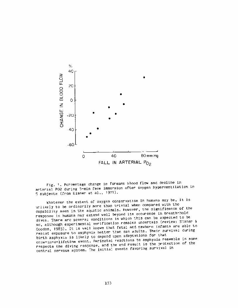
40
0
20
OO
0
LLJ
z -20
V-40
-60
40 80 mm Hg
FALL IN ARTERIAL P02
Fig. 1. Per centage change in forearm blood flow and decline inar terial P02 dur ing 5-min face immersion after oxygen hyperventilation in5 subj ects fr om Elsner et al ., 1971 ! .
Whatever the extent of oxygen conservation in humans may be, it isunlikely to be ordinarily more than tr ivial when compared with thecapability seen in the aquatic animals. However, the significance of theresponse in humans may extend well beyond its occurence in breath-holddives. Ther e are several conditions in which this can be expected to beso, although exper imental verif ication remains uncer tain review: Elsner &Gooden, 1983!. It is well known that fetal and newbor n inf'ants are able tor esist exposur e to asphyxia better than can adults. Their survival duringbir th asphyxia is likely to depend upon adaptations for thatonce-in-a-lifetime event. per inatal reactions to asphyxia resemble in somerespects the diving r esponse, and the end result is the protection of thecentr al nervous system. The initial events favoring sur vival in
153
near-drowning accidents might be related to vigor ous diving responseswhich precede rapid cooling of the central ner vous system and consequentreduction in oxygen demand. The clinical utility of face immersion inreversing paroxysmal tachycardia has been established. Observations onexperimental animals of the vigor with which lung inflation during divingepisodes stimulates cardiac action suggests that activation of' pulmonarystretch r eflexes during car dio-pulmonary resuscitation may have usefuleffects on heart function in addition to its respiratory benefits.insights into the possible causes of sudden infant death syndrome may beprovided by consider ation of the neur al mechanisms governing the divingresponse. Those mechanisms have been identif'ied in a variety ofexper imental animals, including both marine and terrestrial species.
Circulator contr ol of metabolism7 Its possible role in asphyxial defense
The basic priority of the circulation is that blood flow should besupplied to tissues in accordance with their needs. This primary roleleads one to think of the circulation as the servant of tissue metabolism.The circulation may also act as a controller of' metabolic rate, as can bedemonstrated in limbs . The effect of decreased blood flow on tissuemetabolism has been studied in skeletal muscle, pappenheimer �941! foundthat at f'low rates greater than 40 ml/min in a 10 kg dog, the rate ofoxygen consumption in the isolated perfused hindlimb of the dog wasindependent of blood flow. However, below this value the oxygenconsumption decreased, reaching 50$ of its normal value when the floodflow was r educed to about 10 ml/min. Fales, Heisey 5 Zierler �962!studied the dog gastrocnemius-plantaris muscle in situ during partialvenous occlusion and compar ed this procedure with the effects of ar ter ialocclusion. They found that, within limits, muscle oxygen consumption wasdependent on or limited by blood flow. Such conditions may well haveexisted in the trained seals in which abdominal aorta blood flow was muchdecreased, although not, to the degree seen in long or forced dives,despite a modest bradycar dia Elsner et al., 1966!.
154
REFERENCES
Butler, P. J. & Woakes, A. J. �979! . Changes in heart rate andrespirator y frequency during natural behavior of ducks withparticular r efer ence to diving. J. Exper. Biol. 79r283-300.
Denison, D. M. & Kooyman, C. L. �973! . The structure and function of thesmall air ways in pinniped and sea otter lungs. Respir. Physiol.17:1-10.
Elsner, R. �965! . Hear t r ate response in for ced versus trainedexperimental dives in pinnipeds. Hvalradets Skrifter, NorskeVidenskaps-Akad., Oslo 48r24-29,
Elsner, R., Franklin, D. L,, Van Citters, R. L. & Kenney, D. W, �966!,Car di ovascular de f ense aga i nst asphy xia. Sc } ence 153: 941-949.
Elsner, R, & Gooden, B.A. �983!. Diving and Asphyxia, A Comparative Studyof Animals and Man, Monogr aphs of the Physiological Society,Cambridge University Press. 168 pp.
Elsner, R., Gooden, B. A. & Robinson, S. N. �971a!. Arter ial blood gaschanges and the diving response in man. Aust. J. Exp. Biol. Med.Sc i. 49:435-444.
Cooden, B. A., Stone, H. L. & Young, S. �974!. Cardiac responses to snoutimmersion in trained dogs. J. Physiol, 242:405-414.
Hammel, H. T., Elsner, R., Heller, H. C,, Maggert, J. A. & Bainton, C. R.�977! . Thermor egulator y responses to altering hypothalamictemperatur e in the harbor seal. Am. J. Physiol. 232:R18-26.
Heistad, D. D. & Wheeler, R. C. �970!. Simulated diving during hypoxiain man . J. Appl. Physiol. 38:652-656.
Heistad, D. D,, Abboud, F. N. & Eckstein, J. W. �968! . Vasoconstr ictorresponse to simulated diving in man. J. App 1. Phys iol. 25:542-549.
Hong, S. K., Lin, Y. C., Lal1y, D. A., Y im, B. J. B., Kominami, N., Hong,P. W. & Moore, T. 0. �971!. Alveolar gas exchanges andcardiovascular functions during breath holding in air. J. Appl.Phys iol. 30: 540-547.Irving, L. �963! . Bradycardia in human divers. J. Appl. Physiol.18:489- 491,Irving, L., Scholander, P.F. & Grinnell, S.W. �941!. The respiration ofthe porpoise Tur siops truncatus. J. Cell. Comp. Physiol. 17:145-168.
Kanwisher, J. W., Gabr ielsen, C. & Kanwisher, N. �981!. Free and forceddives in birds. Science Zll:717-719.Kjekshus, J. K., Blix, A. S., Elsner, R., Hol, R. & Arnundsen, E. �982! .hfocardtal blood flow and metabolism in the diving seal. Am. J.Physiol. 242:R79-R104.Kooyman, G. L. &. Campbell, W. B. �972!. Heart rates in freely divingWeddell seals Leptonychotes weddelli. Comp. Biochem. Physiol.43A:31-36.Kooyman, G. L., Wahrenbr ock, E. A., Castellini, M. A., Davis, R. W.
155
Sinnett, E. E. �980! . Aer obic and anaerobic metabolism duringvoluntary diving in Weddell seals: evidence of preferred pathwaysfrom blood chemistry and behavior . J. Comp. Physiol. 138:335-346.
Olsen, C. R., Fanestil, D. D. 4 Scholander, P. F. �962!. Some effects ofapneic underwater diving on blood gases, lactate and pressur e in man.J. Appl. Physiol. 17:938-942.
Pappenheimer, J. R, �941! . Blood flow, ar ter ial oxygen saturat ion andoxygen consumption in the isolated perfused hind limb of the dog.Physiol. 99:283-303.
Raper, A. J., Richardson, D. W., Kontos, H, A. I Patterson, J. L., Jr.�967! . Circulatory responses to breath-holding in man. J. Appl,Phys iol. 22:201-206,
Scholander, P. F. �940!. Experimental investigations on the respiratoryfunction in diving mammals and bir ds. Hvalradets Skrifter, NorskeV idenskaps-Akad., Oslo22: 1-131.
Scholander, P. F., Irving, L. & Grinnell, S. W. �942a!. Aer obic andanaerobic changes in the seel muscles during diving, J, Biol. Chem.142:431-440.
Scholander, P. F., Irving, L. A Grinnell, S. W. �942b! . On thetemperature and metabolism of the seal dur ing diving. J. Cell. Comp.Physiol. 19:67-78,
Song, S. H., Lee, W. K., Chung, Y. A. tk Hong, S. K. 1969!. Mechanism ofapneic bradycardfa in man. J. Appl, Physiol. 27:323-327.
oxygen uptake and oxygen transport in skeletal muscle. Am. J.Physiol, 206:858-866.
156
Discussion following Dr. Blsner's presentation
Dr. Hong: There are many interesting comparisons that can be madebetween diving marnrnals and humans, but we can r eserve those f orgeneral discussion.
Dr. Data: What about the pulmonary circulation during breath � holddiving?
Dr. Blsner: Not much is known about it. There is littleinf orrnation. It is very sluggish during the dive, it f alls to verylow values, but some f low is maintained. pulmonary pr essur e is notchanged during diving.
Dr. Siesjo: I wonder if there's any inf ormation about the oxygencontent or oxygen tension and glucose concentration in the plasma ofthese animals during prolonged dives.
Dr. Elsner: Yes there is, there is in all of those. The blood gasesand pH, glucose< lactate, free fatty acids and such things.
Dr. Siesjo: What P02 do you get after prolonged breath-hold dives in seals!?
Dr. Elsner: At the end of a long dive, a very long dive, a divethat's carried to the point of the beginning of slow wave and highamplitude activity on the KEG, the P02 gets down to about 9 torr.
Dr. Siesjo: Is it that Low2
Dr. Blsner: Yes.
DX'. Sies jo: And at that level, what is the plasma glucoseconcentration2
Dr. Blsner: I can't tell you off the top of my head. The changes inplasma glucose are not spectacular, there is a small decrease.Dr. Arnold: If a seal dives for l5 minutes, and it's got bloodpooled in the inferior vena cava, do they have a mechanism to avoidclotting2
Dr. Blsner: That's a very good point, and a student of mine, LoriWickharn, is studying that point right now. Yes, the point is thatthe blood is sitting in those large venous pools, the kind that Dr.Craig talked of yesterday. It's not completely stopped, it' sflowing through at a very sluggish rate, but certainly sedimentationis taking place and it. turns out that the viscosity of seal blood atlow shear rates is especially low so that it could be that there issome rnechanisrn to inhibit the aggregation of red cells in thatcondition.
157
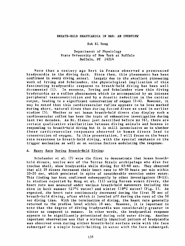
BREATH-HOLD BRADYCARDIA IN MAN: AN OVERVIEW
Suk Ki Hong
Depar tment of PhysiologyState University of New York at Buffalo
Buf f alo, NY 14214
Note than a century ago Bert in France observed a pronouncedbradycardia in the diving duck. Since then, this phenomenon has beenconf irmed in every d iving animal. Largely due to the excel lent p ioneer ingwork of Irving and Scholander, the physiological implication of thisfascinating bradycardic response to breath-hold diving has been welldocumented �!. In essence, Irving and Scholander view this divingbradycardia as a reflex phenomenon which is accompanied by an intenseperipheral vasoconstriction and by a drastic reduction in the cardiacoutput, lead.ing to a signif icant conservation' of oxygen �-4!. However, itmay be noted that this cardiovascular reflex appears to be less markedduring short, natural dives than during forced dives as used in earlierstudies �!. Whether or not human breath-hold divers also display such acardiovascular reflex has been the topic of exhaustive investigation duringlast two decades. As Dr. Elsner just described before me �!, there arecertain qualitative similarities between diving animals and humans inresponding to breath-hold diving but it is still inconclusive as to whetherthese cardiovascular responses observed in human divers lead toconservation of oxygen. In this presentation, I will focus on the heartrate responses to breath � hold diving, with the special emphasis on thetrigger mechanism as well as on various factors modulating the response.
R. Beget Rate ~Dnrin Breath-Bold ~Divin
Scholander et al. �! were the first to demonstrate that human breath-
hold divers, native men of the Torres Strait archipelago who dive fortrochus shell, show bradycardia while diving for 45-60 sec. They observedthat all 19 divers decreased their heart rate to 40-50 beats per min within20-30 sec, which persisted in spite of considerable exercise under water.This finding has been confirmed subsequently by other investigators 8-11!.In studies reported by Hong et al. �1! using Korean women divers, theheart rate was measured under various breath-hold maneuvers including thedive in both summer �7 C water! and winter �0 C water! Fig. 1!. Asexpected, the heart rate continuously decreased during the first 20 sec ofbreath-hold diving, after which it leveled off during the rest of the 50sec diving time. With the termination of diving, the heart rate generallyreturned to the predive level within l0 sec. However, it is important tonote that the degree of diving bradycardia was considerably greater inwinter as compared to summer. In other words, the bradycardial responseappears to be significantly potentiated during cold water diving. Anotherimportant observation was that a virtually identical pattern of bradycardiawas observed even during either breath-hold surface swim with the facesubmerged or a simple breath-holding in water with the face submerged.
158
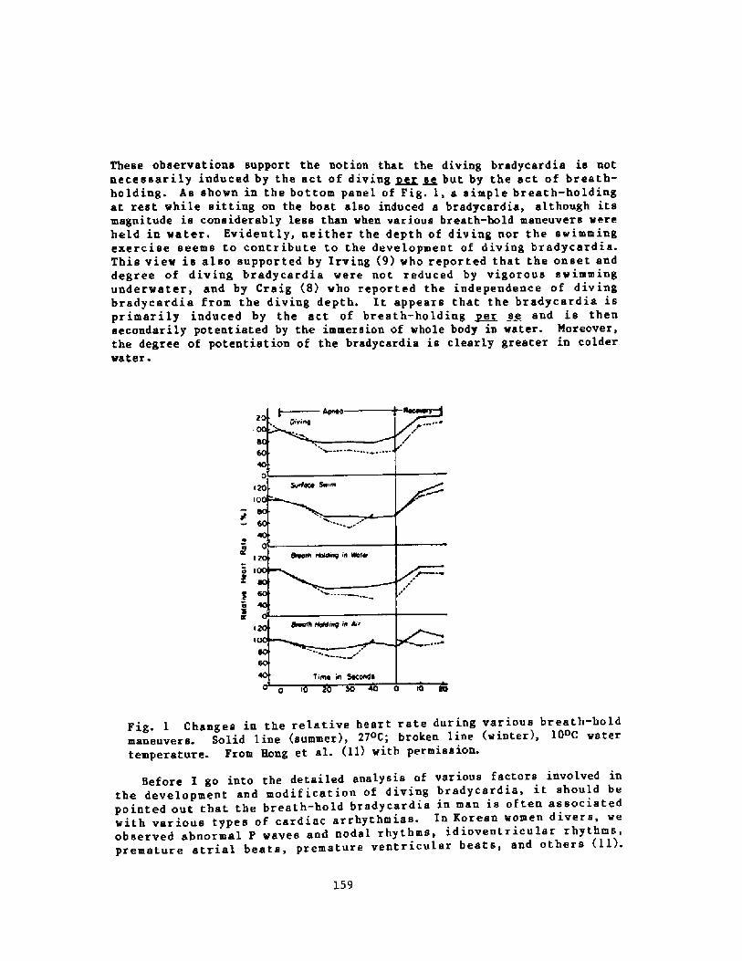
These observations support the notion that the diving bradycardia is notnecessarily induced by the act of diving ~ ~s but by the act of breath-holding. As shown in the bottom panel of Pig. 1, a simple breath-holdingat rest while sitting on the boat also induced a bradycardia, although itsmagnitude is considerably less than when various breath-hold maneuvers wereheld in water. Evidently, neither the depth of diving nor the swimmingexercise seems to contribute to the development of diving bradycardia.This view is also supported by Irving 9! vho reported that the onset anddegree of diving bradycardia were not reduced by vigorous swimmingundervater, and by Craig 8! who reported the independence of divingbradycardia from the diving depth. It appears that the bradycardia isprimarily induced by the act of breath-holding ~e se and is thensecondarily potentiated by the immersion of vhole body in water. Moreover,the degree of potentiation of the bradycardia is clearly greater in colderwater.
Pig. 1 Changes in the relative heart rate during various breath-holdmaneuvers. Solid line summer!, 21oC; broken line winter!, 10oC watertemperature. Prom Hong et al. �1! with permission.
Before I go into the detailed analysis of various factors involved inthe development and modif ication of diving bradycardia, it should bepointed out that the breath-hold bradycardia in man is often associatedwith various types of cardiac arrhythmias. In Korean women divers, weobserved abnormal P waves and nodal rhythms, idioventriculsr rhythms,premature atrial beats, premature ventricular beats, and others �1!.
159
Moreover, the incidence of these arrhythmias wss considerably higher inwinter �2X! than in summer �32!. Scholander et sl. �! were alsosurprised to f ind that a high incidence of arrhythmia developed duringdiving. Thirteen out of 18 divers developed anomalies visible on the ECG,such as inversion of T waves, ventricular premature contractions,idioventricular rhythms, and one case of atrial fibrillation, Theseobservations in human divers are in total contrast to those in divinganimals in which arrhythmias have not been observed during diving.
As discussed above, the diving bradycardia appears to be triggered bybreath holding and is then potentiated by immersion of the body in water especially in cold water!. In fact, Wyss �2! already reported nearly 30years ago that ECG s showed bradycardis in men while submerged and duringapnea. Whatever is the mechanism underlying this brsdycardic response, theparasympathetic. nerve supply seems to play s critical role because divingbradycsrdia in man can be blocked by atropine �3!. It could be thatinhibition of the respiratory center induced by the cessation ofrespiratory movements excites cardiac vagal motoneurones leading tobradycardia �4!.
Although the whole body immersion in water appears to potentiate thebradycardial response to breath holding, it soon became evident that simpleface immersion in water alone also similarly potentiates the response.Although Elsner et al. �5! used the latter approach for the first time in1963 to induce a simulated diving bradycardis, the quantitative similarityof apneic bradycardia induced by whole-body immersion to that induced byface immersion alone was not established until 1972 �6!. In order todefine if a certain region of the face is involved in potentiating breath-hold bradycardia upon immersion, face immersion experiments were carriedout with a nose clip or with a face mask. However, the results obtainedfrom different laboratories are conflicting and thus no definite conclusioncan be drawn regarding the particular role played by the stimulation of theskin in the region of the eyes and nose �7,18!. Moreover, it is not evenunequivocally established that the face needs to be wetted during faceimmersion to potentiate breath-hold bradycardia. Separating the face fromwater by thin plastic sheet either attenuated �9,20! or had no significanteffect �6! on the breath-hold bradycardia. However, it is generallyaccepted that thermal receptors in the facial skin surface seem to play animportant role see below!.
Despite the lack of the understanding of the mechanism underlying 'theunique role of face immersion in simulating the diving bradycardia in manithis technique has been widely utilized during last two decades byinvestigators to study diving bradycardia. In fact, most of the currentknowledge of diving bradycardia is derived from such studies-
As stated above, the magnitude of breath-hold bradycardia in Koreanwomen divers was significantly greater in winter than in summer I I! iin icating the important role of temperature in determining the degree ofi d'
160
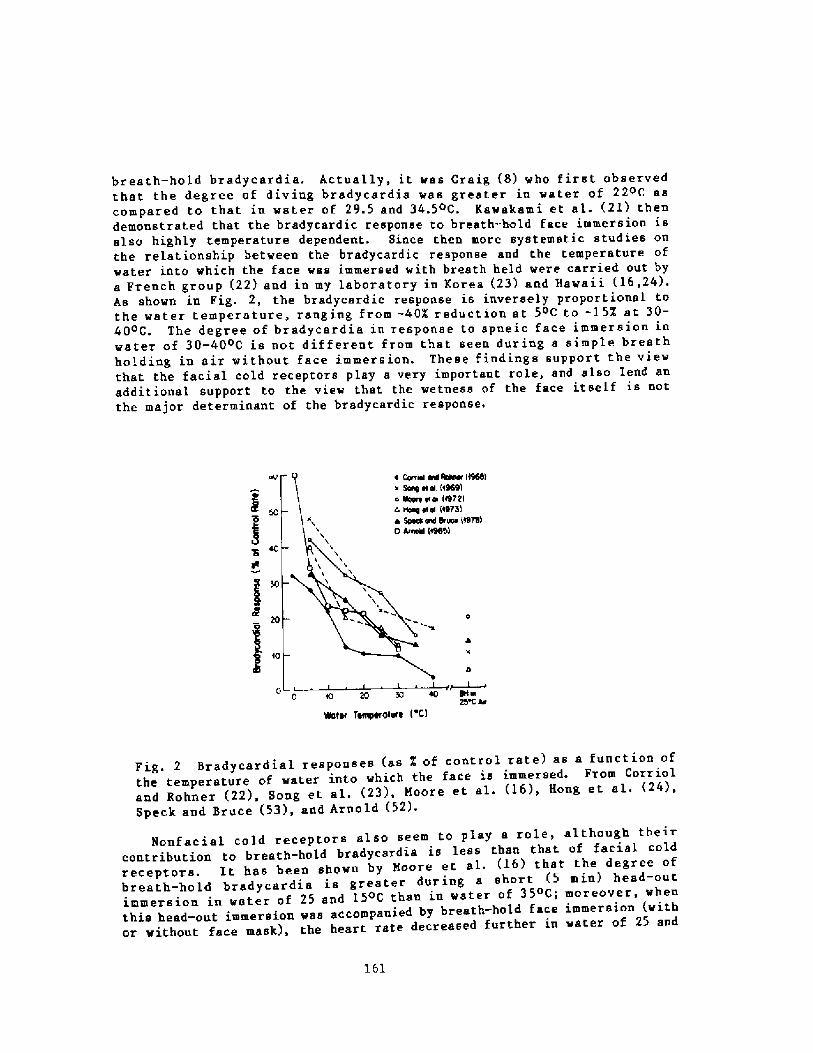
breath-hold bradycardia. Actually, it vas Craig 8! who first observedthat the degree of diving bradycardia was greater in water of 22oC ascompared to that in water of 29.5 and 34.5oC. Kavakami et al. �1! thendemonstrated that the bradycardic response to breath-hold face immersion isalso highly temperature dependent. Since then more systematic studies onthe relationship between the bradycardic response and the temperature ofwater into which the face was immersed with breath held vere carried out bya French group �2! and in my laboratory in Korea �3! and Hawaii. �6,24!.As shovn in Fig. 2, the bradycardic response is inversely proportional tothe water temperature, ranging from -40% reduction at 50C to -15X at 30-40 C. The degree of bradycardia in response to spneic face immersion inwater of 30-40 C is not d if f erent f rora that seen during a simple breathhold ing in air vithout f ace immersion. These f indings support the viewthat the facial cold receptors play a very important role, and also lend anadditional support to the view that the wetness of the face itself is notthe major determinant of the bxadycardic response,
Arleer r969Is989IIr972III%73 Isrvee IreraI
msaI
10 0 Q 20 N 40 &e23 Ckr
Water Temperature 'C!
Fig. 2 Bradycardial responses as X of control rate! as a function ofthe temperature of water into vhich the face is immersed. From Corrioland Rohner �2!, Song et al. �3!, Moore et al. �6!, Hong et al. �4!,Speck and Bruce �3!, and Arnold �2!.
Nonfacial cold receptors also seem to play a role, although theircontribution to breath-hold bradycardia is less than that of facial coldreceptors. It has been shovn by Moore et al. �6! that the degree ofbreath-hold bradycardia is greater during a short � min! head-outimmersion in vater of 25 and 15 C than in vater of 35 C; moreover, when'this h d- t immersion was accompanied by breath-hold face immersion vithis ea -ou immer 25or without face mask!, the heart rate decreased further in water of an
161
15 C but not in water of 35 C. However, the heart rate responses in thelatter experiment were not different from those observed during breath-holdface immersion alone in water of corresponding temperatures, indicating thedominant role of facial cold receptors. In this connection, it should bepointed out that a face immersion in cold vater without breath holding alsoinduces a short, slight bradycardia �1,25-27!.
Although it is quite clear that the combination of breath holding andthe stimulation of facial cold receptors acts synergistically, theunderlying mechanism is not understood. Typically, a breath-hold faceimmersion in cold water increases the blood pressure especially thediastolic pressure! and decreases the peripheral blood flow �1,23!, butthe correlation between the bradycardic response and these hemodynamicchanges is not evident. Moreover, the same hemodynamic changes may beinduced by immersion of the hands or feet in cold water without breath-holding and/or face immersion and yet the magnitude of bradycardia, if any,ie much smaller �1,23!. In order to accentuate the degree of cold-inducedhemodynamic changes, Moore et al. �6! studied the bradycardic response tobreath-hold face immersion in water of 25, 15 and 5 C in human subjects wearing only swimming trunks! resting in a 25 or 5oC room. As compared tothat in the 25 C room, the mean skin temperature was lower byapproximately 5oC! while the diastolic blood pressure was higher byapproximately 10mmHg! in the 5 C room. Moreover, the control heart rate without any breath-hold maneuvers! was lower by 13 bpm in 5oC than in 25 Croom, suggesting a cold-induced increase in vagal tone. During apneic faceimmersion, the temperature-dependent bradycardic response vas observed atboth room temperatures, but the relative degree of bradycardic response toa given apneic maneuver wss clearly less in 5 C room. However, there wasno difference in terms of the lowest absolute heart rate during 60 secapnea between the two room temperatures, These findings indicate that anincreased vagal tone induced prior to an apneic face immersion in coldwater by exposing to cold air for 30 min reduces the magnitude of apneicface immersion-dependent vagal activation. In other words, the breath-holdface immersion in cold water alone appears to increase the vagal tone tothe same maximal level. Once the vagal tone is increased by means otherthan breath-hold maneuvers, the magnitude of the breath holding-inducedincrease in vagal tone deer eases correspondingly.
C. Role of Intrathoracic Pressure:
In msn, diving is usually preceded by a near maximal inspiration equivalent to -85% of the vital capacity! folloved by breath holding �8!.The inspiratory muscles are then relaxed, and the intrathoracic pressure isincreased at least to the extent of the relaxation pressure of the lungsand thorax. Moreover, diving often involves a position change which wouldalter the pattern of venous return, the cardiac output and certainhemodynamic parameters such aa arterial pressure. Any of these diving-induced changes could alter the heart rate. Craig 8,54! observed thatmaneuvers which increased venous return at the beginning of the breath holdproduced a bradycardia during the apnea, and conversely when venous return
l62
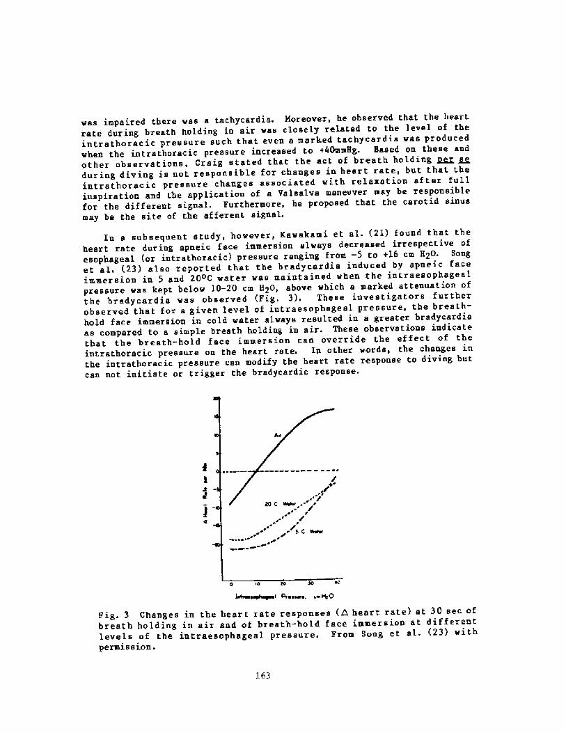
was impaired there was a tachycardia. Moreover, he observed that the heartrate dur ing breath holding in air was closely re1ated to the level of theintrathoracic pressure such that even a marked tachycardia was producedwhen the intrsthoracic pressure increased to +40mmHg. Based on these andother observations, Craig stated that the act of breath holding ~r seduring diving is not responsible for changes in heart rate, but that theintrathoracic pressure changes associated with relaxation after fullinspiration and the application of a Valsalva maneuver may be responsiblefor the different signal. Furthermore, he proposed that the carotid sinusmay be the site of the afferent signal.
In a subsequent study, however, Kawakami et al. �I! found that theheart rate during apneic face immersion always decreased irrespective ofesophageal or intrathoracic! pressure ranging from � 5 to +16 cm H20. Songet al, �3! also reported that the bradycardia induced by apneic faceimmersion in 5 and 20 C water was maintained when the intraesophagealpressure was kept below l0-20 cm H20, above which a marked attenuation ofthe bradycardia was observed Fig. 3!. These investigators furtherobserved that for a given level of intraesophageal pressure, the breath-hold face immersion in cold water always resulted in a greater bradycardiaas compared to a simple breath holding in air. These observations indicatethat the breath-hold face immersion can override the ef feet of theintrathoracic pressure on the heart rate. In other words, the changes inthe intrathoracic pressure can modify the heart rate response to diving butcan not initiate or trigger the bradycardic response.
O IO m 30
~eaphaym ~ PF eaare, c~ Hg 0
Fig. 3 Changes in the heart rate responses A heart rate! at 30 sec ofbreath holding in air and of breath-hold face immersion at differentlevels of the intraesophageal pressure. From Song et al. �3! withpermi s s ion.
163
El.sner et al. �5! originally reported that the cardiovascular response i.e., bradycardia and peripheral blood flow! to apneic face immersion wasaccentuated when performed in expiratory position. This finding isinteresting in that diving mammals are known to dive in expiratoryposition. Although Asmussen and Kristiansson �6! reported a similarobservation, Kawakami et al. �1! failed to confirm this interestingobservation. The latter investigators, in fact, observed that themagnitude of bradycardic response to breath-hold face immersion in coldwater tended to be lower when performed in expiratory position with thefunctional residual capacity', FRC! than in inspiratory position with thetotal lung, capacity; TLC!. Kawakami et al. �1! further observed that, insimple breath holding in air vithout face immersion! at FRC, there vas nochange in heart rate, whereas a decrease of 10 bpm from the resting levelwas seen during breath holding at TLC. Similarly, Song, et al. �3!reported that the reduction in heart rate during simple breath holding inair is proportional to the initial lung volume. In fact, at the residualvolume RV!, the heart rate even tended to increase rather than decreaseduring breath holding. The reason for these dif ferences in observationsregarding the ef f ect of the lung volumes on breath-hold bradycardia amonginvestigators is not obvious. Nevertheless, it may be parentheticallynoted that Angelone snd Coulter �9! demonstrated an inverse relationshipbetween the heart rate and the lung volume and suggested that stretchreceptors located in the lungs or thoracic wall initiate a reflexresponsible for this phenomenon. On the other hand, the attenuation ofbreath-hold bradycardia at FRC or RV, observed by Kawakami et al. �1! andSong et al. �3!, may be related to a possible increase in theintrathoracic pressure associated with the sct of expiration prior tobreath holding see above!.
An act of breath holding is associated w ith the cessation ofrespiratory movements and the development of both hypoxia and hypercapnia.It is, therefore, possible that hypoxia and/or hypercapnia could beresponsible for the bradycardic response. However, Olsen et al. �0,31!pointed out that diving bradycardia in man occurred before sny majorchanges in blood gas pressure could be expected; moreover, the degree ofbradycardia did not correlate with the duration of breath holding.Kavakami et al. �1! also observed that breathing 5X 02 � 20X CO2 mixturefor 2 min did not cause bradycardia either with face out or vith faceimmersion. It thus appears that asphyxia ~r se does not produce abradycardia. However, it is still possible that hypoxia and /or hypercapniamight either potentiate or attenuate the breath-hold bradycardia. Infact, it was proposed that hypercapnia followed by hypoxia during breathholding may contribute to the maintenance of the diving bradycardia �1!-Based on the experiments in which alveolar 02 and CO2 pressures vereselectively altered prior to breath holding with or without face immersionin water!, Moore et sl. �2! reported that the breath-hold bradycardia waspotentiated in the presence of low alveolar P02, attenuated under
164
conditions of high alveolar POp, and appeared to be independent of alveolarpCO2 levels. These observations suggest that the peripheral chemoreceptorscan modify the intensity of the diving response.
Recently, Lin et al. �3! designed 5 series of breath-holdingexperiments to partition the effect of apnea ~er se, hypercapnia, andhypoxia on the development and maintenance of breath-hold bradycardia induced by face immersion in 25 C water!. According to their analysis, a31X reduction in the heart rate induced by this breath-hold maneuverrepresented an arithmetic sum of 19X reduction by apnea, 18X reduction byhypoxia, and 6X increase by hypercapnia �9+18-6=31!. These resultsindicate that the hypoxia which develops during the course of breathholding has a major potentiating effect while hypercapnia has only a minorattenuating effect on breath-hold bradycardia.
F. Effect of ~Ph eicei Exetciee:
Studies conducted in the field using professional breath-hold diversindicated that diving bradycardia persisted in spite of considerableexercise under water �,9,11,30!. These observations led manyphy siologists to believe that even a severe physical exercise may not beable to override the mechanism leading to diving bradycardia. In fact,Stromme et, al. �7! found that breath-hold face immersion during exerciseinduced a nearly 50X reduction in heart rate, independent of work load overa DV02 the difference between the resting metabolic rate and themetabolic rate during steady state of swimming! range of 5-30 ml 02/Kg permin. On the other hand, Craig 8! z epor ted that the typical divingbradycardia was almost completely reversed during underwater exercise moving flippers up and down at a moderate rate and. force for 35 sec! in adiver who hyperventilated using 100X 02 for about 5 min and then passivelydescended while breath holding! to the pool bottom and remained quiet for1 min. In fact, the heart rate increased to about the same rate as whenthe subject was at the surface and breathing. However, the recovery periodwas much shorter when the subject was submerged. The reason for thisdisagreement between Stromme et al. �7! and Craig 8! is most likelyattributed to the difference in work load. According to Paulev and Hansen�4!, breath holding produced bradycardia at relatively low rates ofsteady-state cycling! work Es VDg/V02max below 40%! just as during rest,and as relative work rates increased, the bradycardia graduallydisappeared. In this connection, it is interesting to note that the heartrate during exercise in apneic divers is dependent on the type of exercise.For instance, decreases in heart rate induced by apneic face immersion in15 C water! were much more marked during dynamic exercise than during restor isometric exercise �5!. However, the exact physiological mechanismresponsible for the differing influence of isometric and dynamic exerciseon the diving bradycardia is obscure at present.
Whether or not physical conditioning or fitness! modifies the breath-hold bradycardia is still controversial. Originally, Bove et al. �6!reported that a physical training program appeared to augment thebradycardia and, on this basis, suggested that the breath-hold maneuvers
165
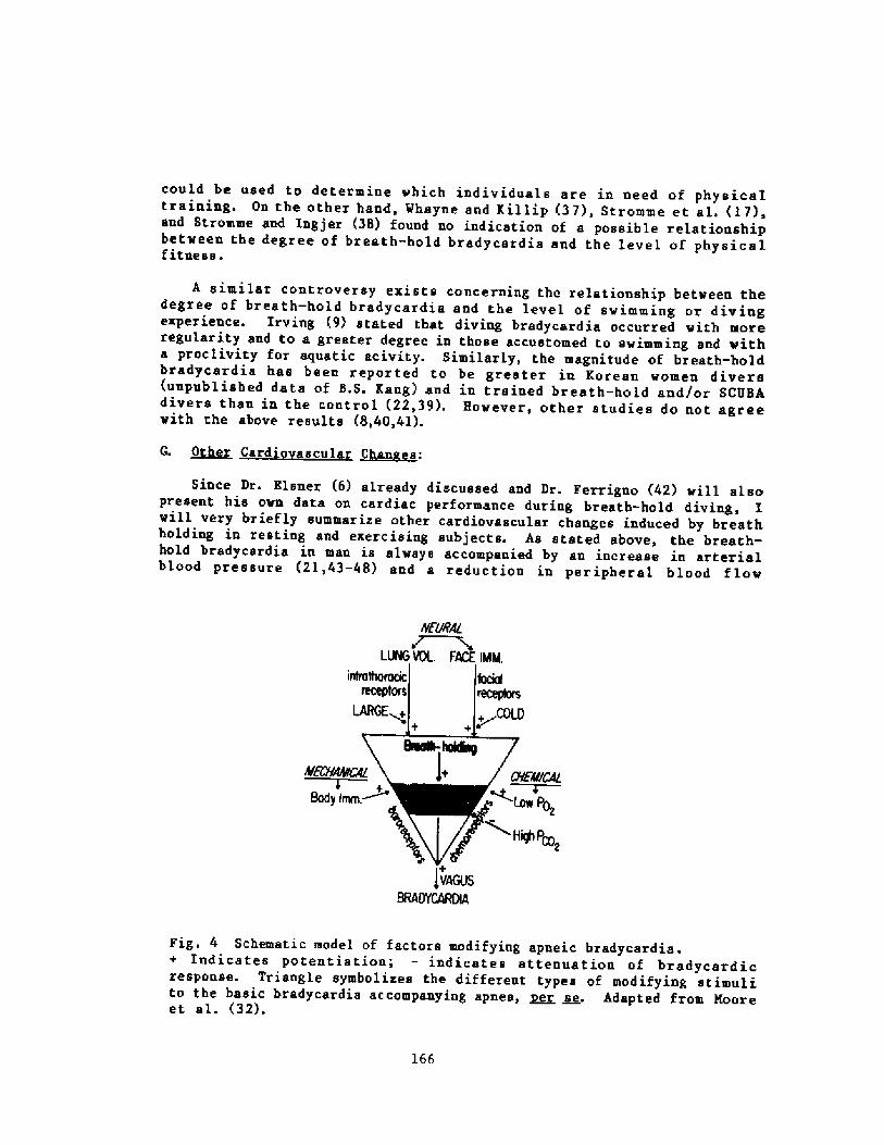
could be used to determine which individuals are in need of physicaItraining. On the other hand, Whayne and Kil lip �7!, Stromme et al. �7!,and Stzomme and Ingjer �8! found no indication of a possible relationshipbetween the degree of breath-bold bradycardia and the level of physicalfitness.
A similar controversy exists concerning the relationship betveen thedegree of breath � hold bradycardia and the level of swimming or divingexperience. Irving 9! stated that diving bradycardia occurred vith moreregularity and to a greater degree in those accustomed to swimming and witha proclivity for aquatic acivity. Similarly, the magnitude of breath-holdbradycardia has been reported to be greater in Korean vomen divers unpublished data of B.S. Kang! and in tzained breath-hold and!or SCUBAdivers than in the control �2,39!. However, other studies do not agreevith the above results 8,40,41!.
G. ~Ot Card' scular !Laaes:
Since Dr. Blsner �! already discussed and Dr. Ferrigno �2! vill al.sopresent his ovn data on cardiac performance during breath-hold diving, Iwill very briefly summarize other cardiovascular changes induced by breathholding in resting and exercising subjects. As stated above, the breath-hold bradycardia in man is always accompanied by an increase in arterialblood pressure �1,43-48! and a reduction in peripheral blood flov
hEl&AL
LLNG UOL. FACE IM
JvAGusBRAlN~DN
Fig. 4 Schematic model of factors modifying apneic bradycardia.+ Indicates potentiation; � indicates attenuation of bradycardicresponse. Triangle symbolizes the different types of modifying stimulito the basic bradycardia accompanying apnea, 2' se. Adapted from Mooreet al. �2!.
166
�5,23,44,45,48,49!, although the latter changes are not necessarilyresponsible for the development of bradycardial response see above!. Onthe other hand, the cardiac output during breath holding with or withoutf ace immersion either remained unchanged �4,47!, decreased by -20Z �1,48!or increased by 14X �0!. Therefore, total peripheral resistance has beenshown to increase by 26 to 53Z �6,48!. More recently, Sjertnaes et al.�1! studied cardiovascular responses to face immersiou and apnea duringsteady state muscle exercise and reported that the cardiac output duringthe last half of breath-hold face immersion was reduced by 49X whilesystemic vascular resistance increased by 200X. Moreover, theseinvestigators found that myocardial 02 demand determined by the heart rate� pressure double product decreased by 42X during breath-hold faceimmersion. However, they also noted that the rise in systemic vascularresistance is not closely adjusted to the reduction in cardiac output, andthat the 02-saving potential is reduced because of rise in cardiacafterloads.
S. ~Summa
The breath-hold bradycardia is tr iggered by the cessation ofrespiratory movements, but there are many neural, mechanical and chemicalfactors known to modify the degree of the bradycardia. Eased on the dataavailable in the current literature, a modif ied version of previouslypublished "multiple factor theory" �3,32! is shown in Fig.4.
167
REFERENCES
Irving L, Scholander PF, Grinnel SW. The regulation of arterial bloodpressure in the seal during diving. Am J Physiol 1942; 135: 557-566.
Scholander PF. Physiological adaptation to diving in animals and man.Harvey Lect Ser 1961-62; 57: 93-110.
3.
Scholander, PF. The master switch of life. Sci Am 1963; 209: 92-106.
Elsner R. Heart rate response in forced versus trained experimentaldives in pinnipeds. Hvalradets Skrifter, Norshe Videnskaps-Akad, Oslo1965; 48: 24-29.
5 ~
Elsner R. Diving r esponse: A. compar ison of animals and man. In:Lundgren C. ed. Proceedings of the Workshop on the Physiology ofBreath-hold Diving. Undersea Med Soc: Be the ada, 1985.
6.
Scholander PF, Hammel HT, LeMessurier H, Hemmingsen E, Garey W.Circulatory adjustment in pearl divers. J Appl Physiol 1962; 17: 184-190.
Craig AB Jr. Heart rate response to apneic underwater diving and tobreath holding. J Appl Phys iol 1963; 18: 854-862.
Irving L. Sradycardis in human divers. J Appl Physiol 1963; 18: 489-491.
Sasemoto H. The electrocardiogram pattern of the diving AMA. In: RahnH, Yokoyama T, eds. Physiology of Breath-Hold Diving and the AMA ofJapan. NAS-NRC Publication 1341, 1965; 271-280.
10.
Hong SK, Song SH, Kim PK, Suh CS. Seasonal observations on the cardiacrhythm during diving in the Korean AMA. J Appl Phsyiol 1967; 23: IS-22.
Wyss V. Electrocardiogram of apneic subjects during immersion in waterat various depth. Boll Soc Ital Biol Sper 1956; 32: 503-506.
12.
Heistad DO, Abboud FM, Eckstein JW. Vasoconstrictor response tosimulated diving, in man. J Appl Physiol 196S; 25: 542-549.
13.
14. Daly MB, Angell-James JE. The diving response and its possibleel inical implicat ion. Internat Med 1979; 1: 12-19.
Elsner RW, Ger ey WF, Sebo lander PF. Selective ischemis in diving man.Amer Heart J 1963; 65: 571-572.
168
Anderson HT. Physiological adaptation in diving vertebrates. PhysiolRev 1966; 46: 212-243.
16.
Stromme SB, Kerem D, Elsner R. Diving bradycardis dur ing rest endexercise and its relation to physical f itness. J Appl Physiol 1970;28: 614-62]..
17.
Gooden BA, Feinstein R, Skutt HR. Heart rate response of scuba diversvia ultrasonic telemetry. Undersea Biomed Res 1975; 2: 11-19.
18.
Wolf S, Schneider RA, Grover ME, Further studies on the circulatoryand metabolic alterations of the oxygen-conserving diving! reflex inman. Trans Assn Am Physns 1965; 78: 242-254.
19.
Gooden BA, Holdstock G, Hampton JR. The magnitude o f the bradycardiainduced by face immersion in patients convalescing from myocardialinfarction. Card iovas Res 1978; 12: 239-242.
20.
Kawakami Y, Natelson BH, DuBios AB. Cardiovascular effects of faceimmersion and factors af f ecting diving reflex in man. J Appl Physiol1967; 23: 964-970.
21.
Corriol J, Rohner JJ. Role de la temperature de 1 eau dans labradycardie d immersion de la face. Arch Sci Physiol 1968; 22: 265-274.
22.
23. Song SH, Lee WK, Chung YA, Hong SK. Mechanisms of apneic bradycardiain man. J Appl Physiol 1969; 27: 323-327.
24. Hong SK, Moore TO, Lally DA, Morlock JF. Heart rate response to apneicface immersion in hyperbaric heliox environment. J Appl Physiol 1973;34: 770-774.
Brick I. Circulatory response to immersing the face in water. J ApplPhys io 1 1966; 21: 33-36.
Asmussen E, Kristiansson NG. The "diving bradycardia" in exercisingman. Acta Physiol Scand 1968; 73: 527-535.
26.
27. Paulev PE. Cardiac rhythm during breath-holding and water immerison inman. Acta Physiol Scand 1968; 73: 139-150.
28. Hong SK, Rahu H, Kang DH, Song SH, Kang BS. Diving pattern, lungvolumes and alveolar gas of the Korean diving women AMA!. J ApplPhys io 1 1963; 18: 457-465.
29. Angelone A, Coulter NA Jr. Heart rate response to held lung volume. JAppl Physiol 1965; 20: 464-468.
169
Moore TO, Lin YC, Lally DA, Hong SK. Effects of temperature,immer sion, and ambient pressure on human apneic bradycardis. J App 1Physio 1 1972; 33: 36-41.
30.
Olsen CR, Fanestil DD, Scholander PF. Some effects of apneicunderwater diving on blood gases, lactate, and pressure in man. J ApplPhys iol 1962; 17: 938-942.
31.
Moore TO, Elsner R, Lin YC, Lally DA, Hong SK. Effects of alveolar P02and PCOZ on apneic bradycardia in man. J Appl Physiol 1973; 34: 795-798.
32.
Lin YC, Shida KK, Hong SK. Effects of hypercapnia, hypoxia andrebreathing on heart rate response during apnea. J Appl Physiol 1983;54: 166-171.
33.
Paulev PK, Heusen HG. Cardiac response to apnea and water immersionduring exercise in man. J Appl Physiol 1972; 33: 193-198.
Bergman SA Jr, Campbell JK, Wildenthal K. "Diving reflex" in man'- itsrelation to isometric and dynamic exercise. J Appl Physiol 1972; 33:27-31.
35.
Bove AA, Lynch PR, Connell JV Jr, Harding JM. Diving reflex afterphysical training. J Appl Physiol 1968; 25: 70-72.
36.
Hhayne TF Jr, Rillip TK III. Simulated diving in man: Comparison offacial stimuli and response in arrhythmia. J Appl Physiol 1967; 22:800-807.
37.
Stromme SB, Ingjer P. Comparison of diving bradycardia and maximalaerobic power. Aviat Space Environ Med 1978; 49: 1267-1270.
38.
39. Hong SK, Moore TO, Seto G, Park HK, Hiatt MR, Bernauer EM. Lungvolumes and apneic bradycardia in divers. J Appl Physiol 1970; 29:172-176.
Harding PE, Roman D, Hhelan RF. Diving bradycardia in man. J Physiol1965; 181: 401-409.
40.
Elener RM, Franklin DL, Van Citters RL, Kenney DW. Cardiovasculardefense against asphyxia. Science 1966; 153: 941-949.
41.
Ferrigno M. Cardiac performances during breath hold diving. Zn:Lundgren C, ed. Proceedings of the Workshop on the Physiology ofBreath-hold Diving. Undersea Med Soc: Bethesda, 1985.
42.
Gross PM, Terjung RL, Lohman TG. Left-ventricular performance in manduring breath-holding and simulated diving. Undersea Biomed Res 1976j3; 351-360.
43.
l70
Olsen CR, Fanestil DD, Scholander PF. Some ef fects of breath-hoMingand apneic underwater diving in man. J Appl Physiol 1962; 17: 461-466.
44 ~ Heistad DO, Abboud FM, Eckstein JW. Vasoconstrictor response tosimulated diving in man, J Appl Phys iol 1968; 25: 542-549.
45. Hei.stad DO, Wheeler RC. Simulated diving during hypoxia in man. JAppl Physiol 1970; 28: 652-656.
46. Hong SK, Lin YC, Lally DA, Yim BJB, Kominami N, Hong PW, Moore TO.Alveol.ar gas exchanges and cardiovascular functions during breathholding with air. J Appl Physiol 1971; 30: 540-547.
47. Lin YC, Shida KK, Hong SK. Effects of hypercapnia, hypoxia andrebreathing on circulatory response to apnea. J Appl Physiol. 1983;54: 172-177.
48. Whayne TF, Smith NTY, Eger El II, Stoelting RK, Whitcher CE. Reflexcardiovascular responses to simulated diving. Angiologia 1972; 23:500-508.
49. Folinsbee A. Cardiovascular response to apneic immersion in cool andwarm water. J Appl Physiol 1974; 36: 226-232.
50. Paulev PE, Wettergvist H. Cardiac output during breath-holding in man.Scand J Clin Lab Invest 1968; 22: 115-123.
51. Bjertnaes L, Hauge A, Kjekshus J, Soyland E. Cardiovascular responsesto face immersion and apnea during steady state muscle exercise. Aheart catheterization study on humans. Acta Physiol Scand 1984; 120:605-612.
52. Arnold RW. Extremes in human breath hold, facial immersionbradycardia. Undersea Biomed Res 1985; 12: 183-190.
53. Speck DF, Bruce DS. Effects of varying thermal and apneic conditionson the human diving reflex. Undersea Biomed Res 1978; 5: 9-14.
54. Craig AB Jr. Effects of submersion and pulmonary mechanics oncardiovascular function in man. In: Rahn H, Yokoyama T, eds-Physiology of breath-hold diving and the AMA of Japan. NAS-NRCPublication 1341, 1965; 295-302.
171
Discussion folloving Dr. Hong's presentation
Dr. Ferrigno: You talked about the relationship between lung volumeand heart rate, and you also said that when the subject performed aValsalva maneuver, the bradycardial response was reduced< andconversely, when he did the Mueller maneuver, the bradycardia wasaccentuated.
Dr. Hong: Yes. In fact, during the Valsalva maneuver, even atac hy car dia develops.
Dr. Perrigno: Of course even if the subject did not intend to do aValsalva or Muller maneuver, but he just held his breath atdifferent lung volumes with relaxed respiratory muscles, thatitself would have influenced the intrathoracic pressure, so would youcomment on the effect of lung volume on the heart rate, independentlyfrom the intrathoracic pressure.
Dr. Hong: There are two notions. One notion is that when we startthe breath-holding f ollowing a maximal inspiration, of cour se<intrathoracic pressure becomes more negative during the act ofmaximal inspiration, which could play a role by increasing the venousreturn. According to Dr. Craig's model, this would potentiate thebradycardial response. The other possibility is that there is a paperindicating the presence of some sort of intrathoracic receptorssensitive to the lung volume which modulate the heart rate.
Dr. Arnold: The ef f ect of lung volume has been studied w ithmeasurement of intrathoracic pressure and without, and whenintrathoracic pressure is maintained constant, lung volume still hasthe effect that Dr. Hong mentioned.
Dr. Craig: Dr. Hong, as you mentioned, you can change the heart rateduring apnea by changing position. I think an awful lot of resultscan be explained in terms of baroreceptor response in light ofchanging arterial resistance and the other aspects of changing venousreturn and cardiac output.
Dr. Hong: In my last slide I mentioned the mechanical ef feet.involving bar oreceptor s.
Dr ~ Craig: I'm not sure we have to look for some triggeringmechanism. It would just depend on the usual physiology.
Dr. Lin: We talk about baroreceptor that can modify the response,but not triggering the response< and we determined it in this way.We did some experiments with rats where we depleted theircatecholamines and we dunk his head in the water, now his bloodpressure goes down, but the bradycardial response still appears.Normally when you dunk the head of the rat in the water, you wouldhave bradycardia and the blood pressure rise, but now you decreasedits catecholamines, and doing the same thing, blood pressure goesdown, the bradycardia is still there. Therefore, we conclude that thebaroreceptor activity has nothing to do with initiation, however thisdoes not deny its modifying role.
172
Dr. Arnold: The Cold Pressor Test can cause essentially the sameamount of peripheral vasoconstriction and the heart rate stays thesame.
Dr. Hong: Yes, the blood pressure goes up tremendously, but it alonedoesn't necessarily potentiate the breath-hold bradycardia.
Dr. Ferrigno: Dr. Hong, you showed also that the response of the heartrate related to the temperature of the water. Some Japaneseresearchers Kobayasi, S. and T. Ogawa. Effect of water temperature onbradycardia during nonapneic facial immersion. Jap. J. Physiol.23:613-624, l973! did experiments with water warmer thanthermoneutral, which is 35 C at rest and they saw that the trend wasinverted at higher temperatures; instead of seeing a smallerbradycardial response, the response was increasing again. Will youcomment on that'P
Dr. Arnold: I looked at that. On either end of the spectrum, in icewater and above thermoneutral, nociceptors are activated, whereasbetween about 27 degrees and 5 degrees cold receptors, and between 25degrees and 35 warm receptors are activated. So nociceptive receptorsare probably important afferents in the diving response at lowtemperatures and at temperatures above thermoneutral.
173
CARDIAC PERFORNANCE NRIN6 BREATH-HI.D OIVINm IN MANiAN OlfENI94
Massimo Ferrfgno~ Mats H. Liner and Claes E. G. Lundgren
Oepariment of Physiology' State Un1vers1ty of New YorkBuffalo> New York 14214
The classical description of the dfvfng responsei elicited fn dfvfng animalsby apnea and submersion> includes a vagal ly induced bradycardiai asympathetically controlled per1pheral vasoconstrfction and a decreased cardfacoutput. A diving responsei al though attenuated or mod1f fed> and especf al ly thedfv1ng bradycardfa> have been descrfbed also in man and a review of thfs topicwas recently done by Elsner and Gooden 9!r Lfn �1!> Hfckey and Lundgren �2!and by Hong �3!.
W fth regard to the ef fects of breath-hol df ng on cardf ac output fn man>earl fer studies have shown dffferfng results. Kawakamf and hfs colleagues �7!measured rfght cardf ac output w1th a N~&plethysmograph fc method 1 n sevensubjects durfng breath-holdfng with face immersion. In this condition strokefndex and cardiac index decreased by 14% and 22%> respectfvely. The decrease incard1ac output was accompanfed by a sfgnff1cant decrease 1n heart rate> morepronounced when the subjects' face was fmmersed in cold rather than in coolwater. Paulev and Wetterqvfst �3!> using a dye-dflutfon techniques showed a 20-40% reduct1on in cardiac output fn four subjects holding the1r breath wfthrelatfvely h1gh fntrapulmonfc pressures. In contrasts cardf ac output wasunchanged or fncreased fn four other subjects who hei d the1r breath with1 ntrapulmon1c pressures around zero. Hong and his colleagues �5! d1d notobserve any changes fn cardfac outputs measured by the Cardfo-green dflutfonmethods during 2 minute breath-holds. In thefr experiments' durfng wh1ch theirsubjects rebreathed 1nto a bag for sequentfal alveolar gas sampl fng. heart ratetended to decrease. while stroke volume tended to increase. Whayne and hfscolleagues �2! measured> by means of a ballfstocardfogram Beg!-analog computersystem> stroke volume and cardiac output fn efght subjects who held thefr breathw1th a col d cloth � C! on thef r face. After an fnft1al transfent fncreaseicardiac output decreased because of a parallel change fn heart rate. Ling Shfdaand Mong 1n 1983 �2! obta1ned stroke volume with an impedance cardiogram during90 second breat&holds wfth face 1mmers1on fn 22-24 C ~ater, and they observeda 33% increase fn stroke volume which was counterbalanced by a 30% reduction 1nheart rate. One year lateri Bjertnaes and his colleagues �!, us1ng thethermodflut1on technfque~ observed a 49% reduction fn cardiac output wfth faceimmersion 1n fce water and apnea dur1ng steady state exercfse.
Some of these dfscrepancfes were probaly caused by the presence> or absence>of the col d-induced cf rculatory components of the diving response �8!. Anotherimportant consf deratfon fs the possfbf1 fty that differences in the 1ntrathoracfcpressures of the subjects in the various studfes were responsible for thedfssfm1lar cardfac output measurements dur1ng breath-hol d1ng
It fs predictable that ff a subject takes a deep inspf ration and holds hisbreath with closed glottfs and relaxed respiratory muscles> hfs fntrathoracfcpressure will increase. This increase fn 1ntrathoracfc presure fs caused by the
174
inward chest-wall recoil at large lung volume and it fs 1 ikely to hinder venousreturn and therefore reduce cardfac output. Howevers the sftuatfon probablychanges during actual diving, Once the subject starts hfs descent underwater>the increasing hydrostatfc pressure surrounding hfm w ill compress his chest.Because of this, the inward recoil of his chest w fl 1 decrease~ and, at asufficient depth> ft will even be reversed~ thus reducing the subject~sintrahoracfc pressure. Consequentlyr hfs venous return should no more be impededand hfs cardiac output should be either restored to> or fncreased over, pre-breath-hol d level s.
Submersf on~ which f s an f ntegral part of breath-hol d div ing. is al solikely to f nfl uence venous return �i10!. As you can see in Ff g. Ir thewater pressure counteracts the blood pooling fn the dependent regions, which fspresent in af r and due to the gravity force. The hydrostatic pressure on theoutside of the body will redistrfbute blood into the thorax and the filling ofthe heart will be increased. Arborel fus and his colleagues �! observed fn threesubjects> during head-out fmmersfon fn 35 "C waterr a mean increase fn centralblood volume by 700 ml. In the same conditions' they also showed a 32K increasefn cardiac output fn ten subjects> with a 35% increase fn stroke volume while
heart rate dfd not change. Fig. 1. Schematic fl 't ustratfon of theeffect of immersion with the head abovewater on the di'stributfon of bl oodbetween dependent regfons of the bodyand the thorax. The springs indicate theel a stf city of the 1 ung tf ss ue. Dottedareas: fncompressfbl e but resil fenttissues. Hatched areas: blood. The leftmanometer black fluid column! indicatesthe pl eural pressure, the rf ghtmamometer hatched fl uf d col umn!fndfcates the right atrial pressure. Thedifference between rf ght atrial pressureand pl eural pressure ref 1 ects atrf altransmural pressure gradient. A: erectbody position fn the non immersedsituation; blood pooled fn the vascularbed below the heart. Atrial pressure fsalmost zero and pleural pressure fsnegative. Rf ght atrf al transmuralpressure gr adfent is small. B: erectbody position during immersion with thehead above water; redistribution ofblood tow ar ds the heart andintrathoracfc vascular bed~ distendfngthese structures. Atrf al press ure i sraised and pleural pressure almost zero.Rf ght atrf al transmural pressuregradfent fs increased. From �! withpermf ssf on.
175
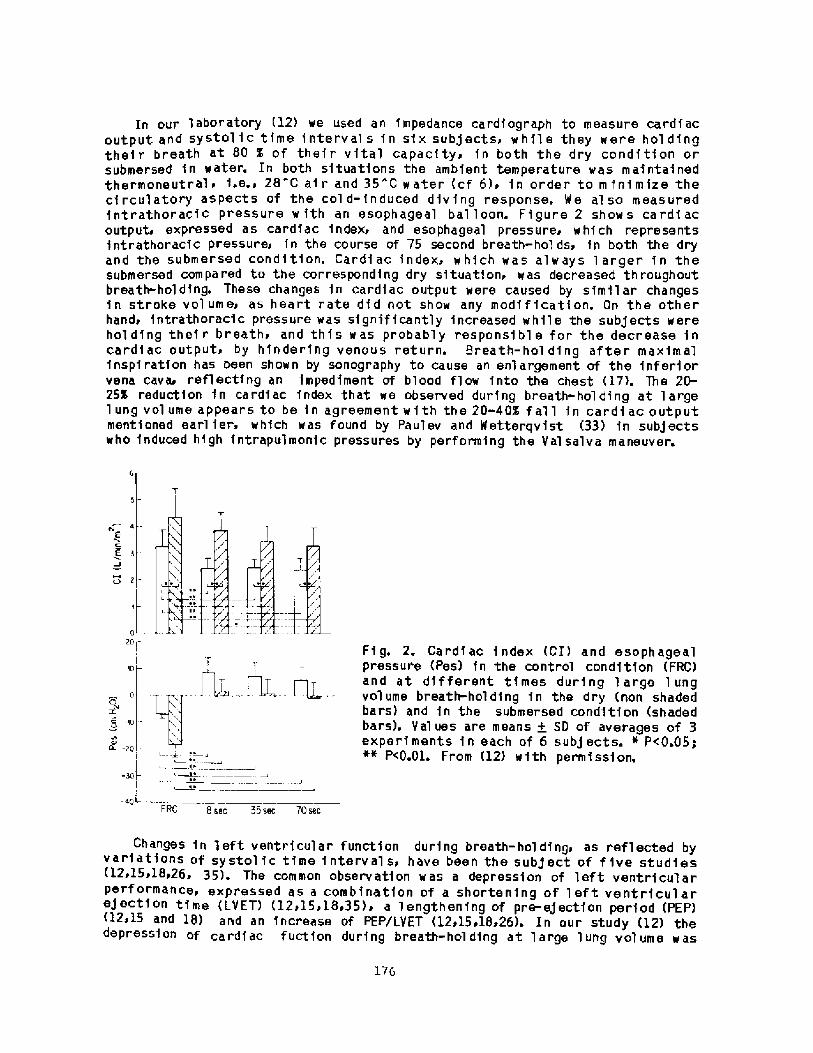
In our laboratory �2! we used an impedance cardfograph to measure cardiacoutput and systol f c tfme f nterval s f n sf x subj ectsi w hi 1 e they were hol d f ngtheir breath at 80 % of thefr vftal capacfty> fn both the dry condftfon orsubmersed in water. In both sftuatfons the ambfent temperature was maintainedthermoneutral4 f.e.4 28 c air and 35 "c water cf 6!> in order to m fnfmize thecirculatory aspects of the col d-induced dfvfng response. We al so measuredfntrathoracfc pressure w fth an esophageal bal loon. Figure 2 shows cardf acoutput expressed as cardiac index and esophageal pressure> which representsfntrathoracfc pressures fn the course of 75 second breath-hol ds> fn both the dryand the submersed condition. Cardiac fndeX4 whfCh waS always 1 arger in thesubmersed compared to the correspondfng dry sftuatfoni was decreased throughoutbreath-holding. These changes fn cardfac output were caused by simf1 ar changesf n strOke V ol um e4 as h ea rt r ate d f d not sh ow any mod f f f cat f on. On th e oth e rhand4 fntrathOraCic preSsure was sfgnff fcantly fnCreased while the Subjects Wereholding thef r breathi and thfs was probably responsible for the decrease incardf ac outputs by hindering venous return. Breath-hol ding after maximalfnspf ration has been shown by sonogr aphy to cause an enlargement of the fnferforvena cava reflecting an impediment of blood flow fnto the chest �7!. The 20-25% reduction fn cardfac fndex that we observed during breath-holding at largelung volume appears to be fn agreement w 3 th the 20-40$ f all fn cardf ac outputmentioned earl fer. whfch was found by Pau'lev and Wetterqvfst �3! fn subjectswho fnduced high fntrapulmonfc pressures by performing the Val salva maneuver.
CV 4e
2
0Zpr
Fig. 2. Cardiac index CI! and esophagealpressure Pes! fn the control condition FRC!and at different tImes durIng large lungvolume breathholdfng fn the dry non shadedbars! and fn the submersed condftfon shadedbars!. Values are means + SD of averages of 3experiments fn each of 6 subjects. ~ P<0.05;~" P<0.01. From �2! with permf ssf on.
ip
Kh n0COC4
10
+I 92 ~
40LFFZC 8 sec 35 sec 70 sec
ChangeS fn left ventricular funCtion during breath-holdfng4 as reflected byvar fatfons of systol fc time fnterval s have been the subject of f fve studies<12415i18426. 35!. The common observation was a depression of left ventricularPe««mance~ expressed as a combfnatfon of a shortening of left ventricularejection time LYET! �2>15.18435� a lengthenfng of pre-eJectfon period PEP!
and 18> and an increase of PEP/LVET <12>15418>26! ~ In our study �2! thedepressfon of cardiac fuctfon during breath-holding at large lung volume was
176
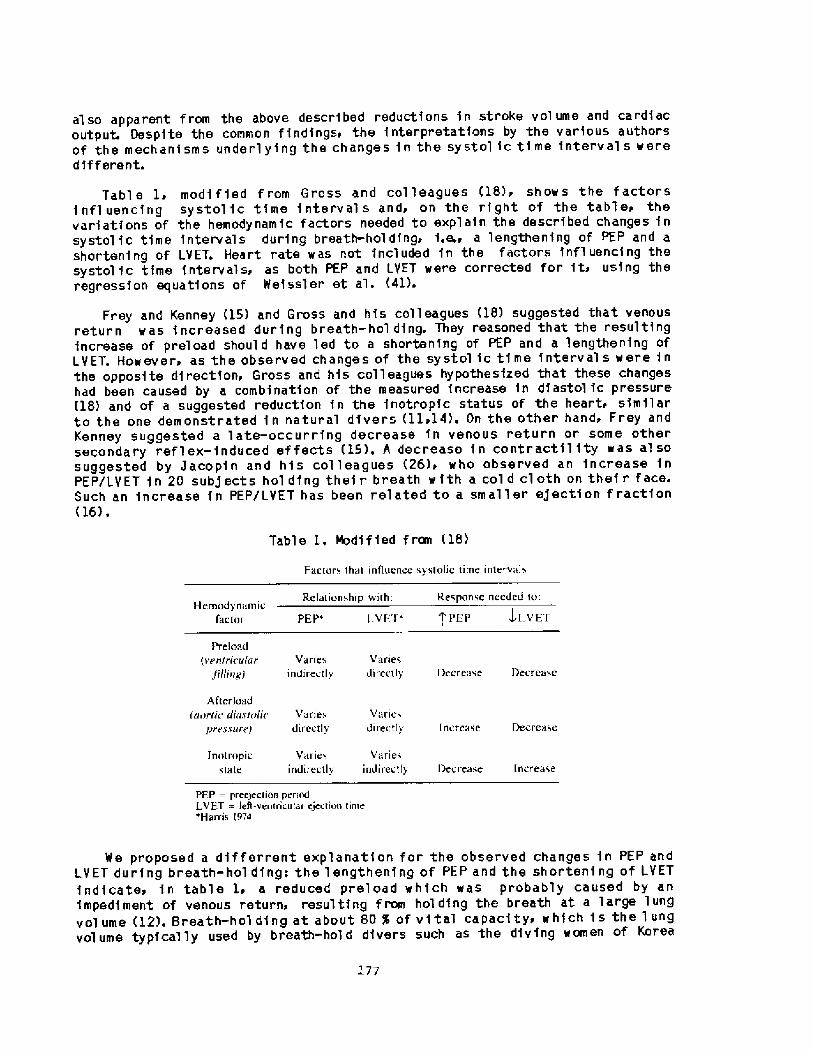
also apparent from the above described reductions in stroke volume and cardiacoutput. Despite the common findingsi the interpretations by the various authorsof the mechanisms underlying the changes in the systol ic time interval s weredif f erent.
Table 1i modf f ied f rom Gross and col leagues �8>i show s the factorsi nfl uencing systol ic time interval s andi on the right of the tablei thevariations of the hemodynamic factors needed to explain the described changes insystol ic time intervals during breath-hol ding. i.e.i a lengthening of PEP and ashortening of LVET. Heart rate was not included in the factors influencing thesystolic time intervalsi as both PEP and LVET were corrected for iti using theregression equations of Weissl er et al. �1>.
Frey and Kenney �5> and Gross and his colleagues �8! suggested that venousreturn was increased during breath-hol ding. They reasoned that the resultingincrease of preload should have led to a shortening of PEP and a lengthening ofLVET. Howeveri as the observed changes of the systol ic time interval s were inthe opposite directioni Gross and his colleagues hypothesized that these changeshad been caused by a combination of the measured increase in diastolic pressure�8! and of a suggested reduction in the inotropic status of the heart> similarto the one demonstrated in natural divers llil4!. On the other hand> Frey andKenney suggested a late-occurring decrease in venous return or some othersecondaty reflex-induced ef fects �5!. A decrease in contractility was al sosuggested by Jacopin and his colleagues �6!i who observed an increase inPEP/LVET in 20 subjects hol ding their breath with a col d cloth on their face.Such an increase in PEP/LYET has been related to a smaller egection f raction�6! .
Table 1, Modified f reel �8!
Faetnrs that intlnenee sySlOliC lime intervals
Re talion shi p wi h: Response needed to:Hernody namic
Iactol l.VI I' f'PEP J L VE'IPEP'
Preload > en/ricu ar
Jilting!
Variesdirectly
Vanesin direct l s DecreaseDecrease
Afterloadiunrric riiairri/ic
pres. cureiVariei
directtsVaries
directly DecreaseIncrease
Variesindirectly
Jnot ropicslate
V,'ll I esindirect ty l bee rease Increase
PFP = preejection penodLVET = lert-ven ricntai ejection time*Harris t 974
17'
We proposed a differrent explanation for the observed changes in PEP andLVET during breath-hol ding: the lengthening of PEP and the shortening of LVETindicatei in table 1i a reduced preload which was probably caused by animpedilnent of Venous return> reSulting from holding the breath at a large lurlgvolume �2!. Breath-hol ding at about 80 % of vital capacity, which is the lungvolume typically used by breath-hald divers such as the diving women of Korea
�4!i can on theoretical grounds be expected to generate an fntrathoracfcpressure of about 6 cm H20 Ref 1!. This fs in agreement with our observat1onof an esophageal pressure of about 7 cm H20 �2!> and this increasedfntrathoracfc pressure probably impeded venous return> and therefore reducedcardf ac output.
Further support to the suggestion that changes fn venous return wereresponsfble for the modff1catfons of the systol fc time intervals during breath-hol dfng> mfght be derived from the different temporal pattern of changes>described by Frey and Kenney �5!> 1 n what they called the 1notropf c and thechronotropfc response to apnefc face 1mmersfon, These authors observed that theappearance of the change fn left ventricular performance> as shown by anfncrease fn PEP, lagged behfnd that of bradycardf a. We suggested that thistemporal separation was the result of the different nature of the two responses�2!: the negative chronotrop1c fnfluence occurred earl fer because ft was reflexmedfatedi while ft took longer time for the change fn PEP to become evfdent> asft was induced by a gradual reduction fn venous return.
In our hyperbarfc chamber we ai so measured cardiac funct1on and1ntrathoracfc pressure fn sfx subjects durfng simulated breath-hold dives to 20meters �3!i a depth routinely reached by the Japanese female divers �9!.Experfments were performed fn both the dry and the submersed condition and thfsisa to our knowledge. the only study of cardiac performance fn man duringbreath-hold divfng. Cardfac output~ whfch was decreased by 20-25% during breath-hol d1ng at the surfacer was restored to control val ues at depth. Furthermorepthe changes 1n systol fc tfme Intervals> fnduced by breath-hol dfng+ had beenreversed by compression. We suggested that this restorat1on of card1acperformance> when the breath-holding subjects were compressed to 20 meters> wasthe result of the concom1tant fall fn fntrathoracic pressure whIch waspredictable from the compression of the chest to one th1rd of fts orfgfnalvolume. Some additional volume reduction probably resulted from alveolar gasexchange at depth �0!. Thereforer with the fall 1n 1ntrathoracfc pressure theimpediment to venous returns whfch had been present at the surface> was removedand cardiac output was restored.
It fs of interest to consider how the chest wali reco11. responsible for thefntrathoracfc pressure changes during breath-hold diving, was influenced by thedry and the submersed dives. Figure 3 serves such a theoretical comparison. Itshows schematfc pressur~volume curves for the chest wall during relaxatfon fnthe dry condf tf on conti nuous 1 f ne! and 1 n head-out immersf on dotted 1 fne! modified from Agostonf et al.> 1! It also depicts the pred1cted posftfons of asubject's lungs on these curves> dur fng breath-hold diving fn the dry and 1n thesubmersed mode respectively. An important difference between the dry and thesubmersed condition fs that fntrathoracfc blood volume fs 1 fkely to be largerduring submersion. The pulmonary afr volumes used fn desfgnfng Ffg. 3 were theones employed fn our experfments �3!: resfdual volume plus 80% of "dry" vftalcapacfty fn the non-submersed condition and residual volume plus 85% of "wet"vftal capac1ty 1n the submersed cond1tfon. Thus> the difference between dry andimmersed vftal capac1ty measurements was considered> and the subject initiatedthe dry and submersed dfves with essentially the same absolute volume of afr fnhfs lungs.
For the purpose of the calculatfons on which Ffg. 3 fs based~ the meanval ues for lung volumes recorded in our study �3! were used, f.e., a "dry"vital capacfty of 5.7 L and a residual volume of 18 L. For s1mpl fcfty> changes
178
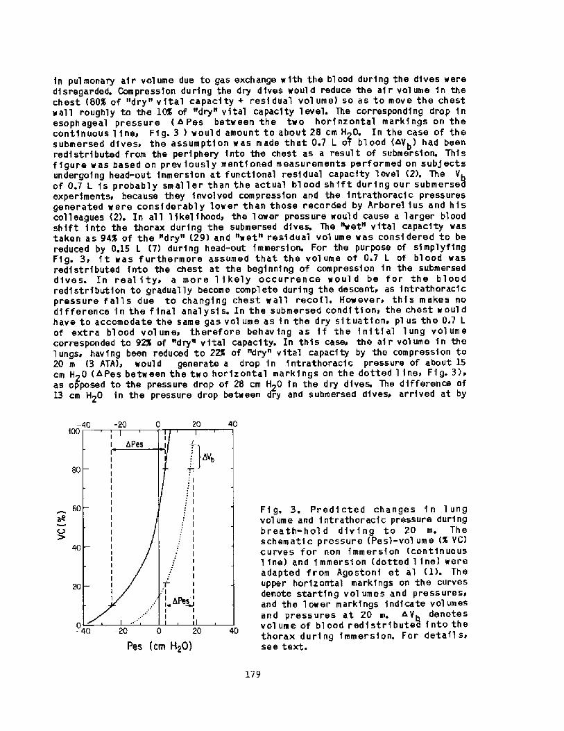
in pulmonary air volume due to gas exchange w1th the blood during the dives weredisregarded. Compression during the dry dives would reduce the air volume in thechest 80$ of "dry" v1tal capacity + residual vol ume! so as to move the chestwall roughly to the 10% of "dry" vital capacity level. The corresponding drop inesophageal pressure 4 Pes between the two horinzontal markings on thecont1nuous line> Fig. 3 ! would amount to about 28 cm H>0. In the case of thesubmersed dives> the assumption was made that 0.7 L o7 blood <Vb! had beenredistributed from the periphery into the chest as a result of submersion. Thisf1gure was based on previously mentioned measurements performed on subjectsundergoing head-out immers1on at functional residual capacity level �!. The Vof 0.7 L is probably smaller than the actual blood shift during our submersedexperiments> because they involved compression and the 1ntrathoracic pressuresgenerated were considerably lower than those recorded by Arborel ius and hiscolleagues �!. In all likelihood~ the lower pressure would cause a larger bloodshift into the thorax during the submersed dives. The "wet" vital capacity wastaken as 94% of the "dry" �9! and "wet" res1dual vol ume was considered to bereduced by 0.15 L �! during head-out 1mmersion. For the purpose of simplyfingFig. 3i it was furthermore assumed that the volume of 0.7 L of blood wasredistributed into the chest at the beginning of compression in the submerseddives. In reality> a more 1 ikely occurr ence woul d be for the bloodredistribution to gradual ly become complete during the descent> as intrathoracicpressure falls due to chang1ng chest wall recoil. However> thi's makes nod1fference in the f1nal analysis. In the submersed condition> the chest wouldhave to accomodate the same gas vol ume as in the dry situations plus the 0.7 Lof extra blood vol ume~ therefore behaving as if the ini t1al lung volumecorresponded to 92I of "dry" v1tal capacity. In th1s caser the a1r volume in thelungsi having been reduced to 22$ of "dry" vital capacity by the compression to20 m � ATA!> would generate a drop in intrathoracic pressure of about 15cm H20 hPes between the two hor1zontal mark1ngs on the dotted 1 ine> Fig- 3!ras opposed to the pressure drop of 28 cm H20 in the dry dives. The difference of13 cm H20 in the pressure drop between dry and submersed dives> arrived at by
20 40-20� 40i00
60
C3
Fig. 3. Pred1cted changes in lungvolume and intrathoracic pressure duringbreath-hol d div ing to 20 m. Thes chem ati c pr essure Pe s!-vol um e I VC!curves for non immersion <continuous1 ine! and immersion dotted 1 ine! wereadapted from Agostoni et al <1!. Theupper horizontal markings on the curvesdenote starting vol umes and pressuresiand the lower markings indicate volumesand pressures at 20 m. 4V denotesvolume of blood redistribute3 into thethorax during immersion. For detail sisee text.
40
20
-40 -20 00
20
PeS Cm H20!
179
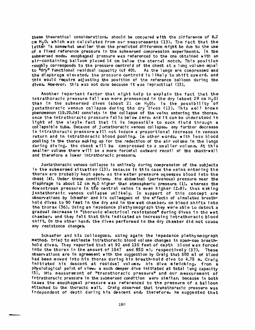
these theoretical consfderatfonsi should be compared w fth the difference of 8.2cm H20. which was calculated from our measurements �3!. The fact that thelatter is somewhat smaller than the predfcted dffference might be due to the useof a fixed reference pressure fn the submersed compressfon experfments. In thesubmersed mode> esophageal pressure was referenced to the one obtained with anair-containing balloon placed 14 cm below the sternal notch. This positionroughly corresponds to the pressure centrofd of the chest at a lung volume equalto "dry" functional resf dual capacity <cf 40!. As the lungs are compressed andthe d1aphragm elevated> the pressure centroid 1 s 1 fkely to shift upward. andthis would requ1re adjustfng the positfon of the reference balloon during thedives. However. thfs was not done because ft was fmpract1cal �3!.
Another important f actor that mf ght hei p to expl af n the f act that theintrathoracic pressure fal 1 was more pronounced f n the dry about 29 cm H20!than 1 n the submersed dives about 21 cm H20!~ 1s the possibf1 fty ofjuxtathoracfc venous collapse during the dry dfves �3!. This wel 1 knownphenomenon �9i20>34! consists in the collapse of the veins entering the thorax>once the fntrathorac1c pressure fal ls bel ow zero> and it can be understood f n11ght of the sfmple fact that it is impossible to suck fluid through acollapsible tube. Because of juxtathoracfc venous col lapsed' any further decreasein fntrathoracic pressure w 111 not induce a proportional fncrease in venousreturn and in intrathoracic blood pooling. In other words> with less bloodpooling in the thorax making up for the reduction of the air volume in the lungsduring dfvfng, the chest w fll be compressed to a smal ler volume. At thfssmaller volume there will be a more forceful outward reco11 of the chest"walland therefore a lower fntrathoracfc pressure.
Juxtathoracfc venous collapse fs unlikely dur1ng compression of the subjectsfn the submersed sf tuatf on <13! i because in th fs case the veins enter fng thethorax are probably kept opens as the ~ater pressure squeezes blood 1 nto thechest �!. Under these conditions, the abdominal perfvenous! pressure near thedfaphragm is about 12 cm M20 higher than atmospheric pressure l!i whereas thedownstream pressure fn the central veins 1 s even higher �i8! ~ thus makingjuxtathoracfc venous coll apse un11kely. In support of this concept areobservations by Schaefer and hfs colleagues of the effects of simulated breath-hol d dives to 90 feet fn the dry and fn the wet chamber> on blood shifts intothe thorax �6!. Usf ng an impedance plethysmograph they were able to detect agradual decrease fn "thoracic electrical resfstance" durfng dives in the wetchamber> and they fel t that this indicated an increasing f ntrathoracfc bloodsh1ft. On the other hand> the dfves performed in the dry chamber d1d not causeany resf stance changes,
Schaefer and hfs colleagues. using again the impedance plethysmographmethod> tried to estimate fntrathoracfc blood volume changes fn open-sea breath-hol d dives. They reported that at 90 and 130 feet of depth blood was forcedinto the thorax fn the amount of 1047 and 850 mls respectively �7!. Theseobservations are fn agreement with the suggestion by Graf g that 600 ml of bloodhad been moved into his thorax during h1s breath-hol d dive to 4.75 m. Craig1 nf tf ated h1s descent at resf dual vol umei hfs dive mimicking> fromphysiological pofnt of vfewi a much deeper dfve init1ated at total lung capacity<5!. His measurement of "transthoracic pressure" and our measurement offntrathoracfc pressure fn the submersed condftfon were similar~ because fn bothcases the esophageal pressure was referenced to the pressure of a balloonattached to the thoracic wall. Cra1g observed that transthoracfc pressure wasindependent of depth during hfs descent and> therefore> he suggested that
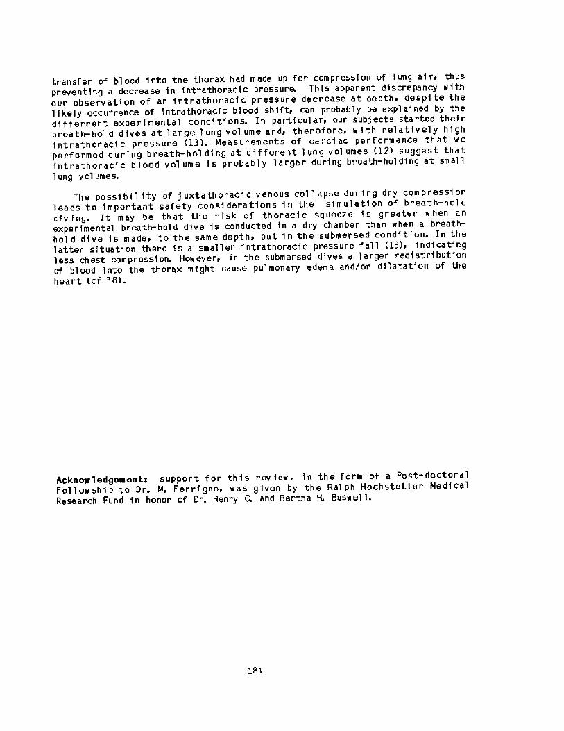
transfer of blood into the thorax had made up for compression of lung afri thuspreventing a decrease in fntrathoracfc pressure. This apparent discrepancy withour observation of an fntrathoracfc pressure decrease at depth. despite thelikely occurrence of fntrathoracfc blood shift> can probably be explained by thedf f ferrent expertmental conditions. In partfculari our subjects started theirbreath-hol d dfves at 1 ar ge lung vol ume andi therefore. w f th relatively hf ghfntrathoracic pressure �3!. Measurements of cardi ac performance that weperformed during breath-hol ding at different lung vol umes �2! suggest thatfntrathoracfc blood vol ume f s probably larger during breath-holdfng at smalllung volumes.
The possibil fty of juxtathoracfc venous collapse durfng dry compressionleads to important safety considerations fn the sfmulatfon of breath-hol ddf v f ng. It m ay be th at the rf sk of thoracic squeeze f s greater w hen anexperimental breath-hold dfve fs conducted in a dry chamber than when a breath-hol d dive is madei to the same depthi but in the submersed condftfon. In thelatter situation there is a smaller fntrathoracfc pressure fall �3!. indicatingless chest compression. However, in the submersed dives a larger redistributionof blood into the thorax mfght cause pulmonary edema and/or dilatation of theheart cf 38!.
Acknoyledgeaent: support for this rev iew ~ fn the form of a Post-doctoralFellowship to Dr. M. Ferrf gnoi was given by the Ral ph Hochstetter MedicalResearch Fund in honor of Dr. Henry C. and Bertha H. Buswel l.
181
REFERENCES
Agostonir E.r G. Gurtnerr G. Torrf and H. Rahn. Respiratory mechanicsdur'f ng submersion and negati ve-pressure breathing. J ~ Appl ~ Physi ol ~ 21:251-258, 1966.
Arborel ius~ M. J r.i U.L Ball din~ B. Lilga and C.E.G. Lundgren. Hemodynam1cchanges 1 n man dur1ng immersion w 1th the head above water. Aeros pace Med.43 592 598' 1972
3, Bjertnaest L t A Haugei J ~ Kgekshus and E. Soyland. Cardiovascularresponses to face immersion and apnea dur1ng steady state muscle exercise. Aheart, catheterization study on humans. Acta Phyisol ~ Scand. 120:605-612> 1984.
4. Bgurstedt> H.> G. Rosenhameri C.M. Hesser and S. Lindberg. Response tocontinuous negat1ve pressure breathing in man at rest and dur1ng exercise. J.Appl ~ Phys1ol. 48:977-981> 1980.
5. Craig AB. J r. Depth l imits of breathhol d diving. Respi rationPhysi ol ogy 1968; 5:14-22.
6. Craigr A.B. J r. and M. Dvorak. Thermal regulation during water immersion.J. App l. Physi ol. 21:1577-1585' 1966.
7. Dahl back G.O.i Jon sson E. and M.H. Liner. Inf l uence of hydrostaticcompression of the chest and intrathoracic blood pool1ng on static lungmechanics during head-out immersion. Undersea Biomed Res 1978; 5:71-85.
8. Echtr M.i L. Lange and O.H. Gauer, Changes of peripheral venous tone andcentral transmural venous pressure during immersion in a thermo-neutral bath.Pf1 uegers Arch. 352:211-217>1974.
9. El snerr R. and B. Gooden. Diving and asphyxi a. New York: Cambri dgeUniversity Press' 1983.
10, Farhi, L.E. and D. L innarsson. Cardiopul monary reap ustments during gradedimmersion 1n ~ater at 35 C. Resp. Phys. 30:35-50> 1977.
ll. Ferrante> F.L. and D.F. Opdyke. Mammal ian ventricul ar function duringsubmersion asphyxi a. J. Appl. Physi ol. 26:561-570' 1969.
12. Ferrigno M., Hickey D.D.> L incr M.H. and C,E.G. Lundgren. Cardi acperformance in humans during breath-hol d1ng. J Appl Physi ol 60;1871-1877>1986.
Ferri gnome M.t D.D. Hickeyr M.H. Liner and C.E.G. Lundgren. Simul atedbreath-hold diving to 20 meters: cardiac performance in man. J. Appl. Physiol,00: 0000-0000r 1987m
14. Folkow> B.> B. Lisander and B. Oberg. Aspects of the cardiovascularnervous control in a mammalian diver Myocastor coypus!. Acta Physi ol. Scand.82:439-446, 1971.
15. Freyi M.A.B. and R.A. Kenney. Changes in left ventricular activity duringapnea and face immersion. Undersea Biomed. Res. 4:27-37i 1977.
IB2
16. Garrard C I. Jr ~ Wefssler A M. and H. Dodge. The rel atfonshfp ofalterations in systol fc time f nterval s to eJectfon fraction fn patfents wfthcardfac disease. Circulation 1970; 9:455-462.
17. Granti E.i F. Rendanor E. Sevfnci J. Gammelgaard> H,H Holm and S. Gronvall.Normal fnferfor vena cava: cal fber changes observed by dynamic ultrasound JQR135:335-338' 1980.
18. Gross> P.M., R.L. TerJ ung and T.G. Lohman. Left-ventrfcul ar performance fnman during breath-holdfng and simulated diving. Undersea Biomed. Res. 3:351-360' 1976.
19. Guyton> AC. Venous return. In: Handbook of Physiology. Circul atf on.Washfngton> DC; Am. Physiol. Soc.; sect. 2>vol. 2~ chapt. 32; 1963:1099-1133.
20. Guyton> A.C.r A.W. L indsey> B. Abernathy and T.Q. Rf ch ardson. Venousreturn at various right atrial pressures and the normal venous return curve. Am.J. Phy sf ol . 189:609-615 > 1957.
21. Harrfsr W.S. Systol ic Tfme Interval s in the Noninvasive Assessment of LeftVentricular Performance fn Man. Chap. 9 In: Mfrsky> I.> Ghfsta~ D.N. and H.Sandier, eds. Cardiac Mechanics: Physiological~ Cl inical and MathematfcalConsiderations. John Wf1 ey and Sons' 1nc. 1974:233-292.
22. Hfckeyi D.D. and C.E.G. Lundgren. Breath-hol d diving. Chapter sectionin: The Physician's guf de to dfvf ng medicine. Edf tors: Shill fngjCoWCarl stonrC.B. and Math fasiR.A. New York: Pl enum Press; 1984:206-221
23. Hong' S.K. Breath-hol d bradycardf a in man: an overvf ew. In: LundgrenC.E.G. and M. Ferrigno> eds. Proceedings of the Workshop on the Physiology ofBreath-hol d Diving fn Man. Undersea and Hyperbaric Medical Society> Bethesda>MDp 1987.
24. Hong>S.K. Hae-Nyo. The Diving Women of Korea. In: Physfol ogy of Breath-hol dDiving and the Ama of Japan. H. Rahn, ed. Washington~ D.C.: National Academy ofScfences~ 1965; Publ fcatfon 1341: 99-11I.
25. Hong, S.K., Y.C. Lin> D.A, Lal lyi et al. Al veol ar gas exchanges andcardiovascular functions during breath holding with air. J. Appl. Physfol.30:540-547' 1971.
26. Jakopin. J.i B. Pohar and B. Sasel. Delovanje srca in ozflJa pri prostempotaplJanJu. [Cardiovascular function f n breath-hol dingl. Pomorska MedicfnaIII' Bel grade> Yugoslav fa> Pomorska B fbi foteka Sveska 33!i 267-274' 1983. English Summary!.
27. Kawakamf~ Y�B.H. Natel son and A.B. DuBof s. Cardiovascular effects offace immersion and factor affecting diving reflex in man. J. Appl. Physiol.23:964-970' 1967.
28. Kobayasf> S. and T. Ogawa. Effect of ~ater temperature on the bradycardfaduring nonapnefc facfal immersion fn man. Jap. J ~ Physiol. 23:613-624' 1973.
29. Kurssi D,I.~ C.E.G. Lundgren and A.J. Pasche. Effect of water temperatureon vital capacity in head-out immersion. In: A.J. Bachrach and M.M. Matzen~
l83
eds. Underwater Physfology YII. Proceedfngs of the 7th Symposfum on UnderwaterPhysfology. Bethesda: Undersea Med. Soc.» 1981, pp. 297-301.
30. Lanphfer E.H. and H. Rahn. Alveolar gas exchange durfng breath-holddiving. J Appl Physfol 1963; 18:471-477.
31. Lfn» Y.C, Circulatory functfons during fmmersfon and breath-hold dfves fnhumans. Undersea Bfome* Res. 11:123-138» 1984.
32. Lin» Y.C.» K.K. Shfda and S.K. Hong. Ef fects of hypercapnf a» hypoxfa»rebreathfng on cf rculatory response to apnea. J. Appl. Physi ol.: Respf rat,Environ. Exercfse Physfol. 54:172-177» 1983.
33. Paulev» P. and H. Wetterqvfst. Cardf ac output durfng breath holding fnman. Scand. J. Cl fn. Lab. Invest. 22:115-123» 1968.
34. Permutt» S. and R.L. Rfley. Hemodynamfcs of collapsfble vessel s with tone:the vascul ar waterf al 1. J. Appl. Phy sf ol. 18:924-932» 1963.
35. Rodgers, K.L.» P.R. Lynch and B.D. Franks. Effects of apnea and faceimmersion fn water on the time components of the cardiac cycle. Res. Quarterly47 i7 83 7 87» 1 976
36. Schaefer K.E.» All fson R.D.» Carey C,R. and R. Strauss. The ef fects ofsimulated breathholdfng dives fn the dry and wet chambers on blood shffts fntothe thorax. Naval Submarfne Medi cal Research Laboratory: Report No. 729» 26October 1972
37 ~ Sch aef er» K.E.» R.D. Al 1 f son» J.H. Do ugherty» J r.» C.R. Ca rey» R. W al ke r»F. Yost and D. Parker. Pulmonary and Circulatory Adjustments Determfnfng theLimfts of Depth fn Breathhold Dfvfng. Scfence 162:1020-1023»1968.
38. Stf gler» R. Die Kraft unserer Inspf ratfonsmuskulatur. Pf1 ugers Arch1911! 139:234-254.
39. Teruoka G. Dfe Ama und fhre Arbeft. Arbeftsphysfologfe 5:239-251» 1932.
40. Thalmann» E.D.» D.K. Sponholtz and C.E.G. Lundgren. Effects of fmmersfonand statfc lung loadfng on submerged exercise at depth. Undersea Bfomed. Res6i259-290» 1979.
41. Wefssler. A.M.» W.S Harris and C.D, Schoenfel* Systol fc time fntervals fnheart failure in man. Cfrculatfon 37:149-159, 1968.
42. Whayne» T.F. J r. ~ N.T. Smith» E.I. Eger» R.K. Stoel tfng and C.E. WhftcherReflex cardiovascular response to sfmulated dfvfng. Angiology 23:500-508» 1972
184
Discussion following Dr. Ferrigno's presentation
Dr. Slsner: If I understand correctly, the left ventricular ejectiontime increased in the dive, is that right7
Dr. Pe r r igno: Ye s.
Dr ~ Slsner: That would suggest that there was an incr ease incontractility.
Dr Per r igno The f act I s that the sy stol ic time intervals are notspecif ically a measurement of contractility, which is only one of thedeterminants of cardiac performance. In the table I showed you, thecoupling of the increase in left ventricular ejection time and thedecrease in pre-ejection period observed during compression of thebreath-holding subjects, suggests an increase in preload. Gettingback to your question, it is just the opposite. An increase incontractility will shorten the left ventricular ejection time, if allthe other determinants of cardiac performance are kept constant,because the contraction will be faster. It will also shorten the pre-ejection period.
Dr. Slsner: I asked this because in ducks, in diving seals, indiving dogs, in diving nutria those are the four species we knowabout, contractility during the dive as measured by dP/dt max isdecreased by 20 to 50 percent.
Dr. Ferrigno: Yes, that's a very interesting observation. In divingmammals invasive measurements of contractility have been done andthere is sometimes a problem when they measure the dP/dt max: thisindex should be corrected for the end-diastolic ventr icular volumebecause an increase in preload will cause an increase in cardiacperformance but not an increase in contractility.
Dr. Slsner: For the seals we had a better idea of what the volumeswere and there's no change in left ventricular end-diastolic pressureuntil way up at the end of the longest dives.
Dr. Ferrigno: There is also a difference in the vagal innervation ofthe ventricles in man and in some diving animals. These divinganimals have a definite vagal innervation of the ventricles, while inman it doesn't seem particularly pronounced.
Dr. Boysen: I have a question, but let me just say something first.There's a clinical quota of what you' ve done, that has come out overthe last year in patients who are undergoing extra-corporeal shockwaves for lithotripty, and these patients are worse when immersedhead � out in water. The idea was that they had to be immersed inwater to be able to better tolerate the shock waves, and if you dothat on patients who have borderline cardiac function, it isreasonable to use a Swan-ganz catheter to monitor their cardiacfunction during this procedure, and we' ve done that. And when youimmerse these patients, usually up to no more than the suprasternalnotch, you see a dramatic increase in pulmonary capillary wedgepressure, as an index of left ventricular diastolic pressure. These
185
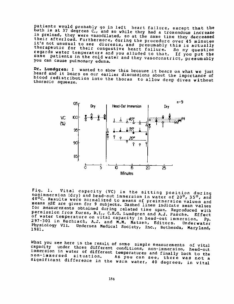
Dr. Lundgren: I wanted to show this because it bears on what we justheard and it bears on our earlier discussions about the importance ofblood redistribution into the thorax to allow deep dives withoutthoracic squeeze.
n=9I05
DryHead-OuI ImmersionDry
~ b,
0VC IOO
85
20 25I0 l5
Minutes
Fig. l. Vital capacity VC! in the sitting position duringnonimmersion dry! and head-out immersion in water of 20, 35, and40 C. Results were normalized to means of preimmersion values andmeans ++E are given for 9 subjects. Dashed lines indicate mean valuesfor measurements obtained during related time span. Reproduced withpermission f rom Kurss, D.I., C.E.G. Lundgren and A.J. Pasche. Effectof water temperature on vital capacity in head-out immersion. Pp.297-301 in Bachrach, A.J. and N.N. Natzen, Editors. UnderwaterPhysiology Vl I. Unde r sea Nedica 1 Soci e ty, Inc., Bethesda, Nary land,1981.
What you see here is the result of some simple measurements of vitalcapacity under three different conditions, non-immersion, head-outimmersion in water of different temperatures and finally back to thenon-immersed si tuation. As you can see, ther e was not asignificant difference in the warm water, 40 degrees, in vital
186
patients would probably go in left heart failure, except thatbath is at 37 degrees C., and so while they had a tremendous increasein preload, they were vasodilated, so at the same time they decreasedtheir af texload. Furthermore, during the procedure over 45 minutesit's not unusual to see diuresis, and presumably this is actuallytherapeutic for their congestive heart failure. So my questionregards water temperatuxe and you alluded to that. If you put thesame patients in the cold water and they vasoconstrict, pxesumablyyou can cause pulmonary edema.
capacity compared to non-immersion, while in 35 degree water,thermoneutral, you have the classical 5 or 6 percent reduction invital capacity which has always been ascribed to blood redistributioninto the chest. In colder water, 20 degrees C, you have a furtherdecrease in vital capacity averaging some l0 percent. The point hereis twofold, one is that it is rather surprising that one caneliminate or reduce the hydrostatic effect of the external watercolumn on your capacitance vessels by warming the water. The otherpoint regards the potential consequence for the fall in intrathoracicpressure relative to ambient pressure during a deep breath hold dive.These observations suggest that you may have a greater intrathoracicpressure drop in warm water than in cold water.
Dr. Boysea: In patients with kidney stones, some are breathingspontaneously with an epidural anesthetic, and I haven't had thembreath-hold, but there are some patients that who are under generalanesthetic and sometimes it's helpful to give an inflation to fixthe position of the diaphragm in order to set up the kidney, so youcan focus the stone. This situation is the opposite of breath-holddi.ving in my view, based on what you just said, because you make thepleural pressure more positive and, although they are at the sametime immersed, they are exquisitely sensitive to that technique andtheir arterial pressure just drops out.
Dr. Craig: In regard to your comment about the response of peoplewith congestive failure and so forth to immersion, I remind you thata number of years ago John Enolls and others looked at the heart rateresponse to a Valsalva type maneuver in normals and in people withcongestive failure. That was a classic paper for that period.
t%0IFI CAT IONS IN THE VASOMOTOR AfN
OF THE HINAN DI VIN6 RESPONSE
by Robert W, Arnold, M.D,
current «the
Mayo Graduate School of Medicine
Department of Ophthalmology
Rochester, Minnesota 55905
188
NSIFicATIONS IN THE vAsoNOToR ARf OF T% NNAH DIVINs REsP0NSE
ABSTRACTNIMxi e terial pressure is maintained during the human diving resfxese because the vagal
bradieerdia is accompanied by an intense peripheral vasminstrlction. If the vasomotorcomponent of the diving ~ense of soma diving animals is altered, blood pressure regulation isdisrupted.
To test the hypothesis that blood pressure regulation takes priority over braditcardla in thehuman diving respomxi, healthy subjects were exposed to conditions which altered their abilityto vtmonstrict, Intense snblant heating gadually attenuated seated diving bradycardia hChange Of pOSture frIXn SeaM tO Supine raxnrered a diving bradyCardia Superimpasedthiazide-induced hypovolemia and ilnbient heating resulted in e rapid elimination of divingbradyCardia The CharacteriStic forearm blOOd flow dm.else that is nOrInaliy a part Of thediving rissfonsa was radijced by enbient heat exposure, At room temperature, the peripheralvasodilators hydralazine an arteriolar smooth muscle relaxant! and particularly pramsin analphai -adrenergic antagonist} reduced seated diving bradfcardia. These results emphasize theImportance of adequate cardiac pre-load and after-load to the br+erdie of the human divingresponse
lNTRODUCT loNTrigemlnal nerve thermoreceptors stimulated by facial immersion and
breath hold comprise the primary afferent arms of the diving response.The two efferent arms of the diving response are vagal bradycardia andselective adrenergic peripheral vasoconstriction. As a result of theconcommitant vasoconstriction, the diving response is an unique vagotonicmaneuver since central mean arterial pressure is maintained, even duringextremes in bradycardia . Figure l is a diagram of the components ofblood pressure. Blood pressure is a function of cardiac output andperipheral resistance. During apneic facial immersion, cardiac outputfalls in proportion to heart rate while total peripheral resistance risesby selective shifts in vasomotor tone in the parallel vascular beds.Syncope would follow a brief period of hypotension in the cerebralcirculation.
If total peripheral resistance is fixed or reduced during apneic facialimmersion, there could be at least two possible outcomes depending on thepriority of cardiovascular control of the organism. Either blood pressurewould fall during bradycardia resulting in light-headedness or syncope, orthe preservation of blood pressure would be favored at the expence ofdiving bradycardia. The following non-invasive experiments were designedto test the hypothesis that blood pressure takes priority over diving
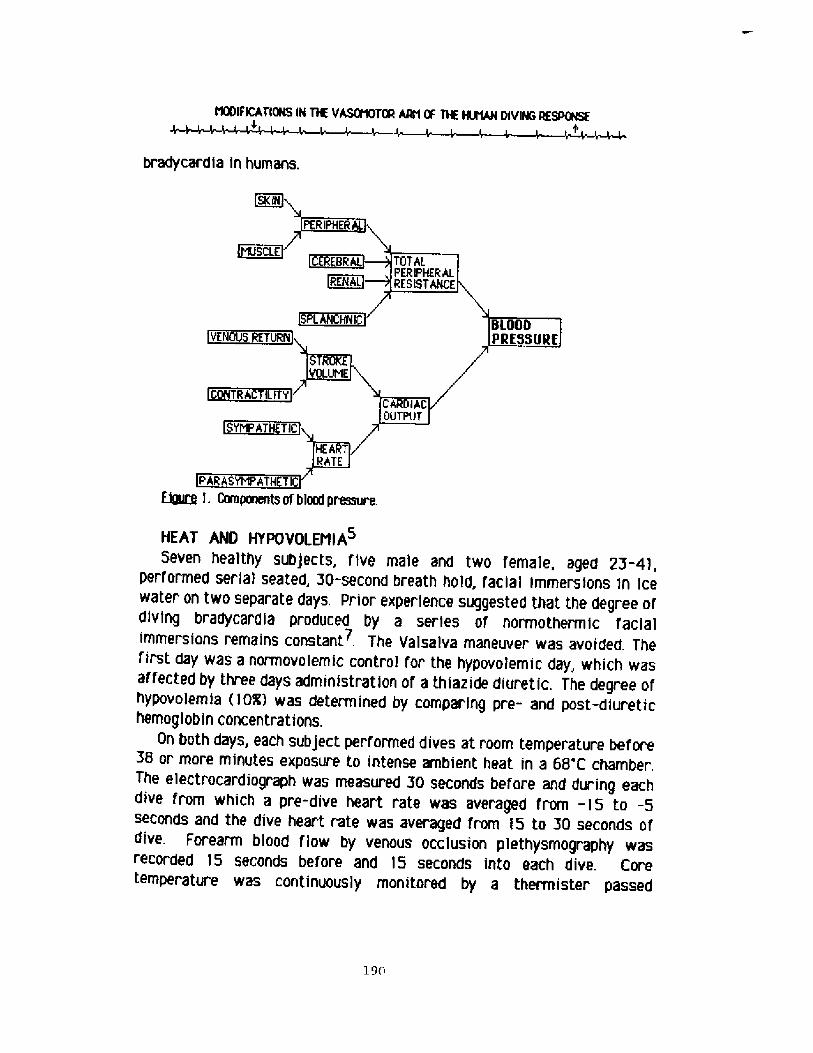
NNFICATIONS IN T% VAS9%TOR AAti OF T% fNNAM DIVING RESPNSE
bradycardia in humans.
Bguce l. Components of blaxf pressure.
HEAT AND HYPOVOLENIA
Seven healthy subjects, five male and twa female, aged 23-4!,performed serial seated, 30-second breath hold, facial immersions in icewater on two separate days, Prior experience suggested that the degree ofdiving bradycardia produced by a series of normothermic facialimtTIersionS remainS COnStant . The ValSalva maneuver was avOided. Thefirst day was a normovolemic control for the hypovolemic day, which wasaf fected by three days administration of a thiazide diuretic. The degree ofhypovolemia �QR! was determined by comparing pre- and post-diuretichemoglobin concentrations.
Qn both days, each subject performed dives at room temperature before58 or more minutes exposure to intense ambient heat in a 68'C chamber.The electrocardiograph was measured 30 seconds before and during eachdive from which a pre-dive heart rate was averaged from -15 to -5seconds and the dive heart rate was averaged from 15 to 30 seconds ofdive. Forearm blood flow by venous occlusion plethysmography wasrecorded 15 seconds before and 15 seconds into each dive. Coretemperature was continuously monitored by a thermister passed
190
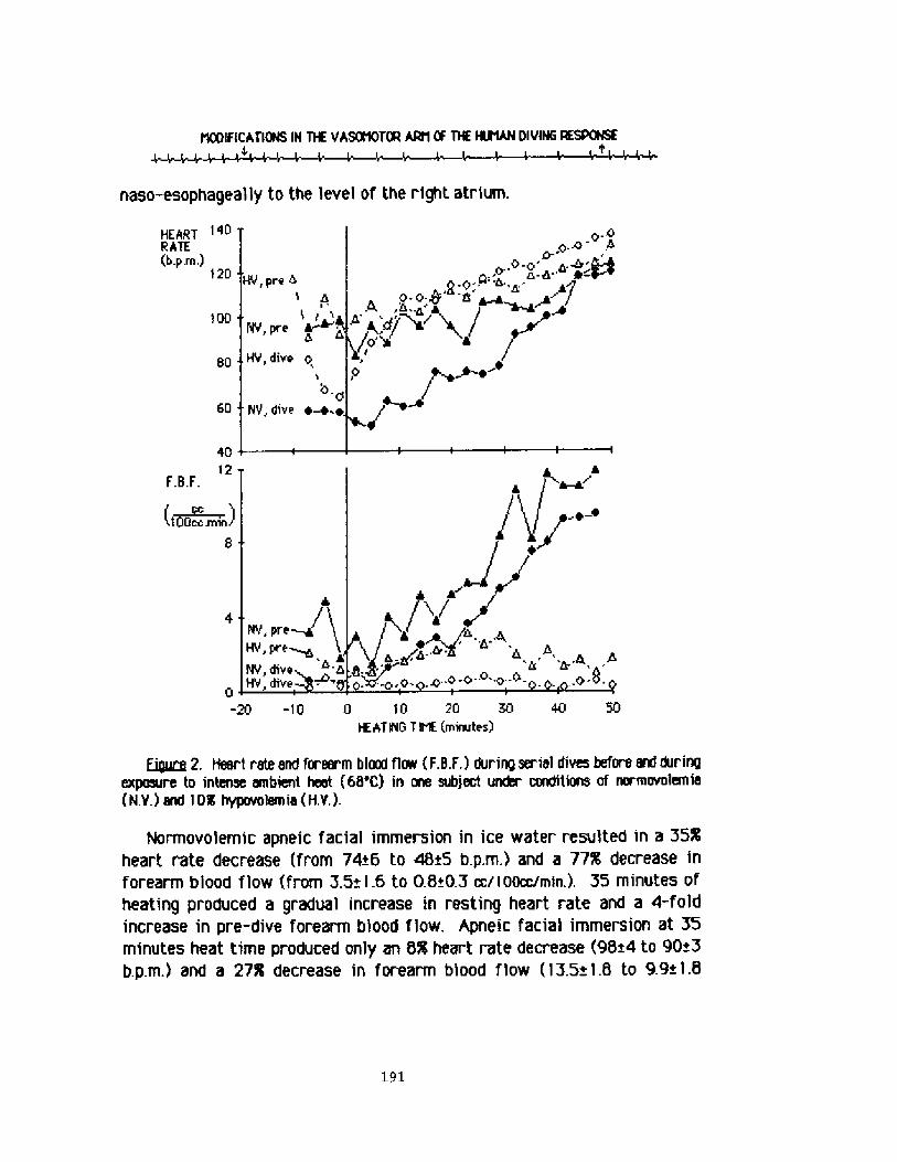
NSIFICATIONS IN T% VASQ%TOR ARt1 Ã THE NNAN DIVIN6 RESPONSE
naso-esophageally to the level of the right atrium.
HEART 140RATE
b.p,ln.! 120
40'l2
F.B.F.
fOQcc.min!
S 0 -20 -10 l3 10 20 R3 40 50HEAT ING I II' mimrtes!
Eiguce 2, ffeart rate end forearm blood flow F.B.F.! during serial dives before and duringexpaaure tO intenSe INIbient heat �B'C! in me SubjeCt uxor conditiee of m.mOvstemie N.V.! and I M hypavolemia H.Y,!
Normovo'lemic apneic facial immersion in ice water resulted in a 35%heart rate decrease from 74+6 to 48 5 b.p.m.! and a 77K decrease inforearm blood flow from 3.5+ 1 6 to 0.8<0.3 cc/1oocc/min.!. 35 minutes ofheating produced a gradual increase in resting heart rate and a 4-foldincrease in pre-dive forearm blood flow, Apneic facial immersion at 35minutes heat time produced only an 8% heart rate decrease 98s4 to 90+3b.p.m.! and a 27R decrease in forearm blood flow I3.5+1.8 to 9.9+l,8
191
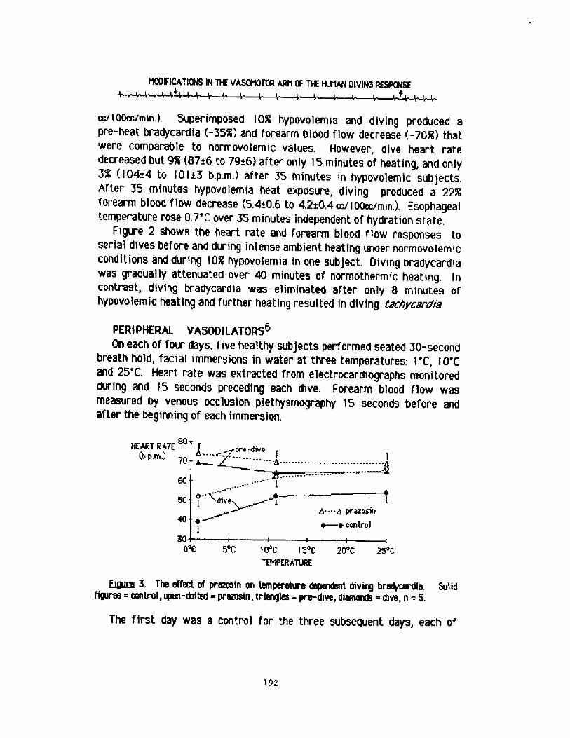
NGIFIcATIONS IN T% vARNQToR NN 0F T% NNAN DlvING REsPGNsE
cc/IOOoc/min.!, Superimposed 10K hypovolemia and diving produced apre-heat bradycardia -35%! and forearm blood flow decrease -70%! thatwere comparable to normovo'Iemic values. However, dive heart ratedecreased but 9% 87+6 to 79+6! after only 15 minutes of heating, and only3% �04~4 to I01+3 b.p.m ! after 35 minutes in hypovolemic subjects.After 35 minutes hypovolemia heat exposure, diving produced a 22Kforearm blood flow decrease �,4+0.6 to 42+0,4 cc/I oocc/min,!. Esophagealtemperature rose 0,7'C over 35 minutes independent of hydration state,
Figure 2 shows the heart rate and forearm blood flow responses toserial dives before and during intense ambient heating under normovolemicconditions and during 10% hypovolemia in one subject. Diving bradycardiawas gradually attenuated over 40 minutes of normothermic heating, Incontrast, diving bradycardia was eliminated after only 8 minutes ofhypovolemic heating and further heating resulted in diving tachyca.gaia
PERIPHERAL VASODILATORS6On each of four days, five healthy subjects performed seated 30-second
breath hold, facial immersions in water at three temperatures: 1'C, 10'Cand 25'C. Heart rate was extracted from electrocardiographs monitoredduring and IS seconds preceding each dive. Forearm blood flow wasmeasured by venous occlusion plethysmography 15 seconds before andafter the beginning of each immersion.
HEART RhTE b.p.m.! 7G
50
3o G C 5 C 1O c 15 C 2o c 25 cTEHPERATURE
Qyzg 3. The effect of premein on temperature dependant diving b~~erdia Solidfigures mntrol, open-dotted pramsin, triangles = pre-dive, diamonds = dive, n = S.
The first day was a control for the three subsequent days, each of
l92
NSIFICATIONS IH T% VARNOT% NN OF T% NNAM OIVIHG AESPNSE
which characterized by administration of a low dose peripheralvasodilator.. 50 mg hydralazine by mouth, 2 mg prazosin by mouth and then15 mg nitroglycerine paste transderrnally. Due to the characteristicindividual variability of resting heart rates and bradycardia, data areanylyzed by percentage change from baseline using analysis of variance ANOVA! and paired t-Test,
In the absence of drugs, apneic facial immersion produced a consistent,temperature dependent bradycardia -34R 0 1'C, -12R 0 IO'C, -8R e25'C! and forearm blood flow decrease -62'R 0 I 'C, -40R 0 10'C, -21R 925'C!. Nitropaste produced little change in dive heart rate. Hydralazineproduced a reduction in diving bradycardia at I'C -34R to -28R, p<.05!.Prazosin produced a reduction in temperature dependent bradycardia F, �-11, p<,05! speci f ica Ily at 10'C -12R to - I R, p<.05! and 1'C -34R to-17R, p<.02! shown1n Figure 3. Doses of peripheral vasodilators was keptsufficiently low to limit changes In Forearm blood fiow,
HEAT ANO POSTURE4One healthy 23 year old male subject performed a series of 30-second
breath hold facial immersions in a basin of water at 2-4 C. At least threeminutes separated each dive. The Valsalva maneuver was avoided,Electrocardiograph was recorded for the 15-seconds preceding each diveand during the 30-second dive. Three dives were performed at an ambienttemperature of 22'C followed by seven dives at 86'C. t1ost of the diveswere performed in a seated posture, however the dive at 26 minutesheating was performed supine. At 32 minutes of heating, a 30-secondbreath hold without immersion was performed, then the experiment wasterminated at the request of the subject,
F igure 4 shows ambient temperature Ta!, immersion watertemperature TII2O!, reCtal temperature Tr!, and 15-SeCOnd pre-dive anddive mean and standard deviation heart rate before and during heatexposure, The 44R pre-heat dive heart rate decrease from 77+5 to 43i2b.p.m.! was slightly greater than the average for the previous groups.After only 22 minutes at this high ambient heat, rectal temperature hadrisen 0.7'C, and the seated diving bradycardia had been eliminated. At 26minutes of heating, a change of posture to supine reduced pre-dive heartrate and resulted in an 8R diving bradycardia. At 32 minutes of heat,breath hold alone produced a 5R heart rate increase
193
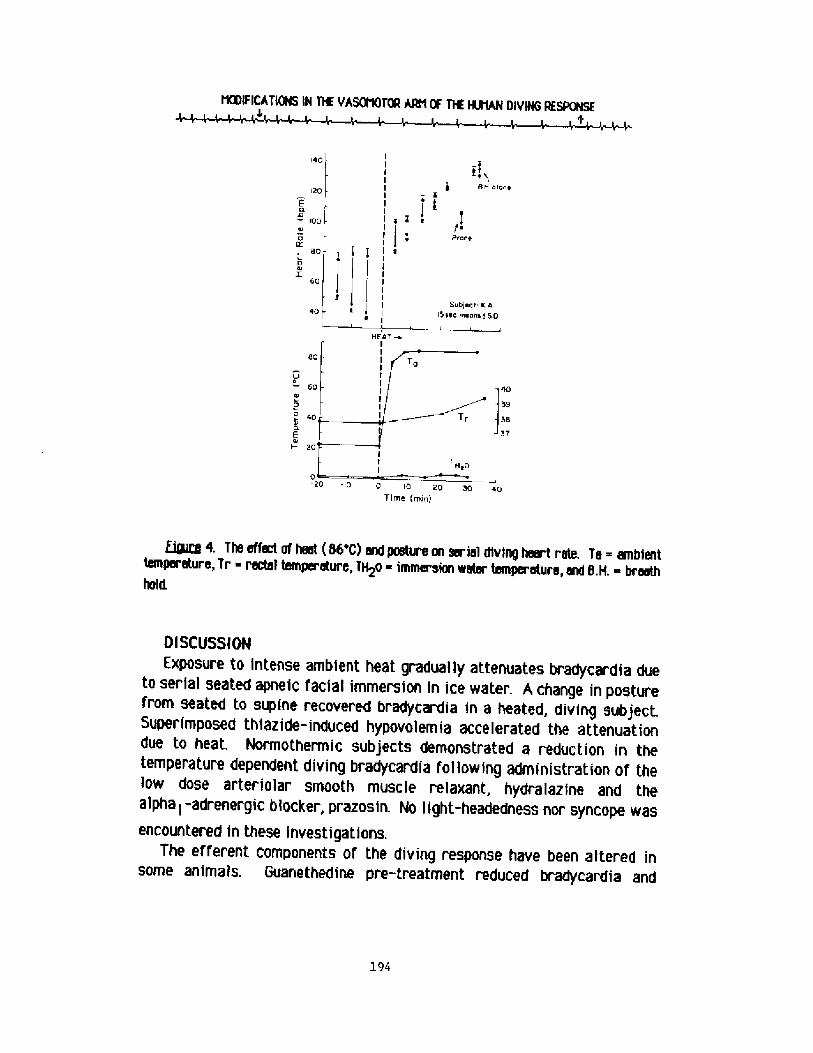
NNFICAT[ NS iN T% VARMTN AAN% T% %HAM Div NG RESPONSE
IIIIII
I 4 ! e
[ l I eIIIIII
|,~BH e luce120
f a� 100 l
600Z
60
Subject K A15 eee ere cue t 6 0
HEATeI
V 060
te
40et0.EI- 20
0 10 20 30 4pT irtte rttirr!
QgZe 4. The effect of heat 86'C! and posture m serial diving heart rate. Ta ambienttemperature, Tr rectal temperature, TH2o - immarsie water temperature, and S.H. - breathhold.
l94
Ol SCUSSIOM
Exposure to intense ambient heat gradually attenuates bradycardia dueto serial seated apneic facial immersion in ice water. A change in posturefrom seated to supine recovered bradycardia in a heated, diving subject.Superimposed thiazide-induced hypovolemia accelerated the attenuationdue to heat. Normothermic subjects demonstrated a reduction in thetemperature dependent diving bradycardia following administration of thelow dose arteriolar smooth muscle relaxant, hydralazine and thealpha l -adrenergic blocker, prazosin. No light-headedness nor syncope wasencountered in these investigations.
The efferent COmponents Of the diVing respOnse haVe been altered inSOme animals. Guanethedine pre-treatment reduCed bradycardia and
CSIFICATIONS IN T% VASNOTOR ARti OF T% NNAN DIVING RESPNSE
induced hypotension in diving ducks. Reserpine pre-treatment resulted infatal hypotensive diving response in these birds . Reserpine produceddiving hypotension in rats, aiso . When the bradycardia of young divingseals was overpowered by electrical cardiopacing, hyper tension occurredand one seal died
The effect on the diving response of such manipulations as exercise,change in posture or intra-pleural pressure, heat and mental stimulationhas been studied in humans. One paper reports on the effect ofalpha-adrener gic agents in the human diving response. Finley, et al, foundrelatively little effect of phentolamine on diving bradycardia during abrief, eight second immersion . However, diving produced significantbradycardia from isoproterenol-induced tachycardia.
Diving bradycardia persists during exercise, in fact some of the lowesthuman dive heart rates have been recorded in exercising subjects . Ithas been pro osed that venous return during exercise permits divingbradycardia . Diving bradycardia is greater in dynamic than isometricexercise presumably because of venous return
intense ambient heat exposure produces some cardiovascular changessimilar to exercise: tachycardia and an increase in core temperature.Relatively few papers address the effect of heat on the diving response.Pulse decreased during shower cooling after sauna bathing but a measureof the effect of such cooling on normothermic subjects was notmentioned, Bachmann, et al, using intra-arteriai blood pressuretelemetry found a normotensive bradycardia following a transient markedhypertension �00/240 mm. Hg! when a subject was totally immersed in a15'C pool after sauna~, Caputa and Cabanac reported a moderatebradycardia in response to eupneic facial immersion in subjects who hadbeen warmed in a 40'C bath, however they did not include normothermiccomparison data
The heart rate decrease during apneic immersion is greater with lowerintra-pleural pressures . Lower diving heart rates accompany head-downposture than head up . Venous return is favored by both of thesemaneuvers.
One of the few factors which reduces human diving bradycardia ismental preoccupation such as performing mental arithmetic during facialimmersion, Nental arithmetic produces a dilatation of peripheral
l95
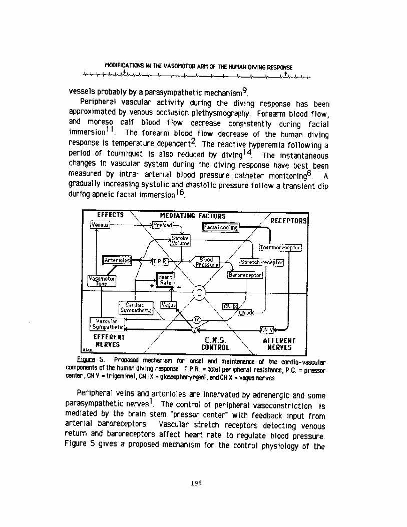
NRIFICATioNs IN THE VAQNOTOR ARt OF THE HH1AN OIYING RESPONsE
vessels probably by a parasympathetic mechanism,Peripheral vascular activity during the diving response has been
approximated by venous occlusion plethysmography, Forearm blood flow,and moreso calf blood flow decrease consistent ly during facialimmersion . The forearm blood flow decrease of the human divingresponse is temperature dependent . The reactive hyperemia following aperiod of tourniquet is also reduced by diving . 7he instantaneouschanges in vascular system during the diving response have best beenmeasured by intra- arterial blood pressure catheter monitoring . Agradual ly increasing systolic and diastolic pressure follow a transient dipduring apneic facial immersion' .
Lyly 5. P~ mechenism for onset end meintenence of the cerdio-vescuiercomponents of the human diving resporme. T.P.R. = tote! peripheral resistance, P,G. = pressorcenter,CNY trigemineI,CNIX giossopheryngeei,nndCNX vegusnerves.
Peripheral veins and arterioles are innervated by adrenergic and someparasympathetic nerves . The control of peripheral vasoconstriction ismediated by the brain stem "pressor center with feedback input fromarterial baroreceptors. Vascular stretch receptors detecting venousreturn and baroreceptors affect heart rate to regulate blood pressure,Figure 5 gives a proposed mechanism for the control physiology of the
196
t1OGIFICATioNS IN THE VASOfQTOR JUN OF T% NNAN OIVING RESPONSE~W~l- ~ ~l~
onset and maintenance of the human diving response. The primary afferentis fac ial cooling mediated by trigeminal thermoreceptors and enhanced bybreath hold. The two ef ferent arms are vagal bradycardia and sympatheticperipheral vasoconstriction. The brain stem control center s! for thediving response has not been clearly defined. Only instantaneousmonitoring of the vascular system can delineate whether bradycardiaprecedes vasoconstriction or vice versa during the first few seconds afterimmersion. Vascular stretch receptors and baroreceptors continuallymonitor cardiac pre- load and af ter-load respectively, and provideadditional afferent input to the maintenance of the diving response.Chemoreceptors, pulmonary stretch receptors, higher cortical centers, andothers evidently also provide input to the controi of the diving response.
Venous return and peripheral vasoconstriction have a major infiuenceon the degree and duration of the diving response. Adequate cardiacpre-load and after-load are prerequisites to human diving bradycardia.The control mechanism for the diving response in some animals seems tofavor bradycardia and vasoconstriction in response to immersion even ifblood pressure regulation goes awry, ln contrast, it appears that bloodpressure maintenance and avoidance of syncope takes priority overbradycardia in the human diving response.
ACKNOWLEDBEMENTSThe guidance end facilities of Dr. Robert Elsner, University of Alaska-Fairbanks and Dr.
Ethen R. Nadel, John 8. Pierce Foundation and Yale University Schooi of Medicine have beenmuch appreciated.
REFERENCES1 Abboud,FM., Eckstein phf, Reflex vasoconstrictor end vasodilator respoees in man.
Circulation Res. 29: suppl l96-103, 19662 Arnold,R'W. A vasomotor comperison of the human diving response and the cold pressor test.
Alaska Medicine 28: accepted, 1986.Arnold,RW. Extremes in human breath hold, fILial immersion bradltcardia. Under'Biomed. Res. 'I 2: 185-190, 1985.
4 Ar nold,RW. Modifications in the vasmnotor component of the human diving resporee. M.D.Thesis, Yale, 1984.
5 Arnold,RW., Nadel,EN. The attenuating effs:t of heat end hypovolemia on the human divingre~. Submitted.
6 Arnold,A W., Lister,6., Madel,EN. The effect of peripheral vmdilators m the human divingrespon.'e. Submittett
197
rtNIFIcATIONS IN T% vAsoMoTOR ARM QF T% %RAN DlvlNG RESPQ6E
7 Arnold,RW. The human diving br+mrdia end its raerery time. B.S, Thesis, University afAlaska-fairbanks, 1979.
8 Becheae,K,, Hoffme,H., Gunther,W,2erzawy,R. Ergebnisse telamitrischer Kreis-
9 Bancroft,H,, Brad p., Hej 1g., Hirsjavi,EA., KitchinpH. Mechanism of vasodilatation infar«x'm muscle during stress mental « ithmetic!. Clin. Sci. 19: 577-586, 1960.
10 Beqynrm.SA., Cempbell pK,, Wildsnthaf,K, Diving reflex inman; its relation to isOmetriCand dynamic exercise. J. Appl. Physiol. 33: 27-31, 1972.
I I Campbell,LB,, Gooden,BA., Lahm«r,RO., Pym P. Simultaneous calf end forearm blood flowduring immersion in mrN, Aust.J. Exp. Bio!. Mel, Sci, 47: 747-754, 1969.
12 Caputa,M., Qsbenec,M. Bradycerdie during fae cooling in man mey be produced by selectivebrain calling. J, Appl. Physiol. 46: 905-907, 1979.
13 Craig jS. Heart rate rashes to apneic urxhrwatar diving end to breath holding in mrN. J.Appl. Physiol. 18: 854-862, 1963.
14 Elsner,R., Oaadsn JA. Reduction of reactive hyperemia in the human fare«'m by faximmcrsim J, Appl. Physial, 29 627-630, 1970.
15 Finleyp>., Bonnet/F., WaxmrN,MB. Autan«nic pathways reyesible f«br+cardia onfix:Ial Immersion. J. Appl. Physiok 47: 1218-1222, 1979.
16 Kewak«ni,Y., Netelsan,B., DuBolspB. Cardioveeul«effects of face immersion end factorsaffecting diving reflex in m«r. J, Appl. Physiol, 23: 964-970, 1967.
17 Kobinger,W., Oda,M. Effects of sympathetic blacking substances on the diving reflex inducks. Eur.J. Phermrx'. 7: 289-295, 1969.
18 Lin,YC. Autonomic mvous cretrol af cerdiavascul«respanse during diving in the r at. Am.J. Physlol. 227: 601-605, 1974.
19 Matousek p., Pribil,M, Reaction of pulse frequency to various methods of aeling in sauna.Fyslat. Vestn. 56: 208-2'I 2, 1978.
20 Murdaugh,HV., Craes,CE., Millen+., Ges,JBL., Robin,ED. Dissociation of bredyoerdia anda terial constrictian during diving in the seel, Phm vith/iver Science 162: 362-363,1968.
21 Olsen CR., Fanestil,DD., Scholandsr,PF. Some effects of epneic underwater diving on bloodgases, lactate end pressure in mrN. J. Appl, Physiol. 17: 938-942, 1962.
22 Ross4, Steptoe4 Attenuation of the diving reflex in m«r by mental stimuiatirN. J.Physiol. Landon! 302: 387-393, '1 980.
23 Song,DH,, Lar,WK., Chung,YA., Hong,SK. Mechanism of apneic br~erdia in man. J. Appl.PhysIol. 27: 323-327, 1969,
24 Stramme,SB., Kerem,D, Elsrer,R, Diving bradycardia during rest and exercise and itsrelation to physical fitness. J. Appl. Physiol, 28: 614-62 I, 1970.
25 Williams,Ci,, Bernausr,EM. Diving reflex during exerci«r in m«r, Med. Sci, Sports 8:63-64,1976
l98
Discussion following Dr. arnold's presentation
Dr. Van Llew: I'm very interested in your talk. It seems to me therole of science is to predict and control< and you' ve described astory that is so complex that I don't think you' re rea'ly able topredict or control. Experiments done under the circumstances of yourstudy can fit into a scheme all right, but it's hard to use theinformation for predictions about experiments done under differentcircumstances'
Dr. hrnold: I agree. I have seen some healthy people with extremebradycardia while seated. I' ve used diving response to treatpa r oxysm al supr avent r i cul ar tachyca rdi a in olde r people too. Since theblood pressure is maintained during this vagotonic response, it may bea unique vagotonic maneuver and is very useful and probably quite safein inducing bradycardia because of that. Carotid sinus massage,Valsalva and other maneuvers are of ten associated with syncope,because blood pressure is not maintained. In the animals for whomdiving response is probably more important for their livelihood thanit is for humans, it appears that their diving response takes priorityover blood pressure. If they' re cardiac paced, or if their' vasomotorresponse is inhibited, they favor the diving response< i.e ~bradycardia or vasoconstriction, continues at the expense of bloodpressure. I suspect that the human does better maintaining bloodpressure at the expense of bradycardia or vasoconstriction in divingresponse.
Dr. Hong: In humans, the increase in blood pressure is quitesubstantial and that presents some problem in terms of oxygenconservation. One question that I was interested in is to explorethe role of stretch-receptors.
Dr. hrnold: There's A and B and they' re frequently found in theatrial walls. It's not clear exactly their role but it's shown inanimals that it's of f and on, but mainly the B type receptors caninduce bradycardia when they' re activated. They also regulate urineflow quite a bit< but I haven't been as interested in that.
Dr. Hong: Because in human subjects during head-out immersion acephalad blood shift takes place and the cardiac stretch receptors arepresumably stimulated, heart rate decreases even without breath-holding. That has been our f inding.
Dr. Elsner: You can impose! a thermal load on a diving seal byheating or cooling the hypothalamus, in a technique for looking at thepossible conflict between vasodilation of heating and vasoconstrictionof diving, and when you do this you f ind that the seal generallyfavors the vasoconstriction of diving.
199
The cardiac response to breath-hold diving at rest and while exercising, A comparison between man and diving homeotherms!
P, J, Butler and A. J. WoakesDepartment of Zoology and Comparative Physiology,
University of Birmingham, Birmingham B15 2TT, U.K.
The "classical" divin res onse
A reduction in hear t rate below the resting level br adycardia ! occursin man and in all air breathing vertebrates that habitually dive underwater, when they are exper imentally submerged in a resting state Lin 1982;Butler 5 Jones 1982 !. Complete submersion is not necessary; merely placingthe head under water is all that i.s required to elicit this response. Thesignificance of the br adycardia in aquatic air breathing vertebrates waselucidated by Irving �939 ! and Scholander 1940 !. Parts of the body thatcan withstand oxygen lack, e.g. gut, muscles, are effectively eliminatedfr om the circulation by intense vasoconstr iction while the oxygen-dependenttissues such as the heart and brain continue to be perfused. An increase inarterial blood pressure, as a result of the selective vasoconstriction, isavoided by a reduction in cardiac output, brought about predominantly bythe so-called "diving bradycardia" Fig la!. Thus oxygen within the body isconserved for the heart and CNS while the rest of the animal metabolisesanaerobically and accumulates lactic acid. The total, oxygen conservingresponse to involuntary submersion in aquatic homeotherms has been termedthe "classical" diving response Butler 1982!. Bradycardia is the typicalelement of the cardiovascular, and hence of the metabolic, adjustments toinvoluntary diving and is often taken as an indicator of the remainder ofthe response taking place Pasche A Krog 1980!, It is worth investigatingthe validity of this assumption in a little more detail.
Heart rate and ox en conservation durin breath-hold submersion atrest
There is ample evidence that the rate of oxygen consumption does infact decrease during such experimental submersion in air breathing aquaticvertebrates and that anaerobic metabolism increases Scholander 1940;Andersen 1966; Butler A Jones 1982; Klsner k Gooden 1983 !. It is alsoapparent that temperature of the water has no effect on the response indiving birds Andersen 1963; Butler & Jones 1968! or mammals Dykes 1974 !.Although both bradycardia and vasoconstriction in the per ipher alcirculation occur during apnoeic face immersion in man Wyss 1956; Heistadet al 1968; Campbell et al 1969 ! their intensities appear to be greater atlower water temperatures Kawakami et al 1967; Burch A Giles 1970 !. The'critical' water temperature in this respect is around 35 C and if thewater is colder than this there is a temperature-related bradycardia duringnon-apnoeic face immersion, whereas in warmer water little change in heartrate occurs Kobayasi 4 Ogawa 1973!. Despite the existence of thesecardiovascular adjustments during submersion, which in aquatic birds andmammals are concerned with conservation of oxygen, there is some doubt asto whether they are sufficiently intense in man to cause a reduction in the
200
rate of oxygen consumption .
Wolf et al �965 ! demonstrated that all the signs of reduced aer obicmetabolism and increased anaerobiosis are present in man during headsubmersion when a clear bradycardia from 85 to 50 beats per minute ! ispresent. However, in the absence of bradycardia during a similar period�0 s ! of breath-holding under ~ater, oxygen saturation of the blooddeclines more markedly and ther e is little post-dive accumulation oflactate in the blood. Similarly, Elsner et al �971! found that there is agood inverse relationship between the decline in partial pressure of oxygenin arterial blood and the fall in forearm blood flow during face immersionafter hyperventilation of oxygen. On the other hand, Hong et al 1971!found that the rate of removal of oxygen from the lungs and blood during a4 min breath-hold in air is slightly greater than pr'e-apnoeic restingoxygen consumption. However, it must be noted that at 2 min into the breathhold at least, there were no significant reductions in heart rate andcardiac output nor an increase in peripheral resistance see Table 6 inHong et al 1971!. Even when a bradycar dia was reported to have occurr ed,the lowest mean heart rate was approximat,ely 75 beats per minute see Fig 4in Hong et al 1971!. To add to the apparent confusion it has recently beendemonstr ated that breath-hold dur ation in man is no longer when submer sedin water at 32 C than it is in air despite the fact that heart rate is
0
significantly lo~er during the former Ster ba 4 Lundgren 1985!. This resultwould tend to support the contention. that, unlike the situation in aquatichomeotherms, the period of breath-hold diving in man does not exceed thatwhich would be expected on the basis of usable oxygen stores and restingoxygen consumption Lin 1982 !. The situa.tion is complicated, however, bythe fact that afferent information from the respiratory muscles themselvesmay stimulate respiratory activity during apnoea before cr itical levels ofoxygen and/or carbon dioxide are reached Fowler 1954;Campbell et al 1967 !.
Heart rate durin breath-hold divin while exercisin
As long ago as 1960 Eliassen, on the basis of his own observations ofnatural dive durations in aquatic birds and of his calculations of the
amount of usable oxygen that they could store in their body, argued that itis not necessary for these habitual divers to undergo the intensecardiovascular adjustments of the "classical." diving response duringnatural dives. They should be able to metabolise completely aerobicallywhile swimming under water. This paper and its provocative suggestion werelargely ignored for almost 20 years, but when it was possible to recordphysiological data fr om freely diving birds and mammals it turned out thatreduction of heart rate below the resting level and anaerobiosis are notcomponents of routine dives in these animals .
By using implanted radiotransmitters it has been demonstrated forseveral species of birds that, unlike the situation dur ing submersion ofthe head of a resting animal, there is no maintained reduction of heartrate below the resting level during spontaneous dives Butler h Woakes
large reduction in heart rate to approximately 30% of the pre-dive valuewithin 30 s of involuntary submer sion of the head Fig 1a! and this is
20l
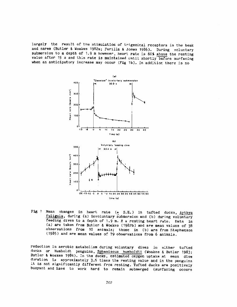
largely the result of the stimulation of tr igeminal receptors in the beakand nares Butler 4 Woakes 1982a; Furilla I Jones 1986!. Dur ing voluntarysubmersion to a depth of 1.9 m however, heart rate is 60% above the restingvalue after 15 s and this rate is maintained until shortly before sur facingwhen an anticipator y increase may occur Fig 1b!. In addition there is no
e!Cfage!ngl' invelunl ~ 12 gubmereien
g00 300 g
300
C e es! 200
e 100e
Z 0 -10 -5 0 5 10 15 2D 25 30 35 AOTime ie!
Ib!Volenfery feeding dive
500 c' 23rg g
gOOm 2 m
300ess
2 DOe
100
Z 0 -20 -15-10 .5 0 5 10 15 20 25 30 35 gO g 5 50 55 50yfme m!
Fig 1 nean changes in heart rate + S,E.! in tufted ducks, ~Ath a~fuli uia, during a! involuntary submersion and b! during voiuntar yfeeding dives to a depth of 1.9 m. R = resting heart rate. Data in a! are taken from Butler 4 Woakes �982b! and are mean values of 38observations from 10 animals; those in b! are from Stephenson�985 ! and are mean values of 79 observations from 6 animals.
reduction in aerobic metabolism during voluntary dives in either tuftedducks or Humboldt penguins, S heniscus humboldti Woakes 4 Butler 1983;Butler 4 Woakes 1984 !. In the ducks, estimated oxygen uptake at mean diveduration is approximately 3.5 times the resting value and in the penguinsit is not significantly different from resting . Tufted ducks are positivelybuoyant and have to work hard to remain submerged surfacing occurs
202
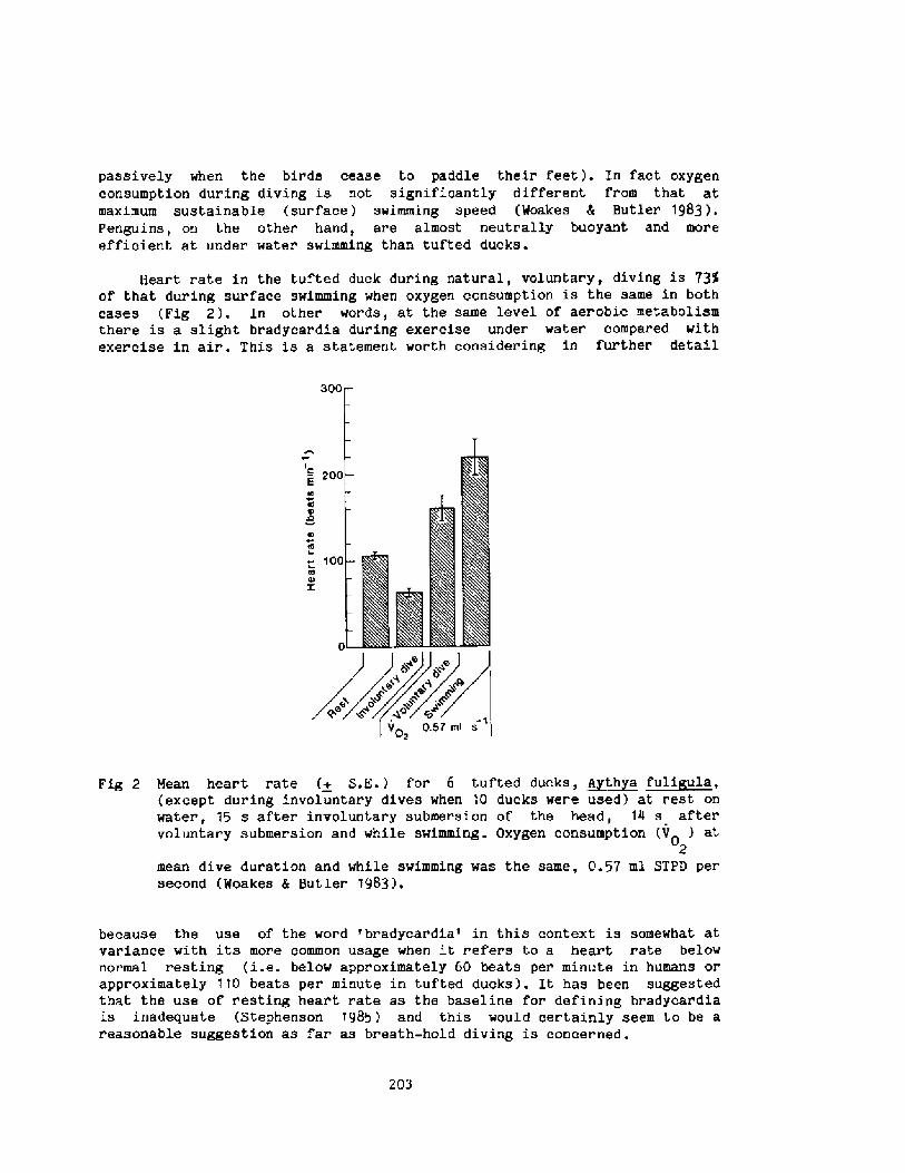
passively when the birds cease to paddle their feet !. In fact oxygenconsumption during diving is not significantly different from that atmaximum sustainable surface ! swimming speed Woakes 4 Butler 1983 !.Penguins, on the other hand, are almost neutrally buoyant and moreefficient at under water swimming than tufted ducks .
Heart rate in the tufted duck during natural, voluntary, diving is 73%of that dur ing surface swimming when oxygen consumption is the same in bothcases Fig 2 !. In other words, at the same level of aerobic metabolismthere is a slight bradycardia during exercise under water compared withexercise in air. This is a statement worth considering in further detail
ffg 2 Ncaa heart rate + E.E.! for 6 tufted ducks, ~Ath a ~felt ula, except during involuntary dives when 10 ducks were used! at rest onwater, 15 s after involuntary submersion of the head, 14 s aftervoluntary submer sion and while swimming. Oxygen consumption V ! at
0
mean dive dur'ation and while swimming was the same, 0.57 ml STPD persecond Woakes 5 Butler 1983!.
because the use of the word 'bradycardia' in this context is somewhat atvariance with its more common usage when it refers to a heart r ate belownormal resting i.e. below appr oximately 60 beats per minute in humans orapproximately 110 beats per minute in tufted ducks!. It has been suggestedthat the use of resting heart rate as the baseline for defining bradyoardiais inadequate Stephenson 1985 ! and this would certainly seem to be areasonable suggestion as far as breath-hold diving is concer ned.
203
The fact that heart rate in tufted ducks is lower during naturaldi.ving than during exer cise in air at the same level of aerobic metabolismand yet is substantially above resting Fig 2! has led Butler �982 ! iosuggest that the cardiovascular response to natural diving in these birdsis a balance between the "classical" diving response and the response toexercise, with the bias towards the latter. This implies that exercisemodifies the "classical" diving response or vice versa! in tufted ducks atleast to the extent that the active locomotory muscles, as well as the CNSand heart, receive sufficient blood and hence oxygen ! to metaboliseaerobically. The oxygen used from body stores is quickly r eplaced uponsurfacing. Other' par ts of the body, such as the respirator'y muscles and thelarge flight. muscles, may well reduce the rate at which they consumeoxygen, as postulated by the "classical" hypothesis, but whether or notthey metabolise anaerobically and produce lactic acid remains to be seen.
Data from seals indicate that they also metabolise aerobically, usingstored oxygen, during the vast majority of their natur al dives. In greyseals Halichoerus r us! heart rate is approximately 30 beats per minuteduring submersion and 1 10 beats per minute when breathing at the surface,irrespective of whether the animal is resting or swimming. As swimmingspeed increases, the time spent submerged decreases so that overall meanheart rate increases with oxygen uptake in the typical fashion. There is noincrease in blood lactate during such swimming activity. However, if theanimal is alarmed in any way then heart rate falls to very low rates of�0 beats per minute whether it is r'esting or swimming. This is typicaL ofthe classical oxygen conserving response Fedak et al 1984 !. The majorityof dives per formed by WeddeLL seals, Le ton chotes weddelli, in nature areless than 25 min in duration and completely aer obic . Only after those fewdives which are longer than 25 min is there an incr ease in lactic acid inthe blood, the level of which is related to the duration of the dive Kooyman et al 1980!.
Man, of course, is not a habitual diver so the difficulty indemonstrating some functional significance of the car diovascular responsesthat occur during facial submersion in resting subjects is perhaps notsurprising. Breath-hold diving for the "fruits of the sea" is stillpractised by a diminishing! number of humans and many of these performunassisted dives. That is, the diver carries no weights and {like thetufted duck! has to over come his/her own drag and buoyancy during descent.Unlike the tufted duck, ascent is only partly assisi,ed by the diver' sbuoyancy. Thus, during these dives the individual is exercising and in viewof the dai,a from naturally diving birds and mammals it was decided to lookmore closely at the cardiac r esponses during active submersion in humans.
The change in heart rate in man during apnoeic exercise in water hasbeen reported on a number of occasions but there is no consistency in thetechniques that wer e used and there are quantitative as well as qualitativedifferences in the results that wer'e obtained. A number of authors havefound that heart rate falls gradually to below the resting value duringsubmersion in man while exercising. The subjects in the study by Scholander'
204
et al �962! began from r est in water and ther e was an initial increase inheart rate followed by the bradycar dia. In other studies the subjects werealready exercising, either" in water Irving 1963; Stromme et al 1970! or ona cycle ergometer in air Asmussen & Kristiansson 1968; Bergman et al 1972;Oldr idge et al 1978; Bjertnaes et al 1984; Smeland et al 1984 !, beforesubmerging their head and becoming apnoeic. Heart rate was, therefore,already elevat,ed above the resting level before the "dive", but it. oftenfell to the same level as that recorded during "diving" under restingconditions . On the other hand, Craig �963 ! found that when a man restingquietly on the bottom of' a pool began to exercise, his heart rate increasedabove its resting value and declined at the end of' the exercise. Thetachycardia of exercise, although attenuated, was still present in subjectsexercising on a cycle ergometer completely submerged in water ,Craig 5 Nedd1968 !. In Korean ama, heart rate, 30 s after the start of breath-holddiving or of swimming, just below the surface in summer, was slightly higheror similar' to resting, heart rate Hong et al 1967!.
In the present study hear t rate was monitored from 6 male subjects ofmean age + S.E.! of 24 + 1 years and mean mass 73.'5 + 2.4 kg. A singlechannel PIN r adiotransmitter Butler 4 @cakes 1982b! was tucked into theback of the subjects' trunks and leads attached via waterproof connectorsto two electrodes; one electrode was attached over the sternum and theother approximately 20 cm lower on the left-hand side of' the chest.Measurements were made in a 25 m swimming pool at a vater temperature of028 C. Resting heart rate was obtained from the subjects wrapped in a gownand sitting quietly on a seat and was 67 + 3.7 beats per minute. When thesubjects with a nose clip! submerged themselves completely in the pool,but remained inactive, there was a prompt but gradual reduction in heartr ate which r cached 72% of the r esting value within 30 s and 604 of therest,ing rate at mean dive duration of 59 + 5 .6 s Fig 3 !. Hear t rate wassignificantly below the resting rate within 15 s of submer sion. Thesubjects then dried, wrapped themselves in a gown and sat on the seat untilthey had fully recovered. This procedure was repeated between each test.When they propelled themselves under water for 33 s by kicking their legswhile breathing through a snorkel tube, there vas a progressive incr ease inheart rat,e which was 76% above the resting rate at 28 s after commencingthe exercise Fig 3!. However, when they performed the same manoeuvrewithout the snorkel tube i.e. while holding their breath! there was aninitial increase in heart rate to 58K above the resting rate within 10 s.This was followed by a decline in heart rate to 72'l of the resting value atmean dive duration of 33 + 1.8 s Fig 3!. The fact that heart rate reachedthe same level during active dives as it did during inactive dives meansthat it declined at a higher rate during the f'ormer. Deceleration ratesbetween 10 s after submersion and at either 30 s af'ter submersion or atsurfacing were -2.7 beats per minute per second during the active dives and-0.7 beats per minute per second during the inactive dives.
The time under water and the distances tr avelled in the pool weresimilar during both types of active dives, so the work performed must havebeen similar in both cases . Although heart rate was not significantly belowthe resting level until just before surfacing during the active dives withbreath-hold, it was significantly below the corresponding value dur ing
205
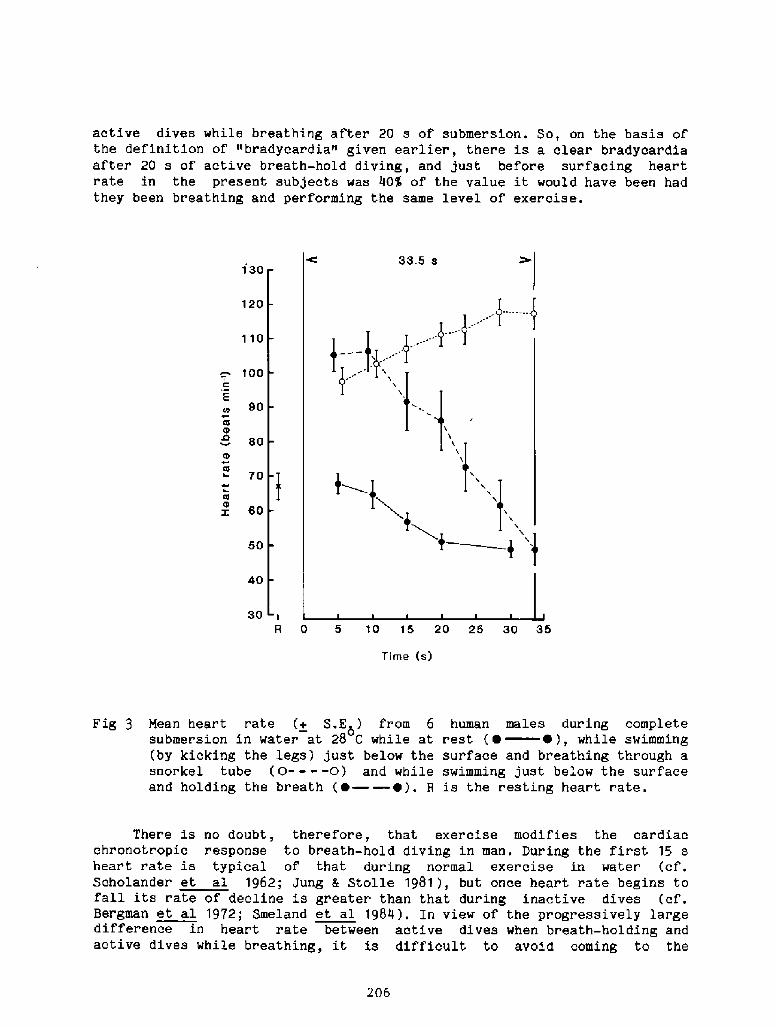
active dives while br eathing after 20 s of submersion. So, on the basis ofthe definition of "bradycardia" given earlier, there is a clear bradycardiaafter 20 s of active breath-hold diving, and just before surfacing heartrate in the present subjects was 40% of the value it would have been hadthey been breathing and perfor'ming the same level of exercise.
130
120
110
100
E90
60
70
60
50
40
30R 0 5 10 15 20 25 30 35
Time s1
Ther e is no doubt, therefore, that exeroise modifies the car diacchronotropic response to breath-hold diving in man. During the first 15 sheart rate is typical of that during normal exercise in water cf.Scholander et al 1962; Jung 5 Stolle 1981!, but once heart rate begins tofall its rate of decline is greater than that during inactive dives cf.Bergman et al 1972; Smeland et al 1984!. In view of the progressively largedifference in heart rate between active dives when breath-holding andactive dives while breathing, it is difficult to avoid coming to the
206
Fig 3 Mean heart rate + S.K>! fr om 6submer sion in water at 28 C while at by kicking the legs! just below thesnor kel tube 0- - - -0! and whileand holding the breath ~ ~ !. 8
human males during completerest � ~ !, while swimmingsurface and breathing through aswimming just below the surfaceis the resting hear t r ate.
conclusion that during the for mer there is a progressive involvement of anoxygen conserving response. Certainly some of the subjects in the presentstudy noted that discomfort pain! in the legs was greater when swimmingunderwater with breath-hold than when breathing. It seems, therefore, that
may be different fr om naturally diving homeotherms in that he enlistsoxygen conserving r esponse to a greater extent during active
breath-hold dives than dur ing inactive dives. It is worth noting, however,that seal.s and ducks can habituate to submersion of the head in that thefall in heart rate is reduced or abolished if the animal is taught to«dive« or if it is submerged repeatedly for the same per'iod of time Elsner1965; Gabbott A Jones 1986 !. Also, during unusually long voluntary divesboth seals and ducks do exhibit a lower heart rate than during dives ofmore normal duration Kooyman A Campbell 1972; Butler et al 1986 !. Thus,the awareness of the subject man or animal! of what is happening dur ingpassive head submersion and the size of usable oxygen stores relative tothe amount of oxygen being used during active breath-hold dives, togetherwith the effect of temperature, may be the major factors involved in theapparent differences in the response to diving inactive or active! betweenman and habitually diving homeotherms.
References
Andersen, H. T. 1963. Factors determining the cir culatory adjustments todiving. I . Water immersion. Acta physiol. Scand . 58, 173-185 .
Andersen, H. T. 1966. Physiological adaptations in diving vertebrates.Physiol. Rev. 46, 2'I2-243.
Asmussen, E. and Kristiansson, N.-G. 1968. The "diving bradycar dia« inexercising man . Acta physiol. Scand. 73 527-535 .
Bergman, S. A., Campbell, J. K. and Wildenthal, K. 1972. "Diving reflex" inman: its relation to isometric and dynamic exercise. J. appl. Physiol.33, 27-31-
Bjertnaes, L., Hauge, A., Kjekshus, J. and Soyland, E. 1984. Car diovascularresponses to face immersion and apnea during steady state muscleexercise. Acta physiol. Scand. 120, 605-612.
Burch, G. E. and Giles, T. D. 1970. A digital rheoplethysmographic study ofthe vasomotor response to 'simulated diving' in man. Cardiology 55,257-271.
Butler, P. J 1982. Respiratory and car diovascular control during diving inbirds and mammals. J. exp. Biol. 100, 195-221.
Butler. P. J. and Jones, D. R. 1968. Onset of and recovery fr om divingbt adycardia in ducks. J. Physiol. 196, 255-272.
Butler, P. J. and Jones, D, R. 1982. The comparative physiology of divingin vertebrates. In: Advances in Comparative Physiology andBiochemistry, Vol. 8 ed. 0. E. Lowenstein !, pp. 179-364. New York,Academic Press .
Butler, P. J. and Woakes, A. J. 1979. Changes in heart rate and respiratoryfrequency during natural behaviour of ducks, with particular referenceto diving. J. exp. Biol. 79, 283-300.
P. J. and Woakes, A, J. 1982 . Telemetry of physiological variablesfrom diving and flying birds. Symp, zool. Soc . Lond. 49, 107-128,
P. J. and Woakes, A. J. 1982. Control of heart rate by carotid bodychemoreceptors during diving in tufted ducks. J. appl. Physiol.:Respirat. Environ. Exercise Physiol. 53, 1405-'I410.
207
Butler, P. J. and Woakes, A. J. 1984. Hear t rate and aerobic metabolismHumboldt penguins, S heniscus humboldti, during voluntary dives. J,exp. Biol. 108, 419-428.
Butler, P. J., Stephenson, R. and Woakes, A. J. 1986. Variability of thehear t rate response during voluntary diving in the tufted duck, Aythva~full ula. O. physlol. in press!.
Campbell, E. J, H,, Freedman, S,, Clark, T. J. H., Robson, J, G. andNorman, J. 1967, The effect of muscular paralysis induced bytubocurarine on the duration and sensation of breath-holding. Clin.Sci, 32, 425-432.
Campbell, L. B., Gooden, B. A. and Horowitzy J. D. 1969. Cardiovascularresponses to partial and total immersion in man. J. Physiol. 202,239-250 '
Craig, A. B. Jr. 1963. Heart rate responses to apneic underwater diving andto breath holding in man, J. appl. Physiol. 18, 854-862.
Craig, A. B. Jr . and Medd, W. L. 1968. Han's responses to breath-holdexercise in air and in water . J. appl. Physiol. 24, 773-777.
Dykes, R. W. 1974. Factors related to the dive reflex in har bor seals:sensory contributions from the trigeminal region . Can . J. Physiol .Pharmacal. 52, 259-265.
Elsner, R. 1965 !. Heart rate response in forced versus trainedexperimental dives in pinnipeds. Hvalradets Skrifter, NorskeVidenskaps-Akad ., Oslo 48, 24-29 .
Elsner, R. and Gooden, B. 1983. Diving and Asphyxia. Cambridge, UniversityPress.
Elsner, R., Gooden, B. A. and Robinson, S, H. 1971. Arterial blood gaschanges and the diving response in man. Aust. J. exp. Biol. med. Sci.49, 435-444.
Eliassen, E. 1960. Cardiovascular responses to submer sion asphyxia in aviandivers . Arbok Univ. Bergen Hat . Naturv . Ser . 2, 1-100 .
Fedak, M. A., Kanwisher, J. and Lapennas, G. 1984. Patte ns of breathingand heart rate in relation to metabolic rate of swimming and divingseals. Proc. 1st Congress Comp. Physiol. and Biochem. Section of IUBS,p.A86.
Fowler, W. S. 1954. Breaking point of breath-holding . J. appl, Physiol. 6,539-545.
Furilla, R. A. and Jones, D. R, 1986. On the initiation of submersionbradycardia in diving compared with dabbling ducks. In prepar ation!.
Gabbott, G. R. J. and Jones, D. R. 1986. Psychogenic influences on thecardiac response of the duck Anas lat rh nchos ! to forcedsubmersion. J. Physiol. in press !.
Heistad, D. D., Abboud, F. M. and Eckstein, J. W. 1968 . Vasoconstrictorresponse to simulated diving in man . J . appl, Physiol . 25, 542-549 .
Hong, S. K., Song, S. H., Kim, P. K. and Suh, C. S. 1967. Seasonalobservations on the cardiac rhythm during diving in the Korean ama. J.appl. Physiol. 23, 18-22.
Hong, S. K., Lin, Y. C., Lally, D. A., Yim, B. J, B., Kominami, N., HoagyP. . and Moore, T. O. 1971. Alveolar gas exchanges and cardiovascula~P.W.a
functions dur ing br eath holding with air, J. appl, Phys iol. 3o >540-547.
Irving, L. 1939. Respiration in diving mammals. Phys. Rev. 19, 112-134.Irving, L. 1963 . Bradycardia in human divers. J, appl. Physiol.
489-491.
208
Jung, K. and Stolle, W. 1981. Behaviour of heart rate and incidence ofarrhythmia in swimming and diving. Biotelemetry patient Monitg. 8,228-239.
Kawakami, Y., Natelson, B. H. and DuBois, A, B. 1967. Cardiovasculareffects of face immersion and factors affecting diving reflex in man.
appl. Physiol. 23, 964-970.Kobayasi, S. and Ogawa, T. 1973. Effect of water temperature on br adycardia
during nonapneic facial immersion in man. Jap. J. Physiol . 23,613-624.
Kooyman, G. L. and Campbell, W. B. 1972. Heart rates in freely divingWeddell seals, Le ton chotes weddelli. Comp. Biochem. Physiol. 43,31-36.
Kooyman, G. L., Wahrenbrock, E. A., Castellini, M. A., Davis, R. W. andSinnett, E . E. '1980 . Aerobic and anaerobic metabolism during voluntarydiving in Weddell seals: evidence of preferred pathways from bloodchemistry and behavior . J. comp. Physiol. 138, 335-346.
Lin, Y-C. 1982. Breath-hold diving in terrestrial mammals. In: Exercise andSport Sciences Reviews, Vol. '10. ed. R. L. Terjung!, pp. 270-307,Philadelphia: Franklin Pt ess.
Oldridge, N. B., Heigenhauser, G. J. F., Sutton, J. R. and Jones, N. L.1978. Resting and exercise heart rate with apnea and facial immersionin female swimmers. J. appl. Physiol.: Respirat. Environ. ExercisePhysiol. 45, 875-879.
Pasche, A. and Krog, J. 1980. Heart rate in resting seals on land and inwater . Comp. Biochem. Physiol. 67, 77-83.
Scholander, P. F. 1940. Experimental investigations on the respiratoryfunction in diving mammals and birds. Hvalradets Skrifter, NorskeVidenskaps-Akad., Oslo 22, 1-131.
Scholander, P. F., Hammel, H. T., LeMessur ier, H., Hemmingsen, E. andGarey, W. 1962. Circulatory adjustment in pearl divers. J. appl.Physiol. 17, 184-190.
Smeland, E. B., Owe, J. 0. and Andersen, H. T. 1984. Modification of the'diving bradycardia' by hypoxia or exercise. Resp. Physiol. 56,245-251 .
Stephenson, R. 1985 . The Physiology of Natural Diving Behaviour in theTufted Duck A th a fuli ula!. M. Sc. qual. ! thesis, University ofBirmingham, England.
Sterba, J. A. and Lundgren, C. E. G. 1985. Diving bradycardia andbreath-holding time in man. Undersea Biomed. Res. 12, 139-150.
Stromme, S. B., Kerem, D. and Elsner, R. 1970. Diving bradycardia duringrest and exercise and its relation to physical fitness. J. appl.Physiol. 28, 614-621.
Woakes, A. J. and Butler, P. J. 1983. Swimming and diving in tufted ducks,A th a fuli ula, with par ticular reference to heart rate and gasexchange. J. exp. Biol . 107, 31 1-329.
Wolf, S., Schneider, R. A. and Groover, M. E. 1964. Further studies on thecirculatory and metabol.ic alterations of the oxygen-conserving diving! reflex in man. Trans. Assn. Am. Physns. 78, 242-254.
Wy» > V- 1956. Electr ocardiogramma di soggetti in apnea durante immersionein acqua a profondita diver se. Boll. Soc. Ital., Biol. Sper. 32,503-506.
209
Discussion following Dr. Butler's presentation
Dc. Bong: I have several points that need clarification. Number onerwhat is the level of exercise? Have you tried dif ferentwork3.oads'? paulev and Hansen Cardiac response to apnea and waterimmersion dur ing exercise in man. J. Appl. Physiol. 33:193-l98, 1972!found that when the exercise load is heavy, then they are able tooverride the bradycardial response. The other issue is what Bergmanet al. " Diving reflex in man: its relation to isometric and dynamicexercise. J. Appl. Physiol. 33:27-31i 1972! described about thedifference between the responses to static and dynamic exercise atsame oxygen consumptions. Why is there this difference?
Dr. Butler: First of all, we did not measure oxygen consumptionduring swimming, but we got the same individuals to work on a bicycleergometer, so that their' heart rate was at the same level as when the>were swimming and breathing. Their oxygen uptake was about one and ahalf liters per minute, which was about f ive times their restingvalue. So vhen they were swimming, just kicking thei r legs underwater, we can say that oxygen ccusumption was approximately five timesthe resting level, so that's the first thing. We had actually donesome preliminary work where the individuals used their arms as well aatheir legs when swimming, so that the workload was increased, andyou' re absolutely right, the period of tachycardia is much longer,it's almost extended to the end of the period they can stayunderwater. Then towards the end, the heart rate starts to decline<but even so it doesn't go down to the resting level. Anotherinteresting thing is that, if the individual is working swimming! andbreathing before breath-hold, the bradycardia that occurs is muchfaster than when breath-hold swimming starts f rom rest. So, whether asubject is working before breath-hold submersion, or whether work andbreath-hold submersion start together, does seem to have an effect onthe cardiac response. Maybe it has something to do with blood flow+muscle contraction.
Dc. hrnold: Bergman et al. J. Appl. Physiol. 33: 27-31, 1972!compared isometric and dynamic exercise, and found that. isometricexercises like hand grip were good vasoconstrictors but do not enhancevenous return, whereas dynamic exercise does. There's much mor ebradycardia with dynamic than isometric exercise. Dr. Elsner's lowestheart rate person Stromme et al., J. Appl. Physiol. 28: 614-621,1970! was swimming in 25 degrees C and breath-holding ~i~ immersingthe face. With swimming, breath-hold, followed by facial immersion,the bradycardia onset was immediate. The last point is that, theheart rate drops from 120 bpm when your exercising swimmers work.Exercise is associated with peripheral vasodilation and an increase inheart rate just as is exposure to heat, but it is due to a differentrequirement of the cardiovascular system. lt's interesting to notethat the subjects in my study who were ~ exposed to an oxygendemanding stress, eliminated their bradycardia, whereas in exercise,the same degree of bradycardia is produced dramatically by facialimmersion.
Dr. Butler: From our work on birds we just modified the classicalScholander story by saying that as well as the heart and the centraL
210
Dr. Rennie: I would like to mention some data f rom! the expeditionthat Dr. Data invited me to join, off the fantail of the French shipTrident, in the Ustica Harbor, in Sicily, about six years ago duringlunch hours. The divers were instructed to swim in a vertical
attitude watching a flag on a suspended line and were swimming againstan imposed load of 3, 6< and 9 kilograms> while f ree breathing f romSCUBA and af ter they spat out their mouthpiece. The purpose was toplot the heart rate as a function of time for each of these threeloads. This particular little study showed that with exactly the samewoLkloads, when divers breath-held at exactly the same depth, theheart rate dropped down to 40-45 beats per minute compared to l60-l70when f ree-breathing. We called that the underwater step test.
Dr. Rennie, was the face exposed to the cold water orDz. hrncld:not7
Dr. Rennie: The water wasn't very cold, this is Ustica in Sicily. Itwas 22 to 24 degrees centigrade and the mask, from my recollection,was left on. They just spat out the mouthpiece.
nervous system, the active muscles also get an adequate blood supply.The heart is an active muscle, and when the birds are swimmingunderwater, their leg muscles are also active, so they get sufficientblood supply to enable them to maintain aer'obic metabolism. You cando the calculations in terms of the amount of usable stored oxygenthese birds have, and the duration of their dives and, generallyspeaking, they keep well within their aerobic limits. The same goesfor the Weddell seal as well. It certainly does appear that thecardiovascular response to breath-hold diving in humans depends uponwhether there is breath � hold first then exercise, or exercise firstthen breath-hold, or if the two events begin together.
CARDIAC ARRHYTf&fIAS AS A PRECURSORTO DROWNING ACCIDENTS
J.R. MCDONOUGH, M.D., J.P. BARUTT> B.S., AND J.C. SAFFRON, R.N ~ *
Introduction
The principal investigator first became interested in the possibilityof water-immersion induced arrhythmia when, as an attending physician inthe early 1970's, he participated in the care of a high school girl admit-ted to hospital because of fresh water near drowning. She was on theschool swimming team and was swimming laps in a routine fashion when syn-cope occurred; and she sank to the bottom of the pool. Recovery was un-eventful; subsequent Ecg monitoring, echocardiography and exercise tread-mill were negative except for an occasional premature ventricular contrac-tion. Years later, as a medical student, she collapsed while showering,could not be resuscitated by the Seattle Fire Department Medic-One rescueunit, and expired of apparent cardiac arrest.
The investigators propose that cardiac arrhythmias, some of which maypredispose to loss of consciousness and accidental drowning, occur duringwater immersion; that such arrhythmias may be much more common than ex-pected; and that they may comprise a significant proportion, perhaps themagori.ty, of unexplained sudden death during water immer'sion.
Specific Aims
To defin.e prevalence of arrhythmia during scuba diving and tocontrast this with arrhythmia prevalence during pre-dive andpost-dive periods in order to find out whether arrhythmia occurswith greater frequency while diving.To search for potential predictors of diving arrhythmias such aspresence of arrhythmia on 12-lead Ecg, treadmil.l exercise Ecg andduring facial immersion in tepid and chilled water; and bloodpressure and heart rate responses to exercise and facial immer-sion.
�!
Significance
A considerable literature exists which deals with cardiocirculatoryadaptations to water immersion in animals, particularly diving birds andmammals. The diving reflex results in vasoconstriction and bradycardia,
212
*Funds provided by Cardiology Associates, Inc., P.S., Allenmore Medi-cal Center B-6007, Tacoma, WA 98405 and a grant from the Fraternal Orderof Eagles. Presented October 29, 1985, University of Buffalo, NY, Workshopon the Physiology of Breath-hold Diving. Consultants to the study: DarwinD. Jorgensen, Asst. Prof. Biology Comparative Physiology!, Univ. of PugetSound, Tacoma, WA; R. Bruce blind, Chairman, Dept. of >iathematics and Com-puter Science, Univ. of Puget Sound, Tacoma, WA; and Richard Dunford,Operations Director, Virginia Mason Hyperbaric Unit, Seattle, WA.
altering the circulation to essentially a heart-braIn circuit, and sparingoxygen by the use of anaerobic metabolism for per formance of musc1e workwhile submerged �-6!. The afferent limb of this reflex is activated bysensory receptors in the distribution of the fifth cranial nerve primarilyby cold and wet stimuli of the eyes and nostrils. Efferent components ofthe reflex involve the sympathetic nervous system for vasoconstriction ofmost vascular beds, and cardiac vagus nerves to slow the heart �-9!.Facial immersion in cold water with breath-holding will activate this re-flex in humans, and has been shown to effectively convert paroxysmal supra-ventricular tachycardia to normal sinus rhythm � 2, 13!.
Head-out water immersion causes a reduction in vital capacity by dis-placement of blood volume from peripheral to central areas. Right atrialand pulmonary arterial pressures, and stroke volume subsequently increase.More frequent extrasystoles occur during the first. few minutes of head-outimmersion, and a suggested mechanism involves right atrial and right ven-tricular stretch as a result of increased blood volume in these structures
�4,15!. One study of patients who survived myocardial infarction foundan increased frequency of extrasystoles during underwater swimming, andcautioned that water submersion for patien.ts with coronary disease may behazardous �.6!.
Accidental drowning while scuba diving is not a rare occurrence. Aseven year study conducted by the University of Rhode Island NationalUnderwater Accident Data Center for the years 1970-1976 reported 893 scubarelated diving deaths in the United States. The state of Washington hadthe third highest number oi such deaths at 65. A search by the investi-gators of death certificate records involving scuba related deaths unex-plained by nitrogen bubble disease, trauma, or other recognized causes!for the state of Washington during a four year period 1979-1982 revealed35 such deaths, with most having occurred in Puget Sound.
A panel discussion entitled "Unexplained Sudden Death During Diving"was presented at the annual meeting of the Undersea '.iedical Societ San Antonio, Texas, June 2, 1984!. Medical examiners from Dade and PalmBeach counties in Florida presented a series of cases with autopsy resultson divers who died unexpectedly while diving. Unexplained death was foundto occur in otherwise healthy divers irrespective of age, level of train-ing, or duration of diving experience.
If, as suspected, cardiac arrhythmia is a mechanism of unexplainedsudden death during water immersion, it becomes important to identify thefrequency and types of arrhythmia during water immersion, those havingpotential hazard, and to look for tests which might predict which individ-uals are likely to develop such arrhythmias.
Methods
Preliminary observation and measurements on 20 healthy scuba-divingvolunteers, carried out in the laboratory, consisted of a self � administered
213
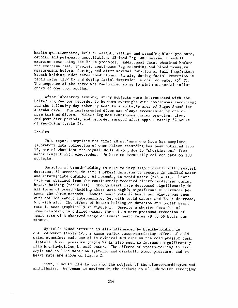
health questionnaire, height, weight, sitting and standing blood pressure,cardiac and pulmonary auscultatio~, 12-lead Ecg, and maximal treadmillexercise test using the Bruce protocol. Additional data, obtained beforethe exercise test, involved continuous Ecg recording and blood pressuremeasurement before, during, and after maximal duration of full inspiratorybreath holding under three conditions: In air, during facial immersion intepi.d water �8 C! and during facial immersion in chilled water � C!.The sequence of the three was randomized so as to minimize serial influ-ences of one upon another.
After laboratory testing, study subjects were instrumented with theHolter Ecg 24-hour recorder to be worn overnight with continuous recording;and the fallowing day taken by boat to a suitable area of Puget Sound fora scuba dive. The instrumented diver was always accompanied by one ormore trained divers. Holter Ecg was continuous during pre-dive, dive,and post-dive periods, and recorder removed after approximately 24 hoursof recording table I!.
Results
This report comprises the first 20 subj ects who have had completelaboratory data collection of whom Holter recording has been obtained from16, one of whom lost the signal while diving due to "shorting-out" fromwater contact with electrodes. We hope to eventually collect data on 100subjects.
Duration of breath-holding is seen to vary significantly with greatestduration, 81 seconds, in air; shortest duration 53 seconds in chilled waterand intermediate duration, 65 seconds, in tepid water table II!. Heartrate was obtained from the continuously recorded electrocardiogram duringbreath-holding table III!. Though heart rate decreased significantly inall forms of breath-holding there were highly significant differences be-tween the three methods. Lowest heart rate 42 beats per minute was seenwith chilled water; intermediate, 54, with tepid water; and least decrease,6l, with air. The effect of breath-holding on duration and lowest heartrate is seen. graphically in figure 1. Despite a shorter duration ofbreath-holding in chilled water, there is a more profound reduction ofheart rate with observed range of lowest heart rates 29 to 59 beats perminute.
Systolic blood pressure is also influenced by breath-holding inchilled water table IV!, a known reflex vasoconstricting effect of coldwater sometimes made use of in clinical medicine as the cold pressor test.Diastolic blood pressure table V! is also seen to increase significantlywith breath-holding in cold water. The effects of breath-holding in air,tepid and chilled water on systolic and diastolic blood pressure, and onheart rate are shown on figure 2.
Next, I would like to turn to the subject of the electrocardiogram andarrhythmias. We began as novices in the techniques of underwater recording'
214
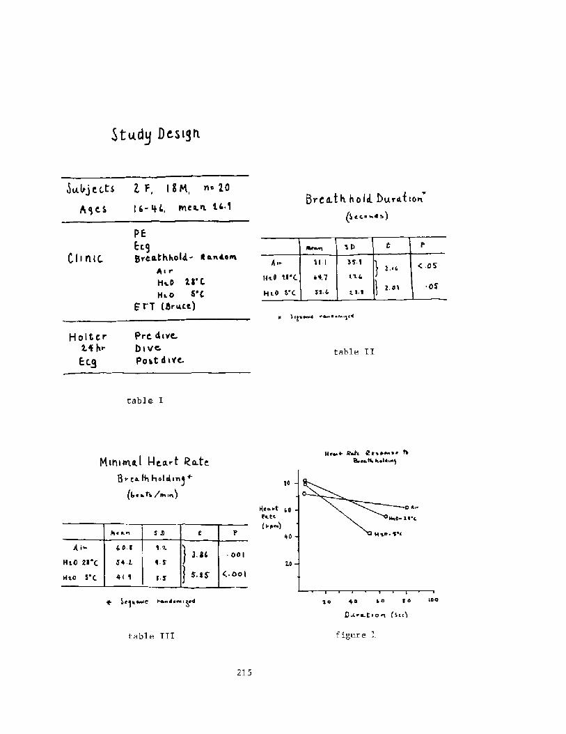
C I i st< c.
à I tpw4 wL ~ lw
Pre 4iVe.
9>vc
Post diVc,
HoItcr2.f gr
Ecgtable II
table I
pi c*t4 hotdin!+ ~. t./
'RO
Hea iteat~ s p~!
f iI;ure ltable III
215
5tgdg Dcsi]h
JulrjcLCS 2 F, I EM, < 20h,pcs l6- tL, ~c«xa1
PE
Ecggreathhold.- k Laiosl
her
Hie il' CHa o 5'C
Fr T S.~ca!
H nival I-Isa.rt Ra.fg
+ t]glvlcc I'ILm Jy&s I �4
9rCa.th ho ll Durah~o~� ec.~ ~a>!
4 e~ 4- Rat< l e > yae s c h8a ra% k elk us!
'40 +0 ko 1d
D~ia.t i o ~ �c~!
!qggp le 8 laos 8esSLArCQ gag.Q ho Id
w~ Hq!
D i a a tolrc B I a<4 t'ress u t<
St8 urn<a r~Er <pre! P8qmcr v~iemig<4
table IV table V
and it took a year of trial and error to obtain diagnostic-quality record-ings underwater. I will be showing several examples of these recordings,the first is a normal electrocardiogram using the two-channel recorder figure 3!. This was taken during Holter monitoring prior to a dive.
I � I sec I
1%0
80
BPR Lp
Penn! gfttr
figure 3figure 2
Next figure 4! an electrocardiogram recorded during Stage II of theBruce multistage maximal exercise test. We see 2:1 sino-atrial exi.t blockwith the heart rate of 120 during sub-maximal exercise transiently droppingto 60. Next figure 5! we see the same 2:1 sino-atrial exit block during
216
'I 2.0
H! i00
O.X ~el<~ Pl Cn~h<0 PyL- 0 ivy.
gorman.l
reatl,k el4>n!+ ~~ H!!
facial immersion xn chilled water. This occurred early, prior to thedevelopment of bradycardia or heart rate slowing.
Facial i me e i bio n O'C Ha 0E'FT ota~c K
CH 2.LQ
hh E~it Bio<~
2'I Si Zan 4t ca.
FA
gcc+
figure 5figure 4
The next figure 6! shows facial immersion in chilled water with a non-conducted junctional contraction, a premature ventricular contraction, anda premature atrial contraction. Another subject figure 7! during facialimmersion in chilled water shows sinus bradycardia or heart rate slowing,but also shows frequent unifocal premature ventricular contractions, and abrief run of ventricular tachycardia defined as 3 or more consecutiveventricular contractions.
F inal i emir> >o n 4 C Hl.0
FK mi t% iaaf ~e a etc'Facial Irnmiriian g't ti~o
IEIB LO K
+I k~c+
i a ~ ~
figure 6
iipure 7
217
Next figure 8! we see a single premature ventricular contraction duringHolter monitoring. This occurred before the dive. Another figure 9!pre-dive Holter shows two unifocal premature ventricular contractions.There is also a single premature atrial cont~action. Next figure l0!during pre-dive we see multiform premature ventricul.ar contractions. Thetracing is not continuous i.n order to bring in from a later portion ofthe tape the premature ventricular contraction which originated from adifferent vnetricular focus than the first. Jn clinical cardiology prac-tice we regard multiform premature ventricular contractions as more seriousthan unifocal patterns.
pre D ive
GC 33M
Scc +
Prc Dive.
Pal 35 8 +I Jet.+
figure 9
fi~uro 8
The next figure ll! occurs during a dive, and shows frequent prema-ture atrial contractions and a cluster of paired premature atrial contrac-tions. In clinical practice we regard occasional premature atrial contrac-tions as less serious than unifocal premature ventricular contractions.However, frequent premature atrial contractions, and especially pairs,often precede more serious forms of supzaventricular arrhythmia such asatrial tachycardia, atrial flutter, or atrial fibrillation. Next figure l2! shows a single premature ventricular contraction during adive. Next figure L3! is another premature ventricular contraction re-corded during a dive..
Next figure L4! we see multiform premature ventricular contractionsduring a dive. The next figure l5! shows paired premature ventricularcontractions during a dive. I would like to remind the audience that allof these examples of abnormal cardiac rhythms, or arrhythmias, occurredin healthy young persons.
Let us now turn to a classification of these arrhythmias table VI!.We can classiry all observed arrhythmias into 5 categories: Prematuresu raventricular contractions include atrial and junctional, and we sawexamples of both. Com lex su raventzicular arrh thmias include sino-atrialexit block, which was seen, and several varieties not observed: Includingatrial tachycardia, atrial flutter, and atrial fibrillation. Unifocal
218
Prt Di v' t
mglt< form PVC.
Oi vccc � i
1<d F08
figure 10
JH are
I'VC.
Qrve
figure 12
219
Dive
Sec+
5L 3ov
I I LK+
figure 11
figure 13
D tv'
PB 2AM
f � f 5ec+D tye.
gttftg fnrppg P V C'>!g.g.nd. PV<$ CC.� }xc L8F
figure 14
figure 15
premature ventricular contractions have also been presented. ~Com lesventricular arrh thmias include multiform and paired premature ventricularcontractions, and ventricular tachycardia, all seen; and ventricular fib-
e r is that of auses,rillation, fortunately not seen. The last category s a pdefined as exceeding 1. 6 seconds in length, and exceeding the precedingcycle interval y more an pb than 25 ercent. Next table VII! we will examinethe frequency of these categories for all methods of detection. First,let us examine the totals. Of 252 specific arrhythmic events, the mostcommon represente orm was prd f remature supraventricular contractions, Ithe next most connnon unifocal premature ventricular co ' . 4 , hntractions. 45, then
ventricular arrhythmias 37. Five instances of complex supra-ventricular arrhythmias were seen and all were 2:I sino-atria al exit block.Six pauses were seen. When arrhythmia frequency is exalnined by type ofrecor ing we seed that only 2 of 252 arrhythmic events were seen in
e detected bstandard 12-lead Ecg, a rather poor showing, while 36 were detec e yexercise testing, 60 with breath-holding, and 154 during Holter recording.Next table VIII! we will examine the distribution of these 5 categoriesof arrhythTnia by breath-holding category. Notice that the maj ority, 38 of60, are detected by chilled water immersion whereas air �0! and tepidwater �2! detecte ar ewer.d f f r. Ail pauses occurred with breath-holding,that is, none were detected during Holter recording, including dive,and none were seen uring s an ard t d rd resting or exercise electrocardiography.Of the more serious categories of arrhythmia, that is the uni f oca1
220
C 1 a~S I f Icat jOn of A vvhg t h~iaa
PSVC PVCvnatu. VC Supra VCnt Vi IX a rCOvltra t iovIa - at viala.nd gunct Iaj
CSVA E.'amp/ex Duple.vents ic ala.rAvrhq<hm i<.~ � 5A Exit 0/0 k A Fil FlIj.ttev, A/BIO k AOtseevi!
HPttC Uviifoca.l Premature Vent vicu/arCo nt ractl cvIS
C V4 CaviIple>< Vcnivi cola~ Avvhqthmi apo,irea YIultitovin PVC's, VTC V'F rIot sccvt,!
PQIISCS !I ~ Scc I.vid o l 2SxP v c c c c 4 I vI 9 R R j. vi t c v vcL j
table VI
Arr hqthmia. A nels S I5T~pc ~a plaf4r
table VII
premature ventricular contractions and the complex ventricular arrhythmias,18 of 2I occurred during cold water immersion. Next table IX! we see thedistribution of 5 categories of arrhythmia by Halter category, that ispre-dive, dive, and post.-dive components. First, note that the categoriesare now reduced to three, as complex supraventricular arrhythmias andpauses were not seen.
Of 154 arrhythmic events noted, 97 occurred pre-dive, 45 during dive,and 12 during post-dive recording. Also note the important differencesin time of recording, with pre-dive 267 hours, dive 6.5 hours, and post-dive 89 hours. We shall have more to say about this later when we discussarrhythmic events per hour,
A rr hq thrnia A na lq s izb y iioitei Co.kqor~
Arr hg t he < a Aria,l!S iSBrcathhaki >n] go~ erg
table IXtable VIII
221
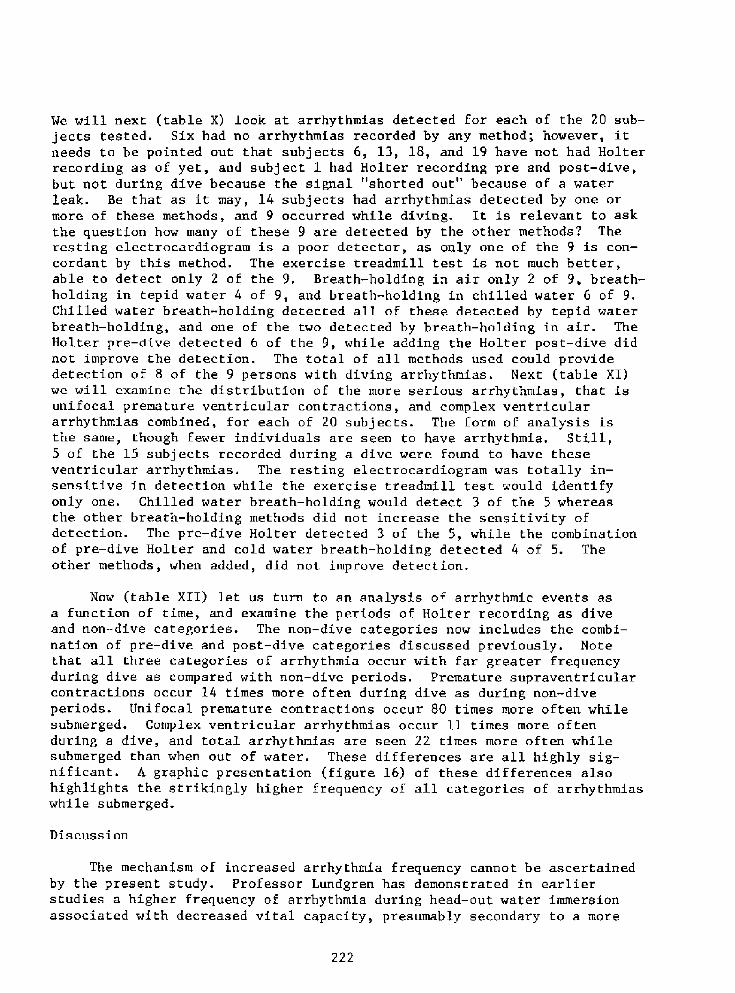
We will next table X! look at arrhythmias detected Eor each of the 20 sub-jects tested. Six had no arrhythmias recorded by any method; however, itneeds to be pointed out that subjects 6, 13, 18, and 19 have not had Holterrecording as of yet, and subject 1 had Halter recording pre and post-dive,but not during dive because the signal "shorted out" because of a waterleak. Be that as it may, 14 subjects had arrhythmias detected by one ormore of these methods, and 9 occurred while diving. It is relevant to askthe question how many of these 9 are detected by the other methods? Theresting electrocardiogram is a poor detector, as only one of the 9 is con-cordant by this method. The exercise treadmill test is not much better,able to detect only 2 of the 9. Breath-holding in air only 2 of 9, breath-holding in tepid water 4 of 9, and breath-holding in chilled water 6 of 9.Chilled water breath-holding detected all of these detected by tepid waterbreath � holding, and one of the two detected by breath-holding in air. TheHolter pre-dive detected 6 of the 9, while adding the Holter post-dive didnot improve the detection. The total of all methods used could providedetection of 8 of the 9 persons with diving arrhythmias. Next table XI!we will examine the distribution of the more serious arrhythmias, that isunifocal premature ventricular contractions, and complex ventriculararrhythmias combined, for each of 20 subjects. The form of anaIysis isthe same, though fewer individuals are seen to have arrhythmia. Still,5 of the 15 subjects recorded during a dive were found to have theseventricular arrhythmias. The resting electrocardiogram was totally in-sensitive in detection while the exercise treadmill test would identifyonly one. Chilled water breath-holding would detect 3 of the 5 whereasthe other breath-holding methods did not increase the sensitivity ofdetection. The pre-dive Holter detected 3 of the 5, while the combinationof pre-dive Holter and cold water breath-holding detected 4 of 5. Theother methods, when added, did not improve detection.
Now table XII! let us turn to an analysis of arrhythmic events asa function of time, and examine the periods of Holter recording as diveand non-dive categories. The non-dive categories now includes the combi-nation of pre-dive and post-dive categories discussed previously. Notethat all three categories of arrhythmia occur with far greater frequencyduring dive as compared with non-dive periods. Premature supraventricularcontractions occur 14 times more often during dive as during non-diveperiods. Unifocal premature contractions occur 80 times more often whilesubmerged. Complex ventricular arrhythmias occur 11 times more oftenduring a dive, and total arrhythmias are seen 22 times more often whilesubmerged than when out of water. These differences are all highly sig-nificant. A graphic presentation figure 16! of these differences alsohighlights the strikingly higher frequency of all categories of arrhythmiaswhile submerged.
Discussion
The mechanism of increased arrhythmia frequency cannot be ascertainedby the present study. Professor Lundgren has demonstrated in earlierstudies a higher frequency of arrhythmia during head � out water immersionassociated with decreased vital capacity, presumably secondary to a more
222
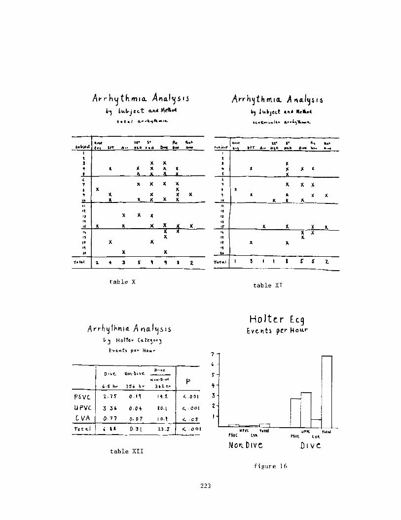
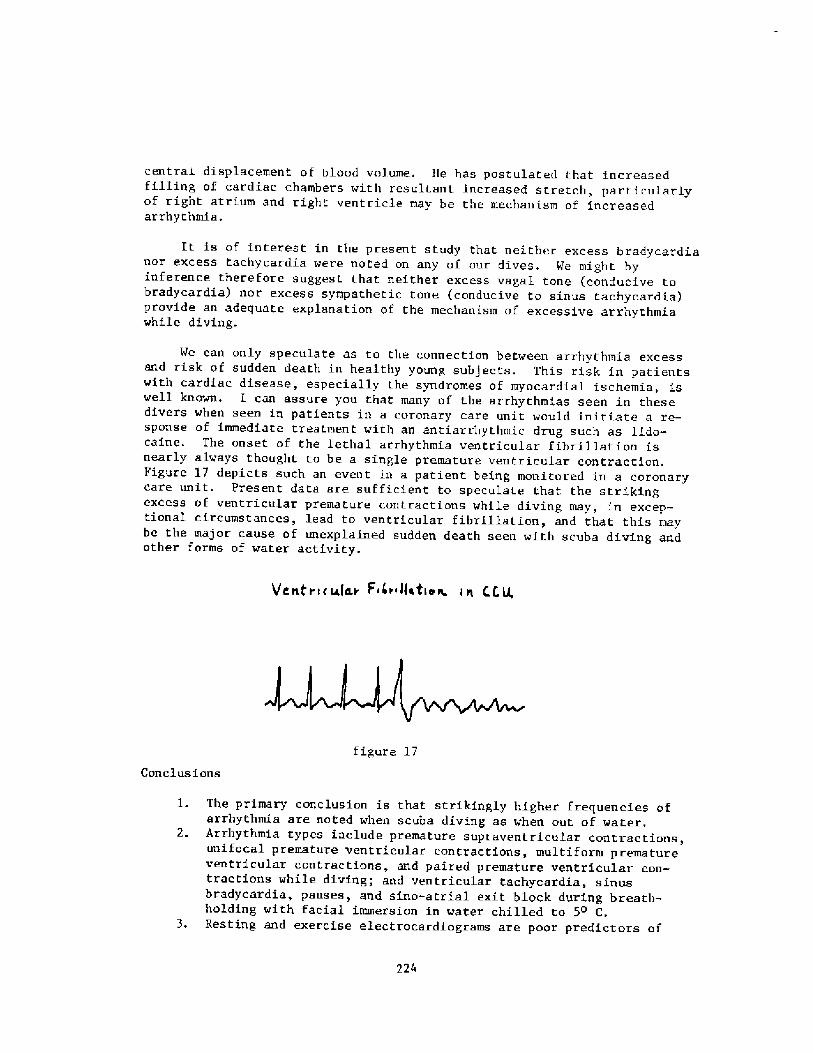
centrai displacement of blood volume. He has postulated that increasedfilling of cardiac chambers with resultant increased stretch, particularlyof right atrium and right ventricle may be the mechanism of increasedarrhythmia.
It is of interest in the present study that neither excess bradycardianor excess tachycardia were noted on any of our dives. We might byinfer'ence therefore suggest that neither excess vagal tone conducive tobradycardia! nor excess sympathetic tone conducive to sinus tachycardia!provide an adequate explanatio~ of the mechanism of excessive arrhythmiawhile diving.
We can only speculate as to the connection between arrhythmia excessand risk of sudden death in healthy young subj ects. This risk in patientswith cardi.ac disease, especially the syndromes of myocardial ischemia, iswell known. I can assure you that many of the arrhythmias seen in thesedivers when seen in patients in a coronary care unit would initiate a re-sponse of immediate treatment with an antiarrhythmic drug such as lldo-caine. The onset of the lethal arrhythmia ventricular fibrillation isnearly always thought to be a single premature ventricular contraction.Pigure 17 depicts such an event in a patient being monitored in a coronarycare unit. Present data are sufficient to speculate that the strikingexcess of ventricular premature contractions while diving may, in excep-tional circumstances, lead to ventricular fibrillation, and that this maybe the major cause of unexplained sudden death seen with scuba diving andother forms of water activity.
V C et r «slav F < < i ~tatty s- i h C t; U,
figure 17
Conclusions
1. The primary conclusion is that strikingly higher frequencies ofarrhythmia are noted when scuba diving as when out of water.
2. Arrhythmia types include premature supraventricular contractions,unifocal premature ventricular contractions, multiform prematureventricular contractions, and paired premature ventricular con-tractions while diving; and ventricular tachycardia, sinusbradycardia, pauses, and sino-atrial exit block during breath-holding with facial immersion in water chilled to 5o C.
3. Resting and exercise electrocardi.ograms are poor predictors of
224
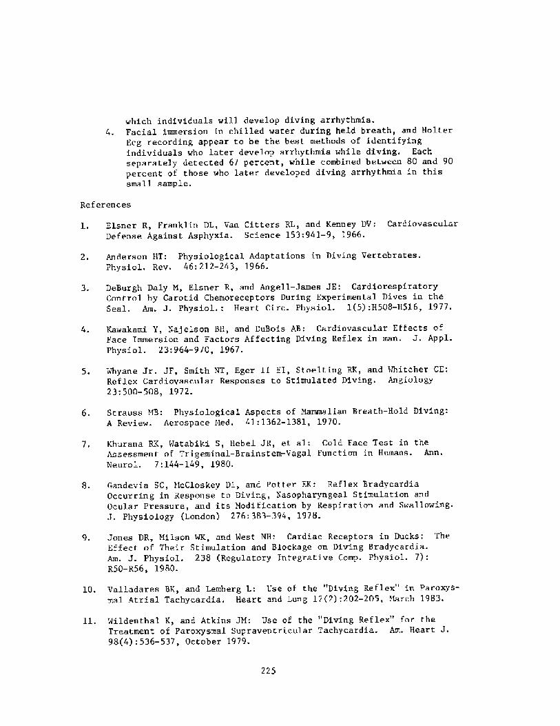
which individuals will develop diving arrhythmia.4. Facial immersion in chilled water during held breath, and Holter
Ecg recording appear to be the best methods of identifyingindividuals who later develop arrhythmia while diving. Eachseparately detected 67 percent, while combined between 80 and 90percent of those who later developed diving arrhythmia in thissmall sample.
References
Elsner R, Franklin DL, Van Citters RL, and Kenney DV; CardiovascularDefense Against Asphyxia. Science 1S3:941-9, 1966.
Anderson HT: Physiological Adaptations in Diving Vertebrates.Physiol. Rev. 46:212-243, 1966.
DeBurgh Daly M, Elsner R, and Angell-James JE: CardiorespiratoryControl by Carotid Chemoreceptors During Experimental Dives in theSeal. Am. J. Physiol.: Heart Circ. Physiol. 1�!:H508-H516, 1977.
Kawakami Y, Najelson BH, and DuBois AB: Cardiovascular Effects ofFace Immersion and Factors Affecting Divi.ng Reflex in man. J. Appl.Physiol. 23:964-970, 1967.
Whyane Jr. JF, Smith NT, Eger II El, Stoelting RK, and Whitcher CE:Reflex Cardiovascular Responses to Stimulated Diving. Angiology23:500-508, 1972.
Strauss MB: Physiological Aspects of Mammalian Breath-Hold Diving:A Review. Aerospace Med. 41:1362-1381, 1970.
Khurana RK, Watabiki S, Hebel JR, et al: Cold Face Test in theAssessment of Trigeminal-Brainstem-Vagal Function in Humans. Ann.Neurol. 7:144-149, 1980.
Gandevia SC, McCloskey DI, and Potter EK: Reflex BradycardiaOccurring in Response to Diving, Nasopharyngeal Stimulation andOcular Pressure, and its Modification by Respiration and Swallowing.J. Physiology London! 276:383-394, 1978.
Jones DR, Milson WK, and West NH: Cardiac Receptors in Ducks: TheEffect of Their Stimulation and Blockage on Diving Bradycardia.Am. J. Physiol. 238 Regulatory Integrative Comp. Physiol. 7!:R50-R56, 1980.
10.
Wi.ldenthal K, and Atkins JM: Use of the "Diving Reflex" for theTreatment of Paroxysmal Supraventricular Tachycardia. Am. Heart J.98�!:536-S37, October 1979.
225
Valladares BK, and Lemberg L: 92;se of the "Diving Reflex" in Paroxys-mal Atrial Tachycardia. Heart and Lung 12�!:202-205, ."iarch 1983.
12. Condry P, Jain A, marshall R, and Bowyer A: Ventricular TachycardiaCaused by the Diving Reflex. Lancet 2�947!:1263, December 20, 1975.
13. Hughes T, Carter J, and Wolf S: Disorders of Cardiac ConductionAccompanying the Dive Reflex in Man. Pav. J ~ Biol. Sci.16�!:25-33, January-March 1981.
14, Arborelius Jr. M, Balldin UI, Lilj a B, and Lundgren CEG: HemodynamicChanges in Man During immersion wi.th Head Above Water. Aerospace Med.43�!: 592-598, 1972.
15. Kurss DI, Lundgren CEG, and Pasche AJ: Effect of Water Temperatureon Vital Capacity in Head-Out Immersion. Proceedings of the SeventhSymposium on Underwater Physiology, Eds. Bachrach and Matzen,Undersea Med. Soc, Inc., Bethesda, MD, 1981.
16. Gi.esler H, Jungmann H, and Stein G: Sind Koronarkranke beim Tauchendurch Herzrhythmusstorungen geflhrdet? Med Klin 75:518-523, 1.980.
226
Discussion following Dr. NcDonougb's presentation
Dr. Lanphier: Well I wasn't quite clear on what proportion of SCUBAdiving vs breath-hold diving was involved here. Can you clarify thatas the first questions
Dr. IIcDonougb: They were separate, and at different times. Thebreath-hold testing was conducted in the laboratory as part of a busycardiology practice on a day off and and three types of breath-holding were done. These were done in sequence with intervals ofprobably 5 to 10 minutes in between, and because it was suspectedthat perhaps there might be an influence of one upon the other whendone serially, the sequencing was randomized. I'm pleased that we didthis, because of what I heard yesterday f rom Dr. Arnold's commentsabout a second breath-hold bradycardia being more profound than afirst. The SCUBA dive itself was done on a day when an individual wasconnected to a continuously recording electrocardiogram obtained for24 hour s. The SCUBA dives were usually down to somewher e between 50and 110 feet, and they lasted 25 to 35 minutes.
Dr Lanphier: We}l, I wanted to see if we could figure out, from whatDr. NcDonough has done, what role hypoxia might have had in promotingthese arrhythmias, but obviously not during scuba diving.Furthermore, face immersion might have had a role, but it's hard tosay. Now one final thing, one of the big puzzles in loss ofconsciousness in divers is what happens to these individuals who madea significant dive and are now heading for shore on the surface,perhaps snorkeling, and all of a sudden they just go dead in the waterand are supposed to have had some kind of fatal cardiac event. Atleast, that seems the best explanation. And, one of my thoughts aboutthis, is that the change in position might have something to do withthis: that they' ve been immersed where position doesn't make all thatmuch difference, they come up in a vertical position, and then whenthey reach the surf ace, they go to the horizontal position to swim.If they have trouble, they may go vertical head-out! again, whichchanges quite a few things. 1 just wonder if you have any thoughtsabout that.
Dr. NcDonougb: None really beyond what I' ve expressed, except that weknow the posture changes have important influences on circulatorydynamics and you know, it's conceivable that that posture change mayhave an important role.
Dr. Lanpbier: I should add that most of these recorded cases havebeen in r'elatively cold water.
Dr. Luadgren: As a SCUBA diver comes up the surface to return toshore, he/she is often low on air and likely to take out themouthpiece and change to snorkel; this will cause a rather heftyimmersion effect on their circulation, with atmospheric pressure inthe lungs and more or less the full immersion effect on thecirculatory system.
Dr. Lanphier: I would assume very few people would try to swim toshore using their tank.
227
Dr. Lundgren: Right.
Dr. Shiraki: I have noted many cases of arrhythmias during head-outimmer sion.
Dr. Lin: Your high frequency of arrhythmias during scuba diving seemsto agree well with Dr. Hong's studies, especially in cold water> but<I am quite surprised to see in your study that face immersion had ahigh incidence of arrhythmias. We, in 10 to 15 years of laboratorystudies, rarely observed arrhythmias during face immersion even duringcold water immersion. In the field, however, arrhythmia seems to be acommon occurrence.
Dr. NcDonougb: We see a lot of arrhythmias during facial immersion.
Dr. Lin: That's surprising.
Dr. Lamdsberg: A similar study was done in 1970 by Sem-Jacobsen inNorway. At that stage he suggested a Panic Button for Aircraft Pilotsto sound an alarm when they developed Cardiac Arrhythmias. The PanicButton was wired to a lead which monitored the heart beat and wouldsound an alarm when the Arrhythmia occurred. He recorded severalpotentially fatal Arrhythmias in operational divers and actuallydescribed Ventricular Fibrillation in one diver who was particularlyapprehensive during a dive. To what extent are we justif ied insubjecting prospective divers to this type of selection testing, whichcould be fatal, in order to exclude them from operational diving?
Dr. NcDonougbr It's a very interesting question. One of the thingsthat we would be interested in doing as a continuing study is torepeat the dive using Holter electrocardiogram recording in the samesubjects, to see whether the same individuals develop arrhythmia withrepeat testing.
Dr. Boysen: Just a couple of quick questions. First of all, havethese subjects discontinued nicotine, caffeine, and that type of thingbefore they were tested?
Dr. NcDoaough: They were leisure time divers in the young age groupsthat were tested, Generally they were very careful about not usingbeverages or eating before they went in the water. Smoking wasvirtually unknown for most of them, alcohol was in light use by someof them. In general, there was no use of coffee, other beverage orfood within probably about 2 or 3 hours of diving.
Dr. Boysen: Was there any correlation between time of the dive andthe occurrence of these arrhythmias7
Dr. NcDonougb: I did not see a pattern.
Dr. Boysen: The other issue was, as you said, the importance ofrepetitive dives. Can you reliably reproduce arrhythmias with a facialimmersion in subjects'
Dr. NcDonoogb: We haven't done this yet.
228
Dx. Arnold: I have done repetitive studies and though I wasn' tlooking for it, I have found arrhythmias in several healthy subjects.Arrhythmias frequently occur at a heart rate which one would expect tobe the ventricular inherent automaticity, around 40 beats perminute. When the hear t rate is slowly aiming towards 40 and goingbeyond, there are many subjects who will start throwing ventricularescape rhythm at about that point, so that they' re going to bottom outat 40 bpm even though it looked like they were aiming for 30 bpm. AndI' ve seen atrial premature contractions also. It appears that agiven individual may have a tendency towards a stereotyped arrhythmia,my brother for instance. He, on repeated dives, 3 minutes apart wouldshow ventricular escape at 40 beats per minute.
Dr. NcDonough: Let me comment upon that, because I think that's animportant distinction. We did see escape rhythms, junctional orventricular, during breath-hold facial imroersions on some occasions.During the SCUBA dives I thought we would see a lot of escape rhythms,but we didn' t. The SCUBA diving arrhythmias were all ectopic.Furthermore, we did not see bradycardia during SCUBA diving. Thesewere free swimming persons whose ambient heart rates generally rangedfrom 80 to 140 beats per minute.
Dr. Axnold: And they had a face mask.
Dr. NcDonough: Yes, a face mask. It is also of interest to me thatour divers, who had these recordings, were unaware of the occurrenceof arrhythmic heart beats while they were diving, they were totallyunaware and there was no apparent effect of these arrhythmias upontheir underwater overall performance. I would tend to agree then thatthe arrhythmic heart beats did not importantly intexfere with cardiacor circulatory performance in the water. Conversely, they may be aprecursor to something more severe, and potentially lethal, such assustained ventricular tachycardia or ventricular fibrillation.
229
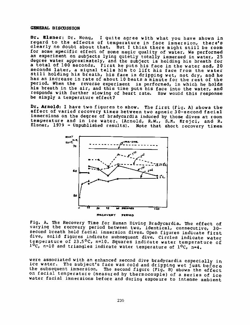
emIBRAI, DISCDSSION
Dr. Blsner: Dr. Hong, I quite agree with what you have shown inregard to the effects of temperature in face immersion< there' sclearly no doubt about that. But I think there might still be roomfor some specific effect of some magic quality of water. We performedan experiment on subjects lying quietly totally immersed in water< 25degree water approximately, and the subject is holding his breath fora total of 100 seconds. First he puts his face in the water and, 20seconds later, a signal tells him to lift his face from the waterstill holding his breath, his face is dripping wet, not dry, and hehas an increase in rate of about 10 beats a minute for the rest of theperiod. When the reverse experiment is performed, in which he holdshis breath in the air, and this time puts his face into the water, andr'esponds w ith f ur ther slow ing of heart rate. How would this responsebe simply a temperature effect2
Dr. hraold: I have two f igures to show. The f irst Fig. A! shows theeffect of varied recovery times between two apneic 30-second facialimmersions on the degree of bradycardia induced by those dives at roomtemperature and in ice water. Arnold, RW., SM. Krejci< and R.Elsner, l979 - Onpublished results!. Note that short recovery times
~INST
Caw E. 70 3o %$ M $4ce~sRKcovc$tY pRR'too
Fig. A. The Recovery Time f or Human Diving Bradycardia. The ef f ect ofvarying the recovery period between two, identical, consecutive, 30-second breath hold facial immersion dives. Open figures indicate firstdive, solid f igures indicate subsequent dive. Ci rcles indicate watertemperature of 23.5 C, n=10. Squares indicate water temperature ofl C, n=l0 and triangles indicate water temperature of l C, n=4.
were associated with an enhanced second dive bradycardia especially inice water. The subject's face was cold and dripping wet just beforethe subsequent immersion. The second figure Fig. 8! shows the effecton facial temperature measured by thermocouple! of a series of icewater facial immersions bef ore and during exposure to intense ambient
23G
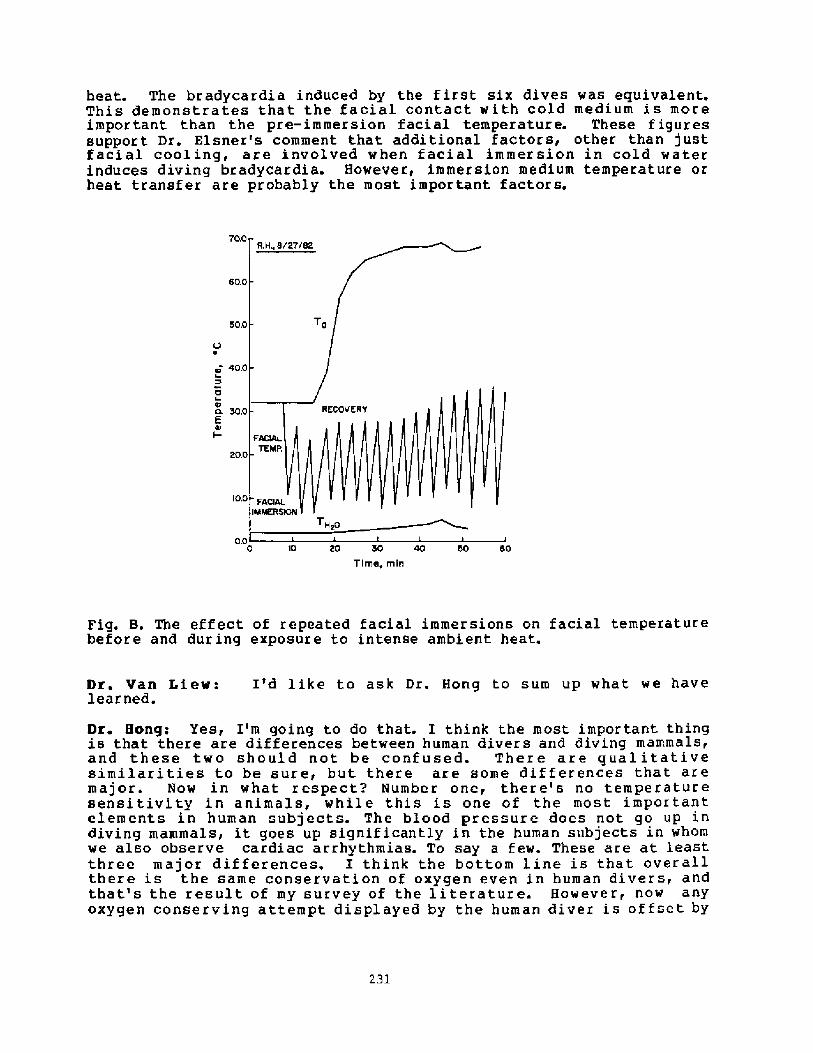
heat. The bradycardia induced by the first six dives was equivalent.This demonstrates that the facial contact with cold medium is moreimportant than the pre-immersion facial temperature. These figuressupport Dr. Elsner's comment that additional factors, other than justfacial cooling, are involved when facial immersion in cold waterinduces diving bradycardia. However, immersion medium temperature orheat transfer are probably the most important factors.
70.0
60,0
50.0
10.0
0.0 0 10 60 30 40 50 60Time, min
Fig. B. The effect of repeated facial immersions on facial temperaturebefore and during exposure to intense ambient heat.
Dr. Van Liew: I'd like to ask Dr. Hong to sum up what we havelearned.
Dr. Hong: Yes, I'm going to do that. I think the most impor tant thingis that there are differences between human divers and diving mammals,and these two should not be confused. There are qualitativesimilarities to be sure, but there are some dif f erences that aremajor. Now in what respect? Number one, there's no temper'aturesensitivity in animals, while this is one of the most importantelements in human subjects. The blood pressure does not go up indiving mammals, it goes up signif icantly in the human subjects in whomwe also observe cardiac arrhythmias. To say a few. These are at leastthree major differences. I think the bottom line is that overallthere is the same conservation of oxygen even in human divers, andthat's the result of my survey of the literature. However, now anYoxygen conserving attempt displayed by the human diver is offset bY
231
the increase in blood pressure, which offsets the advantage one gainsby bradycardia, so it is fair to say that, yes, we can describe someoxygen conservation in human divers, but not. to the extent that wefind in animals. Even diving mammals, sornetirnes, don't have oxygenconservation. So maybe the difference is no big deal.
Dr. Lin: Well, I couldn't agree with that; the reason is this: wecan describe reduced oxygen consumption experimentally. However< thisdoesn't necessarily mean oxygen conservation. For example, the bodycons urnes less oxygen unde r many phys iolog ical condi t i ons such ashibernation, torpor, or in accident in ice cold water. Such levels of02 consumption are appropriate f or these condi tions. Oxygenconsumption is reduced because demand of oxygen is reduced< but, arethey really conserving oxygen? At a body temperature of 37 C, 02consumption of 250 ml/min is normal, at 10oC, 50 ml/min is normal-Cont.rary to these, diving mammals obviously use less oxygen duringdiving where physical activity increased. In order to describe 02conservation, an equivalent physiological demand should exist and usea reduced amount of oxygen.
Dr hrnoM: I think another point is that the human diving responseis quite variable. When trained seals are forced to dive, theirresponses are quite similar whereas when humans are asked to dive,their cardiovascular response can be reasonably variable from subjectto subj ect.
Dt. Hong: Another important issue is that there is a myth in divingphysiology introduced by several physiologists: in case of drowningin cold water, the diving reflex saves one's lif e. I don't know ifthis is true. This is a big issue, actually. I was hoping to getinto that discussion, but we ran out of time. But maybe sometime wecan spend 10 to 15 minutes and really talk to each other. This is adangerous myth and we' ve got to do something about it.
232
PATHOPHYSIOLOGY OF NEAR-DROWNING:
ASPIRATION VS . NON-ASPIRATION
P. G. Boysen, N. D.
Various Responses to Submersion
The same sequence of events does not occur in all victims
of submersion. In fact, what ultimately happens depends on the
patient's degree of laryngospasm, type and length of immersion,
and underlying physiological condition.
It appears that most victims of sub~ersion accidents
sustain laryngospasm initially. In about 80% of cases, the
subsequent carbon dioxide build-up results in laryngeal
relaxation before the onset of respiratory paralysis. This
sequence allows water to enter the lungs, resulting in "wet
drowning" � a process that usually occurs in patients who
struggle in the water, hold their breath, and make violent,
terminal inspiratory efforts as they asphyxiate �!.
Alternatively, some 10-20% of victims develop sustained
laryngospasm until the onset of respiratory paralysis t3!. This
effectively bars the entrance of water into the lungs. As a
result, early resuscitative efforts in these latter patients
often succeed, and most of them recover without complications,
unless aspiration of water entering the stomach occurs during
resuscitation. This type of episode, which is sometimes termed
"dry drowning," typically presents in patients who accident. ally
233
fall in the water, as opposed to those who suffer from
exhaustion following prolonged swimming or struggling l,2!.
Also of importance in submersion accidents are the type and
length of immersion. For example, a sudden immersion in cold
water may result in cardiac arrest mediated through vagal
mechanisms. This happens most commonly in patients depressed by
alcohol l,6!. On the other hand, some patients become
profoundly hypothermic due to prolonged exposure in water. The
water in which the submersion accident occurs need not be
particularly cold in order to cause hypothermia. In contrast to
the drowning deaths when the Titanic sank in icy seas, the
Laconia disaster proves that hypothermia can incapacitate
victims even in 15 C <60 F! water in the summertime �!.o 0
Moreover, actively struggling or swimming a long distance can
cause the more rapid onset of hypothermia, since such physical
activity leads to peripheral vasodilation and increased body
heat loss.
Another type of submersion accident can occur in swimmers
who purposely alter their physical condition before swimming .
As a means of increasing their endurance, these swimmers
hyperventilate, hoping that by becoming hyperoxic they will be
able to further their underwater swimming distance during a
breath-hold. But they also are obviously hypocapnic <more so
than hyperoxic! and may lapse into unconsciousness before the
234
rising arterial carbon dioxide tension initiates inspiration or
prompts them to surface and terminate the breath-hold.
Mul,tisystem Pathophysiology
It has been difficult to understand pathophysiological
changes that occur in near-drowning victims. Indeed,
extrapolation from early animal experiments helps little and,
more often than not, Leads only to confusion and
misinterpretation. Early dog experiments dealt with aspiration
volumes exceeding 22 ml/kg of fluid. Clinical stud.ies, however,
now suggest that only about 15% of humans involved in drowning
accidents suffer physiologic changes consistent with this degree
of aspiration 8,9!. Because of this disparity and the many
myths generated by reliance on such experimental studies, we
must take a fresh look, on a system-by-system basis, at
pathophysiologic changes occurring in most near-drowning
patients.
PULMONARY. Although fresh and salt water aspiration cause
somewhat differing effects on the lungs, some changes occur
despite the type of fluid. Among those common lesions are
factors that cause severe hypoxia, including alveolar
inflammation, right-to-left shunting, and injured alveolar
membranes due to foreign materials. When patients aspirate
fresh water, however, two characteristic events occur: a!
surfactant abnormalities arise, which result in a lowered
235
functional residual capacity, unstable alveoli, and stif f,
noncompliant lungs, and b! the lung absorbs much of the fluid
rapidly �0,11!. But since sea water is hypertonic, this fluid
remains in the lung for a period of time and exerts an osmotic
gradient, which causes a further influx of physiologic fluid
into affected alveoli.
NEUROLOGIC. Near-drowning victims frequently present with
signs of cez.ebral hypoxia, especially coma, and the metabolic
acidosis produced by tissue hypoxia further complicates this
problem by compromising cerebral circulation. The cumulative
effects of these insults can be devastating and include
increased intracranial pressure due to brain swelling and,
possibly, brain stem herniation and sudden death �2,14!.
However, the degree to which neurological changes occur varies
considerably, depending on: a! the extent and time period of
anoxia, b! the previous condition of the patient, and c! the
temperature of the water. Obviously, the shorter the period of
anoxia and the better the patient's underlying condition, the
more favorable is the outlook. Likewise, the colder the water,
the better is the chance of neuron survival. The evidence for
this latter effect comes from the numerous reports of successful
resuscitation and the complete return of normal neurological
function following prolonged �0-40 minutes! submersion in cold
water 8,12,13,15!. The importance of such reports is that the
236
absolute anoxic time limit for neurological survival is unknown
in such cases 8,l4!.
HRNATOLOGIC. Two myths surrounding near-drowning are: a!
that aspiration of fresh water decreases the patient' s
hemoglobin and hematocrit by hemodilution and hemolysis and b!
that sea water aspiration increases these same values due to a
positive fluid shif't into the lungs. Actually, clinical
observations now make it clear that neither of these two effects
occur in practice. The reason is that most near-drowning
patients aspirate far smaller quantities than required to cause
such changes, and equilibration of any fluid shifts occurs
rapidly. In fact, a decreased hemoglobin level should initiate
a search for a source of blood loss l!.
CARDIOVASCULAR' While dogs that aspirate huge quantities
�4 ml/kg! of water develop ventricular fibrillation, one rarely
encounters this phenomenon in normothermic near-drowning
victims. As with hemodilutional changes, ventricular
fibri.llation occurs rarely because most survivors of submersion
accidents aspirate a lesser quantity than this. Moreover,
patients who aspirate larger quantities of fluid probably die
before receiving medical therapy �6, 17!. The cardiovascular
change that can occur, however, is arterial hypotension. In
general, the degree to which this manifests primarily depends on
the patient's vascular tone, which is affected by the degree of
blood loss, the patient's peripheral body temperature, and
237
levels of circulating catecholamines and carbon dioxide. Ne
have assessed the hemodynamic changes that occur in a laboratory
model during asphyxia, fresh water aspiration and salt water
aspiration. Ho matter which of the above occurs, there is
arterial oxygen desaturation and mixed venous oxygen
desaturation. During asphyxia there is no change or an increase
in arterial pressure, pulmonary artery hypertension and a
fluctuating central venous pressure with continued inspiratory
efforts. Fresh water aspiration is followed by hypotension,
increased PA pressure and a rising CVP; cardiac output falls by
20-30%. After salt water aspiration hypotension is severe but
PA pressure and CVP are elevated; cardiac output is severely
reduced by 50%.
In each case, the most notable changes are l! arterial and
mixed venous oxygen desaturation; 2! elevated PA pressure
secondary to ¹l! and 3! fall in cardiac output, also in part
due to hypoxemia. Clearly the most important aspect of early
treatment of the near-drowned is management of the airway and
maintenance of oxygenation.
METABOLIC. Metabolic acidosis is one of the most
significant abnormalities occurring in near-drowning victims,
and in most cases, it results almost totally from hypoxia rather
than poor tissue perfusion. This problem can be so severe that.
some patients present with a pH in the range of 6 .8-6 .9. The
severity of the metabolic acidosis, however does not reliably
238
predict survival, since some patients with a pH in this low
range do survive if they receive appropriate treatment.
As mentioned above, hypothermia acts as a double-edged
sword with victims of submersion accidents. On the one hand, it
may rapidly claim the patient's life. On the other, it may
provide a protective effect by preserving both brain and cardiac
tissue function for short periods of time, despite severe
hypoxia �!.
Finally, the potential for electrolyte abberations with
near-drowning victims is minimal. As is the case with other
unfounded fears concerning near-drowning, reports of severe
electrolyte imbalance with drowning arise frm early animal
experiments using large volumes of water. In practice, only
slight changes in the serum sodium and chloride occur following
salt water aspiration, and electrolyte changes following fresh
water are usually negligible. In fact, when compared to the
profound hypoxemia and metabolic acidosis that most victims
experience, these electolyte changes pose no significant
management problems 8!.
Restoring Vital Physiological Function
The goals of therapy for near-drowning victims basically
encompass the restoration of vital physiological function. In
particular, this means reversing the hypoxemia and metabolic
acidosis, protecting the brain from further insult, and treating
239
both the pulmonary complications associated with aspiration and
hypothermia, when present. But because of the controversies
surrounding which patients to resuscitate and the need for lung
drainage, one first must be clear on the relative value of these
procedures.
RESUSCITATION. The controversy surrounding resuscitation
stems from the conflicting conclusions of several series of
drowning and near-drowning victims l2,13,18,19!. The fear is
that resuscitation of near-drowning victims may result in a
living but permanently brain-damaged patient �0!. In fact,
this concern has merit, since many near-drowning victims do
survive in a vegetative state. The problem, however, is that
there exists no simple way to predict any given patient' s
outcome during the initial resuscitative period and, in some
cases, even with specialized monitoring during the first 24-48
hours �8,19,21!. This fact leaves little choice but to employ
aggressive resuscitation with all victims.
Unfortunately, the question of draining fluid from the
lungs is not so easily answered. On one side of the debate are
those who advocate an abdominal thrust Heimlich maneuver! as
the in.itial resuscitative procedure �2!. Theoretically, this
clears the lungs of excess fluid and, thus, allows for better
gas exchange. On the other side are those who find this
maneuver unnecessary and problematic �3,24!. In the first
place, the lung rapidly absorbs the fluid in patients sustaining
240
fresh water aspiration, and in those with salt water
aspiration, the excess fluid rarely appears to cause significant
problems with ventilation. In the second place, an abdominal
thrust may cause the forceful expulsion of the gastric contents
from the patient's stomach, risking pulmonary acid aspiration
and all the terrible consequences that it engenders. For these
reasons, most authorities feel that the abdominal thrust
maneuver may waste time, and they reserve it solely for patients
in whom solid material obstructs the larynx and prevents the
establishment of a patent airway.
OXYGENATION is the primary treatment consideration in
near-drowning patients.
When recovering a submersion victim who has no spontaneous
respirations, the need for immediate ventilation is clear.
Anyone who is present should clear the airway of food or foreign
objects, support the airway by elevating the jaw and soft
tissues, and begin mouth-to-mouth or mouth-to � nose ventilation.
In some cases, an oral or nasopharyngeal airway proves necessary
to maintain an adequate airway, but it is better to avoid such
devices when possible because of the chance that their insertion
may precipitate laryngospasm or vomiting �5!. As soon as
possible, all patients should also receive supplemental oxygen,
since even conscious patients may suffer significant pulmonary
damage with severe hypoxemia and still be conversant.
241
METABOLIC ACIDOSIS is partially resolved by treating
the patient's hypoxia, but sometimes this does not correct
the problem rapidly enough. To correct this problem, use
intravenous sodium bicarbonate in patients with a pH of
less than 7.2, calculating the amount needed with the
following formula: [HCO3 deficit] = body weight kg! x0.2 x j desired HCO>! measured HCO3! ] .
CEREBRAL RESUSCITATION. After initiating the above
resuscitative procedures and stabilizing patients from a
cardiopulmonary and metabolic standpoint, evaluation of
neurological status is essential. Patients who are awake and
alert and have minimal cardiopulmonary changes may only require
observation for 24 hours as indicated above. Alternatively,
patients who remain unconscious, but maintain normal pupillary
and pain withdrawal responses, need hourly neurological
monitoring to detect any further changes or deterioration in
function. Those with signs of increased intracranial pressure
or severe neurological dysfunction, however, require more
definitive therapeutic intervention. Intravenous mannitol l
g/kg!, dexamethasone 80 mg/day! and hyperventilation are all
effective in lowering intracranial pressure to maintain
patient's PaCO2 in the range of 30 torr!. After instituting
these measures, most authorities recommend the placement of
intracranial pressure ICP! and cerebral perfusion pressure
CPP! monitors to gauge the need and effect of further
intervention �8,29!. Such intervention may include
242
hypothermia, barbiturate coma, and muscle relaxants. However,
the precise benefits of this kind of therapy for near-drowning
victims remains unclear. Unfortunately, those patients who
develop an ICP greater than 20 mm Hg and a CPP less than 50 mm
Hg generally fare poorly despite therapy, and most either die or
persist in a vegetative state �9!.
PULMONARY COMPLICATIONS. Early oxygenation and ventilation
are important in terms of outcome. Once transported to the
hospital, manipulation of positive end-expiratory pressure
PEEP!, which may require invasive hemodynamic monitoring, can
be initiated to improve gas exchange.
HYPOTHERMIA. The treatment for hypothermia is somewhat
controversial, but proves necessary in all near-drowning
patients with a core temperature below 35 C. However, one0
should employ different techniques depending on the degree of
hypothermia, and they also should be aware of special
considerations when managing dysrhythmias in these victims. For
patients with mild hypothermia �2 -35 C!, passive external0 o
warming techniques usually suffice, as long as these victims are
able to produce endogenous heat. This method generally involves
placing victims at room temperature and covering them with dry,
warm insulating blankets. For patients with a core temperature
below 32 C, one can institute active external or coreo
rewarming techniques. Active exter nal rewarding techniques
include the use of electric blankets, heated objects, such as
243
hot-water bottles, and, possibly, immersion in warm �0 C!0
water using a Hubbard tank. When utilizing active external
rewarming techniques other than immersion, however, heat the
trunk only, since extremity rewarming may cause hypotension due
to peripheral vasodilation and also "afterdrop," which may
result from the sudden return of cold, hypokalemic, acidotic
blood to the core organs. Immersion in warm water proves
controversial because of both these latter problems with
extremity rewax'ming and the need for arterial blood gas, blood
pressure, pulse, and CVP monitoring, which are cumber'some in
this setting. In addition to nonimmersion external techniques,
active core rewarming methods can be used. The most practical
of these methods encompasses the inhalation of heated �5 C!, 0
humidified oxygen via a mask or endotracheal tube and irrigation
of the stomach, bladder, and/or peritoneum, using an appropriate
solution at a temperature near 42 C.o
The treatment of dysrhythmias in hypothermic victims
differs considerably from that used in normothermic patients.
First, atrial arrhythmias need no therapy, since they generally
resolve spontaneously as the patient's core temperature rises.
Ventricular fibrillation, on the other hand, presents greater
therapeutic problems. In patients with this problem,
defibrillation usually proves unsuccessful unless their core
0temperature is above 3G C. However, resuscitative techniques
should be continued until patients have a core temperature in
244
excess of 35 C. Likewise, the use of some antiarrhythmic0
agents, including lidocane and procainamide, helps little. In
hypothermic patients, lidocaine works poorly and procainamide
may actually increase ventricular irritability. As a result,
one should consider the use of bretylium Bretylol! in
fibrillating, hypothermic patients at an initial intravenous
dose of 5 mg/kg in children or 350-500 mg in adults, given over
8-10 minutes.
PREVENTION. The magnitude of submersion accidents in
numbers, as well as in terms of morbidity and mortality, makes
prevention a critical social issue. Though improved medical
management certainly saves lives, the fact that 9L% of pediatric
drowning victims are deceased at the scene speaks for the need
for a solid program of water and pool safety.
Education is of primary importance in order to implement
safety principles whether it involves monitoring and fencing
backyard pools, breath-hold diving, SCUBA diving, or any other
water sport. Reaction to urgent or emergent situations in the
event of an accident is important to preserve life and function.
245
References
2.
3.
4.
6.7.
8.
9.
10.
12.
13.
14.
15.
16.
17.
246
Donaldson JC, Royall JD: Drowning and near-drowning.Postgrad Med 64:71, 1968.Clark EB, Niggemann EH: Near-drowning. Heart Lung4:946, 1975.Rowe MI, Arongo A, Allington G: Profile of pediatricdrowning victims in water-oriented society. J Trauma17:587, 1977.Pleuckhahn VD: Drowning: Community aspects. Med JAust 2:226, 1979.Hunter TB, Whitehouse WM: Fresh water near-drowning:Radiological aspects. Radiology 112:51, 1974.Miles S: Drowning . Br Med J 3:597, 1968.Conn AW: The near-drowned child. Emerg Med 12:16,1980.Modell JH, Graves SA, Ketover A: A clinical course of91 consecutive near-drowning victims. Chest 70:231,1971.Modell JH, Boysen P: Respiratory crises. CriticalCare: State of the Art. Shoemaker WC, Thompson WL eds!, Fullerton, California, Society of Critical CareMedicine, 1980.Giammona ST, Modell JH: Drowning by total immersion:Effects on pulmonary surfactant of distilled water,isot,onic saline and sea water. Am J Dis Child 114:612,1967.Modell JH, Calderwood HW, Ruiz BC, et al: Effects ofventilatory patterns on arterial oxygenation afternear-drowning in sea water. Anesthesiology 40:376,1974.Peorn JH, Bort RD, Yamaok AR: Neurologic sequelaeafter childhood near-drowning: A total populationstudy from Hawaii. Pediatrics 64:187, 1979.Peterson B: Morbidity of childhood near-drowning .Pediatrics 59:364, 1977.Conn AW, Fdmonds JF, Barker GA: Cerebral resuscitationin near-drowning. Ped Clin North Am 26:691, 1979.Siebke H, Breivik H: Survival after 40 minutes'submersion without cerebral sequelae. Lancet 1:1275,1975.Modell JH, Moya F: Effects of volume of aspiratedfluid during chlorinated fresh water drowning .Anesthesiology 27:662, 1966 .Modell JH: The Pathophysiology and Treatment ofDrowning and Near-drowning . Springfield, Illinois,Charles C Thomas, 1971.
18,
19.
20.
21.
22.
25.
26.
27.
28.
29.
30.
31.
32.
33.
34.
Oakes DD, Sherck JP, Maloney JR, Charters AC III:Prognosis and management of victims of near-drowning.J Trauma 22:544, 1982.Dean JM, McComb JG: Intracranial pressure monitoring insevere pediatric near-drowning. Neurosurgery 9:627,1981.Frates RC Jr: Analysis of predictive factors in theassessment of warm-water near-drowning in children. AmJ Dis Child 135:1006, 1981.Modell JH, Graves SA, Kuck EJ: Near-drowningcorrelation of level of consciousness and survival.Can Anaesth Soc J 27:211, 1980.Heimlich JH: Subdiaphragmatic pressure to expel waterfrom the lungs of drowning persons. Ann Emerg Med10:476, 1980.23. Wilder RJ, Wedro BC: "HeimlichManeuver" for near-drowning questioned letter!. AnnEmerg Med ll:ill, 1982.Orlowski JP: "Heimlich Maneuver" for near � drowningquestioned letter!. Ann Emerg Med 11:ill, 1982.Modell JH: Drowning and near-drowning . Disorders ofthe Respiratory Tract in Children. Kendig EL, ChurnickD eds!, Philadelphia, WB Saunders, 1983.Gong H Jr: Toward a better understnading of ARDS. FamMed Reports 1:79, 1983.Boysen PG: Individualizing mechanical ventilation topatient requirements. ER Reports 2:63, 1981.Pfenninger J, Sutter M: Intensive care after freshwater immersion accidents in child.ren. Anaesthesia37:1157, 1982.Nussbaum E, Galant SP: Intracranial pressuremonitoring as a guide to prognosis in the nearlydrowned, severely comatose child. J Pediatrics102:215, 1983.Petty TL, Fowler AA: Another look at ARDS. Chest82:101, 1981.Nicholson DP: Corticosteroids in the treatment ofseptic shock and the adult respiratory distresssyndrome. Med Clin North Am 67:717, 1983.Sibbald WJ, Anderson RR, Reid B, et. al:Alveolar-capillary permeabil.ity in human septic ARDS.Chest 79:138, 1981.Wolfe JE, Bone RC, Ruth WE: Effects of corticosteroidsin the treatment of patients with gastric aspiration.Am J Med 63:719, 1977.Calderwood HW, Modell JH, Ruiz BC: The ineffectivenessof steroid therapy for treatment of fresh waternear-drowning. Anesthesiology 43:642, 1975.
247
ASPHYXiA
200
Art.100
rnmHg!
40
PA20
rnmHg!
40
CVP20
rnrnHg! ~,.N,P,>! I1 0 p r. r,'. i:
CORKC.O. 6.0 ~ 3.6 L/min
Fig. la
ASPHYXIA: EEMODYNAPCICS
Arterial Art! pulmonary artery PA! and central venouspressure CVP! tracings during a brief period of upper airwayocclusion and asphyxia. Calculated systemic and pulmonaryvascular resistance increases. Note negative pressure swings inthe PA and CVP tracing with continued inspiratory effort. Thehemodynamic changes, including the fall in cardiac output arereversed with relief of hypoxernia.
ASPHYXIA
100
80
60
40
20 Sv02-FiberapticFig. lb
ASPHYXIA
An indwelling pulmonary artery catheter equipped with afiberoptic system to measure Sv02 continuously shows markedmixed venous oxygen desaturation during upper airway obstructionand continued inspiratory effort. With removal of theobstruction the SvO immediately improves. The overshootimplies not only improved gas exchange but also a temporaryaugmentation of cardiac output.
249
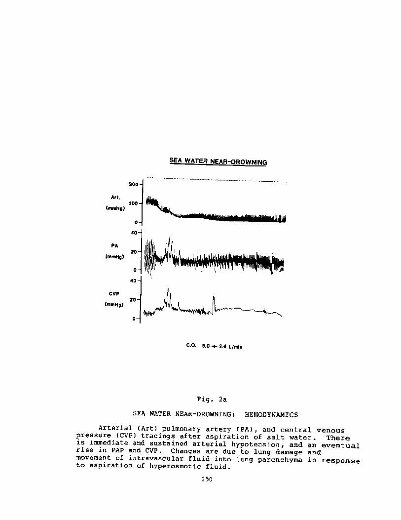
SEA WATER NEAR-DROWNING
200
Art,100
mmH0!
PA
mmH0!
io
CVP20
mmH2! C,O. 5.0 ~ 2.4 I./minFig. 2a
SEA WATER NEAR-DROWNING: HENODYNAMICS
Arterial Art! pulmonary artery PA!, and central venouspressure <CVP! tracings after aspiration of salt water. Thereis immediate and sustained arterial hypotension, and an eventua,lrise in PAP and CVP. Changes are due to lung damage andmovement of intravascular fluid into lung parenchyma in responseto aspiration of hyperosmotic fluid.
250
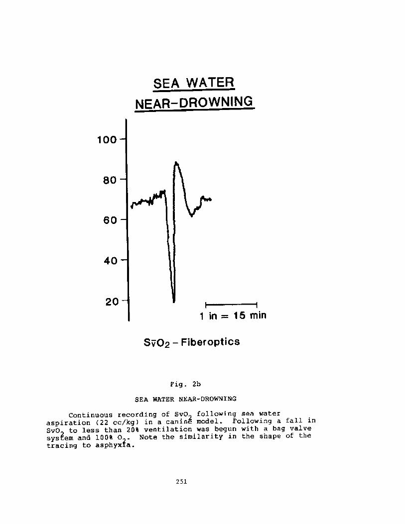
SEA WATER
NEAR � DROWNING
100
80
40
20
1 in= 15 min
Sv02 � Fiber optics
Fig. 2b
SEA WATER NEAR-DROWMIhlG
Continuous recording of Sv02 following sea wateraspiration �2 cc/kg! in a canine model. Following a fall inSvO~ to less than 20% ventilat.ion was begun with a bag valvesystem and 100% 0 . Note the similarity in the shape of thetracing to asphyx/a.
251
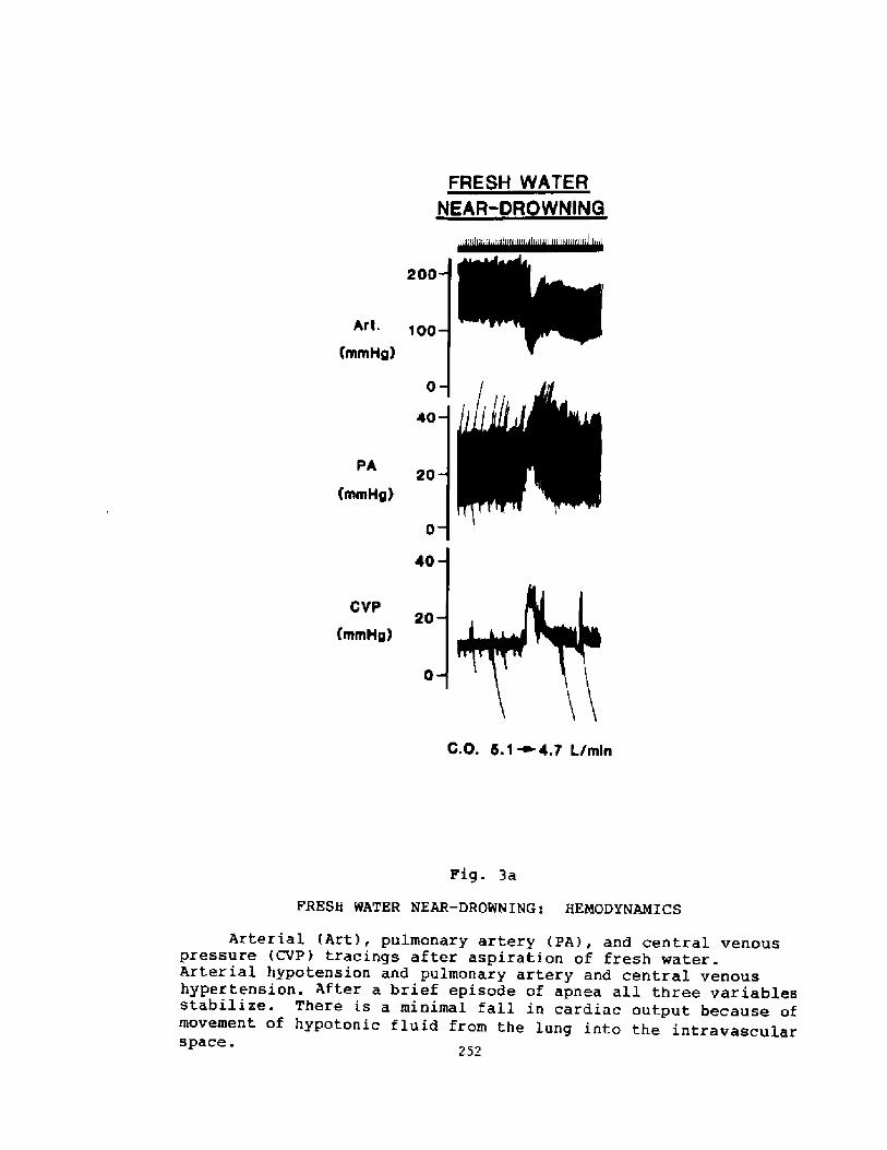
FRESH WATER
NEAR-DROWNING
200
AI't. q 00
mmHg!
40
PA
mmHg!
40
cvp20
mmHg! G.O. 5.1~4.7 Lfm[nPig. 3a
FRESH WATER NEAR-DROWWrNG: HEXODYNmXCS
Arterial Art!, pulmonary artery PA!, and central venouspressure CVP! tracings after aspiration of fresh ~ater.Arterial hypotension and pulmonary artery and central venoushypertension. After a brief episode of apnea all three variablesstabilize. There is a minimal fall in cardiac output because ofmovement of hypotonic fluid from the lung into the intravascularspace. 252
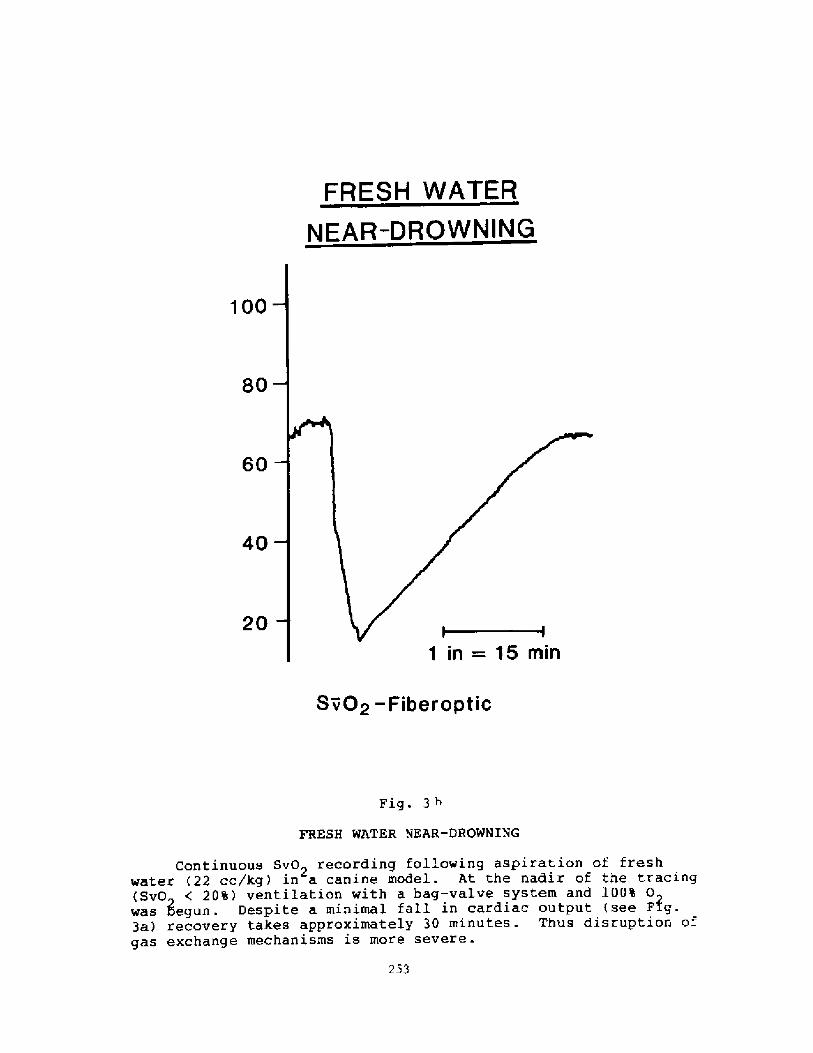
Discussion following Dx. Boysen's presentation
Dr. Norf lect: Did you observe any seizure activity in yourexperimental model, and, if so, was it related to the type of insultP
Dr. Boysen: We didn't observe any seizure activity. That question hascome up before, we may have masked it; these dogs were anesthesized.
Dr. Horfleet: What was the anesthesia'P
Dr. Boysen: Pentobarbital, intravenous. So that may have influencedthat.
Dr. Siesjo: I was wondering about the blood pressure at the end ofthe asphyxia.
Dr. Boysen: The blood pressure depends on the length of the asphyxiaDr. Siesjo: Yes. If you have a long period of asphyxia, yet shortenough not to interfere with adequate resuscitation, what would theblood pressure be?
Dx. Boysen: The blood pressure is still normal or elevated. Once theblood pressure starts to go down in the asphyxiant, usually you don' tget much in the way of recovery. It just starts to go down befoxe tenminutes. The asphyxia time, and I'm sure it's the same for patients,tends to be about four to six minutes. Now the issue is when weintervene, it's typical when patients are pulled from the water, theparamedic will find that they have a laryngospasm. You can break alaryngospasm with continuous positive airway pressure, but in generalair cannot be insuff lated with a bag-mask valve system. And so thepoint is that, once they relax, it's important to insuff late theairway and intubate the patient almost immediately.
Dr. Siesjo: I was just w'ondering if you can clarify whether or notthe ca.rdiac reaction to the asphyxia is related to what type ofanesthesia you use.
Dr. Boysen: I haven't done it with a dif ferent anesthetic, I can' tanswer that.
Dr. Blsnex: Could I bring up a question which may have criticalbearing here'? Some years ago, when we were doing studies of theneural control mechanisms in the diving response of the seal, we werevery much impressed by the power of the reflex of lung inflation andstimulation of pulmonary stretch receptors in producing a tachycardia.So it might be that a by-product of CPR and lung inflation is torestart or to stimulate the heart as well.
Dr. Boysen: We'll, I think that is absolutely true. Although if youlook at the patient histories, of most of the patients that are pulledf rom the water and survive, there is not a great incidence, or' atleast recorded incidence, of arrhythmias which I might expect if thereis some stimulant ef f ect.
254
Dx. Slsner: Yes, but they do likely respond ta the lung inflation ofCPR with reversing of cardiac arrest.
Dx. Boysen: That would be hard to know because I think the majorthing is reversal of hypoxia, and cardiac function in our experiencetends to improve immediately once you relieve the hypoxia, so I don' tknow if we can separate the two.
255
HYPERVENTILATION: AN UNPREDICTABLE DANGER TO THESPORTS DIVER
PIETER GUILLAUME LANDSBERG, MB.ChB. Rand!,M.F.G.P. S.A.!M.D.'! Geneva Street Lakefield BENQNI, SOUTH AFRICA 1 501
INTRODUCTION
Under conditions of normal atmospheric pr'essure, breath-holdingresults in important changes in the mechanism whereby C02 is transported andexchanged in the lungs, but a reversal of the COz gradient from the lungs tothe blood does not occur unless the inspired air contains a highconcentration of CO. Lanphier 8 Rahn 1 963!.
Under conditions of rai sed atmospheric pressure, as occurs i nbreath-hold diving, the whole aspect of COz transport and exchange ismarkedly alter'ed and cannot be equated with apnoea at normal atmosphericpressure Lanphier & Rahn 1963!. Prolonged apnoea under raised atmosphericpressur e, results in a reversal of CO~ exchange from the lungs to the blood Lanphier & Rahn 1963!. During apnoea a shrinkage of lung volume occur s,suggesting that CO~ output decreases while 0~ uptake is continuous Craig1961 !. Arterial PCOi wi 11 increase and exceed venous PCQz during apnoea, asa result of the oxygenation of Hb in the presence of falling COz output tothe lungs Haldane effect!. Schaefer �975! showed the flow of CQ~ into thelungs duri ng ascent from a di ve is regulated in part by the speed of ascent.Mithoefer �959! offered an explanation for the " aberrations in CO~transport and exchange" which occur during apnoea with Oz .
Craig 1961 ! showed the unconsciousness following hyperventilation andbreath-hold di ving, was probably caused by hypoxi a.
In 1963, Lanphier, working with four divers under simulatedbreath-hold diving conditions in a compression chamber, established thecourse of alveolar gas exchange under pressure and also determined from thenomogramme of Di 1 1 the time course of mixed venous CO~ content duringsimulated dives. He worked in a pressure chamber in order to determineaccurately the pressure-time relationships and to facilitate the taki ng ofalveolar gas samples. He concluded that hypercapnia CO~ narcosis! may causeunconsciousness in prolonged, deep breath-hold dives. Kong �963! in a fieldstudy of the Korean diving women Ama!, noted that they did nothyperventilate to any great extent and followed the relatively safe patter nof the 60 second dive to 10 meters in which neither hypoxi a or C02accumulaton is a serious hazard.
Smit � 967, 74, g] ! determined the alveolar CQz and =Q~ X afterbreath-hold dives in a swimming bath and diving tank, but no blood CQ2levels were determined. 8]ack-outs occurred when the P~ 0~ dropped to below4 5 kl'a, �3 � 34 mmHg!. In divers whose exhaled values came close to thisf<gure but wer e not below 4,5 kPa, hypoxi c black-outs did not occur e.g. at4.8 and 5,3 kPa!. The probable limit of hypoxic stress during apnoea may beat a PtIQ~ of 4,0 � 4,5 kPa, which, when transformed, will be close to thecritical arterial oxygen pr essure P a02 ! of 4 kPa �0 mmKg! associated with
256
loss of consciousness.
An early prelim~nary experiment on 5 experienced breath-hold diversindicated the possibility that the effects of hyperventilation on the COzcontent of peripheral venous blood may be unpredictable in different diversunder the same diving conditions and also in the same diver from day to day Landsberg 1971 !.
More recently it was shown that the partial pressure of CO> inarterial ised peripheral venous blood, duri ng the hyperventilationbreath-hold-dive cyc'le under a steady state condition, is unpredictable andthat hypoxi a PgP~ = 4 � 6 kPa! superimposed on hypocarbi a PvCOz = 4 - 5kPa! may enhance loss of consciousness underwater Landsberg 198Z,1 984!.
Unpredictable, wide differ ences in the alveolar PCO~ of SCUBA diversin the sea at 1 0 m and 30 m have also been found Owyer 8 Pi lmani s 1 975!.Unpredictably the hi ghest alveolar PCO~ of 8,9 kPa �8 mmkg ! was found atthe shallowest depth i n the most experienced diver. CO> retention was notconsistent at any workload or depth. Dwyer found 3 groups of SCUBA divers;those that eliminate COz normally, those that retain COz and those thathyperventilate and eliminate excessive amounts CO~. He also found thatdivers may shift from one group to another in a manner not related toworkload or depth, as well as a wide individual variation.
Morrison et al., �978!, investigating loss of consciousness in 2SCUBA di vers, noted a low r espi ratory response to inspired CO~ in thesedivers: i.e. insensitivity to COz must have been inher ent in the divers.They inferred from their experimental results that loss of consciousness wasassociated with hypercapni a, although hyper capni a alone could not havecaused unconsciousness. In their experiments loss of consciousness was notthe direct result of hypoxia. They quote Case 8 Haldane �941! who observedthat divers 1 ost consciousness at very di fferent partial press~res of CO~,which indicates a large range of individual susceptibility.
Several physiological mechanisms can potentially cause uriconsciousnessunderwater duri ng breath-hold and SCUBA diving: � hypoxia Craig 1961 !,hypercapni a - COz narcosis � Lanphier 8 Rahn 1963; Paulev 1 969!, variouscombinations of hypoxi a with hyper- or hypocapni a or even a normal Gi bloodtension wi th hypocapnia Rahn, Oti s 8 kodge, 1946; Dheni n et al ., 1978!,cardiac arrhythmi as, ventricular fibrillation Sem Jacobson 8 Styri 1 970,Brown 1974!, bradycardia Landsberg 197Z, Bove et al., 1973!.
Brown 1974! maintains that " shall ow water blackout" has beenconfusing the diving community long enough and that underwater blackout i san entity with a number of possible causes, which he classifies.
Wolf �970!, has postulated the mechanism of sudden death duringsubmergence as follows: the stimulus is anticipation or fear of diving orforced immersion which can cause extrasystoles; the response duringsubmergence wi 11 be the oxygen conserving "diving reflex" consisting ofbradycardi a initiated by parasympathetic vagal activity; blood pressure willdecr ease, but peripheral vasoconstriction brought about by sympathet ic
257
activity, will maintain blood pressure; skin, muscle and peripheral bloodflow will decrease with lactic acid and COz accumulation causi ng metabolicacidosis with low pH, high K levels and hypoxia; all these factors combinedwi11 cause extreme bradycardia, extrasystoles, arrhythmi as, ventricularfibrillation and cardiac arrest. Anderson and Blix �974! have confirmedthis mechanism in ducks wi th alpha and neuronal adrenergic blockadeexperiments. They have demonstrated the pharmacological components in theautonomic control of the diving reflex.
UNPREDICTABLE EFFECTS OF HYPERVENTILATION LANDSBERG, 1984!
This study was initiated by the drowning of an expert and world classSouth African spearfisherman on the 9th March, 1968. Mhat factorscontributed to the tragic and unnecessary death of a superbly fit and veryexperienced diver? Spearfishermen admit they hyperventilate, otherwise theirdeep diving in search of fish becomes physiological ly impossible.Hyperventilation gives them 10 � 20 seconds extra bottom time", as shown byLanphier 8 Rahn �963!, but increases the danger of hypoxic black-out Craig1 961!. They appear to understand the dangers of hypoxi c "black� -out" onascent. They control their hyperventilation prior to breath-hold diving andappear to heed the physiological alarms set up in thei r bodies urgi ng themto surface to breathe. In spite of this, several have arrived on the surfaceunconscious, automatically breathing a mixture of water and air throughtheir snorkels until they overcome their hypoxia and regain consciousness,coughing, cyanosed and bewildered, yet conti nui ng to dive after a period ofrest . Some "dive on their watches" taki ng 75 seconds to di ve + 20 m surfaceto surface!. They have worked out their individual dive times by experiencewith previ ous hyperventilation and near black-outs. The effects ofhyperventilation in divers are unpredicabl e as we cannot predi ct a hi gh orlow level of COr at any point in the hyperventilation-breath-hold-divecycle. It is therefore very difficult for an experienced diver who has beenhyperventilating prior to breath-hold diving for many years, to be dogmaticabout his ability to heed the physiological alarms that he may experi enceduring a dive. One day he may have a perfect CO~ accumulation andstimulation of his respiratory centre wi th rapid diffusion of COz from bloodto alveolar gas taking place long befor e hypoxia causes unconsciousness. Thenext day CO~ accumulation may be slow due to excessi ve hyperventilation, orCO~ diff'usion into the alveolar gas may be slow due to rapid ascent causingexcessive CO~ accumulation in the blood, producing CO> narcosishypercapnia Paulev 1969!. Hypoxia, which occurs due to the breath-holdingand underwater exercise combined with COz narcosis will causeunconsciousness and drowning. Another factor involved is the over-ridingcompetitive spirit that prevails at spearfishing competitions, which causesthe di ver to wilfully shut off his physiol ogi cally protective alarm systems
Craig 1961, Deppe 1971 !.
THE PROBABLE CAUSES OF UNCONSCIOUSNESS DURING THEHYPERVENTILATION-BREATH-HOLD-DIVE CYCLE H-B-H-D-CYCLE!
During descent
On the surface a di ver may have a high level of CO~ in his lungs
2S8
2. On the bottom
Unconsciousness is due to prolonged submergence more than 80 seconds!which causes hypoxia and CO~ narcosis. This occurs due to the d~ver wilfullyignoring his desi re to br eathe Craig 1961, Deppe 1971!.
3. On ascent
Unconsciousness is caused by the drop in the arterial 02 tensionproducing hypoxia. A very rapid ascent may also cause CO~ accumulation inthe blood, and possibly CO~ narcosis, by not allowing suffi cient time forthe COz to enter the alveolar gas Schaefer 1975! However, the PACO~decreases because of air expansion i n lungs on ascent. Therefore thi s cannotblock COz diffusion i nto the alveoli Guyton 1982!.
DISCUSSION
An analysis of the dive patterns found in 30 apnoeic diver s in thecompr essi on chamber ! andsberg 1984! show various unpredictabl e blood andalveolar gas tensions all potentially capable of causing near loss ofconsciousness near black-out! at different breaking points. Some blood andalveo1 ar gas tensi on combinations wi 11 be more dangerous than others.
Because of the relationship between PgCO~ and the PvCO~ in thearteri alised peripheral venous blood of divers, it may be concludedthat i n general the arterial bood COo levels in trained divers arereduced by hyperventilation and increased by repeatedbreath-hold-diving.
Z. This is not true in all cases as divers show a var~at~on in thearteri ali sed PvC02 Landsberg 1 984!. This may possibly explain theunpredi ctable nature of the effects of hyperventilation in divers.
The alveolar Oz partial pressur e, Pa0~, incr eases afterhyperventilation. This is due to the loss of CO~ from the alveolar gasvolume during hyperventilation and the partial pressure space 1eft bythe exhaled CO> is take~ up rapidly by Oz Green 1 978!. In one di ver,at the end of 100 s breath-hald-dive, the PA Oz is 4,5 kPa, theaccepted lower limit of nornral maintenance of consciousness SmitGiesel 1981!.
3.
259
after a strenuous swim. If he does not hyperventilate to blow off the excessCO~ and decided to dive to 10 m the blood COr tension may rise above 6 kPa�6 mmHg! reaching levels of 6,5 - 9,2 kPa �0 -70 mmHg! the range of "C02narcosi s" Lanphi er lr Rahn 1963, Paulev 1 969!. This high level of CO~ maycause unconsciousness on descent. This may also occur if the diver decidesto di ve deeper than 1 0 m after exercising at that level. It may also occurif the diver does not allow an adequate rest period during repeatedhyperventilation-breath-hold-dive cycles Schaefer 1975 !.
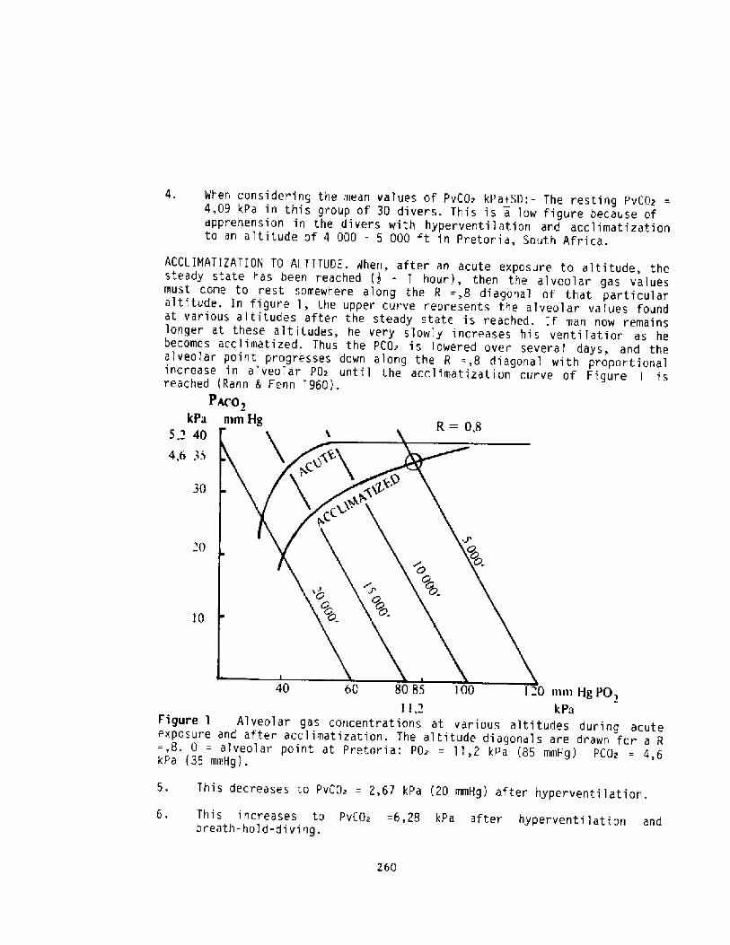
When considering the mean values of PvCO~ kPatSD: � The resting PvCO~4,09 kPa in t!iis group of 30 divers. This is a low figure because ofapprehension in the divers with hyperventilation and acclimatizat.ionto an altitude of 4 000 � 5 000 ft in Pretori a, South Africa.
ACCL IMATIZATION 10 AL.TITUOE. When, after an acute exposure to altitude, thesteady state has been reached � - 1 hour!, then the alveolar gas valuesmust come to rest somewhere along the R =,8 diagonal of that particularaltitude. In figure 1, the upper cur ve represents the alveolar values foundat various altitudes after the steady state is reached. If man now remainslonger at these altitudes, he very slowly increases his venti lation as hebecomes acclimatized. Thus the PCO~ is lowered over several days, and thealveolar poi nt progresses down along the R =,8 di agonal wi th proportionalincrease in alveolar PO~ until the acclimatization curve of Figure 1 i sreached Rahn 8 Fenn 1960!.
PAC0,kPa rnm Hg R=0,8
S,~ 40
4,6 35
30
>0
10 40 60 80 85 l00 120 I»i» HgPO,I I 2 kPa
Figure l A'Iveol ar gas concentrations at various altitudes during acuteexposure and after acclimatization. The altitude diagonals are drawn for a R=,8. 0 = alveolar point at Pretoria: PO~ = 1 1,2 kPa 85 mmHg! PCOpkPa �5 mmHg!.
5. This decreases to PvCO~ = 2,67 kPa �0 mmHg! after hyperventilation.
6. This increases to PvCOz =6,28 kPa after hyperventilation andbreath-ho!d-diving.
260
FLUCTUATIONS IN PvCOz OF ARTERIALISED PERIPHERAL VENOUS BLOOD DURINGHYPERVENTILATION AND SREATH-HOLD-DIVING IN THE SAME DIVERS ON DIFFERENTOCCASIONS LANDSBERG 1984!.
A comparison of 30 apnoeic divers shows that these divers haddifferent PvCO~ values during the hyper ventil ation breath-hold-dives,showing that it is impossible to predict the PCO~ changes that take placeduring a breath-hold-dive. The factors responsible for this are:�
1. The different tidal volumes in divers Miles 1971!.2. The var~able rates of COz diffusion in and out of the lungs Donald
1971!.3. The physical exertion of hyperventilation Hong et al., 1 963, Schaefer
8 Carey 1962!.4. The amount of CO~ produced by the exercise of hyperventialtion which
is added to the COz produced during the dive Schaefer 1975!.5. The activity of the diver while submerged; this may be considerable in
a spearfisherman and an underwater hockey player.6. The time that a diver is submerged; this may be up to 90 seconds
Lanphier 8 Rahn 1963, Craig 1961!, and 158 seconds Landsberg 1984!.7. The rate of ascent Schaefer 1975!.8. The length of recovery period on the surface before the next dive;
adequate time must be allowed to blow off the accumulated CO> from alarge reservoi r Schaefer 1975!.
9. The diver may wilfully "shut-off" or ignore the physiological alarmset up by the high blood COz stimulating the respiratory centre andurgi ng him to breathe Craig 1 961, Oeppe 1971 !.
Loss of consciousness. There is a close relationship in hypoxia between theoxygen tension of the cerebral venous blood and the level of consciousness.Consciousness is lost when the jugular venous oxygen tension is reduced to2,23 � 2, 50 kPa � 7 - 1 9 mmHg !. The corresponding arterial oxygen tensionvaries wi th the cerebral blood flow which depends i n turn on the arterialtensions of oxygen and carbon dioxide.
The arterial oxygen tension which produces a cerebral venous tensionof 2,32 - 2,50 kPa �7 � 19 mmHg! and unconsciousness, can lie between 2,63- 4,60 kPa �0 and 35 mmHg! depending on the degree of hypocapnia. Whilst,on average, a man becomes unconscious when the alveolar oxygen tension isreduced to 4 kPa �0 rnmHg! or below for a significant period of time, he maylose consciousness at an alveolar oxygen tension as high as 5,26 kPa �0rnmHg! if there is marked hyperventilation, and retain consciousness at analveolar oxygen tension as low as 3,28 kPa �5 rnmHg! if there is nohypo-capnia Dhenin et al., 1978!.
Using t,hese physiological facts as a basis and integrating the objectof thi s study � to show that duri ng the hyperventilation-breath-hold-divecycle, the human diver' s brain i s exposed to wilfully imposed blood gaschanges which, depending on thei r particular, unpredictable summation andcombination, will cause loss of consciousness black-out!, near drowning anddeath if immediate rescue precautions are not ~~stituted � an analys~s ofthe di ve patter ns found in 30 apnoeic divers show various unpredictableblood and alveolar gas tensions all potentially capable of causing near lossof consciousness near black-out! at different apnoeic breaki ng poi nts. Someblood and alveolar gas tension combinations will be mor e dangerous thanothers.
261
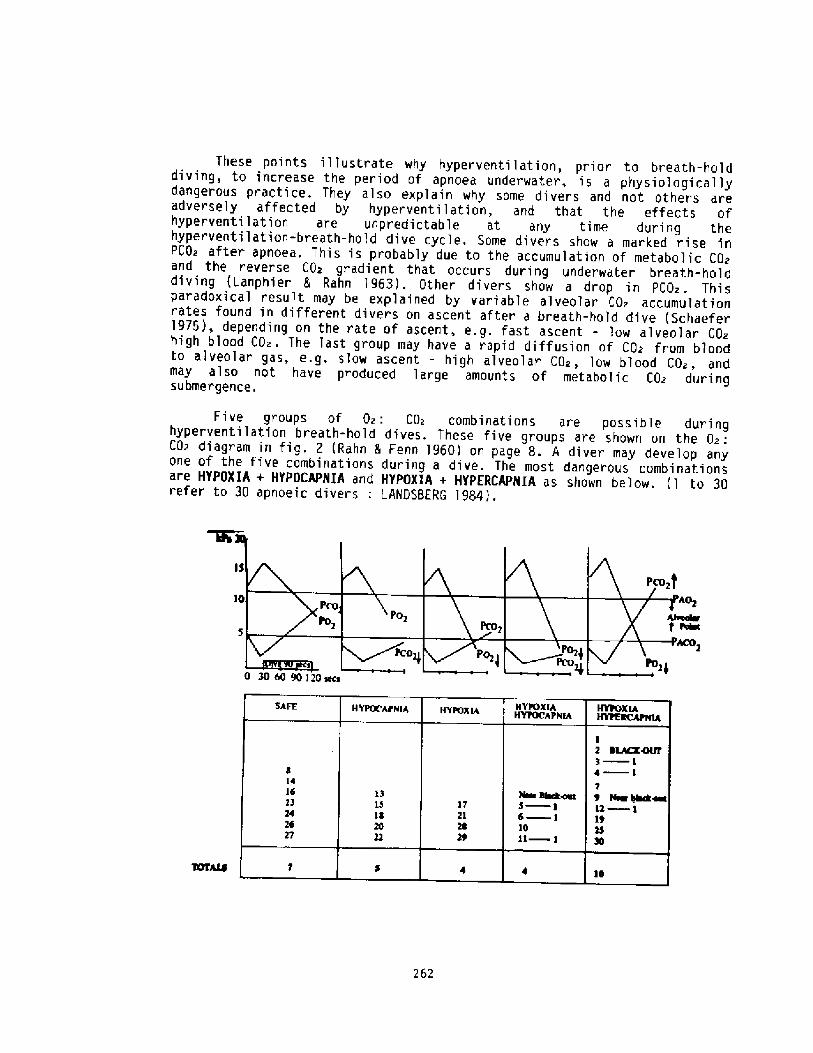
These points illustrate why hyperventilation, prior to breath-holddiving, to increase the period of apnoea underwater, is a physiologicallydangerous practice. They also explain why some di vers and not others areadversely affected by hyperventilation, and that the effects ofhyper ventilation are unpredictable at any time during thehyperventilation-breath-hold dive cycle. Some divers show a marked rise inPCO~ after apnoea. This is probably due to the accumulation of metabolic CO~and the reverse CO> gr adient that occurs during under water breath-holddiving Lanphier & Rahn 1963!. Other divers show a drop in PCO~ . Thisparadoxical result may be explained by variable alveolar CO~ accumulationrates found in different divers on ascent after a breath-hold dive Schaefer1975!, depending on the rate of ascent, e.g. fast ascent - low alveolar COzhigh blood CO~. The last group may have a rapid diffusion of CQ~ from bloodto alveolar gas, e.g. slow ascent � high alveolar C02, low blood COz, andmay also not have produced large amounts of metabolic COz duringsubmergence.
Five groups of Oz . CO> combinations are possible duringhyperventilation breath-hold dives. These five groups are shown on the 0~:CO~ di agram in fig� . Z Rahn & Fenn 1 960! on page 8. A diver may develop anyone of the five combinations during a dive. The most dangerous combinationsare HYPOXIA + HYPOCAPNIA and HYPOXIA + HYPERCAPNIA as shown below. � to 30refer to 30 apnoeic divers: LANDSBERG 1984!.
262
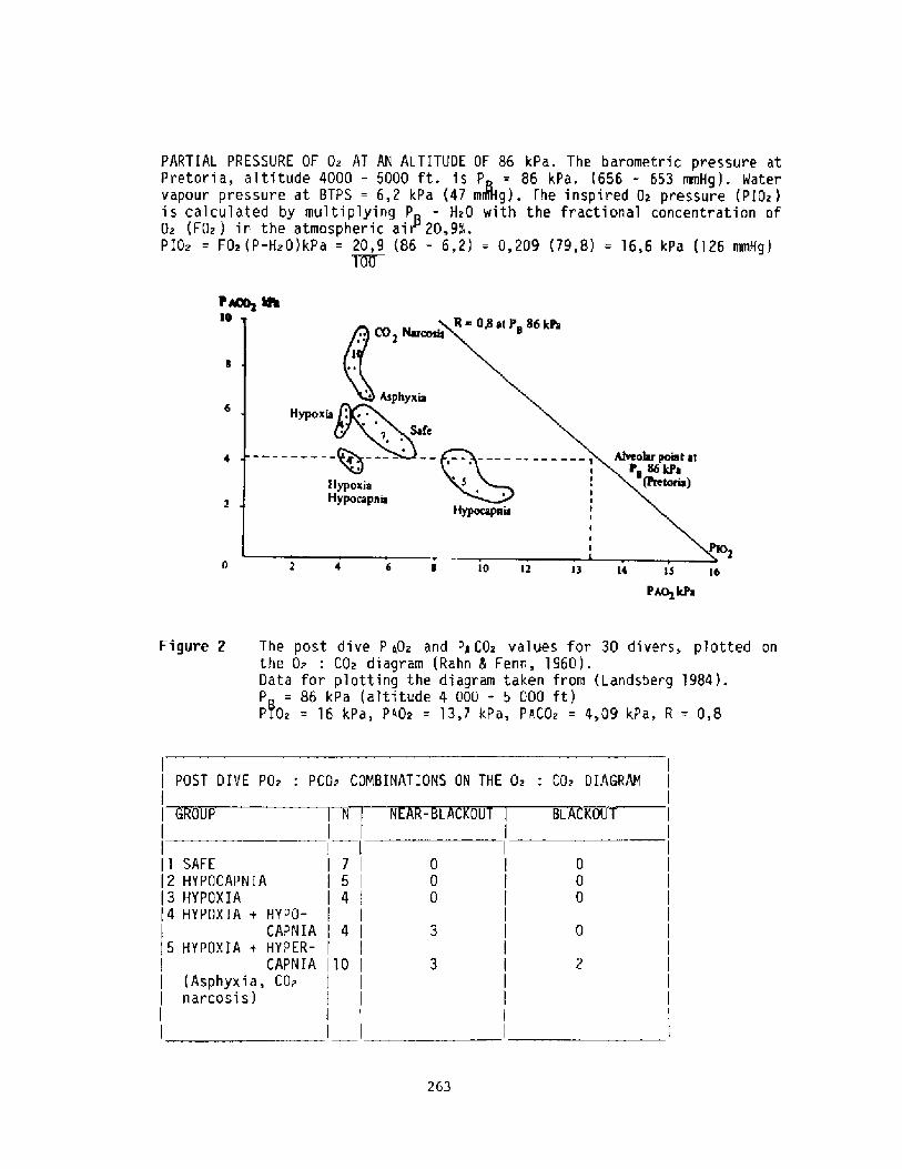
l~&10
10 IZ 13 i6 33 16
PAo kPW
Figure 2 The post dive P q0z and PqCO~ values for 30 divers, plotted onthe 0~ : COa diagram Rahn 5 Fenn, 1960!.Data for plotting the diagram taken from Landsberg 1984 !.P = 86 kPa altitude 4 000 - 5 000 ft!PIO~ = 16 kPa, P40~ = 13,7 kPa, PaCO~ = 4,09 kPa, R = 0,8
POST DIVE POz . 'PCOz COMBINATIONS ON THE Oz . COz DIAGRAM
0 P N N -L 0U BL KO
754
11 SAFE12 HYPOCAPNIA13 HYPOXIA14 HYPOXIA + HYPO-
CAP N IA 415 HYPOXIA + HYPER-
CAPNIA 1'10 Asphyxia, COznarcosis!
263
PARTIAL PRESSURE OF Oz AT AN ALTITUDE OF 86 kPa. The barometric pressure atPretoria, altitude 4000 - 5000 ft. is P = 86 kPa. �56 - 653 mmHg!. Watervapour pressure at BTPS = 6,2 kPa �7 mJItg!. The inspired Di pressure PIO~!is calculated by multiplying P - HzO with the fractional concentration ofO~ FOz ! in the atmospheric air 20,9%%d.PIOz = F02 P-Hz0!kPa = 20,9 86 � 6,2! = 0,209 �9,8! = 16,6 kPa �26 mmH9!
Trr
CONCLUS ION STo prohibit hyperventilation during breath-hold diving, is not a
successful safety measure as Lanphier & Rahn �963! have shown that thispractice gives a diver an extra 10 to 20 seconds diving time. A strict buddysystem and ensuring that the divers are not negatively buoyant will be moreeffective safety measures. However, the unpredictable nature of the effectsof hyperventilation should be explained to the divers who should beencouraged to work out thei r own indi vidual safe di ve profi 1 es usi ng a depthgauge and watch. SCLIBA divers should be made aware of the adverse,unpredictable effects of hyperventilation and breath-holding followingexhaustion and anxi ety while swimming underwater Dwyer & Pi 1 mani s 1 975 ! .
The CO~ changes in the blood of breath-hold di vers dur i ng thehyperventilation-breath-hold-dive cycle are unpredictable, show individualvari ations and vary from day to day Landsberg 1 971, 1 982, 1984 !.
Similar unpredictable wide differences in the alveolar PCOz of SCU8Adi vers in the sea at 1 0, 20 and 30 m have also been found Dwyer & Pilmani s1975!,
Morrison et al ., 1978 !, investigati ng loss of consciousness in ZSCUBA divers, noted a low repir atory response to i nspi red CD~ in thesedivers, i . e. insensi tivity to CO~ must have been i nher ent i n the divers .
In the se lection and trai ning of divers both breath-hold and SCUBA!the following should be added to the medical screening process:-
Alveolar CD> and 0~ responses during hyperventilation breath-hold-divecycles Lanphier & Rahn 1963, Craig 1961, Hong 1963 Landsberg 1982,1984.!
Z. Respiratory response to inspi red CO~ Morrison et al., 1981 !.3. Assessment of psychological motivation Deppe 1971 !.
REFERENCES
ANDERSON, H T. & BLIX, A.S. �974!: Pharmacological exposure of thecomponents in the autonomic control of the diving reflex. Acta.Physiol. Scand. 90,381.
BOVE, A.A., PIERCE, A.L., BARRERA, F., et al �973!: Diving bradycardia as afactor in underwater blackout. Aerospace med. 44 : 3,245 � 248.
BROMN, C.V. �974! : Shallow water blackout. Pressure, 3 : 1,5, IN: UnderseaBiomed. Research, 1 : 3.
CRAIG, A.B. Jr., �961! : Causes of loss of consciousness during underwaterswimming. J. Appl. Physiol., 16 : 4,583 - 586.
DEPPE, A.H.�971! : Psychological factors relevant to the selection of SCUBAdivers� . D. Litt. et. Phi l., thesi s, Uni versity of South Africa, 44 .
DHENIN, G.,SHARP, G.R. and ERNSTINE, J. �918!: Aviation Medicine IPhysiology and Human Factors. P.77 - 82. Trimed Books Ltd., London.
DONALD, K.M. June, 1971 ! : Personal communication . Professor of medicine,The Royal Infirmary, Edinburgh, Scotland.
DMYER, H.J. & PILNANIS, A.A. �975! : Phys~ological studies of diversworking at depths to 99 ft in the open sea. IN ; Proceedings of theSixth symposium on underwater physiology, Federation of Americansoci eties for experimental biology, 9659 Rockville Pike, Bethesda,Maryland 20014, U.S.A.
GREEN, J.H. �978! : An Introduction to Human Physiology, 4th S.I.!Edition, Oxford University Press . 78 - 80.
264
GUYTON, A.C. �982!: Professor, Department of Physiology University ofMi ssi s si ppi . Per son a 1 Communicat i on.
HONG, S.K., RAHN, H., I KANG, D.H. et al., �963! : Diving pattern, lungvolumes and alveolar gas of the Korean diving women Ama!. J.Appl.Physiol. 18 : 457-465.
LANDSBERG, P.G. �911! : COi changes in hyperventilation and breath-holddi vi ng . IN: Abstracts of the first scientific congress of thePhysiological and Biochemical Society of Southern Africa, Universityof the Witwatersrand.
LANDSBERG, P.G. �972! : Bradycardi a during human di ving, IN : Abstracts,Science week '72, University of the Wi twatersrand. 43.
LANDSBERG, P.G.�982! : Alveolar Gas and Peripheral Yenous Blood ChangesDuring Breath-Hold-Diving . S.Afr. Med . J .61 .
LANDSBERG,P.G.�984!: Hyperventilation: An unpredictable danger to thesports di ver. Thesis: Dept. of Physiology University of Pretoria,SouthAfrica.
LANPHIER,E.H. 5 RAHN,H. �963!: Alveolar gas exchange in breath-hold diving.J. Appl. Physiol 18: 3, 471 � 477.
LANPHIER,E.H. 5 RAHN,H. �963!: Alveolar gas exchange during breath-holdingwi th a i r. J. App 1 . Physi o 1 . 18: 3, 418 � 482.
MITHOEFER, J.C. �959!: Mechanism of pulmonary gas exchange and COztransport during breath-holding. J. Appl. Physiol, 14: 5, 706 - 710.
MORRISON, J.B. et al., �978!: Observations after loss of consciousnessunder water. Undersea Biomed. Res. 5�! : 179 � 187.
MORRISON, J.B. et a1.,�981! : Effects of COz Intensitivity and RespiratoryPattern on Respiration i n Divers. Undersea Biomed Res. 8�!:209-2'l7.
MILES, S July 1971! : Personal communication. Dean of post graduatestudies, University of Manchester, England.
PAULEV, P.E. � 969!: Respiratory and cardiovascular effects ofbreath-holding. Acta. Physiol Scand. Suppl . 324: 1 - 1 16.
RAHN, H., OTIS, A.B. HODGE, M., et al � 946 !: The effect of hypocapnia onperformance. J. Aviation hied. 17:164 .
RAHN,H. IL FENN, M.O.� 960!: A Graphical Analysis of the Respiratory GasExchange, The 0~ � CO~ Diagram. The American Physiological Society,Washington, D.C.
SCHAEFER, K.E. 5 CAREY, C.R. �962 !: Alveolar Pathways during 90 ftbreath-hold dives� . Science, N.Y . 1 37 : 1 051 � 1052.
SCHAEFER,K.E. � 975 ! : CO~ effects under conditions of rai sed environmentalpressure. IN: The physiology and medicine of diving and compressed airwor k, �nd ed! Eds. Bennett, P.B. 5 El liott, D.H., Bai llers Tindall,London.
SEM-JACOBSEN. C.M. 5 STYRI. O.B. � 970!: E .K. G. arrhythmi as monitored fromfree swimming SCUBA divers. IN. Proceedings, Journees intcrnationalesd ' hyperbaric et de physiologic subaquatique. Marseilles. 8-1 1 Juin,'! 970.
SMIT,P.J. �967!: Die invloed van apnoise duike op sekere fisiologieseprosesse. Voeding, 28:8, 380- 409.
SHIT,P.J.�974!: Alveolar O~ content of apnoeic divers, IN: Proceedings ofthe 3rd South African symposium for underwater sciences Ed.Smit,P.J.!, Baralogia, University of Pretoria,9.
SMIT,P.J. 5 Giesel,J �981 ! ;Apnoeic diving Safety � Experimental approachesto 0~ augmentation. S. Afr.Med. J. 60.959.
'WOLF, S. �970!: The Oz - conserving dive ref lex. Abbotempo,8:2, 14-1 7,
265
Discussion folloviag Dr. Landaberq's presentation
Dr. Rahn: You' ve opened up a whole new window for me in terms of highCO2 at the start of a dive. I was not prepared for that. Are these thedivers who cannot wait long enough to go down for their next fish, isthat a way of putting itP
Dr. Laadsberg: We actually lost one of our champion spearfishermenduring an individual spearfi shing competition. He was lost on thef irst breath-hold dive � went down and never came up. His body wasrecovered on the sea bed.
Dr. Rahn: On the first dive7
Dr. Landsberg: On the first, yes.
Dr. Rahu: That wouldn't be hypercarbia
Dr. Landsberg; But he was swimming towards the diving site, I don' tthink he gave himself enough time to recover.
Dr. Boysen: When you talked about dive profiles, can a diver changehis profiles depending on his dive historyP
Dr. Landsberq: No, it's completely unpredictable. In fact I' ve foundthat the same diver, under different diving conditions, produceddifferent breath-hold dive profiles. There is no standard profile, itchanges from one group to the other. When you interview thesespearfishermen, when you M~ them about this unpredictability, theysay:- 'Look Doctor, I' ve been diving for l0-15 years and I' ve nevercome near to blacking out" ... and then in the very next competitiongthat same diver is drowned. This is what had happened in SouthAfrica, we have lost most of our champion spearfishemen, so we came upwith this warning. We now also have a buddy system and surface coverduring competitions.
Dz. NcDonough: Wouldn't duration of dive be an important determinantof whether or not risk was higher, and if that is so, shouldn't thetypes of divers that you' re talking about time their dives2
Dr. Landsberg: Yes, I didn't have time to go into this; it willappear in the paper that I have submitted, but in effect, this is whatwe are doing now. We are giving them a "saf e" dive prof ile. Thespearfishermen and underwater hockey players are completely differentanimals to the Scuba diver in South Africa. They, the spearfishermenand hockey players, are the supreme athletes in underwater sport. Thespearf ishermen do use a watch and work out a safe dive prof ile; theytry and stick to their profile and do not digress from it, even ifthey shoot some magnificent fish at the bottom � when the dive prof ilesays to come up, they just forget about the f ish and surface. Thishas been the saving grace in preventing more fatalities.
Dr. Hong: In your schemes diving bradycardia is detrimental, right?Because it reduces cardiac output, and cerebral blood flow
266
Dr. Landsberg: But, it's the quality of blood which is being pumped tothe brain � very low 02 partial pressure, high CO2 partial pressure/which causes unconsciousness. This is the extreme case, I think thatis already beyond the dive profile on the way to asphyxia and death.
267
BREATH HOLD DIV ING ACCIDENTS IN THE MEDITERRANEAN AREA
Damf ano Zannf nf> M.D.
Breath hold diving became a mass sport in 1950 fn theMediterranean area and the main countries involved were naturallyItaly~ France> Spa f n and Yugosl av f a. Thf s 1 ed to th eorganfzati on of na tf onal and f nterna tf onal spear-f f shin gcontests. which soon became part of the Worl d-wide Champf onshfp.
Of course both doctors interested in sports medicine anddoctors who practiced this spor t them sel ves got f nvol ved f nprobl ems of preventfon and safety f n the f f el d of breath hol ddivfng. as wel 1 as Scuba dfvfng thef r interest was al mostf nevi tabl ei consf derfng that the technique of d fv in g f s tf ghtlytf ed to phy sf ol ogy!.
Datfng back to the second hal f of the 50isi medfcal groupswere organized fn Sports Associations with the participation ofdoctors interested in Sports Medi cf ne. Studf es and publ fcmessages about diving safety probl ems were developed at nationaland f nterna tf onal meetf ngs, us i ng al 1 means on han di f.e.>artf cl es on newspapers, d fving magaz f nes> T.V. show s, etc.>especf ally at the beginnf ng of summer. Technf cal manual s wer'eal so publf shed> as well as scientff f c 1 iterature and proceedingsof th e meetf ngs hei d.
For some time physfcfans of countries on the south-easternside of the Mediterranean sea such as Greece> Turkey> Egypt>Israel ~ Tunf sf a! kept fn contact with thef r colleagues in theEuropean countrf es.
In effects what we have accompl fshed fs a betterunderstandfng of opfnfons among doctors of the Medfterraneanarea. w ith hei p from CMAS Wor 1 d Con f ederati on of UnderwaterActfvftfes!> FFESSM Federation Francafse Etudes et SportsSousmarf nes! and SIMSI Ital f an Socf ety of Underwater andHy perbarf c Medf cf ne!i and al so thanks to EUBS European Undersea8 fomedf ca l Socf ety!.
Through these societies new research contributions about thephysiology of breath hol d diving became avaf1abl e to severalunf versf ti es~ such as Marsef 1 1 e and Genoa. Sta rtf ng f rom 1960on ~ many up-to-date traf nfng cl asses for physi cf ans wereor ganf z ed, f f rst. f n Genoa~ th en f n Napl es. Hun dr eds of doctorsattended. At the same tf mei school s f or sport d fvf ng devel opedand doctors were cal led to collaborate in teaching divers how toavof d accf dents.
In 1976 with the constf tutf on of the SIMSI and then theCocchf Association for Safety in Water, meetfngs among doctors orbetw een doctors and technf ci ans mul tf pl fed.
Posters were prepared f or primary and hf gh school s. Legal
268
procedur es rel atf ng to death by accf dents fn pool s andresponsfbf1 fty of the 1 ffe-guards were studied fntensfvelyiespecf al 1 y wfth regard to rescue procedures.
Durfng meetings we have always been astonished by the greatnumber of fatal accf dents that occur fn breath hold diving. Th1srisk fs generally undervalued by the practfcfng sportsmen andoverestf mated by the propagandf sts of preventi on.
In the Medf terranean area, as well as 1n other parts of theworl d, statf stf c data th at, are exacts or fndicatfve of the numberof deaths> are not yet avaf1 abl ei because of the fact that breathhol d dfv fng acci dents are usual ly 1 fsted under "death by drow nf ngor by submersf on".
To know these data waul d be extremely usaf ul ~ both toeval uate the exact seriousness of the risk> and 1 1 kew 1 se~ tof m prove our know 1 edge on cause s of syncope other th an pr ot ongedapnea. A better propaganda f or publf c awareness of th! s probl emw oui d be possf bl e.
All th e Eur opean countries that touch the Medf terranean seaare now making efforts to organize a systematic gatherfng of dataon these accf dents, so most probably there shoul d be morerel fable data avaflable next year.
These data coul d probabl y be used f n preparf ng 1 aws so as toachf eve safer breath-hol d dfv1ng, but this is a jurf df cal andtechnical probl em for examples even 1f accfdent rate fn mountafnactfv 1 tfes 1 s much h1gher than accf dent rate f n the seai yetnothing has been done by Iaw!.
The safest rul esr such as spear- ffshfng fn coupl e, watchingf rom above' presence of a scuba d fv er~ connectf on w 1 th thesurf ace by a 'I 1 ne and buoy al 1 means wh1ch doctors agree shoul dbe appl fed! are too often considered non-appl fcablei becausef 1sherm en state they act as antf f 1 shing 1 mpl ements> especf al lyduring the corn pet1tf ons. As a matter of fact, thf s summer ayoung Ital fan diver died durfng a competf tf on.
At presents the only numerfcal data that can be obtained arefrom a few insurance companies that take care of divers> membersof a sport federatf on fn France and S pain>; but these regardonl y th e members' who general ly represent a m1norfty of thosepractf sing 1mmersfon 1 n apnea; e.g.. fn Italy 30 ~ 000 dfvers aremembers of dffferent Sport Federatfonsr while those practisingare certainly over 200.000. In France there are about 260 ~ 000members of the Sport Federatf on> fn Spa1n 6,500.
I got these data f rom colleagues of dffferent MediterraneanCountr 1es ff g. I! and recef ved most of the f nform atf on f romItaly.
269
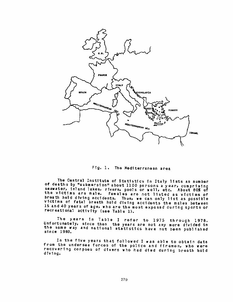
Ff g. I. The Medf terranean area
The Central Instftute of Statf stf cs 1 n Italy 1 1 sts as numberof deaths by "submers1on" about llDO persons a yeari comprfsfngseawater> 1 nl and lakes~ rfversi pool s or well. etc. About 80% ofthe v1ctf ms are mal e. Fee al es are not 11sted as v 1ct1 ms ofbreath hold d fvfng accf dents. Thus> we can only 1 f st as possf bl evf ctfms of fatal breath hold dfvfng acc1dents the males between15 and 40 year s of age. who are the most exposed durfng sports orrecreatf onal actfvfty see Tabl e I!.
The years fn Tabl e I refer to l975 through 1978 ~Unf ortunately~ sfnce then the years are not any more dfv1ded 1 nthe same way and natf onal statf stf cs have not been publ f sheds1n ce I980.
In the f fve years that followed I was abI e to obtafn datafrom the undersea forces of the pol fee and f1 remen~ who wererecover 1 ng cor pses of df vers w ho had df ed dur 1 ng breath hol ddfvfng.
270
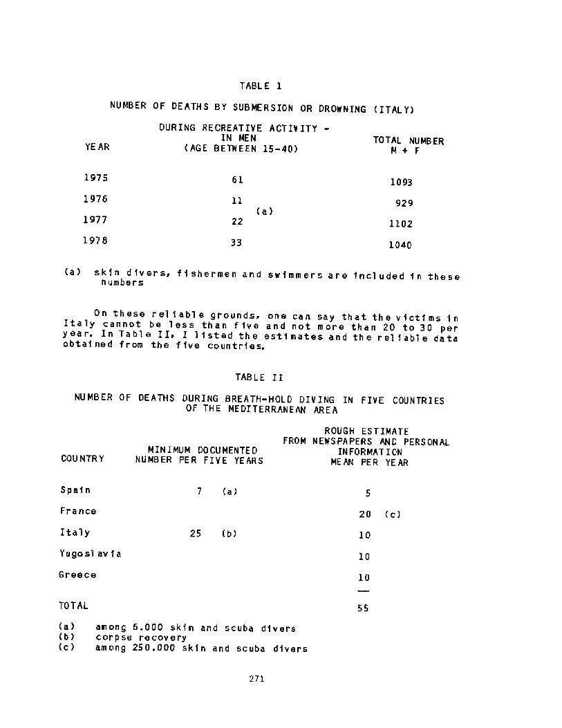
TABL E 1
NUMBER OF DEATHS BY SUBMERSION OR DROWNING ITAL Y!
DURING RECREATIVE ACTIY ITYIN MEN
AG E BE TW E EN 15-40!TOTAL NUMB ER
M+ FYE AR
1975 61 1093
1976 929 a!
1977 22 1102
1978 33 1040
a! sk1n divers, fi shermen and swimmers are included in thesenumbers
TABLE I I
NUMBER OF DEATHS DURING BREATH-HOLD DIVING IN FIVE COUNTRIESOF THE MEDITERRANEAN AREA
ROUGH ESTIMATEFROM NEWSPAPERS AND PERSONAL
IN FOR MAT I ONMEAN PER YEAR
MINIMUM DOCUMENTE DNUMBER PE R FI V E YE ARSCOUNTR Y
7 a!Spain
France
Italy
Yugosl av 1 a
Greece
20 c!
1025 b!
10
l0
55TOTAL
a! among 6.000 skin and scuba divers b! corpse recovery c! among 250.000 skin and scuba divers
271
On these rel tabl e grounds, one can say that the v icti ms 1nItaly cannot be less than f ive and not more than 20 to 30 peryea r. In Ta bl e I I. I l 1 sted th e esti mates an d th e r el 1abl e dataobta1ned f rom the f ive countries.
In ef feet> it invol ves a minimum of about 5S deaths per yeari n al 1 the control led European area of the Mediterranean sea.This excludes the south and eastern coasts> which are much lessfrequented by touri sts.
In addi ti on to these data I can menti on th at about 70% ofIta] ian divers who participate in national and internationalcompetitions have reported one or more presyncopal symptoms intheir career.
Inasmuch as regards Italy and Greece. I can say that thevictims of breath hol d diving are mostly competition diver s orpersons w ith much exper i ence.
Now> do ol d divers present more ri sks th an y oung peopl e?Can we hypothesize some factors as: CO> retention and lowersensitivity to CO>i too deep or too 1 ong divest heedlessness ofthe danger~ age-re1ated causes of syncope?
To conclude> I woul d 1 ike to express a certain amount ofper pl exi ty which I f eel f or two o p po si te f acts. Fi r str d iv er sw il 1 not f ol 1 ow proper rul es of sa f ety to avoi d f atalconsequences; secondly> researchers in the physi ol ogical fi el dare devel oping an al ways deeper know ledge. Let us hope thatthis knowledge w ill hei p the divers to consider more seriouslythe ri sks th ey face.
Since the number of accidents seems to be decreasing ratherthan increasing even though the practice of breath hol d divinghas been increasing> especi al ly af ter th e proh i bi ti on to useSCUBA for fi shing!> I woul d like to believe that all the studiesand propaganda has at least contributed to save a certain numberof persons. Rel iabl e indivi dual observations al ready confirmth i s.
272

Discussion folloving Dr. Xannini's presentation
Dr. Hong: Do you have any breakdown of accidents by seasons, forexample, winter versus summer?
Dr ~ Xannini: No, I don' t; but the accidents are more f requent insummer when most people are on vacation. However, also in winter alot of people dive regularly along the coasts, as the watertemperature does not drop below 10-15 C.
273
LOSS OF CONSCIOUSNESS DURING BREATH HOLD DIVING
Pier Giorgio Data
Institute of Physiological Science and School of
Underwater and Hyperbaric medicine. Universityof Chieti, Chieti, Italy.
The best known, most frequent and most dangerous type ofaccident connected with apnea diving is the sudden loss ofconsciousness under water. It is most common at depths lessthan 2 m and hence during distance underwater swimming inpools when it may occur at distances from 45 m or more 1,2!
These accidents are due to hypoxia and more common after'
hyper ventilation which decreases CO tension and prolongsbreath-holding time without increasing 0 supply significant-
2ly. Very well-trained divers may however have hypoxic syncopewithout significant hyper ventilation 2 ! . The same type of'"black outs" may occur also after deep apnea dives if thediver stops ascent at depths between 5 m and surf'ac..
We have however recorded abnormal heart rhythms with ECC'during a series of deep apnea dives conducted over several
years which most probably were the main cause of loss ofconsciousness �!.
Material and Methods
ECG recordings during apnea diving wer e obtained in about150 healthy age 20-40 years! spor t divers with r epeated divesto di fferent depths in each. As several dives were followed in
each a total of close to 500 have been r ecorded.
In 18 subj ects measur ements wer e obtained with a per cutan-eous right heart catheter during 70 dives from 3 to 30 m of
depth. None of the subjects had a history of cardiac arrhythmiasor other heart disease, neither' has such occur red later . All
sub jects gave informed consent to the study.
274
The divers hyperventilated for one minute at surface beforethe dive. They were pulled head first to the desired depth by alead weight and ther eafter turned head up and held on to a ropeduring the apnea. They were followed under water by exper iencedScuba-divers in case of accidents. All used wet suits. Thedepths varied between 3 and 72 meters.
Two lead precordial EGG and pulmonary artery pr essures wer erecorded with a waterproof tape recorder tied to the back ofthe subject. Pulmonary artery pressure PAP ! and surroundinghydrostatic pressure were recorded with piezo electric tr ans-ducer s inside the shielding, with subtraction of' hydrostaticpressure from the PAP
Results
During diving var ious degrees of bradycardia were seen in allexperiments. The bradycardia increased with time at depths downto 20 m. At gr eater depths the decrease in heart rate was morerapid and related to the increase in depth. However, after 60 sthe same degree of bradycar dia was final ly seen at all depths f=26-46 min. ! . Down to 20 m sinus br adycar dia was most commonwhile at greater depths disappearance of the P-wave oftenindicated a nodal rhythm or a His'bundle pacemaker. Single orseries of 3 to 5 ventricular extrasystole were often recorded.
Four subjects lost consciousness during ascent. They haddived to 20, 20, 23 and 30 m of depth, respectively. One ofthose ascending from 20 m had a normal heart rythm but ascend-ed slowly and got unconscious at 4 m of depth af'ter 92 s. Heorobably had hypoxic syncope. Anothe subject swimming up from20 m had several extrasystole before ascent and ventricular
tachycardia when he lost consciousness at a depth of 6 m. Hewas brought to surf'ace after 52 s and rapidly regainedconsciousness. Two of those with heart catheterization had
"black outs" during ascent. One had dived to 30 m of depth andgot unconscious a f ter 95 s also wi th ventri cu lar arrhythmia wi tha high fr ecjuency. One subject diving to 23 m with a total timeof submersion of 118 s is shown in the following illustrations.Sinus tachycardia was seen after hyper ventilation with adecrease in heart rate during descent fig. 1!. Immediatelyafter arriving to 23 n of depth the heart rate was down to
60 /min. Ther e was probably a nodal rhythm and series of extr a-
275
systole occur r ed f ig. 2!, while PAP had incr eased to
30-40 mm Hg. After 42 seconds a ser ies of ventr icular extra-
systole occurred which turned into a ventr'icular fibr illation
on EGG with a maximum frequency of 584/min. and an averagefrequency of 230/min. fig. 3! . However, there was still a
dynamic frequency on the PAP registr ation of 90 and the pulmon-ary arter y pr essure showed a small decrease. After 50 s to 60 s
EGG showed ventricular fibrillation while small pulmonaryartery systole were seen with a frequency around 60/min. The
subject still felt well as related later on. He started climb-
ing up the rope but lost consciousness when reaching 9.5 mafter 90 s Fig. 4!. Electrocardiographically he st.ill had ven-tricularr fibrillation but now with very irregular PAP systolewith a rate of ll0. When he was brought to the surface he hadan electrocardiographic sinus rhythm but with no dynamic rightheart action and a pulmonary artery pressure of close to80 mm Hg. He was resuscitated with oxygen on mask and cardiaccompressions at 142 s Fig. 5!. After half a minute he had
regained electrocardiographic sinus tachycardia with synchron-ous pulmonary artery pressure fluctuations. The pulmonaryartery pressure was still high and he had single extrasystolebut was mentally alert and no further complications werenoted fig. 6!.
Discussion
It cannot be excluded that i.rr itation from the r ight heartcatheter has contributed to the ventricular tachycardia orfibrillation seen in some of the subjects.
However, vent.icu'ar extrasystole occurr'ed in most subjectsalso without heart catheterization and it seems probable thatthe extensive acute cardiac dilation, at depths below 20 m fig. 7!, contributes to the increased irritability . Hypoxiaduring extended apnea may also increase irritability and echo-car diogr aphic registrations at our laboratory during apnea atRV in air have actually shown VES excited by each diaphragmalcontraction occurring near breaking point. It seems contra-dictional with sustained right ventricular function duringtachycardia/fibrillation. we have recorded the same phenomenonin dogs with ventricular fibrillation caused by local applica-
276
tion of aconitin fig. 8! . Likewise, everybody ~orking withresuscitation will have noticed patients with electr ocar dio-
gr aphi ca 1 ly nor mal s inus rhythm but with no measurable pulse orblod pr essur e.
Conclusions
seems good as no comp 1 iincidents and only one
References
1. Cr aig AB. Causes of loss of consciousness dur'ing underwaterswimming. J Appl Physiol 1961;16�!:583-586.
2. Arborelius N Jr. Om medvetsloshet vid under vattenssimning. About unconsciousness during underwater swimming!.Sv Lakartidningen 1962:59:1-7.
3. Data PG. La sincope anossica. Aspetti fisiopatologici. In:6icurezza in acqua. Atti del 1' convegno nazionale sulla
sicurezza in acqua. Prato 5-5-1979. Nistri-Lischi Editori,
Pisa, 1981. Page 83-98.
277
Heart distension, hy
deep apnea diving seem
may cause ex tr asystole,
f'ibrillation with loss
poxia or other f actors connected to
to incr ease myocardial sensitivity andventricular tachycardia and ventricular
of consciousness. However, prognosiscations were noted in four such
of four sub j ects needed r'esusc i tat ion.
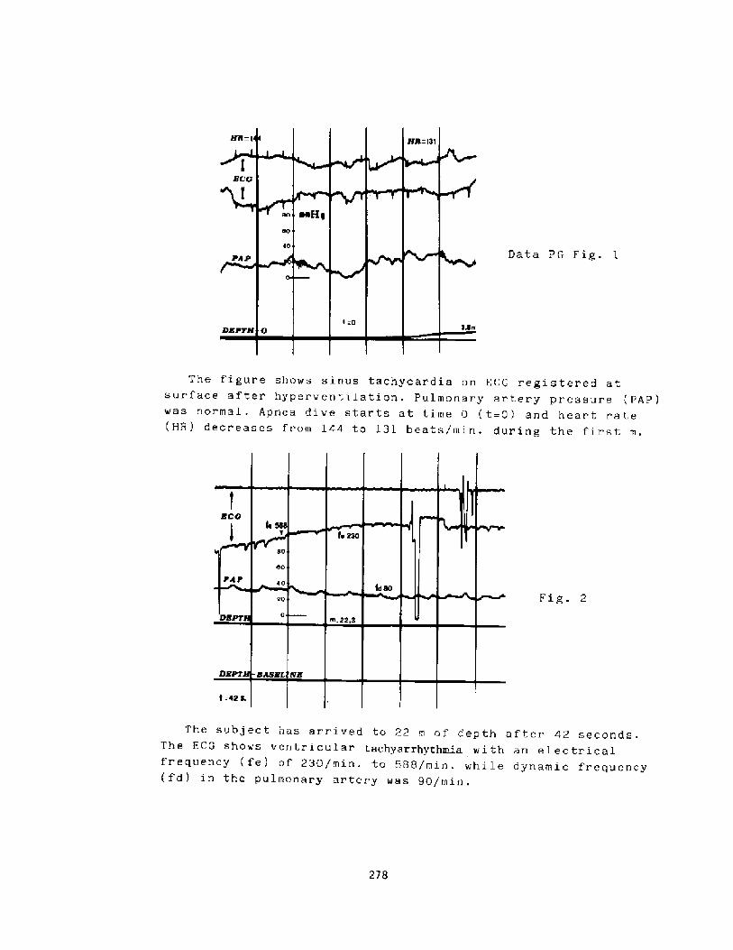
Data PG Fig. 1
The figure shows sinus tachycardia on ECG registered atsurface after hyperventilation. Pulmonary artery pressure PAP!was normal. Apnea dive starts at time 0 t=0! and heart rate HH! deer eases from 144 to 131 beats/min. during the first m.
Fig. 2
The subject has arr ived to 22 m of depth after 42 seconds.The KCG shows ventr icular tachyarrhythmia with an electricalfrequency fe! of 230/min. to 589/min. while dynamic frequency fd! in the pulmonary ar tery was 90/min.
278
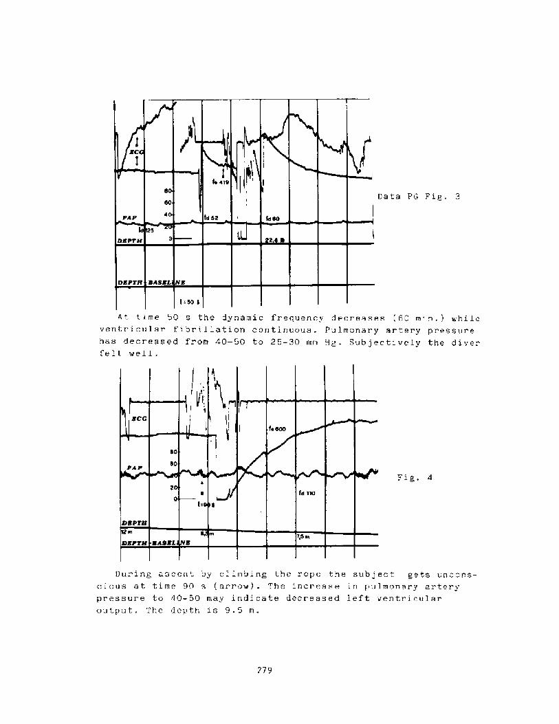
Data PG Fig. 3
A t t ime 50 s the dynami c frequency decreases 60 min�. ! while
ventr icular f i br i 1 lation continuous. Pulmonar y ar ter'y pressure ehas deer eased from 40-50 to 25-30 mm Hg. Subjectively the diverfelt well.
Fig.
Dur'ing ascent by cl imbing the rope the sub j ect gets uncor.s-
cious at time 90 s ar row! . The increase in pulmonary ar terypressure to 40-50 may indi cate decreased left ventr'i euler
output. The depth xs 9.5
279
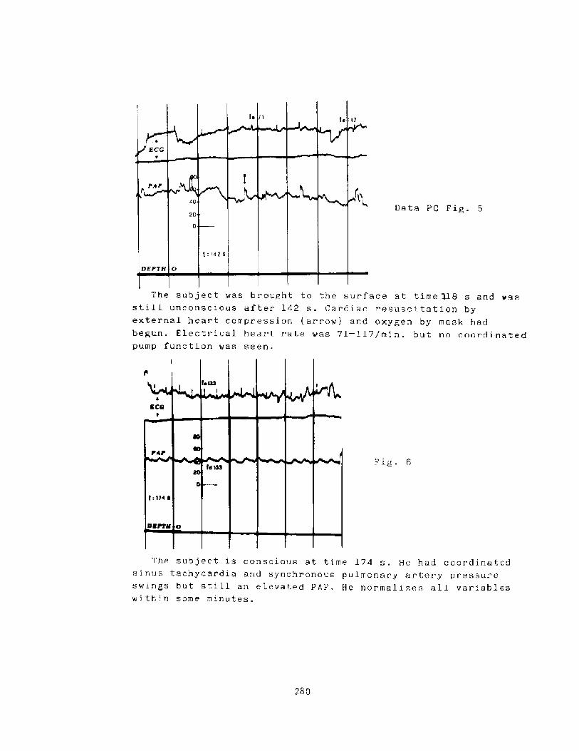
Data PG Fig. 5
The subject was brought to the surface at time M.B s and was
still unconscious after 142 s. Cardiac resuscitation byexternal heart compression arrow! and oxygen by mask hadbegun. Electrical heart rate was 71 � 117/min. but no coordinatedpump function was seen.
The subject is conscious at time 174 s. He had coordinated
sinus tachycar dia and synchr onous pulmonary artery pressur eswings but still an elevated PIP. He normalize - all variableswithin some minutes.
280
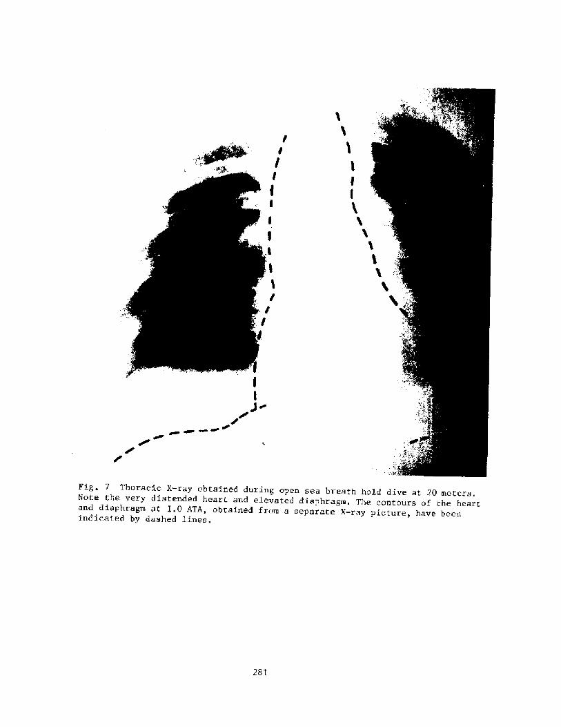
rr
r
Fig. 7 Thoracic X-ray obtained during open sea breath hold dive at 20 meters.Note the very distended heart and elevated diaphragm, The contours of the heartand diaphragm at I.O ATA, obtained from a separate X-ray picture, have beenindicated by dashed lines.
281
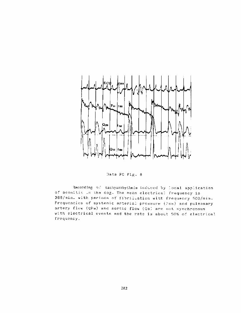
Data PG Fig. 8
Record
o aconi ti n
388/min. w i t
Frequencies
artery flow
with electr i
frequency
282
in' o i tachyarrhythmia i educed by 1 ocal app 1 i cat i onin the dog. The mean electr ical frequency ish per iods of fibr illation with frequency 500/min.of systemic ar ter i al p r essure 2 s a ! arrd pulmonary QPa! and aortic flow Qa! ar e not synchr onouscal events and the rate is about .~OX of electrical
Discussion following Dr. Data's presentation
Dr. Lundgren: It is certainly food for thought and quite impressivethat you would be able to get these recordings under those conditions;there's really only one measurement that I'm missing and that's theheart rate of the experimenter as this was happening.
Dr. NcDonough: Were these Swan-ganz catheters, that is, balloontipped catheters floating in the pulmonary artery, following insertionthrough a peripheral vein?
Dr. Data: Yes.
Dr. NcDonough: What was your access route, was it antecubital, neck,or groin?
Dr. Data: Antecubital.
Dr. Soysen: Did you get fairly good tracings when your subjects weretrying to swim with that catheter in the arm? When we tried to doexperiments like that with arm movement, the catheter tends to moveback and forth.
Dr. Data: We had no problems in this regard, as the arm was splinted,so that they could not bend it.
283
GENBRAL DISCUSSIOH
Dr. Bong: I proposed this morning that we should discuss more aboutthe issue dealing with cold water drowning. Now we heard a number ofpapers showing the development of bradycardia and cardiac arrhythmiasiwhich are potentiated by cold water. That's one phenomenon. But theother story involves cold water drowning of little babies, who weresuccessfully resuscitated after 40 minutes. There is a general myth/statement made by some people that this is due to the diving reflex-The media tell their story that, as soon as a baby drowns in coldwater> he behaves like a diving seal, reducing his oxygen consumption-Does this view have a scientif ic basis? Since most of us who areinterested in this question are in this room, maybe we should getsome feedback from you about this issue.
Dr. Slener: Yes, I' quite agree. There's a preamble, though, thatI'd like to suggest. You know, I think we ought to finally discard>stamp out, and throw away the term diving reflex.
Dr. Hong: Right.
Dr. Elsner: There's no way that that makes physiological sense. Buton the other hand, is it possible that all of the events that you seein the child who falls into cold water, and is revived, could beexplained simply on the basis of the rapid cooling of the centralnervous system and having nothing whatever to do with bradycardialresponse?
Dx. Arnold: I think there are probably a lot of components. Braincooling is important. There are two similar situations: the case ofasphyxia as opposed to the case of water inhalation. The asphyxialcase has a much better prognosis. For the same length of submergencetime and the same temperature of water, one individual will survivewell and another will either be severely compromised or will notsurvive. So I suspect that there's a variability in our ability, oneperson to another, to survive. En the period of submergence, bef oreachieving brain cooling which may take longer in an adult than in achild!, the diving response may maintain cerebral integrity.
Dr. Hong: The reason why this diving reflex or response is invokedby some people is the notion that, because of this response, onlybrain and coronary blood flow is maintained at the expense of otherregions. However, are we justif ied to say that the bloodredistribution developed in human subjects during cold water faceimmersion, as it seemed to develop in diving mammals, is responsiblef or the conservation of oxygen?
Dr. planer: There's not one scrap of data for that. All I'm saying isthat there is not a single measurement to suppor t this statement.However, the diving response may be involved, for example, in theinitiation of apnea and the early distribution of blood flow.
Dr. Bong: Number two, what we need is an hypothetical experimentwith human subjects, in which we let the subject fall into coldwater, without his knowledge. This procedure may induce a severe
284
bradycardia and the distribution of blood flow similar to that indiving seals, whereby conserving oxygen.
Dr. Lundgren: Forced diving.
Dr. Hong: That's right.
Dr. Siesjo: I have a question to Dr. Boysen and a comment which maybe relevant to the variability we are discussing. I was a bitintrigued by the fact that you can have asphyxia with maintained bloodpressure for, say, eight minutes and brain damage does not result.The reason of course is that during all that time you must have someoxygen delivery to the brain. I'm wondering what your actual PO2'swere. When working with rats we have always been rather impressedwith the resistance of the brain to hypoxia, as long as the perfusionpressure is maintained. However, there is one factor which gives alot of variation to the outcome in hypoxia and also in ischemia.That's the blood glucose concentration. For example, if you induceincomplete ischemia in a fasted animal which would maintain a bloodglucose concentration which is normal or below normal, then the braincan sustain perhaps l0 or l2 minutes of virtual anoxia without anyreal brain damage. But if you perform the same experiments on a fedanimal� so that hyperglycemia develops, then perhaps four or fiveminutes may be sufficient to give irreversible brain damage. Ofcourse the difference is that in the last case you get exaggeratedlactic acidosis in the brain. So it means that in your experiments,where the blood pressure is maintained, the reaction of the brainwould depend very much on the blood glucose concentration. Since youhave maintained circulation to the hypoxic brain, conditions are athand f or enhanced lactic acidosis. I wonder, f irst, what was the PO2in your experiment and, second, what was the blood glucoseconcentration.
Dr. Boysen: The PO2 at the end of by four or five minutes of asphyxiawould be less than twenty torr.
Dr. Siesjo: And at the end of, say, seven or eight minutes'7
Dr Boysen: Usually> at the end of seven or eight minutes, if themeasur ement were correct, it was nine or ten torr, something likethat. I don't know the answer to the rest of your question because 1didn't measure blood glucose, but having known about what you said,these were fasted animals that had water but no food overnight, and wedid not use glucose infusion when we administered the anesthetic.
Dr. Siesjo: So they were fasted.
Dr. Boysen: Yes, they were.
Dr. Hong: However, blood glucose could considerably go up becausepossible increase in catecholamine release. I can see thatglycogenolysis might be stimulated because of the catecholaminerelease due to frightening.
Dr, Siesjo: Yes, that's absolutely true, but if you fast a rat for
2S5
24 hours, there would be virtually no liver glycogen left. Sa it meansthat your bload glucose concentration cannot rise much.
Dr. Hong: But here's another thing we' ve been talking about, well-fedchildren walking around a lake and suddenly drowning in cold icewater, so in that case blood glucose level could considerably ga up.
Dr. Siesjo: That could be patentially dangerous.
Dr. Rahn: Could some very simple experiments in man be dane on theinduction of laryngospasm with cold and warm water?
Dr. Boysen: We did that with cats, nat with dogs, and you' reabsolutely right. Almost any way to manipulate the airway can inducelaryngospasm, and even if I haven't any clinical experience with coldwater drowning, when you put cold water versus warm water into a cat' sair way, the cold water has a much higher incidence of instituting orinitiating a significant degree af laryngospasm. So in the cold waterthat may be another factor, irrespective of whatever the reflexes wetry to name.
Dr. Rabn: But don't you think these rather simple experiments couldbe done in man.
Dr. Boysen: I don't think we cauld get any volunteers. Thesubjective feeling is not a very nice one.
Dr. Luadgrea: Is anything known about the possible influence ofhypacapnia ar hypercapnia, for that matter, on the susceptibility tolaryngospasm?
Dr. Boyaeu: Yau mean as it starts?
Dr. Luadgren: Yes, realizing that hypocapnia is related withincreased tendency for spasm in other skeletal muscle, I was justwondering.
Dr. Boysen: Yes, it's the same in the upper airway.
286
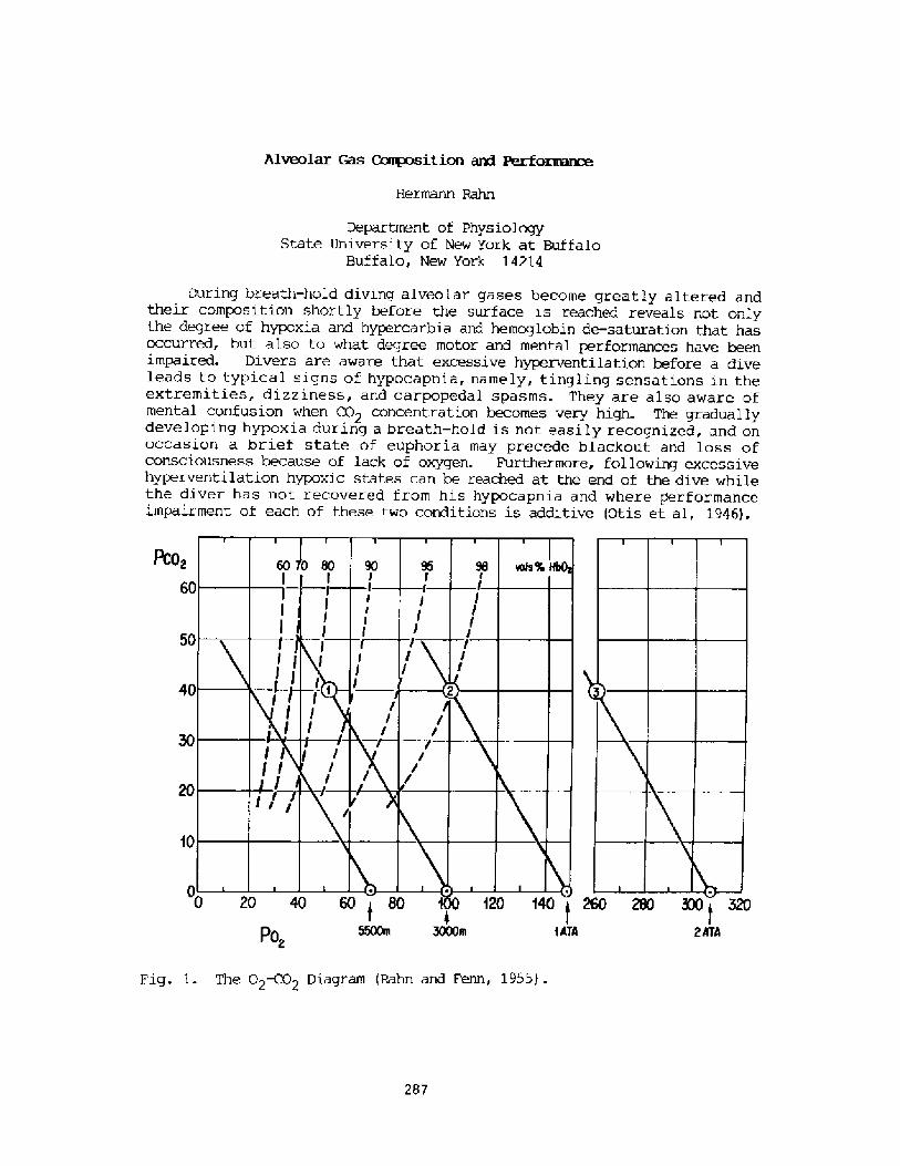
Alveolar Gas Ga~osition and Perfom~
Departrrent of PhysiologyState University of New York at Buffalo
Buffalo, New York 14214
During breath-hold diving alveolar gases become greatly altered andtheir composi.tion shortly before the surface is reached reveals not onlythe degree of hypoxia and hypercarbia and hemoglobin de-saturation that hasoccurred, but also to what degree motor and mental performances have beenimpaired. Divers are aware that excessive hyperventilation before a diveleads to typical signs of hypocapnia, namely, tingling sensations in theextremities, di.zziness, and carpopedal spasms. They are also aware ofmental confusion when CO2 concentration becomes very high. The graduallydeveloping hypoxia during a breath-hold is not easily recognized, and onoccasion a brief state of euphoria may precede blackout and loss ofconsciousness because of lack of oxygen. Furthermore, following excessivehyperventilation hypoxic states can be reached at the end of the dive whilethe diver has not recovered from hi.s hypocapnia and where performanceimpairment of each of these two conditions is additive Otis et al, 1946!.
R :02 60
50
40
f0
2ATAt ATAPo,
Fig. 1. The 02~2 Diagram Rahn and Fenn, 1955! .
287
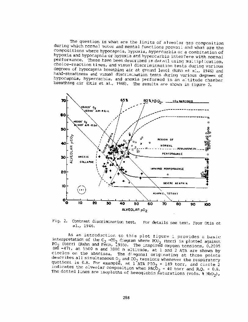
The question is what are the limits of alveolar gas compositionduring which normal motor and mental functions prevail and what are thecompositions where hypocapnia, hypoxia, hypercarbia or a combi.nation ofhypoxia and hypocapnia or hypoxi.a and hypercarbia interfere with normalperformance. These have been described in detail using multiplication,choice-reaction times, and visual discriminmation tests during vari.ousdegrees of hypocapnia breathing ai.r at ground level Rahn et al., 1946! andhand-steadiness and visual discriminati.on tests during various degrees ofhypocapnia, hypercarbia, and anoxia performed in an altitude chamberbreathing ai.r Otis et al., 1946!. The results are shown in figure 2.
eo
I0 40 SO 60 70 BO 90 IOOALVEOEAR pQp
Fig. 2. Contrast discrimination test. For details see text. From Otis etal., 1946.
As an i.ntroduction to this plot fi.gure 1 provides a basicinterpretation of the 02 -CO~ diagram where PCO2 torr! is plotted againstPO2 torr! Rahn and Fenn, 2955!. The inspi.red oxygen tensions, 0.2095 pB -47!, at 5500 m and 3000 m altitude, at 1 and 2 ATA are shown bycircles on the abscissa. The diagonal origi.nating at these pointsdescribes all simultaneous 0 and CO tensions whenever the respiratoryquotient is 0.8. For example, at 1 ATA pIO = 149 torr, and circle 2indicates the alveolar compositi.on when PACE> = 40 torr and R.Q. = 0.8.The dotted li.nes are i.sopleths of hemoglobin saturations vols. % Hb02!
288
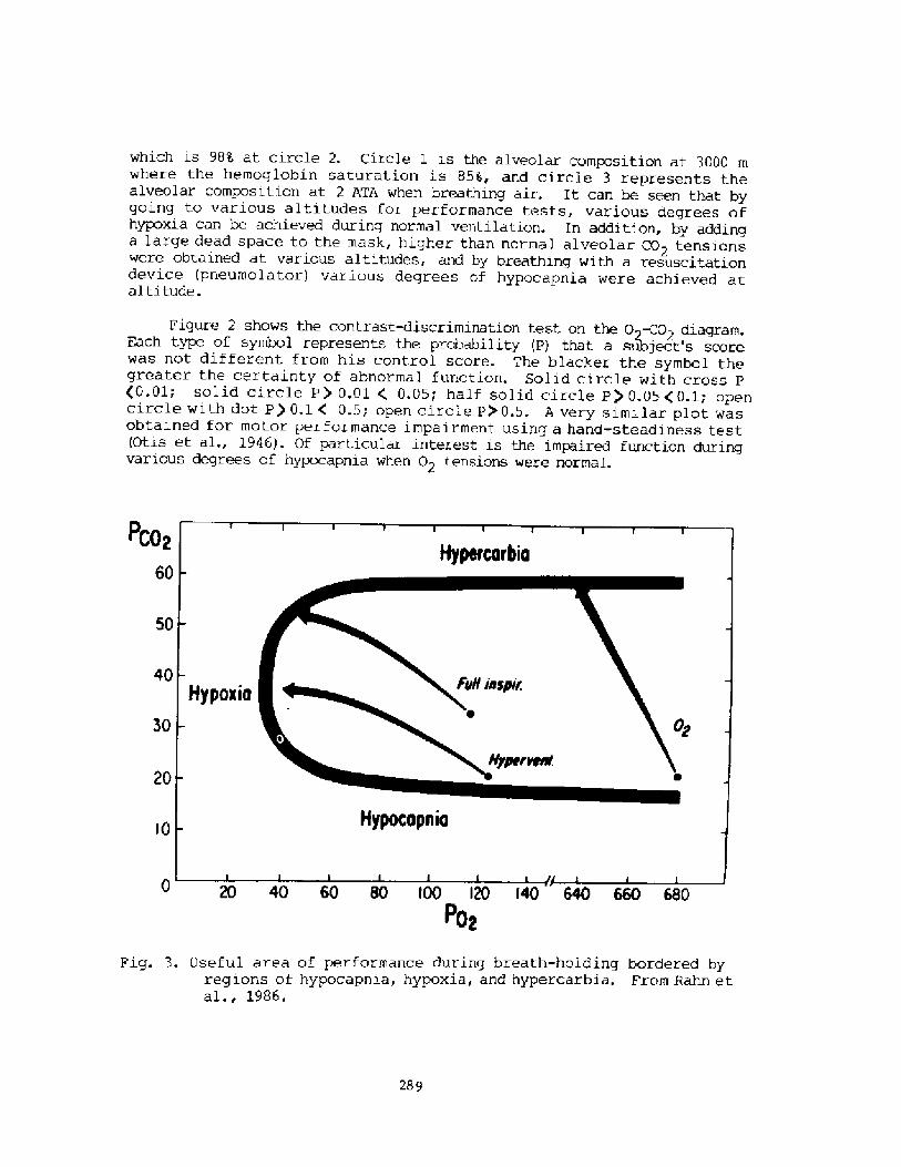
which is 98'4 at circle 2. Circle 1 i.s the alveolar composition at 3000 mwhere the hemoglobin saturation is 858, and circle 3 represents thealveolar composition at 2 ATA when breathing air. It can be seen that bygoing to various alti.tudes for performance tests, various degrees ofhypoxia can bc achieved during normal ventilation. In addition, by addinga large dead space to the mask, higher than normal alveolar CO2 tensionswere obtained at various altitudes, and by breathing with a resuscitationdevice pneumolator! various degrees of hypocapnia were achieved atalti,tude.
Figure 2 shows the contrast-discrimination test an the 0>-CO2 diagram.Each type of symbol represents the probability P! that a subject's scorewas not different from his control score. The blacker the symbol thegreater the certainty of abnormal function. Solid ci.rcle with cross P�.01; solid circle P! 0.01 0.05; half solid circle P! 0.054'0.1; opencircle with dot P! 0.1 C 0.5; open circle PO 0.5. A very similar plot wasobtained for motor performance impairment using a hand-steadiness test Otis et al., 1946!. Of particular interest is the impaired function duringvarious degrees of hypocapnia when 02 tensions were normal.
Pco~60
50
40
30
20
� 0 t00 0 �0 640 660 680Po~
Fig. 3. Useful area of performance during breath-holding bordered hyregions of hypocapnia, hypoxia, and hypercarbia. From Bahn etal., 1986.
289
Assuming that these performance criteria are applicable to the breath-hold diver, then figure 3 charts the approximate boundaries for safeperformance on the O2-CO2 diagram Rahn et al., 1986!. The pathways forthree breath-holds are shown, all originating in the useful performancearea. For example, after a full inspiration breathing air at sea level,the alveolar PCO2 = 35 torr and the breaking pornt at the borderline ofsafe performance rs determined by a combination of hypoxic and hypercarbicstimulus. With hyperventilation, in this example the alveolar PC 2 isreduced to 20 torr. If this is done with pure oxygen, the apneic pathwayis shown at the right. It is a straight line ending at the breaking pointof 60 torr and hypercarbia is the only sti.mulus. On the other hand, if thesame degree of hyperventilation is done breathing air at sea level, theonly stimulus to breaking is hypoxia. However, this stimulus alone isfrequently rather weak, and if apnea is prolonged, alveolar 02 willconti.nue to fall to levels of 30 � 35 torr, where black-out andunconsciousness set in.
References
Otis, A. B., H. Rahn, M. A. Epstein and W.O. Fenn. 1946. Performance asrelated to composition of alveolar air. Am. J. Physiol. 146: 207-221.
Rahn, H., and W. O. Fenn. 1955. A graphical analysis of the respiratorygas exchange. The 02-CO2 diagram. Am. Physiol. Soc., Washington,D.C.
Rahn, H., A. Olszowka and C. Lundgren. 1986. Shallow water and ascentblackout during breath-hold diving. In: Adaptive Physiology toStressful Environments, Edited by J. S. Samueloff and M. K. Yousef.CRC Press in press!.
Rahn, H., A.B. Oti.s, M. Hodge, M. A. Epstein, S. W. Hunter, and W.O. Fenn.1946. The effects of hypocapnia on performance. J. Aviation Med.17: 164-172.
290
Depth records: practf cal consf deratfons
Enzo Maf orca> Si racusa> Italy
In 1913 Georgh fos Kaggfir a Greek sponge diver~ made severalbreath-hol d dives down to 80 m of depth more than 260 feet!, tor ecover the 1 ost anchor of the Ital fan battl esh fp "ReginaMargherf ta" �!. Mow ever~ tt was not untf1 1949 that deep breath-hol d dfv ing started as a sport. In that year Ref mondo Bucher> anItalian dfveri made and won a wager: holding his breath andpul 1 ed underw ater by a wef ght> he reached 30 m of depth about100 feet!. At the bottom> an astonished hard-hat diver gave hfma w ater proof envel ope co nta f ning the priz e: twenty thousandital fan 1 i ra. Since that time many deep breath hol d divers. bothmen and w omen> have descended to incr easf ng depth s r ecentl ydiving down to more than 100 m �30 feet!. In this brief revf ewof breath-hol d dives to record depths I would like to mention.besf des Raimondo Bucher~ Ennio Fal coi Al berto Novell f~ Amerf coSantarel 1 i, Bob Croft~ Jacques Mayol, Marf a and G ful f anaTrel eaning Angel a Bandin f~ my two daughters Patr fz fa and Rossanaand mysel f �!. Rossana has recently established the new worldr ecord dive for women> descending to 68 m.
Unluckily> fn 1960 the Worl d Underwater Federation and in1981 the Italian Underwater Federatfon ceased to acknowledgethese records, because thef r Medical Commissions fudged theseattem pts dangerous. I heartily df sagree w f th thf s opf ni on thatw as based on th e threat of th or aci c squeez e. The depth at w hichth or aci c squeez e was pre df cted ke pt m ov f n g deeper and deeper w f thevery new successf ul record dive.
In Tabl e 1 you can see the three principal techniques fordeep breath-hol d diving: free diving, assf sted-descent divingand assi sted df v ing. In free dfv ing both descent to the bottomand ascent to the surface are performed by the dfver by his ownef f ort> th at is sw fmmfng dow n during descent> and pul 1 f nghimself up al ong a 1 f ne during ascent. Thi s type of diving isper f ormed not only by record dfversi but al so by the much morenumerous spear-f fshermen and by the Ama of Japan> in particularby the Cachido. In the case of assfsted-descent dfving, the diverdescen ds w i th the assi stance of a wef ghti b ut he or she goes bac kto th e sur f ace again usf ng h f s or her muscl es. Thi s technique i sused by record and pearl divers. These are the two types thathave been perf orm fng f or many years and I stf1 1 hol d the 1 astoff fcial records of both types of dfvfng> wfth 87 m �85 feet!reached in 1974 during an assisted-descent breath-hol d dfve> andw i th 60 m �96 feet! during a free breath-hold dive, again in1974. There i s a third type of breath-hol d dfv f ng> and th is hasbeen- used by Mayol in hf s 1 ast record dive to 105 m of depth more than 340 feet! �!: this fs the assisted dfvfng. In this
291
form of dfv f ng the diver fs hei ped f n both th e descent andascent. He f s pulled by a ref ght on hf s way dow ni and he f s1 f f ted by a sel f- fnf1 atabl e bal 1 oon or by a rope corn f ng up. Thistype of d fv fng f s per f ormed by record d fvers and by the Ama Funado!. In the past ft was performed al so by the Greek SpongeD ivers.
TABLE I
Types of Breath-hol d Dives with regard to means ofDescent and Ascent
Perf ormed byTy pe A see ntDescent
I. Free Diving swfmmfng hand-over-handswimming
record diversspear f f shermenAma-Cachf do
2. Assf sted-descent
diving
record dfverspearl df v ers
p ul 1 e d by hand- ov e r- h a n da wef ght 6 sw f mming
3 ~ Assi stedDfvfng
pull ed by 1 if ted by aa wef ght rope or air
bal 1 oon
record diversAm a-F una do
292
Ff gure I depfcts the depth-time prof fle of my deepestbreath-hol d dive> fn which I reached 94 m during traf nf ng. Thiswas an assi sted-descent dive. After an hyperventilation 1 astfng 8min~ I took a f ul 1 fnspf ratf on and I started my head-dow ndescent> pulled by a ref ght of 26 Kg that ran al ong a w fre andthat I coul d stop by means of a brake. Despite the fact that Istarted clearfng my ears from the surface> I had to stop at 30>40 and 50 m dur f ng my desce nt. At th ese depth s I turned h ead-u pfor a few seconds for additional ear-clearing. From 50 to 94 m Idf d not need to cl ear my ears anymore. Once I reached th e maxi maldepth> I 1 eft the weight and I started the ascent> pul 1 fng mysel fup along the wire wfth my hands and swimmfng w fth my legs.Cl oser to the surface> as the wet suit expanded and regaf nedbuoy ancyr ft hei ped my ascent. The total durati on of th f s diveras 3 mfn and 40 sec, with approximately an equal amount of timespent on descent and ascent, at a rate of about I m/sec
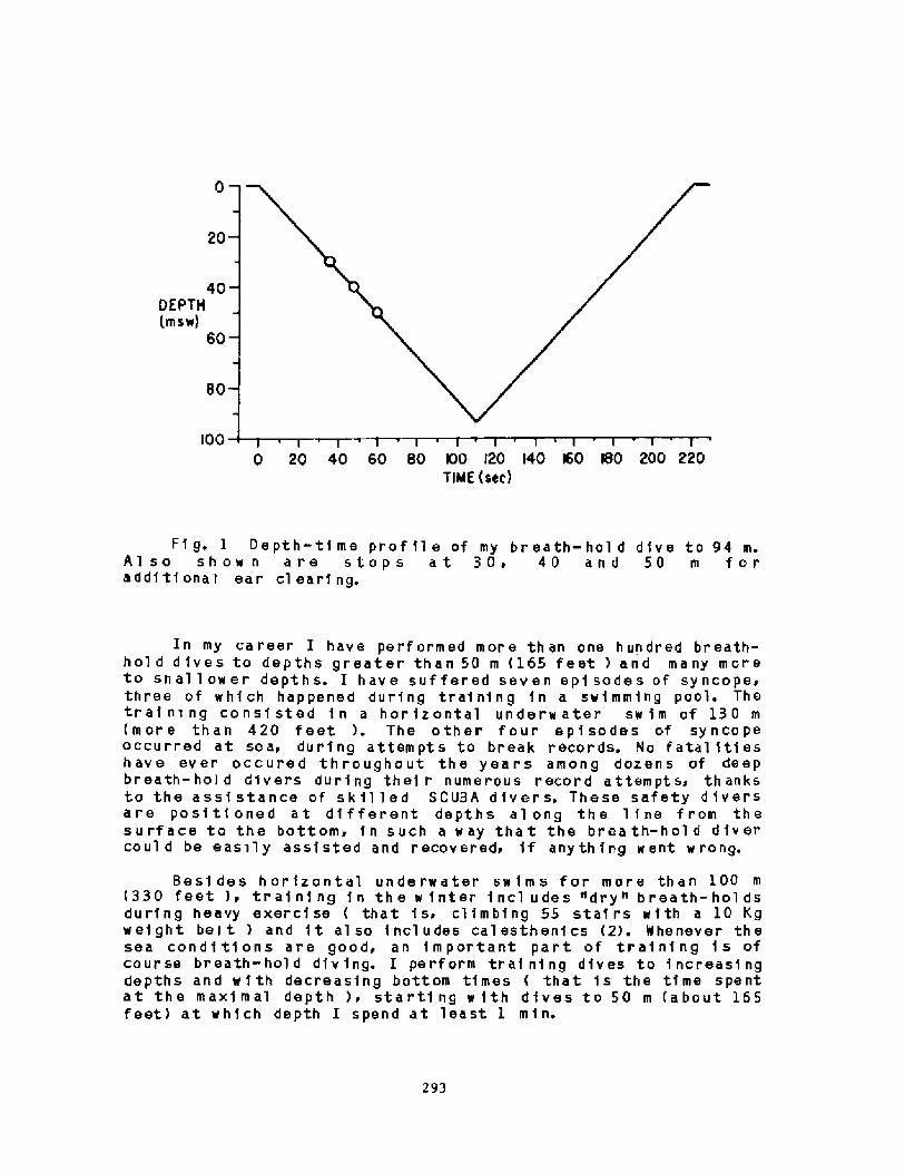
20
40 ! EPTH
mSw! 60
80
l00 0 20 40 60 80 00 �0 �0 l60 180 200 220TIME sec!
Fi g. 1 Depth-time prof 11 e of my br eath-hol d dive to 94 m.Al so show n are stops at 30~ 40 and 50 m foradditional ear clearing.
In my career I have performed more than one hundred breath-hol d dives to depths greater than 50 m �65 feet ! and many moreto sh al 1 ow er depth s. I have suf fered seven epi sode s of sy ncope.three of which happened during training 1n a swimming pool. Thetraining consisted in a horizontal underwater sw1m of 130 m more than 420 feet !. The other four epi sodes of syncopeoccurred at sea> during attempts to break records. No fatal iti eshave ever occured throughout the years among dozens of deepbr eath- hold divers dur1ng th ei r nume r ou s recor 1 attempt s~ th anksto the assi stance of sk il led SCUBA divers. These safety d1versare positioned at different depths al ong the line from thesurface to the bottom, in such a way that the brea th-hol d divercoul d be easily assisted and recovered> if anything went w rang.
Besi des horizontal underwater sw1ms for more than 100 m�30 feet ! > tra1ni ng 1n th e w inter incl u des "dry" breath- hol dsduring heavy exerc1se that 1 s. climbing 55 sta1rs with a 10 Kgweight bel t ! and it al so includes calesthenics �!. Whenever thesea condi tions are good, an important part of training 1 s ofcourse breath-hold diving. I perform training dives to increasingdepths and w1th decreasing bottom times that is the time spentat the maximal depth ! i starti ng w 1th dives to 50 m about 165feet! at wh1ch depth I spend at least 1 min.
293
Finally, I wish to thank all of you Scientists> who workhard everyday for us divers> and I want to give you a picturef rom my experience. Many years ago I w as doi ng a deep breath-hol d diver and the weather was bad even if the sea was calm~ thesky was gray and so there was al most no v isibil ity underwater. Iwas at depth and suddenly saw a fishy a very> very> very big fishand I w as af rai d because I did not know w hat to do. Suddenly> aray of sun fell down from the sky into the sea and hit the bigf ish> which expl oded i n a my ri ad of sm al 1 sil ver f i sh. It w asnot a big fish, it was a school of sardi nes which di sappeared inai l di recti ons. This is the picture th at I give to you.
Ref erences
1. Musenga> G. Un pesca tore di spugne sommozzatore! che senza
apparecchio si tuff a fino a 80 metri. Annali di Medicina
Navale e Co I oni al e> 1915: 163-167.
2. Maiorca> E. Scuola di apnea, Editrice la Cuba, Roma; 1982.
3. Mi ssi rol i> F. M. and B. Rizz ate. Mayol a 105 metr i. In:
Mondo Sommersoi no. 272 Dec. 1983> Jan. 1984!: 32-37.
294
Discussion folloving Nr. Naiorca's presentation
Dr. Lundgren: I think we should begin by thanking Mr. Maiorca foral low ing us to edi t his f ascinating f ilms in this way. Let meacknowledge Mr. Marky and Dr. Ferrigno for doing this. Perhaps a pointworth making is that a deep breath-hold dive like this is no smallfeat from the logistic point of view. The assistance of the SCUBAdivers can be extremely critical for your well being and we heard andsaw that they are positioned along the line, all the way down to thedeepest point where the breath-hold diver turns. In contrast to thebreath-hold diver, they, of course, are f aced with the problems ofaccumulating inert gas and if they had to ascend rapidly in assistingthe breath-hold diver, they might run the risk of decompressionsickness. So it's a very sophisticated operation logistically.
Dr- Perrigno: The other thing is that the SCUBA divers also run therisk of nitrogen narcosis because they are breathing air even below50 meters. These are very experienced divers but still you aretalking about 90 meters in air for SCUBA divers. We should alsoappreciate the technical diff iculties of taking f ilms at these depths.
would also like to add that Mr. Naiorca doesn't f ol low thesuggestion of Dr. Craig to start from outside the surface and thenjump in the water. Instead, he does his hyperventilation in the waterand then he s tar ts hi s des cent.
Dr. Rahn: Mr. Maiorca, do you have to turn head-up for ear-clearing2
Nr. Naiorca: Yes, I have to turn head up for a few seconds at 30, 40and 50 m, and then I resume my head-down descent, despite the factthat I start clearing my ears f rom the surface. For some reason Ineed some additional ear-clearing at these depths.
Dr. Rahn: I'd like to have Mr. Maiorca answer a few questions now.Can you tell us how you hyperventilate before the dive?
Nr. Naiorca: There are two types of hyperventilation, one that wecalled hyperoxygenation, with fast breaths, but then we were told thaihyper ventilation was not usef ul for hyper oxygenation, so we changed tca dif f erent type of hyperventilation, that is, long and slow br eaths.Four br eaths per minute.
Dr Rahn: For how many minutes.
Nr. Naiorca: Eight minutes. At the beginning of training,hyperventilate only for two minutes, but after about one month, I feelthat these two minutes are not enough, so I am forced to add mor e tirrr<for hyperventilation until the day of the record, when I arrive t<eight minutes of hyperventilation. So there is a gradual increase irthe time of hyperventilation. And the same is f elt also by m!daughters and other people in the School for Deep Breath-hold Divin<in Siracusa.
Dr. Rahn: Another question. How careful are you, to completely rela'just before the dive?
295
Nr. Naiorca: I am not totally relaxed because I know I am doingsomething dangerous. I am a man with all my f eelings and mypr eoccupa ti ons, and not a super man.
Dr. Rahn: I was thinking about the C02 production.
Dr. Lundgren: Speaking of hyperventilation. With your method ofhyperventilation you seem to create a high pressure in the thoraxduring expiration. Is that correct?
Nr. Naiorca: Yes.
Dr. Lundgren: I would like to suggest that it ties in with Dr.Craig's suggestion that the best way to start a dive would be to beout. of the water, in order to have as little blood in the chest forstarters as possible, so as to make maximum use of the return of theblood into the thorax during the dive, for compression of pulmonarygas. Now, perhaps this type of breathing, if in the water, would bean alternative to being out of the water in the pxeparatory phase,because the breathing seems to generate repetitive Valsalva maneuvers;that is, high pressure manuevers in the chest that would drive theblood out of the chest and that would provide room for a largerstarting air volume.
Dr. Rahn: The Ama have been doing that f or 2,000 years bywhistling just before they dive.
Dr. Lundgren: Is it not true that all the Amas, when asked why theydo it, answer that they always did it that way.
Dr. Rahn: Yes.
Dr. Lundgren: So, is there a philosophy, a special philosophy, behindthis particular breathing pattern?
Nr. Naiorca: I just feel the need to do it, and we have seen thatwith this hyperventilation, we can obtain the best results.
Dr. Lundqren: In terms of depth or duration?
Nr. Maiorca: For both depth and duration.
Dr. Norfleet: In your record breaking dive, when you came to thesurface and lost consciousness, did any of your companions notice whatcolox your skin was?
Nr. Naiorca: I don't know, no one told me, I don't know.
Dr. NcDonough: Were you breathing spontaneously, or did you havea s si st ed vent i 1 ation'?
Mr. Naiorca: They told me that I was unconscious for about one minuteand thirty seconds, and that I was not breathing at all.
Dr. Rennie: What was the duration of the 130 meters horizontaL
296
underwater swim?
Mr. Maiorca: Two minutes and five seconds.
Dr. Whitelaw: Mr. Maiorca, I would like to know what tricks you useto postpone or reduce the urge to breathe, and also whether you thinkthat your breathing muscles are working toward the end of the dive.
Mr. Maiorca: Coming from a very deep dive, I don't feeldiaphragmatic contractions, but I feel them during the horizontalunderwater swims, even af ter I have swum only twenty meters. So, Ifeel the urge to breathe especially when I swim underwaterhorizontally for training, either at sea or in a swimming pool. And Ithink it's mainly a psychological p~oblem. I fight the diaphragmaticcontractions, and I train myself in doing that throughout the year,for example, climbing stairs �5 steps! while holding my breath andcarrying a 10 Kg weight belt, And, it's much more difficult to holdyour breath while climbing stairs because you have air around you, soyou develop will power, which is essential for these very deep dives.I don't perform any special maneuver, like swallowing or anythingelse, to fight diaphragmatic contractions, which cease after awhile,
Dr. McDonough: Could I ask the question whether you were aware ofany impending loss of consciousness during the dive we have seen orany of the six other dives during which you lost consciousness?
Mr. Maiorca: Not at all. No warning signs. I went from the veryexciting feeling of having reached the record depth to the loss ofconsciousness.
Dr. Hong: When Jacques Mayol came to Japan last year, he told us heuses Yoga for his deep dives. What do you think of this?
Mx. Maiorca: As I told you before, I like to dive as a human being,with all my feelings, and I think that Yoga allows man to become partof the Cosmos and to achieve great results, because it helps from apsychological point of view, but I prefer to go down as a man, and Ilove the sea as an air-breathing human.
Dr. Siesjo: Can I come back to the symptoms of ascent< when Mr,Maiorca is approaching the surface. I wonder if you can notice anydifference in the way you see things, in your vision.
Mr. Maiorca: Nothing at all. At the beginning of my career I used todive without a mask, and my vision improved at depth. Then I startedusing a mask. During the last dives, I have not used masks anymorefbut instead, I now use perforated goggles with 180 dioptric lenses, sothat now I don't have to waste air in clearing the mask.
Dr. Siesjo: I wondered because one of the earlier symptoms inexperimental hypoxia is blurred vision and also narrowed visualf ields.
Mr. Maiorca: AbsoluteLy not. And I know that for sure because I haveto watch the divers, the SCUBA divers around me who give me signalsf
297
for example, they tell me to take of f my goggles close to the surface,because they bother me.
Dr. Iin: I'd like to know, when you are at depth, under such veryhigh pressur e s, do you f eel a ny thing physically?
Nx. Naiorca: I don't have any feelings of pressure at all, but Istart feeling discomfort in my ears, but you have to remember that Ido not clear my ears after 50 meters.
Dr. Lanphier: Do you notice anything like nitrogen narcosis?
Nr. Naiorca: I am perfectly conscious. In fact, I am excited when Ireach the record depth where I have to pick a mark showing the depthfbut then I am very scared that I will not be able to make it back tothe surface. May I have two minutes'? Because I wish to saysomething. Scientists work hard everyday for us divers, as I am adiver today and I wish to thank you, and I want to give you a picturefrom my experience. Many years ago, I was doing a deep breath-holddive, and the weather was bad even if the sea was calm, the sky wasgray and so there was not visibility underwater. I was at depth andsuddenly saw a fish, a very, very, very big fish and I was afraid andI did not know what to do. Suddenly, a ray of sun fell down from thesky into the sea and hit the big fish which exploded into a thousandsmall silver f ish. It was not a big f ish, it was a school ofsardines which disappeared in all directions. This is the picturethat I give to you.
298


![[Hyperbaric oxygen for carbon monoxide poisoning]](https://img.dokumen.tips/doc/110x75/634e376e7c06afa1b60d8395/hyperbaric-oxygen-for-carbon-monoxide-poisoning.jpg)
