Embed Size (px)
Citation preview
J. Pers. Med. 2022, 12, 337. https://doi.org/10.3390/jpm12030337 www.mdpi.com/journal/jpm
Article
Ultrasonographic Confirmation of Nasogastric Tube Placement
in the COVID-19 Era
Vasiliki Tsolaki *, George E. Zakynthinos, Paris Zygoulis, Fotini Bardaka, Aikaterini Malita, Vasileios Aslanidis,
Epaminondas Zakynthinos and Demosthenes Makris
Critical Care Department, University Hospital of Larissa, Faculty of Medicine, University of Thessaly,
Mezourlo, 41335 Larissa, Greece; [email protected] (G.E.Z.); [email protected] (P.Z.);
[email protected] (F.B.); [email protected] (A.M.); [email protected] (V.A.);
[email protected] (E.Z.); [email protected] (D.M.)
* Correspondence: [email protected]; Tel.: +30-69-7270-4419 or +30-24-1350-2964
Abstract: Background: Nasogastric tube (NGT) placement is a daily routine in the Intensive Care
Unit (ICU), and misplacement of the NGT can cause serious complications. In COVID-19 ARDS
patients, proning has emerged the need for frequent NGT re-evaluations. The gold standard tech-
nique, chest X-ray, is not always feasible. In the present study we report our experience with the use
of ultrasonographic confirmation of NGT position. Methods: A prospective study in 276 COVID-19
ARDS patients admitted after intubation in the ICU. Ultrasonographic evaluation was performed
using longitudinal or sagittal epigastric views. Examinations were performed during the initial NGT
placement and every time the patients returned to the supine position after they had been proned
or whenever critical care physicians or nurses considered that reconfirmation was necessary. Re-
sults: Ultrasonographic confirmation of correct NGT placement was feasible in 246/276 (89.13%)
patients upon ICU admission. In 189/246 (76.8%) the tube could be visualized in the stomach (two
parallel lines), in 172/246 (69.9%) the ultrasonographic whoosh test (“flash” due to air instillation
through the tube, seen with ultrasonography) was evident, while in 164/246 (66.7%) both tests con-
firmed correct NGT placement. During ICU stay 590 ultrasonographic NGT evaluations were per-
formed, and in 462 (78.14%) cases correct NGT placement were confirmed. In 392 cases, a chest X-
ray was also ordered. The sensitivity of ultrasonographic NGT confirmation in these cases was
98.9%, specificity 57.9%, PPV 96.2%, and NPV 3.8%. The time for the full evaluation was 3.8 ± 3.4
min. Conclusion: Ultrasonographic confirmation of correct NGT placement is feasible in the initial
placement, but also whenever needed thereafter, especially in the COVID-19 era, when changes in
posture have become a daily practice in ARDS patients.
Keywords: nasogastric tube; POCUS; ultrasonography; intensive care unit
1. Introduction
Nasogastric tube (NGT) placement is performed in every patient hospitalized in the
Intensive Care Unit (ICU) to facilitate enteral nutrition, administer medication, and for
gastric decompression [1,2]. Misplacement of the NGT incidence varies from 0.5 to 89%
and can cause serious complications such as aspiration pneumonia, empyema, pneumo-
thorax, hemothorax, pneumomediastinum, or even intracranial insertion [2,3]. Confirma-
tion of correct placement of the tube can be performed by auscultating air through the
tube (whoosh test), observation of aspirated fluid, and pH measurement of the gastric
aspirate. Yet the gold standard technique is abdominal X-ray confirmation combined with
aspirate testing [3].
The reference pH value is different between the gastric and lung aspirate. Gastric pH
ranges from 1 to 5.5, while lung aspirate is more alkaline. Gastric aspirate is only achieved
Citation: Tsolaki, V.; Zakynthinos,
G.E.; Zygoulis, P.; Bardaka, F.;
Malita, A.; Aslanidis, V.;
Zakynthinos, E.; Makris, D.
Ultrasonographic Confirmation of
Nasogastric Tube Placement in the
COVID-19 Era. J. Pers. Med. 2022, 12,
337. https://doi.org/10.3390/
jpm12030337
Academic Editors: Ioanna V.
Papathanasiou and Evangelos
Fradelos
Received: 31 December 2021
Accepted: 22 February 2022
Published: 23 February 2022
Publisher’s Note: MDPI stays neu-
tral with regard to jurisdictional
claims in published maps and institu-
tional affiliations.
Copyright: © 2022 by the authors. Li-
censee MDPI, Basel, Switzerland.
This article is an open access article
distributed under the terms and con-
ditions of the Creative Commons At-
tribution (CC BY) license (https://cre-
ativecommons.org/licenses/by/4.0/).
J. Pers. Med. 2022, 12, 337 2 of 9
in half of the patients, and the use of proton pump inhibitors, prolonged fasting, and en-
teral feeding may alter the values of gastric pH, complicating the results further [4]. Aspi-
ration of alkaline fluid cannot exclude the presence of the tube in the distant esophagus.
Auscultation, on the other hand, may be tricky when managing COVID-19 patients, as
personal protective equipment may hinder the use of a stethoscope.
On the other hand, radiographies are not always easily available throughout the day,
and certain delays in the initiation of enteral feeding may occur. In the COVID-19 era, a
significant proportion of patients admitted in the ICU are placed in the prone position.
Changes of patients’ posture (from supine to prone and vice versa) occur at least once a
day (according to local protocols, every 16–30 h) [5]. Thus, the NGT position may have to
be rechecked every day or every other day. Performance of daily abdominal X-rays is time
and resource consuming, and it exposes patients to a certain degree of radiation.
Ultrasonographic NGT placement confirmation is increasingly being recognized as a
safe, alternative technique, yet contradictory results from published studies, with mainly
small numbers of included patients, have precluded the wide acceptance of the technique
as a standard procedure [6]. Point Of Care Ultrasonography (POCUS) is gaining place in
everyday clinical practice in ICU practitioners, as it is available at the bedside 24 h a day/7
days a week, and, as it is performed by clinicians caring for the patients, it can answer
clinical questions [7]. Critical care nurses, untrained in the ultrasound technique, can eas-
ily gain skills in accurately performing and interpreting POCUS images, at the patient’s
bedside [8]. In the present study, we report our experience in NGT confirmation using
ultrasonography in COVID-19 ARDS, intubated, ICU patients.
2. Methods
This prospective study was conducted in an 18-bed, COVID-19 dedicated ICU, in a
tertiary hospital in Central Greece. All COVID-19 mechanically ventilated, ARDS patients
admitted from 2 April 2020 until 30 November 2021 were evaluated. Patients having un-
dergone gastric surgery in the past were excluded from the analysis. The study was ap-
proved by the local ethics committee (University Hospital of Larissa) (55949/2020, date of
approval: 23/12/2020), with a waiver for informed consent.
A nasogastric tube was placed in every patient with COVID-19 ARDS, upon ICU
admission. Critical care physicians inserted the NGTs. Confirmation of correct NGT place-
ment was performed with abdominal ultrasonography using a convex probe with low
frequency (2–5 MHz). The probe was positioned in the sagittal and longitudinal epigastric
section. Correct NGT placement was considered if either a single or double parallel lines
of the tube were visualized in the gastric antrum or pylorus (Figure 1) and/or there was a
dynamic appearance of air entering the stomach, instilled through the NGT (ultraso-
nographic whoosh test) (Videos S1 and S2). Confirmation was considered is cases in which
both ultrasonographic tests were present or there was one ultrasonographic test and a
positive “palpation test”. A positive “palpation test” was considered the test when a
“flash” of air was palpated in the epigastrium, after installation of 50 mL of air through
the NGT. When correct NGT placement could not be evaluated with one of the above-
mentioned criteria, an abdominal X-ray was ordered, and enteral nutrition was delayed
until the radiograph was performed. Confirmation of correct NGT position was re-per-
formed using ultrasonography every time the patients returned to the supine position af-
ter they had been proned, or whenever critical care physicians or nurses considered that
reconfirmation was necessary.
J. Pers. Med. 2022, 12, 337 3 of 9
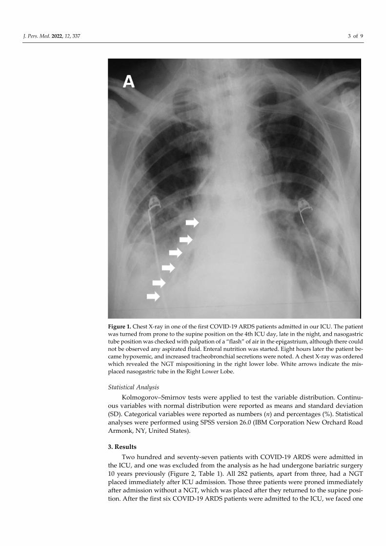
Figure 1. Chest X-ray in one of the first COVID-19 ARDS patients admitted in our ICU. The patient
was turned from prone to the supine position on the 4th ICU day, late in the night, and nasogastric
tube position was checked with palpation of a “flash” of air in the epigastrium, although there could
not be observed any aspirated fluid. Enteral nutrition was started. Eight hours later the patient be-
came hypoxemic, and increased tracheobronchial secretions were noted. A chest X-ray was ordered
which revealed the NGT mispositioning in the right lower lobe. White arrows indicate the mis-
placed nasogastric tube in the Right Lower Lobe.
Statistical Analysis
Kolmogorov–Smirnov tests were applied to test the variable distribution. Continu-
ous variables with normal distribution were reported as means and standard deviation
(SD). Categorical variables were reported as numbers (n) and percentages (%). Statistical
analyses were performed using SPSS version 26.0 (IBM Corporation New Orchard Road
Armonk, NY, United States).
3. Results
Two hundred and seventy-seven patients with COVID-19 ARDS were admitted in
the ICU, and one was excluded from the analysis as he had undergone bariatric surgery
10 years previously (Figure 2, Table 1). All 282 patients, apart from three, had a NGT
placed immediately after ICU admission. Those three patients were proned immediately
after admission without a NGT, which was placed after they returned to the supine posi-
tion. After the first six COVID-19 ARDS patients were admitted to the ICU, we faced one
J. Pers. Med. 2022, 12, 337 4 of 9
unfortunate NGT misplacement in a patient who turned to the supine position after he
had been proned for sixteen hours. A chest X-ray was delayed, and enteral feeding was
initiated after a “palpation” positive test. Eight hours later the patient became severely
hypoxemic with increased secretions. Chest X-ray revealed that the NGT had been posi-
tioned in the right lower lung lobe (Figure 1). After that we decided to adopt a protocol of
ultrasonographic confirmation of correct NGT position.
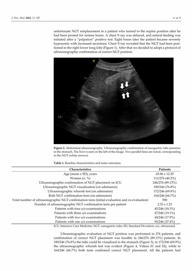
Figure 2. Abdominal ultrasonography. Ultrasonographic confirmation of nasogastric tube presence
in the stomach. The liver is seen on the left of the image. Two parallel lines are noted, corresponding
to the NGT (white arrows).
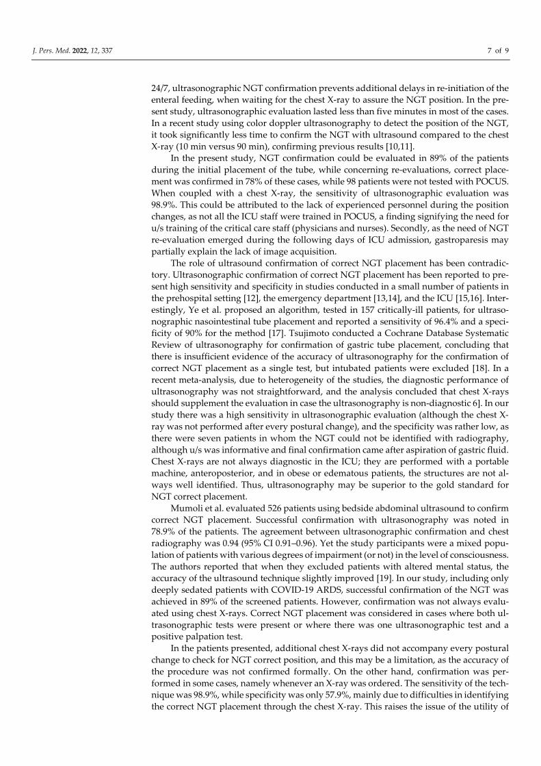
Table 1. Baseline characteristics and main outcomes.
Characteristics Patients
Age (mean ± SD), years 65.86 ± 12.29
Women (n, %) 111/276 (40.2%)
Ultrasonographic confirmation of NGT placement on ICU 246/276 (89.13%)
Ultrasonographic NGT visualization (on admission) 189/246 (76.8%)
Ultrasonographic whoosh test (on admission) 172/246 (69.9%)
Both NGT confirmation tests (on admission) 164/246 (66.7%)
Total number of ultrasonographic NGT confirmation tests (initial evaluation and re-evaluation) 590
Number of ultrasonographic NGT confirmation tests per patient 2.33 ± 1.23
Patients with four u/s examinations 45/246 (18.3%)
Patients with three u/s examinations 47/246 (19.1%)
Patients with two u/s examinations 44/246 (17.9%)
Patients with one u/s examinations 92/246 (37.4%)
ICU, Intensive Care Medicine; NGT, nasogastric tube; SD, Standard Deviation; u/s, ultrasound.
Ultrasonographic evaluation of NGT position was performed in 276 patients, and
confirmation of correct NGT placement was feasible in 246/276 (89.13%) patients. In
189/246 (76.8%) the tube could be visualized in the stomach (Figure 3), in 172/246 (69.9%)
the ultrasonographic whoosh test was evident (Figure 4, Videos S1 and S2), while in
164/246 (66.7%) both tests confirmed correct NGT placement. All the patients had
J. Pers. Med. 2022, 12, 337 5 of 9
additional confirmation with palpation of a “flash” of air in the epigastrium instilled
through the NGT. In the patients in whom the NGT could not be visualized and an ultra-
sonographic whoosh test was negative, a chest X-ray was performed. In all patients the
NGT was located in the stomach.
Five hundred ninety ultrasonographic evaluations, in total including an initial eval-
uation and re-evaluations, were performed during the study period. Four hundred sixty-
two (78.3%) confirmations of correct NGT placement could be evaluated (Table 1, Figure
2). In the 128 cases (21.7%) where the NGT could not be visualized with the ultrasound, a
chest X-ray was ordered. In ninety-eight patients the change in the patients’ position was
performed after midnight, and NGT confirmation was performed palpating an air “flash”
in the epigastrium, inserted through the NGT. In these patients, enteral nutrition was ini-
tiated. The NGT was evaluated as soon as possible, but these patients were not included
in the analysis.
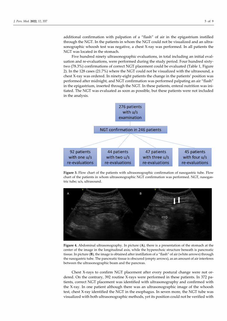
Figure 3. Flow chart of the patients with ultrasonographic confirmation of nasogastric tube. Flow
chart of the patients in whom ultrasonographic NGT confirmation was performed. NGT, nasogas-
tric tube; u/s, ultrasound.
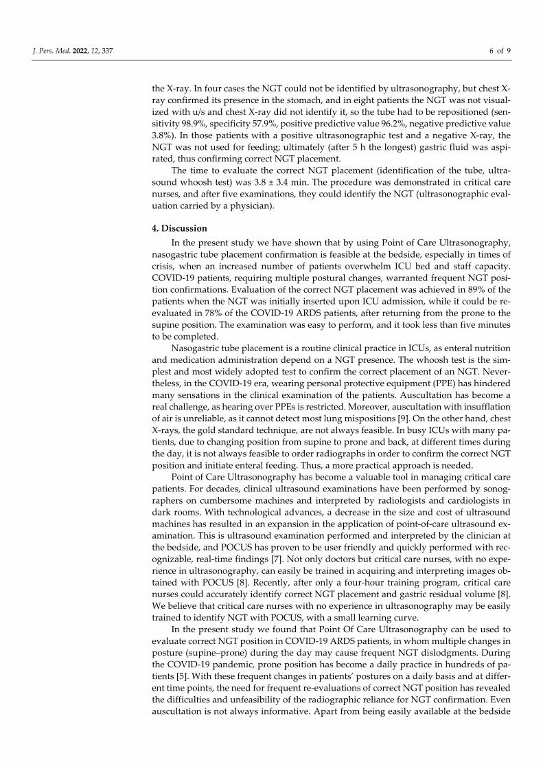
Figure 4. Abdominal ultrasonography. In picture (A), there is a presentation of the stomach at the
center of the image in the longitudinal axis, while the hyperechoic structure beneath is pancreatic
tissue. In picture (B), the image is obtained after instillation of a “flash” of air (white arrows) through
the nasogastric tube. The pancreatic tissue is obscured (empty arrows), as an amount of air interferes
between the ultrasonographic beam and the pancreas.
Chest X-rays to confirm NGT placement after every postural change were not or-
dered. On the contrary, 392 routine X-rays were performed in these patients. In 372 pa-
tients, correct NGT placement was identified with ultrasonography and confirmed with
the X-ray. In one patient although there was an ultrasonographic image of the whoosh
test, chest X-ray identified the NGT in the esophagus. In seven more, the NGT tube was
visualized with both ultrasonographic methods, yet its position could not be verified with
J. Pers. Med. 2022, 12, 337 6 of 9
the X-ray. In four cases the NGT could not be identified by ultrasonography, but chest X-
ray confirmed its presence in the stomach, and in eight patients the NGT was not visual-
ized with u/s and chest X-ray did not identify it, so the tube had to be repositioned (sen-
sitivity 98.9%, specificity 57.9%, positive predictive value 96.2%, negative predictive value
3.8%). In those patients with a positive ultrasonographic test and a negative X-ray, the
NGT was not used for feeding; ultimately (after 5 h the longest) gastric fluid was aspi-
rated, thus confirming correct NGT placement.
The time to evaluate the correct NGT placement (identification of the tube, ultra-
sound whoosh test) was 3.8 ± 3.4 min. The procedure was demonstrated in critical care
nurses, and after five examinations, they could identify the NGT (ultrasonographic eval-
uation carried by a physician).
4. Discussion
In the present study we have shown that by using Point of Care Ultrasonography,
nasogastric tube placement confirmation is feasible at the bedside, especially in times of
crisis, when an increased number of patients overwhelm ICU bed and staff capacity.
COVID-19 patients, requiring multiple postural changes, warranted frequent NGT posi-
tion confirmations. Evaluation of the correct NGT placement was achieved in 89% of the
patients when the NGT was initially inserted upon ICU admission, while it could be re-
evaluated in 78% of the COVID-19 ARDS patients, after returning from the prone to the
supine position. The examination was easy to perform, and it took less than five minutes
to be completed.
Nasogastric tube placement is a routine clinical practice in ICUs, as enteral nutrition
and medication administration depend on a NGT presence. The whoosh test is the sim-
plest and most widely adopted test to confirm the correct placement of an NGT. Never-
theless, in the COVID-19 era, wearing personal protective equipment (PPE) has hindered
many sensations in the clinical examination of the patients. Auscultation has become a
real challenge, as hearing over PPEs is restricted. Moreover, auscultation with insufflation
of air is unreliable, as it cannot detect most lung mispositions [9]. On the other hand, chest
X-rays, the gold standard technique, are not always feasible. In busy ICUs with many pa-
tients, due to changing position from supine to prone and back, at different times during
the day, it is not always feasible to order radiographs in order to confirm the correct NGT
position and initiate enteral feeding. Thus, a more practical approach is needed.
Point of Care Ultrasonography has become a valuable tool in managing critical care
patients. For decades, clinical ultrasound examinations have been performed by sonog-
raphers on cumbersome machines and interpreted by radiologists and cardiologists in
dark rooms. With technological advances, a decrease in the size and cost of ultrasound
machines has resulted in an expansion in the application of point-of-care ultrasound ex-
amination. This is ultrasound examination performed and interpreted by the clinician at
the bedside, and POCUS has proven to be user friendly and quickly performed with rec-
ognizable, real-time findings [7]. Not only doctors but critical care nurses, with no expe-
rience in ultrasonography, can easily be trained in acquiring and interpreting images ob-
tained with POCUS [8]. Recently, after only a four-hour training program, critical care
nurses could accurately identify correct NGT placement and gastric residual volume [8].
We believe that critical care nurses with no experience in ultrasonography may be easily
trained to identify NGT with POCUS, with a small learning curve.
In the present study we found that Point Of Care Ultrasonography can be used to
evaluate correct NGT position in COVID-19 ARDS patients, in whom multiple changes in
posture (supine–prone) during the day may cause frequent NGT dislodgments. During
the COVID-19 pandemic, prone position has become a daily practice in hundreds of pa-
tients [5]. With these frequent changes in patients’ postures on a daily basis and at differ-
ent time points, the need for frequent re-evaluations of correct NGT position has revealed
the difficulties and unfeasibility of the radiographic reliance for NGT confirmation. Even
auscultation is not always informative. Apart from being easily available at the bedside
J. Pers. Med. 2022, 12, 337 7 of 9
24/7, ultrasonographic NGT confirmation prevents additional delays in re-initiation of the
enteral feeding, when waiting for the chest X-ray to assure the NGT position. In the pre-
sent study, ultrasonographic evaluation lasted less than five minutes in most of the cases.
In a recent study using color doppler ultrasonography to detect the position of the NGT,
it took significantly less time to confirm the NGT with ultrasound compared to the chest
X-ray (10 min versus 90 min), confirming previous results [10,11].
In the present study, NGT confirmation could be evaluated in 89% of the patients
during the initial placement of the tube, while concerning re-evaluations, correct place-
ment was confirmed in 78% of these cases, while 98 patients were not tested with POCUS.
When coupled with a chest X-ray, the sensitivity of ultrasonographic evaluation was
98.9%. This could be attributed to the lack of experienced personnel during the position
changes, as not all the ICU staff were trained in POCUS, a finding signifying the need for
u/s training of the critical care staff (physicians and nurses). Secondly, as the need of NGT
re-evaluation emerged during the following days of ICU admission, gastroparesis may
partially explain the lack of image acquisition.
The role of ultrasound confirmation of correct NGT placement has been contradic-
tory. Ultrasonographic confirmation of correct NGT placement has been reported to pre-
sent high sensitivity and specificity in studies conducted in a small number of patients in
the prehospital setting [12], the emergency department [13,14], and the ICU [15,16]. Inter-
estingly, Ye et al. proposed an algorithm, tested in 157 critically-ill patients, for ultraso-
nographic nasointestinal tube placement and reported a sensitivity of 96.4% and a speci-
ficity of 90% for the method [17]. Tsujimoto conducted a Cochrane Database Systematic
Review of ultrasonography for confirmation of gastric tube placement, concluding that
there is insufficient evidence of the accuracy of ultrasonography for the confirmation of
correct NGT placement as a single test, but intubated patients were excluded [18]. In a
recent meta-analysis, due to heterogeneity of the studies, the diagnostic performance of
ultrasonography was not straightforward, and the analysis concluded that chest X-rays
should supplement the evaluation in case the ultrasonography is non-diagnostic 6]. In our
study there was a high sensitivity in ultrasonographic evaluation (although the chest X-
ray was not performed after every postural change), and the specificity was rather low, as
there were seven patients in whom the NGT could not be identified with radiography,
although u/s was informative and final confirmation came after aspiration of gastric fluid.
Chest X-rays are not always diagnostic in the ICU; they are performed with a portable
machine, anteroposterior, and in obese or edematous patients, the structures are not al-
ways well identified. Thus, ultrasonography may be superior to the gold standard for
NGT correct placement.
Mumoli et al. evaluated 526 patients using bedside abdominal ultrasound to confirm
correct NGT placement. Successful confirmation with ultrasonography was noted in
78.9% of the patients. The agreement between ultrasonographic confirmation and chest
radiography was 0.94 (95% CI 0.91–0.96). Yet the study participants were a mixed popu-
lation of patients with various degrees of impairment (or not) in the level of consciousness.
The authors reported that when they excluded patients with altered mental status, the
accuracy of the ultrasound technique slightly improved [19]. In our study, including only
deeply sedated patients with COVID-19 ARDS, successful confirmation of the NGT was
achieved in 89% of the screened patients. However, confirmation was not always evalu-
ated using chest X-rays. Correct NGT placement was considered in cases where both ul-
trasonographic tests were present or where there was one ultrasonographic test and a
positive palpation test.
In the patients presented, additional chest X-rays did not accompany every postural
change to check for NGT correct position, and this may be a limitation, as the accuracy of
the procedure was not confirmed formally. On the other hand, confirmation was per-
formed in some cases, namely whenever an X-ray was ordered. The sensitivity of the tech-
nique was 98.9%, while specificity was only 57.9%, mainly due to difficulties in identifying
the correct NGT placement through the chest X-ray. This raises the issue of the utility of
J. Pers. Med. 2022, 12, 337 8 of 9
chest X-ray as the gold standard method for the confirmation of correct NGT position. The
current report is a presentation of our experience in a busy ICU, overwhelmed with
COVID-19 ARDS patients, during a pandemic period. With this report we want to em-
phasize the value of POCUS in the COVID-19 era, expanding previous knowledge ob-
tained from nine COVID-19 ARDS patients [20], and the potential to be used by critical
care nurses as well. In this regard, with only a limited duration of training, accompanied
with a small number of supervised examinations, critical care nurses can gain the skills
required for POCUS. Although POCUS has been widely used in the critical care setting,
becoming the new “stethoscope” for physicians, only scarce reports have arisen concern-
ing its use by critical care nurses [21,22]. In depth learning of POCUS applicability should
make it an invaluable tool for nurses as well. Another limitation of the study could be that
other ultrasonographic windows to confirm correct NGT placement, such as the esopha-
geal window in the neck, were not used, but this technique cannot exclude the misposition
of the NGT in the esophagus [23].
In conclusion, POCUS can greatly improve everyday clinical practice for critical care
staff, physicians, and nurses concerning the confirmation of correct nasogastric tube place-
ment. It seems that in the settings where X-ray is not readily available, ultrasound may be
useful to detect misplaced gastric tubes. Larger studies are needed to determine the pos-
sibility of adverse events when ultrasound is used to confirm nasogastric tube placement.
Further studies on POCUS may unravel potential advantages of its use among physicians,
nurses, and perhaps physiotherapists.
Supplementary Materials: The following supporting information can be downloaded at
www.mdpi.com/article/10.3390/jpm12030337/s1: Videos S1 and S2. Ultrasonographic whoosh test.
A flash is seen in the center of the image, corresponding to the amount of air pushed through the
nasogastric tube with a syringe. In Video S1, note that the image is vague (structures beneath, after
instillation of the air, are not visualization, as the ultrasound beam does not penetrate the air).
Author Contributions: Conceptualization, V.T. and P.Z.; methodology, E.Z.; software, G.E.Z.; vali-
dation, V.T., G.E.Z., P.Z., and D.M.; formal analysis, D.M.; investigation, V.T., P.Z., G.E.Z., and F.B.;
resources, A.M. and V.A.; data curation, V.T., G.E.Z., and P.Z.; writing—original draft preparation,
V.T. and G.E.Z.; writing—review and editing, E.Z. and D.M.; visualization, A.M.; supervision, E.Z.
and D.M.; project administration, G.E.Z.; funding acquisition, N/A. All authors have read and
agreed to the published version of the manuscript.
Funding: This research received no external funding.
Institutional Review Board Statement: The study was approved by the local (University Hospital
of Larissa) ethics committee (55949/2020, date of approval: 23/12/2020), with a waiver for informed
consent.
Informed Consent Statement: Patient consent was waived due to the COVID-19 pandemic.
Data Availability Statement: All data are available upon request.
Conflicts of Interest: The authors declare no conflicts of interest.
References
1. Stewart, M.L.; Biddle, M.; Thomas, T. Evaluation of current feeding practices in the critically ill: A retrospective chart review.
Intensive Crit Care Nurs. 2017, 38, 24–30. doi: 10.1016/j.iccn.2016.05.004. Epub 2016 Jul 6. PMID: 27395368.
2. Itkin, M.; DeLegge, M.H.; Fang, J.C.; McClave, S.A.; Kundu, S.; d’Othee, B.J.; Martinez-Salazar, G.M.; Sacks, D.; Swan, T.L.;
Towbin, R.B.; et al. Multidisciplinary practical guidelines for gastrointestinal access for enteral nutrition and decompression
from the Society of Interventional Radiology and American Gastroenterological Association (AGA) Institute, with endorsement
by Canadian Interventional Radiological Association (CIRA) and Cardiovascular and Interventional Radiological Society of
Europe (CIRSE). Gastroenterology 2011, 141, 742–765.
3. Boullata, J.I.; Carrera, A.L.; Harvey, L.; Escuro, A.A.; Hudson, L.; Mays, A.; McGinnis, C.; Wessel, J.J.; Bajpai, S.; Beebe, M.L.; et
al. ASPEN Safe Practices for Enteral Nutrition Therapy Task Force, American Society for Parenteral and Enteral Nutrition.
ASPEN Safe Practices for Enteral Nutrition Therapy. JPEN J Parenter Enteral Nutr. 2017, 41, 15–103.
4. Boeykens, K.; Steeman, E.; Duysburgh, I. Reliability of pH measurement and the auscultatory method to confirm the position
of a nasogastric tube. Int. J. Nurs. Stud. 2014, 51, 1427–1433.
J. Pers. Med. 2022, 12, 337 9 of 9
5. Langer, T.; Brioni, M.; Guzzardella, A.; Carlesso, E.; Cabrini, L.; Castelli, G.; Dalla Corte, F.; De Robertis, E.; Favarato, M.; Fo-
rastieri, A.; et al. Prone position in intubated, mechanically ventilated patients with COVID-19: a multi-centric study of more
than 1000 patients. Crit. Care 2021, 25, 128.
6. Lin, T.; Gifford, W.; Lan, Y.; Qin, X.; Liu, X.; Wang, J.; Yang, B.; You, T.; Chen, K. Diagnostic accuracy of ultrasonography for
detecting nasogastric tube (NGT) placement in adults: A systematic review and meta analysis. Int. J. Nurs. Stud. 2017, 71, 80–88.
7. Bledsoe, A.; Zimmerman, J. Ultrasound: The New Stethoscope (Point-of-Care Ultrasound). Anesthesiol. Clin. 2021, 39, 537–553.
8. Brotfain, E.; Erblat, A.; Luft, P.; Elir, A.; Gruenbaum, B.F.; Livshiz-Riven, I.; Koyfman, A.; Fridrich, D.; Koyfman, L.; Friger, M.;
et al. Nurse-performed ultrasound assessment of gastric residual volume and enteral nasogastric tube placement in the general
intensive care unit. Intensive Crit. Care Nurs. 2021, 69, 103183.
9. Metheny, N.; McSweeney, M.; Wehrle, M.A.; Wiersema, L. Effectiveness of the auscultatory method in predicting feeding tube
location. Nurs. Res. 1990, 39, 262–267.
10. Acosta Pedemonte, N.B.; Bagilet, D.H.; Rocchetti, N.S.; Torresan, G.V.; Rodríguez, N.A.; Settecase, C.J. Color doppler ultrasound
is a precise method to evaluate the position of the nasogastric tube in critical ill patients. Med. Intensiva 2021, 45, e11–e14.
11. Vigneau, C.; Baudel, J.L.; Guidet, B.; Offenstadt, G.; Maury, E. Sonography as an alternative to radiography for nasogastric
feeding tube location. Intensive Care Med. 2005, 31, 1570–1572.
12. Chenaitia, H.; Brun, P.M.; Querellou, E.; Leyral, J.; Bessereau, J.; Aimé, C.; Bouaziz, R.; Georges, A.; Louis, F.; WINFOCUS
(World Interactive Network Focused On Critical Ultrasound) Group France. Ultrasound to confirm gastric tube placement in
prehospital management. Resuscitation 2012, 83, 447–451.
13. Kim, H.M.; So, B.H.; Jeong, W.J.; Choi, S.M.; Park, K.N. The effectiveness of ultrasonography in verifying the placement of a
nasogastric tube in patients with low consciousness at an emergency center. Scand. J. Trauma Resusc. Emerg. Med. 2012, 20, 38.
14. Yıldırım, Ç.; Coşkun, S.; Gökhan, Ş.; Pamukçu Günaydın, G.; Özhasenekler, A.; Özkula, U. Verifying the Placement of Naso-
gastric Tubes at an Emergency Center: Comparison of Ultrasound with Chest Radiograph. Emerg. Med. Int. 2018, 2018, 2370426.
15. Zatelli, M.; Vezzali, N. 4-Point ultrasonography to confirm the correct position of the nasogastric tube in 114 critically ill patients.
J. Ultrasound 2017, 20, 53–58.
16. Nedel, W.L.; Jost, M.N.F.; Filho, J.W.F. A simple and fast ultrasonographic method of detecting enteral feeding tube placement
in mechanically ventilated, critically ill patients. J. Intensive Care 2017, 5, 55.
17. Ye, R.; Cheng, X.; Chai, H.; Peng, C.; Liu, J.; Jing, J. A systemic ultrasound positioning protocol for nasointestinal tube in critically
ill patients. Crit. Care. 2021, 25, 213.
18. Tsujimoto, H.; Tsujimoto, Y.; Nakata, Y.; Akazawa, M.; Kataoka, Y. Ultrasonography for confirmation of gastric tube placement.
Cochrane Database Syst. Rev. 2017, 4, CD012083.
19. Mumoli, N.; Vitale, J.; Pagnamenta, A.; Mastroiacovo, D.; Cei, M.; Pomero, F.; Giorgi- Pierfranceschi, M.; Giuntini, L.; Porta, C.;
Capra, R.; et al. Bedside Abdominal Ultrasound in Evaluating Nasogastric Tube Placement: A Multicenter, Prospective, Cohort
Study. Chest 2021, 159, 2366–2372.
20. Qian, A.; Xu, S.; Lu, X.; Tang, L.; Zhang, M.; Chen, X. Rapid positioning of nasogastric tube by ultrasound in COVID-19 patients.
Crit. Care 2020, 24, 568.
21. Chen, L.L. Standardized adult-gerontology acute care nurse practitioner point-of-care ultrasound training: A new perspective
in the age of a pandemic. J. Am. Assoc. Nurse Pract. 2020, 32, 416–418.
22. Brunhoeber, L.A.; King, J.; Davis, S.; Witherspoon, B. Nurse practitioner use of point-of-care ultrasound in critical care. J. Nurse
Practitioners 2018, 14, 383–388
23. Gok, F.; Kilicaslan, A.; Yosunkaya, A. Ultrasound-guided nasogastric feeding tube placement in critical care patients. Nutr. Clin.
Pract. 2015, 30, 257–260.