Embed Size (px)
Citation preview
RESEARCH ARTICLE
Ubr3, a Novel Modulator of Hh SignalingAffects the Degradation of Costal-2 and Kif7through Poly-ubiquitinationTongchao Li1, Junkai Fan2☯, Bernardo Blanco-Sánchez3☯, Nikolaos Giagtzoglou4,5,Guang Lin4, Shinya Yamamoto1,4,5, Manish Jaiswal4,6, Kuchuan Chen1, Jie Zhang2,Wei Wei7,8, Michael T. Lewis1,7,8, Andrew K. Groves1,4,9, Monte Westerfield3, Jianhang Jia2,Hugo J. Bellen1,4,5,6,9*
1 Program in Developmental Biology, Baylor College of Medicine, Houston, Texas, United States ofAmerica, 2 Markey Cancer Center and Department of Molecular and Cellular Biochemistry, University ofKentucky, Lexington, Kentucky, United States of America, 3 Institute of Neuroscience, University of Oregon,Eugene, Oregon, United States of America, 4 Department of Molecular and Human Genetics, Baylor Collegeof Medicine, Houston, Texas, United States of America, 5 Jan and Dan Duncan Neurological ResearchInstitute, Texas Children’s Hospital, Houston, Texas, United States of America, 6 Howard Hughes MedicalInstitute, Baylor College of Medicine, Houston, Texas, United States of America, 7 Department of Molecularand Cellular Biology, Baylor College of Medicine, Houston, Texas, United States of America, 8 Lester andSue Smith Breast Center, Baylor College of Medicine, Houston, Texas, United States of America,9 Department of Neuroscience, Baylor College of Medicine, Houston, Texas, United States of America
☯ These authors contributed equally to this work.* [email protected]
AbstractHedgehog (Hh) signaling regulates multiple aspects of metazoan development and tissuehomeostasis, and is constitutively active in numerous cancers. We identified Ubr3, an E3ubiquitin ligase, as a novel, positive regulator of Hh signaling in Drosophila and vertebrates.Hh signaling regulates the Ubr3-mediated poly-ubiquitination and degradation of Cos2, acentral component of Hh signaling. In developing Drosophila eye discs, loss of ubr3 leadsto a delayed differentiation of photoreceptors and a reduction in Hh signaling. In zebrafish,loss of Ubr3 causes a decrease in Shh signaling in the developing eyes, somites, and sen-sory neurons. However, not all tissues that require Hh signaling are affected in zebrafish.Mouse UBR3 poly-ubiquitinates Kif7, the mammalian homologue of Cos2. Finally, loss ofUBR3 up-regulates Kif7 protein levels and decreases Hh signaling in cultured cells. In sum-mary, our work identifies Ubr3 as a novel, evolutionarily conserved modulator of Hh signal-ing that boosts Hh in some tissues.
Author SummaryHedgehog signaling regulates many important biological processes and has been linked todevelopmental disorders, wound healing, and cancer. Although the major components inthe pathway have been well studied in Drosophila and vertebrates, how the signaling is reg-ulated by different modulators is not well understood. Here, we take advantage of a fly
PLOSGenetics | DOI:10.1371/journal.pgen.1006054 May 19, 2016 1 / 30
a11111
OPEN ACCESS
Citation: Li T, Fan J, Blanco-Sánchez B, GiagtzoglouN, Lin G, Yamamoto S, et al. (2016) Ubr3, a NovelModulator of Hh Signaling Affects the Degradation ofCostal-2 and Kif7 through Poly-ubiquitination. PLoSGenet 12(5): e1006054. doi:10.1371/journal.pgen.1006054
Editor: Claude Desplan, New York University,UNITED STATES
Received: December 13, 2015
Accepted: April 25, 2016
Published: May 19, 2016
Copyright: © 2016 Li et al. This is an open accessarticle distributed under the terms of the CreativeCommons Attribution License, which permitsunrestricted use, distribution, and reproduction in anymedium, provided the original author and source arecredited.
Data Availability Statement: All relevant data arewithin the paper and its Supporting Information files.
Funding: Confocal microscopy was supported in partby the Baylor College of Medicine IDDRC grantnumber 1U54 HD083092 from the Eunice KennedyShriver National Institute of Child Health & HumanDevelopment. https://www.nichd.nih.gov. JJ wassupported by National Institutes of Health GM079684http://www.nih.gov. MTL was supported by BaylorCollege of Medicine Cancer Center grant P30CA125123, National Institutes of Health/Nationalcancer institute grant U54 CA149196 http://www.
forward genetic screen to isolate Ubr3. We show that it is a novel modulator that regulatesHh signaling. Loss of ubr3 leads to Hh signaling defects in developing eyes of Drosophila,and affects eye, and somite and sensory neuron development in zebrafish. However, Hhsignaling is not affected in all cells known to be dependent on Hh signaling as loss of ubr3in the fly wing and zebrafish inner ear are not affected. This suggests that Ubr3 is a mod-ulator that is only required in some Hh dependent organs/cells. We have shown that Ubr3down-regulates the levels of Cos2 and its mammalian homolog Kif7, key negative regula-tors of Hh signaling, through poly-ubiquitination. The poly-ubiquitination of Cos2 byUbr3 is enhanced by Hh activation, suggesting that it functions in a positive feedback thatmodulates Hh activation.
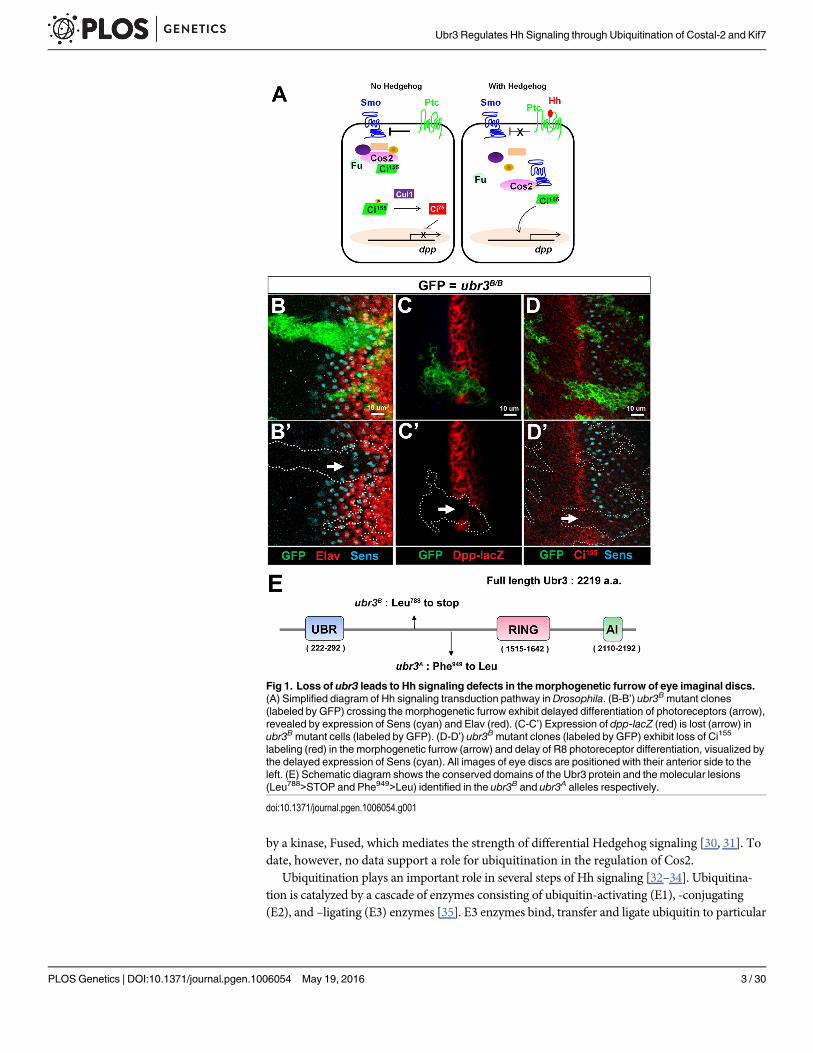
IntroductionHedgehog (Hh) signaling regulates numerous developmental processes and is implicated inmultiple cancers, wound healing and pain sensation in adults [1–3]. The Hh ligand acts as amorphogen to induce differential cell responses based on distinct activity thresholds of its sig-naling transduction cascade [4–6]. Mis-regulation of Hh signaling affects cell specification andproliferation during development and causes several types of cancer such as glioblastoma orbasal cell carcinoma [7, 8]. In the absence of Hh, the receptor Patched (Ptc) inhibits the G-pro-tein coupled receptor Smoothened (Smo) [9]. Inhibition of Smo promotes the assembly of anantagonistic molecular complex composed of Costal 2 (Cos2), a kinesin-related motor protein,Cubitus interruptus (Ci), the key transcriptional effector of Hh [10, 11], and several proteinkinases [12]. This complex phosphorylates the full length, transcriptionally active form of Ci,Ci155. Phosphorylated Ci155 is ubiquitinated by a SCF (Skp1-Cullin1(Cul1)-F-box) E3 ligasecomplex [13] and partially cleaved to generate a transcriptional repressor form, Ci75, whichleads to the transcriptional silencing of Hh target genes [14, 15]. The Hh signaling cascade isactivated by the binding of Hh to Ptc and Ihog (Interference hedgehog) [16], resulting in therelease of Smo inhibition. Activated Smo can interact physically with Cos2 [17–20]. This inter-action prevents the formation of the Hh signaling antagonistic complex and cleavage of Ci155.As a result, levels of Ci155 increase in the cytoplasm, promoting its translocation to the nucleusand the transcription of downstream target genes such as decapentaplegic (dpp) or ptc (Fig 1A).
Previous studies have shown that Cos2 is a key modulator of Hh signaling, and that it facili-tates kinase-mediated phosphorylation of Ci and promotes partial degradation of Ci [21]. Lossof Cos2 leads to ectopic activation of Hh signaling and pattern duplications in the Drosophilawing [11], whereas over-expression of Cos2 inhibits Hh signaling [22], suggesting that Cos2 isboth necessary and sufficient for Hh signaling. In vertebrates, the core components of Hh sig-naling are conserved, including Cos2. Cos2 has two vertebrate orthologs, Kif7 and Kif27 [23,24]. Kif7 has been proposed to function similarly to Cos2, because Kif7 knockout mice and zeb-rafish mutants show an up-regulation of Sonic Hedgehog (Shh) signaling [25–27]. In addition,Kif7 can interact physically and modulate the activity of the GLI transcription factors, themammalian homologs of Ci [27, 28]. Moreover, Cos2 can functionally replace Kif7 [27], dem-onstrating a molecular conservation between vertebrate and invertebrate homologues. Inhumans, patients carrying KIF7 allelic variants display a spectrum of phenotypic severity rang-ing from hydrolethalus or Acrocallosal syndromes to Meckel and Joubert syndromes [28, 29].Hence, proper function of Kif7 activity is essential for correct Hh signal transduction and islikely to be regulated tightly. Previous studies have shown that Cos2 (Kif7) is phosphorylated
Ubr3 Regulates Hh Signaling through Ubiquitination of Costal-2 and Kif7
PLOS Genetics | DOI:10.1371/journal.pgen.1006054 May 19, 2016 2 / 30
cancer.gov and Breast Cancer SPORE P50 CA50183http://trp.cancer.gov/spores/breast. AKG wassupported by National Institutes of Health DC010987nidcd.nih.gov. MW was supported by NationalInstitute of Child Health and Human DevelopmentP01 HD22486 https://www.nichd.nih. and NationalInstitute on Deafness and Other CommunicationDisorders R01 DC004186 http://www.nidcd.nih.gov.HJB was supported by Howard Hughes MedicalInstitute http://www.hhmi.org/scientists/hugo-j-bellen,Friedreich's Ataxia Research Alliance http://www.curefa.org/index.php and Target ALS http://www.targetals.org/index.html. The funders had no role instudy design, data collection and analysis, decision topublish, or preparation of the manuscript.
Competing Interests: The authors have declaredthat no competing interests exist.
by a kinase, Fused, which mediates the strength of differential Hedgehog signaling [30, 31]. Todate, however, no data support a role for ubiquitination in the regulation of Cos2.
Ubiquitination plays an important role in several steps of Hh signaling [32–34]. Ubiquitina-tion is catalyzed by a cascade of enzymes consisting of ubiquitin-activating (E1), -conjugating(E2), and –ligating (E3) enzymes [35]. E3 enzymes bind, transfer and ligate ubiquitin to particular
Fig 1. Loss of ubr3 leads to Hh signaling defects in the morphogenetic furrow of eye imaginal discs.(A) Simplified diagram of Hh signaling transduction pathway in Drosophila. (B-B’) ubr3B mutant clones(labeled by GFP) crossing the morphogenetic furrow exhibit delayed differentiation of photoreceptors (arrow),revealed by expression of Sens (cyan) and Elav (red). (C-C’) Expression of dpp-lacZ (red) is lost (arrow) inubr3Bmutant cells (labeled by GFP). (D-D’) ubr3B mutant clones (labeled by GFP) exhibit loss of Ci155
labeling (red) in the morphogenetic furrow (arrow) and delay of R8 photoreceptor differentiation, visualized bythe delayed expression of Sens (cyan). All images of eye discs are positioned with their anterior side to theleft. (E) Schematic diagram shows the conserved domains of the Ubr3 protein and the molecular lesions(Leu788>STOP and Phe949>Leu) identified in the ubr3B and ubr3A alleles respectively.
doi:10.1371/journal.pgen.1006054.g001
Ubr3 Regulates Hh Signaling through Ubiquitination of Costal-2 and Kif7
PLOS Genetics | DOI:10.1371/journal.pgen.1006054 May 19, 2016 3 / 30
substrates. The two major types of E3 ligase are the Really Interesting New Gene (RING) domainE3s and the Homologous to E6AP Carboxyl Terminus (HECT) domain E3s [36].
We describe the identification and characterization of Ubr3, a novel regulator of Hh signal-ing. Ubr3 belongs to the UBR protein superfamily, characterized by a 70-residue zinc fingerdomain UBR box [37]. Recent studies showed that Ubr3 can polyubiquitinate target proteins[38] involved in multiple biological processes, including olfactory organ function in mice [39],denticle patterning in Drosophila [40], DNA damage repair in yeast [38], apoptosis in flies[41], homoeostasis in the heart [42], and breast cancer [43].
Here we show that Ubr3 promotes Hh signaling by mediating the ubiquitination and degra-dation of Cos2/Kif7. Loss of Ubr3 elevates the levels of Cos2, resulting in a decrease in Ci155
and transcriptional silencing of Hh target genes. Loss of ubr3 in flies and zebrafish affects eyedevelopment, as well as neuronal specification and somite development in zebrafish. Ubr3 reg-ulates the ubiquitination and degradation of Kif7 in mammalian cells, and transcription of theShh target ptch2 is strongly decreased in the retina of ubr3mutant zebrafish. Taken together,our data suggest that Ubr3 is an evolutionarily conserved, positive regulator of Hh signalingthat regulates Cos2/Kif7 ubiquitination and degradation.
ResultsLoss of ubr3 results in Hh signaling defects in DrosophilaTo identify novel components in developmental signaling pathways, we isolated mutations thataffect eye and/or wing morphogenesis in a mosaic forward genetic screen of approximately6000 X-linked lethal mutations inDrosophila [44–48]. We identified an essential complementa-tion group ubr3, consisting of two alleles (ubr3A and ubr3B). Both ubr3A and ubr3B hemizygousmutants die as 1st instar larvae. Homozygous mutant clones of both alleles cause delayed differ-entiation of photoreceptors in the morphogenetic furrow of eye imaginal discs (Fig 1B and S1AFig). This is revealed by the delayed expression of Senseless, an R8 photoreceptor marker [49,50] and Elav (Embryonic lethal abnormal vision), a marker for photoreceptors [51]. Sincedelayed differentiation of photoreceptors is observed when Hh signaling is lost [52], we hypoth-esized that ubr3mutations may impair Hh signaling. To assess the activation of Hh signaling inubr3mutant clones, we examined expression of a Hh reporter, dpp-lacZ [53] and the activeform of Ci, CiA. Both dpp-lacZ and Ci155 are lost in ubr3mutant clones in the morphogeneticfurrow (Fig 1C–1D’ and S1B Fig). We and others also noticed an increase in apoptosis in ubr3mutant cells [41]. To exclude the possibility that the Hh signaling defect in ubr3mutant cells isdue to apoptosis, we over-expressed the anti-apoptotic gene p35 in ubr3 clones. As shown inS1C Fig, the delayed differentiation of photoreceptors is not rescued although apoptosis is sup-pressed (S1D and S1E Fig). Hence, Hh signaling is impaired in ubr3mutant cells.
Alleles of ubr3map to a small deficiency that uncovers ~11 genes including ubr3 (CG42593)and l(1)G0193 (S1F Fig). Both alleles (ubr3A and ubr3B) fail to complement the lethality associ-ated with a P-element insertion in ubr3 (S1F and S1H Fig) [54]. ubr3B carries a Leu788>STOPand ubr3A carries a Phe949>Leu in ubr3 (Fig 1E). No mutations were found in l(1)G0193. Agenomic rescue construct rescued the lethality of both ubr3 alleles (S1F and S1H Fig), andover-expression of the ubr3 cDNA in ubr3Bmutant clones rescued the loss of Ci155 expressionin the morphogenetic furrow (S1G Fig). Together, these data show that ubr3 is required for Hhsignaling.
Ubr3 is a conserved E3 ligase and is expressed in Drosophila eye discsubr3 encodes a 2219 amino acid protein, the Drosophila homolog of the mammalian RING-type E3 ubiquitin ligase n-recognin 3 (UBR3) gene (Fig 1E). Most UBR superfamily member
Ubr3 Regulates Hh Signaling through Ubiquitination of Costal-2 and Kif7
PLOS Genetics | DOI:10.1371/journal.pgen.1006054 May 19, 2016 4 / 30
proteins, including UBR1, UBR2, UBR4 and UBR5, function in the N-end rule pathway, a ubi-quitin-dependent system where E3 ligases recognize N terminal residues of their targets anddegrade them [37]. However, UBR3 does not bind to known N-end rule substrates, suggestinga different molecular function of Ubr3 from N-end rule E3 ligases [55]. Ubr3 contains a UBRmoiety, a RING domain and a C-terminal auto-inhibitory (AI) domain (Fig 1E) [38, 39]. Allthree domains are highly conserved among fly, mouse and human (S1I Fig), suggesting thatthe molecular function of Ubr3 may be conserved.
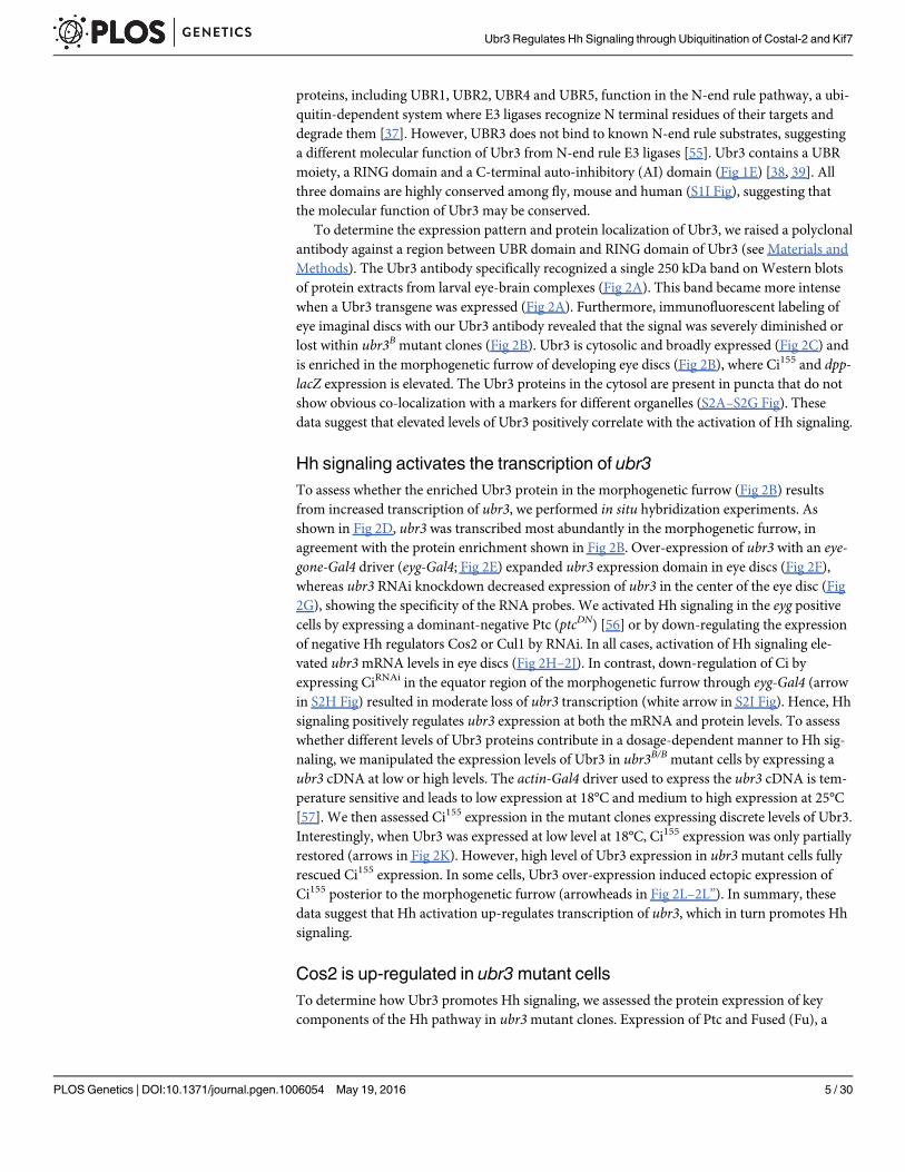
To determine the expression pattern and protein localization of Ubr3, we raised a polyclonalantibody against a region between UBR domain and RING domain of Ubr3 (see Materials andMethods). The Ubr3 antibody specifically recognized a single 250 kDa band onWestern blotsof protein extracts from larval eye-brain complexes (Fig 2A). This band became more intensewhen a Ubr3 transgene was expressed (Fig 2A). Furthermore, immunofluorescent labeling ofeye imaginal discs with our Ubr3 antibody revealed that the signal was severely diminished orlost within ubr3B mutant clones (Fig 2B). Ubr3 is cytosolic and broadly expressed (Fig 2C) andis enriched in the morphogenetic furrow of developing eye discs (Fig 2B), where Ci155 and dpp-lacZ expression is elevated. The Ubr3 proteins in the cytosol are present in puncta that do notshow obvious co-localization with a markers for different organelles (S2A–S2G Fig). Thesedata suggest that elevated levels of Ubr3 positively correlate with the activation of Hh signaling.
Hh signaling activates the transcription of ubr3To assess whether the enriched Ubr3 protein in the morphogenetic furrow (Fig 2B) resultsfrom increased transcription of ubr3, we performed in situ hybridization experiments. Asshown in Fig 2D, ubr3 was transcribed most abundantly in the morphogenetic furrow, inagreement with the protein enrichment shown in Fig 2B. Over-expression of ubr3 with an eye-gone-Gal4 driver (eyg-Gal4; Fig 2E) expanded ubr3 expression domain in eye discs (Fig 2F),whereas ubr3 RNAi knockdown decreased expression of ubr3 in the center of the eye disc (Fig2G), showing the specificity of the RNA probes. We activated Hh signaling in the eyg positivecells by expressing a dominant-negative Ptc (ptcDN) [56] or by down-regulating the expressionof negative Hh regulators Cos2 or Cul1 by RNAi. In all cases, activation of Hh signaling ele-vated ubr3mRNA levels in eye discs (Fig 2H–2J). In contrast, down-regulation of Ci byexpressing CiRNAi in the equator region of the morphogenetic furrow through eyg-Gal4 (arrowin S2H Fig) resulted in moderate loss of ubr3 transcription (white arrow in S2I Fig). Hence, Hhsignaling positively regulates ubr3 expression at both the mRNA and protein levels. To assesswhether different levels of Ubr3 proteins contribute in a dosage-dependent manner to Hh sig-naling, we manipulated the expression levels of Ubr3 in ubr3B/B mutant cells by expressing aubr3 cDNA at low or high levels. The actin-Gal4 driver used to express the ubr3 cDNA is tem-perature sensitive and leads to low expression at 18°C and medium to high expression at 25°C[57]. We then assessed Ci155 expression in the mutant clones expressing discrete levels of Ubr3.Interestingly, when Ubr3 was expressed at low level at 18°C, Ci155 expression was only partiallyrestored (arrows in Fig 2K). However, high level of Ubr3 expression in ubr3mutant cells fullyrescued Ci155 expression. In some cells, Ubr3 over-expression induced ectopic expression ofCi155 posterior to the morphogenetic furrow (arrowheads in Fig 2L–2L”). In summary, thesedata suggest that Hh activation up-regulates transcription of ubr3, which in turn promotes Hhsignaling.
Cos2 is up-regulated in ubr3mutant cellsTo determine how Ubr3 promotes Hh signaling, we assessed the protein expression of keycomponents of the Hh pathway in ubr3mutant clones. Expression of Ptc and Fused (Fu), a
Ubr3 Regulates Hh Signaling through Ubiquitination of Costal-2 and Kif7
PLOS Genetics | DOI:10.1371/journal.pgen.1006054 May 19, 2016 5 / 30
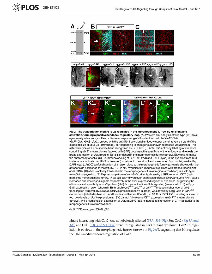
kinase interacting with Cos2, was not obviously affected (S3A–S3B’ Fig), but Cos2 (Fig 3A and3A’) and Cul1 (S3C and S3C’ Fig) were up-regulated in ubr3mutant eye clones. Cos2 up-regu-lation is obvious in the morphogenetic furrow (arrows in Fig 3A’), suggesting that Hh regulatesthe Ubr3-mediated down-regulation of Cos2.
Fig 2. The transcription of ubr3 is up-regulated in the morphogenetic furrow by Hh signalingactivation, forming a positive feedback regulatory loop. (A) Western blot analysis of wild-type (wt) larvaleye-brain lysates from y w flies or flies over-expressing ubr3 under the control ofGMR-Gal4(GMR-Gal4>UAS-Ubr3), probed with the anti-Ubr3 polyclonal antibody (upper panel) reveals a band of theexpected size of 250kDa (arrowhead), corresponding to endogenous or over-expressed Ubr3 protein. Theasterisk indicates a non-specific band recognized by GP-Ubr3. (B) Anti-Ubr3 antibody labeling of eye discscontaining ubr3Bmutant clones (labeled with GFP) document the specificity of the antibody, and reveals thebroad expression of Ubr3 protein. Ubr3 is enriched in the morphogenetic furrow (arrow). Elav (cyan) marksthe photoreceptor cells. (C) Co-immunolabeling of GP-Ubr3 (red) and DAPI (cyan) in the eye disc from thirdinstar larvae indicate that Ubr3 protein (red) localizes to the cytosol and is excluded from nuclei, marked byDAPI (cyan). An XZ confocal section of a region close to the morphogenetic furrow (arrow) is shown, with theanterior side positioned to the left. (D, F-J) In situ hybridization images of eye discs with probes recognizingubr3 cDNA. (D) ubr3 is actively transcribed in the morphogenetic furrow region (arrowhead) in a wild-type(eyg-Gal4/+) eye disc. (E) Expression pattern of eyg-Gal4 driver is shown by a GFP reporter. Ci155 (red)marks the morphogenetic furrow. (F-G) eyg-Gal4 driven over-expression of ubr3 cDNA and ubr3RNAi causeincreased and decreased signals respectively in the over-expressed regions of eye discs, suggesting theefficiency and specificity of ubr3 probes. (H-J) Ectopic activation of Hh signaling (arrows in H to J) in Eyg-Gal4 expressing region (shown in E) through cos2RNAi, ptcDN or cul1RNAi induces higher level of ubr3transcription (arrows). (K, L) ubr3 cDNA expression (shown in green) was driven by actin-Gal4 in ubr3B/B
clones cells (labeled in blue in K and L or dashed lines in K’ and L’) at 18°C or 25°C. Ci155 labeling is shown inred. Low levels of Ubr3 expression at 18°C cannot fully rescue Ci155 expression in ubr3B/Bmutant clones(arrows), while high levels of expression of Ubr3 at 25°C lead to increased expression of Ci155 posterior to themorphogenetic furrow (arrowheads).
doi:10.1371/journal.pgen.1006054.g002
Ubr3 Regulates Hh Signaling through Ubiquitination of Costal-2 and Kif7
PLOS Genetics | DOI:10.1371/journal.pgen.1006054 May 19, 2016 6 / 30
Fig 3. Up-regulation of Cos2 in ubr3mutant clones is responsible for the loss of Hh signaling. (A-A’)ubr3Bmutant cells (marked by GFP) up-regulate Cos2 (red; arrows). (B-B’) Over-expression of Cos2 by eyg-Gal4 (indicated by expression of GFP) leads to loss of Ci155 (arrow in B’) at the morphogenetic furrow. (C-C’)Over-expression of Cul1 by eyg-Gal4 (indicated by expression of GFP) does not cause loss of Ci155 at themorphogenetic furrow. (D-D’) Down-regulation of Cos2 by over-expression of cos2RNAi in ubr3Bmutantclones (marked by the expression of GFP) suppresses loss of Ci155 (red) in the morphogenetic furrow (arrowsin D’). (E-E’) Down-regulation of Cul1 by over-expression of cul1RNAi in ubr3Bmutant clones induces ectopicactivation of Hh signaling anterior to the morphogenetic furrow (shown by expression of Ci155, arrow) butdoes not rescue Ci155 loss in the morphogenetic furrow (arrowheads). (F-F’) Over-expression of ptcDN inubr3Bmutant clones does not rescue Ci155 loss in the morphogenetic furrow (arrowhead).
doi:10.1371/journal.pgen.1006054.g003
Ubr3 Regulates Hh Signaling through Ubiquitination of Costal-2 and Kif7
PLOS Genetics | DOI:10.1371/journal.pgen.1006054 May 19, 2016 7 / 30
Cos2 and Cul1 are both negative regulators of Hh signaling and loss of function of eithergene causes ectopic activation of Hh signaling in eye discs [11, 13, 58]. Because both genes areup-regulated in cells lacking Ubr3, we tested whether over-expression of either gene is sufficientto phenocopy the ubr3mutation. Over-expression of Cos2, but not Cul1, results in loss of Ci155
in the morphogenetic furrow, similar to ubr3mutants (Fig 3B–3C’). Labeling with a Cos2 anti-body showed that a subtle increase of Cos2 is sufficient to inhibit Ci155 expression (S3D andS3D’ Fig), implicating that Cos2 up-regulation in ubr3mutant cells is relevant. Hence, up-regu-lation of Cos2, but not Cul1, is likely to be responsible for the Hh signaling defects observed inubr3mutants. This hypothesis is supported by the observation that reducing Cos2 protein levelsin ubr3mutant clones through cos2RNAi restored Ci155 levels and suppressed the morphogeneticfurrow defects (arrows in Fig 3D and 3D’ and S3E and S3E’ Fig). In contrast, over-expression ofcul1RNAi in ubr3mutant clones did not restore Ci155 expression in the morphogenetic furrow(arrowheads in Fig 3E and 3E’), suggesting that Cul1 up-regulation was not the cause of Ci155
loss. One likely reason why Ci155 expression is not restored by Cul1 RNAi in ubr3mutant clonesin the morphogenetic furrow is that Cul1 RNAi does not completely remove Cul1 in ubr3 clonesand the residual Cul1-Slimb E3 ligase activity may suffice to mediate processing of Ci155. More-over, expression of ptcDN in ubr3mutant clones did not rescue Ci155 loss (arrowhead in Fig 3Fand 3F’). These data show that loss of ubr3 causes a decrease in Hh signaling and a reduction inCi155 that can be restored by Cos2 down-regulation. Hence, ubr3 acts to attenuate the levels ofCos2, which enhances the activity of Hh signaling in the morphogenetic furrow.
Ubr3 possesses Ubiquitin E3 ligase activity and ubiquitinates itselfThe RING domain of Ubr3 is not a canonical RING domain (Fig 4A and 4A’) [59]. To assesswhether Ubr3 has E3 ligase activity, we performed an in vitro ubiquitination assay. Immuno-precipitation-purified Ubr3::GFP fusion proteins were incubated with E1 and E2 enzymes(HR6A) [39] and Flag-tagged Ubiquitin (Flag::Ub) peptides. Interestingly, Ubr3 poly-ubiquiti-nated itself, as shown in Fig 4B. Moreover, the UBR domain fragment may form a dimer whenover-expressed, because a band of twice the molecular weight of GFP::UBR (~80 kDa) isdetected (Fig 4B). Co-immunoprecipitation assays with the over-expressed UBR domain indi-cated that it interacts with the Ubr3 full-length protein present in whole cell extracts of S2 cells(Fig 4C). This suggests that Ubr3 interacts with the UBR domain of another Ubr3 moleculeand that Ubr3 proteins poly-ubiquitinate each other.
Ubr3 binds to and poly-ubiquitinates Cos2To test whether the up-regulation of Cos2 in ubr3mutant cells is due to defective degradationby the proteasome, we performed a degradation assay of Cos2 in Drosophila S2 cells. We founddegradation of Cos2 proteins begins 6 hours after treatment with a translational inhibitorcycloheximide (CHX) and that the level of Cos2 decreased to 10% after 10 hours of treatment(Fig 5A). Addition of the proteasomal inhibitor MG132 suppressed the degradation of Cos2(Fig 5A), suggesting that Cos2 proteins are degraded via the proteasome. The degradation ofCos2 is partially suppressed by down-regulation of Ubr3 by Ubr3RNAi and promoted by over-expression of Ubr3 (Fig 5B), suggesting that Ubr3 mediates the degradation of Cos2.
Because ubiquitination is known to regulate protein abundance through proteasome-medi-ated degradation, Cos2 levels may be regulated via Ubr3-mediated ubiquitination. To deter-mine whether Ubr3 interacts physically with Cos2 and to map which domains are required forthis interaction, we performed co-immunoprecipitation assays. As shown in Fig 5C, both theUBR domain fragment and the full length Ubr3 protein interact with Cos2 (lane 2 and lane 3).To exclude the possibility that Cos2 binds to Ubr3 indirectly via microtubules, we treated S2
Ubr3 Regulates Hh Signaling through Ubiquitination of Costal-2 and Kif7
PLOS Genetics | DOI:10.1371/journal.pgen.1006054 May 19, 2016 8 / 30
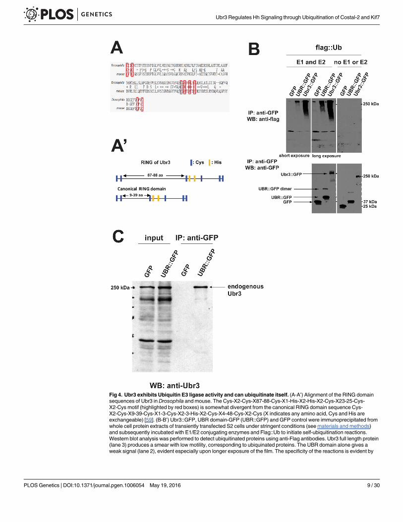
Fig 4. Ubr3 exhibits Ubiquitin E3 ligase activity and can ubiquitinate itself. (A-A’) Alignment of the RING domainsequences of Ubr3 in Drosophila and mouse. The Cys-X2-Cys-X87-88-Cys-X1-His-X2-His-X2-Cys-X23-25-Cys-X2-Cys motif (highlighted by red boxes) is somewhat divergent from the canonical RING domain sequence Cys-X2-Cys-X9-39-Cys-X1-3-Cys-X2-3-His-X2-Cys-X4-48-Cys-X2-Cys (X indicates any amino acid, Cys and His areexchangeable) [59]. (B-B’) Ubr3::GFP, UBR domain-GFP (UBR::GFP) and GFP control were immunoprecipitated fromwhole cell protein extracts of transiently transfected S2 cells under stringent conditions (see materials and methods)and subsequently incubated with E1/E2 conjugating enzymes and Flag::Ub to initiate self-ubiquitination reactions.Western blot analysis was performed to detect ubiquitinated proteins using anti-Flag antibodies. Ubr3 full length protein(lane 3) produces a smear with low motility, corresponding to ubiquinated proteins. The UBR domain alone gives aweak signal (lane 2), evident especially upon longer exposure of the film. The specificity of the reactions is evident by
Ubr3 Regulates Hh Signaling through Ubiquitination of Costal-2 and Kif7
PLOS Genetics | DOI:10.1371/journal.pgen.1006054 May 19, 2016 9 / 30
cells with the microtubule-destabilizing agent Colchicine. The Cos2-Ubr3 interaction is notaffected by Colchicine treatment (lane 4 in Fig 5C), suggesting that Cos2 does not bind Ubr3via microtubules. To identify which domain of Cos2 is critical for the interaction with Ubr3,we tested a series of deletion constructs of Cos2 (Fig 5D) in co-IP assays with the UBR domain.We found that only the fragments bearing the N-terminal motor domain (MD) of Cos2(Cos2ΔC1, ΔC2, and ΔC3) interacted with the UBR domain (Fig 5E). Hence, Ubr3 binds to theN-terminal MD of Cos2 with its UBR domain.
To detect the ubiquitination of Cos2, we performed immunoprecipitation assays and exam-ined the ubiquitination of Cos2 in S2 cells that express ubr3 (Fig 4C). As shown in Fig 5F, theubiquitinated Myc-tagged Cos2 (Myc::Cos2) was detected by an anti-hemagglutinin (HA) anti-body in S2 cells co-transfected with an HA-tagged ubiquitin construct (HA::Ub; Fig 5F, lane 1,top panel). In addition, the HA signal exhibited a lower mobility shift compared to the majorband detected by anti-Myc antibody (Fig 5F), indicating that these bands correspond to theubiquitinated forms of Cos2. Over-expression of an E3 ligase dead form of Ubr3, in which theresidues required for RING domain activity (Fig 4A) were mutated to alanines, did not causean increase in ubiquitination of Cos2 (Fig 5G, lane 1–3), suggesting that the E3 ligase activityof Ubr3 mediates the ubiquitination of Cos2. In addition, removing the Ubr3 binding domainof Cos2, Cos2ΔN1, abolished most of the ubiquitination of full length Cos2 (Fig 5G, lane 4–6).The residual ubiquitination of Cos2ΔN1 may result from endogenous full length Cos2 that co-precipitates with Cos2ΔN1 through dimerization [10, 11].
To determine whether Ubr3 regulates Cos2 ubiquitination, we examined the levels of Cos2ubiquitination when Ubr3 was either over-expressed or knocked down by RNAi. As shown inFig 5F, the co-expression of Ubr3 with Cos2 increased Cos2 ubiquitination, whereas inactiva-tion of Ubr3 by RNAi decreased ubiquitination (lane 2 and lane 3, top panel). A control GFPRNAi (negative control) did not significantly change the level of Cos2 ubiquitination (lane 4,top panel). These results suggest that Ubr3 interacts with and ubiquitinates Cos2.
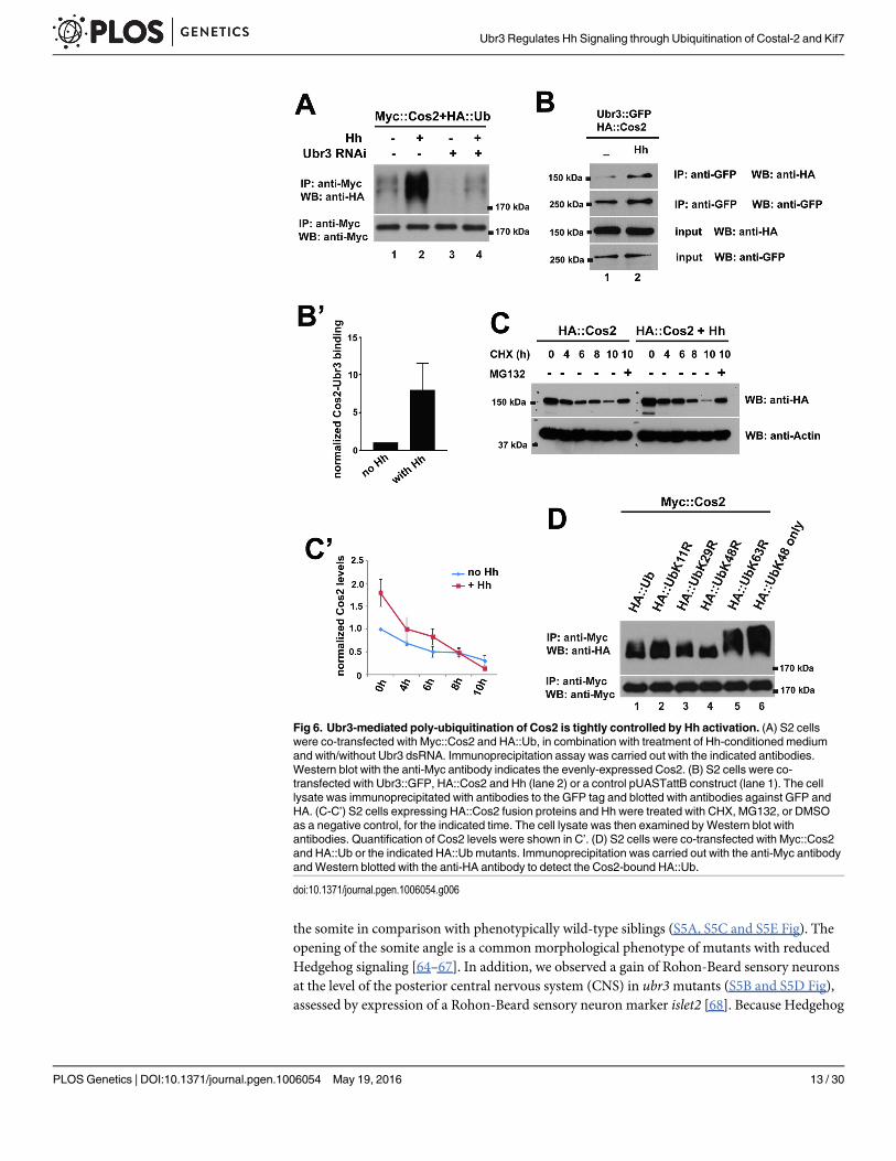
Hh signaling regulates Cos2 ubiquitination by Ubr3We next tested whether Hh signaling regulates the ubiquitination of Cos2. Interestingly, wefound that the ubiquitination of Cos2 was strongly enhanced by Hh treatment (Fig 6A, lane 2).This increased ubiquitination was abolished by down-regulation of Ubr3 (Fig 6A, lane 4), sug-gesting that Ubr3 mediates Hh induced ubiquitination of Cos2. This implied that Ubr3-me-diated ubiquitination of Cos2 was tightly controlled by Hh signaling. Because Ci is notexpressed in S2 cells, Hh-induced ubiquitination of Cos2 cannot be mediated by a positive,transcriptional feedback loop that depends on Ci. We therefore tested whether Hh may pro-mote binding of Ubr3 to Cos2. We performed co-IP assays between the Ubr3 and Cos2 in thepresence or absence of Hh. As shown in Fig 6B, the interactions between Ubr3 full-length pro-tein and Cos2 (lanes 1 and 2 in Fig 6B) were strongly increased by Hh. These data show thatHh induces the ubiquitination of Cos2 by promoting the association of Ubr3 with Cos2. Con-sistent with Hh-induced poly-ubiquitination of Cos2, we also observed a faster degradation ofCos2 upon Hh treatment (Fig 6C and 6C’).
the absence of signal in GFP control samples (lane 1) or upon omitting E1 and E2 enzymes. The membranes werestripped and re-probed with anti-GFP antibodies to ensure the presence of the UBR::GFP or GFP. An additional bandof ~80kDa is observed in UBR::GFP samples, which is approximately twice the size of UBR::GFP (arrow). (C) UBRdomain interacts with endogenous Ubr3. UBR::GFP or GFP alone was immunoprecipitated from lysates of transientlytransfected S2 cells using anti-GFP beads. Western blot was then performed with anti-Ubr3 antibody. Over-expressedUBR::GFP (lane 4), but not GFP alone (lane 3), co-immunoprecipitates with endogenous Ubr3 full length protein.
doi:10.1371/journal.pgen.1006054.g004
Ubr3 Regulates Hh Signaling through Ubiquitination of Costal-2 and Kif7
PLOS Genetics | DOI:10.1371/journal.pgen.1006054 May 19, 2016 10 / 30
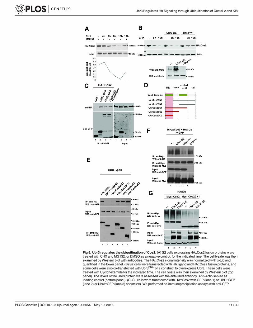
Fig 5. Ubr3 regulates the ubiquitination of Cos2. (A) S2 cells expressing HA::Cos2 fusion proteins weretreated with CHX and MG132, or DMSO as a negative control, for the indicated time. The cell lysate was thenexamined byWestern blot with antibodies. The HA::Cos2 signal intensity was normalized with α-tub andquantified in the lower panel. (B) S2 cells were transfected with Hh ligand and HA::Cos2 fusion proteins, andsome cells were also co-transfected with Ubr3RNAi or a construct to overexpress Ubr3. These cells weretreated with Cyclohexamide for the indicated time. The cell lysate was then examined byWestern blot (toppanel). The levels of the Ubr3 protein were assessed with the anti-Ubr3 antibody. Anti-Actin served asloading control (bottom panel). (C) S2 cells were transfected with HA::Cos2 with GFP (lane 1) or UBR::GFP(lane 2) or Ubr3::GFP (lane 3) constructs. We performed co-immunoprecipitation assays with anti-GFP
Ubr3 Regulates Hh Signaling through Ubiquitination of Costal-2 and Kif7
PLOS Genetics | DOI:10.1371/journal.pgen.1006054 May 19, 2016 11 / 30
The ladder pattern of the HA signal in Fig 5E suggests that Cos2 is poly-ubiquitinated. Wefurther determined the ubiquitination chain pattern by using a panel of ubiquitin mutant con-structs [34]. Compared to wild-type ubiquitin (Fig 6D, lane 1, top panel), a mutated lysine 48in ubiquitin (HA::UbK48R) abolished the formation of the ubiquitin chain (lane 4, top panel),whereas altered lysine 11 (K11R), lysine 29 (K29R), or lysine 63 (K63R) did not affect chainformation. In addition, mutating all of the lysine residues except lysine 48 (HA::UbK48 only)leads to longer ubiquitination chains (Fig 6D, lane 6, top panel). The single sharp band of Cos2ubiquitination by K48R indicates a mono-ubiquitinated Cos2 that cannot be further elongateddue to the lack of K48. Together, these data indicate that Cos2 undergoes K48-linked poly-ubiquitination.
Ubr3 positively regulates Shh signaling in zebrafishTo determine whether Ubr3 plays a conserved function in vertebrates, we created two indepen-dent zebrafish ubr3mutant alleles using CRISPR/Cas9. The ubr3 gene is predicted to encode aprotein of 1808 amino acids, and the ubr3b1250 allele lacks 28 nucleotides (Del 378–405) down-stream of the predicted ATG (S4A Fig) leading to a frameshift and early stop codon. Themutant protein should encode only 129 amino acids (S4C Fig), lacking the UBR and RINGdomains. The second allele, ubr3b1251 carries a 4 nt insertion at position 220 (S4B Fig), alsocausing a frameshift and early stop codon. ubr3b1251 is predicted to encode a 78 aa protein lack-ing all functional domains (S4C Fig). Using an anti-Ubr3 antibody, we detected expression ofUbr3 in the developing retina, central nervous system and trunk, which are lost in ubr3b1250/b1251 mutant zebrafish (Fig 7A and 7B). Three independent crosses between single carriers het-erozygous for the b1250 and b1251 alleles resulted in progeny with a distinguishable and repro-ducible retinal phenotype in a Mendelian frequency (f = 0.22, f = 0.27, f = 0.23, n = 270). At the5-6-somites stage, phenotypically wild-type siblings display optic vesicles characterized by acompacted and stratified epithelium (Fig 7C, 7E and 7G). The optic vesicles of the ubr3 trans-heterozygous mutants failed to form a cohesive and stratified epithelium (Fig 7D, 7F and 7H).Because appropriate levels of Sonic Hedgehog (Shh) signaling are essential for eye morphogen-esis [60, 61], we examined the transcriptional levels of ptch2. In zebrafish, ptch2 is a direct tar-get of Shh signaling [62, 63]. In wild-type embryos, a gradient of ptch2 expression wasobserved within the optic vesicle (dotted area in Fig 7G and 7I). This gradient was character-ized by high levels of ptch2-expressing cells localized in the ventral border of the vesicle, andlow level expressing cells localized in the dorsal border region and vesicle core (Fig 7I). In ubr3mutants, ptch2 expression was strongly decreased (Fig 7H and 7J). Consistent with decreasedShh signaling, ubr3b1250/b1251 trans-heterozygous mutants show a 30% increase in the angle of
agarose followed by western blot assays on the cell lysates. Both UBR domain fragments (lane 2) and theUbr3 full length protein (lane 3) co-precipitate with Cos2, whereas GFP (lane 1) does not. S2 cells co-transfected with HA::Cos2 and Ubr3::GFP were treated with Colchicine for 5 hours prior to harvest. Co-immunoprecipitation assay with anti-GFP agarose beads and western blots were performed from the celllysate. (D) Schematic diagram of Cos2 deletion constructs. (E) Co-immunoprecipitation assays with lysatesfrom S2 cells, transfected with UBR::GFP and the constructs indicated in C reveal that the UBR domain ofUbr3 interacts physically with full length Cos2 and Cos2ΔC1-ΔC3 (red bars in C, lanes 1, 4–6 in D), but notwith Cos2ΔN1-ΔN2 (gray bars in B, lanes 2, 3 in D). (F) S2 cells were co-transfected with Myc::Cos2 and HA::Ub, in combination with Ubr3, Ubr3RNAi or GFPRNAi. Following immunoprecipitation andWestern blot analysiswith anti-HA (upper panel), we found that Cos2 is ubiquitinated, as indicated by the smeary shift of the protein(lane 1). Over-expression of Ubr3 increases ubiquitinated Cos2 (lane 2), whereas down-regulation of Ubr3through Ubr3 RNAi (lane 3) decreases ubiquitinated Cos2. GFPRNAi was used as a negative control. Theasterisks indicate ubiquitinated Cos2 which exhibits a higher molecular weight.(G) S2 cells were co-transfected with Myc::Cos2 or Myc::Cos2ΔN1, and HA::Ub, in combination with wild type Ubr3 or E3 deadform of Ubr3 (Ubr3D), following immunoprecipitation andWestern blot analysis.
doi:10.1371/journal.pgen.1006054.g005
Ubr3 Regulates Hh Signaling through Ubiquitination of Costal-2 and Kif7
PLOS Genetics | DOI:10.1371/journal.pgen.1006054 May 19, 2016 12 / 30
the somite in comparison with phenotypically wild-type siblings (S5A, S5C and S5E Fig). Theopening of the somite angle is a common morphological phenotype of mutants with reducedHedgehog signaling [64–67]. In addition, we observed a gain of Rohon-Beard sensory neuronsat the level of the posterior central nervous system (CNS) in ubr3mutants (S5B and S5D Fig),assessed by expression of a Rohon-Beard sensory neuron marker islet2 [68]. Because Hedgehog
Fig 6. Ubr3-mediated poly-ubiquitination of Cos2 is tightly controlled by Hh activation. (A) S2 cellswere co-transfected with Myc::Cos2 and HA::Ub, in combination with treatment of Hh-conditioned mediumand with/without Ubr3 dsRNA. Immunoprecipitation assay was carried out with the indicated antibodies.Western blot with the anti-Myc antibody indicates the evenly-expressed Cos2. (B) S2 cells were co-transfected with Ubr3::GFP, HA::Cos2 and Hh (lane 2) or a control pUASTattB construct (lane 1). The celllysate was immunoprecipitated with antibodies to the GFP tag and blotted with antibodies against GFP andHA. (C-C’) S2 cells expressing HA::Cos2 fusion proteins and Hh were treated with CHX, MG132, or DMSOas a negative control, for the indicated time. The cell lysate was then examined by Western blot withantibodies. Quantification of Cos2 levels were shown in C’. (D) S2 cells were co-transfected with Myc::Cos2and HA::Ub or the indicated HA::Ub mutants. Immunoprecipitation was carried out with the anti-Myc antibodyandWestern blotted with the anti-HA antibody to detect the Cos2-bound HA::Ub.
doi:10.1371/journal.pgen.1006054.g006
Ubr3 Regulates Hh Signaling through Ubiquitination of Costal-2 and Kif7
PLOS Genetics | DOI:10.1371/journal.pgen.1006054 May 19, 2016 13 / 30
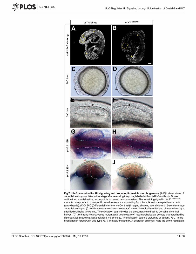
Fig 7. Ubr3 is required for Hh signaling and proper optic vesicle morphogenesis. (A-B) Lateral views ofzebrafish embryos at 18-somites stage after removing the yolks, labeled with anti-Ubr3 antibody. Boxesoutline the zebrafish retina, arrow points to central nervous system. The remaining signal in ubr3b1250/b1251
mutant corresponds to non-specific autofluorescence emanating from the yolk and some peridermal cells(arrowheads). (C-D) DIC (Differential Interference Contrast) imaging showing lateral views of 6-somites stagezebrafish embryos. (C) Wild-type optic vesicle (arrowheads) is morphologically visible and characterized by astratified epithelial thickening. The cavitation seam divides the presumptive retina into dorsal and ventralhalves. (D) ubr3 trans-heterozygous mutant optic vesicle (arrow) has morphological defects characterized bydisorganized tissue that lacks epithelial morphology. The cavitation seam is disrupted or absent. (G-J) In situhybridization for ptch2 in wild-type (G, I) and ubr3mutant (H, J) zebrafish embryos. Note the down-regulation
Ubr3 Regulates Hh Signaling through Ubiquitination of Costal-2 and Kif7
PLOS Genetics | DOI:10.1371/journal.pgen.1006054 May 19, 2016 14 / 30
restricts CNS dorsal fate acquisition [69], this result supports the interpretation that Hedgehogsignaling is decreased in ubr3mutants. This finding is also consistent with our observation ofdecreased retinal ptch2 expression in the absence of ubr3 (Fig 7H and 7J). Because Kif7-depleted zebrafish embryos do not show de-repression of Hh target genes in the CNS [27], ourfindings further suggest that, at least in zebrafish, Ubr3 may regulate not only Kif7 but alsoother intracellular negative regulators of Hedgehog signaling in the CNS. Different zebrafishHh signaling mutants show distinct degrees of severity, highlighting the tissue-specific require-ments of Hh levels during development [60, 70–74]. Similarly, loss of ubr3 does not result incyclopia or inner ear defects, showing that these mutants have a less severe phenotype whencompared to smoothenedmutant animals. Hence, ubr3 zebrafish mutants retain some residualHh signaling. Thus, our data show that Ubr3 positively regulates Hedgehog signaling in tissuessensitive to high levels of Hh like the mesoderm and neuroectoderm. In addition, the transcrip-tion of ubr3 is strongly reduced in smohi1640-/- mutant animals [70], which lose Shh activity(S5F and S5H Fig). In contrast, ectopic activation of the Shh pathway by injection of themRNA encoding a dominant negative form of PKA (dnPKA) [75] expands the expressiondomain of Ubr3 (S5G and S5I Fig). These data suggest that Shh signaling promotes the tran-scription of ubr3 in zebrafish, similar to what we observed in Drosophila. In summary, Ubr3 isrequired for the transduction of Hh signaling and proper eye morphogenesis in zebrafish.
UBR3 negatively regulates protein level of Kif7 through poly-ubiquitination in mammalian cellsTo test whether UBR3 also plays a role in Shh signaling in mammals, we used C3H10T1/2mouse mesenchymal cells. These cells respond to Shh and activate Shh target genes [76]. Wefirst confirmed that Ubr3 is expressed in C3H10T1/2 cells by RT-PCR (see Fig 8B’). We theninfected these cells with a lentivirus bearing 7 tandem binding sites for Gli (the vertebrate homo-logue of Ci) that control the expression of a GFP reporter. Addition of either the Shh ligand orpurmorphamine, an agonist of Smo [77], to C3H10T1/2 cells induced GFP expression in about25% of the cells (Fig 8A and 8A’). To determine whether knockdown of UBR3 impairs Shh sig-naling, we measured the proportion of GFP-expressing C3H10T1/2 cells transfected with one offour different siRNAs against UBR3 or a scrambled siRNA control, followed by purmorpha-mine treatment. Induction of the Gli::GFP reporter by purmorphamine was suppressed whensiRNA reduced the UBR3 levels (Fig 8B), as judged by real time PCR (Fig 8B’). In addition,down-regulation of UBR3 resulted in up-regulation of Kif7 (Fig 8C), the mammalian homologof Cos2. To assess poly-ubiquitination of Kif7, we purified Kif7 through immunoprecipitationand loaded theWestern blot lanes with equal amounts of protein (unlike in Fig 8C where weloaded equal amounts of cells). We observed decreased poly-ubiquitination of Kif7 upon knock-down of UBR3 (Fig 8D). These data indicate that UBR3 regulates Shh signaling through poly-ubiquitination of Kif7 in vertebrate cells, a process that seems to be evolutionarily conserved.
DiscussionNumerous studies have shown that Cos2 plays a central role in Hh signaling [10, 11, 22, 25, 26,78, 79]. Cos2 is both necessary and sufficient to regulate Ci [11, 22] and the level of Cos2 pro-tein is critical for activating Hh signaling [80, 81]. Here, we identified Ubr3 as a novel regulator
of ptch2 in ubr3mutant optic vesicles (dotted area). Dorsal views of flat-mounted zebrafish embryos. (G, H)Cross sections through the optic vesicle of embryos shown in G and H, respectively. All the scale barsrepresent 50μm.
doi:10.1371/journal.pgen.1006054.g007
Ubr3 Regulates Hh Signaling through Ubiquitination of Costal-2 and Kif7
PLOS Genetics | DOI:10.1371/journal.pgen.1006054 May 19, 2016 15 / 30
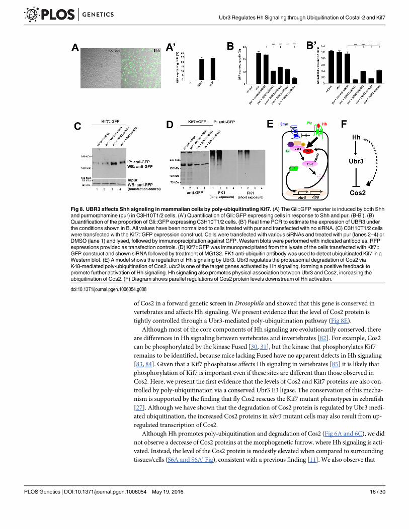
of Cos2 in a forward genetic screen in Drosophila and showed that this gene is conserved invertebrates and affects Hh signaling. We present evidence that the level of Cos2 protein istightly controlled through a Ubr3-mediated poly-ubiquitination pathway (Fig 8E).
Although most of the core components of Hh signaling are evolutionarily conserved, thereare differences in Hh signaling between vertebrates and invertebrates [82]. For example, Cos2can be phosphorylated by the kinase Fused [30, 31], but the kinase that phosphorylates Kif7remains to be identified, because mice lacking Fused have no apparent defects in Hh signaling[83, 84]. Given that a Kif7 phosphatase affects Hh signaling in vertebrates [85] it is likely thatphosphorylation of Kif7 is important even if these sites are different than those observed inCos2. Here, we present the first evidence that the levels of Cos2 and Kif7 proteins are also con-trolled by poly-ubiquitination via a conserved Ubr3 E3 ligase. The conservation of this mecha-nism is supported by the finding that fly Cos2 rescues the Kif7 mutant phenotypes in zebrafish[27]. Although we have shown that the degradation of Cos2 protein is regulated by Ubr3 medi-ated ubiquitination, the increased Cos2 proteins in ubr3mutant cells may also result from up-regulated transcription of Cos2.
Although Hh promotes poly-ubiquitination and degradation of Cos2 (Fig 6A and 6C), we didnot observe a decrease of Cos2 proteins at the morphogenetic furrow, where Hh signaling is acti-vated. Instead, the level of the Cos2 protein is modestly elevated when compared to surroundingtissues/cells (S6A and S6A’ Fig), consistent with a previous finding [11]. We also observe that
Fig 8. UBR3 affects Shh signaling in mammalian cells by poly-ubiquitinating Kif7. (A) The Gli::GFP reporter is induced by both Shhand purmorphamine (pur) in C3H10T1/2 cells. (A’) Quantification of Gli::GFP expressing cells in response to Shh and pur. (B-B’). (B)Quantification of the proportion of Gli::GFP expressing C3H10T1/2 cells. (B’) Real time PCR to estimate the expression of UBR3 underthe conditions shown in B. All values have been normalized to cells treated with pur and transfected with no siRNA. (C) C3H10T1/2 cellswere transfected with the Kif7::GFP expression construct. Cells were transfected with various siRNAs and treated with pur (lanes 2–4) orDMSO (lane 1) and lysed, followed by immunoprecipitation against GFP. Western blots were performed with indicated antibodies. RFPexpressions provided as transfection controls. (D) Kif7::GFP was immunoprecipitated from the lysate of the cells transfected with Kif7::GFP construct and shown siRNA followed by treatment of MG132. FK1 anti-ubiquitin antibody was used to detect ubiquitinated Kif7 in aWestern blot. (E) A model shows the regulation of Hh signaling by Ubr3. Ubr3 regulates the proteasomal degradation of Cos2 viaK48-mediated poly-ubiquitination of Cos2. ubr3 is one of the target genes activated by Hh signaling, forming a positive feedback topromote further activation of Hh signaling. Hh signaling also promotes physical association between Ubr3 and Cos2, increasing theubiquitination of Cos2. (F) Diagram shows parallel regulations of Cos2 protein levels downstream of Hh activation.
doi:10.1371/journal.pgen.1006054.g008
Ubr3 Regulates Hh Signaling through Ubiquitination of Costal-2 and Kif7
PLOS Genetics | DOI:10.1371/journal.pgen.1006054 May 19, 2016 16 / 30
activation of Hh in S2 cells up-regulates Cos2 (Fig 6C). The observation that activation of Hh sig-naling promotes the degradation of Cos2 and that Cos2 protein level is increased, but notdecreased by Hh activation, suggests that some mechanism other than ubiquitination up-regulates the level of Cos2 protein (Fig 8F). Ubr3-mediated degradation of Cos2 may function asa mechanism to prevent aberrantly high levels of Cos2, thereby toning down Hh signaling. Thismay also underlie the observation that not all cells that require Hh signaling are affected in flies.This hypothesis is also supported by the finding that loss of ubr3 in zebrafish affects developmen-tal processes that rely on high levels of Shh signaling but does not affect those that respond tolow Shh signaling (Fig 7 and S4 Fig).
Cul1 functions downstream of Cos2 to process Ci155, one would anticipate that Cul1 is epi-static to Cos2. This is inconsistent with the observation that down-regulation of Cul1 in ubr3clones in the morphogenetic furrow of Drosophila eye discs fails to restore Ci155 expression(Fig 3E), whereas down-regulation of Cos2 restores Ci155 levels (Fig 3D). This may be becausethe RNAi expression does not deplete the protein sufficiently, or because, Cos2 may regulateCi155 through a mechanism independent of Cul1.
Although our data clearly show that Ubr3 plays a role in Hh signaling at the morphogeneticfurrow, we do not observe a loss of Ci155 in ubr3mutant clones in wing discs (S6B and S6B’ Fig).However, we observed a similar up-regulation of Cos2 in ubr3mutant clones in wing discs (S6Cand S6C’ Fig), implying that Ubr3 mediated poly-ubiquitination of Cos2 may be present in wingdiscs. The lack of a Hh phenotype in posterior compartment cells of wing discs may be due toanother E3 ligase that is functionally redundant and downregulates Cos2. Alternatively residualUbr3 in ubr3mutant cells due to perdurance of Ubr3 products may partially downregulateCos2, allowing activation of Hh signaling. When we sensitized the background by over-express-ing ptcDN to ectopically activate Hh signaling, we find that loss of ubr3 strongly suppresses theactivation of Hh signaling in clones, gauged by the reduced clone sizes and Ci155 levels (S6D–S6E’ Fig). This may also be the reason why not all tissues display the typical Shh phenotype inzebrafish. In addition, ectopic activation of Hh signaling leads to up-regulated transcription ofubr3 (S6F–S6J Fig), suggesting that the positive feedback of Ubr3 is present in the wing.
Hh signaling shares many similarities withWnt signaling [86]. Both pathways regulate manydevelopmental processes and induce human cancers when the pathways are aberrantly activated.Moreover, the principal signaling mechanisms are based on similar features. Each pathway isactivated through ligand binding of a G-protein coupled receptor, leading to the downstreamactivation of a transcription factor through phosphorylation-dependent proteolysis. Axin is thescaffold protein that recruits an activation complex inWnt signaling, which mediates phosphor-ylation of β-catenin [87]. This function is similar to that of Cos2 in Hh signaling. Interestingly,previous studies have shown that the levels of Axin protein are also regulated by an E3 ligase,RNF146, through poly-ubiquitination [88–90]. Upon activation of Wnt signaling, Axin under-goes tankyrase-dependent poly ADP-ribosylation, which promotes RNF146-Axin interaction[89]. Ubr3 seems to regulate the poly-ubiquitination of Cos2 in a similar manner, given that Hhactivation promotes the Ubr3-Cos2 interaction and the ubiquitination of Cos2. Hence, our datasuggest further similarities between the Hh andWnt signaling pathways.
Materials and MethodsFly strains and geneticsubr3A and ubr3B mutants were isolated in a forward genetic screen as previously described [45,48]. y w ubr3A FRT19A/FM7c Kr-Gal4, UAS-GFP and y w ubr3B FRT19A/FM7c Kr-Gal4,UAS-GFP flies were crossed to, y w tub-Gal80, eyFLP, FRT19A; actin-Gal4, UAS-CD8::GFP/CyO and y w UbxFLP, tub-Gal80 FRT19A; UAS-CD8::GFP, actin-Gal4 to generate GFP-labeled
Ubr3 Regulates Hh Signaling through Ubiquitination of Costal-2 and Kif7
PLOS Genetics | DOI:10.1371/journal.pgen.1006054 May 19, 2016 17 / 30
ubr3 homozygous mutant clones using the MARCM technique [91]. The ubr3 genomic rescuetransgenic fly strain was generated using the P[acman] system, BAC recombineering and trans-genic platform developed in our laboratory [92]. ubr3 cDNA transgenic flies were generatedthrough φC31-mediated transgenesis [92]. Additional strains used in the study are as follows:dpp-lacZ [93], Df(1)BSC622 [[94], Bloomington Drosophila Stock Center], P[lacW]CG42593G0307a [[54], Bloomington Drosophila Stock Center] cul1EX, FRT42D/CyO [58],FRT42D/CyO [95], UAS-p35 (a kind gift from Andreas Bergmann), eyg-Gal4 [96], UAS-cos2/CyO [81], UAS-cul1/CyO [58], UAS-ubr3RNAi [P{GD12698}, [97], Vienna Drosophila ResourceCenter] UAS-cos2RNAi [[97]; Vienna Drosophila Resource center], UAS-cul1RNAi [TRiP.HM05197, [98]; Bloomington Drosophila Stock Center]; UAS-CiRNAi [TRiP.JF01272, [98];Bloomington Drosophila Stock Center]UAS-ptcDN (a kind gift fromMichael Galko). All flieswere maintained on standard food at 25°C.
Zebrafish strains and husbandryZebrafish strains were AB wild-type, ubr3b1250, ubr3b1251 and smohi1640. The ubr3mutations arerecessive alleles. Phenotypically wild-type siblings were used as controls and labeled as wildtype in Fig 7. Animals were raised in a 10 hour dark and 14 hour light cycle and maintained aspreviously described [99]. Embryos were staged according to the standard series [100]. All ani-mal use protocols were IACUC-approved.
CRISPRmutagenesis and genotypingCRISPR mutagenesis was carried as previously described with minor modifications [101, 102].The zebrafish ubr3 reference sequence used in this study was XM_009304449.1. Identificationof target sequences was done using Zifit software [103, 104]. Candidate sequences were thenblasted against the zebrafish genome (Zv9) and those with unique hits were selected. The fol-lowing target sequences were selected b1250: 5’- GGGGCCTGTGACTGCGGGGA-3’, locatedin the sense strand, and b1251: 5’-GGCGTTATCGTAGGATCGGA3’, located in the antisensestrand (Fig 7, S1 Fig). A guide RNA (gRNA) template was created by PCR. A T7 promoter sitewas incorporated in the gene specific oligonucleotide, followed by the target sequence and thestart of the guide RNA sequence (5’-gttttagagctagaaatagc-3’). The complementary guide RNAscaffold oligonucleotide sequence used was 5’-gatccgcaccgactcggtgccactttttcaagttgataacggac-tagccttattttaacttgctatttctagctctaaaac-3’. PCR was performed using Phusion polymerase (NEB)following the manufacturer’s recommendations. 10μM of each primer was used for the reac-tion. The first denaturation step was carried out at 98°C for 30 sec, followed by 40 cycles ofdenaturation at 98°C for 10 seconds, annealing at 60°C for 10 seconds, and extension at 72°Cfor 15 seconds. A final extension step was introduced at 72°C for 10 minutes. PCR productswere purified using a PCR purification kit (QIAGEN). RNA was transcribed using a MEGA-script T7 kit following the manufacturer recommendations. A volume of 2nl of Cas9 RNA andgRNA were co-injected at a concentration of 100ng/μl each.
Screening of F0 founders and genotyping of F1 carriers were done by PCR and sequencingusing the following primers: Primer b1250F (position 101–119): 5’-CTGCAGGAACTGCTGGATAG-3’; Primer b1250R (position 415–433): 5’-ACCCGCTCTCTCTCATCAC-3’. Primerb1251F (position75-94): 5’-TGACAACAGTTCAGGCTTGC-3’; Primer b1251R (position326-345): 5’-GTGGCGTTATCGTAGGATCG-3’.
Embryo manipulation and RNA injection250 pg of RNA encoding for a dominant negative regulatory subunit of the Protein Kinase A(dnPKA) [75] was injected into 1 cell stage embryos. dnPKA construct was linearized with
Ubr3 Regulates Hh Signaling through Ubiquitination of Costal-2 and Kif7
PLOS Genetics | DOI:10.1371/journal.pgen.1006054 May 19, 2016 18 / 30
NotI and transcribed with SP6 using a mMessage mMachine kit following the manufacturer´srecommendations. Embryos were fixed at 27hpf and processed for in situ hybridization againstubr3.
Immunolabeling and imagingFly tissues were dissected in phosphate-buffered saline (PBS) at room temperature and fixedwith 3.7% formaldehyde in PBS for 20 minutes, followed by permeabilization with 0.2% Tri-ton-X100 in PBS. The primary antibodies and secondary fluorescently-labeled antibodies usedwere: chicken anti-GFP (1:1000, Abcam), rat-Elav [1:1000, 7E8A10, DSHB, [51], guinea piganti-Sens [1:1000, [50]], rat anti-Ci [1:50, 2A1, DSHB [105]], rabbit anti- β-galactosidase (lacZ;1:1000, Abcam), guinea pig anti-Ubr3 (1:1000, this study, see below), mouse anti-Cos2 [1:50,17E11, DSHB [18]], mouse anti-Ptc [1:100, DSHB [106]], mouse anti-Fu [1:100, DSHB [18]],rabbit anti-Cul1 [1:250, [107]], rabbit anti-GM130 (1:500, Abcam), rabbit anti-Rab5 (1:500,Abcam), rabbit anti-Rab7 (1:500) [108], mouse anti-Rab11 (1:100, BD Biosciences) [109],mouse anti-Complex V (1:500) [110], ER-GFP (1:1000 incubate with cells over night, CellLightER-GFP, BacMam 2.0. Thermo Fisher Scientific), PNA-biotin (Vector Laboratories).Alexa488-, Cy3- and Cy5- or DyLight649 conjugated affinity purified donkey secondary anti-bodies (1: 500, Jackson ImmunoResearch Laboratories) and DAPI (0.5 μg/ml, LifeTechnologies).
Zebrafish immunolabeling was performed as previously described [111] with the followingminor modifications. 18-somites stage embryos were fixed in BT-fix overnight at room temper-ature. Embryos were permeabilized in PBS+1% Tween20 for 5 hours at room temperature.Anti-UBR3 antibody (Sigma Prestige, catalogue #HPA035390) was diluted in 1/500. Biotiny-lated anti-rabbit was used at 1/500. To detect signal, ABC kit (Vectorlabs) was used. A and Breagents were mixed together at an 1/100 dilution in PBS- Block and pre-incubated for 20 min-utes at room temperature, then added to the samples for 25 minutes. Tyramide from the TSAkit (Perkin-Elmer) was diluted 1:50 in pre-warmed buffer reagent, and added to samples for 20minutes following the manufacturer’s recommendations. The detection reaction was stoppedby adding cold PBS+0.1%Tween20, followed by 4 washes in PBS+0.1%Tween20.
Microscope image acquisitionImages were acquired using LSM510 and LSM710 confocal microscopes (Zeiss) and examinedand processed using LSM viewer (Zeiss), ZEN (Zeiss) and Photoshop (Adobe) software. Immu-nostained zebrafish embryos were immersed in Vectashield, mounted laterally in a slide cham-ber and imaged with a Zeiss LSM5 confocal microscope. Live embryos were mounted laterallyin 3% methylcellulsose and imaged on a compound microscope using DIC. ISH treatedembryos were dissected in 90% glycerol, flat-mounted in 100% glycerol in a slide chamber andimaged on a compound microscope using DIC.
Cloning, plasmid constructs and antibody productionA ubr3 genomic rescue construct was constructed by cloning a 18.3 kb fragment of genomicDNA that contains the ubr3 gene (X: 7,935,666. . . 7,953,967) [Release 6 Drosophila referencegenome, [112]] into P[acman] [92, 113]. ubr3 cDNA was constructed from exon sequencesand cloned into pUASTattB using a GENEART Seamless Cloning and Assembly Kit (LifeTechnologies). GFP was tagged to the carboxyl terminus of the full length ubr3 sequence or apartial sequence encoding only the UBR domain (aa 222–292). The flag sequence was conju-gated to the carboxyl terminus of the full length ubr3 sequence in the primer. Ubr3::flag wasthen amplified through PCR and cloned into pUASTattB through XhoI and XbaI. To generate
Ubr3 Regulates Hh Signaling through Ubiquitination of Costal-2 and Kif7
PLOS Genetics | DOI:10.1371/journal.pgen.1006054 May 19, 2016 19 / 30
E3 dead form of flag tagged Ubr3 expression construct, mutations results in all residues shownin red box in Fig 4A changed to alanines were introduced through synthesized DNA whichspans 500 bp downstream from RsrII. This synthesized DNA fragment was then clonedtogether with PCR amplified flag tagged carboxyl fragment of Ubr3 into pUASTattB-Ubr3-flagthrough RsrII, BbsI and XbaI. The HA::Cos2N1 to 3 constructs were cloned into pUASTattBthrough EcoRI and XbaI. HA::Cos2ΔN1-2, HA::Cos2ΔC1-3 have been described [114]. Myc::Cos2 was constructed by fusion of 5xMyc tags to the N-terminus of the Cos2 coding sequence.The HA::Ub transgene has been described previously [34]. Kif7::GFP construct is a gift fromDr. Chi-Chung Hui [78].
The 7Gli:GFP reporter of Hedgehog signaling activity contains 7 repeats of the Gli bindingsite (5’-TCGACAAGCAGGGAACACCCAAGTAGAAGCTC) followed by GFP [115]. Theprimer pair (forward 5’-TGAAGCTTGCATGCCCTGCAGGACAAGCAGGGAACGCCCAAGTAG and reverse 5’ CTCGAGTACCGGATCCATTATATACCCTCTGCAGACTTGGGTGTTCCCTGCTTGTCG) was used to amplify the Gli binding sequences from the 8Gli-Lucplasmid by PCR. The reverse primer also contains a TATA sequence, which was used to rebuildthe TATA box after the Gli binding sites. The destination plasmid pRRL.sin-18.ppt.TCF/LEF:GFP.pre [116] was linearized by PstI and BamHI digestion to cut out the TCF/LEF sequenceand the TATA box. The resulting products were recombined into a linearized destination plas-mid by infusion cloning according to the manufacturer’s protocol (Clontech).
For Ubr3 antibody production, the sequence encoding aa 751–1500 of Ubr3 was cloned intopET21 expression construct and expressed in E. coli. Purified inclusion bodies were used toimmunize guinea pigs.
In situ hybridizationubr3 in situ hybridization probes I and II anti-sense sequences contain 2558 to 3576 nt and3775 to 4770 nt of ubr3 cDNA, respectively. Anti-sense sequences were cloned into pGEM-Tvector (Promega). Before transcription, the construct was linearized by SalI. ubr3 RNA in situprobes were transcribed and labeled with a digoxigenin [113] RNA labeling kit (Roche). In situhybridization to whole-mount discs was performed as previously described [117]. Probe I wasused in images shown in Fig 2F, 2G, 2H and 2I and S6J Fig. Probe II was used in images shownin Fig 2E and 2J and S2I, S6F, S6H and S6I Figs.
Zebrafish whole-mount in situ hybridization was carried out as described with minor modi-fications [118]. Digoxigenin-labeled probes were prepared according to manufacturer´sinstructions (Roche). Probe signal was detected using NBT/BCIP mix (Roche). The ptch2probe was kindly shared by Stone Elworthy (University of Sheffield, UK).
Cell culture assaysS2 cells were cultured at 25°C in Schneider’s medium (Life Technologies) plus 10% heat-inacti-vated fetal bovine serum (Sigma), 100 U/mL penicillin (Life Technologies), and 100 μg/mLstreptomycin (Life Technologies). Cells were split every 3 days and plated at a density of 106
cells/well in 12-well cell culture plates for experiments. Transfections were carried out usingEffectene transfection reagent (Qiagen). Ubr3 dsRNA was synthesized against nt 652–1,191.dsRNAs transfection and dsRNA against GFP have been described [114]. CHX (100 μM,Sigma) and MG132 (50 μM, Sigma) in dimethyl sulfoxide (DMSO) were added to S2 cells 48hours after transfection and incubated for indicated time. An equal amount of DMSO wasadded as a negative control (-). 1ug/ml Colchicine (sigma) was incubated with cells for 5 hoursbefore harvest.
Ubr3 Regulates Hh Signaling through Ubiquitination of Costal-2 and Kif7
PLOS Genetics | DOI:10.1371/journal.pgen.1006054 May 19, 2016 20 / 30
C3H10T1/2 cells were cultured at 37°C in 5% CO2 in air in Eagle's Basal medium with Ear-le's BSS, 2 mM L-glutamine, 1.5 g/L sodium bicarbonate and 10% fetal bovine serum, asdescribed by the ATCC (http://www.atcc.org/). Cells were split when reaching 80–90% conflu-ence. Cells were transduced with lentivirus containing Gli::GFP reporter construct for 16h andthen plated on 12-well cell culture plates. On the second day, siRNAs were incubated withLipofectamine RNAiMAX reagent (Invitrogen) overnight. The sequences of siRNAs againstubr3 are as follows: siRNA 1: GTTATAGCTTTGAATCAGT; siRNA 2: CAGAGTTTGCCTCACGACA; siRNA 3: CAAGATTGGTTTGATGCTA; siRNA 4: CAGAAATTGCTCGCAGAGT. Stealth RNAi™ siRNA Negative Control Med GC Duplex (Invitrogen) was used as con-trol siRNA. 3μg/ml Shh (R&D systems) or 10 μM purmorphamine (Calbiochem) in culturemedium, or the same amount of vehicle in culture medium for uninduced controls, was addedto cells on the third day. Cells were photographed for GFP fluorescence and harvested 48 hafter purmorphamine induction. The number of GFP-positive cells was manually counted andstatistical testing was performed with a one way ANOVA followed by Dunnett’s test usinguninduced cells as a control.
Quantitative real-time PCRFor RNA extraction, total RNA from C3H10T1/2 cells was isolated by using Absolutely RNAminiprep Kit (Agilent Technologies). cDNA was synthesized using Superscript III First StrandSynthesis System for RT-PCR (Invitrogen). Quantitative real-time PCR (qPCR) was conductedwith a Master SYBR Green kit (Applied Biosystems) and gene-specific primer sets on a StepOne Plus real-time PCR system (Applied Biosystems). Each experiment was performed withthree biological sample repeats and each PCR was performed in triplicate. L19 was used as anendogenous reference. The gene-specific primer sets used were as follows: L19 (RpL19): 5’-GGTCTGGTTGGATCCCAATG-3’ and 5’-CCCGGGAATGGACAGTCA-3’; UBR3: 5’-CTGATTCATAGAGGAGGCAG-3’ and 5’-ATGGAACAGCTGATTCAGAC-3’.
Co-immunoprecipitation and western blotS2 cells were lysed 48 h after transfection with plasmids in lysis buffer (Tris-HCl 25mM, pH7.5, NaCl 150 mM, EDTA 1mM, NP-40 1%, Glycerol 5%, DTT 1mM) plus Complete protein-ase inhibitor (Roche) for 30 minutes on ice, followed by centrifugation. In these experiments todetect the ubiquitination of Cos2 (Figs 5F, 5G and 6A), we treated S2 cells with 50 uM ofMG132 24 h before harvesting the cells. The supernatant was then immunoprecipitated withagarose beads conjugated to antibodies recognizing different epitope tags, which had been pre-viously equilibrated with lysis buffer, overnight at 4°C. The beads were then washed 3 times inwashing buffer (Tris-HCl 10mM, pH 7.5, NaCl 150mM, EDTA 0.5 mM) before boiling in load-ing buffer. Western blotting was then performed with each sample. The following beads wereused for immunoprecipitation: Chromotek-GFP-Trap Agarose Beads (Allele Biotechnology),Monoclonal Anti-HA−Agarose antibody (Sigma). Protein A resin and anti-Myc (9E10, SantaCruz) were used for Myc immunoprecipitation. To examine the levels of Cos2 ubiquitination,a denaturing method was used as previously described [34]. Briefly, S2 cells were transfectedwith Myc::Cos2 and then lysed with denaturing buffer (1% SDS, 50mM Tris, pH 7.5, 0.5 mMEDTA, and 1 mM DTT) and incubated at 100°C for 5 min. The lysates were then diluted10-fold with regular lysis buffer containing 1.5 mMMgCl2 and subjected to immunoprecipita-tion with the anti-Myc antibody. The proteins were then resolved on an 8% SDS-PAGE, andan immunoblot was performed using an anti-HA antibody to detect the HA::Ub or HA::Ubmutants. The antibodies used in Western blot analysis are as follows: anti-GFP (1:1000 Zymedor 1:1000, Millipore), anti-Myc (1:5000, 9E10, Santa Cruz), anti-HA (1:5000, Santa Cruz, F7 or
Ubr3 Regulates Hh Signaling through Ubiquitination of Costal-2 and Kif7
PLOS Genetics | DOI:10.1371/journal.pgen.1006054 May 19, 2016 21 / 30
1:1000, 16B12, Covance), anti-Ubr3 (1:5000), anti-actin (1:5000, C4, MP Biomedicals), anti-α-tub (1:1000, Cell Signaling). The intensities of the bands in Fig 5B were quantified using imageJ software.
In vitro ubiquitination assayIn vitro auto-ubiquitination assays were performed as described previously [119] with modifi-cations. In brief, S2 cells were first transiently transfected with GFP, UBR-GFP or Ubr3-GFP.S2 cell cultures were collected 48 hours post-transfection and lysed on ice for 45 minutes understringent conditions to minimize interactions with other proteins, using 100 μl RIPA buffer(150 mMNaCl, 1.0% NP-40, 0.5% sodium deoxycholate, 0.1% SDS, 50 mM Tris, pH 8.0) con-taining 1x Complete protease inhibitors cocktail (Roche) for every 106 cells seeded. The lysateswere then added to 30 μl bed volume of Chromotek GFP Trap beads (Allele Biotechnology),previously equilibrated with RIPA buffer, and incubated by rocking at 4°C for 3 hours. Afterwashing with RIPA buffer, 20% of the beads were retained for assessing the expression of GFPprotein by Western blot analysis using anti-GFP (1:1000, Zymed). The remainder of the lysateswere equilibrated by rinsing twice in 1x Ubiquitination Reaction buffer (50 mM Tris-HCl, pH7.4, 5 mMMgCl2, 50 mM NaCl, 1 mM dithiothreitol DTT, 1x protease inhibitors cocktail).The ubiquitination reaction was assembled by adding rabbit UBE1 E1 (Boston Biochem Cat.#302) and human recombinant His6-hHR6A E2 (Boston Biochem Cat. E2-612) conjugatingenzymes and FLAG-Ubiquitin (Sigma) on ice and incubated at 30°C for 30 minutes. The reac-tions were stopped by adding 1x Laemmli buffer, after which the samples were boiled for 10minutes and analyzed by SDS-PAGE and Western blot using anti-FLAGM2 monoclonal anti-body (1:1000, Sigma).
For Cos2 ubiquitination assays, S2 cell culture and RNAi were performed as described pre-viously [114]. Transfections were carried out using Effectene transfection reagent (Qiagen).The immunoprecipitation and immunoblot analysis were performed using standard protocols.Myc-Cos2 was constructed by fusion of 5xMyc tag to the N-terminus of Cos2 coding sequence.HA-Ub and Ub mutants have been described [34]. The HA::UbK48 only has mutations at allof the lysine residues with the exception of K48. GFPRNAi has been described. Ubr3 dsRNAwas synthesized against nucleotides 652–1191. The following antibodies were used: mouseanti-Myc (1:5000, 9E10, Santa Cruz), anti-GFP (1:1000, Millipore), anti-HA (1:5000, F7, SantaCruz), and anti-β-tubulin (1:2000, E7, DSHB).
Supporting InformationS1 Fig. Hh signaling is defective in the ubr3 complementation group. (A) ubr3Amutantclones (labeled by dashed lines) crossing the morphogenetic furrow exhibit delayed differentia-tion of photoreceptors (arrow), revealed by expression of Senseless (cyan) and Elav (red). (B)ubr3A mutant clones (labeled by dashed lines) exhibit loss of Ci155 staining (red) in the mor-phogenetic furrow and delay of R8 photoreceptor differentiation (arrow), visualized by Sense-less expression (cyan). (C) Over-expression of p35 in ubr3B mutant clones (labeled by dashedlines) does not rescue the loss of Ci155 (red) in the morphogenetic furrow (arrow). (D-E)TUNEL assays (red) were performed with eye discs bearing ubr3B/B mutant clones (green in D)or ubr3B/B act>p35 clones (green in E). (F) The ubr3 complementation group maps toCG42593. Both ubr3A and ubr3B alleles fail to complement Df(1)BSC622 and a P-element inser-tion P{lacW}CG42593G0307a. A genomic rescue construct (symbolized by the green box) fullyrescues the lethality of hemizygous ubr3mutants. (G) Over-expression of Ubr3 by actin-Gal4in ubr3Bmutant clones (outlined by dashed lines) fully restores the expression of Ci155 (red) inthe morphogenetic furrow (arrow). (H) Structure of the genomic locus of ubr3 gene. ubr3
Ubr3 Regulates Hh Signaling through Ubiquitination of Costal-2 and Kif7
PLOS Genetics | DOI:10.1371/journal.pgen.1006054 May 19, 2016 22 / 30
genomic rescue sequence is indicated in green box.(I) Identity (I) and similarity (S) of the threeconserved domains between Ubr3 homologues from indicated species.(TIF)
S2 Fig. Subcellular localization of Ubr3. (A-G) S2 cells are co-stained with anti-Ubr3 anti-body (red) and antibodies raised against different proteins associated with different organelles(green) and DAPI (blue). (H) Eye disc from 3rd instar larvae in which eyg-Gal4 drives expres-sion of CiRNAi were stained with anti-Ci155 (red). Ci155 is reduced in the equator region of themorphogenetic furrow (arrow). (I) In situ hybridization experiments with an anti-ubr3 probewere performed on eye discs from 3rd instar larvae in which eyg-Gal4 drove the expression ofCiRNAi. ubr3mRNA is reduced in the equator region of the morphogenetic furrow (arrow).(TIF)
S3 Fig. Cos2 and Cul1 are up-regulated in ubr3mutant cells. (A-C’) Co-immunolabeling ofanti-Ptc (red) and anti-Senseless (cyan) (A-A’), anti-Fu (red) and anti-Senseless (cyan) (B-B’),anti-Cul1 (red) and anti-Senseless (cyan) (C-C’) in eye discs bearing ubr3Bmutant clones(labeled by dashed lines) from 3rd instar larvae shows up-regulated Cul1 (red) in ubr3B mutantcells. (D-D’) High magnification of the boundary region of eyg-Gal4 driven expression of Cos2in the eye disc. A solid line shows the boundary of the loss of Ci155 expression (shown in red).Cos2 levels are indicated by Cos2 labeling (shown in green). Arrows mark regions where Cos2is expressed at low level (green) but is sufficient to inhibit Ci155 expression (red). (E-E’) Eyedisc with ubr3B/B clones that express Cos2 RNAi (shown in green) was labeled with anti-Sens(cyan). Arrows show the suppression of delayed differentiation of photoreceptor cells.(TIF)
S4 Fig. ubr3b1250 and ubr3b1251 mutants produce truncated, non-functional Ubr3 proteins.(A, B) DNA sequences show mutations found in ubr3b1250 and ubr3b1251 mutants. ubr3b1250
mutant carries a 28bp deletion around the CRISPR targeted region b1250. ubr3b1251 carries a4bp insertion within the CRIPSR targeted region b1251. (C) Protein sequences of truncatedUbr3 proteins produced in ubr3b1250 and ubr3b1251 mutants.(TIF)
S5 Fig. ubr3 is required for Hedgehog (Hh) signaling in zebrafish. (A, C) DIC images showlateral views of posterior trunk regions at 24-hours-post-fertilization. Anterior to the left, dor-sal up. The angles of V-shaped somites were shown by the red line. Scale bars: 50μm. (B, D)islet2 in situ hybridization (ISH) of wild type and ubr3b1250/1251 mutant zebrafish. Lateral viewsof posterior trunk regions at 24-hours-post-fertilization are shown. (E) Average somite anglein wild-type siblings (wt sibs) and ubr3b1250/b1251 trans-heterozygous mutants. Wild type sib-lings have a typical V-shaped somite characterized by an average angle of 92°. In the ubr3trans-heterozygous mutants, the angles become more obtuse with an average of 119°. (F-I) ISHagainst ubr3 at 24 and 28 hpf. Lateral views of the somites. (F-G) At 24 hpf (F), ubr3 isexpressed throughout the somites. By 28hpf (G), this expression is restricted to ventral regionsof the somites (red bracket). (H) ubr3 expression is lost in smomutants at 24 hpf. (I) At 28 hpf,the ubr3 expression domain is expanded dorsally when Hh signaling is upregulated by ectopicexpression of dnPKA (green bracket). Bars: SEM. Five angles were measured per larva. Fiveheterozygous and seven ubr3b1250/b1251 larvae were analyzed, P<0.01.(TIF)
S6 Fig. Ubr3 regulates Cos2 in the wing discs. (A-A’) Immunolabeling of wild type eye discwith anti-Cos2 (red) and anti-Ci155 (green). Arrow in A’ shows moderate increase of Cos2 pro-tein levels in the morphogenetic furrow. (B-B’) Wing disc from 3rd larvae with ubr3B mutant
Ubr3 Regulates Hh Signaling through Ubiquitination of Costal-2 and Kif7
PLOS Genetics | DOI:10.1371/journal.pgen.1006054 May 19, 2016 23 / 30
clones (green) was stained with anti-Ci155 (red) and anti-senseless (sens, cyan). (C-C’) Wingdisc from 3rd larvae with ubr3Bmutant clones (green) was stained with anti-Cos2 (red) andanti-senseless (sens, cyan). (D-D’) A wing disc from 3rd larvae with ptcDN expressing clones(green) was stained with anti-Ci155 (red) and anti-senseless (sens, cyan). (E-E’) Wing disc from3rd larvae with ubr3Bmutant clones expressing ptcDN (green) was stained with anti-Ci155 (red)and anti-senseless (sens, cyan). (F, H-J) In situ hybridizations were performed on wing discsfrom 3rd instar larvae with indicated genotypes using an anti-ubr3 probe. (G) A wing disc from3rd larvae in which eyg-Gal4 driven expression of GFP was labeled with anti-GFP (green in G,indicating eyg-Gal4 expression region), anti-Sens (cyan in G, labeling wing margin) and anti-Ci155 (red in G). Arrows indicate elevated ubr3 transcription in eyg-Gal4 expressing domain.(TIF)
AcknowledgmentsWe thank the Bloomington Drosophila Stock Center, the Vienna Drosophila Resource Centerand the Developmental Studies Hybridoma Bank for flies and antibodies. We thank MatthewScott, Graeme Mardon, Andreas Bergmann, Cheng-Ting Chien, Pascal Thérond, Konrad Bas-ler and Michael Galko for sharing fly stocks and molecular biology reagents. We thank SteveDevoto for providing the smohi1640 mutants and Sarah Baxendale and Stone Elworthy for pro-viding Hh signaling reagents. We thank Kartik Venkatachalam and Michael Wangler for help-ful discussions and comments. We thank Honglin Pan and Yuchun He for injections to createtransgenic flies.
Author ContributionsConceived and designed the experiments: TL JF BBS NG JZ MTL AKGMW JJ HJB. Performedthe experiments: TL JF BBS NG GL SY MJ KC JZWW. Analyzed the data: TL JF BBS NG KCJZ JJ HJB. Contributed reagents/materials/analysis tools: WWMTL. Wrote the paper: TL BBSGL SY MJ AKGMW JJ HJB.
References1. Arwert EN, Hoste E, Watt FM. Epithelial stem cells, wound healing and cancer. Nat Rev Cancer.
2012; 12(3):170–80. Epub 2012/03/01. doi: 10.1038/nrc3217 PMID: 22362215.
2. Varjosalo M, Taipale J. Hedgehog: functions and mechanisms. Genes & development. 2008; 22(18):2454–72. Epub 2008/09/17. doi: 10.1101/gad.1693608 PMID: 18794343.
3. Babcock DT, Shi S, Jo J, ShawM, Gutstein HB, Galko MJ. Hedgehog signaling regulates nociceptivesensitization. Curr Biol. 2011; 21(18):1525–33. Epub 2011/09/13. doi: 10.1016/j.cub.2011.08.020PMID: 21906949; PubMed Central PMCID: PMC3262399.
4. Heemskerk J, DiNardo S. Drosophila hedgehog acts as a morphogen in cellular patterning. Cell.1994; 76(3):449–60. PMID: 8313468.
5. Hooper JE, Scott MP. Communicating with Hedgehogs. Nature reviews. 2005; 6(4):306–17. PMID:15803137.
6. Lum L, Beachy PA. The Hedgehog response network: sensors, switches, and routers. Science (NewYork, NY. 2004; 304(5678):1755–9. PMID: 15205520.
7. Jiang J, Hui CC. Hedgehog signaling in development and cancer. Developmental cell. 2008; 15(6):801–12. PMID: 19081070. doi: 10.1016/j.devcel.2008.11.010
8. Pasca di Magliano M, Hebrok M. Hedgehog signalling in cancer formation and maintenance. Nat RevCancer. 2003; 3(12):903–11. PMID: 14737121.
9. Taipale J, Cooper MK, Maiti T, Beachy PA. Patched acts catalytically to suppress the activity ofSmoothened. Nature. 2002; 418(6900):892–7. PMID: 12192414.
10. Robbins DJ, Nybakken KE, Kobayashi R, Sisson JC, Bishop JM, Therond PP. Hedgehog elicits signaltransduction by means of a large complex containing the kinesin-related protein costal2. Cell. 1997;90(2):225–34. Epub 1997/07/25. PMID: 9244297.
Ubr3 Regulates Hh Signaling through Ubiquitination of Costal-2 and Kif7
PLOS Genetics | DOI:10.1371/journal.pgen.1006054 May 19, 2016 24 / 30
11. Sisson JC, Ho KS, Suyama K, Scott MP. Costal2, a novel kinesin-related protein in the Hedgehog sig-naling pathway. Cell. 1997; 90(2):235–45. Epub 1997/07/25. PMID: 9244298.
12. Aikin RA, Ayers KL, Therond PP. The role of kinases in the Hedgehog signalling pathway. EMBOreports. 2008; 9(4):330–6. PMID: 18379584. doi: 10.1038/embor.2008.38
13. Jiang J, Struhl G. Regulation of the Hedgehog andWingless signalling pathways by the F-box/WD40-repeat protein Slimb. Nature. 1998; 391(6666):493–6. Epub 1998/02/14. doi: 10.1038/35154 PMID:9461217.
14. Jia J, Amanai K, Wang G, Tang J, Wang B, Jiang J. Shaggy/GSK3 antagonizes Hedgehog signallingby regulating Cubitus interruptus. Nature. 2002; 416(6880):548–52. Epub 2002/03/26. doi: 10.1038/nature733 PMID: 11912487.
15. Jia J, Zhang L, Zhang Q, Tong C, Wang B, Hou F, et al. Phosphorylation by double-time/CKIepsilonand CKIalpha targets cubitus interruptus for Slimb/beta-TRCP-mediated proteolytic processing.Developmental cell. 2005; 9(6):819–30. Epub 2005/12/06. doi: 10.1016/j.devcel.2005.10.006 PMID:16326393.
16. Zheng X, Mann RK, Sever N, Beachy PA. Genetic and biochemical definition of the Hedgehog recep-tor. Genes & development. 2010; 24(1):57–71. PMID: 20048000.
17. Jia J, Tong C, Jiang J. Smoothened transduces Hedgehog signal by physically interacting with Cos-tal2/Fused complex through its C-terminal tail. Genes & development. 2003; 17(21):2709–20. Epub2003/11/05. doi: 10.1101/gad.1136603 PMID: 14597665; PubMed Central PMCID: PMC280620.
18. Lum L, Zhang C, Oh S, Mann RK, von Kessler DP, Taipale J, et al. Hedgehog signal transduction viaSmoothened association with a cytoplasmic complex scaffolded by the atypical kinesin, Costal-2.Molecular cell. 2003; 12(5):1261–74. Epub 2003/11/26. PMID: 14636583.
19. Ogden SK, Ascano M Jr., Stegman MA, Suber LM, Hooper JE, Robbins DJ. Identification of a func-tional interaction between the transmembrane protein Smoothened and the kinesin-related proteinCostal2. Curr Biol. 2003; 13(22):1998–2003. Epub 2003/11/15. PMID: 14614827.
20. Ruel L, Rodriguez R, Gallet A, Lavenant-Staccini L, Therond PP. Stability and association of Smooth-ened, Costal2 and Fused with Cubitus interruptus are regulated by Hedgehog. Nature cell biology.2003; 5(10):907–13. Epub 2003/10/03. doi: 10.1038/ncb1052 PMID: 14523402.
21. ZhangW, Zhao Y, Tong C, Wang G, Wang B, Jia J, et al. Hedgehog-regulated Costal2-kinase com-plexes control phosphorylation and proteolytic processing of Cubitus interruptus. Developmental cell.2005; 8(2):267–78. PMID: 15691767.
22. Ho KS, Suyama K, Fish M, Scott MP. Differential regulation of Hedgehog target gene transcription byCostal2 and Suppressor of Fused. Development (Cambridge, England). 2005; 132(6):1401–12.PMID: 15750186.
23. Katoh Y, Katoh M. Characterization of KIF7 gene in silico. International journal of oncology. 2004; 25(6):1881–6. PMID: 15547730.
24. Katoh Y, Katoh M. KIF27 is one of orthologs for Drosophila Costal-2. International journal of oncology.2004; 25(6):1875–80. PMID: 15547729.
25. Endoh-Yamagami S, Evangelista M, Wilson D, Wen X, Theunissen JW, Phamluong K, et al. Themammalian Cos2 homolog Kif7 plays an essential role in modulating Hh signal transduction duringdevelopment. Curr Biol. 2009; 19(15):1320–6. PMID: 19592253. doi: 10.1016/j.cub.2009.06.046
26. Liem KF Jr., He M, Ocbina PJ, Anderson KV. Mouse Kif7/Costal2 is a cilia-associated protein that reg-ulates Sonic hedgehog signaling. Proceedings of the National Academy of Sciences of the UnitedStates of America. 2009; 106(32):13377–82. Epub 2009/08/12. doi: 10.1073/pnas.0906944106PMID: 19666503; PubMed Central PMCID: PMC2726420.
27. Maurya AK, Ben J, Zhao Z, Lee RT, NiahW, Ng AS, et al. Positive and negative regulation of Gli activ-ity by Kif7 in the zebrafish embryo. PLoS genetics. 2013; 9(12):e1003955. PMID: 24339784. doi: 10.1371/journal.pgen.1003955
28. Putoux A, Thomas S, Coene KL, Davis EE, Alanay Y, Ogur G, et al. KIF7 mutations cause fetal hydro-lethalus and acrocallosal syndromes. Nature genetics. 2011; 43(6):601–6. PMID: 21552264. doi: 10.1038/ng.826
29. Dafinger C, Liebau MC, Elsayed SM, Hellenbroich Y, Boltshauser E, Korenke GC, et al. Mutations inKIF7 link Joubert syndrome with Sonic Hedgehog signaling and microtubule dynamics. The Journal ofclinical investigation. 2011; 121(7):2662–7. Epub 2011/06/03. doi: 10.1172/JCI43639 PMID:21633164; PubMed Central PMCID: PMC3223820.
30. Nybakken KE, Turck CW, Robbins DJ, Bishop JM. Hedgehog-stimulated phosphorylation of the kine-sin-related protein Costal2 is mediated by the serine/threonine kinase fused. The Journal of biologicalchemistry. 2002; 277(27):24638–47. PMID: 11934882.
Ubr3 Regulates Hh Signaling through Ubiquitination of Costal-2 and Kif7
PLOS Genetics | DOI:10.1371/journal.pgen.1006054 May 19, 2016 25 / 30
31. Ranieri N, Ruel L, Gallet A, Raisin S, Therond PP. Distinct phosphorylations on kinesin costal-2 medi-ate differential hedgehog signaling strength. Developmental cell. 2012; 22(2):279–94. Epub 2012/02/07. doi: 10.1016/j.devcel.2011.12.002 PMID: 22306085.
32. Gulino A, Di Marcotullio L, Canettieri G, De Smaele E, Screpanti I. Hedgehog/Gli control by ubiquitina-tion/acetylation interplay.: Elsevier Inc.; 2012.
33. Li S, Chen Y, Shi Q, Yue T, Wang B, Jiang J. Hedgehog-regulated ubiquitination controls smoothenedtrafficking and cell surface expression in Drosophila. PLoS biology. 2012; 10(1):e1001239. PMID:22253574. doi: 10.1371/journal.pbio.1001239
34. Xia R, Jia H, Fan J, Liu Y, Jia J. USP8 promotes smoothened signaling by preventing its ubiquitinationand changing its subcellular localization. PLoS biology. 2012; 10(1):e1001238. Epub 2012/01/19. doi:10.1371/journal.pbio.1001238 PMID: 22253573; PubMed Central PMCID: PMC3254663.
35. Ordureau A, Munch C, Harper JW. Quantifying Ubiquitin Signaling. Molecular cell. 2015; 58(4):660–76. PMID: 26000850. doi: 10.1016/j.molcel.2015.02.020
36. Metzger MB, Hristova VA, Weissman AM. HECT and RING finger families of E3 ubiquitin ligases at aglance. Journal of cell science. 2012; 125(Pt 3):531–7. PMID: 22389392. doi: 10.1242/jcs.091777
37. Tasaki T, Zakrzewska A, Dudgeon DD, Jiang Y, Lazo JS, Kwon YT. The substrate recognitiondomains of the N-end rule pathway. The Journal of biological chemistry. 2009; 284(3):1884–95.PMID: 19008229. doi: 10.1074/jbc.M803641200
38. Meisenberg C, Tait PS, Dianova II, Wright K, EdelmannMJ, Ternette N, et al. Ubiquitin ligase UBR3regulates cellular levels of the essential DNA repair protein APE1 and is required for genome stability.Nucleic acids research. 2012; 40(2):701–11. Epub 2011/09/22. doi: 10.1093/nar/gkr744 PMID:21933813; PubMed Central PMCID: PMC3258136.
39. Tasaki T, Sohr R, Xia Z, Hellweg R, Hortnagl H, Varshavsky A, et al. Biochemical and genetic studiesof UBR3, a ubiquitin ligase with a function in olfactory and other sensory systems. The Journal of bio-logical chemistry. 2007; 282(25):18510–20. PMID: 17462990.
40. Zanet J, Benrabah E, Li T, Pélissier-Monier A, Chanut-Delalande H, Ronsin B, et al. Pri sORF pep-tides induce proteasome protein processing. Science (New York, NY. 2015.
41. Huang Q, Tang X, Wang G, Fan Y, Ray L, Bergmann A, et al. Ubr3 E3 ligase regulates apoptosis bycontrolling the activity of DIAP1 in Drosophila. Cell death and differentiation. 2014; 21(12):1961–70.PMID: 25146930. doi: 10.1038/cdd.2014.115
42. Zhao C, Wang L, Ma X, ZhuW, Yao L, Cui Y, et al. Cardiac Na 1.5 is modulated by ubiquitin proteinligase E3 component n-recognin UBR3 and 6. Journal of cellular and molecular medicine. 2015.PMID: 26059563.
43. Yang S, Zhang H, Guo L, Zhao Y, Chen F. Reconstructing the coding and non-coding RNA regulatorynetworks of miRNAs and mRNAs in breast cancer. Gene. 2014; 548(1):6–13. PMID: 24979338. doi:10.1016/j.gene.2014.06.010
44. CharngWL, Yamamoto S, Jaiswal M, Bayat V, Xiong B, Zhang K, et al. Drosophila Tempura, a novelprotein prenyltransferase alpha subunit, regulates notch signaling via Rab1 and Rab11. PLoS biology.2014; 12(1):e1001777. PMID: 24492843. doi: 10.1371/journal.pbio.1001777
45. Haelterman NA, Jiang L, Li Y, Bayat V, Sandoval H, Ugur B, et al. Large-scale identification of chemi-cally induced mutations in Drosophila melanogaster. Genome research. 2014; 24(10):1707–18.PMID: 25258387. doi: 10.1101/gr.174615.114
46. Xiong B, Bayat V, Jaiswal M, Zhang K, Sandoval H, CharngWL, et al. Crag Is a GEF for Rab11Required for Rhodopsin Trafficking and Maintenance of Adult Photoreceptor Cells. PLoS biology.2012; 10(12):e1001438. Epub 2012/12/12. doi: 10.1371/journal.pbio.1001438 PMID: 23226104;PubMed Central PMCID: PMC3514319.
47. Yamamoto S, CharngWL, Rana NA, Kakuda S, Jaiswal M, Bayat V, et al. A mutation in EGF repeat-8of Notch discriminates between Serrate/Jagged and Delta family ligands. Science (New York, NY.2012; 338(6111):1229–32. Epub 2012/12/01. doi: 10.1126/science.1228745 PMID: 23197537.
48. Yamamoto S, Jaiswal M, CharngWL, Gambin T, Karaca E, Mirzaa G, et al. A drosophila geneticresource of mutants to study mechanisms underlying human genetic diseases. Cell. 2014; 159(1):200–14. PMID: 25259927. doi: 10.1016/j.cell.2014.09.002
49. Frankfort BJ, Nolo R, Zhang Z, Bellen H, Mardon G. senseless repression of rough is required for R8photoreceptor differentiation in the developing Drosophila eye. Neuron. 2001; 32(3):403–14. Epub2001/11/16. PMID: 11709152; PubMed Central PMCID: PMC3122332.
50. Nolo R, Abbott LA, Bellen HJ. Senseless, a Zn finger transcription factor, is necessary and sufficientfor sensory organ development in Drosophila. Cell. 2000; 102(3):349–62. PMID: 10975525.
Ubr3 Regulates Hh Signaling through Ubiquitination of Costal-2 and Kif7
PLOS Genetics | DOI:10.1371/journal.pgen.1006054 May 19, 2016 26 / 30
51. Robinow S, White K. Characterization and spatial distribution of the ELAV protein during Drosophilamelanogaster development. J Neurobiol. 1991; 22(5):443–61. Epub 1991/07/01. doi: 10.1002/neu.480220503 PMID: 1716300.
52. Strutt DI, Mlodzik M. Hedgehog is an indirect regulator of morphogenetic furrow progression in theDrosophila eye disc. Development (Cambridge, England). 1997; 124(17):3233–40. Epub 1997/10/06.PMID: 9310318.
53. Heberlein U, Wolff T, Rubin GM. The TGF beta homolog dpp and the segment polarity gene hedge-hog are required for propagation of a morphogenetic wave in the Drosophila retina. Cell. 1993; 75(5):913–26. Epub 1993/12/03. PMID: 8252627.
54. Peter A, Schottler P, Werner M, Beinert N, Dowe G, Burkert P, et al. Mapping and identification ofessential gene functions on the X chromosome of Drosophila. EMBO reports. 2002; 3(1):34–8. PMID:11751581.
55. Tasaki T, Sriram SM, Park KS, Kwon YT. The N-end rule pathway. Annu Rev Biochem. 2012;81:261–89. Epub 2012/04/25. doi: 10.1146/annurev-biochem-051710-093308 PMID: 22524314.
56. Johnson RL, Milenkovic L, Scott MP. In vivo functions of the patched protein: requirement of the C ter-minus for target gene inactivation but not Hedgehog sequestration. Molecular cell. 2000; 6(2):467–78.PMID: 10983992.
57. Nagarkar-Jaiswal S, Lee PT, Campbell ME, Chen K, Anguiano-Zarate S, Gutierrez MC, et al. A libraryof MiMICs allows tagging of genes and reversible, spatial and temporal knockdown of proteins in Dro-sophila. eLife. 2015; 4. PMID: 25824290.
58. Ou CY, Lin YF, Chen YJ, Chien CT. Distinct protein degradation mechanisms mediated by Cul1 andCul3 controlling Ci stability in Drosophila eye development. Genes & development. 2002; 16(18):2403–14. Epub 2002/09/17. doi: 10.1101/gad.1011402 PMID: 12231629; PubMed CentralPMCID: PMC187440.
59. Deshaies RJ, Joazeiro CA. RING domain E3 ubiquitin ligases. Annu Rev Biochem. 2009; 78:399–434. Epub 2009/06/06. doi: 10.1146/annurev.biochem.78.101807.093809 PMID: 19489725.
60. Koudijs MJ, den Broeder MJ, Groot E, van Eeden FJ. Genetic analysis of the two zebrafish patchedhomologues identifies novel roles for the hedgehog signaling pathway. BMC developmental biology.2008; 8:15. PMID: 18284698. doi: 10.1186/1471-213X-8-15
61. Neumann CJ, Nuesslein-Volhard C. Patterning of the zebrafish retina by a wave of sonic hedgehogactivity. Science (New York, NY. 2000; 289(5487):2137–9. PMID: 11000118.
62. Wang X, Zhao Z, Muller J, Iyu A, Khng AJ, Guccione E, et al. Targeted inactivation and identificationof targets of the Gli2a transcription factor in the zebrafish. Biology open. 2013; 2(11):1203–13. PMID:24244857. doi: 10.1242/bio.20136262
63. Xu J, Srinivas BP, Tay SY, Mak A, Yu X, Lee SG, et al. Genomewide expression profiling in the zebra-fish embryo identifies target genes regulated by Hedgehog signaling during vertebrate development.Genetics. 2006; 174(2):735–52. PMID: 16888327.
64. Lewis KE, Currie PD, Roy S, Schauerte H, Haffter P, Ingham PW. Control of muscle cell-type specifi-cation in the zebrafish embryo by Hedgehog signalling. Developmental biology. 1999; 216(2):469–80.PMID: 10642786.
65. Schauerte HE, van Eeden FJ, Fricke C, Odenthal J, Strahle U, Haffter P. Sonic hedgehog is notrequired for the induction of medial floor plate cells in the zebrafish. Development (Cambridge,England). 1998; 125(15):2983–93. PMID: 9655820.
66. van Eeden FJ, Granato M, Schach U, Brand M, Furutani-Seiki M, Haffter P, et al. Mutations affectingsomite formation and patterning in the zebrafish, Danio rerio. Development (Cambridge, England).1996; 123:153–64. PMID: 9007237.
67. Varga ZM, Amores A, Lewis KE, Yan YL, Postlethwait JH, Eisen JS, et al. Zebrafish smoothened func-tions in ventral neural tube specification and axon tract formation. Development (Cambridge,England). 2001; 128(18):3497–509. PMID: 11566855.
68. Yeh CW, Kao SH, Cheng YC, Hsu LS. Knockdown of cyclin-dependent kinase 10 (cdk10) geneimpairs neural progenitor survival via modulation of raf1a gene expression. The Journal of biologicalchemistry. 2013; 288(39):27927–39. PMID: 23902762. doi: 10.1074/jbc.M112.420265
69. Nguyen VH, Trout J, Connors SA, Andermann P, Weinberg E, Mullins MC. Dorsal and intermediateneuronal cell types of the spinal cord are established by a BMP signaling pathway. Development(Cambridge, England). 2000; 127(6):1209–20. PMID: 10683174.
70. Aanstad P, Santos N, Corbit KC, Scherz PJ, Trinh le A, Salvenmoser W, et al. The extracellulardomain of Smoothened regulates ciliary localization and is required for high-level Hh signaling. CurrBiol. 2009; 19(12):1034–9. PMID: 19464178. doi: 10.1016/j.cub.2009.04.053
Ubr3 Regulates Hh Signaling through Ubiquitination of Costal-2 and Kif7
PLOS Genetics | DOI:10.1371/journal.pgen.1006054 May 19, 2016 27 / 30
71. Hammond CL, Schulte-Merker S. Two populations of endochondral osteoblasts with differential sensi-tivity to Hedgehog signalling. Development (Cambridge, England). 2009; 136(23):3991–4000. PMID:19906866.
72. Hammond KL, Loynes HE, Folarin AA, Smith J, Whitfield TT. Hedgehog signalling is required for cor-rect anteroposterior patterning of the zebrafish otic vesicle. Development (Cambridge, England).2003; 130(7):1403–17. PMID: 12588855.
73. Wolff C, Roy S, Ingham PW. Multiple muscle cell identities induced by distinct levels and timing ofhedgehog activity in the zebrafish embryo. Curr Biol. 2003; 13(14):1169–81. PMID: 12867027.
74. Wolff C, Roy S, Lewis KE, Schauerte H, Joerg-Rauch G, Kirn A, et al. iguana encodes a novel zinc-fin-ger protein with coiled-coil domains essential for Hedgehog signal transduction in the zebrafishembryo. Genes & development. 2004; 18(13):1565–76. PMID: 15198976.
75. Hammerschmidt M, Bitgood MJ, McMahon AP. Protein kinase A is a common negative regulator ofHedgehog signaling in the vertebrate embryo. Genes & development. 1996; 10(6):647–58. PMID:8598293.
76. Kinto N, Iwamoto M, Enomoto-Iwamoto M, Noji S, Ohuchi H, Yoshioka H, et al. Fibroblasts expressingSonic hedgehog induce osteoblast differentiation and ectopic bone formation. FEBS letters. 1997;404(2–3):319–23. PMID: 9119087.
77. Sinha S, Chen JK. Purmorphamine activates the Hedgehog pathway by targeting Smoothened. NatChem Biol. 2006; 2(1):29–30. PMID: 16408088.
78. Cheung HO, Zhang X, Ribeiro A, Mo R, Makino S, Puviindran V, et al. The kinesin protein Kif7 is a crit-ical regulator of Gli transcription factors in mammalian hedgehog signaling. Science signaling. 2009;2(76):ra29. Epub 2009/06/25. doi: 10.1126/scisignal.2000405 PMID: 19549984.
79. Farzan SF, Ascano M Jr., Ogden SK, Sanial M, Brigui A, Plessis A, et al. Costal2 functions as a kine-sin-like protein in the hedgehog signal transduction pathway. Curr Biol. 2008; 18(16):1215–20. Epub2008/08/12. doi: 10.1016/j.cub.2008.07.026 PMID: 18691888; PubMed Central PMCID:PMC2774813.
80. Wang G, Jiang J. Multiple Cos2/Ci interactions regulate Ci subcellular localization through microtu-bule dependent and independent mechanisms. Developmental biology. 2004; 268(2):493–505.PMID: 15063184.
81. Zhu AJ, Zheng L, Suyama K, Scott MP. Altered localization of Drosophila Smoothened protein acti-vates Hedgehog signal transduction. Genes & development. 2003; 17(10):1240–52. Epub 2003/05/06. doi: 10.1101/gad.1080803 PMID: 12730121; PubMed Central PMCID: PMC196058.
82. Huangfu D, Anderson KV. Signaling from Smo to Ci/Gli: conservation and divergence of Hedgehogpathways from Drosophila to vertebrates. Development (Cambridge, England). 2006; 133(1):3–14.PMID: 16339192.
83. Chen MH, Gao N, Kawakami T, Chuang PT. Mice deficient in the fused homolog do not exhibit pheno-types indicative of perturbed hedgehog signaling during embryonic development. Molecular and cellu-lar biology. 2005; 25(16):7042–53. PMID: 16055716.
84. Merchant M, Evangelista M, Luoh SM, Frantz GD, Chalasani S, Carano RA, et al. Loss of the serine/threonine kinase fused results in postnatal growth defects and lethality due to progressive hydroceph-alus. Molecular and cellular biology. 2005; 25(16):7054–68. PMID: 16055717.
85. Liu YC, Couzens AL, Deshwar AR, LD BM-C, Zhang X, Puviindran V, et al. The PPFIA1-PP2A proteincomplex promotes trafficking of Kif7 to the ciliary tip and Hedgehog signaling. Science signaling.2014; 7(355):ra117. PMID: 25492966. doi: 10.1126/scisignal.2005608
86. Kalderon D. Similarities between the Hedgehog andWnt signaling pathways. Trends in cell biology.2002; 12(11):523–31. PMID: 12446114.
87. Song X, Wang S, Li L. New insights into the regulation of Axin function in canonical Wnt signalingpathway. Protein & cell. 2014; 5(3):186–93. PMID: 24474204.
88. Callow MG, Tran H, Phu L, Lau T, Lee J, Sandoval WN, et al. Ubiquitin ligase RNF146 regulates tan-kyrase and Axin to promoteWnt signaling. PloS one. 2011; 6(7):e22595. PMID: 21799911. doi: 10.1371/journal.pone.0022595
89. DaRosa PA, Wang Z, Jiang X, Pruneda JN, Cong F, Klevit RE, et al. Allosteric activation of theRNF146 ubiquitin ligase by a poly(ADP-ribosyl)ation signal. Nature. 2015; 517(7533):223–6. PMID:25327252. doi: 10.1038/nature13826
90. Zhang Y, Liu S, Mickanin C, Feng Y, Charlat O, Michaud GA, et al. RNF146 is a poly(ADP-ribose)-directed E3 ligase that regulates axin degradation andWnt signalling. Nature cell biology. 2011; 13(5):623–9. PMID: 21478859. doi: 10.1038/ncb2222
91. Lee T, Luo L. Mosaic analysis with a repressible cell marker for studies of gene function in neuronalmorphogenesis. Neuron. 1999; 22(3):451–61. PMID: 10197526.
Ubr3 Regulates Hh Signaling through Ubiquitination of Costal-2 and Kif7
PLOS Genetics | DOI:10.1371/journal.pgen.1006054 May 19, 2016 28 / 30
92. Venken KJ, He Y, Hoskins RA, Bellen HJ. P[acman]: a BAC transgenic platform for targeted insertionof large DNA fragments in D. melanogaster. Science (New York, NY. 2006; 314(5806):1747–51.Epub 2006/12/02. doi: 10.1126/science.1134426 PMID: 17138868.
93. Blackman RK, Sanicola M, Raftery LA, Gillevet T, Gelbart WM. An extensive 3' cis-regulatory regiondirects the imaginal disk expression of decapentaplegic, a member of the TGF-beta family in Drosoph-ila. Development (Cambridge, England). 1991; 111(3):657–66. Epub 1991/03/01. PMID: 1908769.
94. Cook RK, Christensen SJ, Deal JA, Coburn RA, Deal ME, Gresens JM, et al. The generation of chro-mosomal deletions to provide extensive coverage and subdivision of the Drosophila melanogastergenome. Genome biology. 2012; 13(3):R21. PMID: 22445104. doi: 10.1186/gb-2012-13-3-r21
95. Andrews HK, Giagtzoglou N, Yamamoto S, Schulze KL, Bellen HJ. Sequoia regulates cell fate deci-sions in the external sensory organs of adult Drosophila. EMBO reports. 2009; 10(6):636–41. Epub2009/05/16. doi: 10.1038/embor.2009.66 PMID: 19444309; PubMed Central PMCID: PMC2711842.
96. Yao JG, Sun YH. Eyg and Ey Pax proteins act by distinct transcriptional mechanisms in Drosophiladevelopment. Embo J. 2005; 24(14):2602–12. PMID: 15973436.
97. Dietzl G, Chen D, Schnorrer F, Su KC, Barinova Y, Fellner M, et al. A genome-wide transgenic RNAilibrary for conditional gene inactivation in Drosophila. Nature. 2007; 448(7150):151–6. Epub 2007/07/13. doi: 10.1038/nature05954 PMID: 17625558.
98. Ni JQ, Liu LP, Binari R, Hardy R, Shim HS, Cavallaro A, et al. A Drosophila resource of transgenicRNAi lines for neurogenetics. Genetics. 2009; 182(4):1089–100. PMID: 19487563. doi: 10.1534/genetics.109.103630
99. Westerfield M. The Zebrafish Book: A Guide for the Laboratory Use of Zebrafish (Danio rerio):Eugene, University of Oregon Press; 2007.
100. Kimmel CB, Ballard WW, Kimmel SR, Ullmann B, Schilling TF. Stages of embryonic development ofthe zebrafish. Dev Dyn. 1995; 203(3):253–310. PMID: 8589427.
101. HwangWY, Fu Y, Reyon D, Maeder ML, Tsai SQ, Sander JD, et al. Efficient genome editing in zebra-fish using a CRISPR-Cas system. Nature biotechnology. 2013; 31(3):227–9. PMID: 23360964. doi:10.1038/nbt.2501
102. Jao LE, Wente SR, ChenW. Efficient multiplex biallelic zebrafish genome editing using a CRISPRnuclease system. Proceedings of the National Academy of Sciences of the United States of America.2013; 110(34):13904–9. PMID: 23918387. doi: 10.1073/pnas.1308335110
103. Sander JD, Maeder ML, Reyon D, Voytas DF, Joung JK, Dobbs D. ZiFiT (Zinc Finger Targeter): anupdated zinc finger engineering tool. Nucleic acids research. 2010; 38(Web Server issue):W462–8.PMID: 20435679. doi: 10.1093/nar/gkq319
104. Sander JD, Zaback P, Joung JK, Voytas DF, Dobbs D. Zinc Finger Targeter (ZiFiT): an engineeredzinc finger/target site design tool. Nucleic acids research. 2007; 35(Web Server issue):W599–605.PMID: 17526515.
105. Motzny CK, Holmgren R. The Drosophila cubitus interruptus protein and its role in the wingless andhedgehog signal transduction pathways. Mech Dev. 1995; 52(1):137–50. Epub 1995/07/01. PMID:7577671.
106. Capdevila J, Estrada MP, Sanchez-Herrero E, Guerrero I. The Drosophila segment polarity genepatched interacts with decapentaplegic in wing development. EMBO J. 1994; 13(1):71–82. Epub1994/01/01. PMID: 8306973; PubMed Central PMCID: PMC394780.
107. Wu JT, Lin HC, Hu YC, Chien CT. Neddylation and deneddylation regulate Cul1 and Cul3 proteinaccumulation. Nature cell biology. 2005; 7(10):1014–20. Epub 2005/08/30. doi: 10.1038/ncb1301PMID: 16127432.
108. Chinchore Y, Mitra A, Dolph PJ. Accumulation of rhodopsin in late endosomes triggers photoreceptorcell degeneration. PLoS genetics. 2009; 5(2):e1000377. PMID: 19214218. doi: 10.1371/journal.pgen.1000377
109. Khodosh R, Augsburger A, Schwarz TL, Garrity PA. Bchs, a BEACH domain protein, antagonizesRab11 in synapse morphogenesis and other developmental events. Development (Cambridge,England). 2006; 133(23):4655–65. PMID: 17079274.
110. Baqri RM, Turner BA, Rheuben MB, Hammond BD, Kaguni LS, Miller KE. Disruption of mitochondrialDNA replication in Drosophila increases mitochondrial fast axonal transport in vivo. PloS one. 2009; 4(11):e7874. PMID: 19924234. doi: 10.1371/journal.pone.0007874
111. Blanco-Sanchez B, Clement A, Fierro J Jr., Washbourne P, Westerfield M. Complexes of Usher pro-teins preassemble at the endoplasmic reticulum and are required for trafficking and ER homeostasis.Disease models & mechanisms. 2014; 7(5):547–59. PMID: 24626987.
112. dos Santos G, Schroeder AJ, Goodman JL, Strelets VB, Crosby MA, Thurmond J, et al. FlyBase:introduction of the Drosophila melanogaster Release 6 reference genome assembly and large-scale
Ubr3 Regulates Hh Signaling through Ubiquitination of Costal-2 and Kif7
PLOS Genetics | DOI:10.1371/journal.pgen.1006054 May 19, 2016 29 / 30
migration of genome annotations. Nucleic acids research. 2014; 43(Database issue):D690–7. PMID:25398896. doi: 10.1093/nar/gku1099
113. Bischof J, Maeda RK, Hediger M, Karch F, Basler K. An optimized transgenesis system for Drosophilausing germ-line-specific phiC31 integrases. Proceedings of the National Academy of Sciences of theUnited States of America. 2007; 104(9):3312–7. PMID: 17360644.
114. Liu Y, Cao X, Jiang J, Jia J. Fused-Costal2 protein complex regulates Hedgehog-induced Smo phos-phorylation and cell-surface accumulation. Genes & development. 2007; 21(15):1949–63. Epub2007/08/03. doi: 10.1101/gad.1557407 PMID: 17671093; PubMed Central PMCID: PMC1935032.
115. Wei W. Regulatory signaling in breast cancer stem cells (doctoral dissertation). Baylor College ofMedicine, Houston, TX. 2014.
116. Reya T, Duncan AW, Ailles L, Domen J, Scherer DC, Willert K, et al. A role for Wnt signalling in self-renewal of haematopoietic stem cells. Nature. 2003; 423(6938):409–14. PMID: 12717450.
117. Sturtevant MA, Roark M, Bier E. The Drosophila rhomboid gene mediates the localized formation ofwing veins and interacts genetically with components of the EGF-R signaling pathway. Genes &development. 1993; 7(6):961–73. PMID: 8504935.
118. Thisse B, Heyer V, Lux A, Alunni V, Degrave A, Seiliez I, et al. Spatial and temporal expression of thezebrafish genome by large-scale in situ hybridization screening. Methods in cell biology. 2004;77:505–19. PMID: 15602929.
119. Furukawa M, Andrews PS, Xiong Y. Assays for RING family ubiquitin ligases. Methods Mol Biol.2005; 301:37–46. Epub 2005/05/27. doi: 10.1385/1-59259-895-1:037 PMID: 15917624.
Ubr3 Regulates Hh Signaling through Ubiquitination of Costal-2 and Kif7
PLOS Genetics | DOI:10.1371/journal.pgen.1006054 May 19, 2016 30 / 30


