Embed Size (px)
Citation preview
Jiang et al. BMC Genomics (2022) 23:263 https://doi.org/10.1186/s12864-022-08480-1
RESEARCH
Transcriptome sequencing and differential expression analysis of natural and BTH-treated wound healing in potato tubers (Solanum tuberosum L.)Hong Jiang1, Xue Li2, Li Ma1, Yingyue Ren2, Yang Bi2* and Dov Prusky2,3
Abstract
Background: Wound healing is a representative phenomenon of potato tubers subjected to mechanical injuries. Our previous results found that benzo-(1,2,3)-thiadiazole-7-carbothioic acid S-methyl ester (BTH) promoted the wound healing of potato tubers. However, the molecular mechanism related to inducible wound healing remains unknown.
Results: Transcriptomic evaluation of healing tissues from potato tubers at three stages, namely, 0 d (nonhealing), 5 d (wounded tubers healed for 5 d) and 5 d (BTH-treated tubers healed for 5 d) using RNA-Seq and differentially expressed genes (DEGs) analysis showed that more than 515 million high-quality reads were generated and a total of 7665 DEGs were enriched, and 16 of these DEGs were selected by qRT-PCR analysis to further confirm the RNA sequencing data. Gene ontology (GO) enrichment analysis indicated that the most highly DEGs were involved in metabolic and cellular processes, and KEGG enrichment analysis indicated that a large number of DEGs were associ-ated with plant hormones, starch and sugar metabolism, fatty acid metabolism, phenylpropanoid biosynthesis and terpenoid skeleton biosynthesis. Furthermore, a few candidate transcription factors, including MYB, NAC and WRKY, and genes related to Ca2+-mediated signal transduction were also found to be differentially expressed during wound healing. Most of these enriched DEGs were upregulated after BTH treatment.
Conclusion: This comparative expression profile provided useful resources for studies of the molecular mechanism via these promising candidates involved in natural or elicitor-induced wound healing in potato tubers.
Keywords: Potato tubers, Wound healing, BTH, Transcriptome, Metabolic mechanism
© The Author(s) 2022. Open Access This article is licensed under a Creative Commons Attribution 4.0 International License, which permits use, sharing, adaptation, distribution and reproduction in any medium or format, as long as you give appropriate credit to the original author(s) and the source, provide a link to the Creative Commons licence, and indicate if changes were made. The images or other third party material in this article are included in the article’s Creative Commons licence, unless indicated otherwise in a credit line to the material. If material is not included in the article’s Creative Commons licence and your intended use is not permitted by statutory regulation or exceeds the permitted use, you will need to obtain permission directly from the copyright holder. To view a copy of this licence, visit http:// creat iveco mmons. org/ licen ses/ by/4. 0/. The Creative Commons Public Domain Dedication waiver (http:// creat iveco mmons. org/ publi cdoma in/ zero/1. 0/) applies to the data made available in this article, unless otherwise stated in a credit line to the data.
BackgroundWound healing of potato tubers is an event mediated by wound-related signals, which leads to cell regen-eration and deposition at wound sites to form the biopolymer suberin [1, 2]. The main function of suberin deposition is to seal off the injured tissue, and wound-induced suberin production provides protection from
water evaporation and resistance to pathogen infec-tion by acting as a physical barrier [3, 4]. The formation of this physicochemical barrier signifies that complex structural and physiological progress is completed and established on wounded tubers. In general, during long-term storage, potato tubers require wound healing to reduce respiration and decay [5]. However, a process of natural healing without any measures takes a longer time. Therefore, some new strategies for rapid wound healing need to be developed to reduce the postharvest quality deterioration of potato tubers.
Open Access
*Correspondence: [email protected] College of Food Science and Engineering, Gansu Agricultural University, Lanzhou 730070, People’s Republic of ChinaFull list of author information is available at the end of the article
Page 2 of 20Jiang et al. BMC Genomics (2022) 23:263
Benzo-(1,2,3)-thiadiazole-7-carbothioic acid S-methyl ester (BTH), an analogue of salicylic acid (SA), has been reported to induce systemic acquired resistance (SAR) to protect against pathogen infection [6, 7]. The exog-enous application of BTH induced disease resistance in some fruits and vegetables, including apples [8], oranges [9], pitaya [10] and potato [11, 12]. Our previous results showed that BTH-inducible wound healing restricts weight loss and pathogen attack of potato tubers by increasing suberin and lignin deposition at wound sites [13], which was attributed to the elevation of phenylpro-panoid metabolism during wound healing. Moreover, BTH application promoting wound healing was also documented to be related to the involvement of reac-tive oxygen species (ROS) metabolism [14], and it was considered to act as a signalling molecule and cross-link phenylpropanoid-derived monomers to the cell wall during wound healing. Similarly, our recent results found that BTH could promote the wound healing of muskmelons by activating phenylpropanoid metabolism and stimulating ROS production [15].
Regarding the regulatory mechanisms of BTH treatment, the protein profile has been reported dur-ing ripening of muskmelon fruit, revealing a series of defence and stress response proteins [16]. In terms of the mechanism of wound healing on potato tubers, metabolite profiling revealed a broader range of suberin-associated compounds, including organic acids, sugars, amino acids, phenylpropanoids and aliphatics, that contributed the most to wound heal-ing of potato tubers [17]. Proteomic evaluation of wound healing in potato indicated the various meta-bolic mechanisms involved in these processes, such as cell division, cell structure, signal transduction, energy metabolism, defensive reaction and secondary metabolism [18]. Nevertheless, there are few reports on the regulatory analysis of genes participating in the biosynthetic pathways of suberin in potato tubers at the transcriptional level, especially after treatment with BTH.
Hence, we investigated the changes in gene expres-sion profiling related to a series of metabolism such as primary metabolism of starch and sugar and secondary metabolism including fatty acid metabolism, phenylpro-panoid biosynthesis, and terpenoid skeleton biosynthesis. Additionally, we also identified a large number of DEGs of plant hormones, transcription factors, and that related with Ca2+-mediated signal transduction triggered by nat-ural and BTH-induced wound healing. Through the anal-ysis, an abundance of genomic resources for potato and novel molecular insights into wound healing in potato tubers were provided.
Results and discussionOverview of RNA sequencing and mappingA total of 162.54 million (T1), 164.89 million (T2) and 188.47 million (T3) clean reads from transcriptomic librar-ies were generated after the processing of raw reads (Table. S1). Overall, approximately 83.98-91.86% of clean reads were mapped to the reference genome. On average, the mapped numbers (mapped ratios) from T1, T2 and T3 were uniquely 148.71 (91.51%), 142.89 (86.67%) and 159.7 million (84.69%), respectively. These reads were blasted with public databases such as GO and KEGG for further analysis.
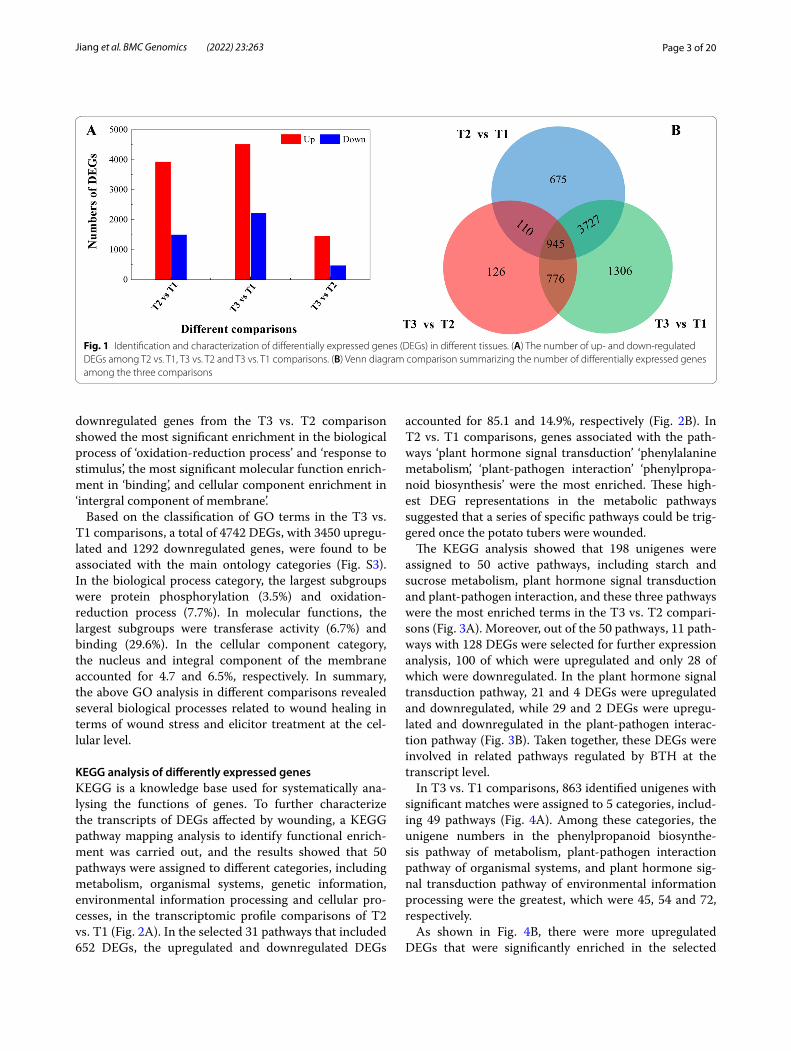
Differentially expressed genes were screened by pair-wise comparison using |FC| ≥ 2 and FDR <0.01 as a threshold. The results showed that the number of DEGs between T1 and T2 was 5457, including 3940 upregu-lated genes and 1517 downregulated genes, and a total of 1957 DEGs, including 1471 upregulated genes and 486 downregulated genes, were identified in the T3 versus T2 comparison. In addition, there were 6754 DEGs between T1 and T3, of which 4524 genes were upregulated and 2227 genes were downregulated (Fig. 1A). A Venn dia-gram of the DEG analysis indicated that 945 genes were shared across the three groups of paired comparisons, which accounted for 1.2% of all the DEGs. The results illustrated that 110 common genes appeared in the T2 vs. T1 and T3 vs. T2 comparisons, 776 genes were shared between T3 vs. T2 and T3 vs. T1, and 3727 genes were shared between T2 vs. T1 and T3 vs. T1 (Fig. 1B). These results indicated that most of the genes were regulated at the transcriptional level during wound healing.
GO annotation and enrichmentGO classification was used to describe the DEG proper-ties by biological process, molecular process and cellular component [19]. To investigate the potential functions of DEGs in the process of wound healing, a GO analy-sis in T2 vs. T1 comparisons was performed. A total of 4787 DEGs were classified into 30 functional categories, including 8 ‘biological process’, 8 ‘molecular functions’ and 14 ‘cellular components’, in which 3550 and 1237 genes were upregulated and downregulated, respectively, in T2 relative to T1 (Fig. S1). In each of these catego-ries, ‘response to stimulus (9.5%)’, ‘binding (28.5%)’ and ‘nucleus (5.3%)’ were the most abundant terms, suggest-ing that both the upregulated and downregulated genes enriched in these categories were related to the process of wound healing.
To evaluate the functions of DEGs in tissues undergo-ing wound healing after treatment BTH, 25 functional categories were classified, including biological process (10), molecular functions (7) and cellular component (8) (Fig. S2). The GO analysis of 1231 upregulated and 231
Page 3 of 20Jiang et al. BMC Genomics (2022) 23:263
downregulated genes from the T3 vs. T2 comparison showed the most significant enrichment in the biological process of ‘oxidation-reduction process’ and ‘response to stimulus’, the most significant molecular function enrich-ment in ‘binding’, and cellular component enrichment in ‘intergral component of membrane’.
Based on the classification of GO terms in the T3 vs. T1 comparisons, a total of 4742 DEGs, with 3450 upregu-lated and 1292 downregulated genes, were found to be associated with the main ontology categories (Fig. S3). In the biological process category, the largest subgroups were protein phosphorylation (3.5%) and oxidation-reduction process (7.7%). In molecular functions, the largest subgroups were transferase activity (6.7%) and binding (29.6%). In the cellular component category, the nucleus and integral component of the membrane accounted for 4.7 and 6.5%, respectively. In summary, the above GO analysis in different comparisons revealed several biological processes related to wound healing in terms of wound stress and elicitor treatment at the cel-lular level.
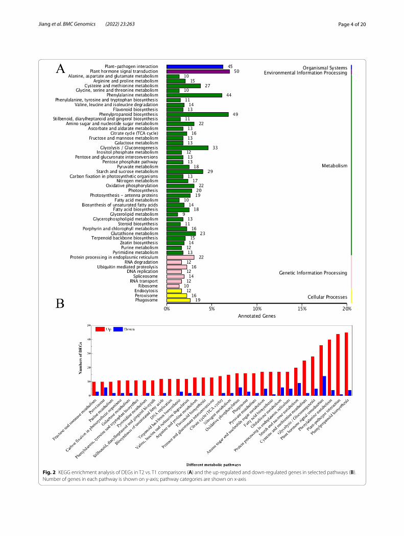
KEGG analysis of differently expressed genesKEGG is a knowledge base used for systematically ana-lysing the functions of genes. To further characterize the transcripts of DEGs affected by wounding, a KEGG pathway mapping analysis to identify functional enrich-ment was carried out, and the results showed that 50 pathways were assigned to different categories, including metabolism, organismal systems, genetic information, environmental information processing and cellular pro-cesses, in the transcriptomic profile comparisons of T2 vs. T1 (Fig. 2A). In the selected 31 pathways that included 652 DEGs, the upregulated and downregulated DEGs
accounted for 85.1 and 14.9%, respectively (Fig. 2B). In T2 vs. T1 comparisons, genes associated with the path-ways ‘plant hormone signal transduction’ ‘phenylalanine metabolism’, ‘plant-pathogen interaction’ ‘phenylpropa-noid biosynthesis’ were the most enriched. These high-est DEG representations in the metabolic pathways suggested that a series of specific pathways could be trig-gered once the potato tubers were wounded.
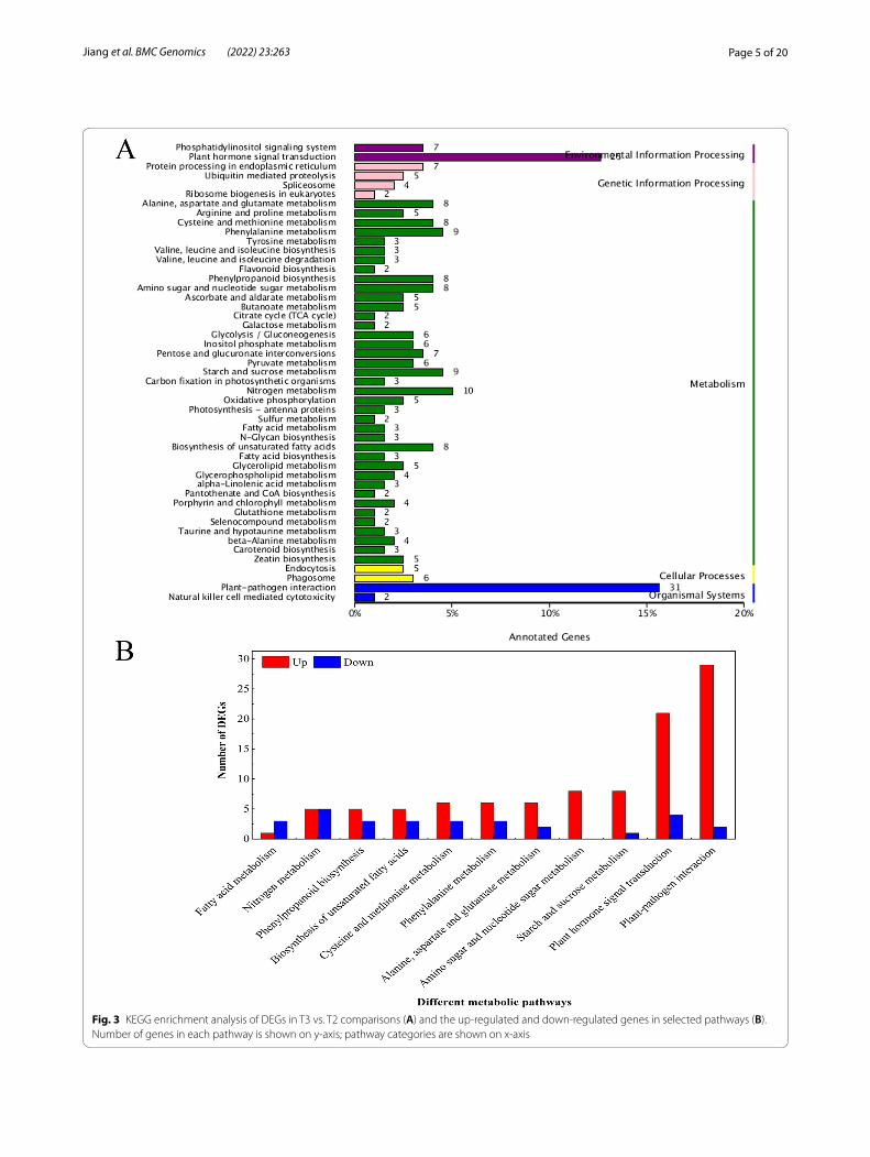
The KEGG analysis showed that 198 unigenes were assigned to 50 active pathways, including starch and sucrose metabolism, plant hormone signal transduction and plant-pathogen interaction, and these three pathways were the most enriched terms in the T3 vs. T2 compari-sons (Fig. 3A). Moreover, out of the 50 pathways, 11 path-ways with 128 DEGs were selected for further expression analysis, 100 of which were upregulated and only 28 of which were downregulated. In the plant hormone signal transduction pathway, 21 and 4 DEGs were upregulated and downregulated, while 29 and 2 DEGs were upregu-lated and downregulated in the plant-pathogen interac-tion pathway (Fig. 3B). Taken together, these DEGs were involved in related pathways regulated by BTH at the transcript level.
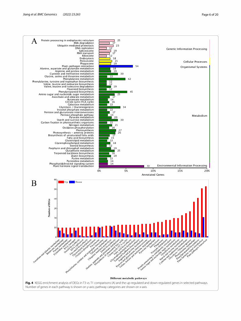
In T3 vs. T1 comparisons, 863 identified unigenes with significant matches were assigned to 5 categories, includ-ing 49 pathways (Fig. 4A). Among these categories, the unigene numbers in the phenylpropanoid biosynthe-sis pathway of metabolism, plant-pathogen interaction pathway of organismal systems, and plant hormone sig-nal transduction pathway of environmental information processing were the greatest, which were 45, 54 and 72, respectively.
As shown in Fig. 4B, there were more upregulated DEGs that were significantly enriched in the selected
Fig. 1 Identification and characterization of differentially expressed genes (DEGs) in different tissues. (A) The number of up- and down-regulated DEGs among T2 vs. T1, T3 vs. T2 and T3 vs. T1 comparisons. (B) Venn diagram comparison summarizing the number of differentially expressed genes among the three comparisons
Page 4 of 20Jiang et al. BMC Genomics (2022) 23:263
Fig. 2 KEGG enrichment analysis of DEGs in T2 vs. T1 comparisons (A) and the up-regulated and down-regulated genes in selected pathways (B). Number of genes in each pathway is shown on y-axis; pathway categories are shown on x-axis
Page 5 of 20Jiang et al. BMC Genomics (2022) 23:263
Fig. 3 KEGG enrichment analysis of DEGs in T3 vs. T2 comparisons (A) and the up-regulated and down-regulated genes in selected pathways (B). Number of genes in each pathway is shown on y-axis; pathway categories are shown on x-axis
Page 6 of 20Jiang et al. BMC Genomics (2022) 23:263
Fig. 4 KEGG enrichment analysis of DEGs in T3 vs. T1 comparisons (A) and the up-regulated and down-regulated genes in selected pathways. Number of genes in each pathway is shown on y-axis; pathway categories are shown on x-axis
Page 7 of 20Jiang et al. BMC Genomics (2022) 23:263
38 pathways than downregulated DEGs, and the plant-pathogen interaction was the pathway with the greatest number of EDGs. These results indicated that various metabolic processes occurred in response to wound-ing healing and that the DEGs in these pathways may play crucial roles during wound healing induced by BTH. Overall, these results highlighted a range of dif-ferent molecular regulation strategies depending on the wound healing of potato tubers, and our data indicated that these candidates involved in various metabolic and biosynthetic pathways may be crucial in the response to wound healing.
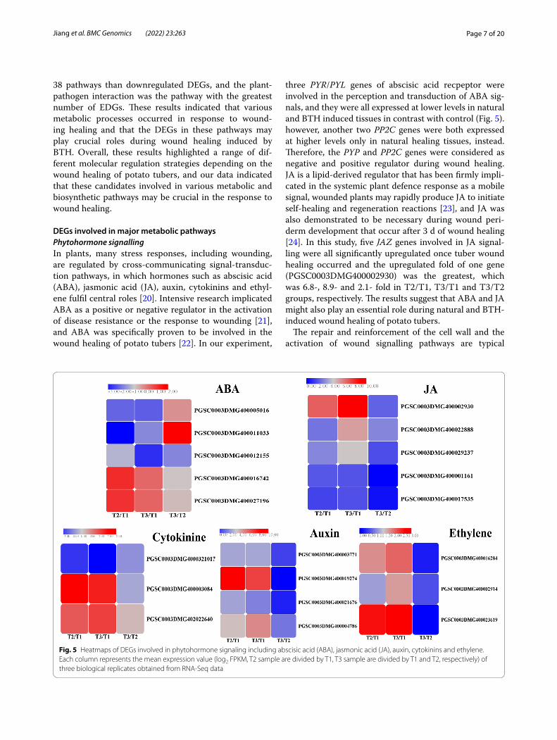
DEGs involved in major metabolic pathwaysPhytohormone signallingIn plants, many stress responses, including wounding, are regulated by cross-communicating signal-transduc-tion pathways, in which hormones such as abscisic acid (ABA), jasmonic acid (JA), auxin, cytokinins and ethyl-ene fulfil central roles [20]. Intensive research implicated ABA as a positive or negative regulator in the activation of disease resistance or the response to wounding [21], and ABA was specifically proven to be involved in the wound healing of potato tubers [22]. In our experiment,
three PYR/PYL genes of abscisic acid recpeptor were involved in the perception and transduction of ABA sig-nals, and they were all expressed at lower levels in natural and BTH induced tissues in contrast with control (Fig. 5). however, another two PP2C genes were both expressed at higher levels only in natural healing tissues, instead. Therefore, the PYP and PP2C genes were considered as negative and positive regulator during wound healing. JA is a lipid-derived regulator that has been firmly impli-cated in the systemic plant defence response as a mobile signal, wounded plants may rapidly produce JA to initiate self-healing and regeneration reactions [23], and JA was also demonstrated to be necessary during wound peri-derm development that occur after 3 d of wound healing [24]. In this study, five JAZ genes involved in JA signal-ling were all significantly upregulated once tuber wound healing occurred and the upregulated fold of one gene (PGSC0003DMG400002930) was the greatest, which was 6.8-, 8.9- and 2.1- fold in T2/T1, T3/T1 and T3/T2 groups, respectively. The results suggest that ABA and JA might also play an essential role during natural and BTH-induced wound healing of potato tubers.
The repair and reinforcement of the cell wall and the activation of wound signalling pathways are typical
Fig. 5 Heatmaps of DEGs involved in phytohormone signaling including abscisic acid (ABA), jasmonic acid (JA), auxin, cytokinins and ethylene. Each column represents the mean expression value (log2 FPKM, T2 sample are divided by T1, T3 sample are divided by T1 and T2, respectively) of three biological replicates obtained from RNA-Seq data
Page 8 of 20Jiang et al. BMC Genomics (2022) 23:263
characteristics of physiological responses to wound-ing and occur through hormones related to wounding, such as auxin, ethylene and cytokinine [25]. Auxin is a hormone that controls the growth and development of plants and functions in recovery from different wounds or organ loss [26]. The four auxin-responsive genes in Fig. 5 were all upregulated in natural and BTH-treated wound healing tissues, among which the GH3 gene PGSC0003DMG400019274 was upregulated by 8.3-fold and 6.7-fold in T2/T1 and T3/T1 groups. This can be reflected by the description of indole-3-acetic acid content increased by 2- and 4-fold after 5 and 7 days of wounding [27]. Another hormone called cytokine plays diverse roles in the defence response to patho-gens, including preinvasive defence, by regulating sto-matal closure and postinvasive defence by inducing callose deposition or gene expression related to defence or phytoalexin generation [28]. In our study, one and two cytokinine genes were down and upregulated in natural and BTH induced tissues in contrast with non-wound tissues, respectively. Ethylene, one of the classical defence hormones, is released when plants are wounded mechanically [26]. Heyman et al. reported that the eth-ylene response factor (ERF) family coordinates stress signalling with wound healing, which initiates the reac-tivation of cell division [29]. In the present experiment, three ERF genes in the ethylene signal were all upregu-lated in natural and BTH-induced wound tissues com-pared with non-wounded tissues. And the ERF gene PGSC0003DMG400023619 was upregulated 2.5- and 2.6-fold in these two groups. ERF genes also involved in disease resistance, it was discovered that CpERF7 was significantly up-regulated after BTH treatment [30]. To sum up, we speculate that the expression of these genes involved in different hormone signalling pathways was triggered by the natural or BTH-induced wound healing of potato tubers. Interestingly, the differential expression of multiple phytohormone signalling genes may indicate that there are not just linear and isolated cascades but also crosstalk reactions between these hormone signals.
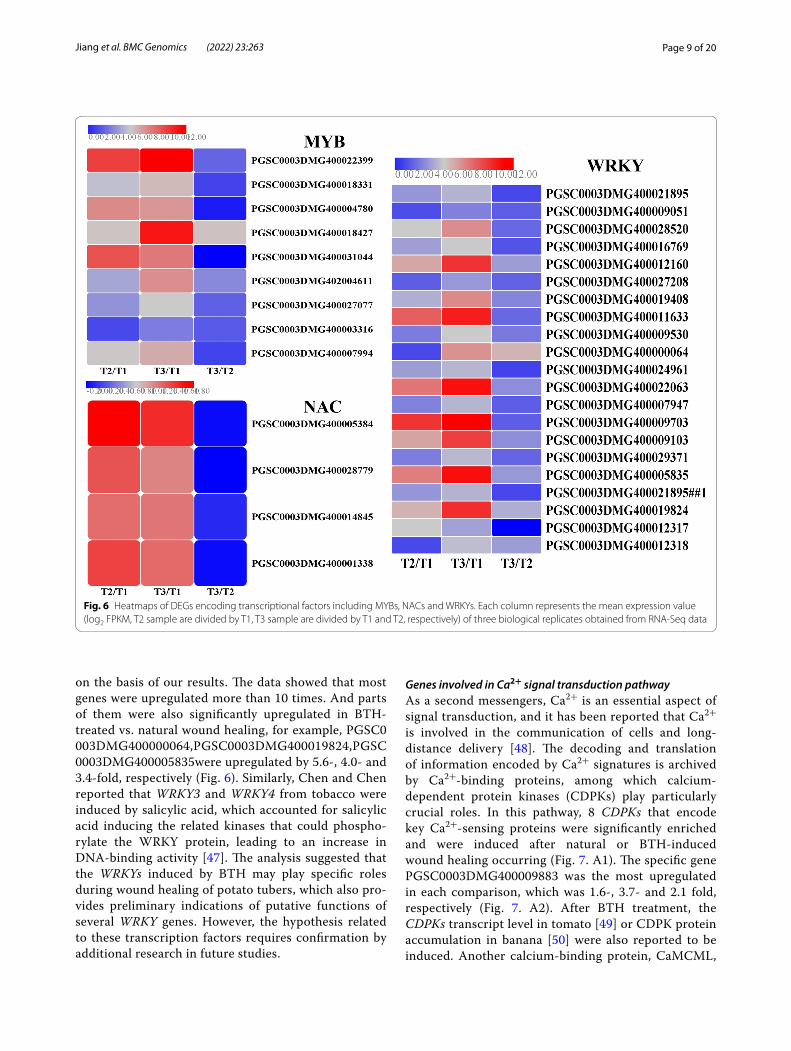
Transcription factorsComplicated transcription regulatory networks require the involvement of a series of transcription factors that belong to important constituents of signalling pathways and play pivotal roles in the response to biotic and abi-otic stresses [31, 32]. These transcription factors include MYB, NAC and WRKY, which are encoded by a large number of genes. MYB transcription factors serve as one of the largest family genes and act as crucial regulators by controlling gene expression in plant development and stress responses [33]. There are 233 MYB family mem-bers that have been identified and analyzed in the potato
genome and were found to confer different responses to abiotic and biotic stresses [34]. In Arabidopsis thaliana, the transcription levels of suberin biosynthetic genes were increased by the overexpression of AtMYB41 and induced ferulate accumulation [35], and the transcrip-tional regulators MYB58 and MYB63 could specifically activate lignin biosynthetic genes during secondary wall formation [36]. Lashbrooke et al. reported that AtMYB9 and AtMYB107 are required for suberin assembly and that the corresponding mutants led to a significant reduction in suberin monomers [37]. In kiwifruit, Ach-nMYB41 and AchnMYB107 play a positive role in the activation of AchnFHT (ω-hydroxyacid/fatty alcohol hydroxycinnamoyl transferase, FHT), while AchnMYB4 works as a negative regulator, by which suberin mono-mer biosynthesis is therefore controlled [38]. In this study, a total of 9 potato MYB genes were upregulated by either natural or BTH-treated wound healing. Except for PGSC0003DMG400003316, other genes exhibited lower FPKM value below 1.0 in non-healing tissues, while they are increased more than 10 times after natural or BTH-induced wound healing (Fig. 6), it was suggested that these upregulated MYB genes in response to wound heal-ing might play a regulatory role in this process.
NAC transcription factors are found in a wide range of plants and are one of the largest families and have been implicated in defensive responses to pathogens or environmental stresses [39]. In Arabidopsis, two NAC transcription factors were documented to be induced after wounding and fungal or bacterial pathogen infec-tion [40, 41]. In potato, 110 NAC genes have been iden-tified by genome-wide analysis, some of which were reported to be expressed under wounding and BTH-treated conditions [42]. In our experiment, 4 NACs transcription factors were all found to be upregulated in natural and BTH-treated wound healing tissues. Simi-lar with WRKY, these four genes in non-healing tissues were not expressed at all, but expressed at higher level in natural and BTH-induced tissues. Especially, the gene (PGSC0003DMG400005384) showed the greatest expression level, which has been proved to repressed the suberin polyester and suberin-associated waxes deposi-tion [43]. Therefore, we hypothesize that the upregula-tion of these NAC genes after wounding may regulate the process of wound healing in potato tubers.
WRKY transcription factors are widely expressed in various organisms and are considered as the largest family of transcription factors [44]. Many studies have shown that WRKY regulators defend against patho-gens and provide resistance to wounding [45]. There are 79 WRKY family members that have been identified in the potato genome [46], 20 of which were analyzed and all of them were induced in wound healing tissues
Page 9 of 20Jiang et al. BMC Genomics (2022) 23:263
on the basis of our results. The data showed that most genes were upregulated more than 10 times. And parts of them were also significantly upregulated in BTH-treated vs. natural wound healing, for example, PGSC0003DMG400000064,PGSC0003DMG400019824,PGSC0003DMG400005835were upregulated by 5.6-, 4.0- and 3.4-fold, respectively (Fig. 6). Similarly, Chen and Chen reported that WRKY3 and WRKY4 from tobacco were induced by salicylic acid, which accounted for salicylic acid inducing the related kinases that could phospho-rylate the WRKY protein, leading to an increase in DNA-binding activity [47]. The analysis suggested that the WRKYs induced by BTH may play specific roles during wound healing of potato tubers, which also pro-vides preliminary indications of putative functions of several WRKY genes. However, the hypothesis related to these transcription factors requires confirmation by additional research in future studies.
Genes involved in Ca2+ signal transduction pathwayAs a second messengers, Ca2+ is an essential aspect of signal transduction, and it has been reported that Ca2+ is involved in the communication of cells and long-distance delivery [48]. The decoding and translation of information encoded by Ca2+ signatures is archived by Ca2+-binding proteins, among which calcium-dependent protein kinases (CDPKs) play particularly crucial roles. In this pathway, 8 CDPKs that encode key Ca2+-sensing proteins were significantly enriched and were induced after natural or BTH-induced wound healing occurring (Fig. 7. A1). The specific gene PGSC0003DMG400009883 was the most upregulated in each comparison, which was 1.6-, 3.7- and 2.1 fold, respectively (Fig. 7. A2). After BTH treatment, the CDPKs transcript level in tomato [49] or CDPK protein accumulation in banana [50] were also reported to be induced. Another calcium-binding protein, CaMCML,
Fig. 6 Heatmaps of DEGs encoding transcriptional factors including MYBs, NACs and WRKYs. Each column represents the mean expression value (log2 FPKM, T2 sample are divided by T1, T3 sample are divided by T1 and T2, respectively) of three biological replicates obtained from RNA-Seq data

Page 10 of 20Jiang et al. BMC Genomics (2022) 23:263
and a total of 15 DEGs were identified in the potato genome, which were all initiated and upregulated by wound healing except for PGSC0003DMG400022693, the upregulation fold of PGSC0003DMG400002993 and PGSC0003DMG400020261 in T2/T1 and T3/T1 reached 4-7 fold.
Respiratory burst oxidase homologues (RBOH) in plasma membranes mainly function in ROS production via signal transduction pathways [51]. Rbohs, a multi-gene family, serve as multispanning transmembrane proteins in the Ca2+ signal transduction pathway and require Ca2+ binding for their activation in response to environmental stresses [52]. It has been reported that early ROS production after potato wounding par-ticipates in the signal cascade reaction, and later ROS are thought to be tied to suberin poly (phenolic) cross-linking in wound healing [53, 54]. In our transcriptomic data, three Rbohs genes were identified in healing tissues,
while two of them (PGSC0003DMG400024754 and PGSC0003DMG400014168) exhibited higher expression levels in healing tissues, the former showed the greatest upregulation fold, especially under the action of BTH application (Fig. 7. A3). Thus, the expression of these pro-teins reflects that the Ca2+-mediated signal transduction pathway via related proteins may be involved in the natu-ral or BTH-induced healing process.
Starch and sugar metabolismSuberization is a complex process including a set of physiological and metabolic events that require the coor-dinated regulation of genes involving primary and sec-ondary pathways both spatially and temporally [55]. The analysis of KEGG enrichment highlighted some func-tional categories: DEGs associated with starch and sugar metabolism, fatty acid metabolism, phenylpropanoid bio-synthesis and terpenoid skeleton biosynthesis. Therefore,
Fig. 7 Heatmaps of DEGs encoding genes related with Ca2+ signal transduction during wound healing of potato tubers (A1) and the up-regulated fold of StCDPKs (A2) and StRBOHs (A3) in different comparisons. (Calcium dependent protein kinase, CDPK; Respiratory burst oxidasehomologue, RBOH; calmodulinc binding protein, CaMCaL. Each column represents the mean expression value (log2 FPKM, T2 sample are divided by T1, T3 sample are divided by T1 and T2, respectively) of three biological replicates obtained from RNA-Seq data)
Page 11 of 20Jiang et al. BMC Genomics (2022) 23:263
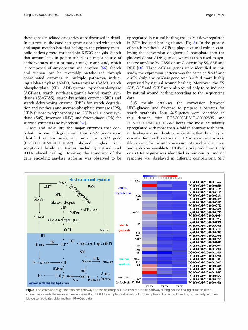
these genes in related categories were discussed in detail. In our results, the candidate genes associated with starch and sugar metabolism that belong to the primary meta-bolic pathway were enriched via KEGG analysis. Starch that accumulates in potato tubers is a major source of carbohydrates and a primary storage compound, which is composed of amylopectin and amylose [56]. Starch and sucrose can be reversibly metabolized through coordinated enzymes in multiple pathways, includ-ing alpha-amylase (AMY), beta-amylase (BAM), starch phosphorylase (SP), ADP-glucose pyrophosphorylase (AGPase), starch synthases/granule-bound starch syn-thases (SS/GBSS), starch-branching enzyme (SBE) and starch debranching enzyme (DBE) for starch degrada-tion and synthesis and sucrose-phosphate synthase (SPS), UDP-glucose pyrophosphorylase (UGPase), sucrose syn-thase (SuS), invertase (INV) and fructokinase (Frk) for sucrose synthesis and hydrolysis [57].
AMY and BAM are the major enzymes that con-tribute to starch degradation. Four BAM genes were identified in our work, and only one BAM gene (PGSC0003DMG400001549) showed higher tran-scriptional levels in tissues including natural and BTH-induced healing. However, the transcript of the gene encoding amylase isoforms was observed to be
upregulated in natural healing tissues but downregulated in BTH-induced healing tissues (Fig. 8). In the process of starch synthesis, AGPase plays a crucial role in cata-lysing the conversion of glucose-1-phosphate into the glucosyl donor ADP-glucose, which is then used to syn-thesize amylose by GBSS or amylopectin by SS, SBE and DBE [58]. Three AGPase genes were identified in this study, the expression pattern was the same as BAM and AMY. Only one AGPase gene was 3.2-fold more highly expressed by natural wound healing. Moreover, the SS, SBE, DBE and G6PT were also found only to be induced by natural wound healing according to the sequencing data.
SuS mainly catalyses the conversion between UDP-glucose and fructose to prepare substrates for starch synthesis. Four SuS genes were identified in this dataset, with PGSC0003DMG400002895 and PGSC0003DMG400013547 being the most abundantly upregulated with more than 3-fold in contrast with natu-ral healing and non-healing, suggesting that they may be essential for starch synthesis. UDPase serves as a revers-ible enzyme for the interconversion of starch and sucrose and is also responsible for UDP-glucose production. Only one UDPase gene was identified in our results, and no response was displayed in different comparisons. SPS
Fig. 8 The starch and sugar metabolism pathway and the heatmap of DEGs involved in this pathway during wound healing of tubers (Each column represents the mean expression value (log2 FPKM, T2 sample are divided by T1, T3 sample are divided by T1 and T2, respectively) of three biological replicates obtained from RNA-Seq data)
Page 12 of 20Jiang et al. BMC Genomics (2022) 23:263
functions in the conversion of fructose into sucrose, and two SPS genes that were significantly downregulated in natural and BTH-treated healing tissues were annotated in our study. Another enzyme, FrK, converts fructose to fructose-6-phosphate, with a downregulation expres-sion pattern in different tissues. Genes related to starch and sugar metabolism have been well researched in other species. However, the key genes and their regula-tory mechanism in the process of wound healing remain unknown. The results presented herein may be helpful for further studying the molecular mechanism underly-ing starch and sucrose metabolism during wound healing of potato tubers.
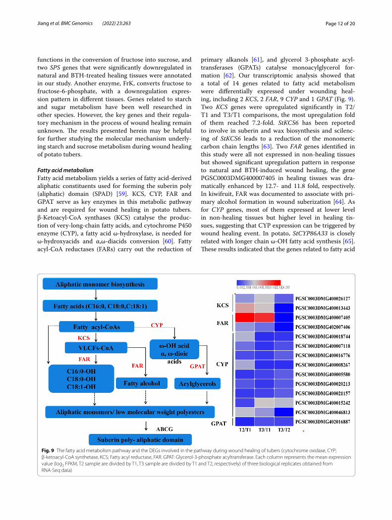
Fatty acid metabolismFatty acid metabolism yields a series of fatty acid-derived aliphatic constituents used for forming the suberin poly (aliphatic) domain (SPAD) [59]. KCS, CYP, FAR and GPAT serve as key enzymes in this metabolic pathway and are required for wound healing in potato tubers. β-Ketoacyl-CoA synthases (KCS) catalyse the produc-tion of very-long-chain fatty acids, and cytochrome P450 enzyme (CYP), a fatty acid ω-hydroxylase, is needed for ω-hydroxyacids and α,ω-diacids conversion [60]. Fatty acyl-CoA reductases (FARs) carry out the reduction of
primary alkanols [61], and glycerol 3-phosphate acyl-transferases (GPATs) catalyse monoacylglycerol for-mation [62]. Our transcriptomic analysis showed that a total of 14 genes related to fatty acid metabolism were differentially expressed under wounding heal-ing, including 2 KCS, 2 FAR, 9 CYP and 1 GPAT (Fig. 9). Two KCS genes were upregulated significantly in T2/T1 and T3/T1 comparisons, the most upregulation fold of them reached 7.2-fold. StKCS6 has been reported to involve in suberin and wax biosynthesis and scilenc-ing of StKCS6 leads to a reduction of the monomeric carbon chain lengths [63]. Two FAR genes identified in this study were all not expressed in non-healing tissues but showed significant upregulation pattern in response to natural and BTH-induced wound healing, the gene PGSC0003DMG400007405 in healing tissues was dra-matically enhanced by 12.7- and 11.8 fold, respectively. In kiwifruit, FAR was documented to associate with pri-mary alcohol formation in wound suberization [64]. As for CYP genes, most of them expressed at lower level in non-healing tissues but higher level in healing tis-sues, suggesting that CYP expression can be triggered by wound healing event. In potato, StCYP86A33 is closely related with longer chain ω-OH fatty acid synthesis [65]. These results indicated that the genes related to fatty acid
Fig. 9 The fatty acid metabolism pathway and the DEGs involved in the pathway during wound healing of tubers (cytochrome oxidase, CYP; β-ketoacyl-CoA synthetase, KCS; Fatty acyl reductase, FAR. GPAT: Glycerol-3-phosphate acyltransferase. Each column represents the mean expression value (log2 FPKM, T2 sample are divided by T1, T3 sample are divided by T1 and T2, respectively) of three biological replicates obtained from RNA-Seq data)
Page 13 of 20Jiang et al. BMC Genomics (2022) 23:263
metabolism could be induced by wound healing and were regulated at the transcriptional level.
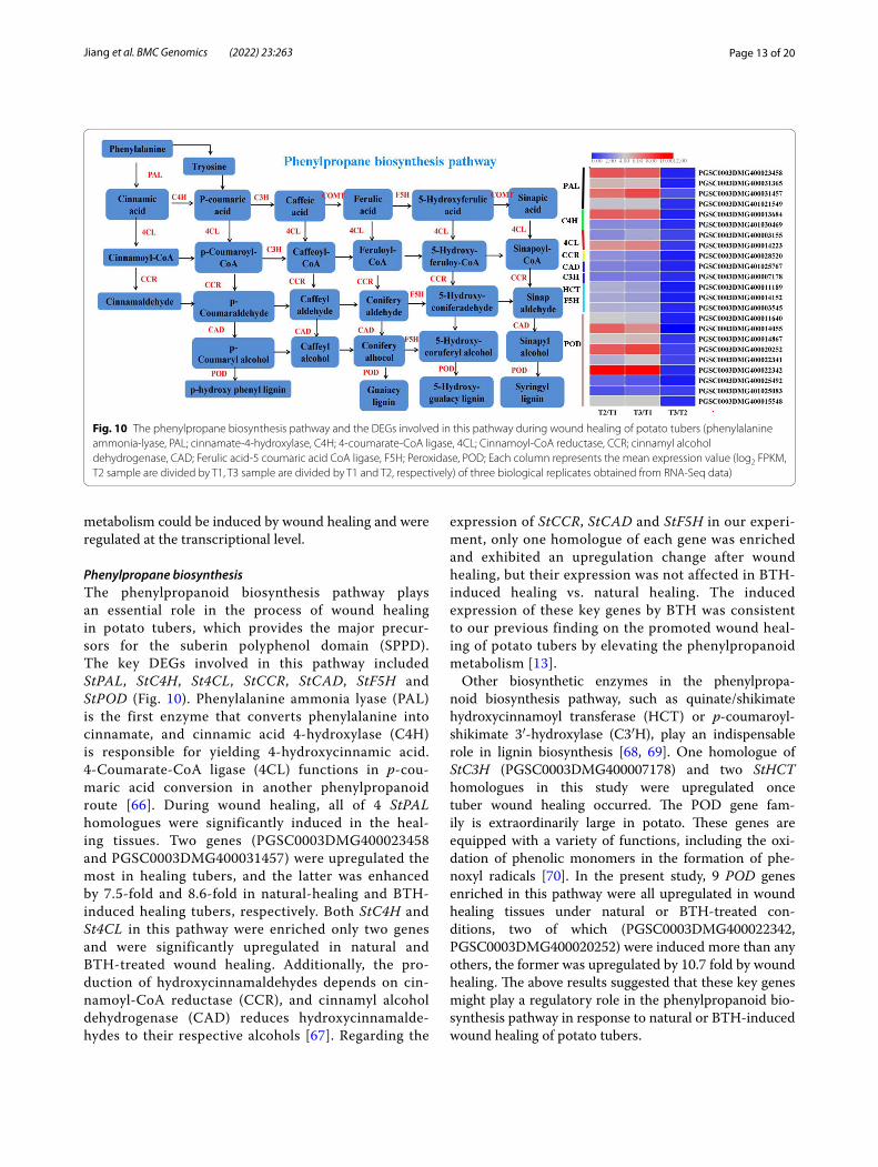
Phenylpropane biosynthesisThe phenylpropanoid biosynthesis pathway plays an essential role in the process of wound healing in potato tubers, which provides the major precur-sors for the suberin polyphenol domain (SPPD). The key DEGs involved in this pathway included StPAL, StC4H, St4CL, StCCR , StCAD, StF5H and StPOD (Fig. 10). Phenylalanine ammonia lyase (PAL) is the first enzyme that converts phenylalanine into cinnamate, and cinnamic acid 4-hydroxylase (C4H) is responsible for yielding 4-hydroxycinnamic acid. 4-Coumarate-CoA ligase (4CL) functions in p-cou-maric acid conversion in another phenylpropanoid route [66]. During wound healing, all of 4 StPAL homologues were significantly induced in the heal-ing tissues. Two genes (PGSC0003DMG400023458 and PGSC0003DMG400031457) were upregulated the most in healing tubers, and the latter was enhanced by 7.5-fold and 8.6-fold in natural-healing and BTH-induced healing tubers, respectively. Both StC4H and St4CL in this pathway were enriched only two genes and were significantly upregulated in natural and BTH-treated wound healing. Additionally, the pro-duction of hydroxycinnamaldehydes depends on cin-namoyl-CoA reductase (CCR), and cinnamyl alcohol dehydrogenase (CAD) reduces hydroxycinnamalde-hydes to their respective alcohols [67]. Regarding the
expression of StCCR , StCAD and StF5H in our experi-ment, only one homologue of each gene was enriched and exhibited an upregulation change after wound healing, but their expression was not affected in BTH-induced healing vs. natural healing. The induced expression of these key genes by BTH was consistent to our previous finding on the promoted wound heal-ing of potato tubers by elevating the phenylpropanoid metabolism [13].
Other biosynthetic enzymes in the phenylpropa-noid biosynthesis pathway, such as quinate/shikimate hydroxycinnamoyl transferase (HCT) or p-coumaroyl-shikimate 3′-hydroxylase (C3′H), play an indispensable role in lignin biosynthesis [68, 69]. One homologue of StC3H (PGSC0003DMG400007178) and two StHCT homologues in this study were upregulated once tuber wound healing occurred. The POD gene fam-ily is extraordinarily large in potato. These genes are equipped with a variety of functions, including the oxi-dation of phenolic monomers in the formation of phe-noxyl radicals [70]. In the present study, 9 POD genes enriched in this pathway were all upregulated in wound healing tissues under natural or BTH-treated con-ditions, two of which (PGSC0003DMG400022342, PGSC0003DMG400020252) were induced more than any others, the former was upregulated by 10.7 fold by wound healing. The above results suggested that these key genes might play a regulatory role in the phenylpropanoid bio-synthesis pathway in response to natural or BTH-induced wound healing of potato tubers.
Fig. 10 The phenylpropane biosynthesis pathway and the DEGs involved in this pathway during wound healing of potato tubers (phenylalanine ammonia-lyase, PAL; cinnamate-4-hydroxylase, C4H; 4-coumarate-CoA ligase, 4CL; Cinnamoyl-CoA reductase, CCR; cinnamyl alcohol dehydrogenase, CAD; Ferulic acid-5 coumaric acid CoA ligase, F5H; Peroxidase, POD; Each column represents the mean expression value (log2 FPKM, T2 sample are divided by T1, T3 sample are divided by T1 and T2, respectively) of three biological replicates obtained from RNA-Seq data)
Page 14 of 20Jiang et al. BMC Genomics (2022) 23:263
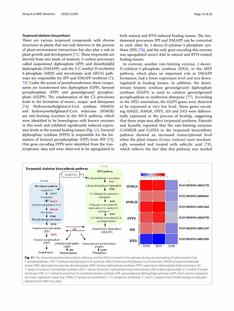
Terpenoid skeleton biosynthesisThere are various terpenoid compounds with diverse structures in plants that not only function in the process of plant-environment interactions but also play a role in plant growth and development [71]. These terpenoids are derived from two kinds of isomeric 5-carbon precursors called isopentenyl diphosphate (IPP) and dimethylallyl diphosphate (DMAPP), and the 2-C-methyl-D-erythritol 4-phosphate (MEP) and mevalonate acid (MVA) path-ways are responsible for IPP and DMAPP synthesis [72, 73]. Under the action of prenyltransferases, these compo-nents are transformed into diphosphate (GPP), farnesyl pyrophosphate (FPP) and geranylgeranyl pyrophos-phate (GGPP). The condensation of the C5 precursors leads to the formation of mono-, sesqui- and diterpenes [74]. Hydroxymethylglutaryl-CoA synthase (HMGS) and hydroxymethylglutaryl-CoA reductase (HMGR) are rate-limiting enzymes in the MVA pathway, which were identified to be homologous with known enzymes in this work and exhibited significantly induced expres-sion levels in the wound healing tissues (Fig. 11). Farnesyl diphosphate synthase (FPPS) is responsible for the for-mation of farnesyl pyrophosphate (FPP) from IPP [75]. One gene encoding FPPS were identified from the tran-scriptomic data and were observed to be upregulated in
both natural and BTH-induced healing tissues. The fun-damental precursors IPP and DMAPP can be converted to each other by 1-deoxy-D-xylulose-5-phosphate syn-thase (IDI) [76], and the only gene encoding this enzyme was upregulated more1-fold in natural and BTH-treated healing tissues.
In contrast, another rate-limiting enzyme, 1-deoxy-D-xylulose-5-phosphate synthase (DXS), in the MEP pathway, which plays an important role in DMAPP formation, had a lower expression level and was down-regulated in healing tissues. In addition, the down-stream terpene synthase geranylgeranyl diphosphate synthase (GGPS) is used to catalyse geranylgeranyl pyrophosphate to synthesize diterpene [77]. According to the DEG annotation, the GGPS genes were detected to be expressed at very low level. These genes encod-ing HMGS, HMGR, FPPS, IDI and DXS were differen-tially expressed in the process of healing, suggesting that these steps may affect terpenoid synthesis. Pateraki and Kanellis reported that the rate-limiting enzymes CcHMGR and CcDXS in the terpenoid biosynthetic pathway showed an increased transcriptional level when the plant tissues (Cistus creticus) were mechani-cally wounded and treated with salicylic acid [78], which reflects the fact that this pathway was needed
Fig. 11 The terpenoid skeleton biosynthesis pathway and the DEGs involved in the pathway during wound healing of tubers (acetyl-CoA C-acetyltransferase, AACT; hydroxymethylglutaryl-CoA synthase, HMGS; hydroxymethylglutaryl-CoA reductase, HMGR; phosphomevalonate kinase, PMK; diphosphomevalonate decarboxylase, MVD; farnesyl diphosphate synthase, FPPS; isopentenyl-diphosphate Delta-isomerase, IDI; 1-deoxy-D-xylulose-5-phosphate synthase, DXS; 1-deoxy-Dxylulose-5-phosphate reductoisomerase, DXR; 4-diphosphocytidyl-2-C-methyl-D-erythritol kinase, ISPE; 2-C-methyl-D-erythritol 2,4-cyclodiphosphate synthase, ISPF; geranylgeranyl diphosphate synthase, GGPS. Each column represents the mean expression value (log2 FPKM, T2 sample are divided by T1, T3 sample are divided by T1 and T2, respectively) of three biological replicates obtained from RNA-Seq data)
Page 15 of 20Jiang et al. BMC Genomics (2022) 23:263
for wound stress. In addition, terpenoid release was reported to be capable of defending against pests [79] and pathogenic fungi [80]. Interestingly, except for ster-oids and carotenoids, a number of phytohormones, including cytokinins, abscisic acid and gibberellins, are derived from terpenoids [72, 81]. Therefore, character-izing these genes further provides a new understanding of the underlying action pattern and molecular mecha-nism of terpenoid biosynthesis during wound healing of potato tubers.
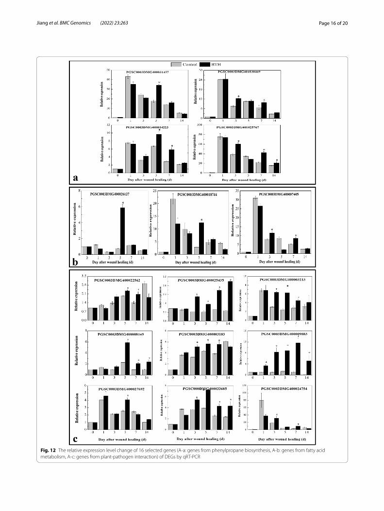
Verification of gene expression related to phenylpropane biosynthesis, fatty acid metabolism and plant‑pathogen interactionsTo validate the expression profiles obtained from RNA-Seq analysis, 16 DEGs from three meta-bolic pathways were randomly selected to explore their expression profiles in wound healing tubers under BTH treatment using qRT–PCR. The efla gene was used as a control for expression nor-malization. These unigenes were involved in phenylpropane biosynthesis and fatty acid and plant-pathogen interaction pathways (Fig. 12). The following genes, StPAL (PGSC0003DMG400031457), StC4H (PGSC0003DMG401030469), St4CL (PGSC0003 DMG400014223) and StCAD (PGSC0003DMG401025767), were induced by BTH at different time points and are involved in phenylpropane biosynthesis. The StKCS (PGSC0003DMG400026127) and StCYP (PGSC0003DMG400018744) genes only exhibited higher expression levels after BTH treatment at 5 days of healing, and another gene, StFAR (PGSC0003DMG400007405), was increased by BTH at 3 and 7 days of healing. How-ever, most of the genes encoding Ca2+-binding proteins (PGSC0003DMG400022562/PGSC0003DMG400025435/PGSC0003DMG400003213/PGSC0003DMG400008149/PGSC0003DMG400013183/PGSC0003DMG400033685) involved in plant-pathogen interactions were significantly induced by BTH from 3 d to 14 d of healing.
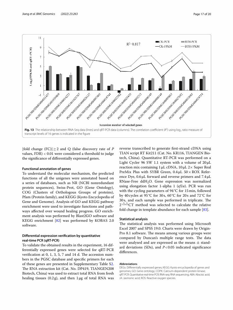
The results showed that although the expression pat-terns of the selected genes varied between RNA-Seq and qRT-PCR analysis, the trend of gene expression changes detected by qRT-PCR largely coincided with the transcriptome sequencing results (Fig. 13). Analysis of Pearson’s correlation coefficient showed that qRT-PCR detection for selected genes and RNA-Seq data at 5 d of healing were highly correlated. The correla-tion coefficient was 0.817, indicating that the RNA-Seq data were positively correlated with the qRT-PCR data, also indicating that the RNA-Seq data in this study are valuable.
ConclusionOur results provide the gene expression patterns related to wound healing of natural or BTH-treated in potato tubers, which also provides clues about important regu-latory mechanisms, especially those involving plant hormones, transcription factors, Ca2+-mediated signal transduction, and a series of metabolic pathways, such as starch and sugar metabolism, fatty acid metabolism, phenylpropane biosynthesis, and terpenoid skeleton biosynthesis pathways, that participate in the healing process. The present research revealed the underlying mechanisms of natural or BTH-treated wound healing of potato tubers, which provided valuable information for understanding the transcriptional regulation processes of wound healing from another perspective.
MethodsPotato tubers and BTH treatmentPotato tubers ‘cv. Longshu No. 3’ free from physical inju-ries and infection was purchased at Gansu Ailan Potato Seed Industry Co. Ltd. and used immediately. The wash-ing, surface sterilization and wounding treatment of tubers were performed as described by Jiang et al. [13]. Tubers were allowed to treat with sterile water and 100 mg/L BTH, which were subsequently stored at 20 °C and a relative humidity of 75–80% for wound healing. The healing tissues of the wounded surface at 0 d and 5 d were collected via manual dissection, frozen with liquid nitrogen, and archived at − 80 °C until further analysis.
RNA extraction and RNA‑SeqHealing tissue sampling and RNA-Seq from three time points, 0 d-Control (T1), 5 d-Control (T2) and 5 d-BTH (T3), were carried out in parallel as three biological rep-licates. Samples were sent to Breeding in Xi’an, where the cDNA library was produced and sequenced using an Illumina HiSeq2500 system. To ensure that the bases of high quality were used for de novo assembly, the raw sequences were filtered by removing adaptor sequences and poor-quality reads and produced 64.49 Gb clean data in total, which were used for subsequent analysis, and the sequenced data were filtered by quality score (Q ≥ 30). The statistics of clean reads are listed in the Supplemen-tary Table. S1. Then, all these clean reads were mapped to contig assemblies using TopHat 2 software and mapped to the reference genome of Solanum tuberosum down-loaded from Potato Genomics Resource (PGSC, http:// solan aceae. plant biolo gy. msu. edu/ pgsc_ downl oad. shtml). The values of fragments per kilobase of transcript per million fragments mapped (FPKM) were calculated by cufflinks software; subsequently, DESeq software was used to analyse differentially expressed genes. Genes with
Page 16 of 20Jiang et al. BMC Genomics (2022) 23:263
Fig. 12 The relative expression level change of 16 selected genes (A-a: genes from phenylpropane biosynthesis, A-b: genes from fatty acid metabolism, A-c: genes from plant-pathogen interaction) of DEGs by qRT-PCR
Page 17 of 20Jiang et al. BMC Genomics (2022) 23:263
|fold change (FC)| ≥ 2 and Q (false discovery rate of P values, FDR) < 0.01 were considered a threshold to judge the significance of differentially expressed genes.
Functional annotation of genesTo understand the molecular mechanism, the predicted functions of all the unigenes were annotated based on a series of databases, such as NR (NCBI nonredundant protein sequences), Swiss-Prot, GO (Gene Ontology), COG (Clusters of Orthologous Groups of proteins), Pfam (Protein family), and KEGG (Kyoto Encyclopedia of Gene and Genome). Analysis of GO and KEGG pathway enrichment were used to investigate functions and path-ways affected over wound healing progress. GO enrich-ment analysis was performed by Blast2GO software and KEGG enrichment [82] was performed by KOBAS 2.0 software.
Differential expression verification by quantitative real‑time PCR (qRT‑PCR)To validate the obtained results in the experiment, 16 dif-ferentially expressed genes were selected for qRT-PCR verification at 0, 1, 3, 5, 7 and 14 d. The accession num-bers in the PGSC database and specific primers for each of these genes are presented in Supplementary Table S2. The RNA extraction kit (Cat. No. DP419, TIANGEN208 Biotech, China) was used to extract total RNA from fresh healing tissues (0.2 g), and then 1 μg of total RNA was
reverse transcribed to generate first-strand cDNA using TIAN script RT Kit211 (Cat. No. KR116, TIANGEN Bio-tech, China). Quantitative RT-PCR was performed on a Light Cycler 96 SW 1.1 system with a volume of 20 μL reaction mix containing 1 μL cDNA, 10 μL 2× Super Real PreMix Plus with SYBR Green, 0.4 μL 50 × ROX Refer-ence Dye, 0.6 μL forward and reverse primers and 7.4 μL RNase-Free ddH2O. Gene expression was normalized using elongation factor 1-alpha 1 (ef1a). PCR was run with the cycling parameters of 94 °C for 15 min, followed by 40 cycles at 95 °C for 30 s, 60 °C for 20 s and 72 °C for 30 s, and each sample was performed in triplicate. The 2-△△CT method was selected to calculate the relative fold change in template abundance for each sample [83].
Statistical analysisThe statistical analysis was performed using Microsoft Excel 2007 and SPSS 19.0. Charts were drawn by Origin-Pro 8.1 software. The means among various groups were compared by Duncan’s multiple range tests. The data were analysed and are expressed as the means ± stand-ard deviations (SDs), and P < 0.05 indicated significance differences.
AbbreviationsDEGs: Differentially expressed genes; KEGG: Kyoto encyclopedia of genes and genomes; GO: Gene ontology; CDPK: Calcium-dependent protein kinase; qRT-PCR: Quantitative real-time PCR; RNA-seq: RNA sequencing; ABA: Abscisic acid; JA: Jasmonic acid; ROS: Reactive oxygen species.
Fig. 13 The relationship between RNA-Seq data (lines) and qRT-PCR data (columns). The correlation coefficient (R2) using log2 ratio measure of transcript levels of 16 genes is indicated in the figure
Page 18 of 20Jiang et al. BMC Genomics (2022) 23:263
Supplementary InformationThe online version contains supplementary material available at https:// doi. org/ 10. 1186/ s12864- 022- 08480-1.
Additional file 1.
Additional file 2.
Additional file 3.
Additional file 4.
Additional file 5.
AcknowledgementsWe would like to thank the editor and reviewers for critically evaluating the manuscript and providing constructive comments for its improvement.
Authors’ contributionsJH was responsible for undertaking experiment, data analysis and writing articles; LX and ML prepared the plant materials and collected samples. RYY assisted to write the manuscript and collected references, BY guided and modified the whole manuscript as the corresponding author. PD contributed to the suggestion of language and grammar in manuscript.
FundingThis work was fully supported by the National Natural Science Foundation (31772040).
Availability of data and materialsAll of data supporting our results are contained within the manuscript. All raw RNA-seq data in this manuscript are available for downloading from the NCBI Sequence Read Archive (BioProject ID: PRJNA757824).
Declarations
Ethics approval and consent to participateThis study did not directly involve humans or animals. Potato seeds were purchased from Potato Seed Industry Co. Ltd. at DingXi city, Gansu Province, China. The plant material used in this experiment is a common variety which is not endangered. And the research conducted complied with all institutional and national guidelines.
Consent for publicationNot applicable.
Competing interestsAll the authors declare that they have no competing interests.
Author details1 College of Horticulture, Gansu Agricultural University, Lanzhou 730070, People’s Republic of China. 2 College of Food Science and Engineering, Gansu Agricultural University, Lanzhou 730070, People’s Republic of China. 3 Department of Posthar-vest Science, Agricultural Research Organization, 7505101 Rishon LeZion, Israel.
Received: 2 September 2021 Accepted: 14 March 2022
References 1. Singh B, Bhardwaj V, Kaur K, Kukreja S, Goutam U. Potato periderm is the
first layer of defence against biotic and abiotic stresses: a review. Potato Res. 2021;4(1):131–46.
2. Vishwanath SJ, Delude C, Domergue F, Rowland O. Suberin: biosynthesis, regulation, and polymer assembly of a protective extracellular barrier. Plant Cell Rep. 2015;34(4):573–86.
3. Lulai EC. Skin-set, wound healing and related defects. In: Verugdenhil H, editor. Potato biology and biotechnology: advances and Prespectives: Elsevier, The Netherlands; 2007. p. 472–500.
4. Doblas VG, Geldner N, Barberon M. The endodermis, a tightly controlled barrier for nutrients. Curr Opin Plant Biol. 2017;39:136–43.
5. Voss RE, Potato. In: Gross KC, Wang CY, Saltveit M, editors. The commercial storage of fruits, vegetables, and florist and nursery stocks. Agricultural Handbook Number 66. U.S: Department of Agriculture, Agricultural Research Service; 2016. p. 506–10.
6. Fu ZQ, Dong XN. Systemic acquired resistance: turning local infection into global defense. Annu Rev Plant Biol. 2013;64(1):839–63.
7. Zhou M, Wang W. Recent advances in synthetic chemical inducers of plant immunity. Front Plant Sci. 2018;9:1613.
8. Li SE, Jiang H, Wang Y, Lyu L, Prusky D, Ji Y, et al. Effect of benzothiadiazole treatment on improving the mitochondrial energy metabolism involved in induced resistance of apple fruit during postharvest storage. Food Chem. 2019;302:125288.
9. Du HY, Sun Y, Yang R, Zhang W, Wan CP, Chen JY, et al. Benzothiazole (BTH) induced resistance of navel orange fruit and maintained fruit quality dur-ing storage. J Food Qual. 2021.
10. Ding X, Zhu X, Zheng W, Li F, Xiao S, Duan X. BTH treatment delays the senescence of postharvest pitaya fruit in relation to enhancing antioxi-dant system and phenylpropanoid pathway. Foods. 2021;10(4):846.
11. Bokshi AI, Morris SC, Deverall BJ. Effects of benzothiadiazole and acetyl-salicylic acid on β-1, 3-glucanase activity and disease resistance in potato. Plant Pathol. 2003;52(1):22–7.
12. Benelli AIH, Denardin ND, Forcelini CA. Ação do Acibenzolar-S-Metil apli-cado em tubérculos e plantas de batata contra canela preta, incitada por Pectobacterium carotovorum subsp. atrosepticum atípica. Tropical. Plant Pathol. 2004;29:263–7.
13. Jiang H, Wang B, Ma L, Zheng XY, Gong D, Xue HL, et al. Benzo-(1, 2, 3)-thiadiazole-7-carbothioic acid s-methyl ester (BTH) promotes tuber wound healing of potato by elevation of phenylpropanoid metabolism. Postharvest Biol Technol. 2019;153:125–32.
14. Jiang H, Wang Y, Li CJ, Wang B, Ma L, Ren YY, et al. The effect of benzo-(1,2,3)-thiadiazole-7-carbothioic acid S-methyl ester (BTH) treatment on regulation of reactive oxygen species metabolism involved in wound healing of potato tubers during postharvest. Food Chem. 2020;309:125608.
15. Wang Y, Yang Q, Jiang H, Wang B, Bi Y, Li YC, et al. Reactive oxygen spe-cies-mediated the accumulation of suberin polyphenolics and lignin at wound sites on muskmelons elicited by benzo (1, 2, 3)-thiadiazole-7-car-bothioic acid S-methyl ester. Postharvest Biol Technol. 2020;170:111325.
16. Li X, Bi Y, Wang J, Dong B, Li H, Gong D, et al. BTH treatment caused physi-ological, biochemical and proteomic changes of muskmelon (Cucumis melo L.) fruit during ripening. J Proteome. 2015;120:179–93.
17. Yang WL, Bernards MA. Metabolite profiling of potato (Solanum tubero-sum L.) tubers during wound-induced suberization. Metabolomics. 2007;3(2):147–59.
18. Chaves I, Pineiro C, Paivai JAP, Planchon S, Sergeant K, Renaut J, et al. Proteomic evaluation of wound-healing processes in potato (Solanum tuberosum L.) tuber tissue. Proteomics. 2009;17(9):4154–75.
19. Götz S, García-Gómez JM, Terol J, Williams TD, Nagaraj SH, Nued MJ, et al. High-throughput functional annotation and data mining with the Blast2GO suite. Nucleic Acids Res. 2008;36(10):3420–35.
20. De Vleesschauwer D, Gheysen G, Hofte M. Hormone defense networking in rice: tales from a different world. Trends Plant Sci. 2013;18(10):555–65.
21. Suttle JC, Lulai EC, Huckle LL, Neubauer JD. Wounding of potato tubers induces increases in ABA biosynthesis and catabolism and alters expres-sion of ABA metabolic genes. J Plant Physiol. 2013;170(6):560–6.
22. Kumar GM, Lulai EC, Suttle JC, Knowles NR. Age-induced loss of wound healing ability in potato tubers is partly regulated by ABA. Planta. 2010;232(6):1433–45.
23. Zhang GF, Zhao F, Chen LQ, Pan Y, Sun LJ, Bao N, et al. Jasmonate-mediated wound signalling promotes plant regeneration. Nat Plants. 2019;5(5):491–7.
24. Ozeretskovskaya OL, Vasyukova NI, Chalenko GI, Gerasimova NG, Revina TA, Valueva TA. Wound healing and induced resistance in potato tubers. Appl Biochem Microbiol. 2009;45(2):199–203.
25. León J, Rojo E, Sánchez-Serrano JJ. Wound signalling in plants. J Exp Bot. 2001;52:1–9.
26. Canher B, Heyman J, Savina M, Devendran A, Eekhout T, Vercauteren I, et al. Rocks in the auxin stream: wound-induced auxin accumulation and ERF115 expression synergistically drive stem cell regeneration. Proc Natl Acad Sci U S A. 2020;117(28):16667–77.
Page 19 of 20Jiang et al. BMC Genomics (2022) 23:263
27. Kolachevskaya OO, Lomin SN, Arkhipov DV, Romanov GA. Auxins in potato: molecular aspects and emerging roles in tuber formation and stress resistance. Plant Cell Rep. 2019;38(6):681–98.
28. Cortleven A, Leuendorf JE, Frank M, Pezzetta D, Bolt S, Schmülling T. Cytokinin action in response to abiotic and biotic stress in plants. Plant Cell Environ. 2018;42(3):998–1018.
29. Heyman J, Cools T, Canher B, Shavialenka S, Traas J, Vercauteren I, et al. The heterodimeric transcription factor complex ERF115-PAT1 grants regeneration competence. Nat Plants. 2016;2(11):16165.
30. Vallejo-Reyna MA, Santamaría JM, Rodríguez-Zapata LC, Herrera-Valencia VA, Peraza-Echeverria S. Identification of novel ERF transcription factor genes in papaya and analysis of their expression in different tissues and in response to the plant defense inducer benzothiadiazole (BTH). Physiol Mol Plant P. 2015;91:141–51.
31. Joshi R, Wani SH, Singh B, Bohra A, Dar ZA, Lone AA, et al. Transcription factors and plants response to drought stress: current understanding and future directions. Front Plant Sci. 2016;7:1029.
32. Li MY, Xu ZS, Tian C, Huang Y, Wang F, Xiong AS. Genomic identification of WRKY transcription factors in carrot (Daucus carota) and analysis of evolu-tion and homologous groups for plants. Sci Rep. 2016;6:23101.
33. Han Z, Shang X, Shao L, Wang Y, Zhu X, Fang W, et al. Meta-analysis of the effect of expression of MYB transcription factor genes on abiotic stress. Peer J. 2021;9:e11268.
34. Roy S. Function of MYB domain transcription factors in abiotic stress and epigenetic control of stress response in plant genome. Plant Signal Behav. 2016;11(1):e1117723.
35. Kosma DK, Murmu J, Razeq FM, Santos P, Bourgault R, Molina I, et al. AtMYB41 activates ectopic suberin synthesis and assembly in multiple plant species and cell types. Plant J. 2015;80(2):216–29.
36. Zhou J, Lee C, Zhong R, Ye ZH. MYB58 and MYB63 are transcriptional activators of the lignin biosynthetic pathway during secondary cell wall formation in Arabidopsis. Plant Cell Online. 2009;21(1):248–66.
37. Lashbrooke J, Cohen H, Levy-Samocha D, Tzfadia O, Panizel I, Zeisler V, et al. MYB107 and MYB9 homologs regulate suberin deposition in angio-sperms. Plant Cell. 2016;28(9):2097–116.
38. Wei XP, Lu WJ, Mao LC, Han XY, Wei XB, Zhao XX, et al. ABF2 and MYB transcription factors dominate feruloyl transferase FHT gene involved in ABA-mediated wound suberization of kiwifruit. J Exp Bot. 2019;71:305–17.
39. Nuruzzaman M, Sharoni AM, Satoh K, Karim MR, Harikrishna JA, Shimizu T, et al. NAC transcription factor family genes are differentially expressed in rice during infections with Rice dwarf virus, Rice black-streaked dwarf virus, Rice grassy stunt virus, Rice ragged stunt virus, and Rice transitory yellowing virus. Front Plant Sci. 2015;6:676.
40. Delessert C, Kazan K, Wilson IW, Van Der Straeten D, Manners J, Dennis ES, et al. The transcription factor ATAF2 represses the expression of pathogenesis-related genes in Arobidopsis. Plant J. 2005;43(5):745–57.
41. Wang XE, Basnayake BVS, Zhang H, Li G, Li W, Virk N, et al. The Arabidopsis ATAF1, a NAC transcription factor, is a negative regulator of defense responses against necrotrophic fungal and bacterial pathogens. Mol Plant-Microbe Interact. 2009;22(10):1227–38.
42. Singh AK, Sharma V, Pal AK, Acharya V, Ahuja PS. Genome-wide organiza-tion and expression profiling of the NAC transcription factor family in potato (Solanum tuberosum L). DNA Res. 2013;20(4):403–23.
43. Soler M, Verdaguer R, Fernández-Piñán S, Company-Arumí D, Boher P, Góngora-Castillo E, et al. Silencing against the conserved NAC domain of the potato StNAC103 reveals new NAC candidates to repress the suberin associated waxes in phellem. Plant Sci. 2020;291:110360.
44. Wani SH, Anand S, Singh B, Bohra A, Joshi R. WRKY transcription factors and plant defense responses: latest discoveries and future prospects. Plant Cell Rep. 2021;40(2):1–15.
45. Hara K, Yagi M, Kusano T, Sano H. Rapid systemic accumulation of tran-scripts encoding a tobacco WRKY transcription factor upon wounding. Mol Gen Genet. 2000;263(1):30–7.
46. Zhang C, Wang DD, Yang CH, Kong NN, Shi Z, Zhao P, et al. Genome-wide identification of the potato WRKY transcription factor family. PLoS One. 2017;12(7):e0181573.
47. Chen CH, Chen ZX. Isolation and characterization of two pathogen- and salicylic acid-induced genes encoding WRKY DNA-binding proteins from tobacco. Plant Mol Biol. 2000;42:387–96.
48. Chand V, Gupta V. Interaction between Ca2+ and ROS signaling in plants [M]//calcium transport elements in plants. Academic Press. 2021:387–410.
49. Hu ZJ, Lv XZ, Xia XJ, Zhou J, Shi K, Yu J, et al. Genome-wide identification and expression analysis of calcium-dependent protein kinase in tomato. Front Plant Sci. 2016;7:469.
50. Cheng ZH, Yu X, Li SX, Wu Q. Genome-wide transcriptome analysis and identification of benzothiadiazole-induced genes and pathways potentially associated with defense response in banana. BMC Genomics. 2018;19:454.
51. Liu JY, Niu YF, Zhang JJ, Zhou YQ, Ma Z, Huang X. Ca2+ channels and Ca2+ signals involved in abiotic stress responses in plant cells: recent advances. Plant Cell Tiss Org. 2017.
52. Vermot A, Petit-Härtlein I, Smith SM, Fieschi F. NADPH oxidases (NOX): an overview from discovery, molecular mechanisms to physiology and pathology. Antioxidants. 2021;10(6):890.
53. Razem FA, Bernards MA. Hydrogen peroxide is required for poly (phe-nolic) domain formation during wound induced suberization. J Agric Food Chem. 2002;50(5):1009–15.
54. Razem FA, Bernards MA. Reactive oxygen species production in associa-tion with suberization: evidence for an NADPH-dependent oxidase. J Exp Bot. 2003;54(384):935–41.
55. Woolfson KN, Haggitt ML, Zhang Y, Kachura A, Bjelica A, Rey Rincon MA, et al. Differential induction of polar and non-polar metabolism during wound-induced suberization in potato (Solanum tuberosum L.) tubers. The. Plant J. 2018;93(5):931–42.
56. Van Harsselaar JK, Lorenz J, Senning M, Sonnewald U, Sophia S. Genome-wide analysis of starch metabolism genes in potato (Solanum tuberosum L.). BMC Genomics. 2017;18(1):37.
57. Zeeman SC, Kossmann J, Smith AM. Starch: its metabolism, evolu-tion, and biotechnological modification in plants. Annu Rev Plant Biol. 2010;61(1):209–34.
58. Kang GZ, Xu W, Liu GQ, Peng XQ, Guo TC, Bell J. Comprehensive analysis of the transcription of starch synthesis genes and the transcrip-tion factor RSR1 in wheat (Triticum aestivum) endosperm. Genome. 2013;56(2):115–22.
59. Nawrath C, Schreiber L, Franke RB, Geldner N, Reina-Pinto JJ, Kunst L. Apoplastic diffusion barriers in Arabidopsis. The Arabidopsis Book. 2013;11:e0167.
60. Franke R, Höfer R, Briesen I, Emsermann M, Efremova N, Yephremov A, et al. The DAISY gene from Arabidopsis encodes a fatty acid elongase con-densing enzyme involved in the biosynthesis of aliphatic suberin in roots and the chalaza-micropyle region of seeds. Plant J. 2009;57(1):80–95.
61. Doan TT, Carlsson AS, Hamberg M, Bülow L, Stymne S, Olsson P. Func-tional expression of five Arabidopsis fatty acyl-CoA reductase genes in Escherichia coli. J Plant Physiol. 2009;166(8):0–796.
62. Yang WL, Pollard M, Li-Beisson Y, Beisson F, Feig M, Ohlrogge JB. A distinct type of glycerol-3-phosphate acyltransferase with sn-2 preference and phosphatase activity producing 2-monoacylglycerol. Proc Natl Acad Sci U S A. 2010;107(26):12040–5.
63. Serra O, Soler M, Hohn C, Franke R, Schreiber L, Prat S, et al. Silencing of StKCS6 in potato periderm leads to reduced chain lengths of suberin and wax compounds and increased peridermal transpiration [J]. J Exp Bot. 2009;60(2):697–707.
64. Wei XP, Mao LC, Wei XB, Xia M, Xu CJ. MYB41, MYB107, and MYC2 pro-mote ABA-mediated primary fatty alcohol accumulation via activation of AchnFAR in wound suberization in kiwifruit. Hortic Res. 2020;7(1):1–10.
65. Bjelica A, Haggitt ML, Woolfson KN, Lee DPN, Makhzoum AB, Bernards MA. Fatty acid ω-hydroxylases from Solanum tuberosum. Plant Cell Rep. 2016;35(12):2435–48.
66. Schneider K, Hövel K, Witzel K, Hamberger B, Schomburg D, Kombrink E, et al. The substrate specificity-determining amino acid code of 4-cou-marate: CoA ligase. Proc Natl Acad Sci U S A. 2003;100(14):8601–6.
67. Bernards MA, Susag LM, Bedgar DL, Anterola AM, Lewis NG. Induced phenylpropanoid metabolism during suberization and lignification: a comparative analysis. J Plant Physiol. 2000;157(6):601–7.
68. Shadle G, Chen F, Srinivasa Reddy MS, Jackson L, Nakashima J, Dixon RA. Down-regulation of hydroxycinnamoyl CoA: shikimate hydroxycinnamoyl transferase in transgenic alfalfa affects lignification, development and forage quality. Phytochemistry. 2007;68(11):1521–9.
Page 20 of 20Jiang et al. BMC Genomics (2022) 23:263
• fast, convenient online submission
•
thorough peer review by experienced researchers in your field
• rapid publication on acceptance
• support for research data, including large and complex data types
•
gold Open Access which fosters wider collaboration and increased citations
maximum visibility for your research: over 100M website views per year •
At BMC, research is always in progress.
Learn more biomedcentral.com/submissions
Ready to submit your researchReady to submit your research ? Choose BMC and benefit from: ? Choose BMC and benefit from:
69. Pu YQ, Chen F, Ziebell A, Davison BH, Ragauskas AJ. NMR characteriza-tion of C3H and HCT down-regulated alfalfa lignin. Bioenergy Res. 2009;2(4):198–208.
70. Arrietabaez D, Stark R. Modeling suberization with peroxidase-catalyzed polymerization of hydroxycinnamic acids: cross-coupling and dimeriza-tion reactions. Phytochemistry. 2006;67(7):743–53.
71. Tholl D. Terpene synthases and the regulation, diversity and biological roles of terpene metabolism. Curr Opin Plant Biol. 2006;9(3):297–304.
72. Amini H, Naghavi MR, Shen T, Wang Y, Nasiri J, Khan IA, et al. Tissue-specific transcriptome analysis reveals candidate genes for terpenoid and phenylpropanoid metabolism in the medicinal plant Ferula assafoetida. Genes|Genomes|Genetics. 2019;9(3):807–16.
73. Falara V, Pichersky E. Plant volatiles and other specialized metabolites: synthesis, storage, emission, and function. In: Vivanco J, Baluška F, editors. Secretions and exudates in biological systems. Signaling and Communi-cation in Plants edited by Springer, Berlin: Heidelberg; 2012. p. 109–23.
74. Ali M, Li P, She G, Chen D, Wan X, Zhao J. Transcriptome and metabo-lite analyses reveal the complex metabolic genes involved in volatile terpenoid biosynthesis in gardensage (Salvia officinalis). Sci Rep. 2017;7(1):16074.
75. Cheng AX, Lou YG, Mao YB, Lu S, Wang LJ, Chen XY. Plant terpenoids: bio-synthesis and ecological functions. J Integr Plant Biol. 2007;49(2):179–86.
76. Shi JW, Fei XT, Hu Y, Liu YL, Wei AZ. Identification of key genes in the syn-thesis pathway of volatile terpenoids in fruit of Zanthoxylum bungeanum maxim. Forests. 2019;10(4):328.
77. Degenhardt J, Köllner TG, Gershenzon J. Monoterpene and sesquiterpene synthases and the origin of terpene skeletal diversity in plants. Phyto-chemistry. 2009;70(15-16):1621–37.
78. Pateraki I, Kanellis AK. Stress and developmental responses of terpenoid biosynthetic genes in Cistus creticus subsp. creticus. Plant Cell Rep. 2010;29(6):629–41.
79. Lin J, Wang D, Chen X, Kollner TG, Mazarei M, Guo H, et al. An (E, E)-a-farnesene synthase gene of soybean has a role in defence against nematodes and is involved in synthesizing insect-induced volatiles. Plant Biotechnol. 2017;15(4):510–9.
80. Langenheim JH. Higher plant terpenoids: a phytocentric overview of their ecological roles. J Chem Ecol. 1994;20(6):1223–80.
81. Chen F, Ludwiczuk A, Wei G, Chen XL, Crandall-Stotler B, Bowman JL. Terpenoid secondary metabolites in bryophytes: chemical diversity, biosynthesis and biological functions. Crit Rev Plant Sci. 2018;1–22.
82. Kanehisa M, Goto S, Kawashima S, Okuno Y, Hattori M. The KEGG resources for deciphering the genome. Nucleic Acids Res. 2004;32:277–80.
83. Livak KJ, Schmittgen TD. Analysis of relative gene expression data using real-time quantitative PCR and the 2-△△C(t) method. Methods. 2001;25(4):402–8.
Publisher’s NoteSpringer Nature remains neutral with regard to jurisdictional claims in pub-lished maps and institutional affiliations.


