Embed Size (px)
Citation preview
pathogens
Review
Tissue Pathogens and Cancers: A Review of Commonly SeenManifestations in Histo- and Cytopathology
Tzy Harn Chua 1,† , Lavisha S Punjabi 1,† and Li Yan Khor 1,2,*
�����������������
Citation: Chua, T.H.; Punjabi, L.S.;
Khor, L.Y. Tissue Pathogens and
Cancers: A Review of Commonly
Seen Manifestations in Histo- and
Cytopathology. Pathogens 2021, 10,
1410. https://doi.org/10.3390/
pathogens10111410
Academic Editors: Jamie Mong
Chen Yee, Anthony Kian-Fong Liou
and Joe Yeong
Received: 26 September 2021
Accepted: 28 October 2021
Published: 30 October 2021
Publisher’s Note: MDPI stays neutral
with regard to jurisdictional claims in
published maps and institutional affil-
iations.
Copyright: © 2021 by the authors.
Licensee MDPI, Basel, Switzerland.
This article is an open access article
distributed under the terms and
conditions of the Creative Commons
Attribution (CC BY) license (https://
creativecommons.org/licenses/by/
4.0/).
1 Department of Anatomical Pathology, Singapore General Hospital, Singapore 169856, Singapore;[email protected] (T.H.C.); [email protected] (L.S.P.)
2 Duke-NUS Medical School, Singapore 169856, Singapore* Correspondence: [email protected]; Tel.: +65-6321-4911† These authors contributed equally to this work.
Abstract: Tissue pathogens are commonly encountered in histopathology and cytology practice,where they can present as either benign mimickers of malignancy or true malignancies. The aimof this review is to provide a timely synthesis of our understanding of these tissue pathogens,with an emphasis on pertinent diagnostic conundrums associated with the benign mimickers ofmalignancy that can be seen with viral infections and those which manifest as granulomas. The onco-genic pathogens, including viruses, bacteria, and parasites, are then discussed with relationship totheir associated malignancies. Although not exhaustive, the epidemiology, clinical manifestations,pathogenesis, and histological findings are included, along with a short review of emerging therapies.
Keywords: pathogen; cancer; virus; bacteria; parasites; pathology; cytology
1. Introduction
Cancer is the leading cause of premature death in 57 countries, along with cardiovascu-lar diseases in 70 countries, and it is anticipated that cancer may supersede cardiovasculardiseases as the leading cause of worldwide premature death in this century [1]. Based onthe GLOBOCAN 2020 estimates, approximately 19.3 million cases of cancer were newlydiagnosed in 2020, with mortality in 10.0 million cases [2]. It has been estimated that 35.0%of cancer deaths were attributable to potentially modifiable risk factors including smoking,alcohol use, low fruit and vegetable intake, overweight and obesity [3], with infectionsagents representing the third leading cause of cancer following smoking and diet [4].
Infection-attributable cancers account for 15.0% to 20.0% of the worldwide cancerburden [5–7]. Newer data reported 2.2 million cancer cases attributable to infection in2018, with the highest incidence rates reported in infections with Helicobacter pylori(H. Pylori) (8.7 cases per 100,000 person-years), human papillomavirus (HPV) (8.0 casesper 100,000 person-years), hepatitis B virus (HBV) (4.1 cases per 100,000 person-years), andhepatitis C virus (HCV) (1.7 cases per 100,000 person-years) [8]. Incidence rates of theseinfection-attributable cancer cases were highest in eastern Asia and sub-Saharan Africa,with an incidence rate of 37.9 cases and 33.1 cases per 100,000 person-years, respectively [8].Furthermore, there is significant economic burden associated with infection-attributable can-cers. A Korean study reported direct costs of these cancers amounting to USD 676.9 millionand indirect costs amounting to USD 2.57 billion in 2014, which accounted for 0.23% of thegross domestic product and 1.36% of the healthcare expenditure [9].
The International Agency for Research on Cancer (IARC) has classified ten infectiouspathogens as carcinogenic to humans (group 1) which include H. Pylori, HBV, HCV, HPV,Epstein–Barr virus (EBV), human herpesvirus type 8 (HHV-8), human T-cell lymphotropicvirus type 1 (HTLV-1), Opisthorcis viverrini (Ov), Clonorchis sinensis (Cs), and Schistosomahaematobium (Sh) [10]. Human immunodeficiency virus (HIV) will be discussed with theassociated pathogens as it causes cancer through immunosuppression and is not oncogenic
Pathogens 2021, 10, 1410. https://doi.org/10.3390/pathogens10111410 https://www.mdpi.com/journal/pathogens
Pathogens 2021, 10, 1410 2 of 24
in itself [11]. Merkel cell polyomavirus (MCPyV) was also described to be implicated inmerkel cell carcinoma in 2008 [12] and will be discussed. The cancers associated with thesegroup 1 pathogens are often encountered in routine histo- and cytopathology practice.While they usually have characteristic morphologies when evaluated histologically orcytologically, ancillary immunochemical and special stains may also help to delineatethe exact phenotype of these cancers. Benign mimickers of malignancies due to viralinfections of the affected cells or granulomatous inflammation are also often encounteredin diagnostic practice and this is especially pertinent in cytopathology.
The emergence of the novel coronavirus SARS-CoV-2 (COVID-19) in late Decem-ber 2019 has shifted much attention to controlling this global health crisis [13]. There isemerging evidence supporting the similarities between COVID-19 and cancers, such as theidentification of oncogenic pathways targeted by SARS-CoV-2 [14,15]. Further, it has beenreported that SARS-CoV-2 encoded proteins can induce the lytic reactivation of HHV-8 inlatently infected cells [16]. As such, a comprehensive understanding of these oncogenicpathogens is warranted, given the current worldwide burden of COVID-19.
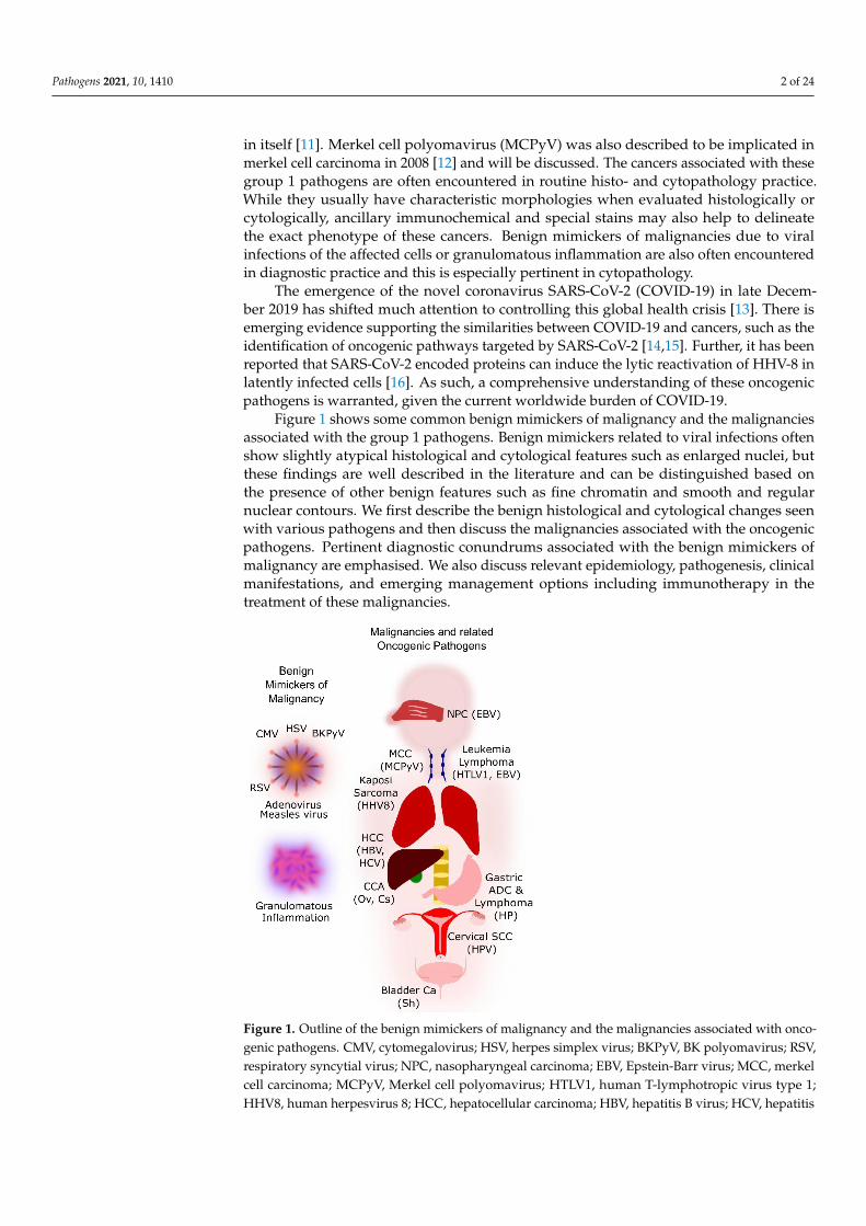
Figure 1 shows some common benign mimickers of malignancy and the malignanciesassociated with the group 1 pathogens. Benign mimickers related to viral infections oftenshow slightly atypical histological and cytological features such as enlarged nuclei, butthese findings are well described in the literature and can be distinguished based onthe presence of other benign features such as fine chromatin and smooth and regularnuclear contours. We first describe the benign histological and cytological changes seenwith various pathogens and then discuss the malignancies associated with the oncogenicpathogens. Pertinent diagnostic conundrums associated with the benign mimickers ofmalignancy are emphasised. We also discuss relevant epidemiology, pathogenesis, clinicalmanifestations, and emerging management options including immunotherapy in thetreatment of these malignancies.
Pathogens 2021, 10, x FOR PEER REVIEW 2 of 26
Schistosoma haematobium (Sh) [10]. Human immunodeficiency virus (HIV) will be dis-
cussed with the associated pathogens as it causes cancer through immunosuppression
and is not oncogenic in itself [11]. Merkel cell polyomavirus (MCPyV) was also described
to be implicated in merkel cell carcinoma in 2008 [12] and will be discussed. The cancers
associated with these group 1 pathogens are often encountered in routine histo- and cyto-
pathology practice. While they usually have characteristic morphologies when evaluated
histologically or cytologically, ancillary immunochemical and special stains may also help
to delineate the exact phenotype of these cancers. Benign mimickers of malignancies due
to viral infections of the affected cells or granulomatous inflammation are also often en-
countered in diagnostic practice and this is especially pertinent in cytopathology.
The emergence of the novel coronavirus SARS-CoV-2 (COVID-19) in late December
2019 has shifted much attention to controlling this global health crisis [13]. There is emerg-
ing evidence supporting the similarities between COVID-19 and cancers, such as the iden-
tification of oncogenic pathways targeted by SARS-CoV-2 [14,15]. Further, it has been re-
ported that SARS-CoV-2 encoded proteins can induce the lytic reactivation of HHV-8 in
latently infected cells [16]. As such, a comprehensive understanding of these oncogenic
pathogens is warranted, given the current worldwide burden of COVID-19.
Figure 1 shows some common benign mimickers of malignancy and the malignancies
associated with the group 1 pathogens. Benign mimickers related to viral infections often
show slightly atypical histological and cytological features such as enlarged nuclei, but
these findings are well described in the literature and can be distinguished based on the
presence of other benign features such as fine chromatin and smooth and regular nuclear
contours. We first describe the benign histological and cytological changes seen with var-
ious pathogens and then discuss the malignancies associated with the oncogenic patho-
gens. Pertinent diagnostic conundrums associated with the benign mimickers of malig-
nancy are emphasised. We also discuss relevant epidemiology, pathogenesis, clinical
manifestations, and emerging management options including immunotherapy in the
treatment of these malignancies.
Figure 1. Outline of the benign mimickers of malignancy and the malignancies associated with
oncogenic pathogens. CMV, cytomegalovirus; HSV, herpes simplex virus; BKPyV, BK polyoma-
virus; RSV, respiratory syncytial virus; NPC, nasopharyngeal carcinoma; EBV, Epstein-Barr virus;
MCC, merkel cell carcinoma; MCPyV, Merkel cell polyomavirus; HTLV1, human T-lymphotropic
Figure 1. Outline of the benign mimickers of malignancy and the malignancies associated with onco-genic pathogens. CMV, cytomegalovirus; HSV, herpes simplex virus; BKPyV, BK polyomavirus; RSV,respiratory syncytial virus; NPC, nasopharyngeal carcinoma; EBV, Epstein-Barr virus; MCC, merkelcell carcinoma; MCPyV, Merkel cell polyomavirus; HTLV1, human T-lymphotropic virus type 1;HHV8, human herpesvirus 8; HCC, hepatocellular carcinoma; HBV, hepatitis B virus; HCV, hepatitis
Pathogens 2021, 10, 1410 3 of 24
C virus; CCA, cholangiocarcinoma; Ov, Opisthorcis viverrini; Cs, Clonorchis sinensis; Sh, Schistosomahaematobium; Ca, carcinoma; ADC, adenocarcinoma; HP, Helicobacter pylori; SCC, squamous cellcarcinoma; HPV, human papillomavirus.
2. Benign Mimickers of Malignancy2.1. Herpes Simplex Virus (HSV)
HSV is a member of the Herpesviridae family of viruses which have enveloped double-stranded DNA genomes [17,18]. This family can be classified into three subfamilies bytissue tropism: alphaherpesviruses, which include herpes simplex virus 1 (HSV-1), herpessimplex virus 2 (HSV-2), and varicella zoster virus (VZV), that infect epithelial cells andremain latent in the neuronal cell body; betaherpesviruses, which include cytomegalovirus(described below), human herpesvirus-6, and human herpesvirus-7, that infect a varietyof cell types; and gammaherpesviruses, which include Epstein–Barr virus and humanherpesvirus-8 (both described below), that infect lymphoid and other cells [19]. A typicalsequence of infection, namely, acute infection followed by latency and variable roundsof viral reactivation, is characteristic of herpesviruses. Approximately 40% to 98% of thegeneral population possess antibodies to HSV-1 [17].
HSV1 is typically implicated in orofacial infections (for example, cold sore of the lip)and sporadic encephalitis, while HSV2 is typically implicated in genital infections (forexample, vesicles and ulcers) and may be transmitted from mother to newborn [20]. As anaide memoire, HSV1 typically causes infections in the superior part of the body, while thereverse holds true for HSV2 (“1 is superior to 2”). Immunocompromised patients may besusceptible to disseminated disease including encephalitis and visceral organ involvement,such as the gastrointestinal and respiratory systems, resulting in clinical manifestations likepneumonia. Similarly, maternal-neonatal transmission, although rare, is associated withdisseminated disease, encephalitis, thrombocytopenia, and disseminated intravascularcoagulation in the neonate. Foetal infection may result in microcephaly, retinitis, andscarring of the skin [20].
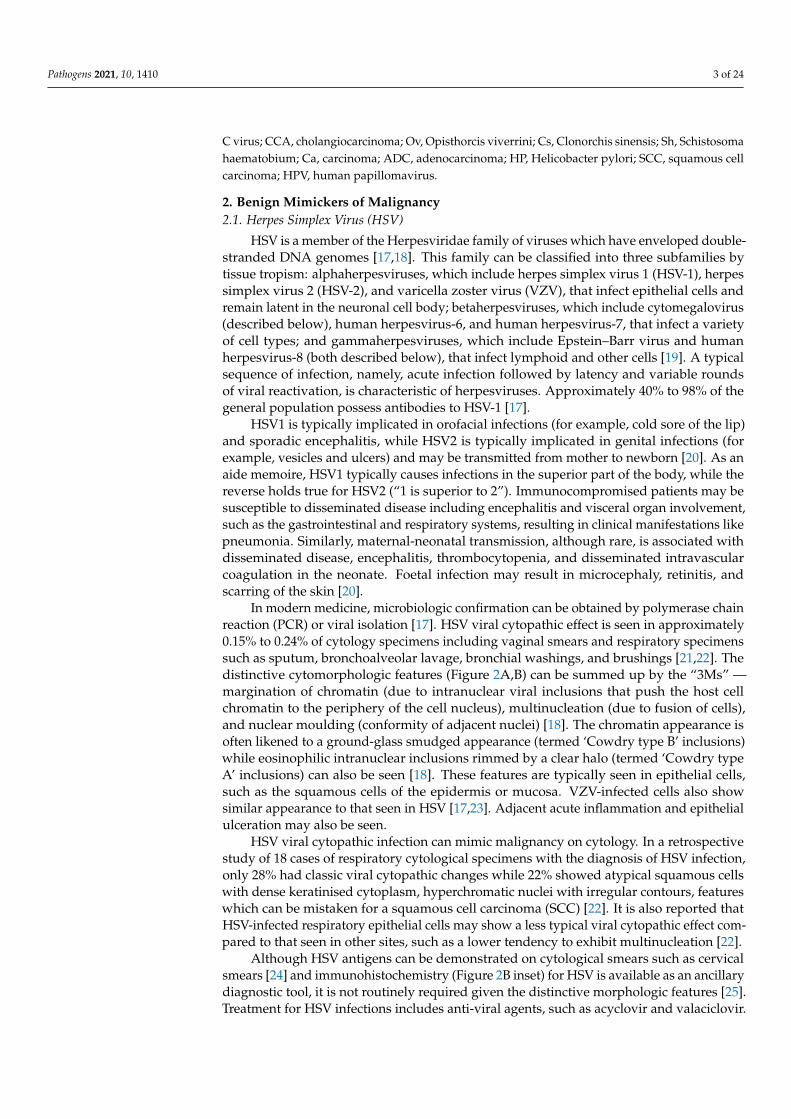
In modern medicine, microbiologic confirmation can be obtained by polymerase chainreaction (PCR) or viral isolation [17]. HSV viral cytopathic effect is seen in approximately0.15% to 0.24% of cytology specimens including vaginal smears and respiratory specimenssuch as sputum, bronchoalveolar lavage, bronchial washings, and brushings [21,22]. Thedistinctive cytomorphologic features (Figure 2A,B) can be summed up by the “3Ms” —margination of chromatin (due to intranuclear viral inclusions that push the host cellchromatin to the periphery of the cell nucleus), multinucleation (due to fusion of cells),and nuclear moulding (conformity of adjacent nuclei) [18]. The chromatin appearance isoften likened to a ground-glass smudged appearance (termed ‘Cowdry type B’ inclusions)while eosinophilic intranuclear inclusions rimmed by a clear halo (termed ‘Cowdry typeA’ inclusions) can also be seen [18]. These features are typically seen in epithelial cells,such as the squamous cells of the epidermis or mucosa. VZV-infected cells also showsimilar appearance to that seen in HSV [17,23]. Adjacent acute inflammation and epithelialulceration may also be seen.
HSV viral cytopathic infection can mimic malignancy on cytology. In a retrospectivestudy of 18 cases of respiratory cytological specimens with the diagnosis of HSV infection,only 28% had classic viral cytopathic changes while 22% showed atypical squamous cellswith dense keratinised cytoplasm, hyperchromatic nuclei with irregular contours, featureswhich can be mistaken for a squamous cell carcinoma (SCC) [22]. It is also reported thatHSV-infected respiratory epithelial cells may show a less typical viral cytopathic effect com-pared to that seen in other sites, such as a lower tendency to exhibit multinucleation [22].
Although HSV antigens can be demonstrated on cytological smears such as cervicalsmears [24] and immunohistochemistry (Figure 2B inset) for HSV is available as an ancillarydiagnostic tool, it is not routinely required given the distinctive morphologic features [25].Treatment for HSV infections includes anti-viral agents, such as acyclovir and valaciclovir.
Pathogens 2021, 10, 1410 4 of 24
As expected, the dose, route and duration of therapy are contingent on the site and severityof infection as well as the host immune system [20].
Pathogens 2021, 10, x FOR PEER REVIEW 4 of 26
Figure 2. Benign mimickers of malignancy. (A) HSV viral cytopathic effect on cytology (arrow),
with intranuclear inclusions and nuclear moulding; (B) HSV viral cytopathic effect (arrow) on
formalin-fixed paraffin-embedded (FFPE) tissue (inset: immunohistochemistry for HSV antigen);
(C) CMV viral cytopathic effect on cytology, with characteristic Owl’s eye nuclear inclusions (ar-
rows); (D) CMV immunohistochemistry (arrow) on FFPE tissue; (E) BKPyV viral cytopathic effect,
also termed ‘decoy cells’ (arrow), showing glassy basophilic nuclear inclusions; (F) cytology of
high-grade urothelial carcinoma, which is an important differential diagnosis of decoy cells; (G)
granulomatous inflammation, consisting of clusters of epithelioid histiocytes; (H) Ziehl–Neelsen
stain for acid-fast bacilli (arrow).
HSV viral cytopathic infection can mimic malignancy on cytology. In a retrospective
study of 18 cases of respiratory cytological specimens with the diagnosis of HSV infection,
only 28% had classic viral cytopathic changes while 22% showed atypical squamous cells
with dense keratinised cytoplasm, hyperchromatic nuclei with irregular contours, fea-
tures which can be mistaken for a squamous cell carcinoma (SCC) [22]. It is also reported
that HSV-infected respiratory epithelial cells may show a less typical viral cytopathic ef-
fect compared to that seen in other sites, such as a lower tendency to exhibit multinuclea-
tion [22].
Although HSV antigens can be demonstrated on cytological smears such as cervical
smears [24] and immunohistochemistry (Figure 2B inset) for HSV is available as an ancil-
lary diagnostic tool, it is not routinely required given the distinctive morphologic features
[25]. Treatment for HSV infections includes anti-viral agents, such as acyclovir and
valaciclovir. As expected, the dose, route and duration of therapy are contingent on the
site and severity of infection as well as the host immune system [20].
2.2. Cytomegalovirus (CMV)
Figure 2. Benign mimickers of malignancy. (A) HSV viral cytopathic effect on cytology (arrow),with intranuclear inclusions and nuclear moulding; (B) HSV viral cytopathic effect (arrow) onformalin-fixed paraffin-embedded (FFPE) tissue (inset: immunohistochemistry for HSV antigen);(C) CMV viral cytopathic effect on cytology, with characteristic Owl’s eye nuclear inclusions (arrows);(D) CMV immunohistochemistry (arrow) on FFPE tissue; (E) BKPyV viral cytopathic effect, alsotermed ‘decoy cells’ (arrow), showing glassy basophilic nuclear inclusions; (F) cytology of high-gradeurothelial carcinoma, which is an important differential diagnosis of decoy cells; (G) granulomatousinflammation, consisting of clusters of epithelioid histiocytes; (H) Ziehl–Neelsen stain for acid-fastbacilli (arrow).
2.2. Cytomegalovirus (CMV)
CMV is an enveloped double-stranded DNA virus [26,27], a member of the beta-herpesviruses subfamily [28]. It is ubiquitous in humans and acquired by most in earlylife [29]. Reports suggest that between half to all adults possess the IgG antibodies toCMV, indicating previous exposure [19]. Inhibition of viral lytic gene expression is a keydriver of latency [30], and the virus may remain latent in a diverse range of cells includingendothelial cells, epithelial cells, stromal cells, and lymphocytes. Subsequent reactivationis thought to occur when the balance between viral load and host immune system causes a“threshold” to be exceeded [30].
As with other infections, the clinical presentation of CMV infection depends uponthe host immune status. In immunocompetent individuals, acute infection may be asymp-tomatic or may present as self-limiting, non-specific febrile illness or as mononucleosis-like
Pathogens 2021, 10, 1410 5 of 24
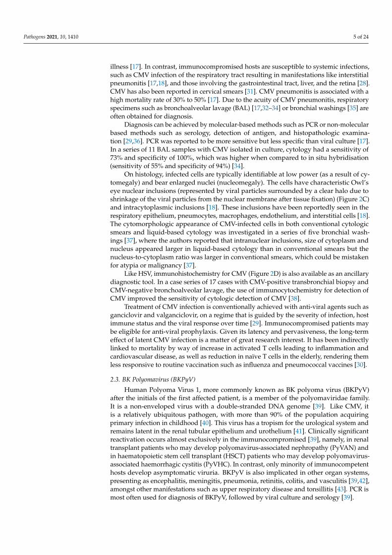
illness [17]. In contrast, immunocompromised hosts are susceptible to systemic infections,such as CMV infection of the respiratory tract resulting in manifestations like interstitialpneumonitis [17,18], and those involving the gastrointestinal tract, liver, and the retina [28].CMV has also been reported in cervical smears [31]. CMV pneumonitis is associated with ahigh mortality rate of 30% to 50% [17]. Due to the acuity of CMV pneumonitis, respiratoryspecimens such as bronchoalveolar lavage (BAL) [17,32–34] or bronchial washings [35] areoften obtained for diagnosis.
Diagnosis can be achieved by molecular-based methods such as PCR or non-molecularbased methods such as serology, detection of antigen, and histopathologic examina-tion [29,36]. PCR was reported to be more sensitive but less specific than viral culture [17].In a series of 11 BAL samples with CMV isolated in culture, cytology had a sensitivity of73% and specificity of 100%, which was higher when compared to in situ hybridisation(sensitivity of 55% and specificity of 94%) [34].
On histology, infected cells are typically identifiable at low power (as a result of cy-tomegaly) and bear enlarged nuclei (nucleomegaly). The cells have characteristic Owl’seye nuclear inclusions (represented by viral particles surrounded by a clear halo due toshrinkage of the viral particles from the nuclear membrane after tissue fixation) (Figure 2C)and intracytoplasmic inclusions [18]. These inclusions have been reportedly seen in therespiratory epithelium, pneumocytes, macrophages, endothelium, and interstitial cells [18].The cytomorphologic appearance of CMV-infected cells in both conventional cytologicsmears and liquid-based cytology was investigated in a series of five bronchial wash-ings [37], where the authors reported that intranuclear inclusions, size of cytoplasm andnucleus appeared larger in liquid-based cytology than in conventional smears but thenucleus-to-cytoplasm ratio was larger in conventional smears, which could be mistakenfor atypia or malignancy [37].
Like HSV, immunohistochemistry for CMV (Figure 2D) is also available as an ancillarydiagnostic tool. In a case series of 17 cases with CMV-positive transbronchial biopsy andCMV-negative bronchoalveolar lavage, the use of immunocytochemistry for detection ofCMV improved the sensitivity of cytologic detection of CMV [38].
Treatment of CMV infection is conventionally achieved with anti-viral agents such asganciclovir and valganciclovir, on a regime that is guided by the severity of infection, hostimmune status and the viral response over time [29]. Immunocompromised patients maybe eligible for anti-viral prophylaxis. Given its latency and pervasiveness, the long-termeffect of latent CMV infection is a matter of great research interest. It has been indirectlylinked to mortality by way of increase in activated T cells leading to inflammation andcardiovascular disease, as well as reduction in naïve T cells in the elderly, rendering themless responsive to routine vaccination such as influenza and pneumococcal vaccines [30].
2.3. BK Polyomavirus (BKPyV)
Human Polyoma Virus 1, more commonly known as BK polyoma virus (BKPyV)after the initials of the first affected patient, is a member of the polyomaviridae family.It is a non-enveloped virus with a double-stranded DNA genome [39]. Like CMV, itis a relatively ubiquitous pathogen, with more than 90% of the population acquiringprimary infection in childhood [40]. This virus has a tropism for the urological system andremains latent in the renal tubular epithelium and urothelium [41]. Clinically significantreactivation occurs almost exclusively in the immunocompromised [39], namely, in renaltransplant patients who may develop polyomavirus-associated nephropathy (PyVAN) andin haematopoietic stem cell transplant (HSCT) patients who may develop polyomavirus-associated haemorrhagic cystitis (PyVHC). In contrast, only minority of immunocompetenthosts develop asymptomatic viruria. BKPyV is also implicated in other organ systems,presenting as encephalitis, meningitis, pneumonia, retinitis, colitis, and vasculitis [39,42],amongst other manifestations such as upper respiratory disease and tonsillitis [43]. PCR ismost often used for diagnosis of BKPyV, followed by viral culture and serology [39].
Pathogens 2021, 10, 1410 6 of 24
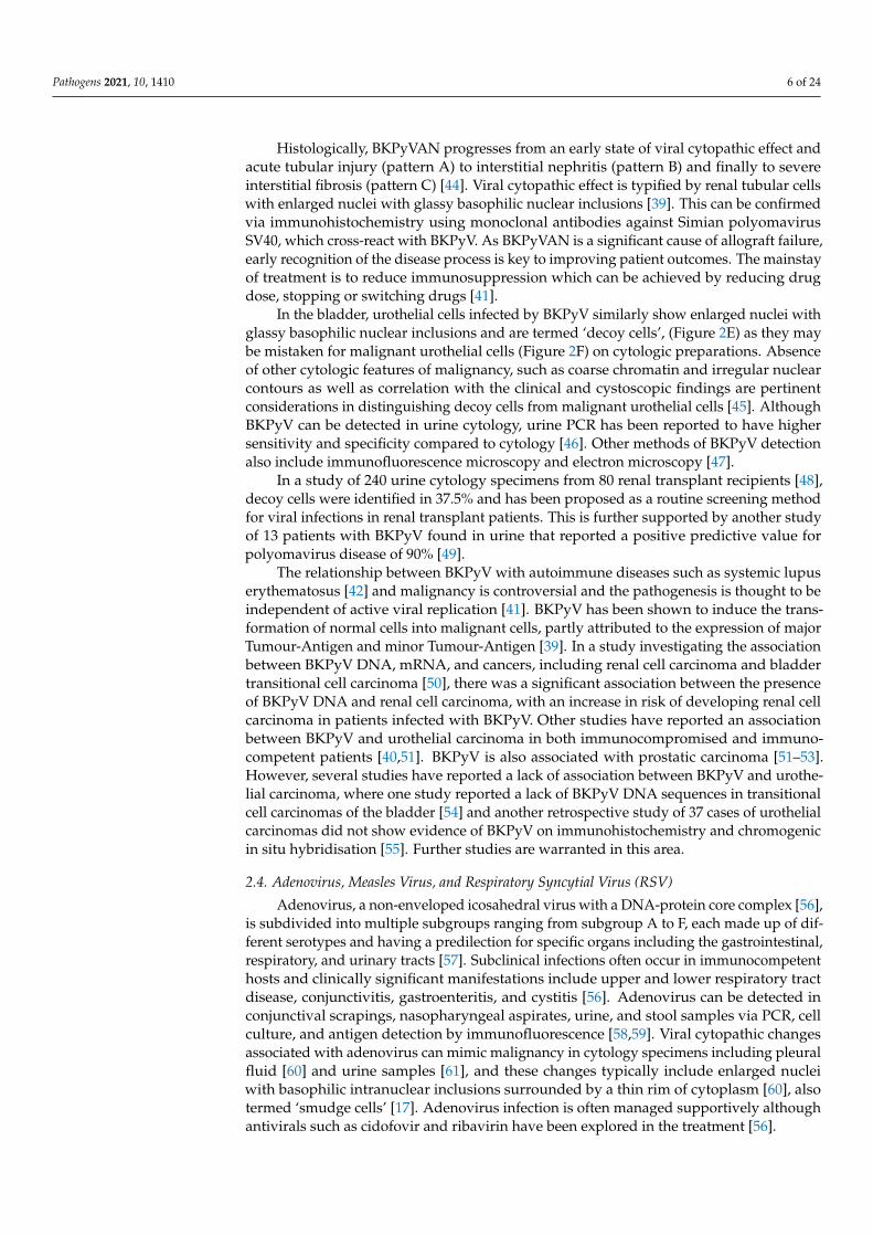
Histologically, BKPyVAN progresses from an early state of viral cytopathic effect andacute tubular injury (pattern A) to interstitial nephritis (pattern B) and finally to severeinterstitial fibrosis (pattern C) [44]. Viral cytopathic effect is typified by renal tubular cellswith enlarged nuclei with glassy basophilic nuclear inclusions [39]. This can be confirmedvia immunohistochemistry using monoclonal antibodies against Simian polyomavirusSV40, which cross-react with BKPyV. As BKPyVAN is a significant cause of allograft failure,early recognition of the disease process is key to improving patient outcomes. The mainstayof treatment is to reduce immunosuppression which can be achieved by reducing drugdose, stopping or switching drugs [41].
In the bladder, urothelial cells infected by BKPyV similarly show enlarged nuclei withglassy basophilic nuclear inclusions and are termed ‘decoy cells’, (Figure 2E) as they maybe mistaken for malignant urothelial cells (Figure 2F) on cytologic preparations. Absenceof other cytologic features of malignancy, such as coarse chromatin and irregular nuclearcontours as well as correlation with the clinical and cystoscopic findings are pertinentconsiderations in distinguishing decoy cells from malignant urothelial cells [45]. AlthoughBKPyV can be detected in urine cytology, urine PCR has been reported to have highersensitivity and specificity compared to cytology [46]. Other methods of BKPyV detectionalso include immunofluorescence microscopy and electron microscopy [47].
In a study of 240 urine cytology specimens from 80 renal transplant recipients [48],decoy cells were identified in 37.5% and has been proposed as a routine screening methodfor viral infections in renal transplant patients. This is further supported by another studyof 13 patients with BKPyV found in urine that reported a positive predictive value forpolyomavirus disease of 90% [49].
The relationship between BKPyV with autoimmune diseases such as systemic lupuserythematosus [42] and malignancy is controversial and the pathogenesis is thought to beindependent of active viral replication [41]. BKPyV has been shown to induce the trans-formation of normal cells into malignant cells, partly attributed to the expression of majorTumour-Antigen and minor Tumour-Antigen [39]. In a study investigating the associationbetween BKPyV DNA, mRNA, and cancers, including renal cell carcinoma and bladdertransitional cell carcinoma [50], there was a significant association between the presenceof BKPyV DNA and renal cell carcinoma, with an increase in risk of developing renal cellcarcinoma in patients infected with BKPyV. Other studies have reported an associationbetween BKPyV and urothelial carcinoma in both immunocompromised and immuno-competent patients [40,51]. BKPyV is also associated with prostatic carcinoma [51–53].However, several studies have reported a lack of association between BKPyV and urothe-lial carcinoma, where one study reported a lack of BKPyV DNA sequences in transitionalcell carcinomas of the bladder [54] and another retrospective study of 37 cases of urothelialcarcinomas did not show evidence of BKPyV on immunohistochemistry and chromogenicin situ hybridisation [55]. Further studies are warranted in this area.
2.4. Adenovirus, Measles Virus, and Respiratory Syncytial Virus (RSV)
Adenovirus, a non-enveloped icosahedral virus with a DNA-protein core complex [56],is subdivided into multiple subgroups ranging from subgroup A to F, each made up of dif-ferent serotypes and having a predilection for specific organs including the gastrointestinal,respiratory, and urinary tracts [57]. Subclinical infections often occur in immunocompetenthosts and clinically significant manifestations include upper and lower respiratory tractdisease, conjunctivitis, gastroenteritis, and cystitis [56]. Adenovirus can be detected inconjunctival scrapings, nasopharyngeal aspirates, urine, and stool samples via PCR, cellculture, and antigen detection by immunofluorescence [58,59]. Viral cytopathic changesassociated with adenovirus can mimic malignancy in cytology specimens including pleuralfluid [60] and urine samples [61], and these changes typically include enlarged nucleiwith basophilic intranuclear inclusions surrounded by a thin rim of cytoplasm [60], alsotermed ‘smudge cells’ [17]. Adenovirus infection is often managed supportively althoughantivirals such as cidofovir and ribavirin have been explored in the treatment [56].
Pathogens 2021, 10, 1410 7 of 24
Measles is an enveloped spherical RNA virus [62] which was notable for its high mor-tality rate of up to millions of deaths each year prior to the introduction of vaccination [62].The initiation begins with infection of the respiratory epithelium of the host [63,64], fol-lowed by an incubation period of 5 to 11 days [63,64], which then leads to the symptomaticstage where clinical manifestations including fever, malaise, conjunctivitis, as well as thecharacteristic Koplik’s spots (white spots on buccal mucosa) [64,65]. Measles can alsopresent as a systemic disease involving other organ systems [63]. The diagnosis of measlescan be confirmed using serologic tests, cultures, and PCR [65]. Eosinophilic cytoplas-mic and intranuclear inclusion bodies are frequently reported in patients with measles,where these inclusions have been reported to occur in the skin, respiratory tract, urinarytract [64,66,67], and central nervous system [63]. Types of specimens include sputum, nasalsecretions, and upper respiratory tract swabbing, and BAL [17]. In addition to inclusionbodies, multinucleated giant cells with overlapping nuclei and variation in shape andsize [17] are also frequently observed [64,67], postulated to occur due to the fusion of type 2pneumocytes [17]. These cytologic features, although non-specific, may mimic giant cellrich malignancies. The management of measles infection is also primarily supportive andantivirals have also been used to treat severe measles [62].
Primarily causing illness in infants and elderly people [68], RSV is an envelopedRNA virus [18] that infects the upper respiratory tract and eyes [68]. RSV infection isclinically apparent with upper respiratory tract signs and symptoms including cough,wheezing, and low grade fever [69], as well as other manifestations including otitis media,bronchiolitis, and pneumonia, which usually happen in children [68]. Depending on theclinical manifestation, radiologic findings may show typical findings such as hyperinfla-tion, diffuse interstitial markings and peribronchial thickening [68]. The diagnosis of RSVis established through detection of RSV antigens in nasopharyngeal aspirates throughimmunofluorescence, enzyme-linked immunosorbent assay (ELISA) or culture [68]. Char-acteristic cytological findings have been described in RSV infections, including presenceof large syncytial cell aggregates with eosinophilic cytoplasmic inclusions [18]. Creolabodies, which are clusters of reactive ciliated bronchial epithelial cells, have also beenreported in RSV bronchiolitis [18]. These clusters of Creola bodies may be confused withadenocarcinoma on cytology but the presence of cilia reinforces the benign nature of thesecells. Treatment in RSV infection is symptomatic management and aerosolised ribavirin isapproved for use in hospitalised infants [70].
2.5. Granulomatous Inflammation
Granulomatous inflammation is defined by the presence of granulomas, which referto aggregates of epithelioid histiocytes (Figure 2G) [71]. Granulomatous inflammation hasbeen extensively discussed in previously published reviews [17,71–73]. Several patternsof granulomatous inflammation have been described, and they include foreign body typereaction, necrotising (with central necrotic material) granulomas, and non-necrotisinggranulomas [71]. Different patterns of granulomatous inflammation are associated withdifferent aetiologies. Foreign body type reaction is often associated with suture materialin excision or resection specimens, as well as other foreign materials such as talc andstarch [71]. Necrotising granulomas are associated with infectious pathogens includingmycobacterium tuberculosis and fungal organisms [17,71], while the differential diagnosesof non-necrotising granulomas encompass both infectious and non-infectious causes includ-ing sarcoidosis [74], Crohn’s disease [75], toxic and drug causes [71]. The Ziehl–Neelsenstain (Figure 2H) is often used clinically to identify acid-fast bacilli in respiratory specimensincluding sputum, with a specificity ranging from 90% to 100%, albeit with a limitedsensitivity [17].
The presence of granulomas associated with tumours is well established, report-edly seen in 4.4% of carcinomas, 13.8% of Hodgkin’s disease, 7.3% of non-Hodgkin lym-phomas [76], and 50.0% of seminomas [77,78]. Distinguishing between a granulomatousinflammation and malignancy has proven to be difficult on fine needle aspiration cytology
Pathogens 2021, 10, 1410 8 of 24
(FNAC) [78], where a study of six cases of neck mass FNAC reported that only one case wasdiagnosed as metastatic carcinoma with extensive granulomatous inflammation while theremaining cases were signed out as “atypical” with a recommendation for tissue biopsy [78].The histopathological diagnoses of the remaining cases included Hodgkin’s disease, lym-phoepithelial carcinoma, diffuse large B-cell lymphoma, and anaplastic carcinoma [78]. Ina subsequent study of 153 patients undergoing endobronchial ultrasound-guided trans-bronchial needle aspiration for mediastinal lymphadenopathy which met radiologicalcriteria for cancer recurrence, 11.0% showed non-caseating granulomas on cytology [79].Granulomatous inflammation also occurs after intravesical Bacillus Calmette-Guerin ther-apy for bladder carcinoma [80].
In particular, granulomatous mastitis, a granulomatous inflammation of breast parenchy-mal tissue, although uncommonly encountered, can be mistaken for a malignancy, bothclinically and radiologically [81]. A series of three studies of patients with granulomatousmastitis reported a mean age of 34 to 44 years [81–83]. In one of these studies, tuberculousinfection accounted for most cases of granulomatous mastitis, followed by foreign bodyand idiopathic aetiologies [82]. Of these three studies, one was a series of 16 patients whichreported clinical impression of a breast abscess in half the patients [81], whereas anotherseries of 18 cases [83] reported a clinical impression of a malignant lesion in all cases. Themost common ultrasonography finding in these studies was a heterogeneous hypoechoiclesion [82]. Cytologic and histological findings were characteristic for granulomas, lympho-cytes, and plasma cells, but differed in the presence or absence of necrosis [82]. An early,limited case series of five patients with granulomatous mastitis [84] reported atypia or‘suspicious for malignancy’ on FNAC in three cases. However, the authors did not specifythe criteria used for a cytologic impression of an ‘atypical’ lesion. There is now a trendtowards performing core needle biopsy over FNAC in sampling breast lesions [85], therebyimproving diagnostic accuracy by histopathology rather than cytology.
3. Oncogenic Pathogens3.1. Hepatitis B and C Viruses (HBV, HCV)
HBV and HCV are hepatotropic viruses implicated in acute hepatitis, chronic liverdisease, and cirrhosis as well as hepatocellular carcinoma (HCC).
Hepatitis B is a member of the Hepadnaviridae family of viruses. It bears a double-stranded DNA genome, which is able to integrate into the DNA of the host genome. Thereare 10 major genotypes of which genotype C and D are associated with a higher risk ofprogression to liver cirrhosis [86]. In childhood, the majority of infections acquired tend topersist as chronic infections, while the reverse holds true for infections acquired in adults,in whom the majority are able to clear the infection [87].
On the other hand, Hepatitis C is a member of the Flaviviridae family of viruses.It bears a single-stranded RNA genome. There are seven major genotypes of whichgenotype 1 is associated with more aggressive disease course and reduced responsivenessto therapy. Once acquired, Hepatitis C persists as a chronic infection in approximately 80%of hosts [86].
Clinical diagnosis of Hepatitis B or Hepatitis C infection can be achieved via serologyor molecular methods. The role of histopathologic examination is primarily in the gradingand staging of liver disease. Of note, the accumulation of Hepatitis B surface antigen(HBsAg) in hepatocytes gives a ground glass appearance to the cytoplasm which stainsmagenta and blue on interrogation with the Shikata’s orcein stain (Figure 3A) and Victoriablue stain, respectively. The other features of active or chronic liver disease in Hepatitis Bmay, to some extent, overlap with other conditions [87]. In contrast, Hepatitis C infectionin the liver is associated with a hallmark triad of bile duct injury, lymphoid follicles withgerminal centres, and small and large droplet steatosis.
Pathogens 2021, 10, 1410 9 of 24Pathogens 2021, 10, x FOR PEER REVIEW 10 of 26
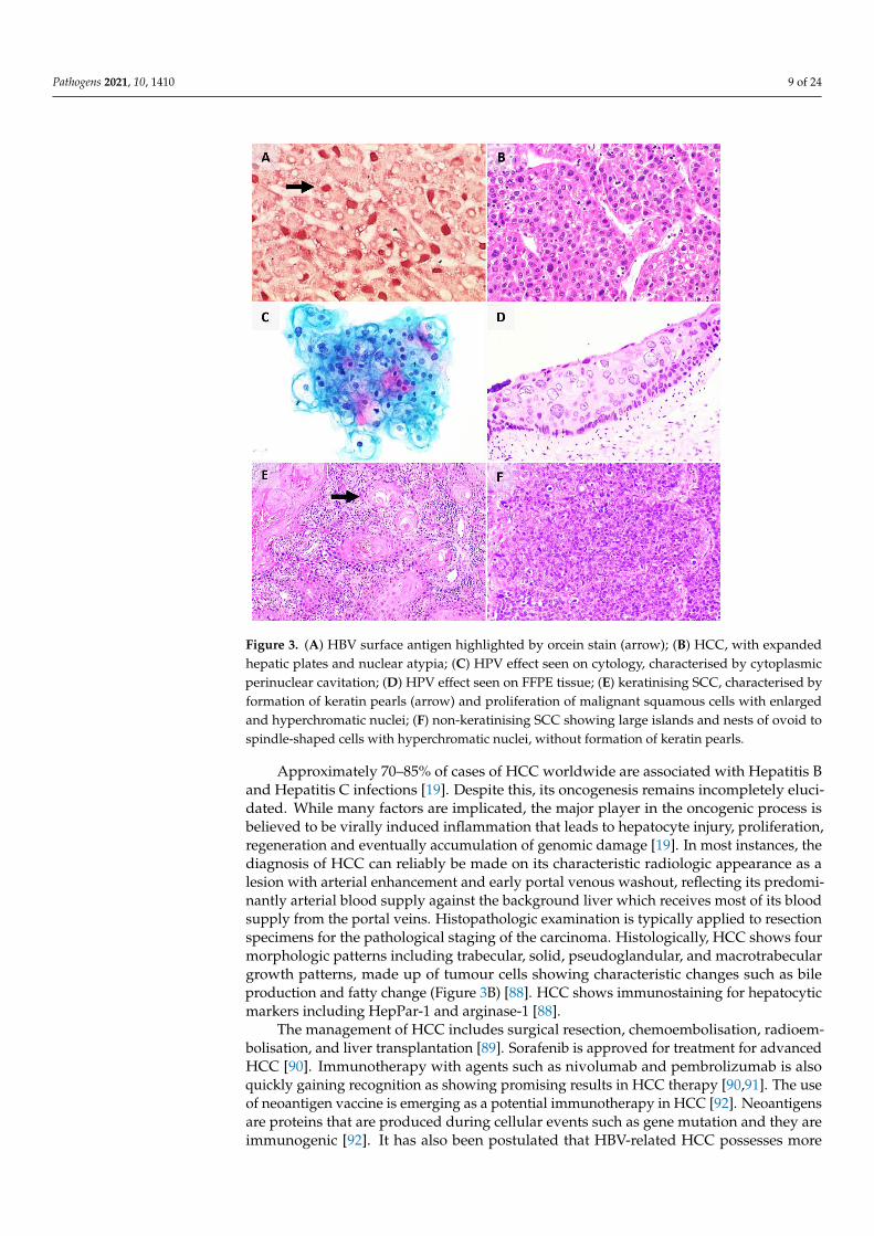
Figure 3. (A) HBV surface antigen highlighted by orcein stain (arrow); (B) HCC, with expanded
hepatic plates and nuclear atypia; (C) HPV effect seen on cytology, characterised by cytoplasmic
perinuclear cavitation; (D) HPV effect seen on FFPE tissue; (E) keratinising SCC, characterised by
formation of keratin pearls (arrow) and proliferation of malignant squamous cells with enlarged
and hyperchromatic nuclei; (F) non-keratinising SCC showing large islands and nests of ovoid to
spindle-shaped cells with hyperchromatic nuclei, without formation of keratin pearls.
The management of HCC includes surgical resection, chemoembolisation, radioem-
bolisation, and liver transplantation [89]. Sorafenib is approved for treatment for ad-
vanced HCC [90]. Immunotherapy with agents such as nivolumab and pembrolizumab is
also quickly gaining recognition as showing promising results in HCC therapy [90,91].
The use of neoantigen vaccine is emerging as a potential immunotherapy in HCC [92].
Neoantigens are proteins that are produced during cellular events such as gene mutation
and they are immunogenic [92]. It has also been postulated that HBV-related HCC pos-
sesses more effective neoantigens compared to non-HBV-related HCC, and this may be a
potential novel target for vaccines in the future [92].
3.2. Human Papillomavirus (HPV)
Human papillomavirus (HPV) is a member of the Papovaviridae family. It is a non-
enveloped DNA virus with a predilection for cutaneous and mucosal epithelium. More
than 90% of people who acquire the virus experience transient infection and can immu-
nologically clear the virus. In contrast, chronic persistent infection, contributed to by var-
ious factors such as immunocompromise, frequent reinfections and co-infections, is asso-
ciated with progression to neoplasia [93]. HIV status was reported to be associated with
higher prevalence of HPV infection [94].
More than 100 types of HPV virus have been isolated and sequenced to date. These
can be broadly subclassified into low-risk and high-risk types, based on malignant poten-
tial. Low-risk types include HPV 6 and 11 which are associated with genital warts and
Figure 3. (A) HBV surface antigen highlighted by orcein stain (arrow); (B) HCC, with expandedhepatic plates and nuclear atypia; (C) HPV effect seen on cytology, characterised by cytoplasmicperinuclear cavitation; (D) HPV effect seen on FFPE tissue; (E) keratinising SCC, characterised byformation of keratin pearls (arrow) and proliferation of malignant squamous cells with enlargedand hyperchromatic nuclei; (F) non-keratinising SCC showing large islands and nests of ovoid tospindle-shaped cells with hyperchromatic nuclei, without formation of keratin pearls.
Approximately 70–85% of cases of HCC worldwide are associated with Hepatitis Band Hepatitis C infections [19]. Despite this, its oncogenesis remains incompletely eluci-dated. While many factors are implicated, the major player in the oncogenic process isbelieved to be virally induced inflammation that leads to hepatocyte injury, proliferation,regeneration and eventually accumulation of genomic damage [19]. In most instances, thediagnosis of HCC can reliably be made on its characteristic radiologic appearance as alesion with arterial enhancement and early portal venous washout, reflecting its predomi-nantly arterial blood supply against the background liver which receives most of its bloodsupply from the portal veins. Histopathologic examination is typically applied to resectionspecimens for the pathological staging of the carcinoma. Histologically, HCC shows fourmorphologic patterns including trabecular, solid, pseudoglandular, and macrotrabeculargrowth patterns, made up of tumour cells showing characteristic changes such as bileproduction and fatty change (Figure 3B) [88]. HCC shows immunostaining for hepatocyticmarkers including HepPar-1 and arginase-1 [88].
The management of HCC includes surgical resection, chemoembolisation, radioem-bolisation, and liver transplantation [89]. Sorafenib is approved for treatment for advancedHCC [90]. Immunotherapy with agents such as nivolumab and pembrolizumab is alsoquickly gaining recognition as showing promising results in HCC therapy [90,91]. The useof neoantigen vaccine is emerging as a potential immunotherapy in HCC [92]. Neoantigensare proteins that are produced during cellular events such as gene mutation and they areimmunogenic [92]. It has also been postulated that HBV-related HCC possesses more
Pathogens 2021, 10, 1410 10 of 24
effective neoantigens compared to non-HBV-related HCC, and this may be a potentialnovel target for vaccines in the future [92].
3.2. Human Papillomavirus (HPV)
Human papillomavirus (HPV) is a member of the Papovaviridae family. It is a non-enveloped DNA virus with a predilection for cutaneous and mucosal epithelium. Morethan 90% of people who acquire the virus experience transient infection and can immuno-logically clear the virus. In contrast, chronic persistent infection, contributed to by variousfactors such as immunocompromise, frequent reinfections and co-infections, is associatedwith progression to neoplasia [93]. HIV status was reported to be associated with higherprevalence of HPV infection [94].
More than 100 types of HPV virus have been isolated and sequenced to date. Thesecan be broadly subclassified into low-risk and high-risk types, based on malignant poten-tial. Low-risk types include HPV 6 and 11 which are associated with genital warts andrespiratory papillomatosis. High-risk types include HPV 16, 18, 31, 33, 45, and 51, which areassociated with malignancies of the cervix, anogenital region, and oral cavity. The centraloncogenic event is the integration of the viral genome into the host genome, which resultsin the loss of the viral E2 repressor leading to increased expression of oncogenic proteinsE6 and E7 that inhibit p53 and Rb proteins, respectively [19].
In the cervix, the neoplastic process is represented by the progression across well-defined morphologic categories of cervical intraepithelial neoplasia (CIN) 1, 2, and 3, andfinally invasive SCC. The hallmark of HPV infection is koilocytes (hollow cell), whichare mature squamous cells that show binucleation, nuclear hyperchromasia, nuclear con-tour irregularities and enlargement, as well as the characteristic perinuclear halo (dueto interaction between the viral E4 protein and cytokeratin filaments causing their shiftaway from the nucleus) that gives the cell its name (Figure 3C,D) [95]. Histologically,HPV-associated SCC (Figure 3E,F) shows a proliferation of squamous cells, with markedlypleomorphic nuclei, and showing features of squamous differentiation such as intercellularbridges [96]. They may show distinctive growth patterns including basaloid, condylo-matous, and papillary patterns [96]. These tumours show positive staining for p16 onimmunohistochemistry [96].
Remarkably, our understanding of the role of HPV in cancer continues to evolve, withthe recognition of new entities such as HPV-related multiphenotypic sinonasal carcinoma(associated with HPV type 33) [97], and the reclassification of established entities, such ascervical adenocarcinomas that are now formally classified by HPV status (HPV-associatedor HPV-independent), as per the latest iteration of the World Health Organization classifi-cation of tumours of the female genital tract [96].
Public health measures such as cervical cancer screening programmes using Papani-colaou smears, liquid-based samples or primary HPV testing by molecular methods, anduniversal HPV vaccination have seen great success in developed countries which havewitnessed a decline in rates of squamous cell carcinoma of the cervix over the years. Thesepublic health measures hold great promise in less developed populations that continue tosuffer from relatively high rates of SCC of the cervix, an increasingly preventable cancer.
The management of cervical cancers depends on the stage of disease, with optionsincluding radical surgery and radiotherapy, as well as neoadjuvant chemoradiotherapy [98].Immunotherapy is currently being explored in treating HPV-associated malignancies [99],and options include the use of immune checkpoint inhibitors and therapeutic vaccines [99].Therapeutic vaccines are postulated to incite inflammatory responses against the viralproteins E6 and E7 [100,101].
3.3. Epstein–Barr Virus (EBV)
Epstein–Barr Virus (EBV) is a member of the gammaherpesviruses subfamily. It is aubiquitous virus acquired by most people by young adulthood. The virus is transmitted by
Pathogens 2021, 10, 1410 11 of 24
saliva and infects epithelial cells and B cells of the oropharynx, causing a predominantlylytic infection in the former while establishing latency in the latter [102].
In immunocompetent hosts, primary infection is typically asymptomatic or presentsas infectious mononucleosis, a classic constellation of fever, tonsillitis/pharyngitis, lym-phadenopathy, and atypical lymphocytosis. The diagnosis can usually be made on clinicalgrounds supported by serologic confirmation. Rarely, atypical clinical presentation leadsto consideration of biopsy of the enlarged tonsil or lymph node [103]. Interpretation ofsuch biopsies is challenging because infectious mononucleosis may manifest as an atypicallymphoid infiltrate with architectural distortion, immunoblastic proliferation, and ReedSternberg-like cells, thus mimicking lymphoma [104]. The polyclonal B cell lymphoprolif-eration is typically curtailed by a robust T cell response in an immunocompetent host.
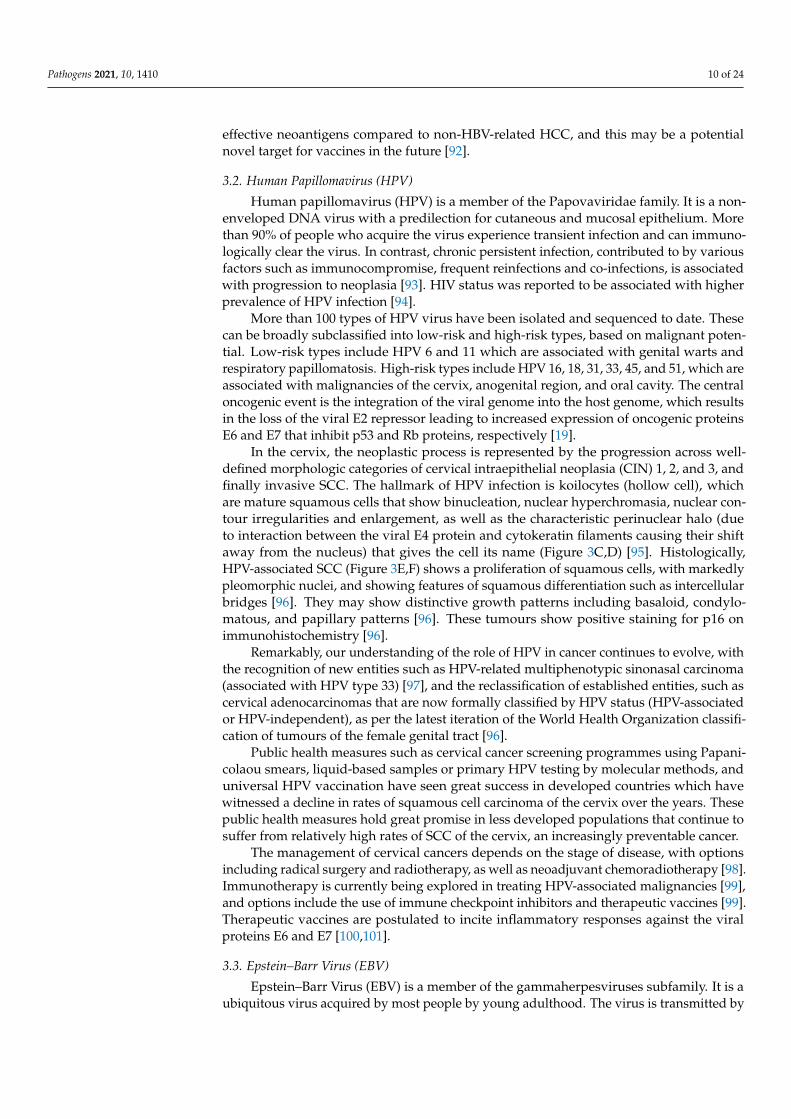
In the immunocompromised, such as in a patient with HIV, however, an uncontrolledB cell lymphoproliferation results, leading to acquisition of additional genomic alterationsculminating in a monoclonal proliferation that amounts to lymphoma [19,105]. Broadly,these are termed EBV-associated lymphoproliferative diseases (including B cell lymphomasand NK/T cell lymphomas). Epithelial malignancies such as nasopharyngeal carcinoma(NPC) (Figure 4A,B) and lymphoepithelial carcinoma of various primary sites and mes-enchymal tumours like EBV positive smooth muscle tumours are also well-documentedEBV-associated malignancies, reflecting the virus’s diverse oncogenic potential. On tissuesections, the EBV status of a tumour can be interrogated by Epstein–Barr-encoded RNA insitu hybridisation (EBER-ISH) (Figure 4C).
The development of prophylactic EBV vaccines for young children (prior to acquisitionof the virus) or in pre-transplant patients is a subject of ongoing research [104]. Therapeu-tic vaccines have also been primarily studied in NPC and have shown promising earlyresults [104,106]. Other immunotherapies are also currently explored in EBV-associatedNPC [106].
3.4. Human Herpes Virus 8 (HHV8)
HHV8, also termed Kaposi sarcoma (KS)-associated herpes virus, belongs to thegammaherpesvirus subfamily and is a large double-stranded DNA within an envelopedcapsid [107]. It is associated with KS, primary effusion lymphoma (PEL), multicentricCastleman disease (MCD), HHV-8 positive diffuse large B-cell lymphoma (DLBCL), andgerminotropic lymphoproliferative disorder (GLPD), and these disorders, with the excep-tion of GLPD, are frequently associated with HIV infection [108].
The incidence of KS is highest in Central Africa [107]. The epidemiology of HHV8 canbe delineated based on the variants of KS, namely, classic, endemic (African), transplantation-associated (iatrogenic), and epidemic (AIDS associated) [109]. Classic KS occurs mainlyin elderly men of Mediterranean, Eastern European, Jewish, and South American de-scent [107,109,110] while endemic KS occurs frequently in certain Central African andsub-Saharan African countries [109,110]. Iatrogenic KS occurs in up to 5% of transplantrecipients [109] and immunocompromised patients [110] while epidemic KS is currentlythe most prevalent form of KS, affecting mainly homosexual males [109]. The prevalenceof HHV8 infection is estimated to be less than 3% to 10% in the United States of America,United Kingdom, and Europe, while it ranges from 4% to 35% in Mediterranean countrieslike Italy and Greece and 30% to 60% in Africa [111].
The virus is transmitted primarily through sexual contact, especially through malehomosexual contact, with other routes of transmission including mother-to-child, saliva,organ transplantation and other unknown routes [109,112]. The clinical manifestations ofHHV8 infection are non-specific and can include fever, maculopapular rash, upper respira-tory tract symptoms, diarrhoea, fatigue, and lymphadenopathy [113]. Classic KS manifestsas bluish-red rashes on the distal lower extremities, which may eventually form multifocalnodules while the endemic KS has four clinical phenotypes, namely, nodular, florid, infiltra-tive, and lymphadenopathic [114]. Iatrogenic KS resembles classic KS on examination andepidemic KS often presents first on the nose, eyelids, ears, and the trunk [114]. PEL often
Pathogens 2021, 10, 1410 12 of 24
presents as a lymphomatous proliferation involving the pleura, peritoneum and/or peri-cardium, typically without discrete masses [109]. HHV-8 is diagnosed based on serologicassays including immunofluorescence, ELISA, and Western blot while in situ hybridisation,PCR, and immunohistochemistry can be performed on tissues [109,113,115].
Pathogens 2021, 10, x FOR PEER REVIEW 12 of 26
Broadly, these are termed EBV-associated lymphoproliferative diseases (including B cell
lymphomas and NK/T cell lymphomas). Epithelial malignancies such as nasopharyngeal
carcinoma (NPC) (Figures 4A and 4B) and lymphoepithelial carcinoma of various primary
sites and mesenchymal tumours like EBV positive smooth muscle tumours are also well-
documented EBV-associated malignancies, reflecting the virus’s diverse oncogenic poten-
tial. On tissue sections, the EBV status of a tumour can be interrogated by Epstein–Barr-
encoded RNA in situ hybridisation (EBER-ISH). (Figure 4C)
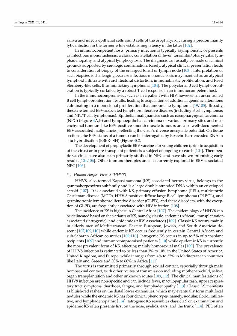
Figure 4. (A) Low power magnification of non-keratinising NPC, made up of a syncytial arrange-
ment of cells; (B) high power magnification of NPC, with cells showing round nuclei, prominent
eosinophilic nucleoli, within an eosinophilic to amphophilic cytoplasm; (C) EBV-encoded RNA in-
situ hybridisation for detection of EBV; (D) low power magnification of KS, showing slit-like vas-
cular spaces (arrow) in the dermis of the skin; (E) high power magnification of KS, showing vascu-
lar spaces (arrow) lined by cells with hyperchromatic nuclei; (F) HHV-8 immunostain, showing
nuclear positivity; (G) low power magnification of MCC, made up of islands of “blue cells” in the
dermis of the skin; (H) high power magnification of MCC, showing cells with increased nu-
cleus/cytoplasm ratio, salt-and-pepper chromatin, and frequent mitoses (arrows); (I) Identification
of HP (arrows) by immunohistochemistry; (J) low power magnification of gastric adenocarcinoma
(arrow), consisting of infiltrative glands in the stomach wall; (K) high power magnification of gas-
tric adenocarcinoma, with glands made up of cells with enlarged nuclei, and prominent nucleoli;
(L) gastric DLBCL, consisting of high-grade lymphoid cells.
The development of prophylactic EBV vaccines for young children (prior to acquisi-
tion of the virus) or in pre-transplant patients is a subject of ongoing research [104]. Ther-
apeutic vaccines have also been primarily studied in NPC and have shown promising
early results [104,106]. Other immunotherapies are also currently explored in EBV-associ-
ated NPC [106].
3.4. Human herpes virus 8 (HHV8)
Figure 4. (A) Low power magnification of non-keratinising NPC, made up of a syncytial arrange-ment of cells; (B) high power magnification of NPC, with cells showing round nuclei, prominenteosinophilic nucleoli, within an eosinophilic to amphophilic cytoplasm; (C) EBV-encoded RNA in-situ hybridisation for detection of EBV; (D) low power magnification of KS, showing slit-like vascularspaces (arrow) in the dermis of the skin; (E) high power magnification of KS, showing vascularspaces (arrow) lined by cells with hyperchromatic nuclei; (F) HHV-8 immunostain, showing nuclearpositivity; (G) low power magnification of MCC, made up of islands of “blue cells” in the dermis ofthe skin; (H) high power magnification of MCC, showing cells with increased nucleus/cytoplasmratio, salt-and-pepper chromatin, and frequent mitoses (arrows); (I) Identification of HP (arrows) byimmunohistochemistry; (J) low power magnification of gastric adenocarcinoma (arrow), consisting ofinfiltrative glands in the stomach wall; (K) high power magnification of gastric adenocarcinoma, withglands made up of cells with enlarged nuclei, and prominent nucleoli; (L) gastric DLBCL, consistingof high-grade lymphoid cells.
The pathogenesis and oncogenesis of HHV8 in the associated malignancies have beenextensively reviewed [112,116–118]. HHV8 is postulated to enter cells mainly throughthe endocytic pathway and can infect various cell types including endothelial cells, andinflammatory cells [117]. The pathogenesis of HHV-8 involves latent and lytic gene expres-sion [112,117,118]. In latent gene expression, the expression of latency associated nuclearantigen (LANA-1) amongst several other genes, is implicated in encouraging cell cycleprogression and halting apoptosis in the malignancies associated with HHV-8 [118]. Lyticreplication of the virus occurs in a small proportion of affected cells, resulting in produc-tion of mature virus [116], and the lytic genes include growth promoting genes like v-IL6
Pathogens 2021, 10, 1410 13 of 24
amongst numerous others [112,116,118]. It is postulated that both a ‘direct’ mechanisminvolving malignant transformation of benign endothelial cells and an ‘indirect’ mechanisminvolving the release of growth factors and cytokines could contribute to pathogenesis andoncogenesis [112].
Histologically, the various KS subtypes show an identical morphologic appearance. Inthe patch stage, there is a proliferation of vascular spaces in the upper reticular dermis, withflattened endothelial cells [119] (Figure 4D,E). In the plaque stage, there is more extensiveproliferation of vessels, with jagged vascular spaces associated with a denser inflammatoryinfiltrate [119]. The nodular stage shows a circumscribed nodular proliferation of spindlecells arranged in fascicles [119]. On immunohistochemistry, the endothelial cells of intrale-sional vessels and lesional tumour cells are highlighted by endothelial markers includingCD31, CD34, and ERG, and show nuclear positivity for HHV-8 [119–121] (Figure 4F).
PEL is a large B-cell neoplasm which shows a spectrum of morphological appear-ances ranging from immunoblastic or plasmablastic to anaplastic appearance, with largenuclei and prominent nucleoli within an abundant basophilic cytoplasm [108]. Some cellsmay appear similar to Reed–Sternberg cells seen in Hodgkin’s lymphoma, with briskmitotic activity [108]. The lesional cells are typically positive for CD45 but negative forB-cell markers including CD19, CD20, CD79a, and PAX-5 [122]. There is nuclear posi-tivity for LANA1 [108]. In MCD, the lymph node and splenic follicles show germinalcentres with prominent mantle zones; “widened concentric rings” of lymphocytes and“prominent penetrating venules” may be seen, and this is typical of MCD (also termed‘onion skinning’) [108]. HHV-8 positive DLBCL shows an effacement of lymphoid ar-chitecture, contributed by the “expansion of small confluent sheets of LANA1-positiveplasmablasts” [108], while GLPD shows a preservation of architecture, with a proliferationof medium- to large-sized plasmablasts-like lymphoid cells [108]. The immunophenotypeof MCD, HHV-8 DLBCL, and GLPD are reviewed elsewhere [108,122].
Chemotherapy with anthracyclines and taxols is the mainstay of treatment for KS [117].Multiple medical therapies have been explored in managing HHV-8 infection and asso-ciated malignancies. Ganciclovir, a HHV-8 DNA synthesis inhibitor, has been proven tosuppress HHV-8 replication and prevent development of KS [123], so its role is primarilypreventative as opposed to therapeutic. Antiretroviral therapy (ART) has also been provento slow down the rates of KS, such as with the use of nucleoside reverse transcriptaseinhibitors (NRTI) and a non-NRTI or a protease inhibitor [123]. Immunotherapy has beenshown to be a promising therapeutic option in HHV-8 related malignancies [124]. Pro-grammed cell death ligand 1 (PD-L1) was reported to be expressed in 36.6% of classic KSand 28.6% of epidemic KS [125]. There have been ongoing trials investigating the use ofnivolumab in cutaneous KS and pembrolizumab in KS [124].
3.5. Human T-Cell Leukemia Virus Type 1 (HTLV-1)
HTLV-1 was the first human retrovirus to be discovered, and consists of an envelopedsingle-stranded RNA [126]. HTLV-1 infection is endemic in the Caribbean, Africa, south-western Japan, Italy, Middle East, South American, the Pacific Melanesian islands, andPapua New Guinea [126,127]. A review of 17 studies reported a prevalence of 36.4%in Japan, followed by 8.5% in Gabon, and 6.6% in Africa, with lowest prevalence ratesreported in Mongolia, Malaysia, and India [128]. The primary mode of transmission ofthe virus is perinatally through breastfeeding, parenterally through blood transfusions orneedle exposures, and sexually [126]. HTLV-1 infection can be detected with the use ofELISA to detect serum antibodies to core, envelop, and tax proteins, as well as Westernblot assays and PCR [127]. The pathogenesis and oncogenesis of HTLV-1 are extensivelyreviewed elsewhere [129–133]. Also presented are the HTLV-1 gene codes for multiplestructural proteins including Gag, Pol, and Env, and regulatory proteins like Tax, whichactivate viral replication and induce the expression of genes responsible for proliferationand anti-apoptosis of ATL cells [133]. The immortalisation of T cells is implicated in theoncogenesis of HTLV-1 [129].
Pathogens 2021, 10, 1410 14 of 24
HTLV-1 infection is associated with adult T-cell leukemia/lymphoma (ATL) andbenign entities such as HTLV-1 associated myelopathy/spastic paraparesis [131]. Thereare four subtypes of ATL, namely, smouldering, chronic, acute, and lymphomatous [108].Histologically, ATL shows a spectrum of morphological appearances, including a leukaemicpattern of proliferation, made up of medium- to large-sized lymphoid cells showingpleomorphic nuclei and blast-like cells may be present, while some cases may appearlike Hodgkin lymphoma with expansion of paracortical areas by small- to medium-sizedlymphocytes [108]. On immunohistochemistry, ATL cells express T-cell markers includingCD2, CD3, CD4, and CD5, while they are mostly negative for CD8 [108].
The current treatment options of ATL include observation, zidovudine and interferon-alfa, chemotherapy, or allogeneic haematopoietic stem cell transplantation [134]. In ameta-analysis of 1767 ATL patients that were managed with allogeneic haematopoieticcell transplantation [135], there was a pooled overall survival of 40% although relapsestill occurred in more than one-third of cases. Emerging therapeutic options include theuse of anti-metabolites such as cladribine, clofarabine, monoclonal antibodies such asmogamulizumab, proteasome inhibitors, immunomodulators such as lenalidomide, andtherapeutic vaccines [134].
3.6. Merkel Cell Polyomavirus (MCPyV)
MCPyV belongs to the family of human polyomaviruses, along with BKPyV. It is arecently identified virus implicated in the pathogenesis of merkel cell carcinoma (MCC),where the genomic sequences of MCPyV were detected in 80.0% of MCC [12]. It is adouble-stranded DNA virus, with an early coding region that expresses three T antigensincluding large T antigen (LT) and small T antigen (ST) [136–138]. Although MCPyV canbe found in the skin of the healthy population, most do not develop MCC [137]. Theprevalence of MCPyV in MCC is variable depending on the region of interest, with 88.5%of MCC in Japanese patients [139], up to 76.0% in the United States population [140], and66.6% in Swiss patients [141] found to be positive for MCPyV DNA. In a study of 37 MCCin patients from North America and Australia [142], it was reported that 69.0% of NorthAmerican patients with MCC were positive for MCPyV DNA, compared to only 24.0% ofthe Australian patients. The authors postulated that increased sun exposure in Australiamay have made viral aetiology a less frequent contributing factor [142]. Aside from MCPyV,ultraviolet exposure and immune deficiencies are also risk factors for the development ofMCC [137], with higher prevalence of MCPyV reported in individuals with HIV [143]. Ameta-analysis of 22 studies reported a pooled risk ratio of 6.32 for MCC associated withMCPyV although there was also a non-negligible proportion of controls with MCPyV [144].Clinically, MCC may present like other skin neoplasms, as a rapidly growing skin nodulein sun-damaged skin or may also present as metastatic disease [137].
The pathogenesis of MCC were previously elaborated in greater details [136,137,145,146].Becker et al. [137] outlined the key pathogenic mechanisms, which are initiated either by“the clonal integration of the MCPyV genome or ultraviolet (UV)-mediated DNA damagecaused by chronic exposure to sunlight”. Key mechanisms involved in the developmentof MCC include the expression of LT and ST proteins, RB1 and TP53 pathways, and UV-induced DNA mutations, although these mechanisms differ depending on the presence ofMCPyV [137].
Histologically, the tumour is located in the dermis and/or subcutis, with cell sizeranging from small to large, and the nuclei showing the characteristic salt-and-pepper chro-matin seen in neuroendocrine neoplasms as well as nuclear moulding (Figure 4G,H) [147].Brisk mitotic activity is often present. On immunohistochemistry, the cells are positivefor epithelial markers including CAM 5.2, AE1/3, and CK20, as well as neuroendocrinemarkers including synaptophysin [137]. A negative TTF-1 differentiates it from a metastaticsmall cell carcinoma [148]. The MCPyV T antigens can also be highlighted on immunohis-tochemistry [137].
Pathogens 2021, 10, 1410 15 of 24
The management of MCC includes wide local excision, assessment of regional lymphnodes with a consideration for sentinel lymph node biopsy if clinically indicated, radiother-apy, chemotherapy, and immunotherapy, with promising results reported in clinical trialsinvestigating the efficacy of anti-PD-L1 therapies such as pembrolizumab [137]. Therapeu-tic vaccines remain a field that is worth exploring in MCC associated with MCPyV [149].
3.7. Helicobacter Pylori (HP)
Helicobacter pylori (H. Pylori) is a member of the Helicobacteraceae family of bacteria.It colonises the gastric epithelium of about half the global population [150]. The bacteriumcontains urease, which allows it to survive the harsh low pH environment of the stomachand flagella enabling it to eventually migrate and reside in the neutral-pH mucous layerof the epithelium [19]. Of those colonised, approximately 10–15% develop gastritis andpeptic ulcer disease, an aetiological link which merited the Nobel Prize in 2005.
While H. Pylori was first discovered through histologic evaluation of biopsies andmicrobiologic cultures, many non-invasive diagnostic tools are available today, for instance,urea breath test and stool antigen test [151]. Nevertheless, histologic evaluation remainsrelevant in patients with indications for endoscopic evaluation (for example, iron deficiencyanaemia and alarm symptoms) [151] and can further provide information about degree ofinflammation, glandular atrophy and intestinal metaplasia, while facilitating less commonbut critical diagnoses such as dysplasia, carcinoma, and lymphoma.
On tissue sections, H. pylori are represented as spiral-shaped organisms, 5µm or lessin length, usually located in the lumen of the superficial antral glands. Additional vigilanceis required in the detection of these organisms in patients on proton pump inhibitor therapywhich allows the organisms to colonise the oxyntic mucosa and the deeper glands. Whenpresent in low quantities, immunohistochemistry for H. Pylori may be used as an ancillarydiagnostic tool (Figure 4I). Accurate detection of H. Pylori allows the clinician to instituteeradication therapy, which typically includes a combination of antibiotics, proton-pumpinhibitor and/or antacid (triple or quadruple therapy).
Chronic H. Pylori infection has been implicated in gastric adenocarcinoma (Figure 4J,K),gastric mucosa-associated lymphoid tissue (MALT) lymphoma and diffuse large B-celllymphoma (DLBCL) (Figure 4L). Gastric carcinogenesis is a complex process contributedby the process of inflammation and epithelial proliferation (similar to that induced byhepatotropic viruses in the liver) as well as H. Pylori strain-specific virulence factorssuch as cytotoxin associated gene A (CagA) and vacuolating cytotoxin A (VacA) [152].Successful eradication of H. Pylori in some populations has led to a reduction in rates ofgastric cancer, ushering a new era of H. Pylori-negative gastric cancers [153]. Histologically,gastric adenocarcinoma has several subtypes including tubular, papillary, poorly cohesive(signet-ring), mucinous, and mixed subtypes [88], and each subtype shows a characteristicmorphologic appearance (Figure 4J,K).
The management of gastric adenocarcinoma includes surgical resection, chemoradio-therapy, targeted therapy, and immunotherapy with the use of agents like pembrolizumab,trastuzumab, and ramucirumab [154]. PD-L1 has been postulated to be involved in thechronicity of H. pylori infection and also in the impaired immune responses against neoplas-tic cells [155]. Furthermore, H. pylori seropositivity has been reported to be associated witha poorer prognosis in non-small-cell lung cancer patients on PD-L1 blockade therapy [156].These findings require validation with larger prospective studies.
3.8. Opisthorcis Viverrine (Ov) and Clonorchis Sinensis (Cs)
Ov and Cs are liver flukes, members of the Opisthorchiidae family [157]. Os is preva-lent in South East Asia [158], including countries like Thailand, Laos, and Cambodia [159],while Cs is prevalent in East Asia, affecting mainly China, Korea, East Russia, Taiwan,and Vietnam [157]. The tradition of consuming raw fish has accounted for most liverfluke infections in Thailand [158]. Humans are host to Ov, among other hosts, includingfresh water intermediate hosts and domesticated cats and dogs [160]. In the infected host,
Pathogens 2021, 10, 1410 16 of 24
the Ov metacercaria excyst in the duodenum, and ascend into the biliary ducts wherethey develop to adult worms which then reside in the biliary system of the host [158]. Csrequires three different hosts for its life cycle (snails, fish, and mammals), with similarpattern of migration to biliary epithelium as Ov [157].
Ov and Cs infections are typically asymptomatic, but mild symptoms such as abdom-inal pain and diarrhoea may occur, and more severe symptoms with hepatomegaly andmalnutrition may be seen with chronic infections [158,161]. Parasitic infection can be con-firmed on stool examination [161] and serological methods using ELISA and DNA-basedmethods via PCR have also been utilised in the diagnosis of these infections [161]. Eggs ofOv and Cs can be identified microscopically in stool specimens, and they are “operculatedand possess prominent opercular ‘shoulders’ and abopercular knob” [162]. The oncoge-nesis of Ov and Cs can be attributed to three main mechanisms, including mechanicaldamage to the biliary epithelium, pathological damage induced by inflammation, andthe toxic effects of parasite secretions [158,160,163]. The parasitic metabolic products areimmunogenic and may interact with biliary epithelium to incite inflammation and promotecellular proliferation [163]. Microarray analysis has revealed upregulation of expression of131 genes during the development of cholangiocarcinoma induced by Ov infection, andthese genes were related to cell proliferation, differentiation, and transformation [164].Oncogenic pathways to cholangiocarcinoma have been reviewed previously [160,165].
Chronic Ov infection shows characteristic histological changes such as periportal andperiductal fibrosis in the liver, and adenomatous hyperplasia and epithelial hyperplasia inthe gallbladder [158], while Cs infection shows changes including adenomatous hyperpla-sia, mucinous metaplasia, periductal inflammation and fibrosis, and dysplasia or neoplasiaof biliary cells [157]. Both Ov and Cs infections are associated with the development ofextrahepatic and intrahepatic cholangiocarcinoma (cancer of the bile ducts). On histology,(Figure 5A,B) intrahepatic cholangiocarcinomas show a glandular proliferation of small- tomedium-sized cuboidal or columnar cells within a desmoplastic stroma [88]. Intrahepaticcholangiocarcinomas can be subdivided into small duct and large duct types, with eachhaving a predilection for either a central or peripheral location and differing based on themorphology and immunohistochemistry of the cells. Extrahepatic cholangiocarcinomasshow irregular glands and small clusters of cells [88]. Cholangiocarcinomas are typicallypositive for cytokeratins CK7, CK19 and negative for CK20 by immunohistochemistry.
The management of cholangiocarcinoma typically involves surgical resection, butmost patients are not surgical candidates at the time of presentation and there is limitedbenefit of systemic chemotherapy [166]. There are emerging therapies aimed at moleculartargets including IL-6 [166] and NF-κB [167]. Promising results have been reported intherapy aimed at the fibroblast growth factors pathways [168,169] as well as targets likeisocitrate dehydrogenase-1 mutations [169].
3.9. Schistosoma Haematobium (Sh)
Sh belongs to the Schistosomatidae family, and are blood flukes [170], located pre-dominantly in the perivesical venous plexus and is implicated in urinary schistosomiasis(bilharziasis) [171]. It is endemic in Africa and the Middle East [171]. These parasites havetwo hosts, namely, a mammalian host and freshwater snails [170], and humans are infectedthrough skin contact with Sh in freshwater, which then enters the cutaneous venules andlymphatic vessels, ultimately making its way to the peri-vesical venous plexus and veins;many parasitic eggs are trapped in bladder wall and incite immunologic reactions [170,171],such as granulomatous inflammation which can progress to fibrosis and malignancy [172].Sh infection may be minimally symptomatic [171] or cause clinical manifestations includ-ing abdominal pain, diarrhoea, portal hypertension, and cognitive impairment [170]. Thepathogenesis and oncogenesis of Sh involves the Sh cell total antigen promoting prolifera-tion of urothelial cells [170,173]. Sh was also postulated to be implicated in the oncogenicmutation of KRAS gene, which is frequently found in bladder carcinomas [174].
Pathogens 2021, 10, 1410 17 of 24
Pathogens 2021, 10, x FOR PEER REVIEW 17 of 26
“operculated and possess prominent opercular ‘shoulders’ and abopercular knob” [162].
The oncogenesis of Ov and Cs can be attributed to three main mechanisms, including me-
chanical damage to the biliary epithelium, pathological damage induced by inflammation,
and the toxic effects of parasite secretions [158,160,163]. The parasitic metabolic products
are immunogenic and may interact with biliary epithelium to incite inflammation and
promote cellular proliferation [163]. Microarray analysis has revealed upregulation of ex-
pression of 131 genes during the development of cholangiocarcinoma induced by Ov in-
fection, and these genes were related to cell proliferation, differentiation, and transfor-
mation [164]. Oncogenic pathways to cholangiocarcinoma have been reviewed previously
[160,165].
Chronic Ov infection shows characteristic histological changes such as periportal and
periductal fibrosis in the liver, and adenomatous hyperplasia and epithelial hyperplasia
in the gallbladder [158], while Cs infection shows changes including adenomatous hyper-
plasia, mucinous metaplasia, periductal inflammation and fibrosis, and dysplasia or neo-
plasia of biliary cells [157]. Both Ov and Cs infections are associated with the development
of extrahepatic and intrahepatic cholangiocarcinoma (cancer of the bile ducts). On histol-
ogy, (Figure 5A,B) intrahepatic cholangiocarcinomas show a glandular proliferation of
small- to medium-sized cuboidal or columnar cells within a desmoplastic stroma [88]. In-
trahepatic cholangiocarcinomas can be subdivided into small duct and large duct types,
with each having a predilection for either a central or peripheral location and differing
based on the morphology and immunohistochemistry of the cells. Extrahepatic cholangi-
ocarcinomas show irregular glands and small clusters of cells [88]. Cholangiocarcinomas
are typically positive for cytokeratins CK7, CK19 and negative for CK20 by immunohisto-
chemistry.
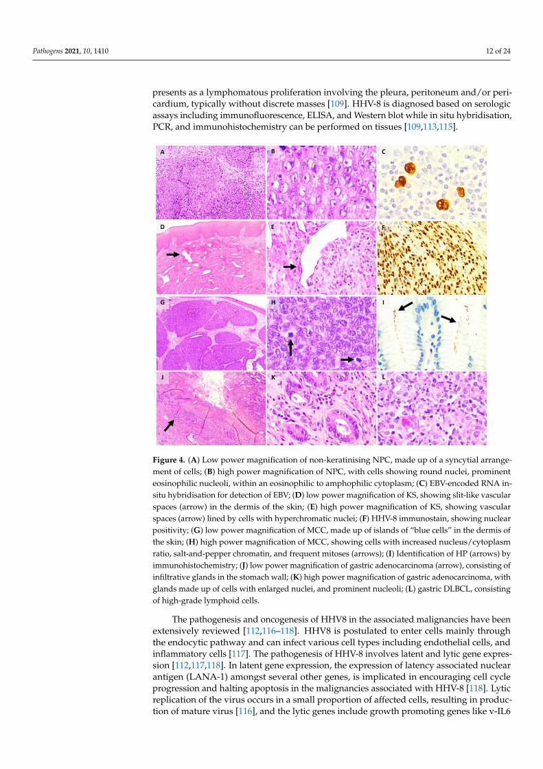
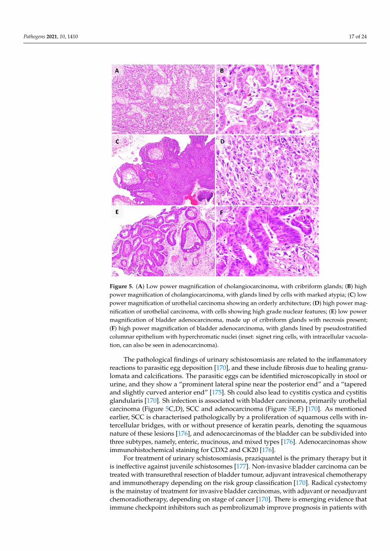
Figure 5. (A) Low power magnification of cholangiocarcinoma, with cribriform glands; (B) high
power magnification of cholangiocarcinoma, with glands lined by cells with marked atypia; (C)
low power magnification of urothelial carcinoma showing an orderly architecture; (D) high power
magnification of urothelial carcinoma, with cells showing high grade nuclear features; (E) low
Figure 5. (A) Low power magnification of cholangiocarcinoma, with cribriform glands; (B) highpower magnification of cholangiocarcinoma, with glands lined by cells with marked atypia; (C) lowpower magnification of urothelial carcinoma showing an orderly architecture; (D) high power mag-nification of urothelial carcinoma, with cells showing high grade nuclear features; (E) low powermagnification of bladder adenocarcinoma, made up of cribriform glands with necrosis present;(F) high power magnification of bladder adenocarcinoma, with glands lined by pseudostratifiedcolumnar epithelium with hyperchromatic nuclei (inset: signet ring cells, with intracellular vacuola-tion, can also be seen in adenocarcinoma).
The pathological findings of urinary schistosomiasis are related to the inflammatoryreactions to parasitic egg deposition [170], and these include fibrosis due to healing granu-lomata and calcifications. The parasitic eggs can be identified microscopically in stool orurine, and they show a “prominent lateral spine near the posterior end” and a “taperedand slightly curved anterior end” [175]. Sh could also lead to cystitis cystica and cystitisglandularis [170]. Sh infection is associated with bladder carcinoma, primarily urothelialcarcinoma (Figure 5C,D), SCC and adenocarcinoma (Figure 5E,F) [170]. As mentionedearlier, SCC is characterised pathologically by a proliferation of squamous cells with in-tercellular bridges, with or without presence of keratin pearls, denoting the squamousnature of these lesions [176], and adenocarcinomas of the bladder can be subdivided intothree subtypes, namely, enteric, mucinous, and mixed types [176]. Adenocarcinomas showimmunohistochemical staining for CDX2 and CK20 [176].
For treatment of urinary schistosomiasis, praziquantel is the primary therapy but itis ineffective against juvenile schistosomes [177]. Non-invasive bladder carcinoma can betreated with transurethral resection of bladder tumour, adjuvant intravesical chemotherapyand immunotherapy depending on the risk group classification [170]. Radical cystectomyis the mainstay of treatment for invasive bladder carcinomas, with adjuvant or neoadjuvantchemoradiotherapy, depending on stage of cancer [170]. There is emerging evidence thatimmune checkpoint inhibitors such as pembrolizumab improve prognosis in patients with
Pathogens 2021, 10, 1410 18 of 24
locally advanced or metastatic urothelial carcinoma with progression after chemother-apy [178]. Vaccine developments have proven to be challenging [177].
3.10. COVID-19
At the time of writing, there are over 200 million cases of COVID-19 worldwide [179].Predominantly an infection of the respiratory tract, it manifests, in severe cases, as acuterespiratory distress syndrome [180]. The association of a deregulated immune responseand ground glass changes on imaging with COVID-19 infection has been postulatedto facilitate cancer initiation and progression [181]. COVID-19 and cancer also involvecommon signalling pathways including cytokine signalling, IL-6 and JAK/STAT signalling,and immune checkpoint signalling amongst numerous other pathways [15]. Several viralinfections such as HIV and HBV also show high expression of programmed cell death-1 receptor [15] and there is also emerging evidence that immune checkpoint receptorsare upregulated in severe COVID-19 cases [182]. Although this evidence demonstratessimilarities between COVID-19 and other pathogens, the true clinical significance of thesesimilarities remains to be determined. With accelerated research and development ofnovel COVID-19 vaccines [183], it is hoped this knowledge can be used to develop similarvaccines against other familiar pathogens.
4. Conclusions
Pathogens are frequently encountered in the practice of anatomical pathology, withsome presenting as benign mimickers of malignancies and others as more commonly seenmalignancies. Considering many of the oncogenic pathogens are endemic in certain partsof the world and are often asymptomatic, there may be a greater need for early detectionof these pathogens as well as identification of the pre-malignant manifestations to initiateprompt medical intervention. With increasing attention given to personalised medicine,molecular diagnostics and therapeutics, it is likely that more knowledge will be uncoveredabout the oncogenic molecular pathways associated with the various oncogenic pathogens.Furthermore, with an accelerated growth in the research around anti-viral and anti-cancervaccines, it is envisioned that more will be known about these pathogens and how theymay be eradicated.
Author Contributions: Conception, T.H.C., L.S.P. and L.Y.K.; writing, T.H.C., L.S.P. and L.Y.K.;critical revision, T.H.C., L.S.P. and L.Y.K.; final approval, T.H.C., L.S.P. and L.Y.K. All authors haveread and agreed to the published version of the manuscript.
Funding: This research received no external funding.
Institutional Review Board Statement: Not applicable.
Informed Consent Statement: Not applicable.
Data Availability Statement: Not applicable.
Conflicts of Interest: The authors declare no conflict of interest.
References1. Bray, F.; Laversanne, M.; Weiderpass, E.; Soerjomataram, I. The ever-increasing importance of cancer as a leading cause of
premature death worldwide. Cancer 2021, 127, 3029–3030. [CrossRef]2. Sung, H.; Ferlay, J.; Siegel, R.L.; Laversanne, M.; Soerjomataram, I.; Jemal, A.; Bray, F. Global Cancer Statistics 2020: GLOBOCAN
Estimates of Incidence and Mortality Worldwide for 36 Cancers in 185 Countries. CA Cancer J. Clin. 2021, 71, 209–249. [CrossRef][PubMed]
3. Danaei, G.; Vander Hoorn, S.; Lopez, A.D.; Murray, C.J.; Ezzati, M. Comparative Risk Assessment collaborating, g. Causes ofcancer in the world: Comparative risk assessment of nine behavioural and environmental risk factors. Lancet 2005, 366, 1784–1793.[CrossRef]
4. De Flora, S.; La Maestra, S. Epidemiology of cancers of infectious origin and prevention strategies. J. Prev. Med. Hyg. 2015, 56,E15–E20.
5. Parkin, D.M. The global health burden of infection-associated cancers in the year 2002. Int. J. Cancer 2006, 118, 3030–3044.[CrossRef] [PubMed]
Pathogens 2021, 10, 1410 19 of 24
6. Pagano, J.S.; Blaser, M.; Buendia, M.A.; Damania, B.; Khalili, K.; Raab-Traub, N.; Roizman, B. Infectious agents and cancer:Criteria for a causal relation. Semin. Cancer Biol. 2004, 14, 453–471. [CrossRef] [PubMed]
7. Kuper, H.; Adami, H.O.; Trichopoulos, D. Infections as a major preventable cause of human cancer. J. Intern. Med. 2000, 248,171–183. [CrossRef]
8. de Martel, C.; Georges, D.; Bray, F.; Ferlay, J.; Clifford, G.M. Global burden of cancer attributable to infections in 2018: Aworldwide incidence analysis. Lancet Glob. Health 2020, 8, e180–e190. [CrossRef]
9. Nguyen, T.X.T.; Han, M.; Ki, M.; Kim, Y.A.; Oh, J.K. The Economic Burden of Cancers Attributable to Infection in the Republic ofKorea: A Prevalence-Based Study. Int. J. Environ. Res. Public Health 2020, 17, 7592. [CrossRef]
10. Cancer IAfRo. IARC Monographs on the Identification of Carcinogenic Hazards to Humans. Available online: https://monographs.iarc.who.int/list-of-classifications/ (accessed on 9 August 2021).
11. Cancer IAfRo. Cancers Attributbable to Infections: Data Sources and Methods. Available online: https://gco.iarc.fr/causes/infections/data-sources-methods (accessed on 22 August 2021).
12. Feng, H.; Shuda, M.; Chang, Y.; Moore, P.S. Clonal integration of a polyomavirus in human Merkel cell carcinoma. Science 2008,319, 1096–1100. [CrossRef]
13. Velavan, T.P.; Meyer, C.G. The COVID-19 epidemic. Trop. Med. Int. Health 2020, 25, 278–280. [CrossRef] [PubMed]14. Tutuncuoglu, B.; Cakir, M.; Batra, J.; Bouhaddou, M.; Eckhardt, M.; Gordon, D.E.; Krogan, N.J. The Landscape of Human Cancer
Proteins Targeted by SARS-CoV-2. Cancer Discov. 2020, 10, 916–921. [CrossRef]15. Zong, Z.; Wei, Y.; Ren, J.; Zhang, L.; Zhou, F. The intersection of COVID-19 and cancer: Signaling pathways and treatment
implications. Mol. Cancer 2021, 20, 76. [CrossRef] [PubMed]16. Chen, J.; Dai, L.; Barrett, L.; Post, S.R.; Qin, Z. SARS-CoV-2 proteins and anti-COVID-19 drugs induce lytic reactivation of an
oncogenic virus. bioRxiv 2020. [CrossRef]17. Baldassarri, R.J.; Kumar, D.; Baldassarri, S.; Cai, G. Diagnosis of Infectious Diseases in the Lower Respiratory Tract: A Cytopathol-
ogist’s Perspective. Arch. Pathol. Lab. Med. 2019, 143, 683–694. [CrossRef]18. Martinez-Giron, R.; Pantanowitz, L. Lower respiratory tract viral infections: Diagnostic role of exfoliative cytology. Diagn.
Cytopathol. 2017, 45, 614–620. [CrossRef]19. Kumar Vinay, A.A.; Aster, J. Robbins Cotrain Pathologic Basis of Disease, 10th ed.; Elsevier: Philadelphia, PA, USA, 2020.20. Whitley, R.J.; Roizman, B. Herpes simplex virus infections. Lancet 2001, 357, 1513–1518. [CrossRef]21. Naib, Z.M.; Nahmias, A.J.; Josey, W.E. Cytology and histopathology of cervical herpes simplex infection. Cancer 1966, 19,
1026–1031. [CrossRef]22. Lapkus, O.; Elsheikh, T.M.; Ujevich, B.A.; Liu, Y.L.; Silverman, J.F. Pitfalls in the diagnosis of herpes simplex infection in
respiratory cytology. Acta Cytol. 2006, 50, 617–620. [CrossRef]23. Blank, H.; Burgoon, C.F.; Baldridge, G.D.; Mc, C.P.; Urbach, F. Cytologic smears in diagnosis of herpes simplex, herpes zoster, and
varicella. J. Am. Med. Assoc. 1951, 146, 1410–1412. [CrossRef] [PubMed]24. Gupta, M.M.; Sharma, B.K.; Singh, V.; Luthra, U.K. Immunocytological demonstration of HSV-II antigen on exfoliated cells from
precancerous and cancerous lesions of the uterine cervix. Diagn Cytopathol. 1988, 4, 48–49. [CrossRef] [PubMed]25. Solomon, I.H.; Hornick, J.L.; Laga, A.C. Immunohistochemistry Is Rarely Justified for the Diagnosis of Viral Infections.
Am. J. Clin. Pathol. 2017, 147, 96–104. [CrossRef]26. Chen, D.H.; Jiang, H.; Lee, M.; Liu, F.; Zhou, Z.H. Three-dimensional visualization of tegument/capsid interactions in the intact
human cytomegalovirus. Virology 1999, 260, 10–16. [CrossRef]27. Gandhi, M.K.; Khanna, R. Human cytomegalovirus: Clinical aspects, immune regulation, and emerging treatments. Lancet Infect. Dis
2004, 4, 725–738. [CrossRef]28. Taylor, G.H. Cytomegalovirus. Am. Fam. Physician 2003, 67, 519–524. [PubMed]29. Dioverti, M.V.; Razonable, R.R. Cytomegalovirus. Microbiol. Spectr. 2016, 4, 151–171. [CrossRef]30. Griffiths, P.; Baraniak, I.; Reeves, M. The pathogenesis of human cytomegalovirus. J. Pathol. 2015, 235, 288–297. [CrossRef]31. Elgert, P.A.; Yee-Chang, M.; Simsir, A. Cytomegalovirus (CMV) in cervical cancer screening tests: A series of 8 cases and review
of the literature. Diagn. Cytopathol. 2018, 46, 593–599. [CrossRef]32. Weldon-Linne, C.M.; Rhone, D.P.; Bourassa, R. Bronchoscopy specimens in adults with AIDS. Comparative yields of cytology,
histology and culture for diagnosis of infectious agents. Chest 1990, 98, 24–28. [CrossRef]33. Woods, G.L.; Thompson, A.B.; Rennard, S.L.; Linder, J. Detection of cytomegalovirus in bronchoalveolar lavage specimens. Spin
amplification and staining with a monoclonal antibody to the early nuclear antigen for diagnosis of cytomegalovirus pneumonia.Chest 1990, 98, 568–575. [CrossRef]
34. Weiss, R.L.; Snow, G.W.; Schumann, G.B.; Hammond, M.E. Diagnosis of cytomegalovirus pneumonitis on bronchoalveolar lavagefluid: Comparison of cytology, immunofluorescence, and in situ hybridization with viral isolation. Diagn Cytopathol. 1991, 7,243–247. [CrossRef]
35. Behrens, H.W.; Quick, C.A. Bronchoscopic diagnosis of cytomegalovirus infection. J. Infect. Dis. 1974, 130, 174–176. [CrossRef]36. Delvenne, P.; Arrese, J.E.; Thiry, A.; Borlee-Hermans, G.; Pierard, G.E.; Boniver, J. Detection of cytomegalovirus, Pneumocystis
carinii, and aspergillus species in bronchoalveolar lavage fluid. A comparison of techniques. Am. J. Clin. Pathol. 1993, 100,414–418. [CrossRef] [PubMed]
Pathogens 2021, 10, 1410 20 of 24
37. Seok, J.Y.; An, J.; Ha, S.Y.; Chung, D.H.; Lee, S.; Kim, H. Morphologic Analysis of Cytomegalovirus Infected Cells in BronchialWashing Cytology: Comparison of Liquid-Based Preparation and Conventional Smear. J. Pathol. Transl. Med. 2016, 50, 147–154.[CrossRef]
38. Solans, E.P.; Yong, S.; Husain, A.N.; Eichorst, M.; Gattuso, P. Bronchioloalveolar lavage in the diagnosis of CMV pneumonitis inlung transplant recipients: An immunocytochemical study. Diagn. Cytopathol. 1997, 16, 350–352. [CrossRef]
39. Siguier, M.; Sellier, P.; Bergmann, J.F. BK-virus infections: A literature review. Med. Mal. Infect. 2012, 42, 181–187. [CrossRef][PubMed]
40. Alexiev, B.A.; Randhawa, P.; Vazquez Martul, E.; Zeng, G.; Luo, C.; Ramos, E.; Drachenberg, C.B.; Papadimitriou, J.C. BKvirus-associated urinary bladder carcinoma in transplant recipients: Report of 2 cases, review of the literature, and proposedpathogenetic model. Hum. Pathol. 2013, 44, 908–917. [CrossRef] [PubMed]
41. Hirsch, H.H. BK virus: Opportunity makes a pathogen. Clin. Infect. Dis. 2005, 41, 354–360. [CrossRef]42. Reploeg, M.D.; Storch, G.A.; Clifford, D.B. Bk virus: A clinical review. Clin. Infect. Dis. 2001, 33, 191–202. [CrossRef]43. Goudsmit, J.; Wertheim-van Dillen, P.; van Strien, A.; van der Noordaa, J. The role of BK virus in acute respiratory tract disease
and the presence of BKV DNA in tonsils. J. Med. Virol. 1982, 10, 91–99. [CrossRef]44. Hirsch, H.H.; Randhawa, P.S.; Practice, A.S.T.I.D.C.o. BK polyomavirus in solid organ transplantation-Guidelines from the
American Society of Transplantation Infectious Diseases Community of Practice. Clin. Transplant. 2019, 33, e13528. [CrossRef]45. Geetha, V.; Rao, L.; Monappa, V.; Susmitha, M.; Prabhu, R. Decoy cells in urine cytology: A useful clue to post-transplant polyoma
virus infection. J. Cytol. 2012, 29, 133–134. [CrossRef] [PubMed]46. Sawinski, D.; Goral, S. BK virus infection: An update on diagnosis and treatment. Nephrol. Dial. Transplant. 2015, 30, 209–217.
[CrossRef]47. Hogan, T.F.; Padgett, B.L.; Walker, D.L.; Borden, E.C.; McBain, J.A. Rapid detection and identification of JC virus and BK virus in
human urine by using immunofluorescence microscopy. J. Clin. Microbiol. 1980, 11, 178–183. [CrossRef] [PubMed]48. Santos, R.L.; Manfrinatto, J.A.; Cia, E.M.; Carvalho, R.B.; Quadros, K.R.; Alves-Filho, G.; Mazzali, M. Urine cytology as a screening
method for polyoma virus active infection. Transplant. Proc. 2004, 36, 899–901. [CrossRef]49. Drachenberg, R.C.; Drachenberg, C.B.; Papadimitriou, J.C.; Ramos, E.; Fink, J.C.; Wali, R.; Weir, M.R.; Cangro, C.B.; Klassen, D.K.;
Khaled, A.; et al. Morphological spectrum of polyoma virus disease in renal allografts: Diagnostic accuracy of urine cytology.Am. J. Transplant. 2001, 1, 373–381. [CrossRef] [PubMed]
50. Bulut, Y.; Ozdemir, E.; Ozercan, H.I.; Etem, E.O.; Aker, F.; Toraman, Z.A.; Seyrek, A.; Firdolas, F. Potential relationship betweenBK virus and renal cell carcinoma. J. Med. Virol. 2013, 85, 1085–1089. [CrossRef]
51. Weinreb, D.B.; Desman, G.T.; Amolat-Apiado, M.J.; Burstein, D.E.; Godbold, J.H., Jr.; Johnson, E.M. Polyoma virus infection is aprominent risk factor for bladder carcinoma in immunocompetent individuals. Diagn. Cytopathol. 2006, 34, 201–203. [CrossRef][PubMed]
52. Das, D.; Wojno, K.; Imperiale, M.J. BK virus as a cofactor in the etiology of prostate cancer in its early stages. J. Virol. 2008, 82,2705–2714. [CrossRef]
53. Das, D.; Shah, R.B.; Imperiale, M.J. Detection and expression of human BK virus sequences in neoplastic prostate tissues. Oncogene2004, 23, 7031–7046. [CrossRef]
54. Rollison, D.E.; Sexton, W.J.; Rodriguez, A.R.; Kang, L.C.; Daniel, R.; Shah, K.V. Lack of BK virus DNA sequences in mosttransitional-cell carcinomas of the bladder. Int. J. Cancer 2007, 120, 1248–1251. [CrossRef]
55. Kumari, K.; Pradeep, I.; Kakkar, A.; Dinda, A.K.; Seth, A.; Nayak, B.; Singh, G. BK polyomavirus and urothelial carcinoma:Experience at a tertiary care centre in India with review of literature. Ann. Diagn Pathol. 2019, 40, 77–80. [CrossRef]
56. Lenaerts, L.; De Clercq, E.; Naesens, L. Clinical features and treatment of adenovirus infections. Rev. Med. Virol. 2008, 18, 357–374.[CrossRef] [PubMed]
57. Kojaoghlanian, T.; Flomenberg, P.; Horwitz, M.S. The impact of adenovirus infection on the immunocompromised host. Rev. Med.Virol. 2003, 13, 155–171. [CrossRef]
58. Avellon, A.; Perez, P.; Aguilar, J.C.; Lejarazu, R.; Echevarria, J.E. Rapid and sensitive diagnosis of human adenovirus infections bya generic polymerase chain reaction. J. Virol. Methods 2001, 92, 113–120. [CrossRef]
59. Matthes-Martin, S.; Boztug, H.; Lion, T. Diagnosis and treatment of adenovirus infection in immunocompromised patients. ExpertRev. Anti Infect. Ther. 2013, 11, 1017–1028. [CrossRef] [PubMed]
60. Harbhajanka, A.; Michael, C.W. Adenoviral cytopathic changes in pleural fluid: A unique cytological presentation mimickingmalignancy. Diagn. Cytopathol. 2021, 49, 657–660. [CrossRef] [PubMed]
61. Zaharopoulos, P. Viral cytopathic changes in urine cytology of ileal conduit attributed to adenovirus: Report of a case. Diagn.Cytopathol. 2004, 30, 284–287. [CrossRef]
62. Moss, W.J.; Griffin, D.E. Measles. Lancet 2012, 379, 153–164. [CrossRef]63. Katz, M. Clinical spectrum of measles. Curr. Top. Microbiol. Immunol. 1995, 191, 1–12. [CrossRef]64. Robbins, F.C. Measles: Clinical features. Pathogenesis, pathology and complications. Am. J. Dis Child. 1962, 103, 266–273.
[CrossRef]65. Bester, J.C. Measles and Measles Vaccination: A Review. JAMA Pediatr. 2016, 170, 1209–1215. [CrossRef] [PubMed]66. Bolande, R.P. Significance and nature of inclusion-bearing cells in the urine of patients with measles. N. Engl. J. Med. 1961, 265,
919–923. [CrossRef]
Pathogens 2021, 10, 1410 21 of 24
67. Lipsey, A.I.; Bolande, R.P. The exfoliative source of abnormal cells in urine sediment of patients with measles. Am. J. Dis. Child.1967, 113, 677–682. [CrossRef] [PubMed]
68. Simoes, E.A. Respiratory syncytial virus infection. Lancet 1999, 354, 847–852. [CrossRef]69. Hall, C.B.; Simoes, E.A.; Anderson, L.J. Clinical and epidemiologic features of respiratory syncytial virus. Curr. Top. Microbiol.
Immunol. 2013, 372, 39–57. [CrossRef]70. Hall, C.B. Respiratory syncytial virus and parainfluenza virus. N. Engl. J. Med. 2001, 344, 1917–1928. [CrossRef]71. Shah, K.K.; Pritt, B.S.; Alexander, M.P. Histopathologic review of granulomatous inflammation. J. Clin. Tuberc. Other Mycobact. Dis.
2017, 7, 1–12. [CrossRef]72. Chopra, A.; Avadhani, V.; Tiwari, A.; Riemer, E.C.; Sica, G.; Judson, M.A. Granulomatous lung disease: Clinical aspects. Expert.
Rev. Respir. Med. 2020, 14, 1045–1063. [CrossRef] [PubMed]73. Williams, G.T.; Williams, W.J. Granulomatous inflammation–a review. J. Clin. Pathol. 1983, 36, 723–733. [CrossRef]74. Mehrotra, R.; Dhingra, V. Cytological diagnosis of sarcoidosis revisited: A state of the art review. Diagn. Cytopathol. 2011, 39,
541–548. [CrossRef] [PubMed]75. Ramzan, N.N.; Leighton, J.A.; Heigh, R.I.; Shapiro, M.S. Clinical significance of granuloma in Crohn’s disease. Inflamm. Bowel. Dis.
2002, 8, 168–173. [CrossRef] [PubMed]76. Brincker, H. Sarcoid reactions in malignant tumours. Cancer Treat. Rev. 1986, 13, 147–156. [CrossRef]77. Tjan-Heijnen, V.C.; Vlasveld, L.T.; Pernet, F.P.; Pauwels, P.; De Mulder, P.H. Coincidence of seminoma and sarcoidosis: A myth or
fact? Ann. Oncol. 1998, 9, 321–325. [CrossRef]78. Khurana, K.K.; Stanley, M.W.; Powers, C.N.; Pitman, M.B. Aspiration cytology of malignant neoplasms associated with granulo-
mas and granuloma-like features: Diagnostic dilemmas. Cancer 1998, 84, 84–91. [CrossRef]79. Kennedy, M.P.; Jimenez, C.A.; Mhatre, A.D.; Morice, R.C.; Eapen, G.A. Clinical implications of granulomatous inflammation
detected by endobronchial ultrasound transbronchial needle aspiration in patients with suspected cancer recurrence in themediastinum. J. Cardiothorac. Surg. 2008, 3, 8. [CrossRef]
80. Betz, S.A.; See, W.A.; Cohen, M.B. Granulomatous inflammation in bladder wash specimens after intravesical bacillus Calmette-Guerin therapy for transitional cell carcinoma of the bladder. Am. J. Clin. Pathol. 1993, 99, 244–248. [CrossRef]
81. Ocal, K.; Dag, A.; Turkmenoglu, O.; Kara, T.; Seyit, H.; Konca, K. Granulomatous mastitis: Clinical, pathological features, andmanagement. Breast J. 2010, 16, 176–182. [CrossRef]
82. Chandanwale, S.; Naragude, P.; Shetty, A.; Sawadkar, M.; Raj, A.; Bhide, A.; Singh, M. Cytomorphological Spectrum ofGranulomatous Mastitis: A Study of 33 Cases. Eur. J. Breast Health 2020, 16, 146–151. [CrossRef]
83. Erhan, Y.; Veral, A.; Kara, E.; Ozdemir, N.; Kapkac, M.; Ozdedeli, E.; Yilmaz, R.; Koyuncu, A.; Erhan, Y.; Ozbal, O. A clini-copthologic study of a rare clinical entity mimicking breast carcinoma: Idiopathic granulomatous mastitis. Breast 2000, 9, 52–56.[CrossRef]
84. Heer, R.; Shrimankar, J.; Griffith, C.D. Granulomatous mastitis can mimic breast cancer on clinical, radiological or cytologicalexamination: A cautionary tale. Breast 2003, 12, 283–286. [CrossRef]
85. Mitra, S.; Dey, P. Fine-needle aspiration and core biopsy in the diagnosis of breast lesions: A comparison and review of theliterature. Cytojournal 2016, 13, 18. [CrossRef]
86. Fletcher, G.J.; Eapen, C.E.; Abraham, P. Hepatitis B genotyping: The utility for the clinicians. Indian J. Gastroenterol. 2020, 39,315–320. [CrossRef]
87. Fiel, M.I. Pathology of chronic hepatitis B and chronic hepatitis C. Clin. Liver Dis. 2010, 14, 555–575. [CrossRef] [PubMed]88. WHO Classification of Tumours Editorial Board. WHO Classification of Tumours: Digestive System Tumours, 5th ed.; International
Agency for Research on Cancer: Lyon, France, 2019.89. Bruix, J.; Sherman, M.; American Association for the Study of Liver. Management of hepatocellular carcinoma: An update.
Hepatology 2011, 53, 1020–1022. [CrossRef] [PubMed]90. Zhu, X.D.; Sun, H.C. Emerging agents and regimens for hepatocellular carcinoma. J. Hematol. Oncol. 2019, 12, 110. [CrossRef]91. Longo, V.; Brunetti, O.; Gnoni, A.; Licchetta, A.; Delcuratolo, S.; Memeo, R.; Solimando, A.G.; Argentiero, A. Emerging role of
Immune Checkpoint Inhibitors in Hepatocellular Carcinoma. Medicina 2019, 55, 698. [CrossRef] [PubMed]92. Chen, P.; Fang, Q.X.; Chen, D.B.; Chen, H.S. Neoantigen vaccine: An emerging immunotherapy for hepatocellular carcinoma.
World J. Gastrointest. Oncol. 2021, 13, 673–683. [CrossRef]93. Asiaf, A.; Ahmad, S.T.; Mohammad, S.O.; Zargar, M.A. Review of the current knowledge on the epidemiology, pathogenesis, and
prevention of human papillomavirus infection. Eur. J. Cancer Prev. 2014, 23, 206–224. [CrossRef] [PubMed]94. Lekoane, K.M.B.; Kuupiel, D.; Mashamba-Thompson, T.P.; Ginindza, T.G. The interplay of HIV and human papillomavirus-related
cancers in sub-Saharan Africa: Scoping review. Syst. Rev. 2020, 9, 88. [CrossRef] [PubMed]95. McManus, L.M. Pathobiology of Human Disease: A Dynamic Encyclopedia of Disease Mechanisms; Elsevier: Amsterdam,
The Netherlands, 2014.96. WHO Classification of Tumours Editorial Board. WHO Classification of Tumours: Female Genital Tumours, 5th ed.; International
Agency for Research on Cancer: Lyon, France, 2020.97. Thompson, L.D.R. HPV-Related Multiphenotypic Sinonasal Carcinoma. Ear. Nose Throat J. 2020, 99, 94–95. [CrossRef] [PubMed]98. Kesic, V. Management of cervical cancer. Eur. J. Surg. Oncol. 2006, 32, 832–837. [CrossRef]
Pathogens 2021, 10, 1410 22 of 24
99. Fakhr, E.; Modic, Z.; Cid-Arregui, A. Recent developments in immunotherapy of cancers caused by human papillomaviruses.Immunology 2021, 163, 33–45. [CrossRef]
100. Skeate, J.G.; Woodham, A.W.; Einstein, M.H.; Da Silva, D.M.; Kast, W.M. Current therapeutic vaccination and immunotherapystrategies for HPV-related diseases. Hum. Vaccin Immunother. 2016, 12, 1418–1429. [CrossRef]
101. Luvero, D.; Lopez, S.; Bogani, G.; Raspagliesi, F.; Angioli, R. From the Infection to the Immunotherapy in Cervical Cancer: CanWe Stop the Natural Course of the Disease? Vaccines 2020, 8, 597. [CrossRef]
102. Shannon-Lowe, C.; Rickinson, A. The Global Landscape of EBV-Associated Tumors. Front. Oncol. 2019, 9, 713. [CrossRef]103. Louissaint, A., Jr.; Ferry, J.A.; Soupir, C.P.; Hasserjian, R.P.; Harris, N.L.; Zukerberg, L.R. Infectious mononucleosis mimicking
lymphoma: Distinguishing morphological and immunophenotypic features. Mod. Pathol. 2012, 25, 1149–1159. [CrossRef]104. van Zyl, D.G.; Mautner, J.; Delecluse, H.J. Progress in EBV Vaccines. Front. Oncol. 2019, 9, 104. [CrossRef] [PubMed]105. Linke-Serinsoz, E.; Fend, F.; Quintanilla-Martinez, L. Human immunodeficiency virus (HIV) and Epstein-Barr virus (EBV) related
lymphomas, pathology view point. Semin. Diagn. Pathol. 2017, 34, 352–363. [CrossRef]106. Hong, M.; Tang, K.; Qian, J.; Deng, H.; Zeng, M.; Zheng, S.; Ding, K.; Du, Y.; Sun, R. Immunotherapy for EBV-Associated
Nasopharyngeal Carcinoma. Crit. Rev. Oncog. 2018, 23, 219–234. [CrossRef] [PubMed]107. Sarid, R.; Olsen, S.J.; Moore, P.S. Kaposi′s sarcoma-associated herpesvirus: Epidemiology, virology, and molecular biology.
Adv. Virus Res. 1999, 52, 139–232. [CrossRef] [PubMed]108. Swerdlow, S.H.; Campo, E.; Harris, N.L.; Jaffe, E.S.; Pileri, S.A.; Stein, H.; Thiele, J.; Arber, D.A.; Hasserjian, R.P.; Le Beau, M.M.;
et al. WHO Classification of Tumours of Haematopoeitic and Lymphoid Tissues, 4th ed.; International Agency for Research on Cancer:Lyon, France, 2017.
109. Sarid, R.; Klepfish, A.; Schattner, A. Virology, pathogenetic mechanisms, and associated diseases of Kaposi sarcoma-associatedherpesvirus (human herpesvirus 8). Mayo Clin. Proc. 2002, 77, 941–949. [CrossRef]
110. Vangipuram, R.; Tyring, S.K. Epidemiology of Kaposi sarcoma: Review and description of the nonepidemic variant.Int. J. Dermatol. 2019, 58, 538–542. [CrossRef]
111. Schulz, T.F.; Cesarman, E. Kaposi Sarcoma-associated Herpesvirus: Mechanisms of oncogenesis. Curr. Opin. Virol. 2015, 14,116–128. [CrossRef]
112. Schulz, T.F. Kaposi’s sarcoma-associated herpesvirus (human herpesvirus 8): Epidemiology and pathogenesis. J. Antimicrob.Chemother. 2000, 45, 15–27. [CrossRef]
113. Bhaduri-McIntosh, S. Human herpesvirus-8: Clinical features of an emerging viral pathogen. Pediatr. Infect. Dis. J. 2005, 24, 81–82.[CrossRef]
114. Geraminejad, P.; Memar, O.; Aronson, I.; Rady, P.L.; Hengge, U.; Tyring, S.K. Kaposi’s sarcoma and other manifestations of humanherpesvirus 8. J. Am. Acad. Dermatol. 2002, 47, 641–655. [CrossRef]
115. Ablashi, D.V.; Chatlynne, L.G.; Whitman, J.E., Jr.; Cesarman, E. Spectrum of Kaposi’s sarcoma-associated herpesvirus, or humanherpesvirus 8, diseases. Clin. Microbiol. Rev. 2002, 15, 439–464. [CrossRef]
116. Mesri, E.A.; Cesarman, E.; Boshoff, C. Kaposi’s sarcoma and its associated herpesvirus. Nat. Rev. Cancer 2010, 10, 707–719.[CrossRef]
117. Dittmer, D.P.; Damania, B. Kaposi sarcoma-associated herpesvirus: Immunobiology, oncogenesis, and therapy. J. Clin. Investig.2016, 126, 3165–3175. [CrossRef]
118. Kaplan, L.D. Human herpesvirus-8: Kaposi sarcoma, multicentric Castleman disease, and primary effusion lymphoma. Hematol.Am. Soc. Hematol. Educ. Program. 2013, 2013, 103–108. [CrossRef]
119. WHO Classification of Tumours Editorial Board. WHO Classification of Tumours: Soft Tissue and Bone tumours, 5th ed.; InternationalAgency for Research on Cancer: Lyon, France, 2020.
120. Cheuk, W.; Wong, K.O.; Wong, C.S.; Dinkel, J.E.; Ben-Dor, D.; Chan, J.K. Immunostaining for human herpesvirus 8 latent nuclearantigen-1 helps distinguish Kaposi sarcoma from its mimickers. Am. J. Clin. Pathol. 2004, 121, 335–342. [CrossRef] [PubMed]
121. Robin, Y.M.; Guillou, L.; Michels, J.J.; Coindre, J.M. Human herpesvirus 8 immunostaining: A sensitive and specific method fordiagnosing Kaposi sarcoma in paraffin-embedded sections. Am. J. Clin. Pathol. 2004, 121, 330–334. [CrossRef]
122. Du, M.Q.; Bacon, C.M.; Isaacson, P.G. Kaposi sarcoma-associated herpesvirus/human herpesvirus 8 and lymphoproliferativedisorders. J. Clin. Pathol. 2007, 60, 1350–1357. [CrossRef]
123. Gantt, S.; Casper, C. Human herpesvirus 8-associated neoplasms: The roles of viral replication and antiviral treatment. Curr. Opin.Infect. Dis. 2011, 24, 295–301. [CrossRef]
124. Broussard, G.; Damania, B. KSHV: Immune Modulation and Immunotherapy. Front. Immunol. 2019, 10, 3084. [CrossRef][PubMed]
125. Gambichler, T.; Koim, S.; Wrobel, M.; Kafferlein, H.U.; Bruning, T.; Stockfleth, E.; Becker, J.C.; Lang, K. Expression of ProgrammedCell Death Proteins in Kaposi Sarcoma and Cutaneous Angiosarcoma. J. Immunother. 2020, 43, 169–174. [CrossRef] [PubMed]
126. Edlich, R.F.; Arnette, J.A.; Williams, F.M. Global epidemic of human T-cell lymphotropic virus type-I (HTLV-I). J. Emerg. Med.2000, 18, 109–119. [CrossRef]
127. Manns, A.; Hisada, M.; La Grenade, L. Human T-lymphotropic virus type I infection. Lancet 1999, 353, 1951–1958. [CrossRef]128. Hlela, C.; Shepperd, S.; Khumalo, N.P.; Taylor, G.P. The prevalence of human T-cell lymphotropic virus type 1 in the general
population is unknown. AIDS Rev. 2009, 11, 205–214.
Pathogens 2021, 10, 1410 23 of 24
129. Ahmadi Ghezeldasht, S.; Shirdel, A.; Assarehzadegan, M.A.; Hassannia, T.; Rahimi, H.; Miri, R.; Rezaee, S.A. Human TLymphotropic Virus Type I (HTLV-I) Oncogenesis: Molecular Aspects of Virus and Host Interactions in Pathogenesis of Adult Tcell Leukemia/Lymphoma (ATL). Iran. J. Basic Med. Sci. 2013, 16, 179–195. [PubMed]
130. Bangham, C.R.M.; Matsuoka, M. Human T-cell leukaemia virus type 1: Parasitism and pathogenesis. Philos Trans. R Soc. Lond. BBiol. Sci. 2017, 372, 20160272. [CrossRef]
131. Tagaya, Y.; Matsuoka, M.; Gallo, R. 40 years of the human T-cell leukemia virus: Past, present, and future. F1000 Res. 2019, 8.[CrossRef]
132. Zhang, L.L.; Wei, J.Y.; Wang, L.; Huang, S.L.; Chen, J.L. Human T-cell lymphotropic virus type 1 and its oncogenesis.Acta Pharmacol. Sin. 2017, 38, 1093–1103. [CrossRef]
133. Tsukasaki, K.; Tobinai, K. Human T-cell lymphotropic virus type I-associated adult T-cell leukemia-lymphoma: New directions inclinical research. Clin. Cancer Res. 2014, 20, 5217–5225. [CrossRef]
134. Hermine, O.; Ramos, J.C.; Tobinai, K. A Review of New Findings in Adult T-cell Leukemia-Lymphoma: A Focus on Current andEmerging Treatment Strategies. Adv. Ther. 2018, 35, 135–152. [CrossRef]
135. Iqbal, M.; Reljic, T.; Klocksieben, F.; Sher, T.; Ayala, E.; Murthy, H.; Bazarbachi, A.; Kumar, A.; Kharfan-Dabaja, M.A. Efficacyof Allogeneic Hematopoietic Cell Transplantation in Human T Cell Lymphotropic Virus Type 1-Associated Adult T CellLeukemia/Lymphoma: Results of a Systematic Review/Meta-Analysis. Biol. Blood Marrow Transplant. 2019, 25, 1695–1700.[CrossRef]
136. Spurgeon, M.E.; Lambert, P.F. Merkel cell polyomavirus: A newly discovered human virus with oncogenic potential. Virology2013, 435, 118–130. [CrossRef]
137. Becker, J.C.; Stang, A.; DeCaprio, J.A.; Cerroni, L.; Lebbe, C.; Veness, M.; Nghiem, P. Merkel cell carcinoma. Nat. Rev. Dis. Primers2017, 3, 17077. [CrossRef]
138. Liu, W.; MacDonald, M.; You, J. Merkel cell polyomavirus infection and Merkel cell carcinoma. Curr. Opin. Virol. 2016, 20, 20–27.[CrossRef]
139. Hattori, T.; Takeuchi, Y.; Takenouchi, T.; Hirofuji, A.; Tsuchida, T.; Kabumoto, T.; Fujiwara, H.; Ito, M.; Shimizu, A.; Okada, E.;et al. The prevalence of Merkel cell polyomavirus in Japanese patients with Merkel cell carcinoma. J. Dermatol. Sci. 2013, 70,99–107. [CrossRef]
140. Duncavage, E.J.; Zehnbauer, B.A.; Pfeifer, J.D. Prevalence of Merkel cell polyomavirus in Merkel cell carcinoma. Mod. Pathol.2009, 22, 516–521. [CrossRef]
141. Mangana, J.; Dziunycz, P.; Kerl, K.; Dummer, R.; Cozzio, A. Prevalence of Merkel cell polyomavirus among Swiss Merkel cellcarcinoma patients. Dermatology 2010, 221, 184–188. [CrossRef]
142. Garneski, K.M.; Warcola, A.H.; Feng, Q.; Kiviat, N.B.; Leonard, J.H.; Nghiem, P. Merkel cell polyomavirus is more frequentlypresent in North American than Australian Merkel cell carcinoma tumors. J. Investig. Dermatol. 2009, 129, 246–248. [CrossRef]
143. Wieland, U.; Kreuter, A. Merkel cell polyomavirus infection and Merkel cell carcinoma in HIV-positive individuals. Curr. Opin.Oncol. 2011, 23, 488–493. [CrossRef]
144. Santos-Juanes, J.; Fernandez-Vega, I.; Fuentes, N.; Galache, C.; Coto-Segura, P.; Vivanco, B.; Astudillo, A.; Martinez-Camblor, P.Merkel cell carcinoma and Merkel cell polyomavirus: A systematic review and meta-analysis. Br. J. Dermatol. 2015, 173, 42–49.[CrossRef]
145. Pietropaolo, V.; Prezioso, C.; Moens, U. Merkel Cell Polyomavirus and Merkel Cell Carcinoma. Cancers 2020, 12, 1774. [CrossRef]146. Grundhoff, A.; Fischer, N. Merkel cell polyomavirus, a highly prevalent virus with tumorigenic potential. Curr. Opin. Virol. 2015,
14, 129–137. [CrossRef] [PubMed]147. Elder, D.E.; Scholyer, R.A.; Willemze, R. WHO Classification of Skin Tumours, 4th ed.; International Agency for Research on Cancer:
Lyon, France, 2018.148. Leech, S.N.; Kolar, A.J.; Barrett, P.D.; Sinclair, S.A.; Leonard, N. Merkel cell carcinoma can be distinguished from metastatic small
cell carcinoma using antibodies to cytokeratin 20 and thyroid transcription factor 1. J. Clin. Pathol. 2001, 54, 727–729. [CrossRef]149. Tabachnick-Cherny, S.; Pulliam, T.; Church, C.; Koelle, D.M.; Nghiem, P. Polyomavirus-driven Merkel cell carcinoma: Prospects
for therapeutic vaccine development. Mol. Carcinog. 2020, 59, 807–821. [CrossRef] [PubMed]150. Hooi, J.K.Y.; Lai, W.Y.; Ng, W.K.; Suen, M.M.Y.; Underwood, F.E.; Tanyingoh, D.; Malfertheiner, P.; Graham, D.Y.; Wong, V.W.S.;
Wu, J.C.Y.; et al. Global Prevalence of Helicobacter pylori Infection: Systematic Review and Meta-Analysis. Gastroenterology 2017,153, 420–429. [CrossRef] [PubMed]
151. Crowe, S.E. Helicobacter pylori Infection. N. Engl. J. Med. 2019, 380, 1158–1165. [CrossRef]152. Kusters, J.G.; van Vliet, A.H.; Kuipers, E.J. Pathogenesis of Helicobacter pylori infection. Clin. Microbiol. Rev. 2006, 19, 449–490.
[CrossRef]153. Katoh, H.; Ishikawa, S. Lifestyles, genetics, and future perspectives on gastric cancer in east Asian populations. J. Hum. Genet.
2021, 66, 887–899. [CrossRef] [PubMed]154. Harada, K.; Lopez, A.; Shanbhag, N.; Badgwell, B.; Baba, H.; Ajani, J. Recent advances in the management of gastric adenocarci-
noma patients. F1000 Res. 2018, 7. [CrossRef]155. Silva, R.; Gullo, I.; Carneiro, F. The PD-1:PD-L1 immune inhibitory checkpoint in Helicobacter pylori infection and gastric cancer:
A comprehensive review and future perspectives. Porto Biomed. J. 2016, 1, 4–11. [CrossRef]
Pathogens 2021, 10, 1410 24 of 24
156. Oster, P.; Vaillant, L.; Riva, E.; McMillan, B.; Begka, C.; Truntzer, C.; Richard, C.; Leblond, M.M.; Messaoudene, M.; Machremi, E.;et al. Helicobacter pylori infection has a detrimental impact on the efficacy of cancer immunotherapies. Gut 2021. [CrossRef]
157. Hong, S.T.; Fang, Y. Clonorchis sinensis and clonorchiasis, an update. Parasitol. Int. 2012, 61, 17–24. [CrossRef] [PubMed]158. Kaewpitoon, N.; Kaewpitoon, S.J.; Pengsaa, P.; Sripa, B. Opisthorchis viverrini: The carcinogenic human liver fluke. World J.
Gastroenterol. 2008, 14, 666–674. [CrossRef]159. Sithithaworn, P.; Haswell-Elkins, M. Epidemiology of Opisthorchis viverrini. Acta Trop. 2003, 88, 187–194. [CrossRef]160. Smout, M.J.; Sripa, B.; Laha, T.; Mulvenna, J.; Gasser, R.B.; Young, N.D.; Bethony, J.M.; Brindley, P.J.; Loukas, A. Infection with the
carcinogenic human liver fluke, Opisthorchis viverrini. Mol. Biosyst. 2011, 7, 1367–1375. [CrossRef] [PubMed]161. Tang, Z.L.; Huang, Y.; Yu, X.B. Current status and perspectives of Clonorchis sinensis and clonorchiasis: Epidemiology, pathogen-
esis, omics, prevention and control. Infect. Dis. Poverty 2016, 5, 71. [CrossRef] [PubMed]162. Centers for Disease Control and Prevention. DPDx—Laboatory Identification of Parasites of Public Health Concern: Opisthorchi-
asis. Available online: https://www.cdc.gov/dpdx/opisthorchiasis/index.html (accessed on 24 August 2021).163. Kim, T.S.; Pak, J.H.; Kim, J.B.; Bahk, Y.Y. Clonorchis sinensis, an oriental liver fluke, as a human biological agent of cholangiocarci-
noma: A brief review. BMB Rep. 2016, 49, 590–597. [CrossRef]164. Wu, Z.; Boonmars, T.; Boonjaraspinyo, S.; Nagano, I.; Pinlaor, S.; Puapairoj, A.; Yongvanit, P.; Takahashi, Y. Candidate genes
involving in tumorigenesis of cholangiocarcinoma induced by Opisthorchis viverrini infection. Parasitol. Res. 2011, 109, 657–673.[CrossRef]
165. Sripa, B.; Brindley, P.J.; Mulvenna, J.; Laha, T.; Smout, M.J.; Mairiang, E.; Bethony, J.M.; Loukas, A. The tumorigenic liver flukeOpisthorchis viverrini–multiple pathways to cancer. Trends Parasitol. 2012, 28, 395–407. [CrossRef] [PubMed]
166. Vaeteewoottacharn, K.; Seubwai, W.; Bhudhisawasdi, V.; Okada, S.; Wongkham, S. Potential targeted therapy for liver flukeassociated cholangiocarcinoma. J. Hepatobiliary Pancreat Sci. 2014, 21, 362–370. [CrossRef]
167. Seubwai, W.; Wongkham, C.; Puapairoj, A.; Khuntikeo, N.; Pugkhem, A.; Hahnvajanawong, C.; Chaiyagool, J.; Umezawa, K.;Okada, S.; Wongkham, S. Aberrant expression of NF-kappaB in liver fluke associated cholangiocarcinoma: Implications fortargeted therapy. PLoS ONE 2014, 9, e106056. [CrossRef]
168. Dabney, R.S.; Khalife, M.; Shahid, K.; Phan, A.T. Molecular pathways and targeted therapy in cholangiocarcinoma. Clin. Adv.Hematol. Oncol. 2019, 17, 630–637. [PubMed]
169. Massironi, S.; Pilla, L.; Elvevi, A.; Longarini, R.; Rossi, R.E.; Bidoli, P.; Invernizzi, P. New and Emerging Systemic TherapeuticOptions for Advanced Cholangiocarcinoma. Cells 2020, 9, 688. [CrossRef]
170. Zaghloul, M.S.; Zaghloul, T.M.; Bishr, M.K.; Baumann, B.C. Urinary schistosomiasis and the associated bladder cancer: Update.J. Egypt Natl. Canc. Inst. 2020, 32, 44. [CrossRef]
171. Smith, J.H.; Christie, J.D. The pathobiology of Schistosoma haematobium infection in humans. Hum. Pathol. 1986, 17, 333–345.[CrossRef]
172. Honeycutt, J.; Hammam, O.; Fu, C.L.; Hsieh, M.H. Controversies and challenges in research on urogenital schistosomiasis-associated bladder cancer. Trends Parasitol. 2014, 30, 324–332. [CrossRef] [PubMed]
173. Botelho, M.; Ferreira, A.C.; Oliveira, M.J.; Domingues, A.; Machado, J.C.; da Costa, J.M. Schistosoma haematobium total antigeninduces increased proliferation, migration and invasion, and decreases apoptosis of normal epithelial cells. Int. J. Parasitol. 2009,39, 1083–1091. [CrossRef] [PubMed]
174. Botelho, M.C.; Veiga, I.; Oliveira, P.A.; Lopes, C.; Teixeira, M.; da Costa, J.M.; Machado, J.C. Carcinogenic ability of Schistosomahaematobium possibly through oncogenic mutation of KRAS gene. Adv. Cancer Res. Treat. 2013, 2013, 876585. [PubMed]
175. Centers for Disease Control and Prevention. DPDx—Laboratory Identificaiton of Parasites of Public Health Concern: Schistoso-miasis. Available online: https://www.cdc.gov/dpdx/schistosomiasis/index.html (accessed on 24 August 2021).
176. Moch, H.; Ulbright, T.M.; Reuter, V.E. WHO Classification of Tumours of the Urinary System and Male Genital Organs, 4th ed.;International Agency for Research on Cancer: Lyon, France, 2016.
177. Molehin, A.J. Schistosomiasis vaccine development: Update on human clinical trials. J. Biomed. Sci. 2020, 27, 28. [CrossRef]178. Sundahl, N.; Rottey, S.; De Maeseneer, D.; Ost, P. Pembrolizumab for the treatment of bladder cancer. Expert Rev. Anticancer Ther.
2018, 18, 107–114. [CrossRef] [PubMed]179. Center JHUoMCR. COVID-19 Dashboard. Available online: https://coronavirus.jhu.edu/map.html (accessed on 24 August 2021).180. Marini, J.J.; Gattinoni, L. Management of COVID-19 Respiratory Distress. JAMA 2020, 323, 2329–2330. [CrossRef]181. Sadhukhan, P.; Ugurlu, M.T.; Hoque, M.O. Effect of COVID-19 on Lungs: Focusing on Prospective Malignant Phenotypes. Cancers
2020, 12, 3822. [CrossRef]182. Sabbatino, F.; Conti, V.; Franci, G.; Sellitto, C.; Manzo, V.; Pagliano, P.; De Bellis, E.; Masullo, A.; Salzano, F.A.; Caputo, A.; et al.
PD-L1 Dysregulation in COVID-19 Patients. Front. Immunol. 2021, 12, 695242. [CrossRef]183. Thanh Le, T.; Andreadakis, Z.; Kumar, A.; Gomez Roman, R.; Tollefsen, S.; Saville, M.; Mayhew, S. The COVID-19 vaccine
development landscape. Nat. Rev. Drug Discov. 2020, 19, 305–306. [CrossRef] [PubMed]