Embed Size (px)
Citation preview
The Orthodontic-Oral Surgery interface: Part One: A service evaluation and an overview of the diagnosis and management of common anomalies
Sharif MO1, Lyne A2, Parker K3, Chia MSY4
1. Mr Mohammad Owaise Sharif
Clinical Lecturer/Honorary Consultant in Orthodontics, MClinDent Deputy
Programme Director, University College London Eastman Dental Institute
2. Miss Alexandra Lyne
Specialty Registrar in Paediatric Dentistry, Guy’s and St Thomas’s Hospitals
NHS Foundation Trust
3. Miss Kate Parker
Post-CCST in Orthodontics, Eastman Dental Hospital and Croydon University
Hospital
4. Mr Matthew SY Chia,
Consultant Orthodontist, Croydon University Hospital
The Orthodontic-Oral Surgery interface. Part One: A service evaluation and overview of the diagnosis and management of common anomalies.
Abstract
The Orthodontic-Oral Surgery interface is important for patients requiring
multidisciplinary management of complex dental anomalies. This two part series
details a service evaluation of a busy district general hospital joint dento-alveolar
clinic, the demographics of the patients attending and their presentations. Part One
details the management of soft tissue anomalies, cysts and supernumerary teeth and
Part Two details the management of anomalies in eruption, transpositions and infra-
occluded primary molars. Both part one and two emphasis the role of the General
Dental Practitioner in the management of these complex anomalies.
Service Evaluation
Introduction
The Joint Dento-Alveolar (JDA) clinic at Croydon University Hospital (CUH) has
been established for more than 15 years. This was in response to the complex nature
of referrals received at CUH. This multidisciplinary clinic supports specialists and
hospital based consultants in the management of complex dental anomalies. The team
comprises of senior members from the hospital Orthodontic and Oral Surgery
departments as well as hospital based Dental Foundation and Specialty Trainees. The
availability of this clinic allows for input from both the Orthodontic and Oral Surgery
specialties at one visit allowing for a holistic, comprehensive treatment plan to be
developed. This process ensures that all of the available treatment options can be
discussed, and enables patients to be fully involved in their decision making, and
therefore able to provide informed consent for their treatment. In addition, a service
evaluation conducted in 2013 revealed that this clinic enhanced the patient
experience.1
The aim of this two part series is to outline the origin of referrals to the JDA clinic
and the presenting anomalies. Furthermore, a summary of the most common and
interesting conditions as well as their management will be provided. Part One will
cover soft tissue anomalies, dentigerous cysts, transpositions and supernumerary
teeth. Part Two will focus on impacted teeth (incisors, canines, second premolars and
molars), as well as generalised delayed eruption, primary failure of eruption,
ankylosed incisors and infra-occluded deciduous molars. Both part one and part two
of the series detail the important role of the General Dental Practitioner (GDP) in
identifying and diagnosing these complex anomalies as well as the importance of
onward referral to secondary care for specialist opinions and management where
required.
Method
A prospective analysis of patient hospital notes was performed for 100 consecutive
new patients attending the JDA at CUH from November 2014 to September 2015.
Data was collected using a pre-designed and piloted data collection sheet. The
following data was collected for each patient:
Age
Gender
Diagnosis / reason for referral
The patient’s pathway from referral to attendance at the JDA clinic
Data was entered into a Microsoft Excel spreadsheet and summary statistics were
produced and evaluated.
Results
To attend the JDA clinic patients are initially referred internally from different
departments within CUH. Table 1 outlines the origin of both the original referral to
the hospital, and the source of the internal referral to the JDA.
The majority of patients were originally referred to CUH by a specialist Orthodontist
in practice (79%), followed by General Dental Practitioners (GDP) (20%), and only
1% were referred by a district general hospital. Internally, the majority of patients on
the JDA were referred from the CUH Orthodontic department (92%), with a much
smaller number referred from the CUH Oral Surgery department (8%).
Origin of new patient referrals to CUH %
Orthodontic Specialist 79
General Dental Practitioners 20
District General Hospital 1
Origin of internal patient referrals to the JDA clinic %
Orthodontic Department 92
Oral Surgery Department 8
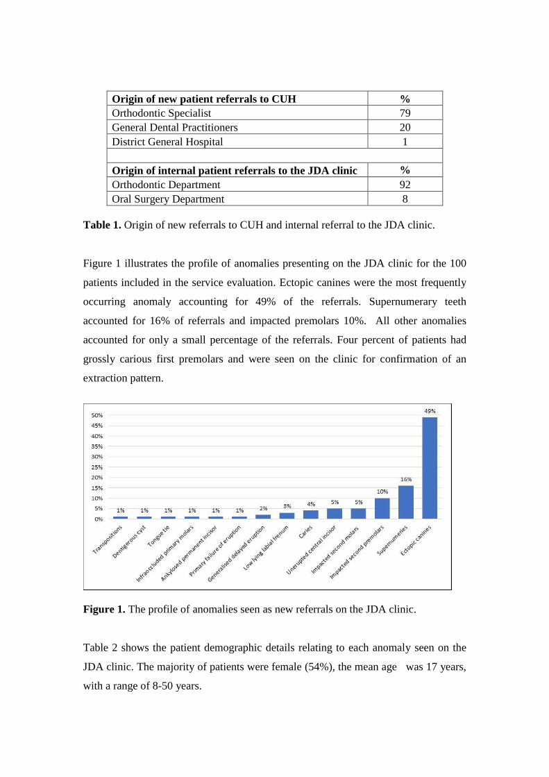
Table 1. Origin of new referrals to CUH and internal referral to the JDA clinic.
Figure 1 illustrates the profile of anomalies presenting on the JDA clinic for the 100
patients included in the service evaluation. Ectopic canines were the most frequently
occurring anomaly accounting for 49% of the referrals. Supernumerary teeth
accounted for 16% of referrals and impacted premolars 10%. All other anomalies
accounted for only a small percentage of the referrals. Four percent of patients had
grossly carious first premolars and were seen on the clinic for confirmation of an
extraction pattern.
Figure 1. The profile of anomalies seen as new referrals on the JDA clinic.
Table 2 shows the patient demographic details relating to each anomaly seen on the
JDA clinic. The majority of patients were female (54%), the mean age was 17 years,
with a range of 8-50 years.

Presenting Anomaly Male:Female Average Age
(Years)
Age range
(Years)
Ectopic Canines 17:32 17 11 to 50
Supernumeries 14:2 13 9 to 18
Impacted second premolars 3:7 15 12 to 28
Impacted second molars 3:2 15 13 to 18
Unerupted central incisors 4:1 10 8 to 14
Caries 2:2 11 8 to 14
Low lying labial frenum 1:2 14 11 to 16
Generalised delayed eruption 0:2 14 13 to 15
Primary failure of eruption 1:0 8 N/A
Ankylosed permanent incisor 0:1 15 N/A
Infraoccluded primary molars 1:0 16 N/A
Tongue tie 0:1 12 N/A
Dentigerous Cyst 0:1 14 N/A
Transpositions 0:1 13 N/A
Total 46:54 15 8 to 50
Table 2. Demographic details of the anomalies seen on the JDA clinic.
Discussion
The majority of new patients attending the JDA clinic were originally referred by
specialist Orthodontists (79%). This supports the view that the throughput of this
clinic comprises a variety of complex cases and that this clinic is invaluable in
supporting primary care Orthodontic services in Croydon.
Ectopic canines (49%) were the most common presenting anomaly, followed by
supernumerary teeth (16%). The majority of patients with ectopic canines were
female and those with supernumeraries were male, which supports previous reports in
the literature. 2, 3
Completion of this service evaluation of the JDA clinics has allowed the department
to recognise the high demand for such multidisciplinary clinics due to the high
number of patients presenting with abnormalities which required multidisciplinary
assessment and management. Following the service evaluation, more Oral Surgery
specialists were allocated for the JDA clinics and the clinics were run more
frequently. The service evaluation also aided the evolution of the JDA clinics by
recognising the need for the use of enhanced imaging modalities such as Cone Beam
CT scans (CBCT) and the development of patient pathways for the multidisciplinary
management of many of these complex patients.
Given the breadth of dento-alveolar pathologies (both common and rare) presenting to
this clinic, we feel that it is important for the GDP to have an understanding of these
conditions, and what types of management may be discussed when these patients are
referred. This article will provide the reader with an overview of the aetiology,
prevalence, classification, diagnosis, features and management of soft tissue
anomalies, cysts, and supernumerary teeth. Part Two in this series focuses on
impacted teeth as well as generalised delayed eruption, primary failure of eruption,
ankylosed incisors and infra-occluded deciduous molars.
Common Anomalies
Low Lying Labial Frenum
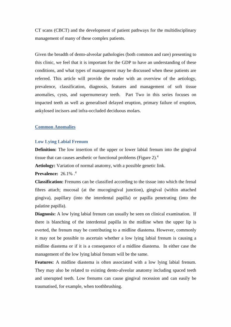
Definition: The low insertion of the upper or lower labial frenum into the gingival
tissue that can causes aesthetic or functional problems (Figure 2).4
Aetiology: Variation of normal anatomy, with a possible genetic link.
Prevalence: 26.1% .4
Classification: Frenums can be classified according to the tissue into which the frenal
fibres attach; mucosal (at the mucogingival junction), gingival (within attached
gingiva), papillary (into the interdental papilla) or papilla penetrating (into the
palatine papilla).
Diagnosis: A low lying labial frenum can usually be seen on clinical examination. If
there is blanching of the interdental papilla in the midline when the upper lip is
everted, the frenum may be contributing to a midline diastema. However, commonly
it may not be possible to ascertain whether a low lying labial frenum is causing a
midline diastema or if it is a consequence of a midline diastema. In either case the
management of the low lying labial frenum will be the same.
Features: A midline diastema is often associated with a low lying labial frenum.
They may also be related to existing dento-alveolar anatomy including spaced teeth
and unerupted teeth. Low frenums can cause gingival recession and can easily be
traumatised, for example, when toothbrushing.
Management: Low labial frenums may occasionally resolve spontaneously. Even if
they do not resolve spontaneously the majority of low lying frenums will not require
treatment. Interventional management (frenectomy) may be indicated if the low frenal
attachment is associated with a midline diastema, if there is gingival recession or if
there is difficulty maintaining oral hygiene around the frenum.
If a low lying frenum is associated with a midline diastema and is assessed as being
likely to prevent orthodontic closure of the diastema a frenectomy is indicated. The
frenectomy can be carried out prior to orthodontic treatment, during treatment, or
following removal of the orthodontic appliances. A frenectomy during orthodontic
treatment carried out just prior to space closure allows good surgical and space
closure can be started following the frenectomy and contraction of the scar tissue will
aid closure of the diastema. A frenectomy prior to orthodontic treatment allows for
better surgical access, however, scar tissue may have formed once the patient is ready
to commence orthodontic space closure and this scar tissue may impede space
closure. Frenectomies can also be carried out following orthodontic treatment. In this
instance, surgical access may be difficult but there is the advantage that scar tissue
contraction may contribute to keeping the diastema space closed. Despite the
proposed advantages and disadvantages of the different timing of carrying out a
frenectomy there is little evidence to support the type and timing of any surgical
intervention.5
Role of the GDP: To diagnose the presence of low lying frenums and give
appropriate oral hygiene advice to prevent recession and gingival trauma. Referral
onward to secondary care if the frenum is causing aesthetic or functional concerns.
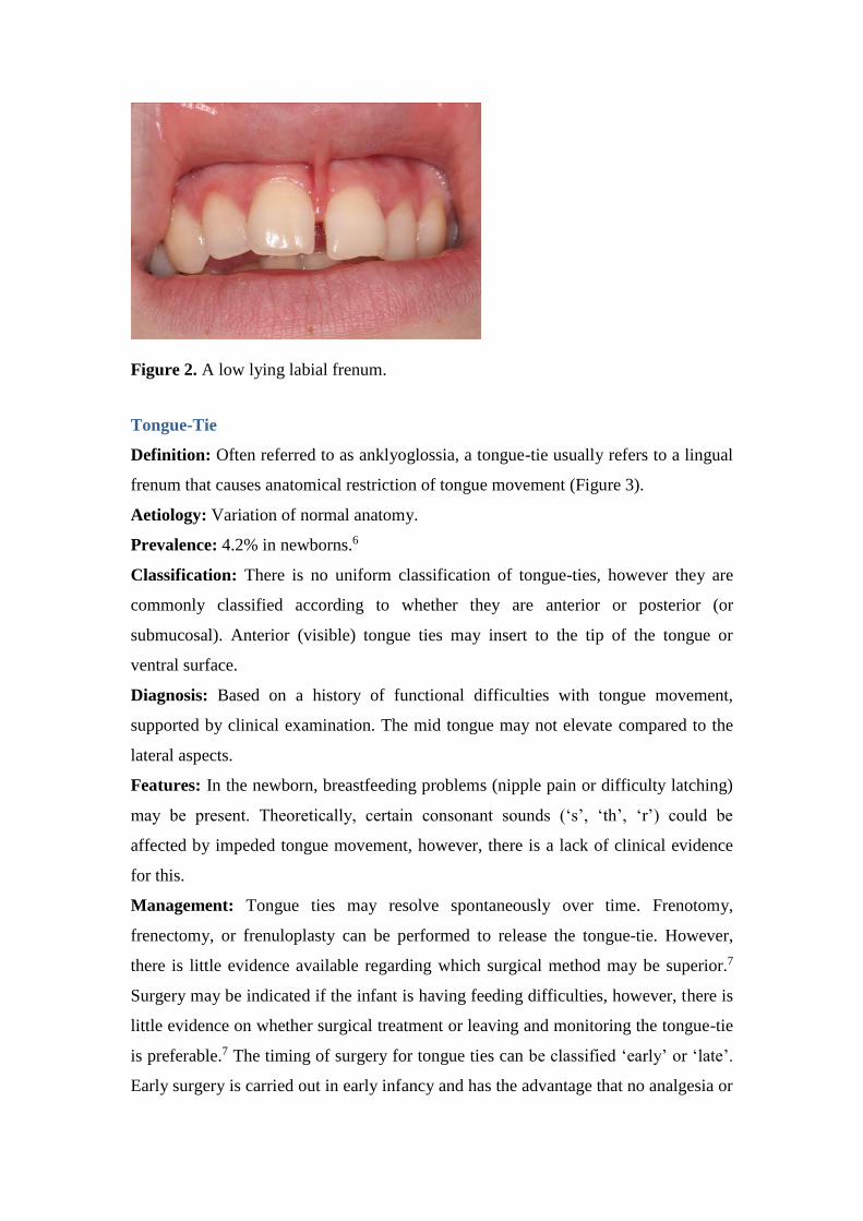
Figure 2. A low lying labial frenum.
Tongue-Tie
Definition: Often referred to as anklyoglossia, a tongue-tie usually refers to a lingual
frenum that causes anatomical restriction of tongue movement (Figure 3).
Aetiology: Variation of normal anatomy.
Prevalence: 4.2% in newborns.6
Classification: There is no uniform classification of tongue-ties, however they are
commonly classified according to whether they are anterior or posterior (or
submucosal). Anterior (visible) tongue ties may insert to the tip of the tongue or
ventral surface.
Diagnosis: Based on a history of functional difficulties with tongue movement,
supported by clinical examination. The mid tongue may not elevate compared to the
lateral aspects.
Features: In the newborn, breastfeeding problems (nipple pain or difficulty latching)
may be present. Theoretically, certain consonant sounds (‘s’, ‘th’, ‘r’) could be
affected by impeded tongue movement, however, there is a lack of clinical evidence
for this.
Management: Tongue ties may resolve spontaneously over time. Frenotomy,
frenectomy, or frenuloplasty can be performed to release the tongue-tie. However,
there is little evidence available regarding which surgical method may be superior.7
Surgery may be indicated if the infant is having feeding difficulties, however, there is
little evidence on whether surgical treatment or leaving and monitoring the tongue-tie
is preferable.7 The timing of surgery for tongue ties can be classified ‘early’ or ‘late’.
Early surgery is carried out in early infancy and has the advantage that no analgesia or
anaesthesia is usually required due to the tongue having limited nerve endings at this
time. Late surgery often requires a local or general anaesthetic, however, there is
limited evidence on whether early or late surgery is superior.
Role of the GDP: The GDP can diagnose tongue ties and reassure parents of the
diagnosis. If there are feeding concerns with infants an onward referral is indicated.
Figure 3. A tongue-tie.
Dentigerous Cyst
Definition: A cyst is an abnormal, lined, fluid filled cavity within the body. A
dentigerous cyst is a developmental cyst that is associated with the crown of an
unerupted or partially erupted tooth (Figure 4).
Aetiology: Proliferation of enamel epithelium after enamel formation. The
pathogenesis is poorly understood; one theory is that proliferation is induced by the
osmotic pressure during an extended period of impaction.
Prevalence: 1.2%.8
Classification: According to the teeth associated with the cyst.
Diagnosis: A provisional diagnosis is made from clinical and radiographic findings of
an unerupted tooth with an associated radiolucency enveloping the tooth. Cone beam
CT (CBCT) imaging may help determine the size and anatomical structures
associated with a cyst. A definitive diagnosis is made through histological
examination of the lining of the cyst.9
Features: Unerupted or impacted teeth, displaced or mobile adjacent teeth, bony
destruction, root resorption of adjacent teeth, crowding, spacing.
Management: Depending on the size and location, cysts may be managed by either
enucleation or marsuplisation (decompression) followed by enucleation after the
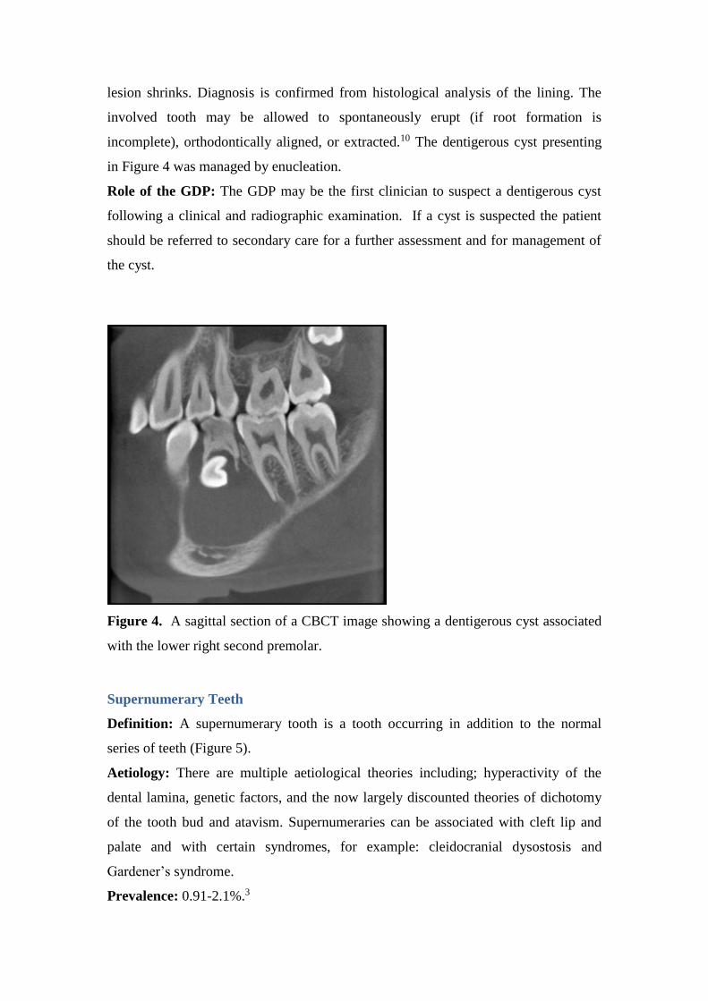
lesion shrinks. Diagnosis is confirmed from histological analysis of the lining. The
involved tooth may be allowed to spontaneously erupt (if root formation is
incomplete), orthodontically aligned, or extracted.10 The dentigerous cyst presenting
in Figure 4 was managed by enucleation.
Role of the GDP: The GDP may be the first clinician to suspect a dentigerous cyst
following a clinical and radiographic examination. If a cyst is suspected the patient
should be referred to secondary care for a further assessment and for management of
the cyst.
Figure 4. A sagittal section of a CBCT image showing a dentigerous cyst associated
with the lower right second premolar.
Supernumerary Teeth
Definition: A supernumerary tooth is a tooth occurring in addition to the normal
series of teeth (Figure 5).
Aetiology: There are multiple aetiological theories including; hyperactivity of the
dental lamina, genetic factors, and the now largely discounted theories of dichotomy
of the tooth bud and atavism. Supernumeraries can be associated with cleft lip and
palate and with certain syndromes, for example: cleidocranial dysostosis and
Gardener’s syndrome.
Prevalence: 0.91-2.1%.3
Classification: According to morphology or location (Table 3).
Diagnosis: Erupted supernumeraries can be detected by conducting a dental
examination. Unerupted supernumeraries are most commonly detected as an
incidental find on radiographs. If plane film radiographs are unclear a CBCT scan
may be required to confirm the presence, morphology and location of some
supernumerary teeth. Supernumerary teeth may also be unclear or undetected on
Dental Panoramic Tomograph (DPT) radiographs if they are outside of the focal
trough. In these situations, further imaging, either with intra-oral plain films or
CBCTs may be indicated. It is important to appreciate that supernumeraries can
develop later in life.11 Therefore, the absence of any supernumeraries in the early
years does not rule out their development in the later years.
Features: Complications of the adjacent permanent teeth, such as dilaceration, failed
or delayed eruption, impaction, rotation, root resorption, crowding, spacing, and
retained primary teeth. The supernumerary itself may undergo cystic change, or
rarely, migration into the nasal cavity or maxillary sinus.12
Management: If there is no associated pathology or underlying malocclusion, these
supernumeraries may be left in situ. If unerupted, periodic radiographic review is
recommended to assess for any cystic change or damage to adjacent structures.
Alternatively, supernumeraries may require extraction, if there is any associated
pathology or if orthodontic treatment is required and if the supernumerary would
interfere with tooth movement. Unerupted supernumeraries can be extracted
surgically or occasionally supernumerary teeth erupt and then a simple extraction can
be undertaken.
Role of the GDP: The GDP can identify and diagnose supernumerary teeth and they
can also often identify if there is any associated pathology. Referral to secondary care
for a specialist opinion is advisable. If supernumerary teeth are left in situ, the GDP
can carry out regular reviews with radiographic monitoring and re-refer if required. If
the supernumerary is to be extracted, simple extractions of erupted supernumeraries
may be undertaken by the GDP. However, surgical extraction of unerupted
supernumeraries is often undertaken in secondary care by a specialist Oral Surgeon.
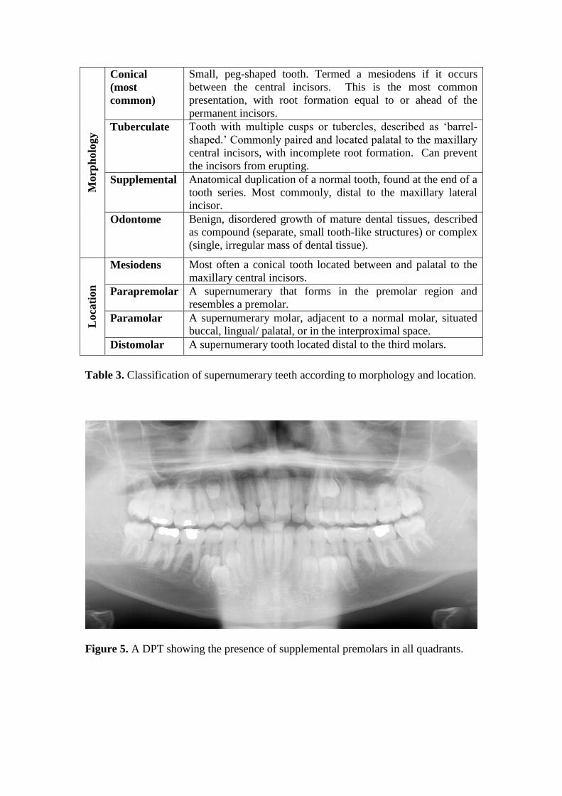
Morp
holo
gy
Conical
(most
common)
Small, peg-shaped tooth. Termed a mesiodens if it occurs
between the central incisors. This is the most common
presentation, with root formation equal to or ahead of the
permanent incisors.
Tuberculate Tooth with multiple cusps or tubercles, described as ‘barrel-
shaped.’ Commonly paired and located palatal to the maxillary
central incisors, with incomplete root formation. Can prevent
the incisors from erupting.
Supplemental Anatomical duplication of a normal tooth, found at the end of a
tooth series. Most commonly, distal to the maxillary lateral
incisor.
Odontome Benign, disordered growth of mature dental tissues, described
as compound (separate, small tooth-like structures) or complex
(single, irregular mass of dental tissue).
Loca
tion
Mesiodens Most often a conical tooth located between and palatal to the
maxillary central incisors.
Parapremolar A supernumerary that forms in the premolar region and
resembles a premolar.
Paramolar A supernumerary molar, adjacent to a normal molar, situated
buccal, lingual/ palatal, or in the interproximal space.
Distomolar A supernumerary tooth located distal to the third molars.
Table 3. Classification of supernumerary teeth according to morphology and location.
Figure 5. A DPT showing the presence of supplemental premolars in all quadrants.
Conclusion
This article has summarised the profile of the cases seen on the JDA at CUH and
given an overview of the common soft tissue anomalies, dentigerous cysts, and
supernumerary teeth including their diagnosis, management and the role of the GDP.
It has also highlighted the invaluable role of the GDP in identifying and aiding the
diagnosis of these anomalies so that patients can be referred for specialist opinion in a
timely manner. Part Two of this series focuses on anomalies in eruption, as well as
transpositions and infra-occluded deciduous molars.
Declaration of Interest
The authors do not have any conflicts of interest.
References
1. Khamashta-Ledezma L, Kordi Z, Radecki J, Davenport-Jones L, Chia M. Patient
satisfaction with Croydon MDT dento-alveolar clinics. British Orthodontic Society
Clinical Effectiveness Bulletin 2014; 8-10.
2. Mossey P A, Campbell H M, Luffingham J K. The palatal canine and the adjacent
lateral incisor; a study of a west of Scotland population. BJO 1994; 21:169 -174.
3. Shah A, Gill D S, Tredwin C, Naini F B. Diagnosis and Management of
Supernumery Teeth. Dent Update 2008; 35:510-520.
4. Boutsi E A, and Tatakis D N. Maxillary labial frenum attachment in children. Int J
Paediatr Dent 2011; 21:284-8.
5. Devishree S K G, Shubhashini P V. Frenectomy: a review with the reports of
surgical techniques. J Clinc Diagn Res 2012; 6:1587-92.
6. Ricke L A, Baker N J, Madlon-Kay D J, DeFor T A. Newborn tongue-tie:
prevalence and effect on breast-feeding. J Am Board Fam Med 2005; 18:1-7.
7. Suter V G, Bornstein M M. Ankyloglossia: facts and myths in diagnosis and
treatment. J Periodontol 2009; 80:1204-19.
8. Tortorici S, Amodio E, Massenti M F, Buzzuano F, Vitale F. Prevalence and
distribution of odontogenic cysts in Sicily: 1986-2005. J Oral Sci 2008; 50:15-18.
9. Scholl R J, Kellett H M, Neumann D P, Lurie A G. Cysts and cystic lesions of the
mandible: clinical and radiologic-histopathologic review. Radiographics 1999;
19:1107-24.
10. Motamedi M H, Talesh K T. Management of extensive dentigerous cysts. Br Dent
J 2005; 198:203-206.

11. Shah A, Gill D S, Tredwin C, Nani F B. Diagnosis and management of
supernumerary teeth. Dental Update 2008; 35:510-520.
12. Garvey M T, Barry H J, Blake M. An overview of classification, diagnosis and
management. J Can Dent Assoc 1999; 65:612-6.