Embed Size (px)
Citation preview
Urology & Kidney Disease NewsGlickman Urological & Kidney Institute | A Physician Journal of Developments in Urology and Nephrology
Vol. 23 | Winter 2014
Pg17
Pg24
Pg9
Page 9 Electronic Health Records: A Tool for Research and for Improving Patient Care
Page 24 First Mutation Identified That Increases DHT Synthesis to Promote Hormone Therapy Resistance
Page 17 Advances in Zero-Ischemia Robotic Partial Nephrectomy: Sequential Preplaced Suture Renorrhaphy Technique
2 Urology & Kidney Disease News
Urology & Kidney Disease News
Chairman’s Report ....................................................................4
News from the Glickman Urological & Kidney InstituteUrological & Kidney Institute National Locations ..........................5
Urology Services Begin in Las Vegas ...........................................5
New National Staff ....................................................................5
New Staff .................................................................................6
Endowed Chairs ........................................................................7
CME Upcoming Events — Save These Dates ...............................8
Electronic Health Records: A Tool for Research and for Improving Patient Care ...................................................9
Best Practices Positioning Cleveland Clinic to Deliver Excellence in an Era of Change ................................................10
Shared Medical Appointments Offer Opportunity to Enhance Chronic Care ........................................................Online
Analyses in Laparoscopic Nephrectomy and Prostatectomy: Mechanical Bowel Preparation ............................................Online
Center for Robotics and Image-Guided Surgery Robot-Assisted Intracorporeal Ileal Conduit Urinary Diversion: Applying Principles of Open Reconstruction to a Minimally Invasive Approach ...................................................................11
Robot-Assisted Intracorporeal Ileal Neobladder: Initial 14 Cases .......................................................................12
Robot-Assisted Laparoscopic Renal Artery Aneurysm Repair with Selective Arterial Clamping ................................................15
Neoadjuvant Therapy for Downsizing of Renal Tumors Prior to Complex Robotic Nephron-Sparing Surgery ....................16
Advances in Zero-Ischemia Robotic Partial Nephrectomy: Sequential Preplaced Suture Renorrhaphy Technique .................17
Robotic Partial Nephrectomy for Completely Endophytic Renal Masses: A Single-Institution Experience ...........................18
Repeat Robot-Assisted Partial Nephrectomy: Feasibility and Early Outcomes .................................................20
Robot-Assisted Laparoscopic Retroperitoneal Lymph Node Dissection for Left Clinical Stage I Testicular Cancer ............. Online
Robot-Assisted Laparoscopic Transabdominal Vasovasostomy ..................................................................Online
Minimally Invasive Heminephrectomy: A Specific Definition ...Online
Robotic Partial Nephrectomy in Patients with History of Previous Abdominal Surgery ................................ Online
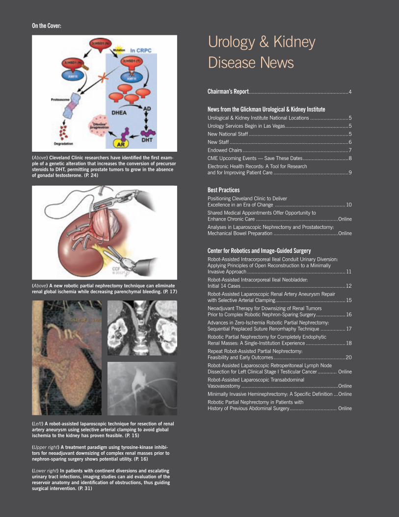
(Above) Cleveland Clinic researchers have identified the first exam-ple of a genetic alteration that increases the conversion of precursor steroids to DHT, permitting prostate tumors to grow in the absence of gonadal testosterone. (P. 24)
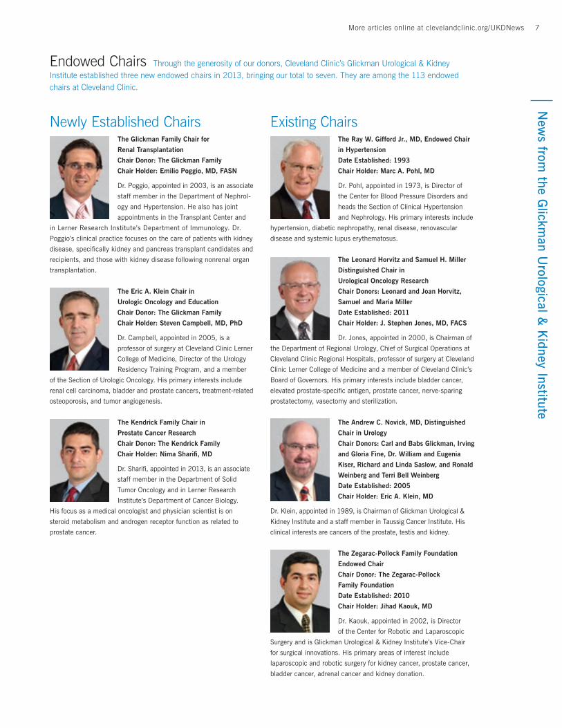
(Above) A new robotic partial nephrectomy technique can eliminate renal global ischemia while decreasing parenchymal bleeding. (P. 17)
(Left) A robot-assisted laparoscopic technique for resection of renal artery aneurysm using selective arterial clamping to avoid global ischemia to the kidney has proven feasible. (P. 15)
(Upper right) A treatment paradigm using tyrosine-kinase inhibi-tors for neoadjuvant downsizing of complex renal masses prior to nephron-sparing surgery shows potential utility. (P. 16)
(Lower right) In patients with continent diversions and escalating urinary tract infections, imaging studies can aid evaluation of the reservoir anatomy and identification of obstructions, thus guiding surgical intervention. (P. 31)
On the Cover:

3More articles online at clevelandclinic.org/UKDNews
Center for Urologic Oncology From Bench to Bedside: Genomics for Active Surveillance Now in Clinical Practice ...........................................................22
Active Surveillance of Localized Prostate Cancer Is Acceptable Management Strategy .............................................23
First Mutation Identified That Increases DHT Synthesis to Promote Hormone Therapy Resistance......................................24
Heat Therapy to Ablate Tumors ...........................................Online
Low Fistula Rate during Hydrodissecton for Cryotherapy ........Online
Novel Molecular Biomarker for Prostate Cancer Screening and Early Detection ............................................................Online
Sustainable Growth Rate Update: We ALL Need to Understand Where We Stand .........................................Online
Center for Blood Pressure and Electrolyte DisordersEvolving Approaches to Resistant Hypertension .........................26
Center for Chronic Kidney DiseasePrognostic Importance of Serum Alkaline Phosphatase in Non-Dialysis-Dependent Chronic Kidney Disease in a Clinical Population ...................................................................28
Consequences of Metabolic Syndrome in Kidney Disease ......Online
Center for Renal DiseasesBaseline Mean Pulmonary Artery Pressure as an Independent Predictor of Acute Kidney Injury After Lung Transplantation .........30
Center for Genitourinary Reconstruction New or Worsening Urinary Infections in Continent Urostomies/Ileocystoplasty .......................................................31
Center for Endourology and Stone DiseaseExpanding the Indications for Tubeless Percutaneous Nephrolithotomy. .....................................................................34
Center for Kidney/Pancreas TransplantationPotential Benefits of Cardiac Resynchronization Therapy in Kidney Disease ....................................................................35
Small Incision, Big Benefits: Anterior Rectus Sheath Approach — Key for Keyhole Kidney Transplants .......................36
Utilization of Arterial Vascular Conduits to Facilitate Renal Transplantation in Patients with Significant Aortoiliac Calcification ............................................................................38
Initiating a Laparoendoscopic Single-Site Live Donor Nephrectomy Program ..............................................Online
Lifestyle Modification as an Opportunity to Improve the Health of Living Kidney Donors ......................................Online
Center for DialysisDialysis Admissions During Weekends and Their Effects .............39
Center for Pediatric UrologyIs Preoperative Hormonal Stimulation Helpful in the Treatment of Proximal Hypospadias? ........................................................40
Physician Resource Guide.......................................................42
The articles in this publication are written for educational purposes only and are presented as a convenience. Cleveland Clinic has no financial interest in nor is it endorsing any product or device mentioned herein.
Urology & Kidney Disease NewsGlickman Urological & Kidney Institute | A Physician Journal of Developments in Urology and Nephrology
Vol. 23 | Winter 2014
Pg17
Pg24
Pg9
Page 9 Electronic Health Records: A Tool for Research and for Improving Patient Care
Page 24 First Mutation Identified That Increases DHT Synthesis to Promote Hormone Therapy Resistance
Page 17 Advances in Zero-Ischemia Robotic Partial Nephrectomy: Sequential Preplaced Suture Renorrhaphy Technique
More Articles Available OnlineWe have even more information available for you online.
To read these articles, visit clevelandclinic.org/UKDNews.
4 Urology & Kidney Disease NewsN
ews
from
the
Glic
kman
Uro
logi
cal &
Kid
ney
Inst
itute
Dear Colleagues,
Welcome to the 2013 Edition of Glickman Urological & Kidney Institute’s Urology & Kidney Disease News. This is a slimmer volume than in years past, representing efforts to move some of the contents online. You will still find between these versions a comprehensive overview of all the exciting things happening in our world.
And our world continues to expand. In April, we opened our newest office in Las Vegas, adding two outstanding urolo-gists, Scott Slavis, MD, and Laurie Larsen, MD (you can read about them on Page 5). Our national footprint of clinical services now covers five states (Ohio, Indiana, West Virginia, Nevada and two cities in Florida, see map on Page 5), and when the new Cleveland Clinic Abu Dhabi outpatient facility and hospital opens in 2015, we will be providing service on two continents.
Chairman’s Report
Eric A. Klein, MDChairman, Cleveland Clinic Glickman Urological & Kidney Institute
On a sad note, our namesake and benefactor Carl Glick-man died this past year after a long illness. Carl’s vision and generosity established the Urological & Kidney Insti-tute in 2002 and his efforts allowed us to build and move into our advanced facility in the Glickman Tower in 2008. Happily, he lived long enough to see both of our special-ties, Urology and Nephrology, ranked No. 1 in U.S. News & World Report’s “America’s Best Hospitals” survey for 2012-2013, and no man ever died prouder of his efforts to help us achieve success. Carl’s bequest allowed us to establish two new endowed chairs in recognition of two outstand-ing staff members, Steven Campbell, MD, PhD, and Emilio Poggio, MD, FASN. You can read about them and all our endowed chair holders on Page 7.
This year also marked an expansion of our scientific endeavors with the hiring of Nima Sharifi, MD, as the inaugural holder of the Kendrick Family Chair in Prostate Cancer Research (you can read about him on Page 6). Nima joins us from the University of Texas at Southwestern and expands our efforts to understand and harness the molecular events underlying the development of prostate cancer.
I hope you enjoy this edition of UKD News.
Eric A. Klein, MD Chairman Glickman Urological & Kidney Institute
Contributors Panel
Edmund Sabanegh Jr., MD Chairman, Department of Urology Glickman Urological & Kidney Institute
Robert Heyka, MD Chairman, Department of Nephrology Glickman Urological & Kidney Institute
Sankar Navaneethan, MD Glickman Urological & Kidney Institute Medical Editor, Nephrology
J. Stephen Jones, MD, FACS Chief of Surgical Operations, Cleveland Clinic Regional Hospitals Medical Editor, Urology

5More articles online at clevelandclinic.org/UKDNews 5N
ews from
the Glickm
an Urological &
Kidney Institute
Urology Services Begin in Las VegasCleveland Clinic’s Glickman Urological & Kidney Insti-tute now offers urology services in Las Vegas. The expan-sion, known as Cleveland Clinic Urology, Las Vegas, and announced in February 2013, results from Cleveland Clinic’s acquisition of the practice of respected Las Vegas urologists Scott Slavis, MD, and Laurie Larsen, MD. The physicians and their nine employees are now employed by Cleveland Clinic. They have moved to a new office with advanced technology that Cleveland Clinic has invested in the practice.
“We’re honored to be recognized as one of the top uro-logical programs in the country, and proud to be able to extend that care to patients in Las Vegas,” said Urological & Kidney Institute Chairman Eric A. Klein, MD. Cleveland Clinic began offering services in Las Vegas in July 2009 at Cleveland Clinic’s Lou Ruvo Center for Brain Health. “Our decision to expand services is based on the success we’ve experienced at the Ruvo Center,” said Dr. Klein. “It is en-couraging to see how the community has welcomed our brand of quality care.”
Laurie Larsen, MD, received her medical degree from the Oregon Health Sciences University School of Medi-cine. She completed general surgery and urologic surgery residencies at the University of California Irvine Medical Center. Dr. Larsen sees patients at Cleveland Clinic Urology, Las Vegas.
Scott Slavis, MD, received his medical degree from the University of Miami School of Medicine. He completed a general surgery internship and residency at Harbor/UCLA Medical Center, a urology residency at the University of California Irvine Medical Center, and a renal transplantation and renovascular surgery fellowship at Cleveland Clinic. He sees patients at Cleveland Clinic Urology, Las Vegas.
Nader Najafian, MD, received his medical degree from the University of Vienna Medical School. He completed an internal medicine residency at the University of Iowa Hospitals & Clinics, a clinical fellowship in nephrology at Brigham and Women’s Hospital, and research fellowships in transplantation at Brigham and Women’s and at Chil-dren’s Hospital, Boston. He is chairman of the Department of Nephrology and Hypertension and heads the Section of Transplant Nephrology at Cleveland Clinic Florida, where he sees patients.
New National Staff Las Vegas Las Vegas Florida
GLICKMAN UROLOGICAL & KIDNEY INSTITUTE NATIONAL LOCATIONS
LAS VEGASNEVADA
WEST PALM BEACHFLORIDA
CHARLESTON WEST VIRGINIA
INDIANAPOLISINDIANA
WESTONFLORIDA
CLEVELANDOHIO

6 Urology & Kidney Disease NewsN
ews
from
the
Glic
kman
Uro
logi
cal &
Kid
ney
Inst
itute Nima Sharifi, MD, received his medical
degree from the University of Pittsburgh. He
completed an internal medicine residency at
the Yale-New Haven Hospital and a medical
oncology fellowship at the National Cancer
Institute. Dr. Sharifi is a member of the De-
partment of Solid Tumor Oncology and holds
the Kendrick Family Chair in Prostate Cancer
Research in the Cleveland Clinic Lerner
Research Institute’s Department of Cancer Biology. He sees patients
at Cleveland Clinic’s main campus.
Sri Sivalingam, MD, received his medical
degree from the University of Toronto.
He completed a urology residency at the
University of Manitoba, and an endourology
and minimally invasive surgery fellowship at
the University of Wisconsin. Dr. Sivalingam
is a member of the Department of Urology
and sees patients at Cleveland Clinic’s main
campus, Hillcrest Hospital and Twinsburg
Family Health and Surgery Center.
Leslie Wong, MD, received his medical
degree from the University of Texas South-
western Medical School. He completed an
internship and residency in internal medicine,
and a fellowship in nephrology and hyperten-
sion, all at the University of North Carolina
Hospitals at Chapel Hill. Dr. Wong is a member
of the Department of Nephrology and
Hypertension and sees patients at Cleveland
Clinic’s main campus.
Ziad Zaky, MD, received his medical degree
from Cairo University School of Medicine
in Egypt. He completed an internship, an
internal medicine residency and a nephrology
fellowship, all at Cairo University School of
Medicine. He also completed an internal
medicine residency at DMC Sinai-Grace Hos-
pital in Detroit and a nephrology fellowship at
the University of Michigan. He is a member of
the Department of Nephrology and Hypertension and sees patients
at Cleveland Clinic’s main campus.
Evamaria Anvari, MD, received her medical
degree from Universidad Autónoma De Gua-
dalajara School of Medicine in Guadalajara,
Mexico. She completed a surgical internship
at Soundshore Medical Center, New Rochelle,
N.Y.; an internal medicine residency at Texas
Tech University Health Sciences Center; and
a nephrology fellowship at the University of
Arizona Medical Center. She is a member of
the Nephrology and Hypertension Department and sees patients at
Cleveland Clinic’s main campus.
Laura Ferreira Provenzano, MD, received her
medical degree from the University of Buenos
Aires School of Medicine in Argentina. She
completed an internal medicine internship and
residency at Caritas St. Elizabeth’s Medical
Center in Boston and a nephrology fellowship
at the University of Pittsburgh Medical Center.
She is a member of the Nephrology and
Hypertension Department and sees patients
at Cleveland Clinic’s main campus.
Hernan Rincon-Choles, MD, received his
medical degree from the Universidad de
Cartagena Medical School in Cartagena,
Colombia. He completed an internal medicine
internship and residency at New York Medical
College and a nephrology fellowship at the
University of Texas Health Science Center. He
is a member of the Department of Nephrol-
ogy and Hypertension and sees patients at
Cleveland Clinic’s main campus.
Fahad Saeed, MD, received his medical degree
from the National University of Sciences
Army Medical College in Punjab, Pakistan.
He completed an internship at Pakistan’s
Jinnah Hospital Lahore and internal medicine
residencies at Jinnah Hospital Lahore, Sheikh
Zayed Hospital Lahore and the University
of Illinois Hospital at Urbana-Champaign.
He completed a nephrology fellowship at
Dartmouth-Hitchcock Medical Center in Lebanon, N.H. Dr. Saeed is
a member of the Department of Nephrology and Hypertension and
sees patients at Cleveland Clinic’s main campus.
New Staff The Glickman Urological & Kidney Institute welcomes the following new staff members:
7More articles online at clevelandclinic.org/UKDNewsN
ews from
the Glickm
an Urological &
Kidney Institute
Existing ChairsThe Ray W. Gifford Jr., MD, Endowed Chair
in Hypertension
Date Established: 1993
Chair Holder: Marc A. Pohl, MD
Dr. Pohl, appointed in 1973, is Director of
the Center for Blood Pressure Disorders and
heads the Section of Clinical Hypertension
and Nephrology. His primary interests include
hypertension, diabetic nephropathy, renal disease, renovascular
disease and systemic lupus erythematosus.
The Leonard Horvitz and Samuel H. Miller
Distinguished Chair in
Urological Oncology Research
Chair Donors: Leonard and Joan Horvitz,
Samuel and Maria Miller
Date Established: 2011
Chair Holder: J. Stephen Jones, MD, FACS
Dr. Jones, appointed in 2000, is Chairman of
the Department of Regional Urology, Chief of Surgical Operations at
Cleveland Clinic Regional Hospitals, professor of surgery at Cleveland
Clinic Lerner College of Medicine and a member of Cleveland Clinic’s
Board of Governors. His primary interests include bladder cancer,
elevated prostate-specific antigen, prostate cancer, nerve-sparing
prostatectomy, vasectomy and sterilization.
The Andrew C. Novick, MD, Distinguished
Chair in Urology
Chair Donors: Carl and Babs Glickman, Irving
and Gloria Fine, Dr. William and Eugenia
Kiser, Richard and Linda Saslow, and Ronald
Weinberg and Terri Bell Weinberg
Date Established: 2005
Chair Holder: Eric A. Klein, MD
Dr. Klein, appointed in 1989, is Chairman of Glickman Urological &
Kidney Institute and a staff member in Taussig Cancer Institute. His
clinical interests are cancers of the prostate, testis and kidney.
The Zegarac-Pollock Family Foundation
Endowed Chair
Chair Donor: The Zegarac-Pollock
Family Foundation
Date Established: 2010
Chair Holder: Jihad Kaouk, MD
Dr. Kaouk, appointed in 2002, is Director
of the Center for Robotic and Laparoscopic
Surgery and is Glickman Urological & Kidney Institute’s Vice-Chair
for surgical innovations. His primary areas of interest include
laparoscopic and robotic surgery for kidney cancer, prostate cancer,
bladder cancer, adrenal cancer and kidney donation.
Newly Established ChairsThe Glickman Family Chair for
Renal Transplantation
Chair Donor: The Glickman Family
Chair Holder: Emilio Poggio, MD, FASN
Dr. Poggio, appointed in 2003, is an associate
staff member in the Department of Nephrol-
ogy and Hypertension. He also has joint
appointments in the Transplant Center and
in Lerner Research Institute’s Department of Immunology. Dr.
Poggio’s clinical practice focuses on the care of patients with kidney
disease, specifically kidney and pancreas transplant candidates and
recipients, and those with kidney disease following nonrenal organ
transplantation.
The Eric A. Klein Chair in
Urologic Oncology and Education
Chair Donor: The Glickman Family
Chair Holder: Steven Campbell, MD, PhD
Dr. Campbell, appointed in 2005, is a
professor of surgery at Cleveland Clinic Lerner
College of Medicine, Director of the Urology
Residency Training Program, and a member
of the Section of Urologic Oncology. His primary interests include
renal cell carcinoma, bladder and prostate cancers, treatment-related
osteoporosis, and tumor angiogenesis.
The Kendrick Family Chair in
Prostate Cancer Research
Chair Donor: The Kendrick Family
Chair Holder: Nima Sharifi, MD
Dr. Sharifi, appointed in 2013, is an associate
staff member in the Department of Solid
Tumor Oncology and in Lerner Research
Institute’s Department of Cancer Biology.
His focus as a medical oncologist and physician scientist is on
steroid metabolism and androgen receptor function as related to
prostate cancer.
Endowed Chairs Through the generosity of our donors, Cleveland Clinic’s Glickman Urological & Kidney Institute established three new endowed chairs in 2013, bringing our total to seven. They are among the 113 endowed chairs at Cleveland Clinic.

8 Urology & Kidney Disease NewsN
ews
from
the
Glic
kman
Uro
logi
cal &
Kid
ney
Inst
itute
March 22, 2014 Tenth Annual Glickman Urological & Kidney Institute Nursing Conference Led by the Nursing Conference Committee
April 4, 2014 Ambulatory Urology Symposium Course co-directors: Edmund Sabanegh Jr., MD,
and J. Stephen Jones, MD, FACS
October 10-11, 2014
Sixth Annual Symposium on Robotic Kidney and Adrenal Surgery Course director: Jihad Kaouk, MD
October 24, 2014
Kidney Stones: Medical, Surgical and Dietary Approaches Course director: Edmund Sabanegh Jr., MD
Please visit ccfcme.org for more details on these events.
Upcoming Events — Save These Dates
9More articles online at clevelandclinic.org/UKDNewsN
ews from
the Glickm
an Urological &
Kidney Institute
Sankar Navaneethan, MD, MPH, and Robert Heyka, MD
In the United States, the Institute of Medicine has been calling for an increase in the use of electronic health records (EHRs) since the 1990s. The adoption rate of EHRs has been low, however, leading to the passage of the Health Informa-tion Technology for Economic and Clinical Health (HITECH) Act in 2009. Now, adoption in the United States has signifi-cantly increased the number of medical practices that use EHRs. The use of EHRs is increasing in both developing and developed countries, leading to the widespread use of EHRs by nephrologists.
When they began in the 1960s, EHRs were simply computer-ized systems that allowed for document storage and retrieval. With the advancement of technology and more widespread adoption, the EHR has evolved into a much more sophis-ticated tool. Today, reporting of health data can be further delineated into areas that support additional use of this data at the individual provider, clinic or health system level in the form of quality reports, clinical research and public health. The ability of EHRs to store and retrieve structured data — demographics (age, gender and ethnicity), laboratory results, clinical data such as blood pressure or heart rate, standard-ized medications, and standardized diagnoses — longitu-dinally is instrumental in data sets required for meaningful clinical research.
Benefits of EHRs for Kidney Disease
Studies have shown that among health centers with EHRs, there is an increased adherence to guideline-based care, enhanced surveillance and monitoring, and a decrease in medication errors. Physicians who use EHRs believe that they improve the quality of patient care and that the data are superior to claims or administrative data for quality report-ing. Effective utilization of EHRs can help improve both the identification of chronic kidney disease (CKD) patients and the quality of care delivered to them. Automated clinical alerts using EHRs may help diagnose CKD earlier and im-prove referral rates.
Recent reports from Canada, Australia and other developed countries have shown promising data indicating that auto-mated estimated glomerular filtration rate (eGFR) report-ing in laboratory results improves nephrology referrals. We developed an EHR-based CKD registry at Cleveland Clinic with the intent of identifying CKD patients earlier and sys-tematically in order to develop programs for these patients and create intervention programs to improve CKD care. We are also developing registries for end-stage renal disease and other kidney disease populations, as this provides an oppor-tunity to develop protocols for value-based care and conduct research studies in that particular area.
Personal Health Records
In addition to the provider-based EHRs, Cleveland Clinic health system offers all patients the ability to access their medical information via a personal health record (PHR) teth-ered to their health system EHRs (Epic MyChart®, Epic Corp., Verona, Wis.). A PHR is generally defined as an electronic application through which individuals can access, manage and share their health information with their healthcare providers. A spectrum of PHRs is available, from stand-alone systems to those that are web-based and interface with the individual health system’s EHRs. Our system allows patients to activate their PHR accounts either in person or via an online authorization process. Once accounts are activated, patients can navigate around this secure web-based applica-tion and manage their health information. They can review and schedule appointments, request prescription renewals, view health summaries, access a current list of medications, and review test results. Patients also receive automated health reminders per gender- and age-based health-mainte-nance schedules, as well as chronic disease -related reminders (e.g., diabetes). Links within the PHR allow patients to access reliable health information about a broad range of topics of personal interest through a third-party vendor such as MedlinePlus. Secure messaging between the patient and the provider is also available via the PHR to facilitate communi-cation. We are conducting studies to examine the potential benefits of PHRs in kidney disease care at our institution.
While novel methods of developing programs that improve patient care and obtain high-quality data for research projects in nephrology have remained a challenge, our experience with EHRs has shown promising results to address some of these issues.
Electronic Health Records: A Tool for Research and for Improving Patient Care
Key Points:
As the adoption rate of EHRs continues to increase, the systems are showing more versatility and sophistication.
Use of EHRs to manage data for CKD patients can improve the overall care of these patients, particularly when inte-grated with a web-based personal health record system.
Bes
t P
ract
ices
10 Urology & Kidney Disease News
Edmund Sabanegh Jr., MD
As our government begins initiatives designed to improve patients’ ability to make more-informed decisions about the cost and quality of their healthcare choices, hospitals must find ways to deliver care more economically.
To help meet this challenge, Cleveland Clinic created a Cost Repositioning Task Force comprising caregivers from across our enterprise. Currently, almost 400 committed profession-als are charged with finding efficiencies that will reposition Cleveland Clinic to meet changes in healthcare reimburse-ment, while ensuring that we can continue to deliver the highest-quality care at prices patients can afford.
As healthcare consumes a growing percentage of our national expenditures (currently almost 18 percent), it is critical that organizations reinvent the way they provide care. Even if we furnish the best health care in the world, it will be irrelevant if no one can afford us.
So how can we make healthcare affordable without com-promising quality? The answer is by ensuring that patients remain at the forefront of everything we do, while striving to maximize care value. Let me explain how this works.
The need for cost repositioning is being driven in part by increasing transparency. As consumers turn to the internet to compare what institutions charge for the same procedure, hospitals must become as efficient as possible. We have been taking a hard look at our indirect costs, or so-called overhead. While these areas, such as finance and administrative sup-port, are critical to smooth care provision, we have identified significant opportunities to provide value with lower costs.
In addition, we have discovered that small changes in sup-ply cost can produce significant savings. Last year, we took a close look at our purchasing practices and began buying larger numbers of items—from surgical and medical equip-ment to office supplies—from fewer vendors. Through these supply chain initiatives, we were able to negotiate better rates that saved us more than $150 million.
As surgeons, we previously did not worry about the cost of medical supplies such as sutures, ordering any that we wanted. Limiting the number of suture vendors enabled us to enjoy economies of scale without compromising care. It required us to change our habits, but when the entire organi-zation adjusted focus, it resulted in tremendous savings.
Treating every disease according to best practices is the op-timal way to ensure that patients receive the highest-quality care, while eliminating unnecessary tests and hospitaliza-tions. Cleveland Clinic’s Department of Urology has taken the lead in an enterprisewide effort to establish care paths for common diseases. For each disease, we are documenting the diagnostic tests that have been shown to be necessary
and useful, along with treatments that produce the best out-comes in certain patient populations, and guidelines for how follow-up should be conducted.
The first care path to be deployed is for clinically localized prostate cancer. It has simplified the diagnostic process by eliminating unnecessary tests and reinforcing evidence-based decisionmaking for all aspects of care. For example, not everyone with prostate cancer needs a CT scan or a bone scan, tests that for some patients may be a waste of resources and cause unnecessary radiation exposure.
We also are reinforcing optimal outcomes by concentrating procedures in the hands of experienced teams. For example, vasectomy patients are being steered to a smaller number of urologists who have a larger percentage of their practices devoted to the procedure.
Because the patient experience extends beyond clinical care, protocols for counseling and follow-up care, as well as for patient education materials, are being made uniform, so that all patients who come to Cleveland Clinic for the same procedure receive the same treatment. This has resulted in improved patient satisfaction rates and reduced complica-tion rates and expenses.
As these innovations spread throughout the institution, we are confident that Cleveland Clinic’s name will remain synonymous with world-class care delivered in an economi-cally sustainable fashion. Cleveland Clinic is known as an innovator in quality of care and patient experience. Now, we can be innovative in efficiency. But we will never sacrifice quality, and we will always remember our mantra, “Patients First.” The changes we make must — and will — make us even better.
Dr. Sabanegh is chairman of the Department of Urology, and of Cleveland Clinic’s Cost Repositioning Task Force.
Positioning Cleveland Clinic to Deliver Excellence in an Era of Change
Key Points:
With increasing healthcare costs, and increasingly cost-conscious consumers, hospitals must maximize care value without compromising patient care.
Cleveland Clinic has created a Cost Repositioning Task Force that is looking for efficiencies. Among the changes instituted are consolidation of vendors to leverage econo-mies of scale; establishing care paths for common diseases to eliminate unnecessary tests and hospitalizations; and reinforcing optimal outcomes by concentrating procedures in the hands of experienced medical teams.
11More articles online at clevelandclinic.org/UKDNewsC
enter for Robotics and Im
age - Guided Surgery
11
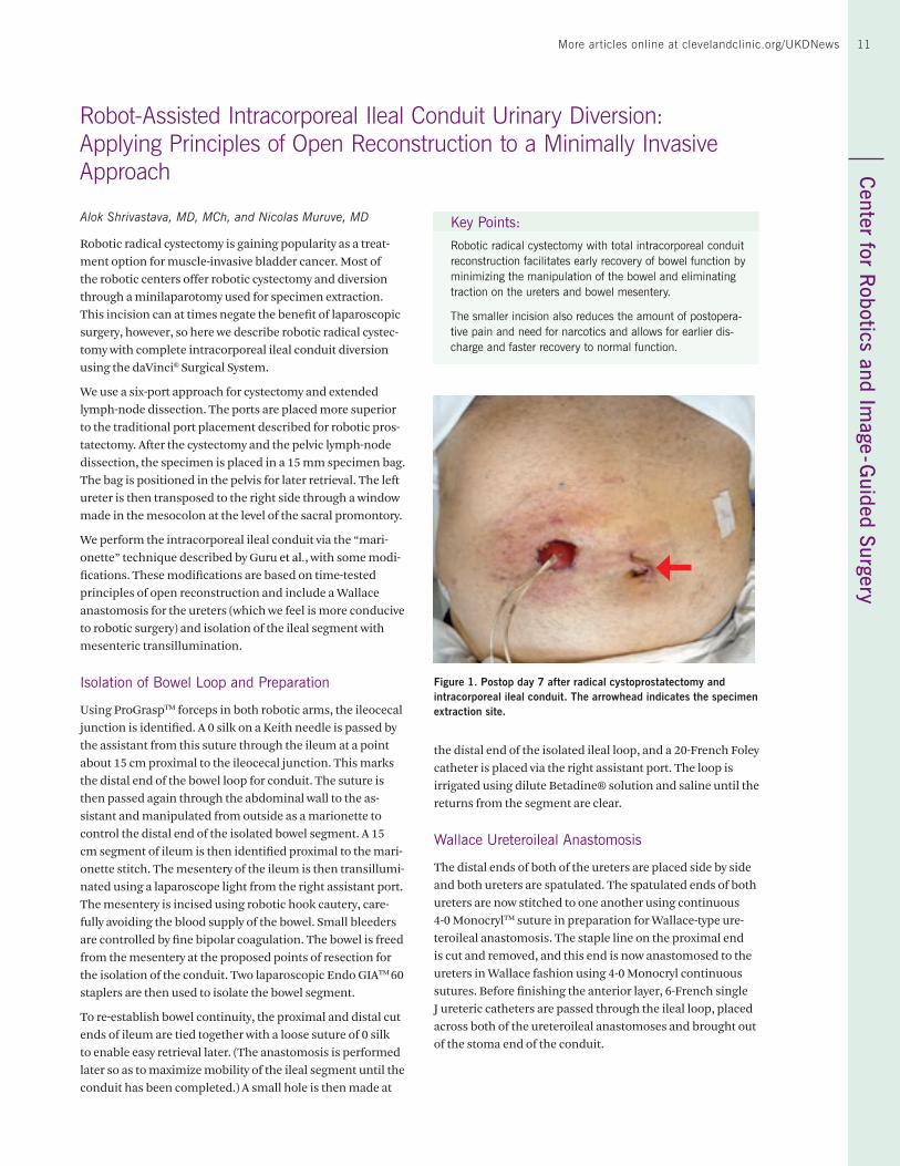
the distal end of the isolated ileal loop, and a 20-French Foley catheter is placed via the right assistant port. The loop is irrigated using dilute Betadine® solution and saline until the returns from the segment are clear.
Wallace Ureteroileal Anastomosis
The distal ends of both of the ureters are placed side by side and both ureters are spatulated. The spatulated ends of both ureters are now stitched to one another using continuous 4-0 MonocrylTM suture in preparation for Wallace-type ure-teroileal anastomosis. The staple line on the proximal end is cut and removed, and this end is now anastomosed to the ureters in Wallace fashion using 4-0 Monocryl continuous sutures. Before finishing the anterior layer, 6-French single J ureteric catheters are passed through the ileal loop, placed across both of the ureteroileal anastomoses and brought out of the stoma end of the conduit.
Robot-Assisted Intracorporeal Ileal Conduit Urinary Diversion: Applying Principles of Open Reconstruction to a Minimally Invasive Approach
Alok Shrivastava, MD, MCh, and Nicolas Muruve, MD
Robotic radical cystectomy is gaining popularity as a treat-ment option for muscle-invasive bladder cancer. Most of the robotic centers offer robotic cystectomy and diversion through a minilaparotomy used for specimen extraction. This incision can at times negate the benefit of laparoscopic surgery, however, so here we describe robotic radical cystec-tomy with complete intracorporeal ileal conduit diversion using the daVinci® Surgical System.
We use a six-port approach for cystectomy and extended lymph-node dissection. The ports are placed more superior to the traditional port placement described for robotic pros-tatectomy. After the cystectomy and the pelvic lymph-node dissection, the specimen is placed in a 15 mm specimen bag. The bag is positioned in the pelvis for later retrieval. The left ureter is then transposed to the right side through a window made in the mesocolon at the level of the sacral promontory.
We perform the intracorporeal ileal conduit via the “mari-onette” technique described by Guru et al., with some modi-fications. These modifications are based on time-tested principles of open reconstruction and include a Wallace anastomosis for the ureters (which we feel is more conducive to robotic surgery) and isolation of the ileal segment with mesenteric transillumination.
Isolation of Bowel Loop and Preparation
Using ProGraspTM forceps in both robotic arms, the ileocecal junction is identified. A 0 silk on a Keith needle is passed by the assistant from this suture through the ileum at a point about 15 cm proximal to the ileocecal junction. This marks the distal end of the bowel loop for conduit. The suture is then passed again through the abdominal wall to the as-sistant and manipulated from outside as a marionette to control the distal end of the isolated bowel segment. A 15 cm segment of ileum is then identified proximal to the mari-onette stitch. The mesentery of the ileum is then transillumi-nated using a laparoscope light from the right assistant port. The mesentery is incised using robotic hook cautery, care-fully avoiding the blood supply of the bowel. Small bleeders are controlled by fine bipolar coagulation. The bowel is freed from the mesentery at the proposed points of resection for the isolation of the conduit. Two laparoscopic Endo GIATM 60 staplers are then used to isolate the bowel segment.
To re-establish bowel continuity, the proximal and distal cut ends of ileum are tied together with a loose suture of 0 silk to enable easy retrieval later. (The anastomosis is performed later so as to maximize mobility of the ileal segment until the conduit has been completed.) A small hole is then made at
Key Points:
Robotic radical cystectomy with total intracorporeal conduit reconstruction facilitates early recovery of bowel function by minimizing the manipulation of the bowel and eliminating traction on the ureters and bowel mesentery.
The smaller incision also reduces the amount of postopera-tive pain and need for narcotics and allows for earlier dis-charge and faster recovery to normal function.
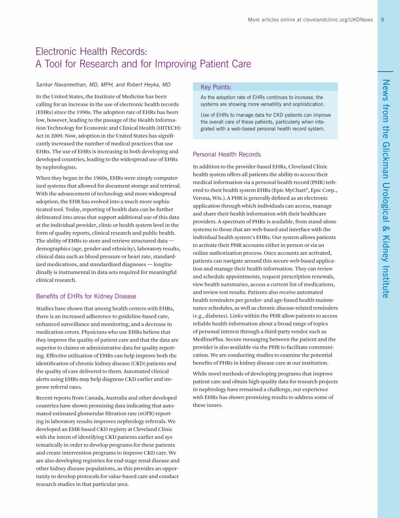
Figure 1. Postop day 7 after radical cystoprostatectomy and intracorporeal ileal conduit. The arrowhead indicates the specimen extraction site.
Cen
ter
for
Rob
otic
s an
d Im
age -
Gui
ded
Surg
ery
12 Urology & Kidney Disease News
Re-establishing Ileum Continuity
The corners of the stapled ends of the bowel are cut, and a 12 mm port is placed in the suprapubic region. Using this port, an Endo GIA 60 stapler is placed within the lumen of the bowel and aligned such that the anti-mesenteric borders of both the proximal and distal segments of the bowel are facing each other. Next, the stapler is fired to attach the two lumens. Another Endo GIA 60 stapler is passed from the right 15 mm assistant port and is used to close the open segment of the side-to-side anastomosis. The anastomosis is thoroughly examined for any possible leaks. A 19-French JP drain is placed from the left 8 mm robotic port site and positioned near the ureteroileal anastomosis.
Stoma Construction and Specimen Retrieval
The marionette suture is cut and removed. The distal end of the ileal conduit with stents is retrieved through a quarter-sized incision made at the proposed stoma site, and the stoma is fashioned in the usual manner. The specimen
within the retrieval bag is removed after enlarging the umbil-ical port incision by 5 cm, and the incision and port sites are closed using PDSTM II (polydioxanone) Suture # 1 (Figure 1).
Postoperative Care
Patients are placed on a general floor with a clear-liquid diet. The diet is advanced after return of bowel function, and patients are discharged at 72 hours postop after removal of the JP drain.
We believe that total intracorporeal conduit reconstruction offers an early recovery of bowel function by minimizing the manipulation of the bowel and eliminating traction on the ureters and bowel mesentery, which is applied in extracor-poreal reconstruction. This may translate into fewer post-operative complications. The smaller incision also reduces the amount of postoperative pain and need for narcotics and allows for earlier discharge and faster recovery to normal function.
Robot-Assisted Intracorporeal Ileal Neobladder: Initial 14 Cases
Georges-Pascal Haber, MD, PhD; Vishnu Ganasan, MS; Idir Ouzaid, MD; Jihad Kaouk, MD; Robert Stein, MD; and Riccardo Autorino, MD, PhD
Robot-assisted laparoscopic radical cystectomy and pelvic lymph node dissection (PLND) have been developed as an extension of the conventional laparoscopic approach, with the primary aim of replicating open surgery principles in a minimally invasive fashion. More recently, robotic intracor-poreal urinary diversions, including ileal conduit (IC) and neobladder (NB), have been performed in a few high-volume robotic centers worldwide. Here we describe our simplified original surgical technique of robotic intracorporeal NB and present our preliminary outcomes.
Surgical Procedure/Perioperative Care
On the day before surgery, the bowel is prepared with osmotic laxative and a stoma site is marked in case conversion to IC is necessary. A single antibiotic shot is administered at the procedure’s onset.
The patient is placed in the lithotomy position, draped in standard sterile fashion, and an 18F Foley catheter is placed. An incision is rendered two fingerbreadths above the umbi-licus and a Veress needle is inserted to establish the pneu-moperitoneum. Through this incision, a 12 mm camera port trocar is placed and the laparoscope is introduced. Three 8 mm robotic ports and a 12 mm assistant port on the left side
Key Point:
Robotic intracorporeal ileal neobladder surgery employing laparoscopic stapling is a minimally invasive, reproducible technique that preserves established open principles of urinary diversion in patients undergoing robotic laparoscopic radical cystectomy and pelvic lymph node dissection for bladder cancer. A simplified step-by-step standardization is needed to facilitate its implementation.
are placed under direct vision. The patient is then placed in steep Trendelenburg position and the robot is docked.
After cystectomy and PLND, specimens are placed in an EndobagTM for later extraction through the midline camera port site in males, and through the vagina in females. A 40 cm distal ileum loop approximately 15 cm from the ileal-colic junction is selected. The bowel loop and its attached mes-entery mobility are tested using traction toward the urethral stump to mimic a tension-free ileo-urethral anastomosis. The mesentery is divided using the Caiman® sealing and cut-ting device, and the distal end is marked with a silk suture. The bowel is divided with the Endo GIA 60TM stapling device.
Bowel continuity is re-established with a standard side-to-side anti-mesenteric ileo-ileal anastomosis using an Endo GIA 60 stapling device cephalad to the transected bowel loop selected for diversion. The open end of the ileum is closed transversely with an additional firing of the Endo GIA 60
13More articles online at clevelandclinic.org/UKDNewsC
enter for Robotics and Im
age- Guided Surgery
13
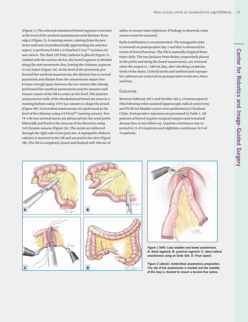
(Figure 1). The selected tubularized bowel segment is incised at the level of the urethral anastomosis (mid-distance from edges) (Figure 2). A running suture, starting from the pos-terior wall and circumferentially approaching the anterior aspect, is performed with a 3-0 barbed V-LocTM incision clo-sure device. The final 22F Foley catheter is placed (Figure 3). Guided with the suction device, the bowel segment is divided along the anti-mesenteric line, leaving the chimney segment (5 cm) intact (Figure 4A). At the level of the previously per-formed ileo-urethral anastomosis, the division line is moved posteriorly and distant from the anastomosis suture line to leave enough space between the two sutures (the already performed ileo-urethral anastomosis and the anterior wall closure suture of the NB to come) at this level. The anterior and posterior walls of the detubularized bowel are sewn in a running fashion using 3-0 V-Loc sutures to shape the pouch (Figure 4B). Ureteroileal anastomoses are performed at the level of the chimney using 4-0 VicrylTM running sutures. Two 7F x 90 mm ureteral stents are advanced into the renal pelvis bilaterally and fixed to the mucosa of the diversion using 3-0 Chromic sutures (Figure 5A). The stents are delivered through the right-side 8 mm port site. A suprapubic Malecot catheter is inserted in the NB and secured to the skin (Figure 5B). The NB is completely closed and flushed with 100 mL of
saline to ensure water-tightness. If leakage is observed, extra sutures must be executed.
Early mobilization is recommended. The nasogastric tube is removed on postoperative day 1 and diet is advanced for return of bowel function. The NB is manually irrigated three times daily. The two Jackson-Pratt drains, respectively placed in the pelvis and along the bowel anastomosis, are removed when the output is < 100 mL/day, after checking creatinine levels of the drain. Ureteral stents and urethral and suprapu-bic catheters are removed on postoperative weeks two, three and four.
Outcomes
Between February 2011 and October 2013, 14 intracorporeal NBs following robot-assisted laparoscopic radical cystectomy and PLND for bladder cancer were performed at Cleveland Clinic. Perioperative outcomes are presented in Table 1. All patients achieved negative surgical margins and remained disease-free at last follow-up. Daytime continence was re-ported in 11 of 14 patients and nighttime continence in 9 of 14 patients.
Figure 1 (left): Loop isolation and bowel anastomosis. A: distal segment. B: proximal segment. C: latero-lateral anastomosis using an Endo GIA. D: Final aspect.
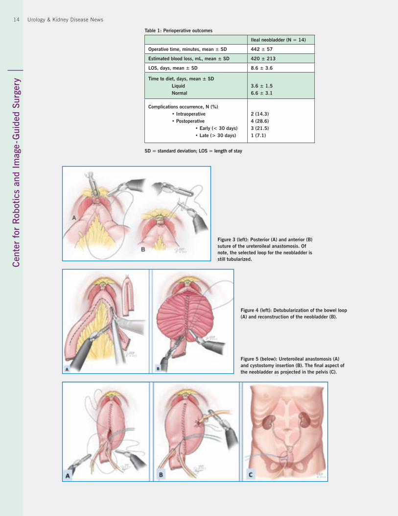
Figure 2 (above): Ureteroileal anastomosis preparation. The site of the anastomosis is marked and the mobility of the loop is checked to ensure a tension-free suture.
Cen
ter
for
Rob
otic
s an
d Im
age -
Gui
ded
Surg
ery
14 Urology & Kidney Disease News
Figure 3 (left): Posterior (A) and anterior (B) suture of the ureteroileal anastomosis. Of note, the selected loop for the neobladder is still tubularized.
Figure 4 (left): Detubularization of the bowel loop (A) and reconstruction of the neobladder (B).
Figure 5 (below): Ureteroileal anastomosis (A) and cystostomy insertion (B). The final aspect of the neobladder as projected in the pelvis (C).
Table 1: Perioperative outcomes
Ileal neobladder (N = 14)
Operative time, minutes, mean ± SD 442 ± 57
Estimated blood loss, mL, mean ± SD 420 ± 213
LOS, days, mean ± SD 8.6 ± 3.6
Time to diet, days, mean ± SDLiquidNormal
3.6 ± 1.56.6 ± 3.1
Complications occurrence, N (%)• Intraoperative• Postoperative
• Early (< 30 days)• Late (> 30 days)
2 (14.3) 4 (28.6)3 (21.5) 1 (7.1)
SD = standard deviation; LOS = length of stay
15More articles online at clevelandclinic.org/UKDNewsC
enter for Robotics and Im
age - Guided Surgery
15
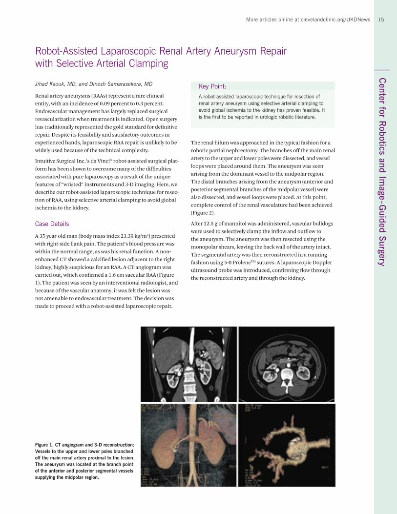
Jihad Kaouk, MD, and Dinesh Samarasekera, MD
Renal artery aneurysms (RAAs) represent a rare clinical entity, with an incidence of 0.09 percent to 0.3 percent. Endovascular management has largely replaced surgical revascularization when treatment is indicated. Open surgery has traditionally represented the gold standard for definitive repair. Despite its feasibility and satisfactory outcomes in experienced hands, laparoscopic RAA repair is unlikely to be widely used because of the technical complexity.
Intuitive Surgical Inc.’s da Vinci® robot-assisted surgical plat-form has been shown to overcome many of the difficulties associated with pure laparoscopy as a result of the unique features of “wristed” instruments and 3-D imaging. Here, we describe our robot-assisted laparoscopic technique for resec-tion of RAA, using selective arterial clamping to avoid global ischemia to the kidney.
Case Details
A 35-year-old man (body mass index 23.39 kg/m2) presented with right-side flank pain. The patient’s blood pressure was within the normal range, as was his renal function. A non-enhanced CT showed a calcified lesion adjacent to the right kidney, highly suspicious for an RAA. A CT angiogram was carried out, which confirmed a 1.6 cm saccular RAA (Figure 1). The patient was seen by an interventional radiologist, and because of the vascular anatomy, it was felt the lesion was not amenable to endovascular treatment. The decision was made to proceed with a robot-assisted laparoscopic repair.
Robot-Assisted Laparoscopic Renal Artery Aneurysm Repair with Selective Arterial Clamping
Figure 1. CT angiogram and 3-D reconstruction: Vessels to the upper and lower poles branched off the main renal artery proximal to the lesion. The aneurysm was located at the branch point of the anterior and posterior segmental vessels supplying the midpolar region.
Key Point:
A robot-assisted laparoscopic technique for resection of renal artery aneurysm using selective arterial clamping to avoid global ischemia to the kidney has proven feasible. It is the first to be reported in urologic robotic literature.
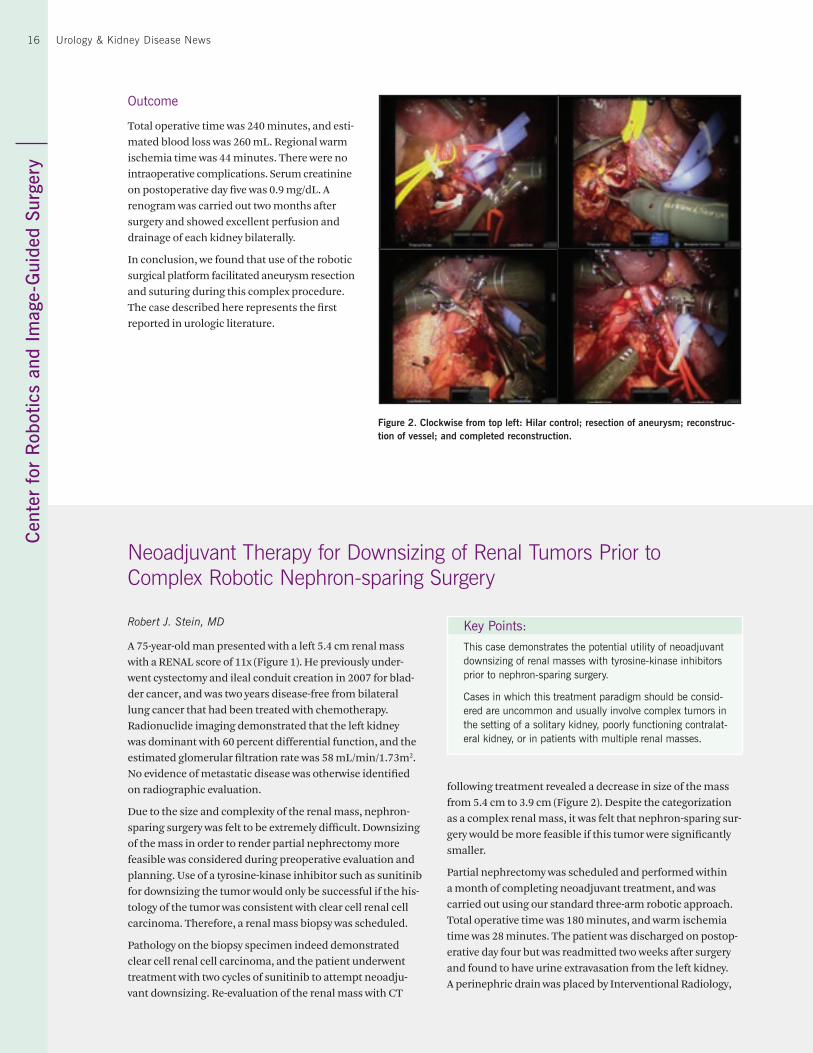
The renal hilum was approached in the typical fashion for a robotic partial nephrectomy. The branches off the main renal artery to the upper and lower poles were dissected, and vessel loops were placed around them. The aneurysm was seen arising from the dominant vessel to the midpolar region. The distal branches arising from the aneurysm (anterior and posterior segmental branches of the midpolar vessel) were also dissected, and vessel loops were placed. At this point, complete control of the renal vasculature had been achieved (Figure 2).
After 12.5 g of mannitol was administered, vascular bulldogs were used to selectively clamp the inflow and outflow to the aneurysm. The aneurysm was then resected using the monopolar shears, leaving the back wall of the artery intact. The segmental artery was then reconstructed in a running fashion using 5-0 ProleneTM sutures. A laparoscopic Doppler ultrasound probe was introduced, confirming flow through the reconstructed artery and through the kidney.
Cen
ter
for
Rob
otic
s an
d Im
age-
Gui
ded
Surg
ery
16 Urology & Kidney Disease News
Outcome
Total operative time was 240 minutes, and esti-mated blood loss was 260 mL. Regional warm ischemia time was 44 minutes. There were no intraoperative complications. Serum creatinine on postoperative day five was 0.9 mg/dL. A renogram was carried out two months after surgery and showed excellent perfusion and drainage of each kidney bilaterally.
In conclusion, we found that use of the robotic surgical platform facilitated aneurysm resection and suturing during this complex procedure. The case described here represents the first reported in urologic literature.
Neoadjuvant Therapy for Downsizing of Renal Tumors Prior to Complex Robotic Nephron-sparing Surgery
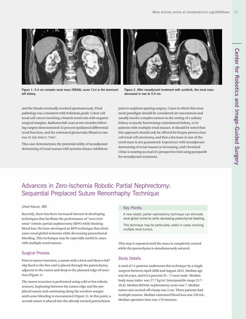
Robert J. Stein, MD
A 75-year-old man presented with a left 5.4 cm renal mass with a RENAL score of 11x (Figure 1). He previously under-went cystectomy and ileal conduit creation in 2007 for blad-der cancer, and was two years disease-free from bilateral lung cancer that had been treated with chemotherapy. Radionuclide imaging demonstrated that the left kidney was dominant with 60 percent differential function, and the estimated glomerular filtration rate was 58 mL/min/1.73m2. No evidence of metastatic disease was otherwise identified on radiographic evaluation.
Due to the size and complexity of the renal mass, nephron-sparing surgery was felt to be extremely difficult. Downsizing of the mass in order to render partial nephrectomy more feasible was considered during preoperative evaluation and planning. Use of a tyrosine-kinase inhibitor such as sunitinib for downsizing the tumor would only be successful if the his-tology of the tumor was consistent with clear cell renal cell carcinoma. Therefore, a renal mass biopsy was scheduled.
Pathology on the biopsy specimen indeed demonstrated clear cell renal cell carcinoma, and the patient underwent treatment with two cycles of sunitinib to attempt neoadju-vant downsizing. Re-evaluation of the renal mass with CT
Key Points:
This case demonstrates the potential utility of neoadjuvant downsizing of renal masses with tyrosine-kinase inhibitors prior to nephron-sparing surgery.
Cases in which this treatment paradigm should be consid-ered are uncommon and usually involve complex tumors in the setting of a solitary kidney, poorly functioning contralat-eral kidney, or in patients with multiple renal masses.
following treatment revealed a decrease in size of the mass from 5.4 cm to 3.9 cm (Figure 2). Despite the categorization as a complex renal mass, it was felt that nephron-sparing sur-gery would be more feasible if this tumor were significantly smaller.
Partial nephrectomy was scheduled and performed within a month of completing neoadjuvant treatment, and was carried out using our standard three-arm robotic approach. Total operative time was 180 minutes, and warm ischemia time was 28 minutes. The patient was discharged on postop-erative day four but was readmitted two weeks after surgery and found to have urine extravasation from the left kidney. A perinephric drain was placed by Interventional Radiology,
Figure 2. Clockwise from top left: Hilar control; resection of aneurysm; reconstruc-tion of vessel; and completed reconstruction.
17More articles online at clevelandclinic.org/UKDNewsC
enter for Robotics and Im
age -Guided Surgery
17
and the fistula eventually resolved spontaneously. Final pathology was consistent with Fuhrman grade 3 clear cell renal cell cancer involving a branch renal vein with negative surgical margins. Radionuclide scan at two months follow-ing surgery demonstrated 42 percent ipsilateral differential renal function, and the estimated glomerular filtration rate was 41 mL/min/1.73m2.
This case demonstrates the potential utility of neoadjuvant downsizing of renal masses with tyrosine-kinase inhibitors
prior to nephron-sparing surgery. Cases in which this treat-ment paradigm should be considered are uncommon and usually involve complex tumors in the setting of a solitary kidney or poorly functioning contralateral kidney, or in patients with multiple renal masses. It should be noted that this approach should only be offered for biopsy-proven clear cell renal cell carcinoma, and that a decrease in size of the renal mass is not guaranteed. Experience with neoadjuvant downsizing of renal masses is increasing, and Cleveland Clinic is nearing accrual of a prospective trial using pazopanib for neoadjuvant treatment.
Figure 1. 5.4 cm complex renal mass (RENAL score 11x) in the dominant left kidney.
Figure 2. After neoadjuvant treatment with sunitinib, the renal mass decreased in size to 3.9 cm.
Advances in Zero-Ischemia Robotic Partial Nephrectomy: Sequential Preplaced Suture Renorrhaphy Technique
Jihad Kaouk, MD
Recently, there has been increased interest in developing techniques that facilitate the performance of “zero-isch-emia” robotic partial nephrectomy (RPN) while limiting blood loss. We have developed an RPN technique that elimi-nates renal global ischemia while decreasing parenchymal bleeding. This technique may be especially useful in cases with multiple renal tumors.
Surgical Process
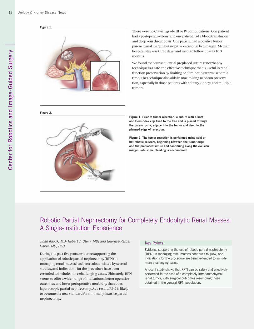
Prior to tumor resection, a suture with a knot and Hem-o-lok® clip fixed to the free end is placed through the parenchyma, adjacent to the tumor and deep to the planned edge of resec-tion (Figure 1).
The tumor resection is performed using cold or hot robotic scissors, beginning between the tumor edge and the pre-placed suture and continuing along the excision margin until some bleeding is encountered (Figure 2). At this point, a second suture is placed into the already excised parenchyma.
Key Points:
A new robotic partial nephrectomy technique can eliminate renal global ischemia while decreasing parenchymal bleeding.
This technique may be particularly useful in cases involving multiple renal tumors.
This step is repeated until the mass is completely excised while the parenchyma is simultaneously sutured.
Study Details
A total of 11 patients underwent this technique by a single surgeon between April 2008 and August 2012. Median age was 66 years, and 63.6 percent (N = 7) were male. Median body mass index was 27.7 kg/m2 (interquartile range 25.7-28.8). Median RENAL nephrometry score was 7. Median tumor size excised off-clamp was 2 cm. Three patients had multiple tumors. Median estimated blood loss was 250 mL. Median operative time was 170 minutes.
Cen
ter
for
Rob
otic
s an
d Im
age-
Gui
ded
Surg
ery
18 Urology & Kidney Disease News
There were no Clavien grade III or IV complications. One patient had a postoperative ileus, and one patient had a blood transfusion and deep vein thrombosis. One patient had a positive tumor parenchymal margin but negative excisional bed margin. Median hospital stay was three days, and median follow-up was 10.3 months.
We found that our sequential preplaced suture renorrhaphy technique is a safe and effective technique that is useful in renal function preservation by limiting or eliminating warm ischemia time. The technique also aids in maximizing nephron preserva-tion, especially in those patients with solitary kidneys and multiple tumors.
Robotic Partial Nephrectomy for Completely Endophytic Renal Masses: A Single-Institution Experience
Jihad Kaouk, MD; Robert J. Stein, MD; and Georges-Pascal Haber, MD, PhD
During the past five years, evidence supporting the application of robotic partial nephrectomy (RPN) in managing renal masses has been substantiated by several studies, and indications for the procedure have been extended to include more challenging cases. Ultimately, RPN seems to offer a wider range of indications, better operative outcomes and lower perioperative morbidity than does laparoscopic partial nephrectomy. As a result, RPN is likely to become the new standard for minimally invasive partial nephrectomy.
Key Points:
Evidence supporting the use of robotic partial nephrectomy (RPN) in managing renal masses continues to grow, and indications for the procedure are being extended to include more challenging cases.
A recent study shows that RPN can be safely and effectively performed in the case of a completely intraparenchymal renal tumor, with surgical outcomes resembling those obtained in the general RPN population.
Figure 1.
Figure 2.Figure 1. Prior to tumor resection, a suture with a knot and Hem-o-lok clip fixed to the free end is placed through the parenchyma, adjacent to the tumor and deep to the planned edge of resection.
Figure 2. The tumor resection is performed using cold or hot robotic scissors, beginning between the tumor edge and the preplaced suture and continuing along the excision margin until some bleeding is encountered.
19More articles online at clevelandclinic.org/UKDNewsC
enter for Robotics and Im
age - Guided Surgery
19
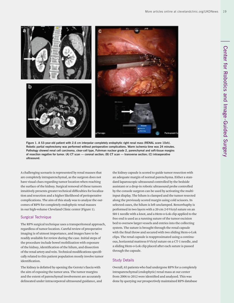
A challenging scenario is represented by renal masses that are completely intraparenchymal, as the surgeon does not have visual clues regarding tumor location when reaching the surface of the kidney. Surgical removal of these tumors intuitively presents greater technical difficulties for localiza-tion and resection and a higher likelihood of perioperative complications. The aim of this study was to analyze the out-comes of RPN for completely endophytic renal masses in our high-volume Cleveland Clinic center (Figure 1).
Surgical Technique
The RPN surgical technique uses a transperitoneal approach, regardless of tumor location. Careful review of preoperative imaging is of utmost importance, and images have to be readily available for review during the case. Initial steps of the procedure include bowel mobilization with exposure of the kidney, identification of the hilum, and dissection of the renal artery and vein. Technical modifications specifi-cally related to this patient population mostly involve tumor identification.
The kidney is defatted by opening the Gerota’s fascia with the aim of exposing the tumor area. The tumor margins and the extent of parenchymal involvement are accurately delineated under intracorporeal ultrasound guidance, and
the kidney capsule is scored to guide tumor resection with an adequate margin of normal parenchyma. Either a stan-dard laparoscopic ultrasound controlled by the bedside assistant or a drop-in robotic ultrasound probe controlled by the console surgeon can be used by activating the multi-input display. The hilum is clamped and the tumor resected along the previously scored margin using cold scissors. In selected cases, the hilum is left unclamped. Renorrhaphy is performed in two layers with a 20 cm 2-0 Vicryl suture on an SH-1 needle with a knot, and a Hem-o-Lok clip applied to the free end is used as a running suture of the tumor excision bed to oversew larger vessels and entries into the collecting system. The suture is brought through the renal capsule with the final throw and secured with two sliding Hem-o-Lok clips. The renal capsule is reapproximated using a continu-ous, horizontal mattress 0 Vicryl suture on a CT-1 needle, and a sliding Hem-o-Lok clip placed after each suture is passed through the capsule.
Study Details
Overall, 65 patients who had undergone RPN for a completely intraparenchymal (endophytic) renal mass at our center from 2006 to 2012 were identified and analyzed. This was done by querying our prospectively maintained RPN database
Figure 1. A 53-year-old patient with 2.6 cm interpolar completely endophytic right renal mass (RENAL score 10xh). Robotic partial nephrectomy was performed without perioperative complications. Warm ischemia time was 24 minutes. Pathology showed renal cell carcinoma, clear-cell type, Fuhrman nuclear grade 2, parenchymal and soft-tissue margins of resection negative for tumor. (A) CT scan — coronal section; (B) CT scan — transverse section; (C) intraoperative ultrasound.
Cen
ter
for
Rob
otic
s an
d Im
age -
Gui
ded
Surg
ery
20 Urology & Kidney Disease News
to consider cases that had been attributed three points for the “E” domain of the RENAL nephrometry score, which is used to describe the exophytic/endophytic properties of the tumor. They accounted for 16.7 percent of RPN cases over the study period.
Demographics and surgical and early postoperative outcomes of the study group were compared with those of controls, represented by patients with “exophytic” mass (i.e., given one point for the “E” domain of the RENAL score), and those of patients with “mesophytic” mass (i.e., given two points for the “E” domain of the RENAL score). As a surrogate of opti-mal outcome, an arbitrary composite outcome (“trifecta”) was considered, consisting of a combination of warm isch-emia time less than 25 minutes, negative surgical margins and no perioperative complications.
Patients with a completely endophytic mass presented a significantly smaller-sized tumor on preoperative imaging (mean 2.6 cm + 1 cm vs. 3.3 cm + 1.8 cm vs. 3.7 cm + 2.1 cm; p = 0.0003) but with a higher RENAL score (mean 8.7 + 1.4 vs. 7.6 + 1.7 vs. 6.4 + 2.2; p < 0.0001). There was no difference between the groups in terms of operative time and estimated blood loss, whereas a lower rate of unclamped cases was noted in the endophytic group (two cases, 3.1 percent) com-
pared with the others (4.8 percent and 18 percent; p < 0.001). Additionally, warm ischemia time was found to be shorter for exophytic (mean 17 minutes + 11.2 minutes) vs. the endo-phytic (21.7 minutes + 9.3 minutes) and the mesophytic (20.2 minutes + 11.5 minutes) tumors (p = 0.0049). No differences were found in terms of intraoperative and postoperative complications or length of hospital stay. A lower pT stage was found for the endophytic masses. There was no differ-ence in terms of positive margin rate between groups. With a similar length of follow-up (mean 12.6 months vs. 15.7 months vs. 14.5 months; p = 0.3), there was a comparable change in terms of estimated glomerular filtration rate and achievement of the trifecta (60 percent for endophytic vs. 59.3 percent for mesophytic vs. 53.6 percent for exophytic; p = 0.5). No differences were found in terms of oncological parameters (cancer-related deaths; tumor recurrence).
In conclusion, RPN can be safely and effectively performed in the case of a completely intraparenchymal renal tumor, with surgical outcomes resembling those obtained in the general RPN population. The accurate use of laparoscopic ultrasound and the unique features of the robotic surgical platform allow optimization of the procedure. Our center is among the few offering this challenging procedure in a minimally invasive fashion on a routine basis.
Repeat Robot-Assisted Partial Nephrectomy: Feasibility and Early Outcomes
Jihad Kaouk, MD; Riccardo Autorino, MD, PhD; Georges-Pascal Haber, MD, PhD; and Robert Stein, MD
Nephron-sparing surgery (NSS) can be a challenging treat-ment option in patients who have undergone a prior NSS and developed a new or recurrent tumor in the same kidney. Although radical nephrectomy has been considered reason-able in this setting, repeat partial nephrectomy (PN) may still be the preferred option, as it maximizes preservation of renal function. This is also supported by the concept that most so-called recurrences actually are due to multifocality and the bilateral nature of the disease, which further supports the role of NSS, if feasible. Repeat open PN has been shown to be associated with good functional and oncological outcomes, but the procedure can be technically challenging because of the increased risk of complications. The challenges become even more significant when a minimally invasive NSS tech-nique is planned (e.g., laparoscopic PN).
Robot-assisted partial nephrectomy (RAPN) seems to offer a more attractive minimally invasive NSS technique com-pared with its standard laparoscopic counterpart. This proposition may be even more applicable when a repeat PN
Key Point:
Repeat partial nephrectomy in patients who have undergone prior open nephron-sparing surgery is challenging. Robotic technology facilitates a minimally invasive approach for these patients.
is indicated. There is a paucity of published data about the outcomes of RAPN in this setting, however. The aim of this study was to demonstrate the feasibility and to report our single-center perioperative outcomes of repeat RAPN.
A Modified Approach
The RAPN surgical technique at Cleveland Clinic has been reported previously. Technical modifications related spe-cifically to this patient population are mostly due to the presence of previous abdominal scars, which require trans-peritoneal access through the most geographically distant quadrant to minimize the risk of inadvertent intra-abdom-inal injury. Moreover, meticulous lysis of intra-abdominal adhesions is required to avoid injury to adjacent organs as
21More articles online at clevelandclinic.org/UKDNewsC
enter for Robotics and Im
age- Guided Surgery
21
well as significant hemorrhaging. If adhesions involving the bowel and its mesentery are present, use of electrocautery should be minimized.
Another problem involves the previously dissected renal hilum and requires a specific approach. Dense adhesions around the hilum are expected, and special care is needed to handle fibrous tissue encasing the renal vessels. The first decision involves whether to clamp, and hilar clamping should be minimized. When deemed necessary, skeleton-izing the artery and vein individually can be too risky and is not advisable. En bloc clamping with the use of a Satinsky can be performed for renal vessels instead of bulldog clamping, which is commonly used for a standard RAPN case.
A previously dissected kidney may have the Gerota’s fascia mobilized or even excised, resulting in tricky remobilization of the kidney. The kidney might be completely lacking in fat and be directly adherent to the undersurface of the abdominal wall. This should be remembered during renal mobilization. Densely adherent perinephric tissue may result in a high likelihood of entering the subcapsular plane, and prevent-ing such stripping of the capsule is of critical importance to facilitate the following surgical step of renorrhaphy. Overall, the limited mobility of the kidney may make tumor excision and renal reconstruction slower and more difficult.
Study Details
From June 2006 to June 2012, 490 patients underwent RAPN for a renal mass at our institution. Of these patients, nine (median age 69 years, six of them female) had undergone previous ipsilateral PN and were included in the present analysis. In all, 12 tumors were removed in these nine patients. A third of the operations were performed on patients with a single kidney. The most common previous ipsilateral NSS
procedure was open PN (five patients), and the median time from the previous NSS procedure was 39.4 months. In all patients, the mass was located in a different portion of the previously treated kidney. The median RENAL nephrometry score for the resected masses was 7, anterior position being the most frequent, and the median tumor size was 2 cm.
The median procedure duration was 153 minutes, with a median warm ischemic time of 17.5 minutes. In three of the nine patients, an unclamped procedure was used. The median (range) estimated intraoperative blood loss was 150 mL (75 mL to 275 mL). No intraoperative complications were registered, whereas only two minor (Clavien I) complications occurred postoperatively — one ileus and one transient el-evation in serum creatinine not requiring dialysis. There was no loss of renal units, and all surgical margins were negative.
Results
For functional outcomes, there was a mean 7 percent decrease in estimated glomerular filtration rate (eGFR) postoperatively, without a significant difference between preoperative and latest postoperative mean eGFR values (70.5 mL/minute/1.73 m2 vs. 63.5 mL/minute/1.73 m2, p > 0.05). The mean follow-up was 8.3 months (standard deviation 13 months). Of the eight patients with a pathology diagnosis of malignant neoplasm, all were alive and free from disease at the latest follow-up.
In conclusion, repeat RAPN can be offered to patients pre-senting with local recurrence after primary NSS for localized renal cell carcinoma. Although technically more demanding, repeat PN can be performed safely and effectively in a mini-mally invasive fashion with the aid of robotic technology in this subset of patients.
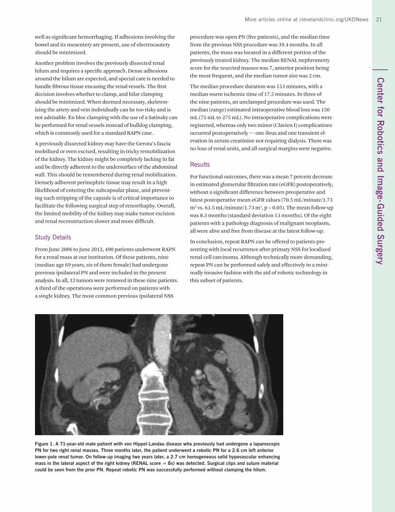
Figure 1. A 71-year-old male patient with von Hippel-Landau disease who previously had undergone a laparoscopic PN for two right renal masses. Three months later, the patient underwent a robotic PN for a 2.6 cm left anterior lower-pole renal tumor. On follow-up imaging two years later, a 2.7 cm homogeneous solid hypovascular enhancing mass in the lateral aspect of the right kidney (RENAL score = 8x) was detected. Surgical clips and suture material could be seen from the prior PN. Repeat robotic PN was successfully performed without clamping the hilum.
Cen
ter
for
Uro
logi
c O
ncol
ogy
22 Urology & Kidney Disease News
Eric A. Klein, MD
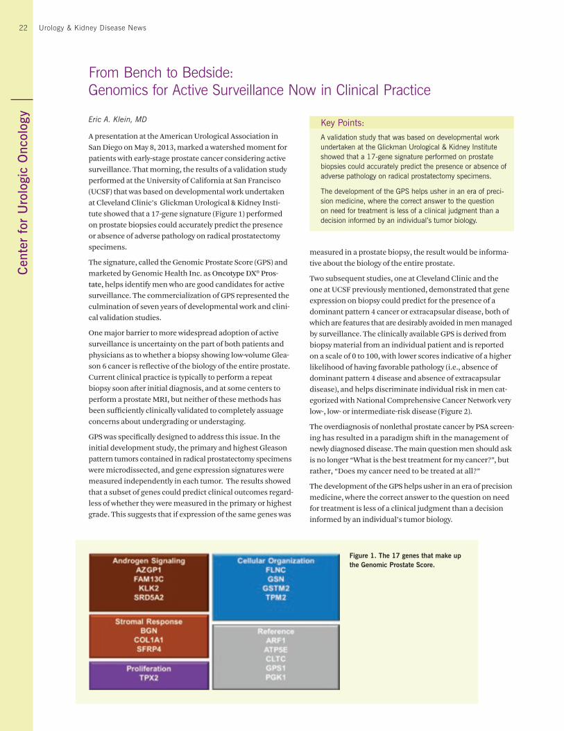
A presentation at the American Urological Association in San Diego on May 8, 2013, marked a watershed moment for patients with early-stage prostate cancer considering active surveillance. That morning, the results of a validation study performed at the University of California at San Francisco (UCSF) that was based on developmental work undertaken at Cleveland Clinic’s Glickman Urological & Kidney Insti-tute showed that a 17-gene signature (Figure 1) performed on prostate biopsies could accurately predict the presence or absence of adverse pathology on radical prostatectomy specimens.
The signature, called the Genomic Prostate Score (GPS) and marketed by Genomic Health Inc. as Oncotype DX® Pros-tate, helps identify men who are good candidates for active surveillance. The commercialization of GPS represented the culmination of seven years of developmental work and clini-cal validation studies.
One major barrier to more widespread adoption of active surveillance is uncertainty on the part of both patients and physicians as to whether a biopsy showing low-volume Glea-son 6 cancer is reflective of the biology of the entire prostate. Current clinical practice is typically to perform a repeat biopsy soon after initial diagnosis, and at some centers to perform a prostate MRI, but neither of these methods has been sufficiently clinically validated to completely assuage concerns about undergrading or understaging.
GPS was specifically designed to address this issue. In the initial development study, the primary and highest Gleason pattern tumors contained in radical prostatectomy specimens were microdissected, and gene expression signatures were measured independently in each tumor. The results showed that a subset of genes could predict clinical outcomes regard-less of whether they were measured in the primary or highest grade. This suggests that if expression of the same genes was
From Bench to Bedside:Genomics for Active Surveillance Now in Clinical Practice
measured in a prostate biopsy, the result would be informa-tive about the biology of the entire prostate.
Two subsequent studies, one at Cleveland Clinic and the one at UCSF previously mentioned, demonstrated that gene expression on biopsy could predict for the presence of a dominant pattern 4 cancer or extracapsular disease, both of which are features that are desirably avoided in men managed by surveillance. The clinically available GPS is derived from biopsy material from an individual patient and is reported on a scale of 0 to 100, with lower scores indicative of a higher likelihood of having favorable pathology (i.e., absence of dominant pattern 4 disease and absence of extracapsular disease), and helps discriminate individual risk in men cat-egorized with National Comprehensive Cancer Network very low-, low- or intermediate-risk disease (Figure 2).
The overdiagnosis of nonlethal prostate cancer by PSA screen-ing has resulted in a paradigm shift in the management of newly diagnosed disease. The main question men should ask is no longer “What is the best treatment for my cancer?”, but rather, “Does my cancer need to be treated at all?”
The development of the GPS helps usher in an era of precision medicine, where the correct answer to the question on need for treatment is less of a clinical judgment than a decision informed by an individual’s tumor biology.
Key Points:
A validation study that was based on developmental work undertaken at the Glickman Urological & Kidney Institute showed that a 17-gene signature performed on prostate biopsies could accurately predict the presence or absence of adverse pathology on radical prostatectomy specimens.
The development of the GPS helps usher in an era of preci-sion medicine, where the correct answer to the question on need for treatment is less of a clinical judgment than a decision informed by an individual’s tumor biology.
Figure 1. The 17 genes that make up the Genomic Prostate Score.

23More articles online at clevelandclinic.org/UKDNewsC
enter for Urologic O
ncology23
Figure 2. A patient’s Genomic Prostate Score (GPS) helps distinguish individual risk of unfavorable pathology across a spectrum of clinical disease categories.
Active Surveillance of Localized Prostate Cancer Is Acceptable Management Strategy
Kiranpreet Khurana, MD, and Andrew Stephenson, MD
In the era of screen-detected prostate cancers, about 50 percent of men diagnosed with prostate cancer (PC) have low-risk disease. The risk that these tumors pose to a man’s longevity and quality of life appears to be very low within approximately 15 years of diagnosis. Thus, active surveil-lance (AS) as a management option is especially appealing for patients with low-risk PC, given the uncertain benefit of treatment in terms of improving survival but the more cer-tain impacts of treatment on health-related quality of life. However, acceptance rates of AS in healthy men with long life expectancy are low (10 percent among low-risk patients in the CaPSURE series) despite accumulating evidence attest-ing to its safety and efficacy.
Several studies have shown that AS is a reasonable option in men with localized PC. It does not preclude desired disease-specific outcomes or the ability for cure. Approximately 30 percent of AS patients advance to definitive treatment, usually on the basis of Gleason score reclassification on repeat biopsy and/or changes in prostate-specific antigen (PSA) kinetics. Among AS patients who ultimately undergo definitive local therapy, PSA failure rates < 20 percent have been reported in most studies, which is comparable to the outcome of patients treated initially with external beam radiation therapy (EBRT) or radical prostatectomy (RP). A recent study of men > 65 years old with localized PC managed without initial curative therapy reported a 6 percent mortal-ity rate from PC, which is substantially lower than that of historical controls. The risk of death from competing causes vastly outweighs the risk of death from PC for men on AS as
Key Point:
Outcomes are no worse over follow-up as long as six years with active surveillance of screen-detected low-risk prostate cancer compared with definitive local therapy. Long-term studies comparing the two strategies are needed.
reported by Klotz, lending support to AS as a relevant initial management option in this cohort.
There are no large studies comparing all-cause mortality and PC-specific mortality among patients managed with AS, EBRT, brachytherapy and RP. AS differs substantially from watchful waiting (WW) as the former involves close monitoring with repeat biopsy to identify important cancers while they are still at a curable stage, whereas WW typically involves administering androgen deprivation therapy at the time of symptomatic local or distant progression. In the Scandinavian randomized trial of RP vs. WW in men with clinically detected (as opposed to screen-detected) cancers, RP was associated with a 6 percent reduction in PC-specific mortality at 15 years (14.6 percent vs. 20.7 percent) and a 6.6 percent improvement in all-cause mortality (46.1 percent vs. 52.7 percent). However, in an unplanned secondary analysis, this benefit was restricted to men < 65 years of age. Consider-ing the five- to 10-year lead time in diagnosis associated with screening, this result would translate to men with screen-detected cancer who are < 60 years old. The recently reported PIVOT trial, which involved 731 American men with screen-detected cancers in a similar trial design, reported no signifi-cant difference in all-cause mortality or PC-specific mortality at 12 years between RP and WW. No difference in all-cause
Cen
ter
for
Uro
logi
c O
ncol
ogy
24 Urology & Kidney Disease News
First Mutation Identified That Increases DHT Synthesis to Promote Hormone Therapy Resistance
Nima Sharifi, MD
The development of castration-resistant prostate cancer (CRPC) occurs in large part by tumors acquiring the capability of synthesizing their own supply of 5α-dihydrotestosterone (DHT) from nongonadal sources, particularly from adrenal precursors. The role and requirement for intratumoral DHT synthesis in the development of CRPC is demonstrated by the efficacy of next-generation hormone therapies that have entered into clinical practice. This includes abiraterone ac-etate, which blocks androgen synthesis, and enzalutamide, which is a potent androgen receptor antagonist.
Despite the long-recognized phenomenon of elevated an-drogens in CRPC, no mutation has yet been described that is responsible for increasing DHT synthesis. Our group has identified the first such example of a genetic alteration that increases the conversion of precursor steroids to DHT,
Key Points:
Our group has identified the first example of a genetic altera-tion that increases the conversion of precursor steroids to DHT, permitting tumors to grow in the absence of gonadal testosterone.
It is possible that this germline variant may play a part in upfront resistance to hormonal therapy.
permitting tumors to grow in the absence of gonadal testos-terone. The enzyme 3β-hydroxysteroid dehydrogenase-iso-enzyme-1 (3βHSD1) is required for the first and rate-limiting step in the conversion of adrenal dehydroepiandrosterone (DHEA) en route to DHT. A mutation occurs in 3βHSD1 in a subset of human CRPC tumors that blocks degradation of this enzyme, increasing the amount of enzyme available
mortality was observed in any of the subgroups analyzed, and only high-risk patients treated by RP had significantly lower risk of PC-specific mortality compared with WW. One would anticipate more favorable outcomes if men in these trials were managed by AS compared with WW, as treatment (when given) is administered with curative intent.
The impact of definitive local therapy vs. AS on all-cause mor-tality and PC-specific mortality for screen-detected cancers was analyzed in a contemporary, multi-institutional cohort of 12,910 patients, 12,458 of whom were treated by either RP (N = 7,672), EBRT (N = 2,467) or brachytherapy (N = 2,319) at one of two high-volume U.S. hospitals and 452 managed by AS at a Canadian hospital (16 percent and 5 percent of whom had intermediate- and high-risk features by D’Amico criteria, respectively). Multivariable regression analyses were used to analyze the impact of treatment on all-cause mortality and PC-specific mortality, adjusting for age, PSA, clinical stage, biopsy Gleason score and year of diagnosis. The median follow-up in the treatment and surveillance groups was 57 and 74 months, respectively, with 1,107 (9 percent) and 89 (20 percent) men with follow-up > 10 years. AS was associated with a significantly reduced risk of all-cause mortality (ad-mission heart rate [aHR] 0.5; 95 percent confidence interval [CI]: 0.4-0.6) and no significant difference in PC-specific mortality (aHR 1.2; 95 percent CI: 0.5-2.8) compared with treat-ment. The increased rate of all-cause mortality associated with treatment was observed among patients receiving EBRT (aHR 2.3; 95 percent CI: 1.03-5.3) and brachytherapy (aHR 2.1; 95 percent CI: 1.5-2.6) but not RP (aHR 1.2; 95 percent
CI: 0.9-1.6). Similar results were observed when the analyses were restricted to men with minimal or no comorbidity and/or low-risk features at diagnosis.
A randomized trial with follow-up of at least 15 years is needed to definitively assess whether treatment for screen-detected PC significantly improves quantity and quality of life compared with AS. However, the existing Scandinavian and PIVOT trials demonstrate the safety and efficacy of WW compared with RP. Our study provides further evidence that AS in appropriately selected patients with screen-detected PC is not associated with diminished all-cause mortality or PC-specific mortality compared with definitive local therapy over a follow-up of 10 years, even among low-risk patients or those with minimal or no comorbidity. A surprising finding of our study was an increased rate of all-cause mortality in men treated by EBRT and brachytherapy, which appears to be the result of a significantly increased risk of death from compet-ing causes. There does not appear to be a healthy cohort bias among AS patients, though we may not have fully adjusted for patient comorbidity and other selection factors (known or unknown) that may have biased our results. Further study is required to assess the durability of these findings at 15 years of follow-up and beyond. However, the accumulating evidence from our study and others indicates that AS is an acceptable management strategy for low-risk patients and need not be restricted to those with limited life expectancy or a high probability of indolent prostate cancer.
For references, please email the editor.
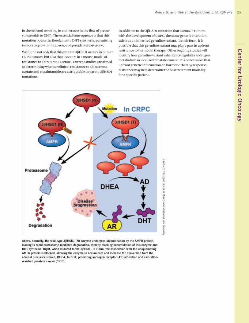
25More articles online at clevelandclinic.org/UKDNews
in the cell and resulting in an increase in the flow of precur-sor steroids to DHT. The essential consequence is that this mutation opens the floodgates to DHT synthesis, permitting tumors to grow in the absence of gonadal testosterone.
We found not only that this mutant 3βHSD1 occurs in human CRPC tumors, but also that it occurs in a mouse model of resistance to abiraterone acetate. Current studies are aimed at determining whether clinical resistance to abiraterone acetate and enzalutamide are attributable in part to 3βHSD1 mutations.
In addition to the 3βHSD1 mutation that occurs in tumors with the development of CRPC, the same genetic alteration exists as an inherited germline variant. In this form, it is possible that this germline variant may play a part in upfront resistance to hormonal therapy. Other ongoing studies will identify how germline variant inheritance regulates androgen metabolism in localized prostate cancer. It is conceivable that upfront genetic information on hormone therapy response/ resistance may help determine the best treatment modality for a specific patient.
Above, normally, the wild-type 3βHSD1 (N) enzyme undergoes ubiquitination by the AMFR protein, leading to rapid proteasome-mediated degradation, thereby blocking accumulation of this enzyme and DHT synthesis. Right, when mutated to the 3βHSD1 (T) form, the association with the ubiquitinating AMFR protein is blocked, allowing the enzyme to accumulate and increase the conversion from the adrenal precursor steroid, DHEA, to DHT, promoting androgen receptor (AR) activation and castration-resistant prostate cancer (CRPC).
Rep
rint
ed w
ith
perm
issi
on f
rom
Cha
ng, et
al.
Cel
l 2013;1
5:1
074-1
084.
25C
enter for Urologic O
ncology
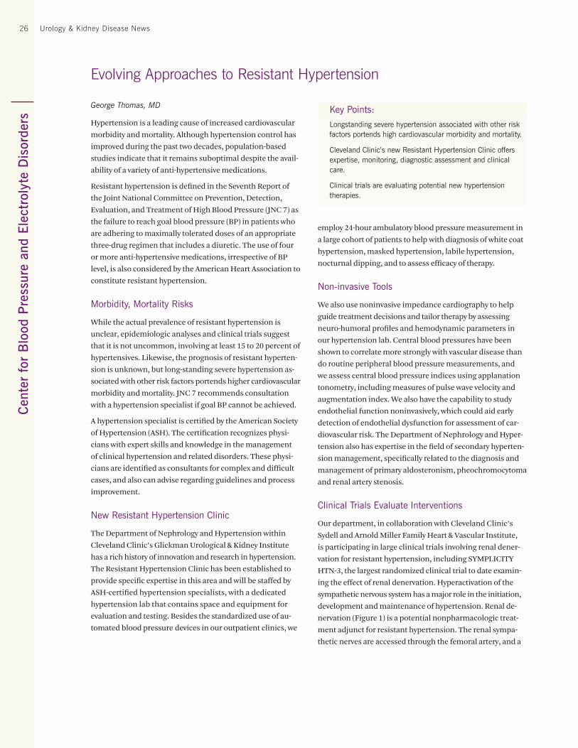
Cen
ter
for
Blo
od P
ress
ure
and
Elec
trol
yte
Dis
orde
rs26 Urology & Kidney Disease News
Evolving Approaches to Resistant Hypertension
George Thomas, MD
Hypertension is a leading cause of increased cardiovascular
morbidity and mortality. Although hypertension control has
improved during the past two decades, population-based
studies indicate that it remains suboptimal despite the avail-
ability of a variety of anti-hypertensive medications.
Resistant hypertension is defined in the Seventh Report of
the Joint National Committee on Prevention, Detection,
Evaluation, and Treatment of High Blood Pressure (JNC 7) as
the failure to reach goal blood pressure (BP) in patients who
are adhering to maximally tolerated doses of an appropriate
three-drug regimen that includes a diuretic. The use of four
or more anti-hypertensive medications, irrespective of BP
level, is also considered by the American Heart Association to
constitute resistant hypertension.
Morbidity, Mortality Risks
While the actual prevalence of resistant hypertension is
unclear, epidemiologic analyses and clinical trials suggest
that it is not uncommon, involving at least 15 to 20 percent of
hypertensives. Likewise, the prognosis of resistant hyperten-
sion is unknown, but long-standing severe hypertension as-
sociated with other risk factors portends higher cardiovascular
morbidity and mortality. JNC 7 recommends consultation
with a hypertension specialist if goal BP cannot be achieved.
A hypertension specialist is certified by the American Society
of Hypertension (ASH). The certification recognizes physi-
cians with expert skills and knowledge in the management
of clinical hypertension and related disorders. These physi-
cians are identified as consultants for complex and difficult
cases, and also can advise regarding guidelines and process
improvement.
New Resistant Hypertension Clinic
The Department of Nephrology and Hypertension within
Cleveland Clinic’s Glickman Urological & Kidney Institute
has a rich history of innovation and research in hypertension.
The Resistant Hypertension Clinic has been established to
provide specific expertise in this area and will be staffed by
ASH-certified hypertension specialists, with a dedicated
hypertension lab that contains space and equipment for
evaluation and testing. Besides the standardized use of au-
tomated blood pressure devices in our outpatient clinics, we
Key Points:
Longstanding severe hypertension associated with other risk factors portends high cardiovascular morbidity and mortality.
Cleveland Clinic’s new Resistant Hypertension Clinic offers expertise, monitoring, diagnostic assessment and clinical care.
Clinical trials are evaluating potential new hypertension therapies.
employ 24-hour ambulatory blood pressure measurement in
a large cohort of patients to help with diagnosis of white coat
hypertension, masked hypertension, labile hypertension,
nocturnal dipping, and to assess efficacy of therapy.
Non-invasive Tools
We also use noninvasive impedance cardiography to help
guide treatment decisions and tailor therapy by assessing
neuro-humoral profiles and hemodynamic parameters in
our hypertension lab. Central blood pressures have been
shown to correlate more strongly with vascular disease than
do routine peripheral blood pressure measurements, and
we assess central blood pressure indices using applanation
tonometry, including measures of pulse wave velocity and
augmentation index. We also have the capability to study
endothelial function noninvasively, which could aid early
detection of endothelial dysfunction for assessment of car-
diovascular risk. The Department of Nephrology and Hyper-
tension also has expertise in the field of secondary hyperten-
sion management, specifically related to the diagnosis and
management of primary aldosteronism, pheochromocytoma
and renal artery stenosis.
Clinical Trials Evaluate Interventions
Our department, in collaboration with Cleveland Clinic’s
Sydell and Arnold Miller Family Heart & Vascular Institute,
is participating in large clinical trials involving renal dener-
vation for resistant hypertension, including SYMPLICITY
HTN-3, the largest randomized clinical trial to date examin-
ing the effect of renal denervation. Hyperactivation of the
sympathetic nervous system has a major role in the initiation,
development and maintenance of hypertension. Renal de-
nervation (Figure 1) is a potential nonpharmacologic treat-
ment adjunct for resistant hypertension. The renal sympa-
thetic nerves are accessed through the femoral artery, and a
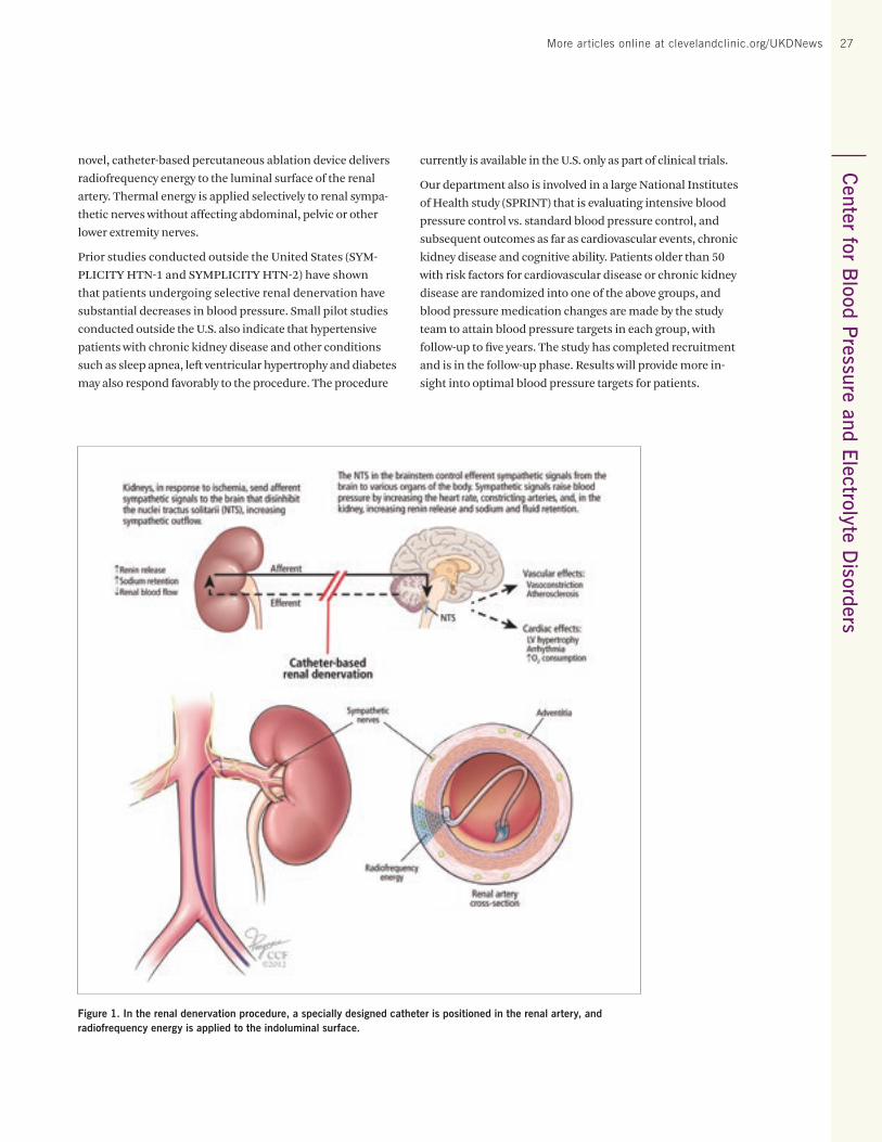
27More articles online at clevelandclinic.org/UKDNews 27C
enter for Blood P
ressure and Electrolyte Disorders
novel, catheter-based percutaneous ablation device delivers
radiofrequency energy to the luminal surface of the renal
artery. Thermal energy is applied selectively to renal sympa-
thetic nerves without affecting abdominal, pelvic or other
lower extremity nerves.
Prior studies conducted outside the United States (SYM-
PLICITY HTN-1 and SYMPLICITY HTN-2) have shown
that patients undergoing selective renal denervation have
substantial decreases in blood pressure. Small pilot studies
conducted outside the U.S. also indicate that hypertensive
patients with chronic kidney disease and other conditions
such as sleep apnea, left ventricular hypertrophy and diabetes
may also respond favorably to the procedure. The procedure
currently is available in the U.S. only as part of clinical trials.
Our department also is involved in a large National Institutes
of Health study (SPRINT) that is evaluating intensive blood
pressure control vs. standard blood pressure control, and
subsequent outcomes as far as cardiovascular events, chronic
kidney disease and cognitive ability. Patients older than 50
with risk factors for cardiovascular disease or chronic kidney
disease are randomized into one of the above groups, and
blood pressure medication changes are made by the study
team to attain blood pressure targets in each group, with
follow-up to five years. The study has completed recruitment
and is in the follow-up phase. Results will provide more in-
sight into optimal blood pressure targets for patients.
Figure 1. In the renal denervation procedure, a specially designed catheter is positioned in the renal artery, and radiofrequency energy is applied to the indoluminal surface.
Cen
ter
for
Chr
onic
Kid
ney
Dis
ease
28 Urology & Kidney Disease News
Jonathan Taliercio, DO, and Sankar Navaneethan, MD, MPH
Elevated serum alkaline phosphatase (ALP) contributes
to the development and progression of vascular calcifica-
tion. Studies conducted in the general population and in
the population of patients on hemodialysis have shown
independent associations between ALP and an increased
risk of cardiovascular events, hospitalization and death. In
selective non-dialysis-dependent chronic kidney disease
(CKD) populations (i.e., African Americans, males), higher
ALP levels are associated with an increased risk of all-cause
mortality. The relationships between ALP and mortality
and between ALP and progression to end-stage renal disease
(ESRD) in a larger heterogeneous population are unclear. We
therefore examined these relationships in a large, diverse
non-dialysis-dependent CKD population followed in our
healthcare system.
Patient Identification
Using the electronic medical record-based CKD registry at
Cleveland Clinic, we identified 28,678 patients who had out-
patient ALP levels measured between the first and second
confirmatory estimated glomerular filtration rate (eGFR)
value < 60 mL/minute/1.73 m2 using the Chronic Kidney
Disease Epidemiology Collaboration (CKD-EPI) equation.
Baseline characteristics were compared between CKD pa-
tients with and without elevated ALP using Chi-square tests
for categorical variables and t-tests or Wilcoxon rank-sum
tests for continuous variables. Kaplan-Meier plots and log-
rank tests were used to analyze survival and ESRD. CKD
patients with measured outpatient ALP values were classi-
fied into quartiles: < 66 units/liter (U/L); 66-81 U/L; 82-101
U/L and ≥ 102 U/L.
Mean age of the study population was 72 (±12) years; 54
percent were female and 12 percent were African American.
After a median follow-up of 2.2 years, 588 patients developed
ESRD and 4,755 died before reaching ESRD. There were 191
patients who developed ESRD and later died. The Kaplan-
Meier and competing risk analyses showed a significant
difference in overall mortality and ESRD among those with
different ALP levels (p < 0.001; Figure 1). The associations
between ALP level and death and ALP level and ESRD were
similar when examined as a continuous variable (Figure 1).
In the multivariate adjusted analysis, each standard devia-
tion (SD) increase in ALP level (42.7 U/L) was associated with
Prognostic Importance of Serum Alklaline Phosphatase in Non-Dialysis-Dependent Chronic Kidney Disease in a Clinical Population
a 16 percent increased hazard for death and a 15 percent in-
creased hazard for ESRD. When the analysis was restricted
to those who had ALP levels within the normal range (< 149
U/L), similar results were observed.
In a subgroup analysis of patients for whom serum phospho-
rus data were available (n = 5,667), when adjusting for serum
phosphorus level, each SD increase in ALP level was associated
with a 10 percent increased hazard for mortality (95 percent
confidence interval [CI]: 1.06-1.14) and a nonsignificant 9
percent increased risk for ESRD (95 percent CI: 0.99-1.19).
In a subgroup analysis of patients with at least one urinary
protein measure (n = 14,116), after adjusting for all covariates
plus proteinuria, each SD increase in ALP level was associated
with a 16 percent increased hazard for mortality (95 percent
CI: 1.12-1.19) and a 17 percent increased hazard for ESRD (95
percent CI: 1.07-1.26).
Proposed Renal Damage Mechanism
Historically, ALP level has only been considered a surrogate
of bone metabolism in patients with CKD and ESRD. ALP is
derived from various tissues but is mostly concentrated in
the liver, biliary ducts, bone and placenta. Tissue-nonspecific
ALP inactivates pyrophosphate, an endogenous inhibitor of
hydroxyapatite formation, resulting in medial arterial vas-
cular calcification. Under conditions such as hypertension,
aging, diabetes and CKD, vascular cells undergo osteoblastic
differentiation and express several bone-associated proteins,
including ALP. Subsequently, this differentiation leads to
mineralization of the endothelium, arterial stiffening and
vascular calcification, thereby contributing to cardiovascular
disease and mortality in CKD. We observed an independent
association between higher ALP levels and ESRD. Apart
from vascular calcification, arterial stiffness as measured
by central pressures might contribute to the progression of
Key Point:
An elevated level of serum alkaline phosphatase (ALP) increases the risk of end-stage renal disease (ESRD) and all-cause mortality in the overall population of patients with chronic kidney disease who are not dependent on dialysis, consistent with data in the dialysis-dependent population and in select groups who are not dialysis-dependent. The associations between ALP and ESRD and mortality remain even when adjusting for serum phosphorus level.
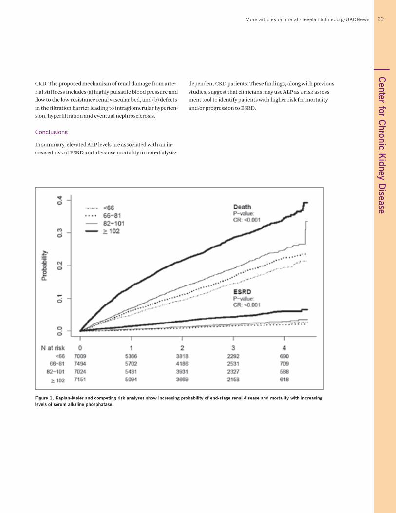
29More articles online at clevelandclinic.org/UKDNews
Figure 1. Kaplan-Meier and competing risk analyses show increasing probability of end-stage renal disease and mortality with increasing levels of serum alkaline phosphatase.
29C
enter for Chronic K
idney Disease
CKD. The proposed mechanism of renal damage from arte-
rial stiffness includes (a) highly pulsatile blood pressure and
flow to the low-resistance renal vascular bed, and (b) defects
in the filtration barrier leading to intraglomerular hyperten-
sion, hyperfiltration and eventual nephrosclerosis.
Conclusions
In summary, elevated ALP levels are associated with an in-
creased risk of ESRD and all-cause mortality in non-dialysis-
dependent CKD patients. These findings, along with previous
studies, suggest that clinicians may use ALP as a risk assess-
ment tool to identify patients with higher risk for mortality
and/or progression to ESRD.
Cen
ter
for
Ren
al D
isea
ses
30 Urology & Kidney Disease News
Brian Stephany, MD
Since the first successful lung transplantation in 1963, national lung transplant volume continues to rise annu-ally. A significant proportion of lung transplant patients develop perioperative acute kidney injury (AKI), reported in the range of 39 to 65 percent, with 5 to 10 percent needing dialysis. Data in the nontransplant population clearly dem-onstrate worse short- and long-term outcomes, including mortality, with even mild and self-limited AKI.
Similarly, recent evidence suggests that AKI immediately after lung transplantation is positively correlated to in-hos-pital and long-term mortality as AKI severity increases. AKI also predicts a longer duration of mechanical ventilation, a longer intensive care unit and hospital stay, an increased cost of care, a faster rate of decline of estimated glomerular filtration rate (eGFR) and an increased risk of chronic kidney disease (CKD).
Some analyses of independent predictors of perioperative AKI in this high-risk group have documented an increased risk in patients undergoing transplantation for idiopathic pulmonary arterial hypertension (IPAH). Furthermore, AKI in patients with IPAH was more likely to be severe, requiring renal replacement therapy (RRT). However, mean pulmonary arterial pressure (mPAP) can be elevated in a multitude of pulmonary diseases that more commonly lead to lung fail-ure requiring transplantation beyond IPAH. To that end, we sought to investigate the association between measured pre-transplant mPAP by pulmonary artery catheterization and perioperative AKI in lung transplant recipients, regard-less of the underlying primary lung diagnosis.
We identified 354 lung allograft recipients transplanted be-tween 1997 and 2009 with available preoperative pulmonary artery pressure measurements. We categorized patients as having no pulmonary hypertension (PH) (mPAP < 25mmHg, n = 162), mild PH (mPAP 25-34 mmHg, n = 133), moderate PH (mPAP 35-45 mmHg, n = 39) or severe PH (mPAP > 45 mmHg, n = 20) prior to transplant. AKI was defined by an absolute rise in creatinine within 48 hours of transplant by > 0.3 mg/dL, according to the Acute Kidney Injury Network classifica-tion schema.
Analysis of this data set identified a graded incidence of perioperative AKI based on baseline measured mPAP: no PH n = 60 (37 percent), mild PH n = 68 (51 percent), moderate PH n = 26 (67 percent), severe PH n = 17 (85 percent), p<0.001. For every 10 mmHg increase in baseline mPAP there was an associated 1.7 (95 percent CI 1.4- 2.2) increased odds of AKI. Controlling for age, gender, race, single vs. double lung transplant, pre-transplant diabetes, baseline creatinine, etiology of lung disease, intraoperative administration of vasopressors and/or inotropes, baseline mPAP, and initial
Baseline Mean Pulmonary Artery Pressure as an Independent Predictor of Acute Kidney Injury After Lung Transplantation
postoperative PaO2/FiO2, those independent covariates that remained significantly associated with AKI post-transplant included vasopressor/inotrope use [odds ratio 2.3 (95 percent CI 1.3-4.3)], baseline creatinine [odds ratio 4.9, (95 percent CI 1.2-21.1) per 1 mg/dL increase], initial PaO2/FiO2 [odds ratio 0.8 (95 percent CI 0.7-0.9) per 50-unit increase], and baseline mPAP [odds ratio 1.5 (95 percent CI 1.2-2.1) per 10 mmHg increase].
These data confirm our suspicion that regardless of the lung diagnosis, elevated baseline mPAP independently predicts an increased risk of AKI immediately after lung transplanta-tion in a dose-dependent manner. These data are clinically relevant when such candidates can be identified and ideally targeted for early employment of renal protective strategies such as avoidance of nephrotoxic medications, overaggres-sive diuresis and contrast use as well as closer hemodynamic monitoring. Future study of the underlying pathophysiologic mechanisms of susceptibility to AKI in addition to novel use of kidney biomarkers that can better characterize impaired renal reserve may lead to improved prophylactic measures that could be instituted before transplantation.
Key Points:
Acute kidney injury is a significant risk in lung transplant patients.
Our analysis of 12 years of preoperative patient data confirms that elevated baseline mean pulmonary arterial pressure independently predicts an increased risk of acute kidney injury immediately after lung transplantation in a dose-dependent manner.
Our finding should aid the identification and targeting of lung transplant candidates for early employment of renal protective strategies.
31More articles online at clevelandclinic.org/UKDNewsC
enter for Genitourinary R
econstruction31
Hadley Wood, MD, and Kenneth Angermeier, MD
Recurrent, escalating urinary infections are by far the most common complaint in patients with continent diversions by any means. They can lead to any of a number of easily treatable conditions, such as bladder stones or diverticula, or may lead to a negative evaluation where the only possible explanation is a capacious, multilobulated reservoir that no longer drains to completion. The former is easily diagnosed and treated, while the latter often requires extensive surgery and recovery with an uncertain outcome with respect to infection.
The first step in getting to a diagnosis is ensuring there are no foreign objects (stones, clips, stents) that may be harbor-ing bacteria. This is easily accomplished with noncontrast CT and/or cystoscopy.
The second step is more complex and involves understand-ing the reservoir. What is the shape of the reservoir? How does it drain? Have the ureters been involved in the past (as with a cutaneous ureterostomy), and what is the condition of the ureterointestinal or uretero-ureteral anastomoses? The only way to understand this includes scrutiny of prior operative reports and judicious use of fluoroscopy and cysto/ureteroscopy. An obstructed upper tract can often lead to silent renal deterioration, and renal function may play an important role in helping to elucidate the area of urinary stasis (as with an obstructed uretero-enteric anastomosis). It may also play a key role in developing a treatment plan for the patient.
Bladder Stones
Incidence of bladder/reservoir stones after enterocysto-plasty or continent supravesical diversion has been variably reported to be 12 to 30 percent in studies that have a median follow-up of 13 years. For large-stone burden, particularly in patients with catheterizable channels, open surgery is pref-erable (Figure 1). Typically, the reservoir is directly under the fascia, often in the location of the prior cecostomy tube. Ac-cess in this location directly into the pouch is usually fairly straightforward. A “temporary vesicostomy,” which is made by suturing the pouch to the fascia, will affix the pouch open and allow the stones to be grasped with Randall stone graspers or an empty sponge stick. This allows removal of the stones without fracturing them. After the stones have been removed, the vesicostomy can be closed with a braided absorbable suture, and fascia can be closed in the usual fashion. We typically leave a 20F-22F Foley catheter or Male-cot tube for secondary drainage for one to three weeks.
For smaller stones, endoscopic approaches through either a 1 to 2 cm incision or a tract dilated up over Amplatz dilators can be used (Figure 2). While some prefer to use a trocar
New or Worsening Urinary Infections in Continent Urostomies/Ileocystoplasty
to enter the pouch, we prefer to make an incision through the fascia, confirm that the pouch has been identified with aspiration via spinal needle and place an Amplatz sheath under direct visualization. Cutting the sheath in half (or in thirds) prior to introducing it allows better mobility of the cystoscope within a capacious pouch. This approach is for optimal stones that can be removed either without fractur-ing via a Nitinol or SeguraTM basket or with limited fractur-ing. At the conclusion of the case, the defect is closed over an 18F-20F Foley catheter with a purse-string Chromic suture and delivered through the abdominal defect as a percutane-ous drain.
Understanding the Reservoir
After a foreign object has been excluded, the next step in-volves defining the patient’s anatomy. Operative reports and historical information from decades prior may include critical information, such as the fact that a patient has had cutaneous ureterostomies for a period of time. This can point to a potential source of obstruction in the proximal or mid-ureters. Defining other patients’ anatomy may require starting from scratch. It is our preference to use a combina-tion of imaging (typically fluoroscopy) and cysto-ureterosco-py to help clarify the situation.
A cystogram can help determine whether there are poorly drained diverticula or a capacious pouch that will not reliably drain to completion via a 14F catheter (Figure 3). To prove poor drainage, we often ask the patient to self-catheterize in his typical position during the study, and shoot an image after he is finished. The cystogram also provides information about the uretero-enteric anastomoses (Figure 4).
When the upper tracts are a source of concern, a combination of antegrade or retrograde pyelography and/or ureteroscopy can be used to help sort out areas of potential obstruction. For these cases, the patient is typically placed on a tilt table (for antegrade studies, Figure 5) or sat upright in the operating room for cross-table images (for retrograde studies). For upper-tract obstruction, nuclear renal scanning can provide
Key Points:
Patients with continent diversions are prone to escalating urinary infections.
Diagnosis requires ruling out foreign objects as the infection source and understanding the reservoir.
Past operative reports, patient histories and a combination of imaging techniques can aid evaluation of the reservoir anatomy and identification of obstructions.
Once the source of urinary stasis has been identified, deci-sions about appropriate surgical intervention can be made.
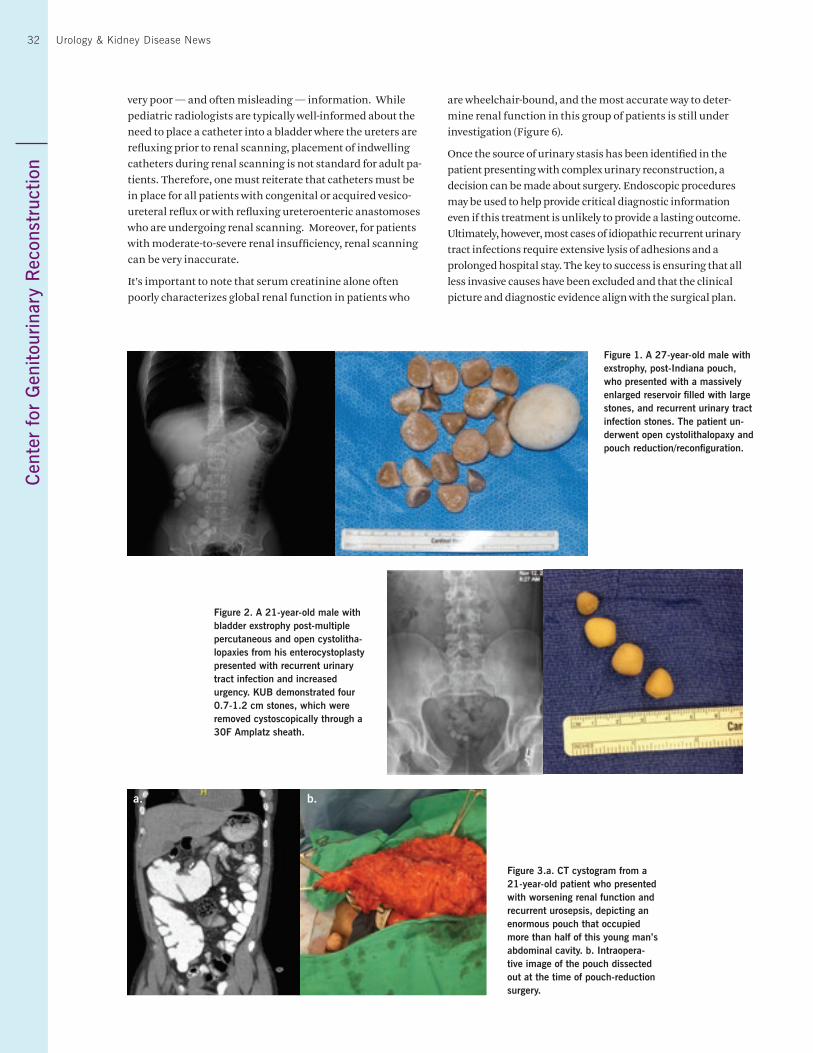
Cen
ter
for
Gen
itour
inar
y R
econ
stru
ctio
n 32 Urology & Kidney Disease News
very poor — and often misleading — information. While pediatric radiologists are typically well-informed about the need to place a catheter into a bladder where the ureters are refluxing prior to renal scanning, placement of indwelling catheters during renal scanning is not standard for adult pa-tients. Therefore, one must reiterate that catheters must be in place for all patients with congenital or acquired vesico-ureteral reflux or with refluxing ureteroenteric anastomoses who are undergoing renal scanning. Moreover, for patients with moderate-to-severe renal insufficiency, renal scanning can be very inaccurate.
It’s important to note that serum creatinine alone often poorly characterizes global renal function in patients who
are wheelchair-bound, and the most accurate way to deter-mine renal function in this group of patients is still under investigation (Figure 6).
Once the source of urinary stasis has been identified in the patient presenting with complex urinary reconstruction, a decision can be made about surgery. Endoscopic procedures may be used to help provide critical diagnostic information even if this treatment is unlikely to provide a lasting outcome. Ultimately, however, most cases of idiopathic recurrent urinary tract infections require extensive lysis of adhesions and a prolonged hospital stay. The key to success is ensuring that all less invasive causes have been excluded and that the clinical picture and diagnostic evidence align with the surgical plan.
Figure 1. A 27-year-old male with exstrophy, post-Indiana pouch, who presented with a massively enlarged reservoir filled with large stones, and recurrent urinary tract infection stones. The patient un-derwent open cystolithalopaxy and pouch reduction/reconfiguration.
Figure 2. A 21-year-old male with bladder exstrophy post-multiple percutaneous and open cystolitha-lopaxies from his enterocystoplasty presented with recurrent urinary tract infection and increased urgency. KUB demonstrated four 0.7-1.2 cm stones, which were removed cystoscopically through a 30F Amplatz sheath.
Figure 3.a. CT cystogram from a 21-year-old patient who presented with worsening renal function and recurrent urosepsis, depicting an enormous pouch that occupied more than half of this young man’s abdominal cavity. b. Intraopera-tive image of the pouch dissected out at the time of pouch-reduction surgery.
a. b.
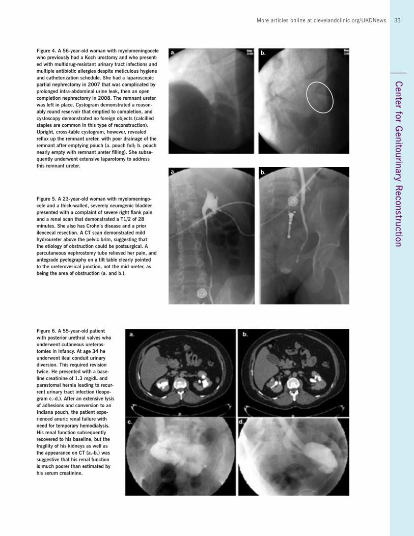
33More articles online at clevelandclinic.org/UKDNewsC
enter for Genitourinary R
econstruction 33
Figure 4. A 56-year-old woman with myelomeningocele who previously had a Koch urostomy and who present-ed with multidrug-resistant urinary tract infections and multiple antibiotic allergies despite meticulous hygiene and catheterization schedule. She had a laparoscopic partial nephrectomy in 2007 that was complicated by prolonged intra-abdominal urine leak, then an open completion nephrectomy in 2008. The remnant ureter was left in place. Cystogram demonstrated a reason-ably round reservoir that emptied to completion, and cystoscopy demonstrated no foreign objects (calcified staples are common in this type of reconstruction). Upright, cross-table cystogram, however, revealed reflux up the remnant ureter, with poor drainage of the remnant after emptying pouch (a. pouch full; b. pouch nearly empty with remnant ureter filling). She subse-quently underwent extensive laparotomy to address this remnant ureter.
Figure 5. A 23-year-old woman with myelomeningo-cele and a thick-walled, severely neurogenic bladder presented with a complaint of severe right flank pain and a renal scan that demonstrated a T1/2 of 28 minutes. She also has Crohn’s disease and a prior ileocecal resection. A CT scan demonstrated mild hydroureter above the pelvic brim, suggesting that the etiology of obstruction could be postsurgical. A percutaneous nephrostomy tube relieved her pain, and antegrade pyelography on a tilt table clearly pointed to the ureterovesical junction, not the mid-ureter, as being the area of obstruction (a. and b.).
Figure 6. A 55-year-old patient with posterior urethral valves who underwent cutaneous ureteros-tomies in infancy. At age 34 he underwent ileal conduit urinary diversion. This required revision twice. He presented with a base-line creatinine of 1.3 mg/dL and parastomal hernia leading to recur-rent urinary tract infection (loopo-gram c.-d.). After an extensive lysis of adhesions and conversion to an Indiana pouch, the patient expe-rienced anuric renal failure with need for temporary hemodialysis. His renal function subsequently recovered to his baseline, but the fragility of his kidneys as well as the appearance on CT (a.-b.) was suggestive that his renal function is much poorer than estimated by his serum creatinine.
a.
a.
b.
b.
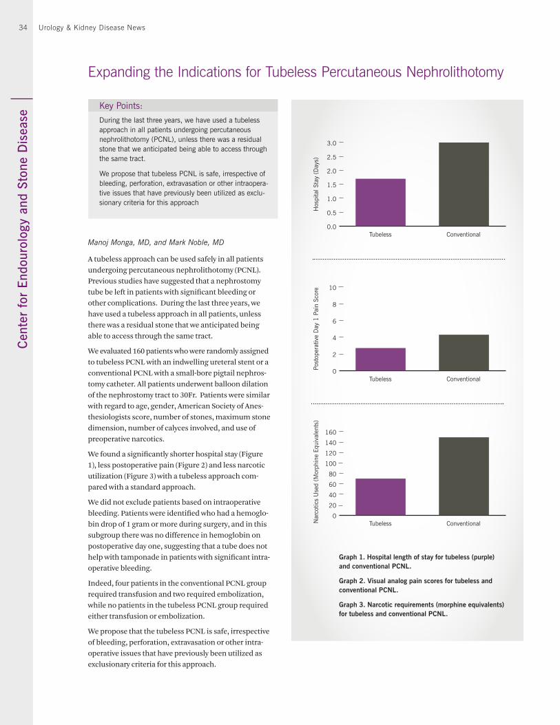
Cen
ter
for
Endo
urol
ogy
and
Ston
e D
isea
se34 Urology & Kidney Disease News
Manoj Monga, MD, and Mark Noble, MD
A tubeless approach can be used safely in all patients undergoing percutaneous nephrolithotomy (PCNL). Previous studies have suggested that a nephrostomy tube be left in patients with significant bleeding or other complications. During the last three years, we have used a tubeless approach in all patients, unless there was a residual stone that we anticipated being able to access through the same tract.
We evaluated 160 patients who were randomly assigned to tubeless PCNL with an indwelling ureteral stent or a conventional PCNL with a small-bore pigtail nephros-tomy catheter. All patients underwent balloon dilation of the nephrostomy tract to 30Fr. Patients were similar with regard to age, gender, American Society of Anes-thesiologists score, number of stones, maximum stone dimension, number of calyces involved, and use of preoperative narcotics.
We found a significantly shorter hospital stay (Figure 1), less postoperative pain (Figure 2) and less narcotic utilization (Figure 3) with a tubeless approach com-pared with a standard approach.
We did not exclude patients based on intraoperative bleeding. Patients were identified who had a hemoglo-bin drop of 1 gram or more during surgery, and in this subgroup there was no difference in hemoglobin on postoperative day one, suggesting that a tube does not help with tamponade in patients with significant intra-operative bleeding.
Indeed, four patients in the conventional PCNL group required transfusion and two required embolization, while no patients in the tubeless PCNL group required either transfusion or embolization.
We propose that the tubeless PCNL is safe, irrespective of bleeding, perforation, extravasation or other intra-operative issues that have previously been utilized as exclusionary criteria for this approach.
Expanding the Indications for Tubeless Percutaneous Nephrolithotomy
0.0
0.5
1.0
1.5
2.0
2.5
3.0
Tubeless Conventional
Hos
pita
l Sta
y (D
ays)
0
2
4
6
8
10
Tubeless Conventional
Post
oper
ativ
e D
ay 1
Pai
n Sc
ore
0
20
40
60
80
100
120
140
160
Tubeless ConventionalNar
cotic
s U
sed
(Mor
phin
e Eq
uiva
lent
s)
Graph 1. Hospital length of stay for tubeless (purple) and conventional PCNL.
Graph 2. Visual analog pain scores for tubeless and conventional PCNL.
Graph 3. Narcotic requirements (morphine equivalents) for tubeless and conventional PCNL.
Key Points:
During the last three years, we have used a tubeless approach in all patients undergoing percutaneous nephrolithotomy (PCNL), unless there was a residual stone that we anticipated being able to access through the same tract.
We propose that tubeless PCNL is safe, irrespective of bleeding, perforation, extravasation or other intraopera-tive issues that have previously been utilized as exclu-sionary criteria for this approach
35
Neha Garg, MD, and Sankar Navaneethan, MD, MPH
The burden of congestive heart failure (CHF) in the popula-tion with chronic kidney disease (CKD) is much higher than in the non-CKD population, with a prevalence as high as 20 percent in CKD stage 4-5 patients. Despite this higher CHF burden, CKD patients are less likely to receive optimal treatment for CHF due to the lack of established treatment guidelines, physician lack of familiarity in managing patients with CHF and concomitant CKD, and an increased incidence of adverse events, notably hyperkalemia, in this population. Cardiac resynchronization therapy (CRT) with or without an implantable cardioverter defibrillator (ICD) device has been proven to reduce the rates of morbidity and mortality in CHF. As such, CRT has been recommended as a Class I indica-tion for patients with left ventricular ejection fraction ≤ 35 percent, QRS ≥ 120 msec, and New York Heart Association functional class III or ambulatory class IV CHF symptoms. A recent update has amended the criteria and expanded the class I indication to include patients with milder class II CHF symptoms. CRT has been shown to improve cardiac struc-ture and function from reverse remodeling in those with both systolic and diastolic CHF. However, evidence is insuf-ficient to support the use of this lifesaving therapy in CKD patients, with device therapy trials excluding patients with moderate to severe kidney disease. As patients with CKD are more likely to die of cardiovascular disease than progress to end-stage renal disease, a greater absolute benefit to CRT in these patients is possible. We therefore conducted a system-atic review to assess the current evidence relating to CRT in the CKD population with CHF.
Eighteen studies met the inclusion criteria and were included in the systematic review: 14 observational studies and four randomized controlled trials (RCTs). Survival outcomes were compared in all CKD patients who had CRT vs. those without CRT. Post-CRT outcomes were compared in patients with baseline CKD vs. those without CKD.
Among RCTs, CRT was associated with a survival benefit over non-CRT modalities (medical therapy or ICD alone) in CKD patients who were eligible to receive CRT. Subgroup analyses of RCTs showed that survival outcomes were not different between the CKD and the non-CKD population with CRT. Most RCTs, however, included patients with stage 3 CKD and only a limited number of stage 4 CKD patients. Observational studies comparing survival between CKD and non-CKD patients after CRT implantation showed inferior outcomes in the CKD group.
There was significant improvement in estimated glomerular filtration rate in CKD patients treated with CRT (four studies; mean difference 2.30 mL/minute/1.73 m2; 95 percent con-
Potential Benefits of Cardiac Resynchronization Therapy in Kidney Disease
fidence interval 0.33-4.27) compared with those not treated with CRT. CRT was associated with preservation of renal function in those with moderate CKD and a modest improve-ment in those with mild CKD at baseline. Taken together, these observations suggest that despite the higher mortality risk in CKD patients, the incremental benefits of CRT are evident in this population. Future studies should include patients with advanced CKD to examine the benefits of CRT. Given the higher costs associated with CRT, additional data on the cost-effectiveness of CRT in CKD are also warranted. We are conducting long-term studies to address the benefits of CRT in the population with kidney disease using Cleveland Clinic’s CKD registry.
Key Point:
In patients with chronic kidney disease and concomitant congestive heart failure, cardiac resynchronization therapy (CRT) with or without an implantable cardioverter defibrilla-tor might be associated with a survival benefit and a modest improvement in kidney function compared with those not treated with CRT.
Center for K
idney/Pancreas Transplantation
35More articles online at clevelandclinic.org/UKDNews
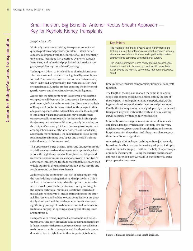
36 Urology & Kidney Disease News
Joseph Africa, MD
Minimally invasive open kidney transplants are safe and quick to perform and provide equivalent — if not better — outcomes compared with the conventional, and essentially unchanged, technique first described by French surgeon Rene Kuss, and refined and popularized by American sur-geon Joseph Murray more than 60 years ago.
Technique: A 2-inch to 5-inch oblique skin incision about 2 inches above and parallel to the inguinal ligament is per-formed. This is carried down to the anterior rectus sheath, which is divided longitudinally. The rectus muscle is then retracted medially, in the process exposing the inferior epi-gastric vessels and the spermatic cord/round ligament.
Access into the retroperitoneum is achieved by dissecting extraperitoneally between the transversalis fascia and the peritoneum, inferior to the arcuate line (linea semicircularis of Douglas). A pocket is then created for the allograft. After adequate exposure of the external iliac vessels, the allograft is implanted. Vascular anastomosis may be performed extracorporeally or in situ (with the kidney in its final posi-tion) or may be done in combination, mostly depending on the recipient’s anatomy. Lich ureteroneocystostomy is then accomplished as usual. The anterior rectus is closed using absorbable monofilament, the subcutaneous tissue is reap-proximated to eliminate dead space and the skin is closed subcuticularly. No drains are used.
This approach ensures a faster, better and stronger musculo-fascial layer closure than the conventional approach, which is done through the external oblique, internal oblique and transversus abdominis muscles/aponeuroses in one, two or sometimes three layers. Due to the fact that muscles are used to hold sutures in the standard technique, these may rip and result in wound dehiscence or hernia.
Additionally, the peritoneum is at risk of being caught with the suture during closing in the standard procedure. This is avoided in the anterior rectus sheath approach because the rectus muscle protects the peritoneum during suturing. In the keyhole technique, minimal dissection is carried out — just what is necessary to fit an allograft and expose the exter-nal iliac vessels and bladder. Wound complications are prac-tically eliminated and the total operative time is shortened significantly (average of two hours vs. three to four hours for traditional surgery) as opening, exposing and closing times are minimized.
Compared with recently reported laparoscopic and robotic transplants, this open procedure is less costly and significant-ly faster to perform (laparoscopic procedures may take four to six hours to perform in experienced hands; robotic proce-dures take four to eight hours). More important, ischemia
Small Incision, Big Benefits: Anterior Rectus Sheath Approach — Key for Keyhole Kidney Transplants
time is shorter, thus not compromising immediate allograft function.
The length of the incision is about the same as in laparo-scopic and robotic procedures, limited only by the size of the allograft. The allograft remains extraperitoneal, avoid-ing complications peculiar to intraperitoneal procedures. Finally, this technique may be easily adopted by experienced transplant surgeons without the costly and risky learning curves associated with high-tech procedures.
Minimally invasive surgeries cause minimal skin, muscle and tissue damage, which means less pain, less scarring, quicker recovery, fewer wound complications and shorter hospital stays for the patient. In kidney transplant surgery, these benefits are magnified.
Interestingly, minimal open techniques have just recently been described but have not been widely adopted. A simple, small-incision technique — without the help of laparoscopic or robotic instruments — using the anterior rectus sheath approach described above, results in excellent renal trans-plant operative outcomes.
Key Points:
The “keyhole” minimally invasive open kidney transplant technique using the anterior rectus sheath approach virtually eliminates wound complications and significantly shortens operative time compared with traditional surgery.
The keyhole procedure is less costly and reduces ischemic time compared with laparoscopic and robotic transplants. It also avoids the learning curve those high-tech procedures entail.
Figure 1. Skin and anterior rectus sheath incisions.
Cen
ter
for
Kid
ney/
Pan
crea
s Tr
ansp
lant
atio
n36
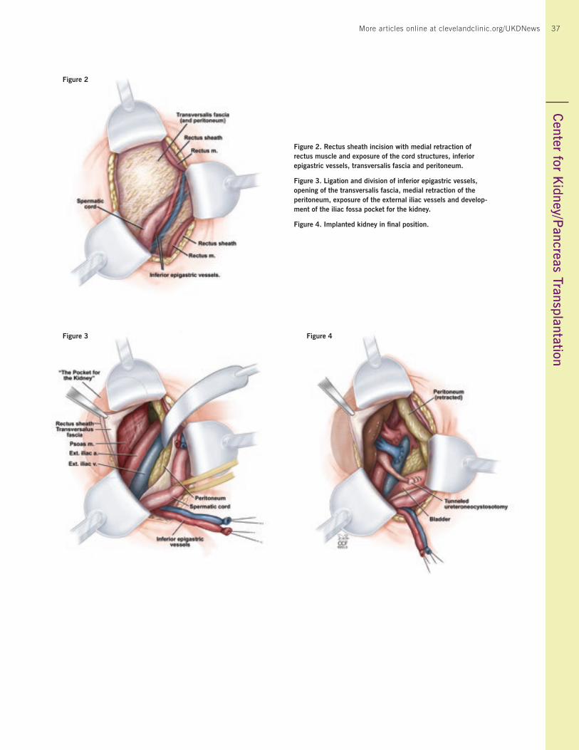
Center for R
obotics and Image – G
uided Surgery37
Figure 2. Rectus sheath incision with medial retraction of rectus muscle and exposure of the cord structures, inferior epigastric vessels, transversalis fascia and peritoneum.
Figure 3. Ligation and division of inferior epigastric vessels, opening of the transversalis fascia, medial retraction of the peritoneum, exposure of the external iliac vessels and develop-ment of the iliac fossa pocket for the kidney.
Figure 4. Implanted kidney in final position.
Figure 2
Figure 3 Figure 4
Center for K
idney/Pancreas Transplantation
37More articles online at clevelandclinic.org/UKDNews
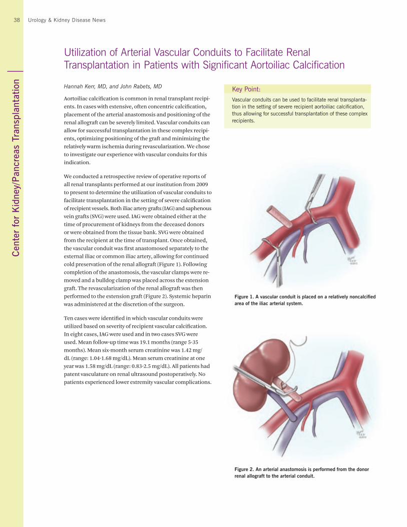
38 Urology & Kidney Disease News
Hannah Kerr, MD, and John Rabets, MD
Aortoiliac calcification is common in renal transplant recipi-ents. In cases with extensive, often concentric calcification, placement of the arterial anastomosis and positioning of the renal allograft can be severely limited. Vascular conduits can allow for successful transplantation in these complex recipi-ents, optimizing positioning of the graft and minimizing the relatively warm ischemia during revascularization. We chose to investigate our experience with vascular conduits for this indication.
We conducted a retrospective review of operative reports of all renal transplants performed at our institution from 2009 to present to determine the utilization of vascular conduits to facilitate transplantation in the setting of severe calcification of recipient vessels. Both iliac artery grafts (IAG) and saphenous vein grafts (SVG) were used. IAG were obtained either at the time of procurement of kidneys from the deceased donors or were obtained from the tissue bank. SVG were obtained from the recipient at the time of transplant. Once obtained, the vascular conduit was first anastomosed separately to the external iliac or common iliac artery, allowing for continued cold preservation of the renal allograft (Figure 1). Following completion of the anastomosis, the vascular clamps were re-moved and a bulldog clamp was placed across the extension graft. The revascularization of the renal allograft was then performed to the extension graft (Figure 2). Systemic heparin was administered at the discretion of the surgeon.
Ten cases were identified in which vascular conduits were utilized based on severity of recipient vascular calcification. In eight cases, IAG were used and in two cases SVG were used. Mean follow-up time was 19.1 months (range 5-35 months). Mean six-month serum creatinine was 1.42 mg/dL (range: 1.04-1.68 mg/dL). Mean serum creatinine at one year was 1.58 mg/dL (range: 0.83-2.5 mg/dL). All patients had patent vasculature on renal ultrasound postoperatively. No patients experienced lower extremity vascular complications.
Utilization of Arterial Vascular Conduits to Facilitate Renal Transplantation in Patients with Significant Aortoiliac Calcification
Key Point:
Vascular conduits can be used to facilitate renal transplanta-tion in the setting of severe recipient aortoiliac calcification, thus allowing for successful transplantation of these complex recipients.
Figure 1. A vascular conduit is placed on a relatively noncalcified area of the iliac arterial system.
Figure 2. An arterial anastomosis is performed from the donor renal allograft to the arterial conduit.
Cen
ter
for
Kid
ney/
Pan
crea
s Tr
ansp
lant
atio
n38
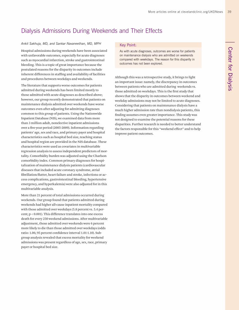
39C
enter for Dialysis
Ankit Sakhuja, MD, and Sankar Navaneethan, MD, MPH
Hospital admissions during weekends have been associated with unfavorable outcomes, especially for acute diagnoses such as myocardial infarction, stroke and gastrointestinal bleeding. This is a topic of great importance because the postulated reasons for the disparity in outcomes include inherent differences in staffing and availability of facilities and procedures between weekdays and weekends.
The literature that supports worse outcomes for patients admitted during weekends has been limited mostly to those admitted with acute diagnoses as described above; however, our group recently demonstrated that patients on maintenance dialysis admitted over weekends have worse outcomes even after adjusting for admitting diagnoses common to this group of patients. Using the Nationwide Inpatient Database (NIS), we examined data from more than 3 million adult, nonelective inpatient admissions over a five-year period (2005-2009). Information regarding patients’ age, sex and race, and primary payer and hospital characteristics such as hospital bed size, teaching status and hospital region are provided in the NIS database. These characteristics were used as covariates in multivariable regression analysis to assess independent predictors of mor-tality. Comorbidity burden was adjusted using the Charlson comorbidity index. Common primary diagnoses for hospi-talization of maintenance dialysis patients (cardiovascular diseases that included acute coronary syndrome, atrial fibrillation/flutter, heart failure and stroke, infections or ac-cess complications, gastrointestinal bleeding, hypertensive emergency, and hyperkalemia) were also adjusted for in this multivariable analysis.
More than 21 percent of total admissions occurred during weekends. Our group found that patients admitted during weekends had higher all-cause inpatient mortality compared with those admitted over weekdays (5.8 percent vs. 5.4 per-cent; p < 0.001). This difference translates into one excess death for every 250 weekend admissions. After multivariable adjustment, those admitted over weekends were 6 percent more likely to die than those admitted over weekdays (odds ratio: 1.06; 95 percent confidence interval 1.01-1.10). Sub-group analysis revealed that excess mortality for weekend admissions was present regardless of age, sex, race, primary payer or hospital bed size.
Although this was a retrospective study, it brings to light an important issue: namely, the discrepancy in outcomes between patients who are admitted during weekends vs. those admitted on weekdays. This is the first study that shows that the disparity in outcomes between weekend and weekday admissions may not be limited to acute diagnoses. Considering that patients on maintenance dialysis have a much higher admission rate than nondialysis patients, this finding assumes even greater importance. This study was not designed to examine the potential reasons for these disparities. Further research is needed to better understand the factors responsible for this “weekend effect” and to help improve patient outcomes.
Key Point:
As with acute diagnoses, outcomes are worse for patients on maintenance dialysis who are admitted on weekends compared with weekdays. The reason for this disparity in outcomes has not been explored.
Dialysis Admissions During Weekends and Their Effects
More articles online at clevelandclinic.org/UKDNews
40 Urology & Kidney Disease News
By Audrey Rhee, MD
Hypospadias is the second most common birth defect involving the male genitalia (first is cryptorchi-dism). The incidence of this defect is 1 in every 250 male births in the United States. Normal sexual dif-ferentiation of the external genitalia is believed to result from a combina-tion of normal genes and a normal hormonal environment. Any disrup-tion of these factors can result in hypospadias.
The Complications of Surgical Repair
The goal of surgical repair is to create a straight phallus and a urethral meatus at the tip of the glans. The risk of complica-tions from surgical correction increases with more proximal meatus location and with smaller phallus or glans size. These complications include glans dehiscence, urethrocuta-neous fistula, meatal stenosis and postoperative scarring.
Considerable controversy surrounds the use of hormonal stimulation to temporarily increase phallic size, specifically glans size, to reduce the risk of complications from surgical repair of hypospadias. Since its successful use in 1973 for micropenis and increase in penile length in prepubertal boys, hormonal stimulation has demonstrated penile en-largement with both topical and parenteral administration and has become common practice for most pediatric urolo-gists. Its side effect profile includes acceleration in bone age, sexual precocity, aggression and premature growth of pubic hair. With cessation of use, most of these side effects will resolve.
In 1999, S. A. Koff and V. R. Jayanthi of the Ohio State Univer-sity Medical Center and Children’s Hospital reported their work with preoperative hormonal stimulation in the proxi-mal hypospadias population. In 12 boys, they found that pre-operative human chorionic gonadotropin (ß-HCG) not only increased penile length and glans size but also reduced the severity of hypospadias and chordee and increased the vas-cularity and thickness of the proximal corpus spongiosum. These preoperative changes permitted a simpler repair. They reported one complication, a urethrocutaneous fistula.
Not So Simple?
In theory, these findings would suggest that reducing the complexity of the hypospadias repair with androgen stimula-tion should result in a decrease in postoperative complica-tions. However, two large retrospective studies published in 2011 suggest that the matter is likely more nuanced.
Gorduza et al. reviewed 300 “severe hypospadias cases” treat-ed with urethroplasty with or without preoperative androgen stimulation during a 10-year period. The indications for androgen stimulation (either ß-HCG or systemic testoster-one) were penis < 25 mm, marked hypoplasia of the ventral tissues of the penis or association of cryptorchidism. There was significant variance in the timing of hormone admin-istration. No difference in complication rates was observed between those who received ß-HCG and those given systemic testosterone. Healing complications occurred in 26 patients; the incidence of healing complications was 30 percent among patients who received androgen stimulation vs. 17.7 percent among patients who did not receive stimulation. The difference was not statistically significant (p = 0.23), which the authors attributed to the power of their study. In looking at the trend, they concluded that androgens inhibit cutane-ous wound healing and promote inflammation, as had been demonstrated in the mouse model. They also suggested that the shorter the interval between androgen stimulation and surgery, the higher the incidence of healing complications.
Snodgrass et al. reviewed 641 patients who underwent trans-urethral incision of urethral plate repair, 56 of whom had proximal hypospadias. Of the total patient population, 32
Is Preoperative Hormonal Stimulation Helpful in the Treatment of Proximal Hypospadias?
Key Points:
Though most pediatric urologists consider preoperative androgen stimulation to temporarily increase phallic size to reduce complications from surgical repair of hypospadias, controversy continues to surround this practice.
Although reducing the complexity of hypospadias repair with androgen stimulation theoretically should reduce postoperative complications, recent large retrospective studies do not bear this out and suggest that androgens may in fact inhibit cutaneous wound healing and promote inflammation.
A recent meta-analysis reveals the low quality and small number of studies addressing this question and underscores the need for randomized, prospective, controlled investigations.
At Cleveland Clinic Children’s, we believe there is merit in the use of androgens in a select group of hypospadias patients, primarily those with a very small glans. C
ente
r fo
r P
edia
tric
Uro
logy
40
41C
enter for Pediatric U
rology41
patients (5 percent) developed postoperative glans dehis-cence, which occurred in 20 of 520 distal repairs (4 percent), 1 of 47 midshaft repairs (2 percent) and 11 of 74 proximal repairs (15 percent). Hormonal stimulation was given to the patients whom the surgeons judged to have an inadequate glans. Comparison of patients who underwent preoperative hormonal stimulation with those who had matched pre-operative factors but did not receive hormonal stimulation revealed no difference in glans dehiscence. Notably, patients with a proximal meatus location had an odds ratio of 3.60 for developing glans dehiscence relative to those with a distal meatus.
The Questions That Remain
This leads to several questions:
• Isn’t the patient with a proximal meatus, small phallus and small glans the same patient who not only is predis-posed to postoperative complications but also is likely to be recommended for preoperative hormonal stimulation?
• Is there a standard regimen that pediatric urologists use?
• Is there a dose that could reduce these apparent complica-tions?
To better answer these questions, Wright et al. recently (2013) published a meta-analysis after reviewing several studies in patients who had undergone proximal hypospa-dias repair. Among the 288 citations, only 11 studies met the researchers’ criteria. The most common method of preopera-tive stimulation was intramuscular testosterone. Of the 11
studies, only four addressed postoperative complications. Dose schedule, time to surgery and indications for stimula-tion were not well-recorded. There were no documented persistent side effects from the hormones. The authors commented on the low quality and small number of studies in the literature but noted that the data suggest a possible relationship between preoperative hormonal stimulation and increased complication rates.
The Bottom Line — For Now
While some retrospective studies have evaluated whether preoperative hormonal stimulation is beneficial and assessed possible complications from its use, the quality of the data is mediocre at best. A randomized, prospective, controlled study is necessary to answer the questions above.
In Cleveland Clinic’s Center for Pediatric Urology, we believe there is merit in the use of androgens in a select group of patients — specifically, those with a very small glans. As we advance in our understanding, there will likely be an evolu-tion of set indications for these select patients (instead of reliance on the surgeon’s subjective assessment), which would be a welcome development.
More articles online at clevelandclinic.org/UKDNews
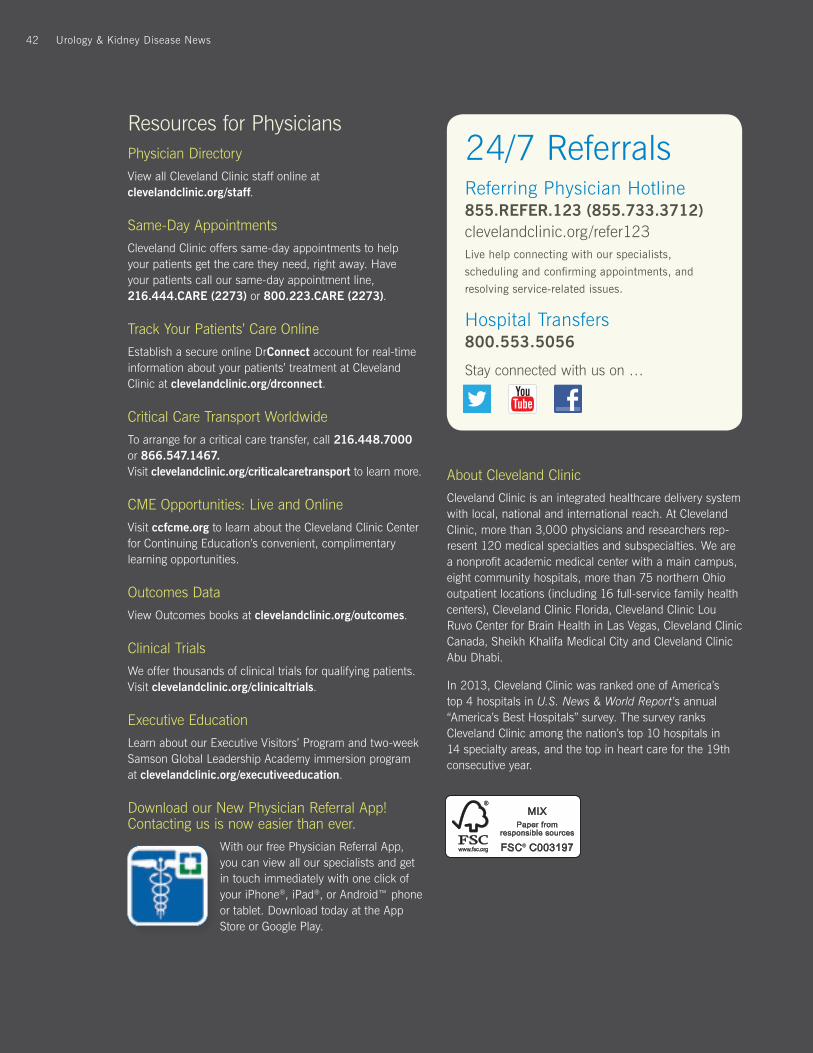
42 Urology & Kidney Disease News
Resources for PhysiciansPhysician Directory
View all Cleveland Clinic staff online at clevelandclinic.org/staff.
Same-Day Appointments
Cleveland Clinic offers same-day appointments to help your patients get the care they need, right away. Have your patients call our same-day appointment line, 216.444.CARE (2273) or 800.223.CARE (2273).
Track Your Patients’ Care Online
Establish a secure online DrConnect account for real-time information about your patients’ treatment at Cleveland Clinic at clevelandclinic.org/drconnect.
Critical Care Transport Worldwide
To arrange for a critical care transfer, call 216.448.7000 or 866.547.1467. Visit clevelandclinic.org/criticalcaretransport to learn more.
CME Opportunities: Live and Online
Visit ccfcme.org to learn about the Cleveland Clinic Center for Continuing Education’s convenient, complimentary learning opportunities.
Outcomes Data
View Outcomes books at clevelandclinic.org/outcomes.
Clinical Trials
We offer thousands of clinical trials for qualifying patients. Visit clevelandclinic.org/clinicaltrials.
Executive Education
Learn about our Executive Visitors’ Program and two-week Samson Global Leadership Academy immersion program at clevelandclinic.org/executiveeducation.
Download our New Physician Referral App! Contacting us is now easier than ever.
With our free Physician Referral App, you can view all our specialists and get in touch immediately with one click of your iPhone®, iPad®, or Android™ phone or tablet. Download today at the App Store or Google Play.
24/7 ReferralsReferring Physician Hotline 855.REFER.123 (855.733.3712) clevelandclinic.org/refer123Live help connecting with our specialists,
scheduling and confirming appointments, and
resolving service-related issues.
Hospital Transfers800.553.5056
Stay connected with us on …
About Cleveland Clinic
Cleveland Clinic is an integrated healthcare delivery system with local, national and international reach. At Cleveland Clinic, more than 3,000 physicians and researchers rep-resent 120 medical specialties and subspecialties. We are a nonprofit academic medical center with a main campus, eight community hospitals, more than 75 northern Ohio outpatient locations (including 16 full-service family health centers), Cleveland Clinic Florida, Cleveland Clinic Lou Ruvo Center for Brain Health in Las Vegas, Cleveland Clinic Canada, Sheikh Khalifa Medical City and Cleveland Clinic Abu Dhabi.
In 2013, Cleveland Clinic was ranked one of America’s top 4 hospitals in U.S. News & World Report’s annual “America’s Best Hospitals” survey. The survey ranks Cleveland Clinic among the nation’s top 10 hospitals in 14 specialty areas, and the top in heart care for the 19th consecutive year.
Urology & Kidney Disease News42
Scale to 21%
43More articles online at clevelandclinic.org/UKDNews
Urology & Kidney Disease NewsVol. 22 / 2013
Urology & Kidney Disease News is a publication of the Cleveland Clinic Glickman Urological & Kidney Institute.
Urology & Kidney Disease News is written for physicians and should be relied on for medical education purposes only. It does not provide a complete overview of the topics covered and should not replace the independent judgment of a physician about the appropriateness or risks of a procedure for a given patient.
Eric A. Klein, MD Chairman Glickman Urological & Kidney Institute
J. Stephen Jones, MD, FACS Chief of Surgical Operations, Cleveland Clinic Regional Hospitals Medical Editor, Urology
Sankar Navaneethan, MD Glickman Urological & Kidney Institute Medical Editor, Nephrology
John Mangels Managing Editor [email protected]
Barbara Ludwig Coleman Graphic Designer
Learn from Cleveland Clinic President and CEO Toby Cosgrove, MD, how blending innovation, efficiency and compassion can lead to success in a challenging healthcare environment.
• Get ideas on teamwork, efficiency and collaboration to share with colleagues.
• Learn how Cleveland Clinic has cut costs and improved quality.
• Discover where healthcare is heading from one of the industry’s leaders.
“A pioneer in American healthcare, Toby Cosgrove shows just how the diligence and innovative thinking behind the Cleveland Clinic has helped solve funda-mental problems most other places barely touch. There are lessons here for everyone — patient, physician, and policymaker alike.”
-Atul Gawande, MD Harvard Medical School professor and bestselling author of The Checklist Manifesto
Visit clevelandclinic.org/ClevelandClinicWay
for details or to order a copy.
44 Urology & Kidney Disease News
clevelandclinic.org/UKDNews
13-U
RL-
315
Cleveland Clinic Urology and Nephrology Ranked No. 2 in the U.S. — U.S. NEWS & WORLD REPORT


![[Recommendations Onco-Urology 2010: Prostate cancer]](https://img.dokumen.tips/doc/110x75/634e210c3bdc8e881007ba9b/recommendations-onco-urology-2010-prostate-cancer.jpg)